Article

A Comprehensive Analysis of Chloroplast Genome Provides New Insights into the Evolution of the Genus Chrysosplenium

1
Hubei Provincial Key Laboratory for Protection and Application of Special Plant Germplasm in Wuling Area of China, College of Life Sciences, South-Central Minzu University, Wuhan 430074, China
2
College of Life Sciences, Zhejiang Normal University, Jinhua 321004, China
3
College of Computer Science, South-Central Minzu University, Wuhan 430074, China
*
Authors to whom correspondence should be addressed.
These authors have contributed equally to this work.
Int. J. Mol. Sci. 2023, 24(19), 14735; https://doi.org/10.3390/ijms241914735
Submission received: 23 August 2023 / Revised: 24 September 2023 / Accepted: 26 September 2023 / Published: 29 September 2023
(This article belongs to the Section Molecular Plant Sciences)

Abstract

:
Chrysosplenium, a perennial herb in the family Saxifragaceae, prefers to grow in low light and moist environments and is divided into two sections of Alternifolia and Oppositifolia based on phyllotaxy. Although there has been some progress in the phylogeny of Chrysosplenium over the years, the phylogenetic position of some species is still controversial. In this study, we assembled chloroplast genomes (cp genomes) of 34 Chrysosplenium species and performed comparative genomic and phylogenetic analyses in combination with other cp genomes of previously known Chrysosplenium species, for a total of 44 Chrysosplenium species. The comparative analyses revealed that cp genomes of Chrysosplenium species were more conserved in terms of genome structure, gene content and arrangement, SSRs, and codon preference, but differ in genome size and SC/IR boundaries. Phylogenetic analysis showed that cp genomes effectively improved the phylogenetic support and resolution of Chrysosplenium species and strongly supported Chrysosplenium species as a monophyletic taxon and divided into three branches. The results also showed that the sections of Alternifolia and Oppositifolia were not monophyletic with each other, and that C. microspermum was not clustered with other Chrysosplenium species with alternate leaves, but with C. sedakowii into separate branches. In addition, we identified 10 mutational hotspot regions that could serve as potential DNA barcodes for Chrysosplenium species identification. In contrast to Peltoboykinia, the clpP and ycf2 genes of Chrysosplenium were subjected to positive selection and had multiple significant positive selection sites. We further detected a significant positive selection site on the petG gene between the two sections of Chrysosplenium. These evolutionary characteristics may be related to the growth environment of Chrysosplenium species. This study enriches the cp genomes of Chrysosplenium species and provides a reference for future studies on its evolution and origin.

Graphical Abstract

1. Introduction

The chloroplast (cp) genome has long been a major source of molecular data for studying plant phylogeny and evolution because of its maternally inherited and relatively conserved nature. The size and structure of cp genomes have been highly conserved during land plant evolution, in contrast to the large variation in the size and structure of plant mitochondrial genomes. In Saxifragaceae species, the cp genome has a highly conserved circular quadripartite structure containing a large single-copy (LSC) and a small single-copy (SSC) divided by two inverted repeat (IR) regions. With the development of sequencing technology, whole-genome sequencing and large-scale phylogenetic analysis of cp genomes of most plants have been achieved, further facilitating plant taxonomic studies [1,2].
Chrysosplenium L., belonging to the family Saxifragaceae, is a small perennial herbaceous plant, usually with flagellate branches or bulbs, whose phyllotaxy is divided into alternate and opposite leaves. There are about 80 Chrysosplenium species in the world, which are mainly distributed in Asia, Europe and North America in the Northern Hemisphere, and a few in temperate regions in the Southern Hemisphere, mainly two species located in and around Chile, namely C. valdivicum and C. macranthum [3,4]. In China, there are about 38 species and 15 varieties, accounting for more than 56% of the total number of Chrysosplenium species in the world, of which 23 species are endemic to China, mainly in northern and southern China [5,6,7]. In addition, the shade-loving and moisture-loving characteristics of Chrysosplenium species make them ideal materials for studying the evolution of low-light and low-temperature adaptations in plants. Taxonomic studies on Chrysosplenium can be traced back as far as the mid-18th century when C. alternifofium L. with alternate leaves and C. oppositifolium L. with opposite leaves were recognized by Linnaeus (1753). Subsequently, at the end of the 19th century, some species were added to Chrysosplenium and classified accordingly [8,9]. In 1877, Maximowicz et al. (1877) divided the Chrysosplenium into subgen. Gamosplenium and subgen. Dialysplenium based on the length of the sepals and stamens [8]. In 1890, the Chrysosplenium was divided into the groups of Alternifolia and Oppositifolia based on opposite and alternate leaves [9]. In 1957, Hara (1957) made a detailed morphological study of Chrysosplenium and identified 55 species divided into sections of Alternifolia and Oppositifolia [3]. In 1986, Pan (1986) identified and studied the Chrysosplenium species in China and classified them into two subgenera (Chrysosplenium and Gamosplenium), as well as five groups and ten lineages [10,11]. Since then, many new Chrysosplenium species have been discovered, and the diversity of Chrysosplenium species has been continuously enriched [6,7,12,13].
Previous phylogenetic studies of Chrysosplenium have mainly used cp fragments and nuclear ribosomal DNA (nrDNA) sequences, while the cp genome has been relatively little studied, and the phylogenetic position of a few species was still controversial. Nakazawa et al. (1997) evaluated the phylogeny of Chrysosplenium species using rbcL and matK sequences and found that matK sequences had a high phylogenetic resolution [14]. Soltis et al. (2001) studied the phylogeny of some Chrysosplenium species based on matK genes and showed that the sections of Alternifolia and Oppositifolia were monophyletic sisters (Figure S1a) [15]. This phylogeny has long been in common use. Afterwards, Xiang et al. (2012) performed a phylogenetic analysis of Saniculiphyllum based on four chloroplast DNA (trnL-trnF, psbA-trnH, matK, rbcL) and two nrDNA fragments (nrITS, rrn26S) [16]. In the Chrysosplenium branch, C. microspermum with alternate leaves clustered with C. nepalense with opposite leaves (Figure S1b). Tkach et al. (2015) investigated the phylogeny of Micranthes based on nrITS and trnL-trnF sequences [17]. In the Chrysosplenium branch, C. microspermum was located at the base of the Chrysosplenium branch (Figure S1c). In the same year, Deng et al. (2015) studied the phylogeny and evolutionary history of Chrysosplenium based on the matrices of cpDNA and nrDNA [18]. The cpDNA-based BI tree showed that Chrysosplenium was mainly divided into three clades, and C. microspermum was located at the base of the Chrysosplenium branch (Figure S1d). The ML tree based on the matrices of cpDNA and nrDNA showed similar results, but the nucleoplasmic-based BI tree showed that Chrysosplenium was divided into two branches corresponding to the sections of Alternifolia and Oppositifolia, and that C. microspermum was clustered in the section Alternifolia branch (Figure S1e,f). Subsequently, Folk et al. (2019) performed a phylogenetic analysis of 627 Saxifragales species based on 301 protein-coding loci, in which Chrysosplenium species were divided into three branches, with the sections of Alternifolia and Oppositifolia not being monophyletic sisters of each other, C. microspermum with alternate leaves clustered in the section Oppositifolia branch, and C. sedakowii with alternate leaves forming a separate branch (Figure S1g) [19]. To date, the phylogenetic position of C. microspermum has not been clarified.
With the publication of the C. aureobracteatum cp genome in 2018 [20], studies on the cp genome of Chrysosplenium were gradually initiated. Then, the six cp genomes of Chrysosplenium species revealed cp genome characteristics of Chrysosplenium [4]. Subsequently more cp genomes of Chrysosplenium species were published [21,22]. Nevertheless, the cp genomes of many Chrysosplenium species are still unknown. Therefore, in order to gain a comprehensive understanding of the phylogenetic relationships among Chrysosplenium species, this study first de novo assembled and annotated the cp genomes of 34 Chrysosplenium species. Together with the published species, a total of 44 Chrysosplenium cp genomes were further performed for comparative genomics and phylogenetic analysis. The primary research questions addressed in this study are as follows. (1) Whether the sections of Alternifolia and Oppositifolia are monophyletic sisters to each other in the phylogeny of the 44 Chrysosplenium species? (2) Where is the phylogenetic location of C. microspermum? (3) Are there significant differences in the cp genomes of Chrysosplenium between species and between the two groups? (4) Is Chrysosplenium under significant positive selection on the cp genome compared to Peltoboykinia?

2. Results

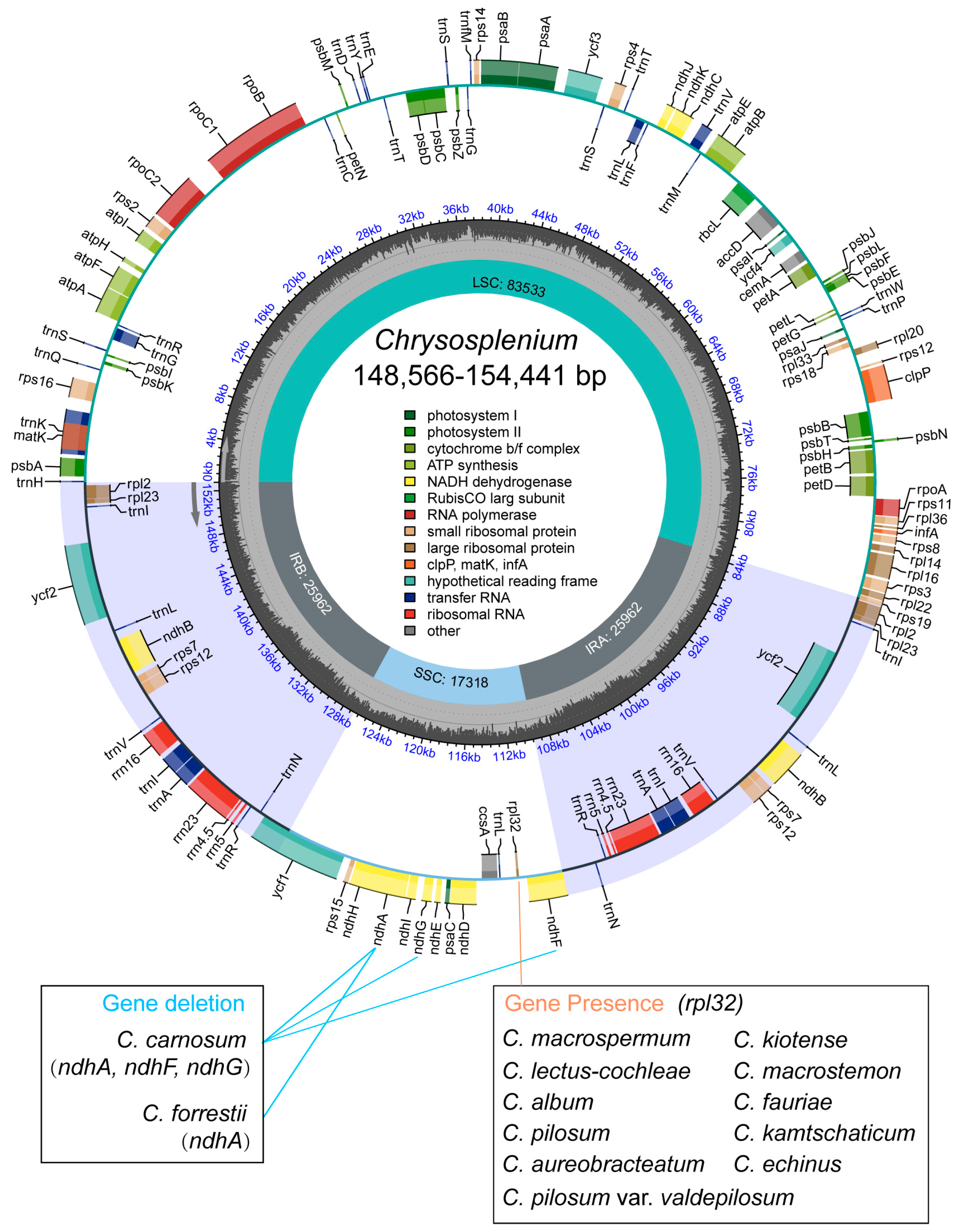
2.1. Structural Characterization of the Chloroplast Genome of Chrysosplenium

All cp genomes of the 44 Chrysosplenium species presented a typical quadripartite structure with a large single-copy (LSC), a small single-copy (SSC), and two inverted repeats (Ira and Irb). The size of cp genome ranged from 148,566 bp to 154,441 bp, with an average of 152,576 bp (Figure 1; Table S1). The GC content of cp genomes ranged from 37.22% to 37.72%, with an average of 37.45%. Among the protein-coding genes (PCGs), 5 genes were responsible for photosystem I (psaA, psaB, psaC, psaI, psaJ), 15 genes for photosystem II (psbA, psbB, psbC, psbD, psbE, psbF, psbH, psbI, psbJ, psbK, psbM, psbN, psbT, psbZ, ycf3), 6 genes for ATP synthase (atpA, atpB, atpE, atpF, atpH, atpI), 9 genes for large ribosomal proteins (rpl2, rpl14, rpl16, rpl20, rpl22, rpl23, rpl32, rpl33, rpl36), and 12 genes for small ribosomal proteins (rps2, rps3, rps4, rps7, rps8, rps11, rps12, rps14, rps15, rps16, rps18, rps19) were found in Chrysosplenium (Table S2). In addition, we found that some PCGs were lost to varying degrees, such as rpl32, ndhA, ndhF, and ndhG. Interestingly, rpl32 was only annotated in some Oppositifolia species, ndhA was missing in both C. carnosum and C. forrestii, and ndhG and ndhF were only missing in C. carnosum (Figure S2).

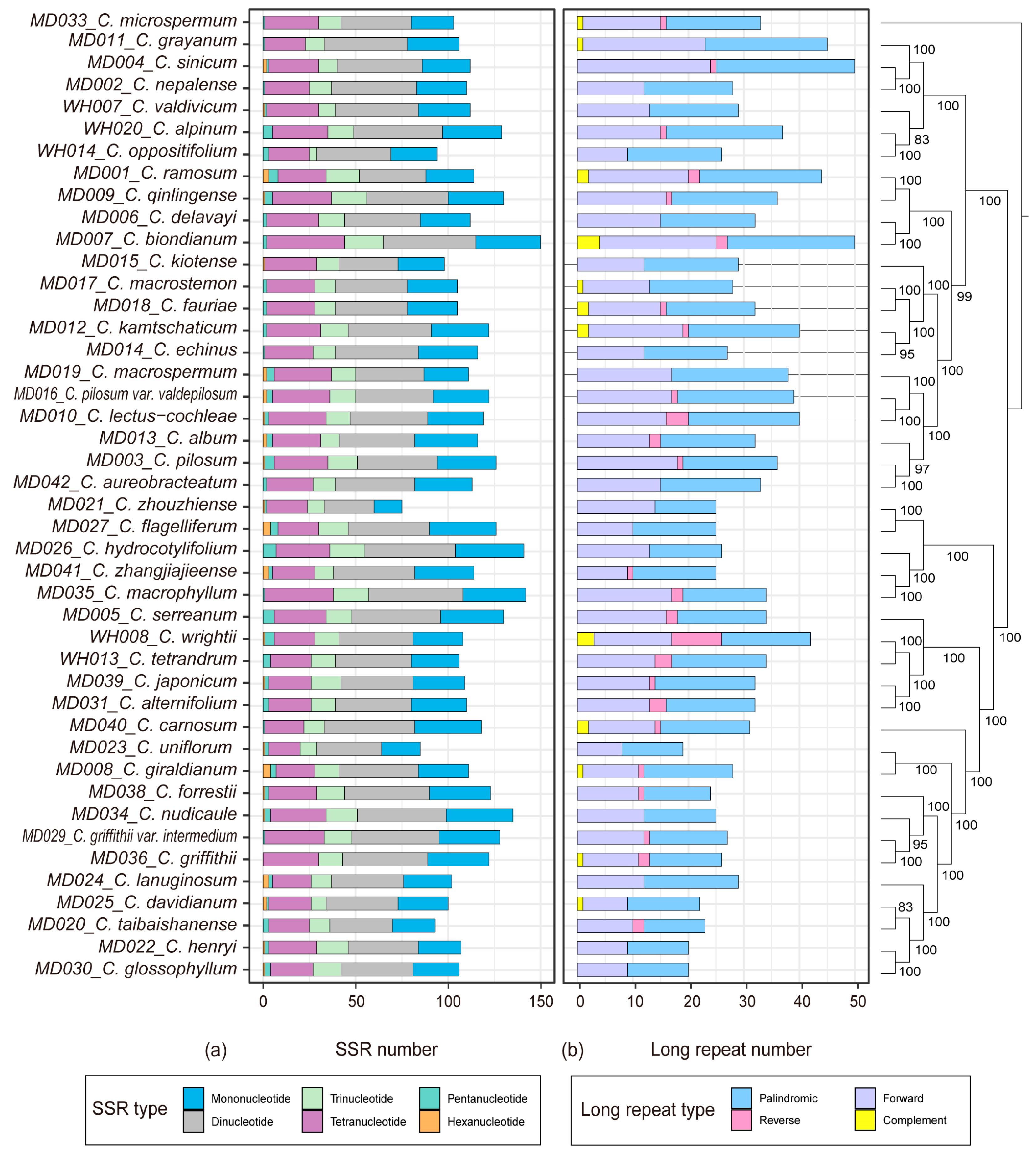
2.2. Repeat Identification

The MISA v. 1.0 software was utilized to detect simple sequence repeats (SSR) in 44 cp genomes of Chrysosplenium (Figure 2a; Table S3). The results of SSR analysis revealed a variation in the number of SSRs, ranging from 75 to 150. These SSRs were predominantly located in the LSC and SSC regions of the gene spacer Among the six types of SSRs, the largest number was dinucleotide repeats, accounting for 36.7%, followed by mononucleotide and tetranucleotide repeats, accounting for 25.5% and 22.9%, respectively. The smallest number was hexanucleotide repeats, accounting for only 0.85%. We examined the number and distribution of long repeats in the cp genomes of 44 Chrysosplenium species, which ranged from 19 to 50, with an average of 31 repeats, mainly in the IR and LSC regions (Figure 2b; Table S4). Fourteen Chrysosplenium species contained only forward and palindromic repeats, namely C. uniflorum, C. henryi, C. glossophyllum, C. zhouzhiense, C. flagelliferum, C. nudicaule, C. hydrocotylifolium, C. echinus, C. nepalense, C. kiotense, C. lanuginosum, C. delavayi, C. aureobracteatum, and C. macrospermum.

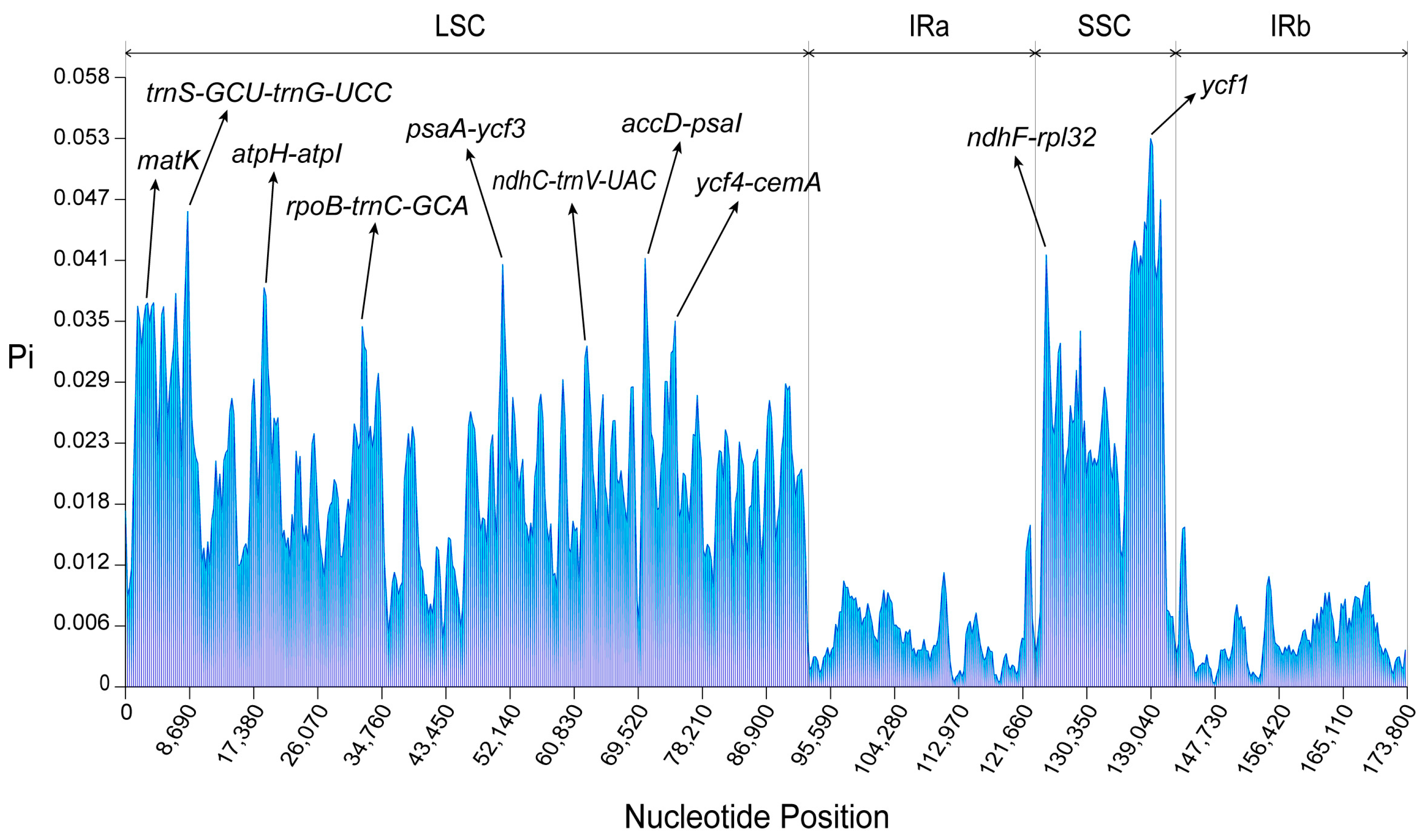
2.3. Divergence Hotspots and Rearrangement Analysis

To evaluate the differences in cp genomes among 44 Chrysosplenium species, we performed mVISTA analysis with the annotated C. ramosum cp genome as a reference (Figure S3). The cp genomes of the 44 Chrysosplenium species showed relatively similar patterns, with the main sequence variations observed in the non-coding regions. On the other hand, the exons and untranslated regions (UTR) exhibited minimal variation across genomes. Nucleotide diversity analysis revealed that coding regions were more conserved than non-coding regions. Among these hot spots, eight intergenic regions (IGSs) (trnS-GCU-trnG-UCC, atpH-atpI, rpoB-trnC-GCA, psaA-ycf3, ndhC-trnV-UAC, accD-psaI, ycf4-cemA, ndhF-rpl32) and two genes (matK, ycf1) showed the highest levels of divergence (Figure 3). Rearrangement analysis indicated that the cp genomes of 44 Chrysosplenium species were relatively conserved, and no significant rearrangements were found (Figures S4 and S5). Intraspecific variation also exists in the genus Chrysosplenium, mainly in the spacer region, e.g., C. sinicum (Figure S6).

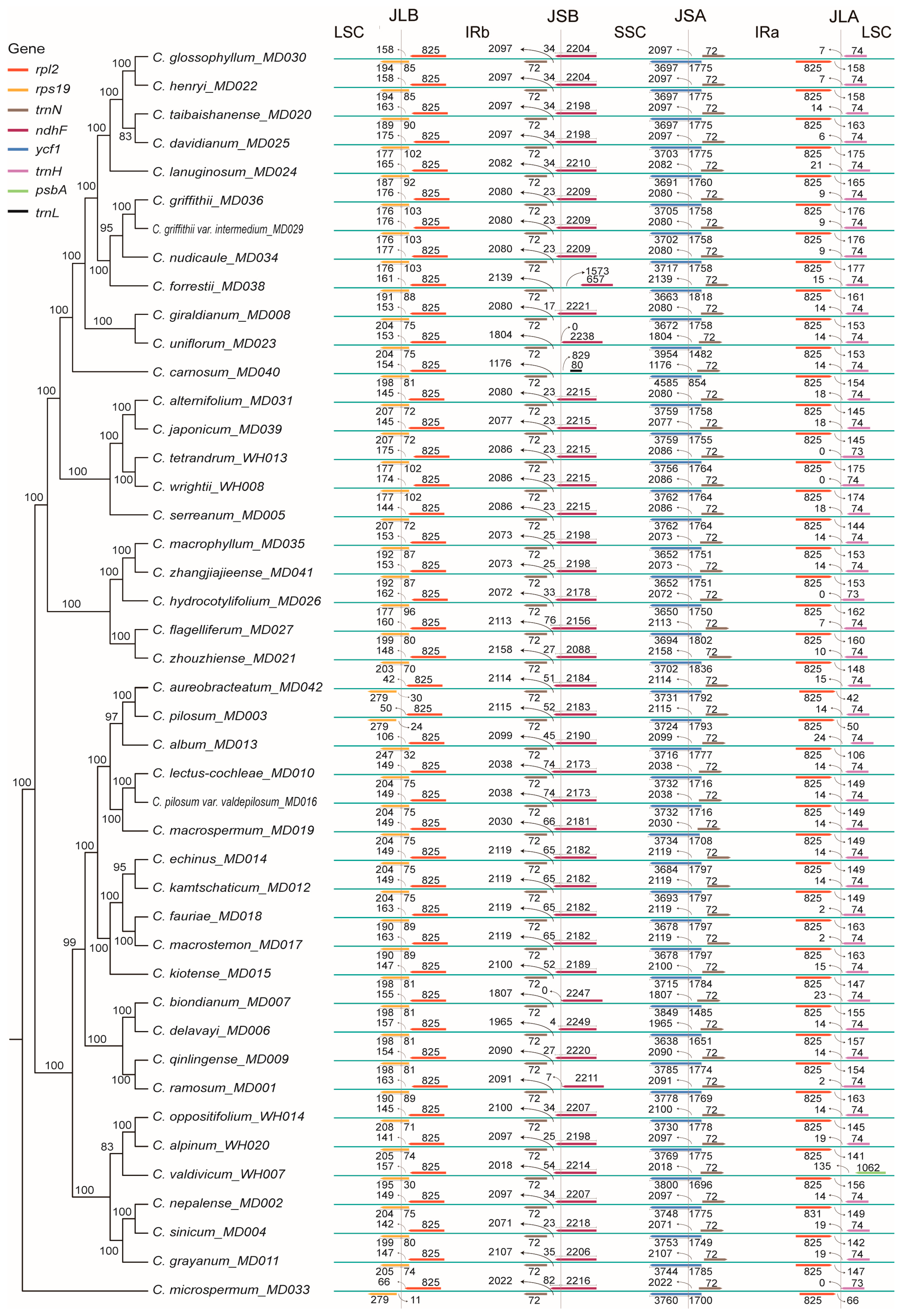
2.4. Dynamic Analysis of the IR Boundary

We analyzed the dynamics of the IR boundaries of the cp genomes of 44 Chrysosplenium species. The boundary situation is different for some species, and the expansion and contraction of the IR regions leads to changes in the cp genes at the IR boundaries, with some genes entering the LSC and SSC regions. The results showed that the four boundaries of the cp genomes of 44 Chrysosplenium species were relatively conserved (Figure 4). The rps19 genes of C. pilosum, C. microspermum, and C. aureobracteatum were located in the LSC region, and the rps19 genes of other 41 species were located in the LSC-IRb boundary. The vicinity of the IRb-SSC boundary mainly contained trnN and ndhF genes. The trnN genes were present in IRb in all Chrysosplenium species, while the ndhF genes of C. ramosum, C. biondianum, C. uniflorum, and C. forrestii were exclusively located in the SSC region. The ndhF genes of the other 40 Chrysosplenium species were located on the IRb-SSC boundary. The SSC-IRa boundary had ycf1 and trnN genes, with the ycf1 gene located on the boundary and the trnN gene located in the IRa region. Near the SSC-IRa boundary, there were also rpl2 and trnH genes, with the rpl2 gene located in the IRa region and the trnH gene located in the LSC region. This phenomenon is similar in all Chrysosplenium species.

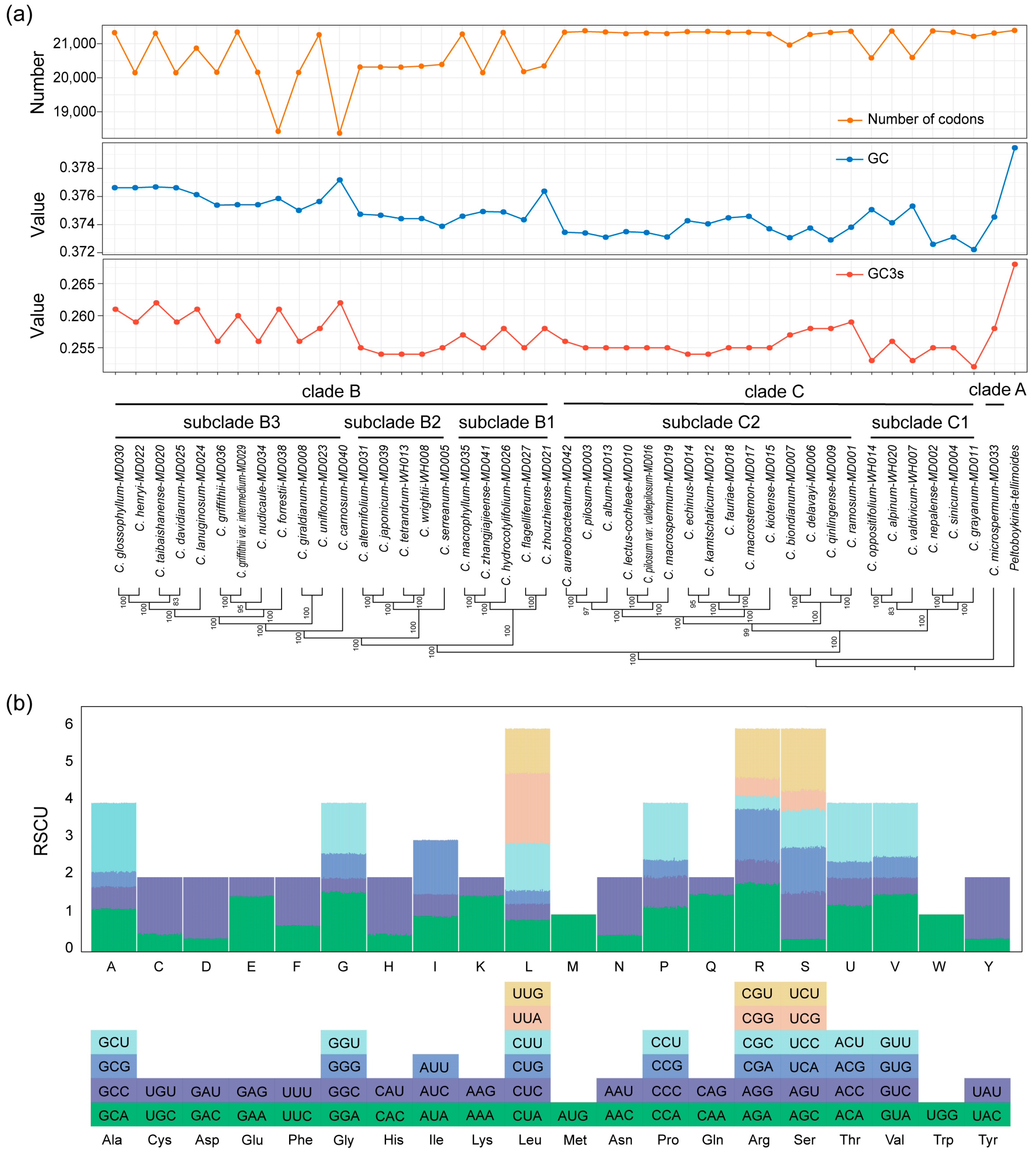
2.5. Codon Usage Analysis

We selected 53 PCGs (>300 bp) for codon usage analysis. Codon analysis revealed some differences in codon usage numbers, GCs, and GC3s among the 44 Chrysosplenium species (Figure 5a; Table S5). The C. forrestii and C. carnosum had lower codon numbers, with clade B showing greater variation in codon numbers than the clade C branch, which was overall more stable. Leucine was found to be the most abundant amino acid in the cp genome, while cysteine was relatively rare. Among the 61 codons, AAU encoded the most frequent occurrence of isoleucine and UGC encoded the least frequent occurrence of cysteine. The trend in GCs and GC3s was generally consistent and lower in 44 Chrysosplenium species than in P. tellimoides. Clade B generally had higher levels of GCs and GC3s than the C branch. Relative synonymous codon usage (RSCU) values of the 44 species were similar, with 61 codons encoding 20 amino acids (Figure 5b; Table S6). The RSCU value for serine encoded by AGC was the lowest, while leucine encoded by UUA had the highest RSCU value. Both tryptophan (UGG) and methionine (AUG) were encoded by only one codon and had RSCU values of one. Furthermore, twenty-nine codons had RSCU values greater than one, indicating biased use.

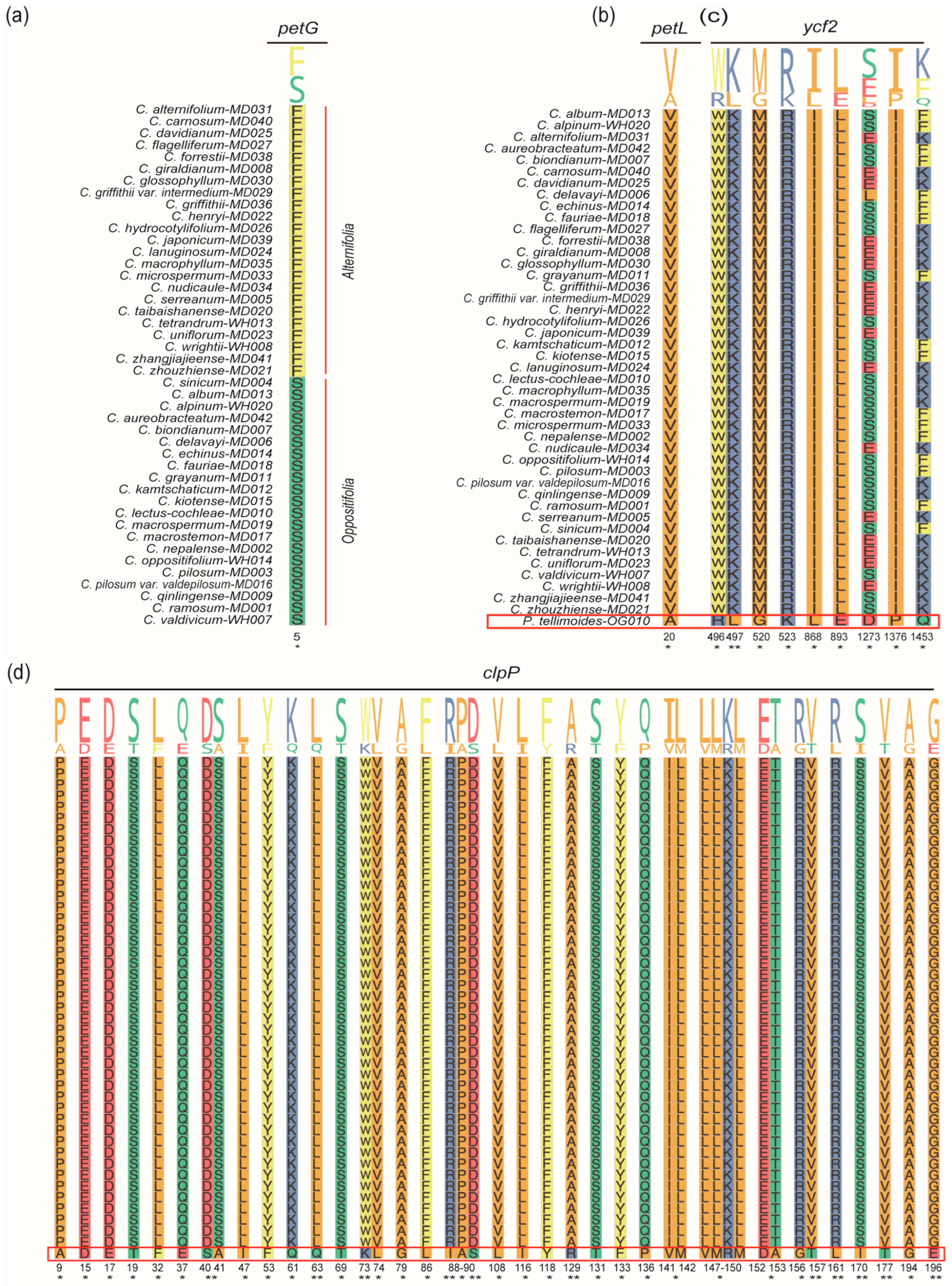
2.6. Selective Pressure Analyses

We analyzed selection pressure on the 44 Chrysosplenium species and P. tellimoides, with a total of 990 combinations. This result showed that the Chrysosplenium species were not subject to positive selection in the species level (Figure S7; Table S7). In the gene level, the LSC region had more positively selected genes (PSGs) than the SSC and IR regions in the sections of Alternifolia and Oppositifolia (Figure 6 and Figure S8; Table S8). PSGs in the LSC region included atpF, matK, ndhJ, psaI, psbK, psbL, rpl33, rps11, rps14, rps16, rps18, rps2, and rps8 (Figure 6a); PSGs in the IR region included ndhB and rps12 (Figure 6b). PSGs in the SSC region included ccsA, ndhE, and rps15 (Figure 6c); The remaining genes were generally subject to purifying selection. In addition, other PSGs were detected in the Alternifolia and Oppositifolia, respectively (Figure S9; Tables S9 and S10), such as: accD, atpE, atpI, cemA, clpP, ndhC, ndhK, petA, petD, psaJ, rbcL, rpl20, rpl22, rpoA, rps3, rps4, ycf3, ycf4, ccsA, ndhD, ndhE, ndhH, ndhI, rps15, ycf1, ndhB, rps12, and ycf2. Compared to P. tellimoides, the ycf2 and clpP genes were positively selected in 44 Chrysosplenium species and had multiple significant positive selection sites, and we also detected one significant positive selection site in the petL gene (Figure 6d, Figure 7b–d and Figure S8; Table S11). These PSGs may be associated with the adaptation of Chrysosplenium species to low-light and low-temperature environments. Furthermore, we did not detect a PSG that could significantly distinguish the two sections in the genus Chrysosplenium, but we detected a significant positive selection site on the petG gene, which may be related to the differential evolution of the two sections (Figure 7a; Table S12).

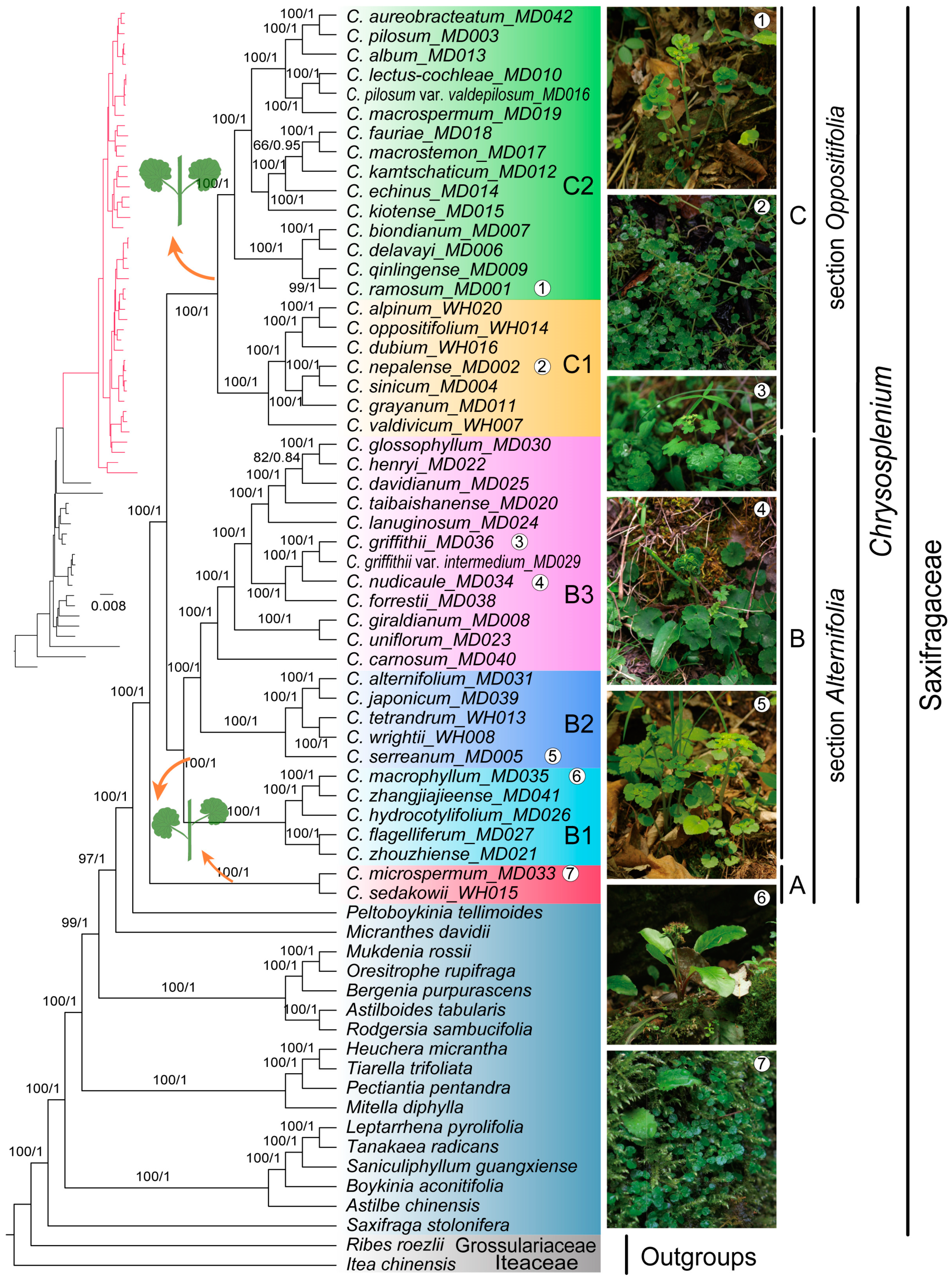
2.7. Phylogenetic Analysis

The cpPCGs matrix length was 72,828 bp, including 8401 parsimony informative sites, 16,649 variable sites and 52,882 conserved sites. The nrDNA matrix length was 6854 bp, including 926 parsimony informative sites, 1326 variable sites and 5169 conserved sites. The cpPCGs + nrDNA matrix length was 79,682 bp, including 9325 parsimony informative sites, 17,974 variable sites and 58,052 conserved sites (Table S14). Phylogenetic trees of the three matrices were constructed by the Maximum Likelihood and Bayesian Inference methods, respectively. The phylogenetic trees of both cpPCGs matrix and cpPCGs + nrDNA matrix have high confidence, while the nrDNA matrix phylogenetic tree as a whole has some branches with low support, which was significantly different from the phylogenetic trees of cpPCGs matrix and cpPCGs + nrDNA matrix (Figures S10–S15). The topology of the phylogenetic tree of cpPCGs + nrDNA matrix obtained by the two methods was similar, and most of the nodes had high support rates and posterior probabilities (Figure 8, Figures S12 and S13). The phylogenetic tree of cpPCGs + nrDNA matrix showed that Chrysosplenium species were more closely related to Peltoboykinia in the family Saxifragaceae. Three branches were formed within the Chrysosplenium, with C. microsperm and C. sedakowii with alternate leaves forming clade A alone, other alternate leaf species forming clade B, and opposite leaf species forming clade C. Clade A and clade B correspond to the Alternifolia, while clade C corresponds to the Oppositifolia, indicating that the two sections were not monophyletic. Clade B can be divided into three subclades, with species in subclade B1 generally distributed at low and middle altitudes, species in subclade B2 more widely distributed, and species in subclade B3 generally distributed at high altitudes in China. Clade C was mainly divided into two subbranches, with species in subclade C1 more widely distributed (e.g., Europe, North America, South America, and Asia), and subclade C2 species originate mainly from the northeastern regions of East Asia (northeastern China, Korea, North Korea, and Japan).

3. Discussion

3.1. Chloroplast Genome Evolution within Chrysosplenium Species

Our study analyzed the cp genomes of 44 Chrysosplenium species and found that they were not highly variable in size. The genomes were conserved in terms of structure, gene composition, and gene order, similar to many angiosperm genera. The distribution of GC content in the cp genome was uneven, with the highest GC content in the IR region and the lowest in the SSC region. The presence of four rRNAs (rrn4.5, rrn5, rrn16, rrn23) in the IR region may lead to the higher GC content [23,24,25]. Additionally, the expansion and contraction of the IR region also played a role in changing the size of the cp genome and the boundary genes. Comparison of the IR/SC boundaries among 44 Chrysosplenium species revealed high similarity. While the boundary region of the cp genome was relatively stable, the expansion and contraction of the IR region may lead to alterations in the ndhF and ycf1 genes located in the boundary region. It was observed that cp genes are rarely lost and are likely transferred to the nuclear genome or functionally replaced by nuclear genes [26,27,28]. Interestingly, we found that ndhA was lost in C. forrestii and C. carnosum, while ndhF and ndhG were lost in C. carnosum. Apart from the deletion of individual genes, no other significant variation was found. The deletion of genes may be related to the environment, such as the loss of the NDH gene in some orchids [29]. Differences between genomes also exist within species, a common phenomenon that may be due to genetic variation and geographic distribution during the evolution of the species. Similar differences exist in the genus Chrysosplenium, where sequence alignment revealed differences in the cp genomes of two different taxa of C. sinicum, mainly in the spacer region. However, there is still a lack of a more comprehensive resolution of genomic differences among intraspecific species in the genus Chrysosplenium.
A comparable number of SSRs and long repeats were identified in 44 different Chrysosplenium species. However, the types of SSRs and long repeats differed among the species. These repeats were predominantly found in the intergenic spacer (IGS) of the large single copy (LSC) region. Mononucleotide and dinucleotide repeats were the most common types of SSRs, while forward and palindromic repeats were the predominant types of long repeats. Since the Pi value of PCGs and IGSs was highest on average in IR, LSC and SSC, we found that matK, trnS-GCU-trnG-UCC, accD-psaI, ycf1, ndhF-rpl32, atpH-atpI, rpoB-trnC-GCA, psaA-ycf3, ycf4- cemA, and ndhC-trnV-UAC had high Pi values and were candidate markers to distinguish Chrysosplenium species, but further experimental studies were needed for specific conclusions.

3.2. Selection Pressure Analysis of Chrysosplenium Species

Species grow in various environments and are often influenced by different climatic factors, such as humidity, light, altitude and temperature. Some genes may be subject to positive selection in response to environmental changes. Our results indicate that the majority of genes exhibited an average Ka/Ks ratio below one. Purifying selection, a prominent mechanism of natural selection, plays a crucial role in continuously removing harmful mutations. These genes hold significant importance in facilitating plant adaptation and ensuring survival. Positive selection is usually associated with adaptive traits. Chrysosplenium species have a wide range of altitudinal distribution, both at low and high altitudes, and prefer shady and humid environments. In Chrysosplenium, most genes were found to be under purifying selection, and only a small number of genes were under positive selection across species, so the purifying selection of most cp genes may be the result of their adaptive evolution. No significant PSGs were detected in combinations of the Oppositifolia and Alternifolia species, and only some combinations were detected to contain PSGs. In terms of the PSG number, the LSC region was more numerous than the SSC and IR regions. Among them, psbL, rps18, ndhB, and rps12 genes showed strong positive selection in most species. Additionally, we detected the petG genes in both the Oppositifolia and Alternifolia to contain a significant locus despite not being under positive selection. The petG genes are primarily associated with photosynthesis [30], suggesting that there may be some differences in photosynthesis between the two sections. The P. tellimoides, ycf2 and clpP genes were subjected to significant positive selection in almost 44 Chrysosplenium species. In angiosperms, the ycf2 genes were susceptible to positive or purifying selection [31,32]. Although the exact function and role of ycf2 remains unclear, studies have shown that ycf2 genes were associated with photosynthesis, leaf patterning, cell survival and ATPase metabolism [33,34,35,36]. The positive selection of ycf2 genes indicated that this gene may be involved in the evolution of low-light adaptations in Chrysosplenium species. The clpP gene encoding clpP protease is also subject to positive selection in some angiosperms, such as Paphiopedilum (Orchidaceae) [37], Acacia (Fabaceae) [38], Bupleurum (Apiaceae) [39], and Ficus (Moraceae) [40], and shows high variability in Amaryllidaceae and Papilionoideae [41,42], suggesting that it may accelerate substitution rates in some angiosperms. The clpP protease can degrade or repair damaged proteins [43] and is important for plant development in response to environmental changes [44]. Thus, the positive selection of clpP gene may help Chrysosplenium species to adapt to low light and low temperature environments. In summary, these PSGs may contribute to the adaptation of different Chrysosplenium species to different environments and can be used as candidate genes to further investigate the adaptive evolutionary mechanism.

3.3. Phylogenetic Relationships of Chrysosplenium Species

The Maximum Likelihood and Bayesian Inference methods were used to construct phylogenetic trees for 44 Chrysosplenium species, and the topology of the phylogenetic trees obtained by these two methods was similar. The phylogenetic trees of cpPCGs and cpPCGs + nrDNA matrices had a similar structure with strong support, the use of nrDNA sequences alone was not well supported for some species, and there was a clear inconsistency between nucleoplasm. Nevertheless, these results suggest that Chrysosplenium was monophyletic, which was supported by previous studies [4,14,15,19,20]. Furthermore, our results showed the division of Chrysosplenium into three main clades, corresponding to the sections of Alternifolia (clade A and clade B) and Oppositifolia (clade C). Clade A (C. microspermum and C. sedakowii) with alternate leaves was located at the base of the Chrysosplenium branch, suggesting that it had a comparable evolutionary position in Chrysosplenium, but this was somewhat at variance with previous studies. Soltis et al. (2001) studied the phylogenetic relationships of some Chrysosplenium species based on matK sequences and showed that Chrysosplenium was divided into two mutually monophyletic branches [15]. However, some Chrysosplenium species such as C. microspermum and C. sedakowii were lacking in this study, and the phylogeny of Chrysosplenium was still not clear enough. A small number of cpDNA and nrITS markers were not sufficiently stable for the phylogenetic position of C. microspermum. Phylogeny using four chloroplast DNA and nrDNA markers showed C. microspermum clustered into a clade with opposite leaf species (C. nepalense), while phylogeny based on nrITS and trnL-trnF markers indicated that C. microspermum was located at the base of the Chrysosplenium. In Deng et al. (2015), ML trees based on partial cpDNAs and nucleoplasmic matrices of 29 Chrysosplenium species all indicated that C. microspermum was located at the base of the Chrysosplenium, but BI trees of the nucleoplasmic matrix did not show consistent results, with C. microspermum clustering with other alternate leaf species [18]. In Folk et al. (2019), although the Saxifragales phylogeny was analyzed using 301 phylogenetic loci, but the molecular data of Chrysosplenium was primarily based on partial chloroplast DNA data from previous studies [19]. This phylogeny showed that C. microspermum was clustered with other opposite leaf species and that C. sedakowii was located at the base of the Chrysosplenium. In this study, we provide more accurate support for the phylogeny of C. microspermum based on the cp genome and nrDNA data of 44 Chrysosplenium species. Our results support that C. microspermum was located at the base of the Chrysosplenium and was more closely related to C. sedakowii. This further showed that two sections of the Chrysosplenium were not monophyletic with each other.
Phylogenetic differences between nucleoplasm and between gene fragments may be due to various reasons, such as hybridization, incomplete lineage sorting, chloroplast capture, and plastid genetic differences. Hybridization occurs frequently in nature. Hybridization has the potential to result in gene trees that are inconsistent with species trees. Many naturally occurring hybrids, including intergeneric hybrids, have been reported in the family Saxifragaceae. Previous studies have suggested that hybridization occurs mainly in intergeneric hybrids between Heuchera and Tiarella, Tellima and Tolmiea, Mitella and Conimitella, and interspecific hybrids in Heuchera [45,46,47,48]. No hybridization events have been reported in Chrysosplenium species, but we cannot exclude the possibility of hybridization here. Incomplete lineage sorting is prevalent in most species phylogenies, where different fragments in the genome have different rates of evolution and conservation. The phylogenetic relationships constructed by different segments may differ somewhat from the true phylogenetic relationships and may also be related to chloroplast capture events. In the family Saxifragaceae, the Tiarella branch has been reported to have an apparent incongruity in the nucleoplasmic phylogeny, and the main reason for this incongruity was due to the fact that the Tiarella branch has captured at least two Heuchera cp genome events through an ancient ancestral hybridization [49]. In contrast, the phylogenetic trees for both nuclear and plastid phylogenies indicated that Chrysosplenium belonged to a monophyletic group, which was less likely for chloroplast capture of Chrysosplenium species with other genera, whereas it was possible to have chloroplast capture events within Chrysosplenium. Genetic differences in plastids may also have an effect on phylogeny, but this has not been reported in Chrysosplenium species. It is widely recognized that plastids are generally inherited matrilineally, but organelle genomes can also be mediated by biparental inheritance in the process of plant evolution. For example, Medicago truncatula and Pelargonium zonale exhibit frequent biparental inheritance [50,51]. Even in plants that are predominantly maternally inherited, such as Nicotiana tabacum and Arabidopsis thaliana, plastid genomes are occasionally inherited through pollen dispersal (paternal leakage) [52,53]. However, the causes and determinants of uniparental inheritance of organelles, as well as the underlying mechanisms of maternal inheritance, remain largely unknown. Previous cytological mechanisms of paternal inheritance of plastids have shown that mild low-temperature stress promotes the entry of paternal plastids into spermatocytes during male gametogenesis and significantly increases the inheritance of paternal plastids [54]. Chrysosplenium species prefer low temperature and low light environments, and it is possible that paternal plastid inheritance could be increased under low-temperature conditions, but further studies are needed. Therefore, in the future, there is a need not only to collect more Chrysosplenium species, but also to study a number of aspects such as nuclear and mitochondrial genomes, population genetics, plastid inheritance patterns, and chloroplast capture, in order to explore more accurate phylogenetic relationships within the genus and to construct a more believable phylogenetic network of the Chrysosplenium.

4. Materials and Methods

4.1. Sampling, DNA Extraction, and Sequencing

A total of 44 Chrysosplenium species covering the major continents of the world were used, of which 34 species of them were newly sequenced in this study. Species specimen number and collection location are listed in Table S1. Leaf tissue was dried in silica gel and genomic DNA was extracted using the modified CTAB method [55]. Whole genome resequencing was performed at Biomarker Technologies Company in Beijing, China. The short insertion library was constructed, and then 2 × 150 bp paired-end reads were obtained from the Illumina NovaSeq platform. The adaptors and low-quality reads were removed using Trimmomatic v. 0.39 [56], and then the filtered reads were quality-controlled using Fastqc v. 0.11.9 [57].

4.2. Chloroplast Genome Assembly and Annotation

The cp genomes of Chrysosplenium were assembled from clean short reads using GetOrganelle v.1.7.5 [58]. The assembly parameters used were “-R 20 -k 21, 45, 65, 85, 105, 127 -F embplant_pt”. Then, we used Bandage to check the integrity of the genomes. The cp genome annotation was performed using CPGAVAS2 [59], PGA [60] and Geneious Prime v. 2022.2.2 [61]. Protein-coding genes (PCGs) were extracted using PhyloSuite v. 1.2.3 [62]. The cp genome map was constructed using CPGview (http://www.1kmpg.cn/cpgview/ (accessed on 10 April 2023)) [63]. For the nrDNA sequences, we also used GetOrganelle v.1.7.5 [58] to assemble them, setting the parameter “-R 7 -k 35, 85, 115 -F embplant_nr” and then annotated with Geneious Prime v. 2022.2.2 [61].

4.3. Repeat Structure Identification

Simple sequence repeats (SSRs) were identified using the MicroSatellite (MISA) [64]. The minimum repeat number was set at 10, 5, 4, 3, 3, and 3 for mononucleotide, dinucleotide, trinucleotide, tetranucleotide, pentanucleotide, and hexanucleotide, respectively. REPuter (https://bibiserv.cebitec.uni-bielefeld.de/reputer (accessed on 10 April 2023)) was used to count the long repeats of the cp genomes, including palindrome sequences and interspersed repeats (complement repeats, forward repeats and reverse repeats) [65]. The minimum repeat and hamming were set to 30 and 3, respectively.

4.4. Codon Usage Analysis

To reduce sampling error, we excluded protein-coding genes (PCGs) shorter than 300 bp when analyzing codon usage patterns. A total of 53 CDSs were used for codon usage analysis. We utilized CodonW v.1.4.4 to determine the GC of the silent 3rd codons, effective number of codons, codon adaptation index, and number of synonymous codons. Additionally, we employed PhyloSuite v1.2.3 [62] to calculate the relative synonymous codon usage (RSCU) value. An RSCU value greater than 1 indicates higher frequency of codon usage than expected, while an RSCU value less than 1 indicates lower frequency of codon usage than expected.

4.5. Sequence Variation Analysis

The cp genomes of 44 Chrysosplenium species were compared using mVISTA in shuffle LAGAN mode, and C. ramosum were used as a reference. Multiple sequence alignment was performed using MAFFT. The DnaSP v. 6.12.03 [66] was used to calculate the nucleotide diversity (Pi) of the cp genome by using the sliding window. The step and window size were set to 200 bp and 600 bp, respectively. The LSC-IRa, IRa-SSC, SSC-IRb, and IRb-LSC boundaries of 44 cp genomes of Chrysosplenium were visualized using IRscope. In addition, mauve and AliTV [67] were used to detect genomic rearrangement events. We also compared the chloroplast genomes of two different taxa of C. sinicum using Geneious Prime v. 2022.2.2 [61].

4.6. Selective Pressure Analysis

The 74 cp PCGs of 44 Chrysosplenium species and one Peltoboykinia species were used to evaluate evolutionary rate variation. Positive selection analysis was performed in four parts, namely within Alternifolia branch, within Oppositifolia branch, between Alternifolia and Oppositifolia, between Chrysosplenium and Peltoboykinia. KaKs_Calculator v. 2.0 with YN model was used to determine the ratio of non-synonymous substitutions (Ka) and synonymous substitutions (Ks) [68]. Ka/Ks < 1 indicates that the gene may be under purifying selection. Ka/Ks > 1 indicates that the gene may be under positive selection. Ka/Ks = 1 indicates that the gene may be under neutral selection. When Ks = 0, the value of Ka/Ks is represented by NA, indicating that the gene has few nonsynonymous sites/substitutions.
We also used the branch-site model in EasyCodeML [69] to further detect the positive selection sites of genes. For the positive selection prediction between Chrysosplenium and Peltoboykinia, we set the Chrysosplenium branch as the foreground branch and the Peltoboykinia branch as the background branch. And within Chrysosplenium, we used the Oppositifolia branch as the foreground branch and the Alternifolia branch as the background branch.

4.7. Phylogenetic Analysis

We selected 19 species as outgroups for phylogenetic analysis (Table S13). Then, 74 common cpPCGs were extracted from the cp genome using PhyloSuite v.1.2.3 [62]. The 74 cpPCGs and nrDNA sequences were aligned separately using MAFFT v.7.4 [70], and then concatenated using PhyloSuite v.1.2.3 [62] to form a cpPCGs matrix, a cpPCGs + nrDNA matrix, and an nrDNA matrix. The phylogenetic tree was conducted using Maximum likelihood (ML) and Bayesian inference (BI) methods, respectively. ModelFinder [71] was used to find the best-fitting model for ML analysis, and the ML tree was further conducted using IQ-TREE v. 2.1.2 [72] with 1000 bootstrap replicates. For the BI tree, we used MrBayes v. 3.2.6 [73] to generate a maximum clade credibility (MCC) tree. The parameters were set as follows: nst = 6, rates = invgamma. The BI tree was performed with the concatenated sequence, using one million generations, two runs, four chains, a temperature of 0.001, and 25% of trees were discarded as burn-in, and trees were sampled every 1000 generations. The resulting tree was visualized using Figtree v. 1.4.4 (https://github.com/rambaut/figtree/Releases (accessed on 4 May 2023)).

5. Conclusions

In this study, we comprehensively performed assembly, comparative genomic, and phylogenetic analyses of multiple Chrysosplenium cp genomes. The analyses revealed that Chrysosplenium species were more conserved in terms of genome structure, gene content and arrangement, SSRs, and codon preference, but differ in genome size and SC/IR boundaries. Phylogenomic analysis showed that plastid data could effectively improve the phylogenetic support and resolution of Chrysosplenium species, strongly supporting Chrysosplenium as a monophyletic taxon and its internal division into three clades. The C. microspermum was not clustered with other Chrysosplenium species with alternate leaves but was clustered with C. sedakowii as the basal branch of Chrysosplenium. In addition, ten mutation hotspot regions were identified, which can be used as potential DNA barcodes for Chrysosplenium species identification. The clpP and ycf2 genes were significantly positively selected in the cp genome of Chrysosplenium compared to Peltoboykinia and had multiple positive selection sites of significance. One significant positive selection site was also detected in the petG gene between the two sections. These positive selection sites may have played an important role in the evolutionary history of the Chrysosplenium species for their low-light adaptation. In conclusion, this study enriches the cp genomes of the Chrysosplenium species and provides a reference for subsequent studies on its evolution and origin.

Supplementary Materials

The following supporting information can be downloaded at: https://www.mdpi.com/article/10.3390/ijms241914735/s1.

Author Contributions

Conceptualization, T.Y. and Z.W.; Formal analysis, T.Y., J.T. and R.Q.; Resources, R.Q. and J.W.; Writing—original draft, T.Y.; Writing—review and editing, Z.W., J.W. and H.L.; Visualization, T.Y. and Z.W.; Project administration, J.T., R.Q., J.W. and H.L.; Funding acquisition, H.L. All authors have read and agreed to the published version of the manuscript.

Funding

This research was funded by the Natural Science Foundation of China (No. 32170207).

Institutional Review Board Statement

Not applicable.

Informed Consent Statement

Not applicable.

Data Availability Statement

The data provided in the study are deposited in GenBank database (https://www.ncbi.nlm.nih.gov/ (accessed on 20 June 2023)) and youdao cloud note (https://note.youdao.com/s/Uf9wZj68 (accessed on 4 August 2023)). Vouchers and GenBank accession numbers are listed in Table S1.

Acknowledgments

We would like to thank Deqing Lan and Guihua Lu for assisting us in obtaining specimens for this study.

Conflicts of Interest

The authors declare no conflict of interest.

References

  1. Gitzendanner, M.A.; Soltis, P.S.; Wong, G.K.S.; Ruhfel, B.R.; Soltis, D.E. Plastid phylogenomic analysis of green plants: A billion years of evolutionary history. Am. J. Bot. 2018, 105, 291–301. [Google Scholar] [CrossRef]
  2. Ruhfel, B.R.; Gitzendanner, M.A.; Soltis, P.S.; Soltis, D.E.; Burleigh, J.G. From algae to angiosperms-inferring the phylogeny of green plants (Viridiplantae) from 360 plastid genomes. BMC Ecol. Evol. 2014, 14, 23. [Google Scholar] [CrossRef]
  3. Hara, H. Synopsis of the genus Chrysosplenium L. J. Fac. Sci. Univ. Tokyo 1957, 7, 1–90. [Google Scholar]
  4. Wu, Z.; Liao, R.; Yang, T.; Dong, X.; Lan, D.; Qin, R.; Liu, H. Analysis of six chloroplast genomes provides insight into the evolution of Chrysosplenium (Saxifragaceae). BMC Genom. 2020, 21, 621. [Google Scholar] [CrossRef]
  5. Pan, J.; Ohba, H. Chrysosplenium L; Science Press: Beijing, China, 2001; Volume 8, pp. 346–358. [Google Scholar]
  6. Fu, L.; Liao, R.; Lan, D.; Wen, F.; Liu, H. A new species of Chrysosplenium (Saxifragaceae) from Shaanxi, north-western China. PhytoKeys 2020, 159, 127–135. [Google Scholar] [CrossRef]
  7. Liu, H.; Luo, J.; Liu, Q.; Lan, D.; Qin, R.; Yu, X. A new species of Chrysosplenium (Saxifragaceae) from Zhangjiajie, Hunan, central China. Phytotaxa 2016, 277, 287–292. [Google Scholar] [CrossRef]
  8. Maximowicz, C.J. Diagnoses plantarum novarum asiaticarum scripsit. Bull. Acad. Imp. Sci. St.-Pétersbg. 1877, 23, 340–350. [Google Scholar]
  9. Franchet, A.R. Monographie du genere Chrysosplenium Tournfort; Nouvelles Archives du Muséum d’ histoire naturelle publiées par les professeurs-administrateurs de cet établissement: Pair, France, 1890; Volume 2, pp. 87–114. [Google Scholar]
  10. Jintang, P. A study on the genus Chrysosplenium L. from China. J. Univ. Chin. Acad. Sci. 1986, 24, 81–97. [Google Scholar]
  11. Jintang, P. A study on the genus Chrysosplenium L. from China (continued). J. Syst. Evol. 1986, 24, 203–214. [Google Scholar]
  12. Fu, L.; Yang, T.; Lan, D.; Wen, F.; Liu, H. Chrysosplenium sangzhiense (Saxifragaceae), a new species from Hunan, China. PhytoKeys 2021, 176, 21–32. [Google Scholar] [CrossRef]
  13. Koldaeva, M.N. Chrysosplenium fallax (Saxifragaceae), a new species from the Russian Far East. Phytotaxa 2021, 491, 35–46. [Google Scholar] [CrossRef]
  14. Nakazawa, M.; Wakabayashi, M.; Ono, M.; Murata, J. Molecular phylogenetic analysis of Chrysosplenium (Saxifragaceae) in Japan. J. Plant Res. 1997, 110, 265–274. [Google Scholar] [CrossRef]
  15. Soltis, D.E.; Tago-Nakazawa, M.; Xiang, Q.Y.; Kawano, S.; Murata, J.; Wakabayashi, M.; Hibsch-Jetter, C. Phylogenetic relationships and evolution in Chrysosplenium (Saxifragaceae) based on matK sequence data. Am. J. Bot. 2001, 88, 883–893. [Google Scholar] [CrossRef]
  16. Xiang, C.; Gitzendanner, M.A.; Soltis, D.E.; Peng, H.; Lei, L. Phylogenetic placement of the enigmatic and critically endangered genus Saniculiphyllum (Saxifragaceae) inferred from combined analysis of plastid and nuclear DNA sequences. Mol. Phylogenet. Evol. 2012, 64, 357–367. [Google Scholar] [CrossRef]
  17. Tkach, N.; Röser, M.; Hoffmann, M.H. Molecular phylogenetics, character evolution and systematics of the genus Micranthes (Saxifragaceae). Bot. J. Linn. Soc. 2015, 178, 47–66. [Google Scholar] [CrossRef]
  18. Deng, J.; Drew, B.T.; Mavrodiev, E.V.; Gitzendanner, M.A.; Soltis, P.S.; Soltis, D.E. Phylogeny, divergence times, and historical biogeography of the angiosperm family Saxifragaceae. Mol. Phylogenet. Evol. 2015, 83, 86–98. [Google Scholar] [CrossRef]
  19. Folk, R.A.; Stubbs, R.L.; Mort, M.E.; Cellinese, N.; Allen, J.M.; Soltis, P.S.; Soltis, D.E.; Guralnick, R.P. Rates of niche and phenotype evolution lag behind diversification in a temperate radiation. Proc. Natl. Acad. Sci. USA 2019, 116, 10874–10882. [Google Scholar] [CrossRef]
  20. Kim, Y.-i.; Lee, J.; Kim, Y. The complete chloroplast genome of a Korean endemic plant Chrysosplenium aureobracteatum Y.I. Kim & Y.D. Kim (Saxifragaceae). Mitochondrial DNA Part B 2018, 3, 380–381. [Google Scholar]
  21. Yan, W.; Liu, H.; Yang, T.; Liao, R.; Qin, R. Complete chloroplast genome sequence of Chrysosplenium ramosum and Chrysosplenium alternifolium (Saxifragaceae). Mitochondrial DNA Part B 2020, 5, 2843–2844. [Google Scholar] [CrossRef]
  22. Yan, W.; Yang, T.; Liao, R.; Wu, Z.; Qin, R.; Liu, H. Complete chloroplast genome sequence of Chrysosplenium macrophyllum and Chrysosplenium flagelliferum (Saxifragaceae). Mitochondrial DNA Part B 2020, 5, 2040–2041. [Google Scholar] [CrossRef]
  23. Lan, Z.; Shi, Y.; Yin, Q.; Gao, R.; Liu, C.; Wang, W.; Tian, X.; Liu, J.; Nong, Y.; Xiang, L.; et al. Comparative and phylogenetic analysis of complete chloroplast genomes from five Artemisia species. Front. Plant Sci. 2022, 13, 1049209. [Google Scholar] [CrossRef]
  24. Peng, J.Y.; Zhang, X.S.; Zhang, D.G.; Wang, Y.; Deng, T.; Huang, X.H.; Kuang, T.H.; Zhou, Q. Newly reported chloroplast genome of Sinosenecio albonervius Y. Liu & Q. E. Yang and comparative analyses with other Sinosenecio species. BMC Genom. 2022, 23, 639. [Google Scholar]
  25. Yan, K.; Ran, J.; Bao, S.; Li, Y.; Islam, R.; Zhang, N.; Zhao, W.; Ma, Y.; Sun, C. The Complete Chloroplast Genome Sequence of Eupatorium fortunei: Genome Organization and Comparison with Related Species. Genes 2022, 14, 64. [Google Scholar] [CrossRef]
  26. Magee, A.M.; Aspinall, S.; Rice, D.W.; Cusack, B.P.; Sémon, M.; Perry, A.S.; Stefanović, S.; Milbourne, D.; Barth, S.; Palmer, J.D.; et al. Localized hypermutation and associated gene losses in legume chloroplast genomes. Genome Res. 2010, 20, 1700–1710. [Google Scholar] [CrossRef]
  27. Gantt, J.S.; Baldauf, S.L.; Calie, P.J.; Weeden, N.F.; Palmer, J.D. Transfer of rpl22 to the nucleus greatly preceded its loss from the chloroplast and involved the gain of an intron. EMBO J. 1991, 10, 3073–3078. [Google Scholar] [CrossRef]
  28. Yagi, Y.; Shiina, T. Recent advances in the study of chloroplast gene expression and its evolution. Front. Plant Sci. 2014, 5, 61. [Google Scholar] [CrossRef]
  29. Lin, C.S.; Chen, J.J.W.; Chiu, C.C.; Hsiao, H.C.W.; Yang, C.J.; Jin, X.H.; Leebens-Mack, J.; de Pamphilis, C.W.; Huang, Y.T.; Yang, L.H.; et al. Concomitant loss of NDH complex-related genes within chloroplast and nuclear genomes in some orchids. Plant J. Cell Mol. Biol. 2017, 90, 994–1006. [Google Scholar] [CrossRef]
  30. Zhang, W.; Wang, H.; Dong, J.; Zhang, T.; Xiao, H. Comparative chloroplast genomes and phylogenetic analysis of Aquilegia. Appl. Plant Sci. 2021, 9, e11412. [Google Scholar] [CrossRef]
  31. Yan, C.; Du, J.; Gao, L.; Li, Y.; Hou, X. The complete chloroplast genome sequence of watercress (Nasturtium officinale R. Br.): Genome organization, adaptive evolution and phylogenetic relationships in Cardamineae. Gene 2019, 699, 24–36. [Google Scholar] [CrossRef]
  32. Zhong, Q.; Yang, S.; Sun, X.; Wang, L.; Li, Y. The complete chloroplast genome of the Jerusalem artichoke (Helianthus tuberosus L.) and an adaptive evolutionary analysis of the ycf2 gene. PeerJ 2019, 7, e7596. [Google Scholar] [CrossRef]
  33. Drescher, A.; Ruf, S.; Calsa, T., Jr.; Carrer, H.; Bock, R. The two largest chloroplast genome-encoded open reading frames of higher plants are essential genes. Plant J. Cell Mol. Biol. 2000, 22, 97–104. [Google Scholar] [CrossRef]
  34. Kikuchi, S.; Asakura, Y.; Imai, M.; Nakahira, Y.; Kotani, Y.; Hashiguchi, Y.; Nakai, Y.; Takafuji, K.; Bédard, J.; Hirabayashi-Ishioka, Y.; et al. A Ycf2-FtsHi Heteromeric AAA-ATPase Complex Is Required for Chloroplast Protein Import. Plant Cell 2018, 30, 2677–2703. [Google Scholar] [CrossRef]
  35. Wang, Q.M.; Cui, J.; Dai, H.; Zhou, Y.; Li, N.; Zhang, Z. Comparative transcriptome profiling of genes and pathways involved in leaf-patterning of Clivia miniata var. variegata. Gene 2018, 677, 280–288. [Google Scholar] [CrossRef]
  36. Zhang, Y.; An, D.; Li, C.; Zhao, Z.; Wang, W. The complete chloroplast genome of greater duckweed (Spirodela polyrhiza 7498) using PacBio long reads: Insights into the chloroplast evolution and transcription regulation. BMC Genom. 2020, 21, 76. [Google Scholar] [CrossRef]
  37. Guo, Y.; Yang, J.; Bai, M.; Zhang, G.; Liu, Z. The chloroplast genome evolution of Venus slipper (Paphiopedilum): IR expansion, SSC contraction, and highly rearranged SSC regions. BMC Plant Biol. 2021, 21, 248. [Google Scholar] [CrossRef]
  38. Dugas, D.V.; Hernandez, D.; Koenen, E.J.; Schwarz, E.; Straub, S.; Hughes, C.E.; Jansen, R.K.; Nageswara-Rao, M.; Staats, M.; Trujillo, J.T.; et al. Mimosoid legume plastome evolution: IR expansion, tandem repeat expansions, and accelerated rate of evolution in clpP. Sci. Rep. 2015, 5, 16958. [Google Scholar] [CrossRef]
  39. Huang, R.; Xie, X.; Chen, A.; Li, F.; Tian, E.; Chao, Z. The chloroplast genomes of four Bupleurum (Apiaceae) species endemic to Southwestern China, a diversity center of the genus, as well as their evolutionary implications and phylogenetic inferences. BMC Genom. 2021, 22, 714. [Google Scholar] [CrossRef]
  40. Zhang, Z.; Yang, X.; Li, W.Y.; Peng, Y.; Gao, J. Comparative chloroplast genome analysis of Ficus (Moraceae): Insight into adaptive evolution and mutational hotspot regions. Front. Plant Sci. 2022, 13, 965335. [Google Scholar] [CrossRef]
  41. Moghaddam, M.; Ohta, A.; Shimizu, M.; Terauchi, R.; Kazempour-Osaloo, S. The complete chloroplast genome of Onobrychis gaubae (Fabaceae-Papilionoideae): Comparative analysis with related IR-lacking clade species. BMC Plant Biol. 2022, 22, 75. [Google Scholar] [CrossRef]
  42. Liu, K.; Sun, L.; Meng, W.; Zhu, H.; Zhang, D.; Wang, J. Comparative genomics and phylogenetic perspectives of six fertile Lycoris species endemic to East Asia based on plastome characterization. Nord. J. Bot. 2022, 4, e03412. [Google Scholar] [CrossRef]
  43. Wicke, S.; Schneeweiss, G.M.; dePamphilis, C.W.; Müller, K.F.; Quandt, D. The evolution of the plastid chromosome in land plants: Gene content, gene order, gene function. Plant Mol. Biol. 2011, 76, 273–297. [Google Scholar] [CrossRef]
  44. Erixon, P.; Oxelman, B. Whole-gene positive selection, elevated synonymous substitution rates, duplication, and indel evolution of the chloroplast clpP1 gene. PLoS ONE 2008, 3, e1386. [Google Scholar] [CrossRef]
  45. Rosendahl, C.; Gortner, R.; Burr, G. A Monograph on the Genus Heuchera; University of Minnesota Press: Minneapolis, MN, USA, 1936; pp. 1–192. [Google Scholar]
  46. Spongberg, S.A. The Genera of Saxifragaceae in the Southeastern United States. J. Arnold Arbor. 1972, 53, 409–498. [Google Scholar] [CrossRef]
  47. Soltis, D.E.; Soltis, P.S. Intergeneric Hybridization between Conimitella williamsii and Mitella stauropetala (Saxifragaceae). Syst. Bot. 1986, 11, 293–297. [Google Scholar] [CrossRef]
  48. Soltis, D.E.; Bohm, B.A. Chromosomal and flavonoid chemical confirmation of intergeneric hybridization between Tolmiea and Tellima (Saxifragaceae). Can. J. Bot. 1985, 63, 1309–1312. [Google Scholar] [CrossRef]
  49. Liu, L.; Du, Y.; Folk, R.A.; Wang, S.; Soltis, D.E.; Shang, F.; Li, P. Plastome Evolution in Saxifragaceae and Multiple Plastid Capture Events Involving Heuchera and Tiarella. Front. Plant Sci. 2020, 11, 361. [Google Scholar] [CrossRef]
  50. Metzlaff, M.; Börner, T.; Hagemann, R. Variations of chloroplast DNAs in the genus Pelargonium and their biparental inheritance. Theor. Appl. Genet. 1981, 60, 37–41. [Google Scholar] [CrossRef]
  51. Matsushima, R.; Hu, Y.; Toyoda, K.; Sodmergen; Sakamoto, W. The Model Plant Medicago truncatula Exhibits Biparental Plastid Inheritance. Plant Cell Physiol. 2008, 49, 81–91. [Google Scholar] [CrossRef]
  52. Ruf, S.; Karcher, D.; Bock, R. Determining the transgene containment level provided by chloroplast transformation. Proc. Natl. Acad. Sci. USA 2007, 104, 6998–7002. [Google Scholar] [CrossRef]
  53. Azhagiri, A.K.; Maliga, P. Exceptional paternal inheritance of plastids in Arabidopsis suggests that low-frequency leakage of plastids via pollen may be universal in plants. Plant J. 2007, 52, 817–823. [Google Scholar] [CrossRef]
  54. Chung, K.P.; Gonzalez-Duran, E.; Ruf, S.; Endries, P.; Bock, R. Control of plastid inheritance by environmental and genetic factors. Nat. Plants 2023, 9, 68–80. [Google Scholar] [CrossRef]
  55. Doyle, J.J.; Doyle, J.L. A Rapid DNA Isolation Procedure for Small Quantities of Fresh Leaf Tissues. Phytochem. Bull. 1987, 19, 11–15. [Google Scholar]
  56. Bolger, A.M.; Lohse, M.; Usadel, B. Trimmomatic: A flexible trimmer for Illumina sequence data. Bioinformatics 2014, 30, 2114–2120. [Google Scholar] [CrossRef]
  57. Simon, A. FastQC: A Quality Control Tool for High Throughput Sequence Data. 2020. Available online: https://www.bioinformatics.babraham.ac.uk/projects/fastqc/ (accessed on 3 February 2023).
  58. Jin, J.; Yu, W.; Yang, J.; Song, Y.; dePamphilis, C.W.; Yi, T.; Li, D. GetOrganelle: A fast and versatile toolkit for accurate de novo assembly of organelle genomes. Genome Biol. 2020, 21, 241. [Google Scholar] [CrossRef]
  59. Shi, L.C.; Chen, H.M.; Jiang, M.; Wang, L.Q.; Wu, X.; Huang, L.F.; Liu, C. CPGAVAS2, an integrated plastome sequence annotator and analyzer. Nucleic Acids Res. 2019, 47, W65–W73. [Google Scholar] [CrossRef]
  60. Qu, X.; Moore, M.J.; Li, D.; Yi, T. PGA: A software package for rapid, accurate, and flexible batch annotation of plastomes. Plant Methods 2019, 15, 50. [Google Scholar] [CrossRef] [PubMed]
  61. Kearse, M.; Moir, R.; Wilson, A.; Stones-Havas, S.; Cheung, M.; Sturrock, S.; Buxton, S.; Cooper, A.; Markowitz, S.; Duran, C.; et al. Geneious Basic: An integrated and extendable desktop software platform for the organization and analysis of sequence data. Bioinformatics 2012, 28, 1647–1649. [Google Scholar] [CrossRef] [PubMed]
  62. Zhang, D.; Gao, F.; Jakovlić, I.; Zou, H.; Zhang, J.; Li, W.X.; Wang, G.T. PhyloSuite: An integrated and scalable desktop platform for streamlined molecular sequence data management and evolutionary phylogenetics studies. Mol. Ecol. Resour. 2020, 20, 348–355. [Google Scholar] [CrossRef] [PubMed]
  63. Greiner, S.; Lehwark, P.; Bock, R. OrganellarGenomeDRAW (OGDRAW) version 1.3.1: Expanded toolkit for the graphical visualization of organellar genomes. Nucleic Acids Res. 2019, 47, W59–W64. [Google Scholar] [CrossRef]
  64. Beier, S.; Thiel, T.; Münch, T.; Scholz, U.; Mascher, M. MISA-web: A web server for microsatellite prediction. Bioinformatics 2017, 33, 2583–2585. [Google Scholar] [CrossRef]
  65. Kurtz, S.; Choudhuri, J.V.; Ohlebusch, E.; Schleiermacher, C.; Stoye, J.; Giegerich, R. REPuter: The manifold applications of repeat analysis on a genomic scale. Nucleic Acids Res. 2001, 29, 4633–4642. [Google Scholar] [CrossRef]
  66. Rozas, J.; Ferrer-Mata, A.; Sánchez-DelBarrio, J.C.; Guirao-Rico, S.; Librado, P.; Ramos-Onsins, S.E.; Sánchez-Gracia, A. DnaSP 6: DNA Sequence Polymorphism Analysis of Large Data Sets. Mol. Biol. Evol. 2017, 34, 3299–3302. [Google Scholar] [CrossRef]
  67. Ankenbrand, M.J.; Hohlfeld, S.; Hackl, T.; Förster, F. AliTV—Interactive visualization of whole genome comparisons. PeerJ Comput. Sci. 2017, 3, e116. [Google Scholar] [CrossRef]
  68. Wang, D.; Zhang, Y.; Zhang, Z.; Zhu, J.; Yu, J. KaKs_Calculator 2.0: A toolkit incorporating gamma-series methods and sliding window strategies. Genom. Proteom. Bioinform. 2010, 8, 77–80. [Google Scholar] [CrossRef]
  69. Gao, F.; Chen, C.; Arab, D.A.; Du, Z.; He, Y.; Ho, S.Y.W. EasyCodeML: A visual tool for analysis of selection using CodeML. Ecol. Evol. 2019, 9, 3891–3898. [Google Scholar] [CrossRef] [PubMed]
  70. Katoh, K.; Standley, D.M. MAFFT multiple sequence alignment software version 7: Improvements in performance and usability. Mol. Biol. Evol. 2013, 30, 772–780. [Google Scholar] [CrossRef] [PubMed]
  71. Kalyaanamoorthy, S.; Minh, B.Q.; Wong, T.K.F.; von Haeseler, A.; Jermiin, L.S. ModelFinder: Fast model selection for accurate phylogenetic estimates. Nat. Methods 2017, 14, 587–589. [Google Scholar] [CrossRef] [PubMed]
  72. Nguyen, L.T.; Schmidt, H.A.; von Haeseler, A.; Minh, B.Q. IQ-TREE: A Fast and Effective Stochastic Algorithm for Estimating Maximum-Likelihood Phylogenies. Mol. Biol. Evol. 2014, 32, 268–274. [Google Scholar] [CrossRef] [PubMed]
  73. Ronquist, F.; Teslenko, M.; van der Mark, P.; Ayres, D.L.; Darling, A.; Höhna, S.; Larget, B.; Liu, L.; Suchard, M.A.; Huelsenbeck, J.P. MrBayes 3.2: Efficient Bayesian phylogenetic inference and model choice across a large model space. Syst. Biol. 2012, 61, 539–542. [Google Scholar] [CrossRef]
Figure 1. Representative chloroplast genome map of Chrysosplenium. The colored boxes in the figure represent genes. Genes located inside the circle are transcribed in a clockwise direction, while genes outside the circle are transcribed in a counter-clockwise direction. The small grey bar graphs in the inner circle indicate the GC contents. Black boxes indicate the absence or presence of individual genes in some Chrysosplenium species.
Ijms 24 14735 g001
Figure 2. Repeat analysis of chloroplast genomes of Chrysosplenium species. (a) SSR statistics of Chrysosplenium species. Different types of SSRs are indicated by different colors. (b) Long repeat statistics of Chrysosplenium species. Different types of repeats are indicated by different colors. The values on the nodes indicate the ML bootstrap support values.
Ijms 24 14735 g002
Figure 3. Nucleotide diversity (Pi) analysis of cp genomes of 44 Chrysosplenium species. The sliding window and step size used for this analysis were set to 600 bp and 200 bp, respectively.
Ijms 24 14735 g003
Figure 4. Dynamic analysis of the IR boundary of cp genomes of the 44 Chrysosplenium species. The values on the nodes indicate the ML bootstrap support values. Arrows indicate the distance of these genes from the IR boundary.
Ijms 24 14735 g004
Figure 5. Codon characterization of PCGs in the cp genomes of 44 Chrysosplenium species. (a) Number of codons used, GC3s and GC content analysis. The values on the nodes indicate the ML bootstrap support values. (b) Codon preference (RSCU) analysis.
Ijms 24 14735 g005
Figure 6. Selection pressure analysis of the genus Chrysosplenium. (a) Positive selection genes between Alternifolia and Oppositifolia and their species combinations in the LSC region. (b) Positive selection genes between Alternifolia and Oppositifolia and their species combinations in the IR region. (c) Positive selection genes between Alternifolia and Oppositifolia and their species combinations in the SSC region. (d) Positive selection genes ycf2 and clpP between Chrysosplenium and Peltoboykinia and their species combinations.
Ijms 24 14735 g006
Figure 7. Analysis of positive selection sites in PCGs in the cp genome. (a) Positive selection sites between Alternifolia and Oppositifolia in the genus Chrysosplenium. (bd) Positive selection sites between Chrysosplenium and Peltoboykinia. One asterisk indicates significance level less than 0.05; two asterisks indicate significance level less than 0.01.
Ijms 24 14735 g007
Figure 8. Phylogenetic tree of Chrysosplenium species using Maximum likelihood (ML) and Bayesian inference (BI) based on cpPCGs + nrDNA matrix. The values on the nodes indicate the ML bootstrap support values (left) and BI posterior probabilities (right). The circle numbers in the species picture correspond to the circle numbers behind the species name, respectively.
Ijms 24 14735 g008
Disclaimer/Publisher’s Note: The statements, opinions and data contained in all publications are solely those of the individual author(s) and contributor(s) and not of MDPI and/or the editor(s). MDPI and/or the editor(s) disclaim responsibility for any injury to people or property resulting from any ideas, methods, instructions or products referred to in the content.

