Article

Dual-Species Biofilms: Biomass, Viable Cell Ratio/Cross-Species Interactions, Conjugative Transfer

by
Marina V. Kuznetsova
1,
Julia S. Pospelova
2,
Irina L. Maslennikova
1 and
Marjanca Starčič Erjavec
3,*
1
Institute of Ecology and Genetics of Microorganisms Ural Branch Russian Academy of Sciences, 614081 Perm, Russia
2
LLC Centralized Clinical Diagnostic Laboratory, 614025 Perm, Russia
3
Department of Microbiology, Biotechnical Faculty, University of Ljubljana, 1000 Ljubljana, Slovenia
*
Author to whom correspondence should be addressed.
Int. J. Mol. Sci. 2023, 24(19), 14497; https://doi.org/10.3390/ijms241914497
Submission received: 30 August 2023 / Revised: 19 September 2023 / Accepted: 19 September 2023 / Published: 24 September 2023

Abstract

:
Biofilms as a form of adaptation are beneficial for bacterial survival and may be hot spots for horizontal gene transfer, including conjugation. The aim of this research was to characterize the biofilm biomass, viable cell ratios and conjugative transfer of the pOX38 plasmid, an F-plasmid derivative, from the Escherichia coli N4i pOX38 strain (donor) into a uropathogenic E. coli DL82 strain (recipient) within dual-species biofilms with one of the following opportunistic pathogenic bacteria: Klebsiella pneumoniae, Enterococcus faecalis or Pseudomonas aeruginosa. Dual-species biofilms of E. coli with K. pneumoniae or P. aeruginosa but not E. faecalis were more massive and possessed more exopolysaccharide matrix compared to single-species biofilms of donor and recipient cells. Correlation between biofilm biomass and exopolysaccharide matrix was rs = 0.888 in dual-species biofilms. In dual-species biofilm with E. faecalis the proportion of E. coli was the highest, while in the biofilm with P. aeruginosa and K. pneumoniae, the E. coli was less abundant. The conjugative frequencies of plasmid transfer in dual-species biofilms of E. coli with E. faecalis and P. aeruginosa were reduced. A decrease in conjugative frequency was also observed when cell-free supernatants (CFSs) of E. faecalis and P. aeruginosa were added to the E. coli conjugation mixture. Further, the activity of the autoinducer AI-2 in the CFSs of the E. coli conjugation mixture was reduced when bacteria or CFSs of E. faecalis and P. aeruginosa were added to the E. coli conjugation mixture. Hence, the intercellular and interspecies interactions in dual-species biofilms depend on the partners involved.

1. Introduction

It is well known that biofilms are a natural form of existence for most bacteria in natural ecosystems and human biotopes [1,2]. Biofilms on biotic and abiotic surfaces are a major problem in medical, environmental, industrial and agricultural settings [3,4,5]. As bacteria in biofilms have an increased resistance to antibiotics, biofilms, including multi-species biofilms, pose a health risk in medical practice. In addition, biofilms can be reservoirs of bacteria that can cause constant reinfection and chronic inflammation, which can also lead to tissue damage, clogging of devices and general resistance to treatment [6,7]. In biofilms associated with urinary tract infections (UTIs), Escherichia coli strains are most prevalent, but other commensals and opportunistic pathogens such as Klebsiella pneumoniae, Enterococcus faecalis and Pseudomonas aeruginosa can also be found [8,9]. Bacterial communication within the biofilm allows them to orchestrate the expression of virulence genes, which further cements the infestation and increases the invasiveness of the infection [10]. These facts emphasize the need to study biofilms of uropathogenic bacteria by describing the mechanisms of interspecies and intraspecies interactions that underlie the formation and functioning of polymicrobial biofilms.
There are a number of studies on the mutual influence of opportunistic pathogenic bacteria on the formation of polymicrobial biofilms. Oliveira et al. (2018) showed that the number of E. coli cells in a mixed biofilm formed during co-cultivation with P. aeruginosa could be higher or lower than in a single-species one, depending on the surface on which the bacteria formed the biofilm (polystyrene 96-well microtiter plate vs. porcine skin explants, respectively) [11]. It was found that the viability of E. coli cells in biofilm is suppressed by K. pneumoniae [12]. Further, it has also been shown that the presence of E. coli can interfere with the attachment of E. faecalis to the surface, slowing the development of a mixed biofilm [13]. Similar data have been obtained with clinical isolates of E. coli which secreted polysaccharides that altered the properties of abiotic surfaces, thereby inhibiting biofilm formation of Gram-positive bacteria, including E. faecalis [14].
Bacterial communication is important both in competition and in synergistic relationships that lead to enhanced metabolic cooperation. The most studied form of cell interaction in bacterial communities is known as quorum sensing (QS). QS response is mediated by different extracellular autoinducer molecules, among which is AI-2 (autoinducer 2; LuxS), a furanosyl borate diester which is an interspecies autoinducer. Its gene luxS is present in many bacterial species (E. coli, K. pneumoniae, E. faecalis). It is known that AI-2 affects virulence and biofilm formation of E. coli, K. pneumoniae, E. faecalis and P. aeruginosa [15,16,17,18,19,20], but the role of AI-2 in mixed cultures is not well understood [21,22].
Interspecies interactions within the biofilm affect the growth and survival of all members, their cellular metabolic processes, and the intra- and interspecific exchange of genetic information. Interspecies interactions therefore affect the development, structure, and functions of multispecies biofilms [23,24,25]. Quorum-sensing autoinducer molecules produced by members of a multispecies biofilm promote horizontal gene transfer [26], which is important for the emergence of multiple drug-resistant bacteria associated with the widespread use of antibiotics in medicine and veterinary medicine [27]. F- and F-like plasmids are of great importance for the maintenance and dissemination of the ecophysiological traits of bacteria: growth, reproduction rate, biofilm formation and resistance to antibiotics and bacteriocins [28,29,30]. Transfer of the conjugative pOX38 plasmid into UPEC strains has been shown to depend on biofilm biomass [31]. It is assumed that the presence of opportunistic pathogenic bacteria or their cell-free supernatants (CFSs) may disrupt the optimal ratio of donor and recipient cells and may affect the efficiency of horizontal gene transfer.
The aim of this work was to characterize the biofilms’ biomass, viable cell ratios/cross-species interactions and conjugative transfer abilities in dual-species biofilms of E. coli with following opportunistic pathogenic bacteria: K. pneumoniae, P. aeruginosa and E. faecalis.

2. Results

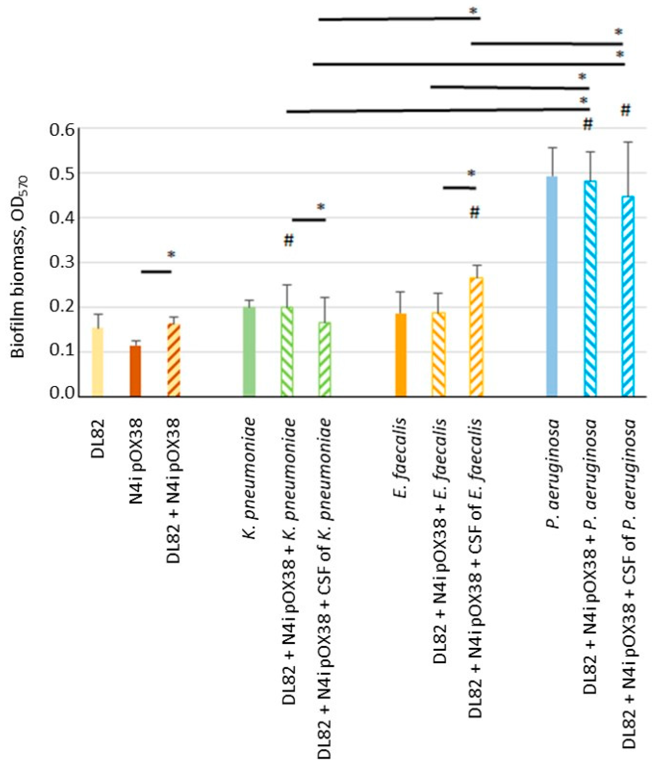
2.1. Biofilm Biomass

The determined biofilm biomass is shown in Figure 1. The biomass of single-species biofilms of the two E. coli strains, recipient DL82 (R) and donor N4i pOX38 (D), K. pneumoniae and E. faecalis were similar to each other and OD570 ranged from 0.1 to 0.2. The biofilm biomass formed by P. aeruginosa was significantly higher and almost reached, on average, an OD570 of 0.5 (p = 0.01). The dual-species biofilms of E. coli with K. pneumoniae and E. coli with P. aeruginosa were significantly more massive than the single-species E. coli biofilm (R + D) (p = 0.022 and p = 0.0001, respectively). In cases where, instead of the cells of opportunistic species, CFSs of opportunistic pathogenic bacteria were added, the E. coli biomass was significantly more massive with the CFSs of E. faecalis and P. aeruginosa than it was with the single-species E. coli biofilm (R + D) (p = 0.0001 and p = 0.0001, respectively). Cell-free supernatant (CFS) of K. pneumoniae as well as CFS of P. aeruginosa decreased the biofilm’s biomass, while the CFS of E. faecalis increased the biofilm’s biomass when compared with dual-species biofilms of E. coli with opportunistic pathogenic bacteria (p = 0.026 and p = 0.0001, respectively). Among studied opportunistic potentially pathogenic bacterial species, P. aeruginosa cells and the P. aeruginosa CFS, when added to E. coli (R + D), contributed to a much higher increase in the biofilm’s biomass compared to K. pneumoniae and E. faecalis cells and CFSs (p = 0.0001).

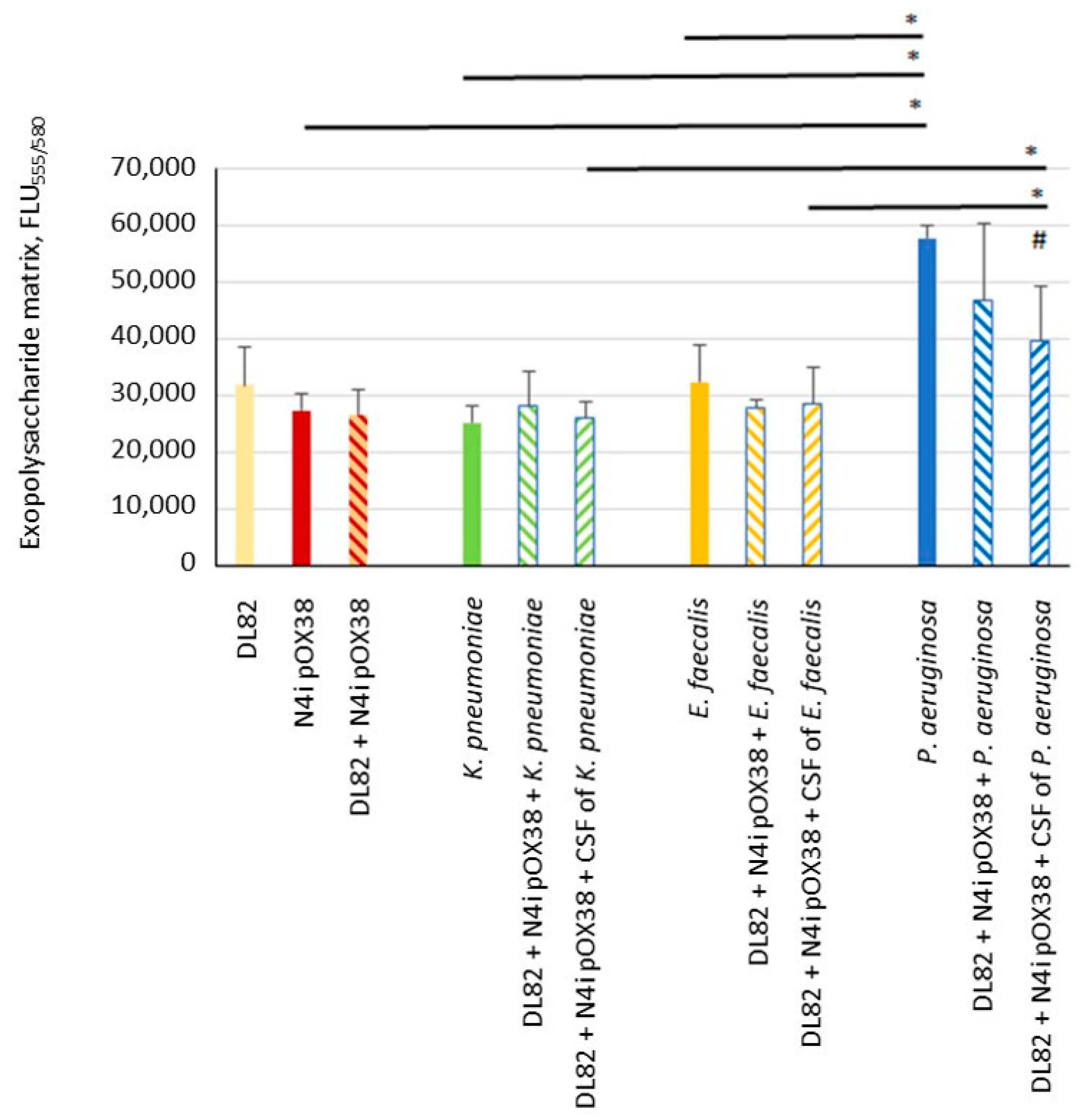
2.2. Biofilm Exopolysaccharide Matrix

The determined massiveness of the exopolysaccharide matrix of biofilms is shown in Figure 2. The exopolysaccharide matrix massiveness of single-species biofilms of the two E. coli strains, recipient DL82 and donor E. coli N4i pOX38, and two opportunistic pathogenic bacteria, K. pneumoniae and E. faecalis, were similar to each other, and FLU555/580 ranged from 25,000 to 32,000. The exopolysaccharide matrix massiveness of the biofilm formed by P. aeruginosa was significantly higher than that of the matrix of donor E. coli N4i pOX38, K. pneumoniae and E. faecalis, and almost reached an FLU555/580 of 60,000 (p = 0.001). The donor and the recipient exopolysaccharide amounts did not differ in single- and mixed-species biofilms, except for the exopolysaccharide amount of the biofilm formed when P. aeruginosa or its CFS was added. The correlation between biofilm biomass and exopolysaccharide matrix was rs = 0.888, indirectly indicating that in highly exopolysaccharide-producing bacteria, the matrix plays a more important role in biofilm biomass than do the cells.

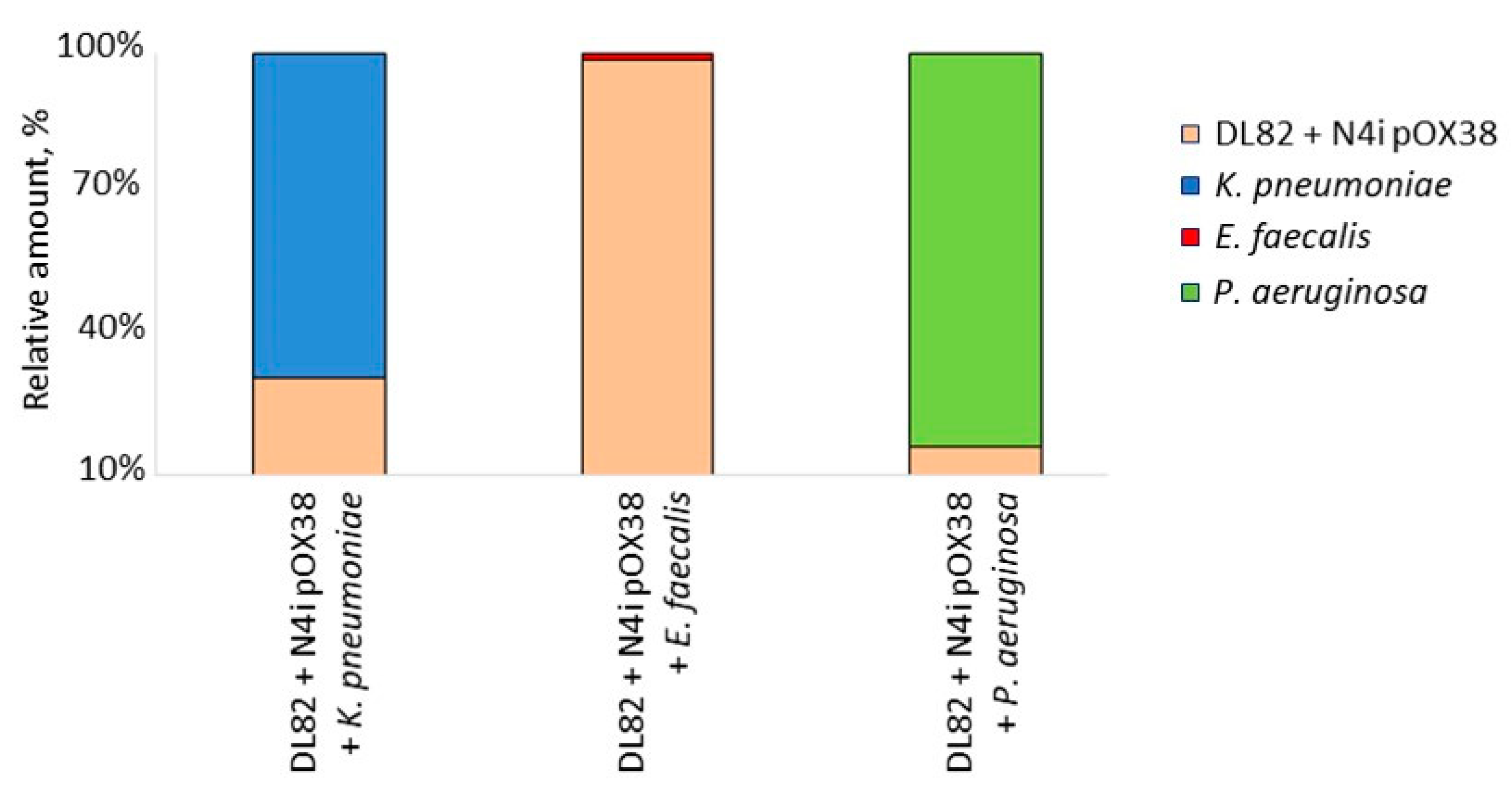
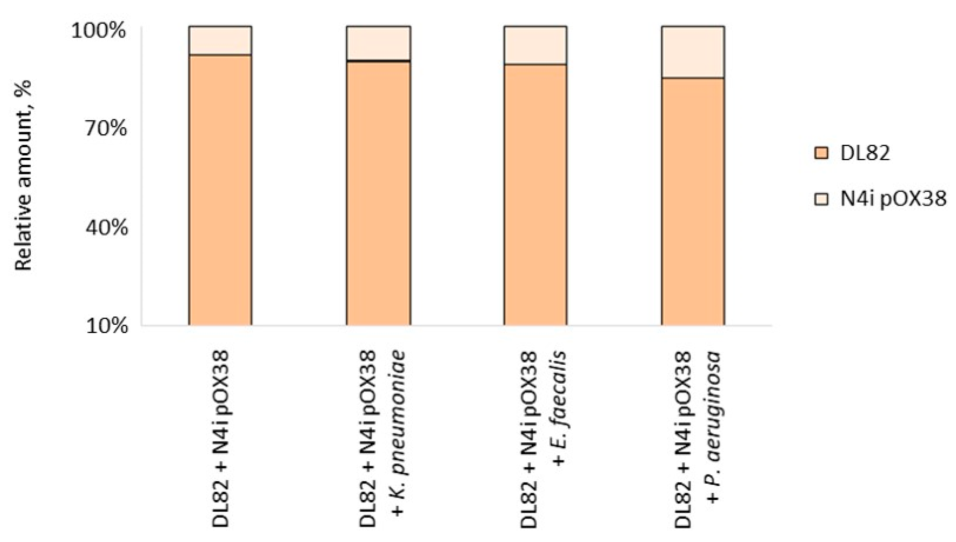
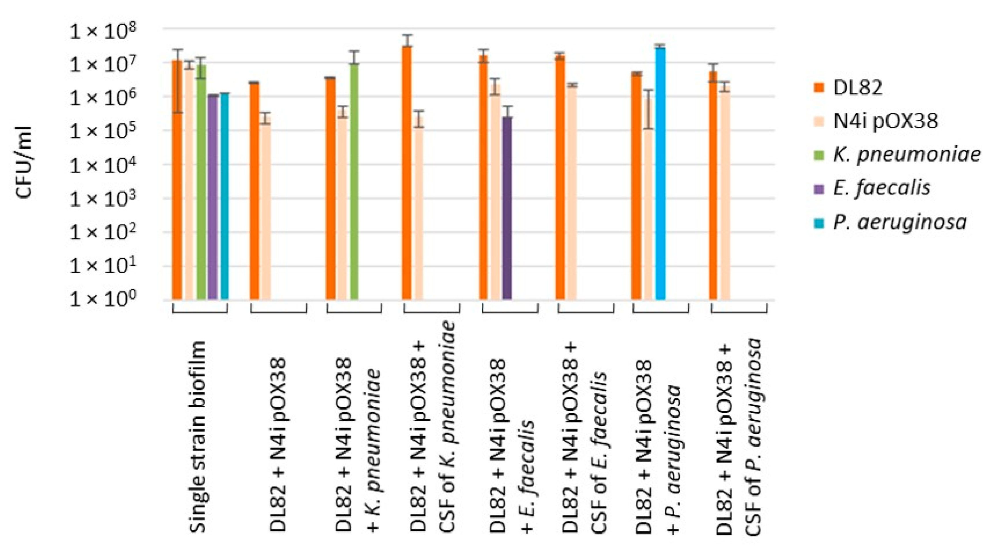
2.3. The Viability of Recipient E. coli DL82, Donor E. coli N4i pOX38 and Opportunistic Pathogenic Bacteria within Dual-Species Biofilms

The ratio of viable E. coli-opportunistic pathogenic bacterial cells in biofilms of dual-species cultures after 24 h differed in biofilms with different opportunistic pathogenic bacteria (Figure 3), even though all cultures initially started with the same ratio of E. coli-opportunistic pathogenic bacteria. After 24h incubation, the proportion of E. coli was the highest (98.76%) in the dual-species biofilm with E. faecalis, while in the biofilm with P. aeruginosa, there was a proportion of only 16.38% E. coli cells. E. coli was also less abundant in the dual-species biofilm formed with K. pneumoniae (30.79%).
It should be noted that despite the different proportion of E. coli in the studied dual-species biofilms, the ratios of recipient and donor cells in the studied biofilms did not change significantly (Figure 4).

2.4. The Frequency of Conjugation within Dual-Species Biofilm

The CFU data used to calculate the conjugation frequency are shown in Figure 5. There were no statistically significant differences in the CFUs of recipient E. coli DL82 and donor E. coli N4i pOX38 in single-species biofilms, dual-species biofilms with opportunistic pathogenic bacteria, or in single-species biofilms with added CFSs of opportunistic pathogenic bacteria (Figure 5). There was also no statistically significant difference in the CFUs of opportunistic pathogenic bacteria in single-species biofilms compared to dual-species biofilms (Figure 5).
The frequencies of conjugative transfer of the F-plasmid derivative from the donor E. coli N4i pOX38 (D) into the recipient E. coli DL82 (R) in biofilms are shown in Table 1. As can be seen from Table 1, the conjugative frequencies of plasmid transfer in dual-species biofilms (E. coli (R + D) + E. faecalis/P. aeruginosa cells) were reduced (U-test: p = 0.0495) in contrast to the K. pneumoniae dual-species biofilm. Addition of CFSs of opportunistic pathogenic bacteria (K. pneumoniae or E. faecalis) to the conjugation mixture resulted in lower conjugative frequencies (decreased by about 1.5 orders); conjugation was completely absent in the presence of P. aeruginosa CFS.
Statistical analysis revealed several negative correlations: between conjugation frequency and total biofilm biomass (rs = −0.440), between conjugation frequency and exopolysaccharide matrix of biofilms (rs = −0.270), between conjugation frequency and CFU of E. coli in biofilm (rs = −0.390) and between conjugation frequency and the total CFU number (R + D + opportunistic pathogenic bacteria) (rs = −0.713).

2.5. AI-2 Activity in CFSs of Conjugation Mixtures

The results of an AI-2 production assay are given in Table 2. It was shown that donor E. coli N4i pOX38 and recipient E. coli DL82 produced a low amount of AI-2 when grown in single-species culture, while in the conjugation mixture (R + D), the production level of AI-2 increased, but did not exceed, the activity of the positive control BB152 strain. The AI-2 activity in the K. pneumoniae CFS was higher than in CFS of E. coli strains: DL82 and N4i pOX38; however, the activity of AI-2 did not change in the CFSs of the conjugation mixture (R + D) in the presence of K. pneumoniae cells or CFS of K. pneumoniae. CFSs of single-species cultures of P. aeruginosa and E. faecalis did not show any autoinducer activity. However, the presence of opportunistic pathogenic bacteria or CFS of these species in the conjugation mixture reduced the activity of the autoinducer (Table 2) when compared to the control (R + D).

3. Discussion

The ecological advantages of microorganisms in biofilms are associated with protection from adverse environmental conditions, biocides and immune effectors, and enhanced metabolic cooperation. Communication between cells of a single-species biofilm mediated by bacterial metabolites (QS regulation systems) can change the expression of genes that provide control over motility, adhesion, biofilm formation and virulence of bacteria, as well as the exchange of genetic information [15,16,17,18,19,20,26,32]. The universal signal molecule AI-2 is well known to regulate inner- and interspecies cell-density-dependent phenotypes, but the role of AI-2 in the establishment of multispecies communities is not well understood [22]. The LuxS/AI-2 system has been implicated in the regulation of numerous bacterial features, including biofilm formation and motility as well as horizontal gene transfer [21]. The low AI-2 level in the media during growth of the recipient and donor E. coli strains in a single culture is probably related to its consumption by the bacteria themselves during the 24 h cultivation. On the other hand, it is known that E. coli strains can take up autoinducers during coexistence [33], which was confirmed in our study, as an increase in the AI-2 level was observed in the mixed culture (R + D), probably due to the blocking of autoinducer transport systems of the competitive strain.
There are two main components in the biofilm structure: bacterial cells and an extracellular matrix; these unite microbial cells into a single system, the architecture of which ensures the efficiency of intercellular contacts during conjugative transfer [34]. Due to close cell-to-cell contact and the presence of DNA and signaling molecules, biofilms could be hot spots for horizontal gene transfer [35]. F-plasmids of E. coli are the most common conjugative plasmids associated with recombinant antibiotic resistance (“R-factors”) [29] and known to be efficiently transferred in biofilms [36,37].
It is well known that uropathogenic bacteria have high adhesion and biofilm-forming ability, which leads to chronic recurrent infections of the urinary system. In addition, the frequent use in urologic practice of biologics and artificial materials, on the surface of which bacterial biofilms form, increases the risk of persistence of the pathogen in the body [9]. For example, UPEC strains have a clear competitive advantage during biofilm growth on catheter surfaces [38]. E. coli, K. pneumoniae, P. aeruginosa (Gram-negative bacteria) and E. faecalis (Gram-positive bacteria) are known inhabitants of the human gastrointestinal tract, which have been found to co-occur in catheter-associated urinary tract infections too [8]. In the present work, interspecies interactions in dual-species biofilms formed by E. coli and either K. pneumoniae, E. faecalis, or P. aeruginosa were studied.
Our study showed that the CFU of E. coli (R + D) did not change in dual-species biofilms of E. coli with K. pneumoniae when compared to single-species E. coli biofilm (R + D) (Figure 5). This result is in contrast to previous data showing that viability of E. coli in biofilm is suppressed by K. pneumoniae [12]. Interestingly, despite a noticeably lower ratio of E. coli in the biofilm during co-growth with K. pneumoniae (Figure 3), the frequency of intraspecific conjugation decreased only slightly (Table 1), which may be due to the preserved donor-to-recipient ratio (Figure 3 and Figure 4) and the spatial structure of biofilms with a less-pronounced exopolysaccharide matrix component in biofilms of these bacterial species (Figure 2). In our study, no effect of Klebsiella CFS on E. coli conjugation in biofilm or on AI-2 activity in CFSs of conjugation mixture (Table 2) was detected. Recently, also, Juarez and Galván (2018) [12] in their study showed that Klebsiella CFS had no pronounced antimicrobial or “antibiofilm” effect on E. coli.
It is known that when E. coli is co-cultured with E. faecalis, the latter significantly enhances biofilm formation and viability of E. coli in vitro and in vivo [39]. This was also observed in our study (Figure 3). The observed effect may be due to E. faecalis exporting L-ornithine, which facilitates biosynthesis of E. coli enterobactin siderophores, enabling E. coli growth and biofilm formation under iron-limiting conditions [39]. On the other hand, E. faecalis is known to be an active producer of AI-2, which stimulates the coaggregation of E. coli cells, leading to the formation of a large number of aggregates of both cell types [22]. In our study, the level of AI-2 was low in the CFS of E. faecalis and in the conjugation mixture of E. coli with this opportunistic pathogenic bacterium, whereas the AI-2 level was higher in the conjugation mixtures with the E. faecalis CSF (Table 2). This may be due to subsequent consumption of AI-2 after 24 h. The low level of AI-2 might affect the conjugative contact efficiency of E. coli strains, leading to the observed decrease in conjugative frequency (Table 1), although the number of E. coli cells, as well as the biomass of the mixed biofilm, were comparable to those of the control (Figure 1 and Figure 5).
Previous studies showed contrasting results of co-culture of E. coli with P. aeruginosa. Some researchers reported that the biomass in the mixed biofilm biomass did not increase, compared to E. coli single culture [40,41], while others reported significantly more-massive biofilms and higher cell numbers of both taxa in dual-species biofilms compared to single-species biofilms [11,42]. Interestingly, indole, which is formed during protein hydrolysis and is the most important extracellular regulator of the Escherichia population, stimulates the formation of surface biofilms of Pseudomonas putida [43]. Many studies have shown that P. aeruginosa inhibits E. coli viability during co-growth [11,44,45], which is consistent with our research findings (Figure 3 and Figure 5), in which, similar to the dual-species biofilm with K. pneumoniae, most cells in the dual-species biofilm with P. aeruginosa were P. aeruginosa cells, but the efficiency of conjugative transfer was significantly lower in the mixed conjugation mixture of E. coli with P. aeruginosa (Table 1). The reason may relate to the influence of CFSs. On one hand, previous data demonstrated that planktonic and biofilm exoproducts of P. aeruginosa did not affect the plankton growth and biofilm formation of E. coli [40], but on the other hand, in our study the E. coli biofilm and biofilm exopolysaccharide matrix formed in the presence of P. aeruginosa CFS were more massive (Figure 1 and Figure 2). Due to limited diffusion to the outside, the biofilm matrix acts as a molecular reservoir, so molecules released by antagonistic bacteria in the biofilm become more concentrated in local areas of the biofilm, resulting in increased deleterious effects on E. coli. In the same way, diffusion of AI-2 molecules in mixed biofilms of E. coli with P. aeruginosa may be prevented, resulting in lower AI-2 activity compared to single-species biofilms (Table 2). A more massive matrix can also prevent cell contact, including mating pair formation, such that lower conjugative frequencies of plasmid transfer are obtained (Table 1). It should be noted that in the present study, complete inhibition of the plasmid transfer process was observed after E. coli exposure to P. aeruginosa CFS.
Interestingly, in experiments with artificial introduction of AI-2 or its precursor analogues (DPD) into the culture medium, an increase in biofilm mass [46] and a decrease in expression of genes associated with conjugation were observed [47]. On the contrary, Cho et al. (2003) showed a positive correlation between conjugation efficiency and increasing AI-2 levels [48], supporting the hypothesis that AI-2 plays a positive regulatory role in bacterial conjugations. Our studies revealed a trend towards a positive correlation between the AI-2 level in the culture medium and the conjugative frequency when adding CFSs of opportunistic pathogens (r = 0.45) or their bacterial cells (0.89). Thus, the ability of AI-2 to regulate horizontal gene transfer appears to be justified under conditions of interspecific competition in microbial communities.

4. Materials and Methods

4.1. Strains and Media

The uropathogenic E. coli DL82 (Ampr) deposited in the collection of the University of Ljubljana [49] was used as the recipient in conjugation assays. E. coli N4i pOX38 (Genr Cmr), constructed on the basis of E. coli Nissle 1917 by introducing the conjugative pOX38 plasmid (a derivative of the E. coli K12 F plasmid) [50], was used as the donor in conjugation assays. Wild type E. coli K12, Klebsiella pneumoniae ATCC®700603 and Pseudomonas aeruginosa ATCC®27853, obtained from “Scientific Centre for Expert Evaluation of Medicinal Products” of the Ministry of Health of the Russian Federation and a clinical strain of Enterococcus faecalis isolated from a fecal sample of a patient with intestinal infection (Perm, Russian Federation) were used as opportunistic pathogenic bacteria to form dual-species biofilms with E. coli. All strains were grown in Luria-Bertani (LB) medium (Amresco, Solon, OH, USA). The AI-2 production assay strains Vibrio harveyi BB170 (luxN::Tn5, sensor AI-2) and BB152 (luxL::Tn5, AI-2 producer) [51] were grown in autoinducer bioassay medium (AB) consisting of 17.5 g/L NaCl, 12.3 g/L MgSO4, 2.0 g/L casamino acids (vitamin-free), 1 M KH2PO4 (pH 7.0), 0.1 M L-arginine and 10 mL/L glycerol [51].

4.2. Cell-Free Supernatant (CFS)

Bacterial cultures were grown for 24 h at 37 °C in LB medium. After the 24-h incubation, 100 µL of bacterial cultures were centrifuged at 8000 rpm (Eppendorf, Hamburg, Germany) for 10 min and then filtered through a Millex®-GS membrane filter (Merck Milli-pore Ltd., Carrigtwohill, Ireland) with a pore size of 0.22 µm to obtain sterile CFSs. The sterility of the CFSs was tested by direct inoculation on LB-agar.

4.3. Conjugation Assay

The overnight cultures of E. coli DL82, E. coli N4i pOX38, K. pneumoniae, E. faecalis and P. aeruginosa were diluted 1:100 in fresh LB media and grown for 3h at 37 °C with shaking (180 rpm). Then, the following conjugation mixtures were prepared: (1) 80 µL of the recipient (E. coli DL82 (Ampr); 106 cells/mL), 40 µL of donor (E. coli N4i pOX38 (Genr Cmr) 2 × 10⁶ cells/mL) and 80 µL of fresh LB (control); (2) 80 µL of the recipient (E. coli DL82 (Ampr) 106 cells/mL), 40 µL of donor (E. coli N4i pOX38 (Genr Cmr); 2 × 10⁶ cell/mL) and 80 µL opportunistic pathogenic bacteria (106 cells/mL) (experimental model 1); and (3) 80 µL of the recipient (E. coli DL82 (Ampr) 106 cells/mL), 40 µL of donor (E. coli N4i pOX38 (Genr Cmr) 2 × 10⁶ cell/mL) and 80 µL of CSFs of opportunistic pathogenic bacteria (experimental model 2). A quantity of 100 µL of the conjugation mixture was added to a 96-well flat bottom polystyrene plate (Medpolimer, Saransk, Russia) and incubated for 24 h at 37 °C without shaking. After the 24 h incubation the 96-well flat bottom plate was washed three times with NaCl (0.9%) and then sonicated (5 times for 1 min at 37 kHz) in an Elma Ultrasonic 30S ultrasonic bath (Elma, Berlin, Germany) and bacterial suspensions were plated on: LB supplemented with chloramphenicol (50 µg/mL) and ampicillin (50 µg/mL) (for transconjugant CFU count); LB supplemented with ampicillin (50 µg/mL) (for recipient CFU count); LB supplemented with gentamicin (40 µg/mL) (for donor CFU count); MacConkey-GRM agar (Federal Scientific Research Center of PMB, Obolensk, Russia) (for K. pneumoniae CFU count); cetrimide agar (Federal Scientific Research Center of PMB, Obolensk, Russia) (for P. aeruginosa CFU count); and enterococcus selective agar (Federal Scientific Research Center of PMB, Obolensk, Russia) (for E. faecalis CFU count). All plates were grown 24 h at 37 °C. After the incubation the CFUs were counted and the frequency of conjugation was calculated as the ratio of the number of CFUs of transconjugants and the number of CFUs of recipients [52]. Experiments were made three times in three technological repeats.

4.4. Biofilm Biomass Analysis

Biofilm biomass formed by single-species culture, dual-species culture (E. coli with opportunistic pathogenic bacteria) or single-species culture with added CFSs (E. coli and CFSs of opportunistic pathogenic bacteria) was determined. The overnight cultures of E. coli DL82, E. coli N4i pOX38, K. pneumoniae, E. faecalis and P. aeruginosa were diluted 1:100 in fresh LB media and grown for 3h at 37 °C with shaking (180 rpm). Then, the following samples were prepared: (1) 80 µL of the recipient (E. coli DL82 (Ampr); 106 cells/mL), 40 µL of donor (E. coli N4i pOX38 (Genr Cmr) 2 × 10⁶ cells/mL) and 80 µL of fresh LB (control); (2) 80 µL of the recipient (E. coli DL82 (Ampr); 106 cells/mL), 40 µL of donor (E. coli N4i pOX38 (Genr Cmr) 2 × 10⁶ cell/mL) and 80 µL opportunistic pathogenic bacteria (106 cells/mL) (experimental model 1); and (3) 80 µL of the recipient (E. coli DL82 (Ampr) 106 cells/mL), 40 µL of donor (E. coli N4i pOX38 (Genr Cmr) 2 × 10⁶ cell/mL) and 80 µL of CSF of opportunistic pathogenic bacteria (experimental model 2). A quantity of 100 µL of the prepared samples was added to wells of a 96-well flat bottom polystyrene plate (Medpolimer, Saint Petersburg, Russia) and incubated for 24 h at 37 °C without shaking. The biofilm biomass was determined according to Merritt J. H. et al. (2005) [53]. The formed biofilms were washed three times with NaCl (0.9%), stained with 0.1% gentian violet for 30 min and washed twice with distilled water, followed by elution of the dye with 200 μL of 96% ethanol to determine biofilm biomass by optical density (OD570) using an Infinite M1000 (TECAN, Grödig, Austria). Experiments were made three times in three technological repeats.

4.5. Biofilm Exopolysaccharide Matrix Analysis

Single-species culture, dual-species culture (E. coli with opportunistic pathogenic bacteria) or single-species culture with added CFSs (E. coli and CFSs of opportunistic pathogenic bacteria) were grown in the wells of a black 96-well plate (Nunc, Roskilde, Denmark) for 24 h at 37 °C without shaking, as stated in the Biofilm Biomass Analysis section. After incubation and washing three times with NaCl (0.9%), the biofilms were stained with 100 μL of conA-tetramethylrhodamine solution (500 μg/mL) for 40 min in the dark; subsequently, the dye was removed and the biofilm was washed once with 100 µL 0.9% NaCl. For control, the conA-tetramethylrhodamine solution was added to an empty well, and then removed and washed as described above. The conA fluorescence was detected at 555/580 nm using an Infinite M1000 (TECAN, Grödig, Austria) [54]. Experiments were made three times in three technological repeats.

4.6. AI-2 Production Assay

Cultures of opportunistic pathogenic bacteria and conjugation mixture were used to prepare CFSs as stated above. The AI-2 production in the CFS was determined as described by [55]. Briefly, the reporter strain V. harveyi BB170 was grown overnight in AB medium at 30 °C with shaking at 120 rpm. On the next day, the reporter-strain culture was diluted 1:5000 in fresh AB medium and the CFSs of a 24 h conjugation mixture or single culture (control) was added at a concentration of 10% (v/v) and incubated for 5 h at 30 °C with shaking at 120 rpm. Positive and negative controls were a CFS of V. harveyi BB152 and AB medium, respectively. After the 5 h incubation, the bioluminescence of each sample was measured in a microplate reader Synergy H1 (BioTek, Santa Clara, CA, USA). AI-2 production in CFS was expressed as the percentage of specific bioluminescence induction of the V. harveyi BB170 reporter strain (relative light unit divided by OD600) compared to the positive control (100%). Experiments were made two times in three technological repeats.

4.7. Statistics

The results were statistically analyzed using Microsoft Office XP Excel and Statistica 6.0. The data are presented as M ± SD or median and quartiles. Student’s t-test (t-test) was used for statistical analysis of biofilm, exopolysaccharide matrix, CFU and AI-2 assay data and the Mann–Whitney test (U-test) was used for the statistical analysis of conjugative frequency data. The threshold for statistical significance was set at p < 0.05. Correlation analysis was performed using Pearson’s coefficient.

5. Conclusions

It is typical for E. coli to function as an integral part of a polymicrobial community attached to the surface in the host biotope. The efficiency of intraspecific conjugation is influenced by biotic factors, including the accompanying microbiota. Based on our results, we can conclude that interspecies interactions in dual-species biofilms formed by two strains of E. coli (conjugative plasmid donor and recipient) and representatives of three different taxa (K. pneumoniae, E. faecalis and P. aeruginosa) affect the conjugative transfer of the pOX38 plasmid into UPEC cells, reducing it by one to two orders of magnitude, regardless of mutual positive (E. coli and K. pneumoniae) or negative (E. coli and P. aeruginosa) influence during biofilm formation of co-culturing bacteria. Of note, complete inhibition of the plasmid transfer between E. coli was observed after exposure to P. aeruginosa CFS. The data obtained indicate a diverse response of bacterial cells in biofilms in symbiotic or antagonistic relationships, a response possibly regulated by AI-2.

Author Contributions

Conceptualization, M.V.K. and M.S.E.; methodology, J.S.P. and I.L.M.; data analysis, J.S.P. and I.L.M.; validation, I.L.M. and M.V.K.; investigation, J.S.P. and I.L.M.; resources, I.L.M. and M.V.K.; data curation, M.V.K. and M.S.E.; writing—original draft preparation, J.S.P., I.L.M. and M.V.K.; writing—review and editing, M.V.K., I.L.M. and M.S.E.; visualization, J.S.P. and I.L.M.; supervision, M.V.K. and M.S.E.; project administration, M.V.K.; funding acquisition, M.V.K. and M.S.E. All authors have read and agreed to the published version of the manuscript.

Funding

The study was carried out within the framework of the state task AAAA-A19-119112290009-1 and with the financial support of the Russian Foundation for Basic Research and the Perm Krai, No. 19-44-590014 r_a.

Institutional Review Board Statement

Not applicable.

Informed Consent Statement

Not applicable.

Data Availability Statement

All raw data are available on request from Marina V. Kuznetsova ([email protected]).

Acknowledgments

We are grateful to B.L. Bassler for providing the V. harveyi strains BB170 and BB152.

Conflicts of Interest

The authors declare no conflict of interest.

References

  1. Besemer, K. Biodiversity, community structure and function of biofilms in stream ecosystems. Res. Microbiol. 2015, 10, 774–781. [Google Scholar] [CrossRef]
  2. Römling, U.; Kjelleberg, S.; Normark, S.; Nyman, L.; Uhlin, B.E.; Åkerlund, B. Microbial biofilm formation: A need to act. J. Intern. Med. 2014, 276, 98–110. [Google Scholar] [CrossRef] [PubMed]
  3. Mizan, M.F.; Jahid, I.K.; Ha, S.D. Microbial biofilms in seafood: A food-hygiene challenge. Food Microbiol. 2015, 49, 41–55. [Google Scholar] [CrossRef] [PubMed]
  4. Xu, Y.; Larsen, L.H.; Lorenzen, J.; Hall-Stoodley, L.; Kikhney, J.; Moter, A.; Thomsen, T.R. Microbiological diagnosis of device-related biofilm infections. APMIS 2017, 125, 289–303. [Google Scholar] [CrossRef] [PubMed]
  5. Wagner, E.M.; Pracser, N.; Thalguter, S.; Fischel, K.; Rammer, N.; Pospíšilová, L.; Alispahic, M.; Wagner, M.; Rychli, K. Identification of biofilm hotspots in a meat processing environment: Detection of spoilage bacteria in multi-species biofilms. Int. J. Food Microbiol. 2020, 328, 108668. [Google Scholar] [CrossRef]
  6. McLean, R.J.; Kakirde, K.S. Enhancing metagenomics investigations of microbial interactions with biofilm technology. Int. J. Mol. Sci. 2013, 14, 22246–22257. [Google Scholar] [CrossRef]
  7. Schulze, A.; Mitterer, F.; Pombo, J.P.; Schild, S. Biofilms by bacterial human pathogens: Clinical relevance—Development, composition and regulation—Therapeutical strategies. Microb. Cell. 2021, 8, 28–56. [Google Scholar] [CrossRef]
  8. Flores-Mireles, A.L.; Walker, J.N.; Caparon, M.; Hultgren, S.J. Urinary tract infections: Epidemiology, mechanisms of infection and treatment options. Nat. Rev. Microbiol. 2015, 13, 269–284. [Google Scholar] [CrossRef]
  9. Mancuso, G.; Midiri, A.; Gerace, E.; Marra, M.; Zummo, S.; Biondo, C. Urinary tract infections: The current scenario and future prospects. Pathogens 2023, 12, 623. [Google Scholar] [CrossRef]
  10. Lila, A.S.A.; Rajab, A.A.H.; Abdallah, M.H.; Rizvi, S.M.D.; Moin, A.; Khafagy, E.S.; Tabrez, S.; Hegazy, W.A.H. Biofilm lifestyle in recurrent urinary tract infections. Life 2023, 13, 148. [Google Scholar] [CrossRef]
  11. Oliveira, A.; Sousa, J.C.; Silva, A.C.; Melo, L.D.R.; Sillankorva, S. Chestnut honey and bacteriophage application to control Pseudomonas aeruginosa and Escherichia coli biofilms: Evaluation in an ex vivo wound model. Front. Microbiol. 2018, 9, 1725. [Google Scholar] [CrossRef] [PubMed]
  12. Juarez, G.E.; Galván, E.M. Role of nutrient limitation in the competition between uropathogenic strains of Klebsiella pneumoniae and Escherichia coli in mixed biofilms. Biofouling 2018, 34, 287–298. [Google Scholar] [CrossRef] [PubMed]
  13. Galván, E.M.; Mateyca, C.; Ielpi, L. Role of interspecies interactions in dual-species biofilms developed in vitro by uropathogens isolated from polymicrobial urinary catheter-associated bacteriuria. Biofouling 2016, 32, 1067–1077. [Google Scholar] [CrossRef] [PubMed]
  14. Rendueles, O.; Travier, L.; Latour-Lambert, P.; Fontaine, T.; Magnus, J.; Denamur, E.; Ghigo, J.M. Screening of Escherichia coli species biodiversity reveals new biofilm-associated antiadhesion polysaccharides. MBio 2011, 2, e00043-11. [Google Scholar] [CrossRef]
  15. Zhu, H.; Liu, H.J.; Ning, S.J.; Gao, Y.L. The response of type 2 quorum sensing in Klebsiella pneumoniae to a fluctuating culture environment. DNA Cell Biol. 2012, 31, 455–459. [Google Scholar] [CrossRef]
  16. Brito, P.H.; Rocha, E.P.; Xavier, K.B.; Gordo, I. Natural genome diversity of AI-2 quorum sensing in Escherichia coli: Conserved signal production but labile signal reception. Genome Biol. Evol. 2013, 1, 16–30. [Google Scholar] [CrossRef]
  17. Li, H.; Li, X.; Wang, Z.; Fu, Y.; Ai, Q.; Dong, Y.; Yu, J. Autoinducer-2 regulates Pseudomonas aeruginosa PAO1 biofilm formation and virulence production in a dose-dependent manner. BMC Microbiol. 2015, 15, 192. [Google Scholar] [CrossRef]
  18. Yang, Y.; Li, W.; Hou, B.; Zhang, C. Quorum sensing LuxS/autoinducer-2 inhibits Enterococcus faecalis biofilm formation ability. J. Appl. Oral. Sci. 2018, 26, e20170566. [Google Scholar] [CrossRef]
  19. Wang, Y.M.; Dong, W.L.; Odah, K.A.; Kong, L.C.; Ma, H.X. Transcriptome analysis reveals AI-2 relevant genes of multi-drug resistant Klebsiella pneumoniae in response to Eugenol at sub-MIC. Front. Microbiol. 2019, 10, 1159. [Google Scholar] [CrossRef]
  20. Chen, L.; Wilksch, J.J.; Liu, H.; Zhang, X.; Torres, V.V.L.; Bi, W.; Mandela, E.; Cao, J.; Li, J.; Lithgow, T.; et al. Investigation of LuxS-mediated quorum sensing in Klebsiella pneumoniae. J. Med. Microbiol. 2020, 69, 402–413. [Google Scholar] [CrossRef]
  21. Pereira, C.S.; Thompson, J.A.; Xavier, K.B. AI-2-mediated signaling in bacteria. FEMS Microbiol. Rev. 2013, 37, 156–181. [Google Scholar] [CrossRef] [PubMed]
  22. Laganenka, L.; Sourjik, V. Autoinducer 2-dependent Escherichia coli biofilm formation is enhanced in a dual-species coculture. Appl. Environ. Microbiol. 2018, 84, e02638-17. [Google Scholar] [CrossRef] [PubMed]
  23. Yang, L.; Liu, Y.; Wu, H.; Hóiby, N.; Molin, S.; Song, Z.J. Current understanding of multi-species biofilms. Int. J. Oral. Sci. 2011, 3, 74–81. [Google Scholar] [CrossRef] [PubMed]
  24. Elias, S.; Banin, E. Multi-species biofilms: Living with friendly neighbors. FEMS Microbiol Rev. 2012, 36, 990–1004. [Google Scholar] [CrossRef] [PubMed]
  25. Rendueles, O.; Ghigo, J.M. Multi-species biofilms: How to avoid unfriendly neighbors. FEMS Microbiol. Rev. 2012, 36, 972–989. [Google Scholar] [CrossRef] [PubMed]
  26. Antonova, E.S.; Hammer, B.K. Quorum-sensing autoinducer molecules produced by members of a multispecies biofilm promote horizontal gene transfer to Vibrio cholerae. FEMS Microbiol. Lett. 2011, 1, 68–76. [Google Scholar] [CrossRef]
  27. Hola, V.; Ruzicka, F. The Formation of Poly-Microbial Biofilms on Urinary Catheters. In Urinary Tract Infections, 2nd ed.; Tenke, P., Ed.; Rendelőintézet: Budapest, Hungary, 2011; pp. 153–172. [Google Scholar] [CrossRef]
  28. Reisner, A.; Höller, B.M.; Molin, S.; Zechner, E.L. Synergistic effects in mixed Escherichia coli biofilms: Conjugative plasmid transfer drives biofilm expansion. J. Bacteriol. 2006, 188, 3582–3588. [Google Scholar] [CrossRef]
  29. Koraimann, G. Spread and persistence of virulence and antibiotic resistance genes: A ride on the F plasmid conjugation module. EcoSal Plus 2018, 8, 1–23. [Google Scholar] [CrossRef]
  30. Stephens, C.; Arismendi, T.; Wright, M.; Hartman, A.; Gonzalez, A.; Gill, M.; Pandori, M.; Hess, D. F Plasmids are the major carriers of antibiotic resistance genes in human-associated commensal Escherichia coli. mSphere 2020, 5, e00709-20. [Google Scholar] [CrossRef]
  31. Kuznetsova, M.V.; Maslennikova, I.L.; Pospelova, J.S.; Žgur Bertok, D.; Starčič Erjavec, M. Differences in recipient ability of uropathogenic Escherichia coli strains in relation with their pathogenic potential. Infect. Genet. Evol. 2022, 97, 105160. [Google Scholar] [CrossRef]
  32. Smith, R.S.; Iglewski, B.H. P. aeruginosa quorum-sensing systems and virulence. Curr. Opin. Microbiol. 2003, 6, 56–60. [Google Scholar] [CrossRef] [PubMed]
  33. Laganenka, L.; Lee, J.W.; Malfertheiner, L.; Dieterich, C.L.; Fuchs, L.; Piel, J.; von Mering, C.; Sourjik, V.; Hardt, W.D. Chemotaxis and autoinducer-2 signalling mediate colonization and contribute to co-existence of Escherichia coli strains in the murine gut. Nat. Microbiol. 2023, 2, 204–217. [Google Scholar] [CrossRef] [PubMed]
  34. Tchebotar, I.V.; Mayanskiy, A.N.; Mayanskiy, N.A. Matrix of microbial biofilms. Clin. Microbiol. Antimicrobial. Chemother. 2016, 18, 9–19. (In Russian) [Google Scholar]
  35. Sørensen, S.J.; Bailey, M.; Hansen, L.H.; Kroer, N.; Wuertz, S. Studying plasmid horizontal transfer in situ: A critical review. Nat. Rev. Microbiol. 2005, 3, 700–710. [Google Scholar] [CrossRef]
  36. Król, J.E.; Wojtowicz, A.J.; Rogers, L.M.; Heuer, H.; Smalla, K.; Krone, S.M.; Top, E.M. Invasion of E. coli biofilms by antibiotic resistance plasmids. Plasmid 2013, 70, 110–119. [Google Scholar] [CrossRef]
  37. Cook, L.C.; Dunny, G.M. The influence of biofilms in the biology of plasmids. Microbiol Spectr. 2014, 2, 0012. [Google Scholar] [CrossRef]
  38. Ferrières, L.; Hancock, V.; Klemm, P. Specific selection for virulent urinary tract infectious Escherichia coli strains during catheter-associated biofilm formation. FEMS Immunol. Med. Microbiol. 2007, 51, 212–219. [Google Scholar] [CrossRef]
  39. Keogh, D.; Tay, W.H.; Ho, Y.Y.; Dale, J.L.; Chen, S.; Umashankar, S.; Williams, R.B.H.; Chen, S.L.; Dunny, G.M.; Kline, K.A. Enterococcal Metabolite Cues Facilitate Interspecies Niche Modulation and Polymicrobial Infection. Cell Host Microbe 2016, 20, 493–503. [Google Scholar] [CrossRef]
  40. Lopes, S.P.; Machado, I.; Pereira, M.O. Role of planktonic and sessile extracellular metabolic byproducts on Pseudomonas aeruginosa and Escherichia coli intra and interspecies relationships. J. Ind. Microbiol. Biotechnol. 2011, 38, 133–140. [Google Scholar] [CrossRef]
  41. Machado, I.; Lopes, S.P.; Sousa, A.M.; Pereira, M.O. Adaptive response of single and binary Pseudomonas aeruginosa and Escherichia coli biofilms to benzalkonium chloride. J. Basic Microbiol. 2012, 52, 43–52. [Google Scholar] [CrossRef]
  42. Cerqueira, L.; Oliveira, J.A.; Nicolau, A.; Azevedo, N.F.; Vieira, M.J. Biofilm formation with mixed cultures of Pseudomonas aeruginosa/Escherichia coli on silicone using artificial urine to mimic urinary catheters. Biofouling 2013, 29, 829–840. [Google Scholar] [CrossRef] [PubMed]
  43. Castonguay, M.H.; van der Schaaf, S.; Koester, W.; Krooneman, J.; van der Meer, W.; Harmsen, H.; Landini, P. Biofilm formation by Escherichia coli is stimulated by synergistic interactions and co-adhesion mechanisms with adherence-proficient bacteria. Res. Microbiol. 2006, 5, 471–478. [Google Scholar] [CrossRef] [PubMed]
  44. Kuznetsova, M.V.; Maslennikova, I.L.; Karpunina, T.I.; Nesterova, L.Y.; Demakov, V.A. Interactions of Pseudomonas aeruginosa in predominant biofilm or planktonic forms of existence in mixed culture with Escherichia coli in vitro. Can. J. Microbiol. 2013, 59, 604–610. [Google Scholar] [CrossRef] [PubMed]
  45. Gritsenko, V.A.; Mrugova, T.M.; Kurlayev, P.P.; Belozertseva, Y.P.; Borisov, S.D. Antagonistic relationship Pseudomonas aeruginosa with gram-negative bacteria. Bull. OSC 2016, 4, 1–5. (In Russian) [Google Scholar]
  46. González Barrios, A.F.; Zuo, R.; Hashimoto, Y.; Yang, L.; Bentley, W.E.; Wood, T.K. Autoinducer 2 controls biofilm formation in Escherichia coli through a novel motility quorum-sensing regulator (MqsR, B3022). J. Bacteriol. 2006, 1, 305–316. [Google Scholar] [CrossRef] [PubMed]
  47. Liu, C.; Goh, S.G.; You, L.; Yuan, Q.; Mohapatra, S.; Gin, K.Y.; Chen, B. Low concentration quaternary ammonium compounds promoted antibiotic resistance gene transfer via plasmid conjugation. Sci. Total Environ. 2023, 887, 163781. [Google Scholar] [CrossRef]
  48. Cho, J.; Jenneson, S.; Lane, M.; Macfadyen, A.; Van Rietschoten, S. The effects of altering autoinducer-2 concentration on transfer efficiencies of the F and RPI plasmids to the Quorum sensing recipient Escherichia coli strain AB 1157. JEMI 2003, 3, 8–14. [Google Scholar]
  49. Rijavec, M.; Rijavec, M.; Starčič Erjavec, M.; Ambrožič Avguštin, J.; Reissbrodt, R.; Fruth, A.; Križan-Hergouth, V.; Žgur-Bertok, D. High prevalence of multidrug resistance and random distribution of mobile genetic elements among uropathogenic Escherichia coli (UPEC) of the four major phylogenetic groups. Curr. Microbiol. 2006, 53, 158–162. [Google Scholar] [CrossRef]
  50. Starčič Erjavec, M.; Petkovšek, Ž.; Kuznetsova, M.V.; Maslennikova, I.L.; Žgur-Bertok, D. Strain ŽP—The first bacterial conjugation-based “kill”-“anti-kill” antimicrobial system. Plasmid 2015, 82, 28–34. [Google Scholar] [CrossRef]
  51. Bassler, B.L.; Greenberg, E.P.; Stevens, A.M. Cross-species induction of luminescence in the quorum-sensing bacterium Vibrio harveyi. J. Bacteriol. 1997, 179, 4043–4045. [Google Scholar] [CrossRef]
  52. Guglielmetti, E.; Korhonen, J.M.; Heikkinen, J.; Morelli, L.; Von Wright, A. Transfer of plasmid-mediated resistance to tetracycline in pathogenic bacteria from fish and aquaculture environments. FEMS Microbiol. Lett. 2009, 293, 28–34. [Google Scholar] [CrossRef] [PubMed]
  53. Merritt, J.H.; Kadouri, D.E.; O’Toole, G.A. Growing and analyzing static biofilms. Curr. Protoc. Microbiol. 2005. [Google Scholar] [CrossRef]
  54. Surette, M.G.; Bassler, B.L. Quorum sensing in Escherichia coli and Salmonella typhimurium. Proc. Natl. Acad. Sci. USA 1998, 95, 7046–7050. [Google Scholar] [CrossRef]
  55. Zorina, A.S.; Maksimova, Y.G.; Demakov, V.A. Biofilm formation by monocultures and mixed cultures of Alcaligenes faecalis 2 and Rhodococcus ruber GT 1. Microbiology 2019, 88, 164–171. [Google Scholar] [CrossRef]
Figure 1. Biofilm biomass of recipient E. coli DL82, donor E. coli N4i pOX38, K. pneumoniae, E. faecalis and P. aeruginosa in single-species biofilms, dual-species biofilms, and under influence of cell-free supernatants (CFSs) of opportunistic pathogenic bacterial species. #—statistically significant difference compared to the biofilm biomass of the DL82+ N4i pOX38 single-species biofilm (t-test; p < 0.05); *—statistically significant difference between denoted values (t-test; p < 0.05).
Ijms 24 14497 g001
Figure 2. The exopolysaccharide of recipient E. coli DL82, donor E. coli N4i pOX38, K. pneumoniae, E. faecalis and P. aeruginosa in single-species biofilms, dual-species biofilms, and under influence of CFSs of opportunistic pathogenic bacterial species. #—statistically significant difference compared to the control (DL82+ N4i pOX38) (t-test; p < 0.05); *—statistically significant difference between denoted values (t-test; p < 0.05).
Ijms 24 14497 g002
Figure 3. Ratio of E. coli (DL82+ N4i pOX38) and opportunistic pathogenic bacterial species (K. pneumoniae, E. faecalis, P. aeruginosa) in dual-species biofilms.
Ijms 24 14497 g003
Figure 4. Ratio of recipient E. coli DL82 and donor E. coli N4i pOX38 in studied single- and dual-species biofilms with opportunistic pathogenic bacterial species (K. pneumoniae, E. faecalis, P. aeruginosa).
Ijms 24 14497 g004
Figure 5. CFU data of recipient E. coli DL82, donor E. coli N4i pOX38 and opportunistic pathogenic bacteria K. pneumoniae, E. faecalis and P. aeruginosa in biofilms in conjugation assays.
Ijms 24 14497 g005
Table 1. Frequency of the conjugative transfer of the pOX38 plasmid from the donor E. coli N4i pOX38 (D) to the recipient E. coli DL82 (R) within studied single- and dual-species biofilms and under influence of CFSs of opportunistic pathogenic bacterial species.
Conjugation MixtureFrequency of Conjugation
R + D + Opportunistic
Pathogenic Bacteria
R + D + CFSs of Opportunistic
Pathogenic Bacteria
E. coli DL82 + E. coli N4i pOX38 14.72 × 10⁻⁴ ± 2.28 × 10⁻⁴
E. coli DL82 + E. coli N4i pOX38+ K. pneumoniae22.69 × 10−4 ± 1.01 × 10−44.93 × 10−5 ± 3.66 × 10⁻⁵
P1–2 = 0.0495 1
E. coli DL82 + E. coli N4i pOX38 + E. faecalis31.78 × 10−5 ± 5.38 × 10⁻⁶
P1–3 = 0.0495
P2–3 = 0.0495
1.93 × 10⁻⁵ ± 4.17 × 10⁻⁷
P1–3 = 0.0495
E. coli DL82 + E. coli N4i pOX38+ P. aeruginosa42.93 × 10−5 ± 3.07 × 10⁻⁵
P1–4 = 0.0495
P2–4 = 0.0495
0.00 × 100
P1–4 = 0.0495
P2–4 = 0.0495
P3–4 = 0.0495
1 Statistically significant differences (P) between conjugation mixtures (designated №) as determined by U-test.
Table 2. The induction of luminescence of Vibrio harveyi reporter strain BB170 by CFSs of V. harveyi BB152, E. coli, opportunistic pathogenic bacteria and conjugation mixtures.
Cell-Free Supernatant (CSF) of Strains/Conjugation MixturesInduction of Luminescence, %
V. harveyi BB1521100.0
E. coli K122386.8 ± 42.9
P1–2 = 0.012 1
E. coli DL8233.2 ± 0.7
E. coli N4i pOX3841.3 ± 0.2
E. coli DL82 + E. coli N4i pOX38573.7 ± 35.5
P3–5 = 0.027
P4–5 = 0.016
K. pneumoniae6190.1 ± 57.9
P3–5 = 0.023
E. coli DL82 + E. coli N4i pOX38+ K. pneumoniae 777.4 ± 42.3
E. coli DL82 + E. coli N4i pOX38 + CFS of K. pneumoniae896.2 ± 6.3
E. faecalis90.1 ± 0.05
E. coli DL82+ E. coli N4i pOX38 + E. faecalis100.7 ± 0.1
P5–10 = 0.027
E. coli DL82+ E. coli N4i pOX38 + CFS of E. faecalis1137.0 ± 4.7
P9–11 = 0.005
P. aeruginosa121.5 ± 1.0
P2–12 = 0.010
E. coli DL82 + E. coli N4i pOX38+ P. aeruginosa1323.5 ± 14.6
P5–13 = 0.040
E. coli DL82 + E. coli N4i pOX38+ CFS of P. aeruginosa1429.4 ± 8.9
1 Statistically significant differences (P) between assay variants (designated as №) as determined by t-test.
Disclaimer/Publisher’s Note: The statements, opinions and data contained in all publications are solely those of the individual author(s) and contributor(s) and not of MDPI and/or the editor(s). MDPI and/or the editor(s) disclaim responsibility for any injury to people or property resulting from any ideas, methods, instructions or products referred to in the content.

