Article

Milk-Derived Small Extracellular Vesicles Promote Osteogenic Differentiation and Inhibit Inflammation via microRNA-21

School of Stomatology, Dalian Medical University, Dalian 116044, China
*
Authors to whom correspondence should be addressed.
These authors contributed equally to this work.
Int. J. Mol. Sci. 2023, 24(18), 13873; https://doi.org/10.3390/ijms241813873
Submission received: 1 August 2023 / Revised: 2 September 2023 / Accepted: 7 September 2023 / Published: 8 September 2023
(This article belongs to the Section Molecular Biology)

Abstract

:
Chronic apical periodontitis (CAP) is a disease with characteristics of inflammation and bone loss. In this study, our objective was to examine the function of small extracellular vesicles (sEVs) obtained from milk in encouraging osteogenic differentiation and inhibiting inflammation by miR-21 in CAP. The expression of miR-21 was detected using qRT-PCR in human CAP samples. The impact of miR-21 on the process of osteogenic differentiation was investigated using CCK-8, qRT-PCR, immunofluorescence staining, and Western blot analysis. The evaluation of RAW 264.7 cell polarization and the assessment of inflammatory factor expression were conducted through qRT-PCR. The influence of sEVs on MC3T3-E1 cells and RAW 264.7 cells was examined, with a particular emphasis on the involvement of miR-21. In human CAP samples, a decrease in miR-21 expression was observed. MiR-21 increased the expression of osteogenesis-related genes and M2 polarization genes while decreasing the expression of M1 polarization genes and inflammatory cytokines. Treatment with milk-derived sEVs also promoted osteogenesis and M2 polarization while inhibiting M1 polarization and inflammation. Conversely, the addition of miR-21 inhibitors resulted in opposite effects. Our results indicated that sEVs derived from milk had a positive effect on bone formation and activation of anti-inflammatory (M2) macrophages and simultaneously reduced inflammation by regulating miR-21 in CAP.

1. Introduction

Chronic apical periodontitis (CAP) is a disease with characteristics of long-term inflammation and alveolar bone loss [1]. The prevalence of CAP varies due to factors such as region, socioeconomic status of the country, and research methods. The results of a meta-analysis indicate that the prevalence of CAP is around 52% [2]. When teeth are subjected to trauma or caries, various microorganisms may enter the pulp cavity or root canal system. These microorganisms release bacterial lipopolysaccharides (LPS) and inflammatory cytokines, leading to inflammation and even necrosis of the dental pulp tissue, ultimately causing CAP [3]. When root canal treatment is incomplete, it can even lead to loosening or loss of teeth, reducing the quality of life of patients [2]. CAP is considered a potential risk for systemic diseases, including cardiovascular diseases [4], obstructive sleep apnea [5,6], and depression [7]. Although CAP has been widely studied, there is limited research on the repair of bone defects associated with CAP [1,8]. Therefore, it is crucial to find novel therapeutic agents that can promote alveolar bone formation and inhibit inflammation in patients with CAP.
MicroRNA-21 (miR-21) is a specific microRNA that plays a vital role in osteogenesis [9] and inflammation [10]. Knockout of the miR-21 gene in mice diminished their bone repair capacity, whereas the addition of miR-21 agonists improved bone defect repair [11]. Other studies have shown that miR-21 promotes the expression of osteogenic markers such as Runx2, OPN, and OCN, thereby facilitating bone formation [12]. Furthermore, miR-21 inhibits the expression of inflammatory cytokines IL-6, IL-1β, and IL-8 while increasing the expression of anti-inflammatory cytokine IL-10 through the PDCD4/NF-κB signaling pathway [13]. The absence of miR-21 leads to more severe gingival inflammation and alveolar bone resorption, indicating that it may be an intervention target for controlling periodontitis [14]. Both periodontitis and CAP have features of inflammation and bone destruction [15]. However, no studies have reported on the role of miR-21 in CAP.
Small extracellular vesicles (sEVs) are lipid bilayer vesicles with an average diameter smaller than 200 nm [16]. Compared to cell-based therapies, sEVs display low immunogenicity, optimal biocompatibility, and analogous biological properties to their originating cells [17,18]. These avoid the limitations of conventional cell therapy, including immune rejection, long-term safety risks, and cellular senescence [19]. They can be isolated from various cells and body fluids such as milk, blood, and urine [20]. Common methods for sEV isolation include differential ultracentrifugation, size exclusion chromatography, and ultrafiltration [21]. sEVs contain many genetic materials, such as DNA, mRNA, microRNA, and protein [22]. Bone mesenchymal stem cell (BMSC)-derived sEVs transfected with miR-23a-3p mimics promote M2 macrophage polarization and facilitate bone healing while reducing inflammation through activation of the IRF1 and NF-κB pathways [23]. Similarly, adipose mesenchymal stem cell (ASC)-derived sEVs enriched with miR-451a exhibit significant efficacy in promoting bone healing, shifting macrophages from the M1 to M2 phenotype and suppressing inflammation via the miR-451a/MIF signaling pathway [24]. Milk-derived sEV-mediated delivery of miR-31-5p improves endothelial cell functions, accelerates angiogenesis, and enhances the healing of diabetic wounds [25]. Compared to sEVs derived from cellular and other bodily fluid sources, milk-derived sEVs can be efficiently mass-produced utilizing abundant milk resources [26]. These vesicles come with reduced production expenses, improved intestinal permeability, and heightened safety profiles and can be administered continuously without provoking allergic reactions [27]. Meanwhile, studies have revealed that sEVs derived from cow’s milk contain abundant miR-21 [28]. These findings offer a promising therapeutic strategy for bone repair in an inflammatory environment. Thus, it is worth investigating whether sEVs derived from bovine milk can exert their effects on CAP through miR-21.
In this study, our objective was to examine the role of milk-derived sEVs in CAP, with the hypothesis that they promoted osteogenic differentiation and inhibited inflammation by modulating miR-21. We discovered that miR-21 increased the expression of osteogenesis-related genes and M2 polarization genes while decreasing the expression of M1 polarization genes and inflammatory cytokines. To further investigate whether milk-derived sEVs exert their effects through miR-21, we successfully isolated sEVs from cow’s milk and analyzed the effect of sEVs derived from milk on promoting osteogenic differentiation of MC3T3-E1 cells, inducing M2 polarization of RAW 264.7 cells, and suppressing inflammation via miR-21. Our results indicated that miR-21 might be a potential therapeutic agent for sEVs in the treatment of CAP.

2. Results

2.1. Expression of miR-21 in CAP

From the clinical samples, we selected 25 cases of CAP samples and 25 cases of healthy periapical membrane tissues. These cases, aged 24 to 55, included 23 females and 27 males, showing no significant gender or age differences (p > 0.05). CAP group lesions were taken from patients with uncontrolled apical periodontitis, undergoing periradicular curettage or tooth extraction, with harvested lesions processed after patient consent. Normal apical tissue in the CON group came from patients needing third molar or healthy premolar extraction for orthodontic reasons. HE staining revealed that inflammatory cells and newly formed capillaries were present in the CAP group (Figure 1A). Analysis by qRT-PCR indicated a decrease in the expression of miR-21 (p-value, 0.011; 95 percent confidence interval, 0.32 to 2.25) and the osteogenic markers OPN, ALP, and Runx2 in the CAP samples compared to the CON group. Conversely, expression of the inflammatory cytokines TNF-α and IL-1β was found to be increased in the CAP group (Figure 1B). Western blot analysis indicated a significant decrease in the expression of OPN and Runx2 in the CAP group compared to the CON group (Figure 1C).

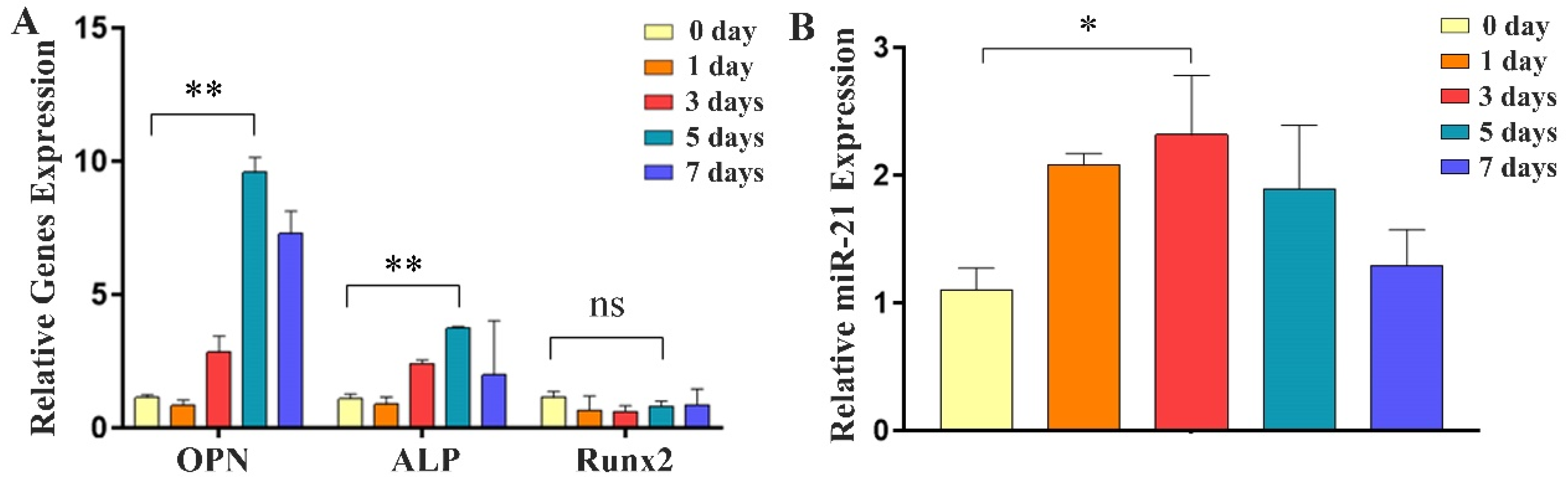
2.2. Expression of Osteogenic Factors and miR-21 during Differentiation of MC3T3-E1 Cells

During the osteogenic differentiation of MC3T3-E1 cells, qRT-PCR analysis was performed on days 0–7. The results revealed a gradual increase in the expression of osteogenic marker genes, OPN and ALP, with the highest expression observed on day 5 (Figure 2A). Furthermore, the expression of miR-21 also exhibited a progressive increase during the osteogenic differentiation process and increased 2.3 times to reach a peak on day 3 (p-value, 0.013; 95 percent confidence interval, −2.01 to −0.42), occurring earlier than the peak expression of OPN and ALP (Figure 2B). These results demonstrated that miR-21 was involved in osteogenic differentiation and might influence the expression of OPN and ALP.

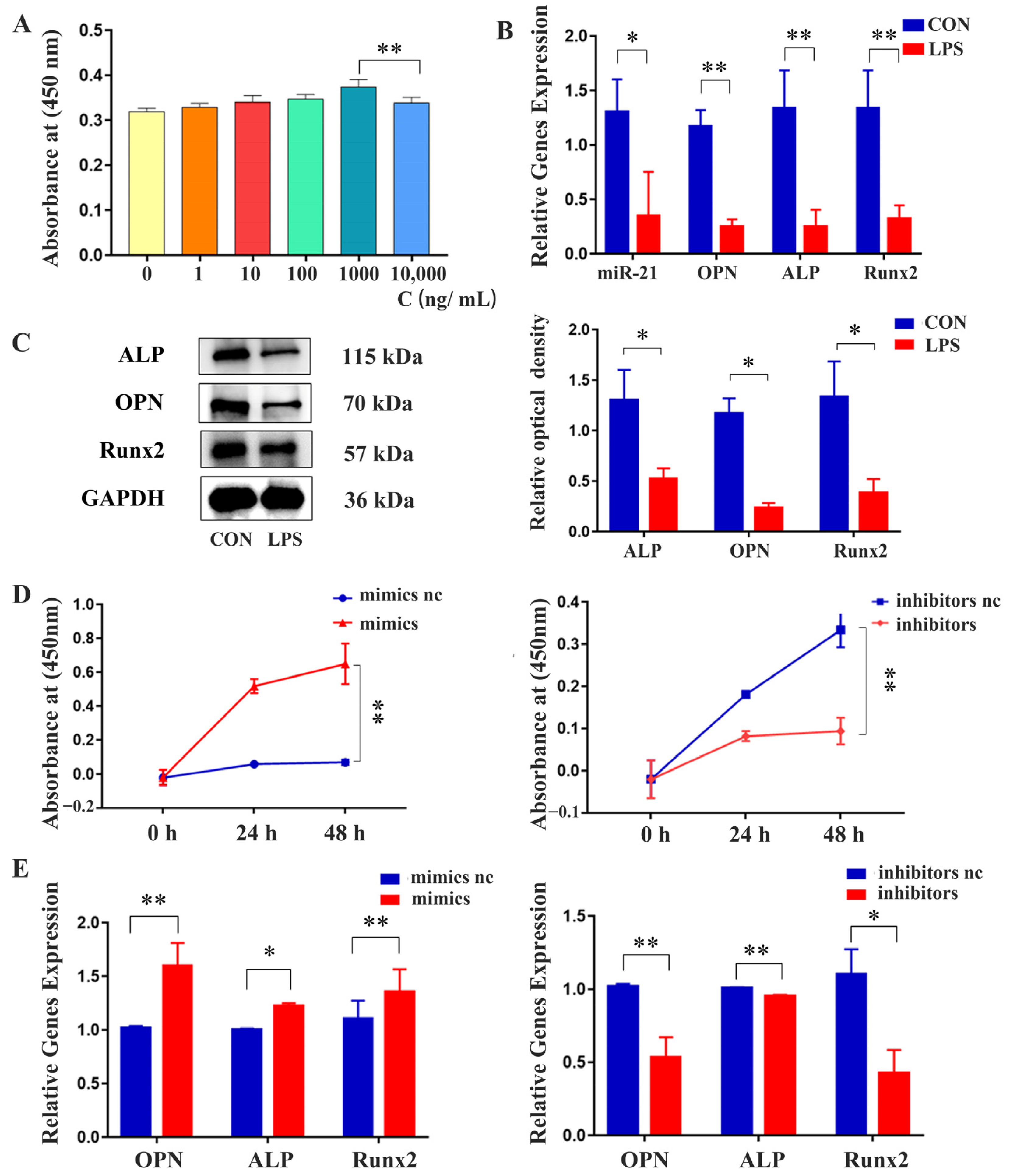
2.3. Effects of miR-21 on the Proliferation and Osteogenic Differentiation of MC3T3-E1 Cells under LPS Stimulation

The results of the CCK-8 assay showed that a concentration of 1000 ng/mL LPS promoted the proliferation of MC3T3-E1 cells without inducing toxicity (Figure 3A). qRT-PCR analysis revealed that LPS-induced inflammatory conditions resulted in reduced gene expression levels of OPN, ALP, Runx2, and miR-21 (Figure 3B). Western blot analysis confirmed that the protein expression levels of OPN, ALP, and Runx2 were reduced in the presence of LPS-mediated inflammation (Figure 3C). However, the CCK-8 assay revealed that MC3T3-E1 cells transfected with miR-21 mimics exhibited increased proliferation compared to NC, which was decreased when MC3T3-E1 cells were transfected with miR-21 inhibitors (Figure 3D). Additionally, qRT-PCR analysis indicated that the transfection of miR-21 mimics increased the expression levels of OPN, ALP, and Runx2. Conversely, inhibiting miR-21 led to a decrease in the expression of these osteogenic markers (Figure 3E).

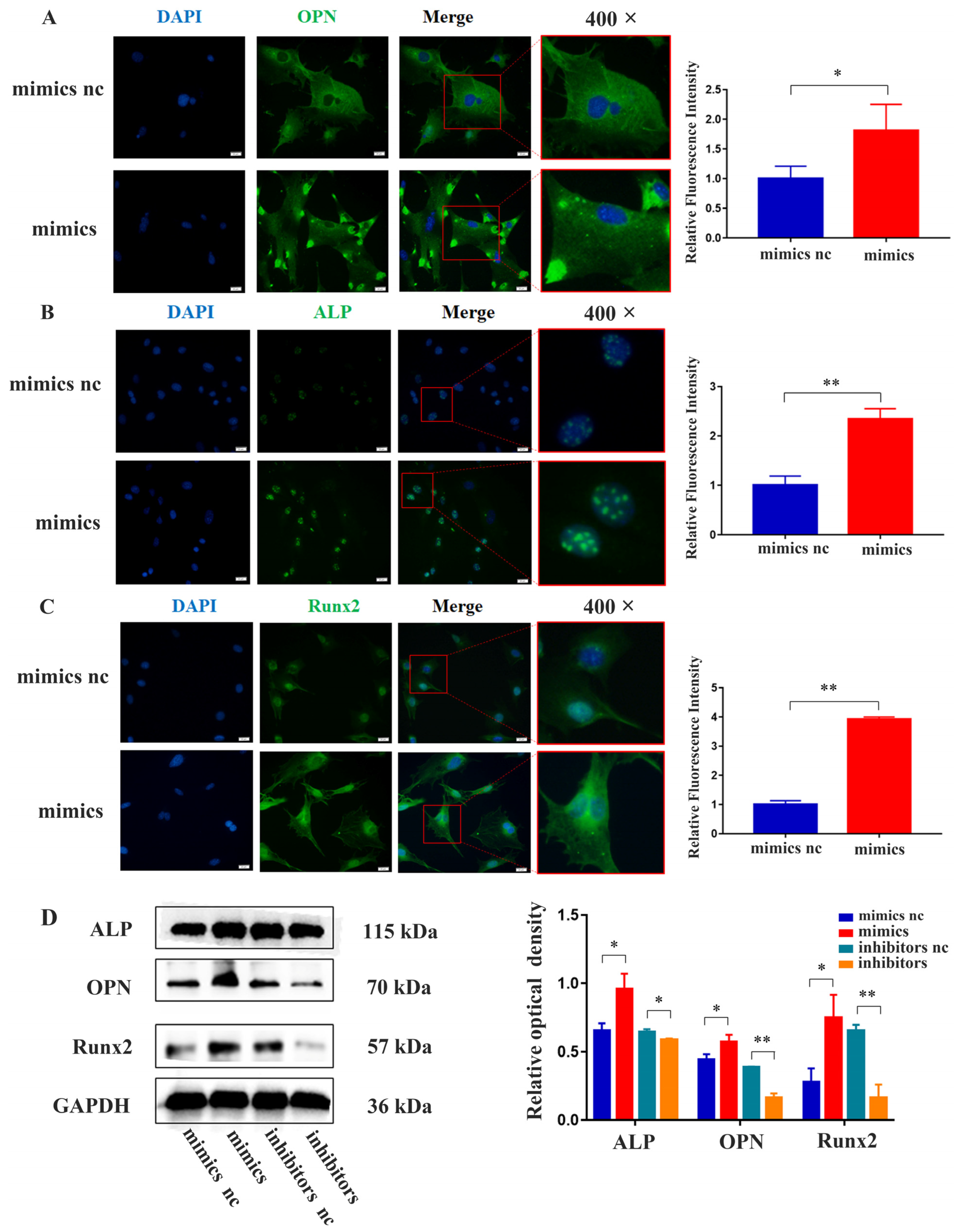
2.4. miR-21 Promoted the Expression of Osteogenic Proteins in MC3T3-E1 Cells under LPS Stimulation

The results of immunofluorescence staining demonstrated that the cytoplasmic fluorescence intensity of OPN was higher in the miR-21 mimics group compared to the NC group. Additionally, the expression level of ALP in the nucleus was higher in the miR-21 mimics group. Furthermore, the expression of Runx2 in both the nucleus and cytoplasm was higher in the miR-21 mimics group (Figure 4A–C). Western blot analysis revealed that MC3T3-E1 cells transfected with miR-21 mimics in the inflammatory environment exhibited increased protein levels of ALP, OPN, and Runx2, while inhibition of miR-21 showed the opposite effect (Figure 4D).

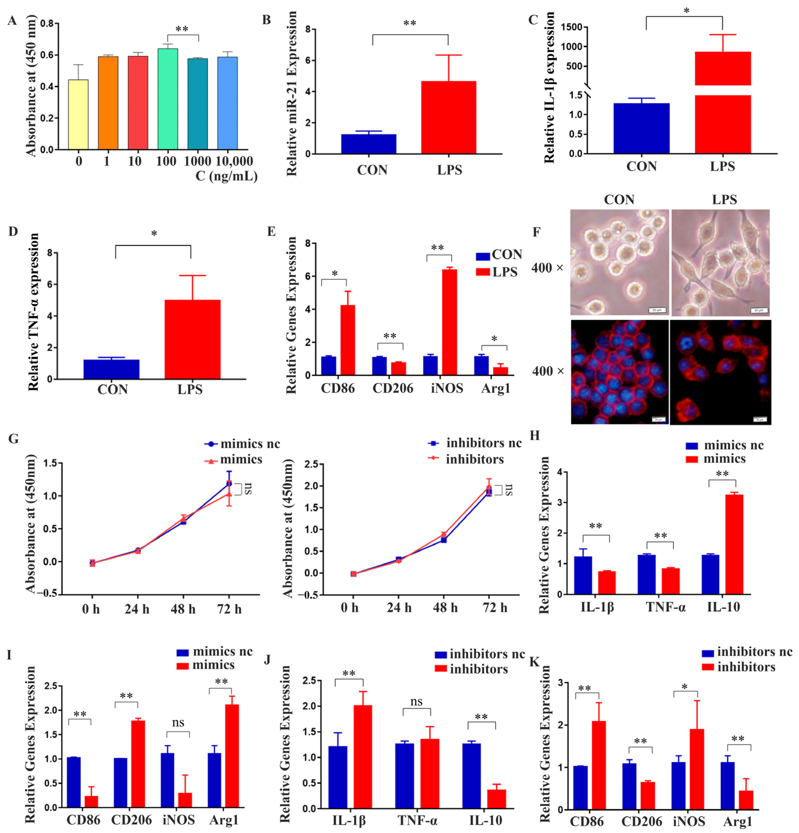
2.5. miR-21 Regulated the Polarization and Expression of Inflammatory Factors in RAW 264.7 Cells under LPS Stimulation

The results of the CCK-8 assay showed that a concentration of 1000 ng/mL LPS promoted the proliferation of RAW 264.7 cells without inducing toxicity (Figure 5A). qRT-PCR analysis revealed that under LPS stimulation, the expression of miR-21, inflammatory cytokines (IL-1β and TNF-α), and M1 polarization markers (CD86 and iNOS) all increased (Figure 5B–E). RAW 264.7 cells exhibited irregular morphology and extended pseudopods in an inflammatory environment, as observed under a 400-times magnification optical microscope and a fluorescence microscope (Figure 5F). The transfection of miR-21 mimics and inhibitors had minimal impact on RAW 264.7 cell proliferation (Figure 5G). However, transfection of miR-21 mimics led to a decrease in the expression of inflammatory cytokines (IL-1β and TNF-α) and M1 polarization markers (CD86 and iNOS), as revealed by qRT-PCR analysis. Additionally, there was an increase in the expression of M2 polarization markers (CD206 and Arg1) and the anti-inflammatory cytokine IL-10 (Figure 5H,I). Conversely, miR-21 inhibition showed the opposite effect (Figure 5J,K).

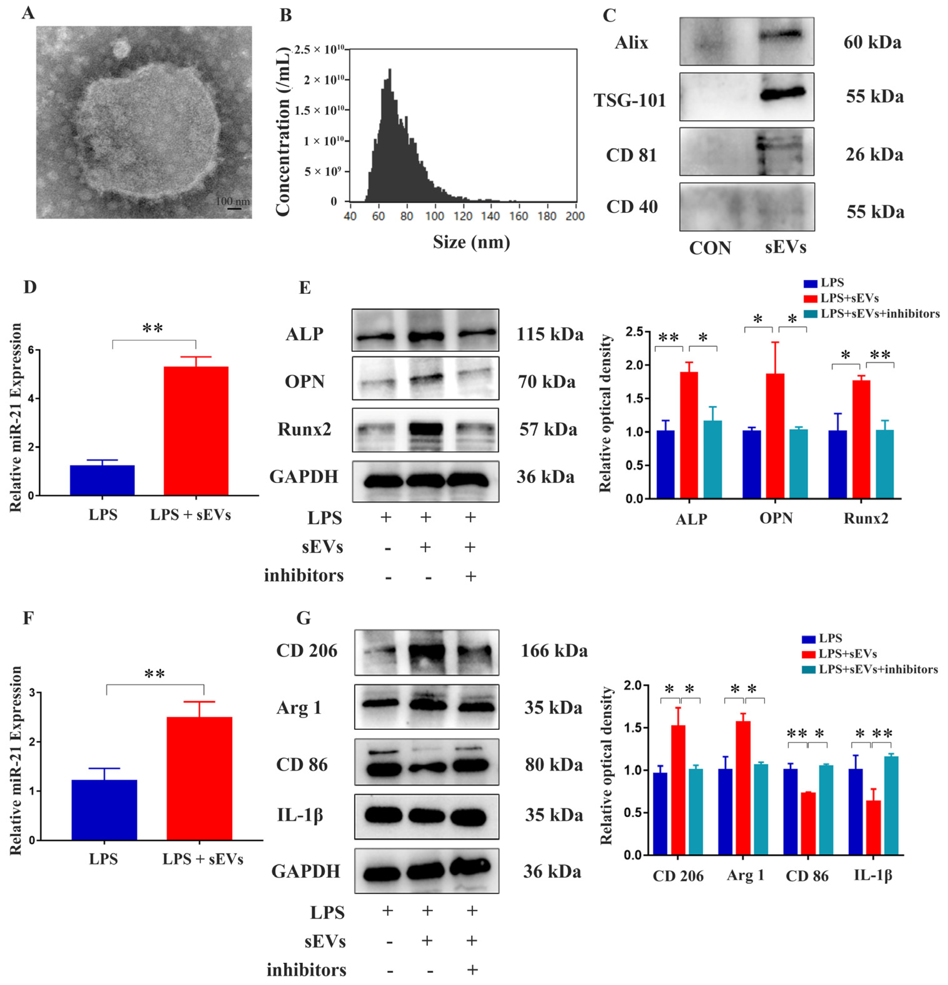
2.6. Isolation and Identification of Milk-Derived sEVs and Their Effects on MC3T3-E1 Cells and RAW 264.7 Cells

sEVs derived from cow’s milk were isolated using ultra-high-speed differential centrifugation. Morphological examination by transmission electron microscopy (TEM) showed that the sEVs were disc-shaped lipid bilayer structures (Figure 6A). Nano-flow cytometric analysis showed that the median particle size of sEVs was 72.2 nm, and the particle concentration was 2.01 × 10¹⁰/mL (Figure 6B). The positive markers CD81, TSG101, and Alix were highly expressed in the sEV group, while the negative marker CD40 was expressed at a low level (Figure 6C). These results indicated that we had extracted milk-derived sEVs successfully. In an inflammatory environment, it was found that expression of miR-21 significantly increased when MC3T3-E1 cells were treated with milk-derived sEVs (Figure 6D). Western blot analysis revealed that sEVs promoted the expression of osteogenic differentiation markers, including ALP, OPN, and Runx2 in MC3T3-E1 cells, but this effect was blocked by inhibiting miR-21 (Figure 6E,F). These results indicated that milk-derived sEVs promoted bone differentiation through miR-21. Furthermore, in an inflammatory environment, the expression of miR-21 was significantly increased when RAW 264.7 cells were exposed to milk-derived sEVs (Figure 6G). The addition of milk-derived sEVs promoted the expression of M2 polarizing factors CD206 and Arg1 while inhibiting the expression of M1 polarizing factor CD86 and inflammatory factor IL-1β in RAW 264.7 cells. However, when miR-21 was inhibited, contradictory results were obtained (Figure 6). This suggested that milk-derived sEVs promoted RAW 264.7 cell polarization towards the M2 phenotype and inhibited the expression of inflammatory factors through miR-21.

3. Discussion

CAP is a disease characterized by persistent infection and the resorption of periapical alveolar bone [29]. It has been shown that LPS enhances the activity of osteoclasts and exacerbates the inflammatory response in an experimental mouse model of CAP by promoting the expression of the low-density lipoprotein receptor in macrophages [30]. The control of infection and the restoration of periapical bone defects are essential aspects of CAP treatment [31]. However, due to the complex microbial environment and unique anatomical structures in root canals, inflammation and bone defects may persist [32]. Therefore, further investigation into potential treatment approaches for CAP that promote osteogenesis and possess anti-inflammatory properties is urgently required.
Numerous studies have demonstrated that miR-21 plays a crucial role in anti-inflammatory responses. For instance, overexpression of miR-21-5p in LPS-induced dental pulp cells suppressed pro-inflammatory cytokines by inhibiting TRAF6 and PDCD4 and downregulating the TLR/NF-κB signaling pathway. This suggested that miR-21 might inhibit inflammation and contribute to pulpitis [33]. The transfection of miR-21 mimics inhibited the production of pro-inflammatory factors by activating NF-κB in macrophages. Knockout of miR-21 resulted in enhanced gingival inflammation and alveolar bone loss, indicating that miR-21 served as a potential target for periodontitis treatment [14]. However, the role of miR-21 in CAP remains unexplored. In this study, we revealed the downregulation of miR-21, as well as of the osteogenic markers OPN and ALP, in human CAP. Conversely, expression of the inflammatory factors TNF-α and IL-1β increased. These findings suggest a potential association between miR-21 and CAP.
MiR-21 also plays a crucial role in promoting bone formation [34]. Previous studies have demonstrated that transfection of miR-21-5p mimics into MC3T3-E1 cells enhanced the expression of Runx2 and Osterix through the miR-21-5p/Dec1 axis [35]. Conversely, inhibiting miR-21-5p resulted in a decrease in ALP activity, the suppression of mineralization, and the downregulation of BMP2, Runx2, and OCN at both mRNA and protein levels [7]. These findings exhibited similarity to our research. In our study, we also found that miR-21 was involved in osteogenic differentiation and might influence the expression of OPN and ALP. However, the effect of miR-21 on the osteogenic differentiation of MC3T3-E1 cells under LPS stimulation remained unclear. Therefore, we further stimulated MC3T3-E1 cells with LPS to induce chronic inflammation and then investigated the impact of miR-21 on cell proliferation and osteogenic differentiation. Subsequently, we observed that miR-21 promoted the proliferation of MC3T3-E1 cells and increased the expression of osteogenic genes and proteins. Conversely, inhibition of miR-21 had the opposite effect. These findings suggest that miR-21 mimics reversed the inhibitory effect of inflammation on MC3T3-E1 cells and promoted their proliferation and osteogenic differentiation.
Macrophages also play a significant role in the pathogenesis of CAP [36] and are closely associated with inflammation [37]. Macrophages can exhibit two main phenotypes: the M1 phenotype, which promotes inflammation [38], and the M2 phenotype, which possesses anti-inflammatory abilities and contributes to immune regulation and repair processes [39]. The expression of M1 polarizing factors is increased in symptomatic CAP, and the M1/M2 ratio influences the severity of CAP [40]. Previous studies have demonstrated that delivery of miR-21-5p inhibited the expression of inflammatory markers in LPS-stimulated RAW 264.7 cells [41]. These findings exhibited similarity to our research. Conversely, controversial research has revealed that overexpressing miR-21 increased pro-inflammatory cytokines significantly, promoting a pro-inflammatory phenotype, partially involving PI3K and NF-ĸB signaling pathways [42]. Moreover, the role of miR-21 in macrophage polarization remains unclear. Therefore, we established an LPS-mediated inflammatory environment to investigate the role of miR-21 in the polarization of RAW 264.7 cells. We found that under LPS stimulation, miR-21 might exert an anti-inflammatory effect. To explore whether the anti-inflammatory effect of miR-21 was related to macrophage polarization, we carried out qRT-PCR, which showed that under LPS stimulation, the expression of M1 polarizing factors increased while the expression of M2 polarizing factors decreased, indicating a polarization of RAW 264.7 cells towards the M1 phenotype. However, transfection of miR-21 mimics in the inflammatory environment produced contradictory results, indicating that miR-21 exerted an anti-inflammatory effect and was associated with the polarization of RAW 264.7 cells towards the M2 phenotype. These results indicated that miR-21 might be a potential target for CAP treatment by promoting osteogenesis and inhibiting inflammation. Nevertheless, despite our research revealing that promoting macrophage M2 polarization suppressed inflammation, scholars have found that laryngopharyngeal reflux (LPR) promoted macrophage M2 polarization, leading to burning mouth syndrome (BMS) [43] and laryngeal squamous cell carcinoma (LSCC) [44]. Therefore, further investigation is needed to explore the detrimental effects of miR-21 in promoting macrophage M2 polarization.
Our previous studies showed that milk-derived sEVs enhanced the proliferation and differentiation of osteoblasts in vitro and promoted bone repair in a rat skull defect model in vivo. This highlighted their potential for application in bone defect repair to treat CAP [45]. Additionally, milk-derived sEVs effectively inhibited the infiltration of inflammatory cells and attenuated the inflammatory response by inhibiting the TLR4-NF-κB signaling pathway and NLRP3 inflammasome activation [46]. Research has shown that sEVs inhibit osteoclast differentiation and alleviate osteoporosis by acting through OPN and miR-21-5p [47]. Furthermore, other studies have indicated that milk-derived sEVs are rich in miR-21 [28]. However, whether milk-derived sEVs promote osteogenesis and inhibit inflammation in CAP through miR-21 remains unexplored. In our study, we observed that sEVs significantly increased the expression of miR-21 in MC3T3-E1 cells and RAW 264.7 cells within an inflammatory environment. Treatment with sEVs promoted the expression of osteogenic differentiation marker proteins, but this effect was blocked by the addition of miR-21 inhibitors. In an inflammatory environment, sEVs promoted the anti-inflammatory M2 polarization of RAW 264.7 cells and inhibited the expression of inflammatory cytokines, effects which were also attenuated by the addition of miR-21 inhibitors. Therefore, we speculated that milk-derived sEVs would promote osteogenic differentiation of MC3T3-E1 cells, induce M2 polarization of RAW 264.7 cells, and inhibit inflammation through the action of miR-21.
However, this experiment has several limitations. First, the specific mechanism by which milk-derived sEVs promote the expression of miR-21 in cells remains unclear, and whether it involves upstream LncRNA is unknown. Second, the downstream targets and signaling pathways of miR-21 require further exploration. Lastly, there is a lack of animal experiments regarding CAP. Therefore, further exploration of the downstream targets and signaling pathways of miR-21 should be detected and validated through database analysis and dual-luciferase reporter gene assays. Moreover, animal experiments need supplementation, such as the utilization of a miR-21 knockout mouse model for CAP. Looking ahead, we intend to utilize engineered milk-derived sEVs loaded with miR-21 to explore their potential application in CAP.

4. Materials and Methods

4.1. Sample Collection

This study received approval from the ethics committee of the Affiliated Stomatological Hospital of Dalian Medical University (2022002). Between April 2022 and April 2023, periapical tissue samples were collected from patients diagnosed with CAP. The CAP group consisted of 25 patients who required apical surgery or tooth extraction, while the control (CON) group comprised 25 healthy premolars or third molars extracted for orthodontic reasons or because they were impacted. Among them, 15 cases from the CAP group and 15 cases from the CON group were subjected to qRT-PCR, 6 for Western blot analysis, and 4 for HE staining. The age range of the participants was 24 to 55 years, with a total of 23 females and 27 males. There were no significant differences in sex or age between the two groups (p > 0.05). Participants with combined periodontal or periodontal pulp lesions, a history of root canal therapy, systemic diseases, and those using non-steroidal anti-inflammatory drugs or antibiotics were excluded from this study [48].

4.2. Hematoxylin and Eosin (HE) Staining

The periapical tissues were fixed in 4% paraformaldehyde and subsequently washed in phosphate-buffered saline (PBS). Dehydration of the tissues was performed using graded alcohols. Xylene was used as a clearing agent to prepare the tissue for further processing. Following the clearing step, the periapical tissues were infiltrated with paraffin wax. Once the infiltration was complete, the tissues were embedded in paraffin wax, and 5 μm continuous sections were obtained using a microtome. HE staining was employed to assess the histological changes in the periapical tissues.

4.3. Cell Culture and Induction

MC3T3-E1 and RAW 264.7 cell lines were obtained from the Center for Excellence in Molecular Cell Science (Shanghai, China) and were cultured in α-MEM and DMEM, respectively, supplemented with 10% fetal bovine serum (FBS) and 1% penicillin/streptomycin. Both cell lines were cultured in the T25 cell culture flask and placed at 37 °C in a humidified incubator with 5% CO2. For osteogenic differentiation, MC3T3-E1 cells were seeded in six-well plates at 2 × 10⁸ cells/well. Osteogenic induction was performed by adding dexamethasone (10 nmol/L), β-glycerophosphate sodium (10 mmol/L), and ascorbic acid (50 μg/mL) to each well of MC3T3-E1 cells every other day following a 24 h culture period. The induction of LPS involved adding LPS from the Porphyromonas gingivalis to the cell culture medium in the range of 0 to 10,000 ng/mL each day.

4.4. Cell Transfection Assay

The miRNA mimics and inhibitors were transfected into MC3T3-E1 cells and RAW 264.7 cells with the Lipofectamine 3000 transfection reagent (Thermo Fisher Scientific, Waltham, MA, USA) according to the manufacturer’s instructions. MiRNA mimics and NC were transfected at a concentration of 50 nM, while miRNA inhibitors were used at 100 nM. The cells were incubated for 24 h. Afterward, the transfection medium was replaced with normal growth medium. Total RNA was then extracted after 24–48 h, and protein extraction was performed after 48–72 h.

4.5. Cell Proliferation Assay

After transfection of MC3T3-E1 cells or RAW 264.7 cells in an inflammatory environment for 24 h, the cells were resuspended and seeded into 96-well plates at a concentration of 4 × 10³ cells per well. Following incubation for 0, 24, or 48 h, the Cell Counting Kit-8 (CCK-8) reagent (APExBIO, Houston, TX, USA) was added to each well at a volume of 10 μL to measure cell proliferation. After incubating for an additional two hours, the optical density (OD) was measured at 450 nm using a spectrophotometer.

4.6. Isolation and Characterization of sEVs Derived from Milk

Milk-derived sEVs were prepared according to a previously published protocol [45]. Briefly, milk was collected from healthy cows (Sanhuan Ranch, Dalian, China). Following the defatting process, the skimmed milk underwent centrifugation at varying speeds: first at 13,000× g for 30 min, then at 100,000× g for 120 min. The resultant middle whey fraction was collected and subjected to further centrifugation at 130,000× g for 90 min, followed by 100,000× g for 120 min. After filtration, the suspension containing milk-sEVs was obtained through centrifugation at 100,000× g for 60 min. The concentration of sEVs was detected using the BCA method. Based on our previous research, 20 µg/mL milk-sEVs were incubated with MC3T3-E1 and RAW 264.7 cells. The milk-sEVs were administered every 24 h. After 24 h, RNA was extracted to assess the levels of osteogenic gene expression and inflammatory cytokines. Following 48 h, the protein expression was tested. The morphology of the sEVs was observed using a transmission electron microscope, the size of the sEVs was measured using nano-flow cytometry, and surface marker proteins were detected by Western blotting using the antibodies anti-CD81 (1:1000, Abcam, Cambridge, MA, USA), anti-CD40 (1:1000, Bioss Antibodies, Woburn, MA, USA), anti-Alix (1:1000, Abbexa Ltd., Cambridge, UK), and anti-TSG-101 (1:1000, Abcam).

4.7. Quantitative Reverse Transcription–Polymerase Chain Reaction (qRT-PCR)

Total RNA was extracted using the UNlQ-10 Column Trizol Total RNA Isolation Kit (Sangon Biotech, Shanghai, China). Subsequently, the RNA was converted into complementary DNA (cDNA) using the Evo M-MLV Reverse Transcription Kit (Accurate Biology, Hunan, China). For miRNA reverse transcription, the stem-loop method was employed. The primers used in the reverse transcription process can be found in Table 1. qRT-PCR analysis was performed using SYBR Green Pro Taq and conducted on a Step Two qRT-PCR System (Bio-Rad, Hercules, CA, USA). The obtained data were analyzed using the 2−ΔΔCt method.

4.8. Immunofluorescence (IF)

After transfecting MC3T3-E1 cells for 48 h, immunofluorescence staining was performed to visualize specific proteins. Cells were fixed in 4% paraformaldehyde for 20 min and blocked with goat serum at room temperature for 1 h. Cells were then incubated with primary antibodies, including anti-OPN (1:200, Proteintech, Wuhan, China), anti-ALP (1:200, Abcam), and anti-Runx2 (1:200, Abcam) overnight at 4 °C followed by secondary antibody for 1 h at room temperature. A fluorescence microscope (Olympus, Tokyo, Japan) was used to visualize the results.

4.9. Western Blotting

Tissue and cell samples were lysed using RIPA buffer to extract proteins, and the protein concentration was determined using a BCA protein assay kit (Beyotime Biotechnology, Shanghai, China). Subsequently, the samples were separated by sodium dodecyl sulfate–polyacrylamide gel electrophoresis (SDS-PAGE) for 1.5 h and then transferred to nitrocellulose (NC) membranes by blotting for 1.5 h. To prevent nonspecific binding, the NC membranes were blocked with fat-free milk at room temperature for 2.5 h and then incubated overnight at 4 °C with the primary antibodies anti-OPN (1:1000, Proteintech, Rosemont, IL, USA), anti-ALP (1:1000, Abcam), and anti-Runx2 (1:500, Abcam). Finally, the membranes were incubated with the secondary antibody (1:5000, ABclonal, Woburn, MA, USA) for 1 h. A gel imaging system (Bio-Rad, Hercules, CA, USA) was used to visualize the protein bands.

4.10. Statistical Analysis

Data are expressed as mean ± SEM. SPSS Statistics (version 17.0) software was used for all statistical analyses. The experiments were repeated independently a minimum of three times. GraphPad Prism 7.0 was employed to create the statistical graphs. Student’s t-test or one-way ANOVA was performed to verify statistical significance. A p-value < 0.05 was statistically significant.

5. Conclusions

In summary, our results show that miR-21 increased the expression of osteogenesis-related genes and M2 polarization genes while decreasing the expression of M1 polarization genes and inflammatory cytokines. This indicated that miR-21 might be a potential target for CAP treatment. In the regulation of osteogenic differentiation and anti-inflammatory effects by milk-derived sEVs, miR-21 played a crucial role since the suppression of miR-21 markedly reduced the regulatory effects of sEVs. Our discovery indicates for the first time that milk-derived sEVs might represent a novel therapeutic tool for CAP by targeting miR-21. For instance, the development of targeted drugs for miR-21, the design of delivery systems for sEVs intended for root canal disinfection and filling, or the utilization of a scaffold-loaded approach to directly implant them into the bone defect associated with CAP. Further studies are required to elucidate the specific mechanisms, downstream targets, and signaling pathways and to confirm the findings in vivo by animal experiments.

Author Contributions

Conceptualization, R.L., M.D. and W.N.; methodology, R.L., S.L., S.W., M.X. and W.L.; software, R.L.; validation, L.W., M.D. and W.N.; investigation, R.L. and S.L.; resources, L.W., M.D. and W.N.; data curation, R.L.; writing—original draft preparation, R.L.; writing—review and editing, S.L., S.W., L.W., M.D. and W.N.; visualization, R.L.; supervision, L.W., M.D. and W.N.; funding acquisition, L.W., M.D. and W.N. All authors have read and agreed to the published version of the manuscript.

Funding

This work was supported by the National Natural Science Foundation of China (grant number 82270971; 82100998); the Provincial Basic Scientific Research Project of the Liaoning Education Department (grant number LJKZ0841); the Science and Technology Plan of Liaoning Province (grant number 2021JH2/10300027); the Liaoning Province Doctor Startup Foundation (grant number 2022-BS-237); and the Dalian Science and Technology Innovation Fund (grant number 2022JJ13SN062).

Institutional Review Board Statement

The study was conducted in accordance with the Declaration of Helsinki and approved by the Ethics Committee of Dalian Medical University (protocol code: 2022002 and 7 March 2022 of approval).

Informed Consent Statement

Informed consent was obtained from all subjects involved in this study.

Data Availability Statement

All data used in this study are included in this article.

Conflicts of Interest

The authors declare no conflict of interest.

References

  1. Luo, X.; Wan, Q.; Cheng, L.; Xu, R. Mechanisms of bone remodeling and therapeutic strategies in chronic apical periodontitis. Front. Cell. Infect. Microbiol. 2022, 12, 908859. [Google Scholar] [CrossRef] [PubMed]
  2. Tibúrcio-Machado, C.S.; Michelon, C.; Zanatta, F.B.; Gomes, M.S.; Marin, J.A.; Bier, C.A. The global prevalence of apical periodontitis: A systematic review and meta-analysis. Int. Endod. J. 2021, 54, 712–735. [Google Scholar] [CrossRef]
  3. Gomes, B.; Herrera, D.R. Etiologic role of root canal infection in apical periodontitis and its relationship with clinical symptomatology. Braz. Oral. Res. 2018, 32 (Suppl. S1), e69. [Google Scholar] [CrossRef]
  4. Gan, G.; Lu, B.; Zhang, R.; Luo, Y.; Chen, S.; Lei, H.; Li, Y.; Cai, Z.; Huang, X. Chronic apical periodontitis exacerbates atherosclerosis in apolipoprotein E-deficient mice and leads to changes in the diversity of gut microbiota. Int. Endod. J. 2022, 55, 152–163. [Google Scholar] [CrossRef] [PubMed]
  5. Berggren, K.; Broström, A.; Firestone, A.; Wright, B.; Josefsson, E.; Lindmark, U. Oral health problems linked to obstructive sleep apnea are not always recognized within dental care-As described by dental professionals. Clin. Exp. Dent. Res. 2022, 8, 84–95. [Google Scholar] [CrossRef]
  6. Maniaci, A.; Ferlito, S.; Lechien, J.R.; Di Luca, M.; Iannella, G.; Cammaroto, G.; Cannavicci, A.; Pollicina, I.; Stilo, G.; Di Mauro, P.; et al. Anxiety, depression and sleepiness in OSA patients treated with barbed reposition pharyngoplasty: A prospective study. Eur. Arch. Oto-Rhino-Laryngol.—Head. Neck Surg. 2022, 279, 4189–4198. [Google Scholar] [CrossRef] [PubMed]
  7. Gomes, C.; Martinho, F.C.; Barbosa, D.S.; Antunes, L.S.; Póvoa, H.C.C.; Baltus, T.H.L.; Morelli, N.R.; Vargas, H.O.; Nunes, S.O.V.; Anderson, G.; et al. Increased Root Canal Endotoxin Levels are Associated with Chronic Apical Periodontitis, Increased Oxidative and Nitrosative Stress, Major Depression, Severity of Depression, and a Lowered Quality of Life. Mol. Neurobiol. 2018, 55, 2814–2827. [Google Scholar] [CrossRef]
  8. Xu, R.; Guo, D.; Zhou, X.; Sun, J.; Zhou, Y.; Fan, Y.; Zhou, X.; Wan, M.; Du, W.; Zheng, L. Disturbed bone remodelling activity varies in different stages of experimental, gradually progressive apical periodontitis in rats. Int. J. Oral. Sci. 2019, 11, 27. [Google Scholar] [CrossRef]
  9. Wang, S.; Xiong, G.; Ning, R.; Pan, Z.; Xu, M.; Zha, Z.; Liu, N. LncRNA MEG3 promotes osteogenesis of hBMSCs by regulating miR-21-5p/SOD3 axis. Acta Biochim. Pol. 2022, 69, 71–77. [Google Scholar] [CrossRef]
  10. Hur, J.; Rhee, C.K.; Lee, S.Y.; Kim, Y.K.; Kang, J.Y. MicroRNA-21 inhibition attenuates airway inflammation and remodelling by modulating the transforming growth factor β-Smad7 pathway. Korean J. Intern. Med. 2021, 36, 706–720. [Google Scholar] [CrossRef]
  11. Wang, H.; Wang, H.; Li, X.; Zhang, Z.; Zhao, X.; Wang, C.; Wei, F. MicroRNA-21 promotes bone reconstruction in maxillary bone defects. J. Oral. Rehabil. 2020, 47 (Suppl. S1), 4–11. [Google Scholar] [CrossRef] [PubMed]
  12. Geng, Z.; Yu, Y.; Li, Z.; Ma, L.; Zhu, S.; Liang, Y.; Cui, Z.; Wang, J.; Yang, X.; Liu, C. miR-21 promotes osseointegration and mineralization through enhancing both osteogenic and osteoclastic expression. Mater. Sci. Eng. C Mater. Biol. Appl. 2020, 111, 110785. [Google Scholar] [CrossRef] [PubMed]
  13. Liu, R.; Du, J.; Zhou, J.; Zhong, B.; Ba, L.; Zhang, J.; Liu, Y.; Liu, S. Elevated microRNA-21 Is a Brake of Inflammation Involved in the Development of Nasal Polyps. Front. Immunol. 2021, 12, 530488. [Google Scholar] [CrossRef] [PubMed]
  14. Zhou, W.; Su, L.; Duan, X.; Chen, X.; Hays, A.; Upadhyayula, S.; Shivde, J.; Wang, H.; Li, Y.; Huang, D.; et al. MicroRNA-21 down-regulates inflammation and inhibits periodontitis. Mol. Immunol. 2018, 101, 608–614. [Google Scholar] [CrossRef] [PubMed]
  15. Silva, T.A.; Garlet, G.P.; Fukada, S.Y.; Silva, J.S.; Cunha, F.Q. Chemokines in oral inflammatory diseases: Apical periodontitis and periodontal disease. J. Dent. Res. 2007, 86, 306–319. [Google Scholar] [CrossRef]
  16. Verweij, F.J.; Balaj, L.; Boulanger, C.M.; Carter, D.R.F.; Compeer, E.B.; D’Angelo, G.; El Andaloussi, S.; Goetz, J.G.; Gross, J.C.; Hyenne, V.; et al. The power of imaging to understand extracellular vesicle biology in vivo. Nat. Methods 2021, 18, 1013–1026. [Google Scholar] [CrossRef]
  17. Bernardi, S.; Balbi, C. Extracellular Vesicles: From Biomarkers to Therapeutic Tools. Biology 2020, 9, 258. [Google Scholar] [CrossRef] [PubMed]
  18. Li, M.; Fang, F.; Sun, M.; Zhang, Y.; Hu, M.; Zhang, J. Extracellular vesicles as bioactive nanotherapeutics: An emerging paradigm for regenerative medicine. Theranostics 2022, 12, 4879–4903. [Google Scholar] [CrossRef]
  19. Du, S.; Guan, Y.; Xie, A.; Yan, Z.; Gao, S.; Li, W.; Rao, L.; Chen, X.; Chen, T. Extracellular vesicles: A rising star for therapeutics and drug delivery. J. Nanobiotechnol. 2023, 21, 231. [Google Scholar] [CrossRef]
  20. Kalluri, R.; LeBleu, V.S. The biology, function, and biomedical applications of exosomes. Science 2020, 367, eaau6977. [Google Scholar] [CrossRef]
  21. Jia, Y.; Yu, L.; Ma, T.; Xu, W.; Qian, H.; Sun, Y.; Shi, H. Small extracellular vesicles isolation and separation: Current techniques, pending questions and clinical applications. Theranostics 2022, 12, 6548–6575. [Google Scholar] [CrossRef] [PubMed]
  22. Pathan, M.; Fonseka, P.; Chitti, S.V.; Kang, T.; Sanwlani, R.; Van Deun, J.; Hendrix, A.; Mathivanan, S. Vesiclepedia 2019: A compendium of RNA, proteins, lipids and metabolites in extracellular vesicles. Nucleic Acids Res. 2019, 47, D516–D519. [Google Scholar] [CrossRef] [PubMed]
  23. Li, Z.; Li, Q.; Tong, K.; Zhu, J.; Wang, H.; Chen, B.; Chen, L. BMSC-derived exosomes promote tendon-bone healing after anterior cruciate ligament reconstruction by regulating M1/M2 macrophage polarization in rats. Stem Cell Res. Ther. 2022, 13, 295. [Google Scholar] [CrossRef] [PubMed]
  24. Li, R.; Li, D.; Wang, H.; Chen, K.; Wang, S.; Xu, J.; Ji, P. Exosomes from adipose-derived stem cells regulate M1/M2 macrophage phenotypic polarization to promote bone healing via miR-451a/MIF. Stem Cell Res. Ther. 2022, 13, 149. [Google Scholar] [CrossRef]
  25. Yan, C.; Chen, J.; Wang, C.; Yuan, M.; Kang, Y.; Wu, Z.; Li, W.; Zhang, G.; Machens, H.G.; Rinkevich, Y.; et al. Milk exosomes-mediated miR-31-5p delivery accelerates diabetic wound healing through promoting angiogenesis. Drug Deliv. 2022, 29, 214–228. [Google Scholar] [CrossRef]
  26. Feng, X.; Chen, X.; Zheng, X.; Zhu, H.; Qi, Q.; Liu, S.; Zhang, H.; Che, J. Latest Trend of Milk Derived Exosomes: Cargos, Functions, and Applications. Front. Nutr. 2021, 8, 747294. [Google Scholar] [CrossRef]
  27. Vashisht, M.; Rani, P.; Onteru, S.K.; Singh, D. Curcumin Encapsulated in Milk Exosomes Resists Human Digestion and Possesses Enhanced Intestinal Permeability in Vitro. Appl. Biochem. Biotechnol. 2017, 183, 993–1007. [Google Scholar] [CrossRef]
  28. Li, D.; Yao, X.; Yue, J.; Fang, Y.; Cao, G.; Midgley, A.C.; Nishinari, K.; Yang, Y. Advances in Bioactivity of MicroRNAs of Plant-Derived Exosome-Like Nanoparticles and Milk-Derived Extracellular Vesicles. J. Agric. Food Chem. 2022, 70, 6285–6299. [Google Scholar] [CrossRef]
  29. do Nascimento, I.V.; Rodrigues, M.I.Q.; Isaias, P.H.C.; Barros-Silva, P.G.; Sousa, F.B.; Nunes Alves, A.P.N.; Mota, M.R.L. Chronic systemic corticosteroid therapy influences the development of pulp necrosis and experimental apical periodontitis, exacerbating the inflammatory process and bone resorption in rats. Int. Endod. J. 2022, 55, 646–659. [Google Scholar] [CrossRef]
  30. Huang, L.; Wu, H.; Wu, Y.; Song, F.; Zhang, L.; Li, Z.; Sun, H.; Huang, C. Pcsk9 Knockout Aggravated Experimental Apical Periodontitis via LDLR. J. Dent. Res. 2022, 101, 83–92. [Google Scholar] [CrossRef] [PubMed]
  31. Amaral, R.R.; Braga, T.; Siqueira, J.F., Jr.; Rôças, I.N.; da Costa Rachid, C.T.C.; Oliveira, A.G.G.; de Souza Côrtes, M.I.; Love, R.M. Root Canal Microbiome Associated with Asymptomatic Apical Periodontitis as Determined by High-Throughput Sequencing. J. Endod. 2022, 48, 487–495. [Google Scholar] [CrossRef]
  32. Sureshbabu, N.M.; Ranganath, A.; Jacob, B. Concentrated Growth Factor—Surgical Management of Large Periapical Lesion Using a Novel Platelet Concentrate in Combination with Bone Graft. Ann. Maxillofac. Surg. 2020, 10, 246–250. [Google Scholar]
  33. Nara, K.; Kawashima, N.; Noda, S.; Fujii, M.; Hashimoto, K.; Tazawa, K.; Okiji, T. Anti-inflammatory roles of microRNA 21 in lipopolysaccharide-stimulated human dental pulp cells. J. Cell. Physiol. 2019, 234, 21331–21341. [Google Scholar] [CrossRef]
  34. Sikora, M.; Śmieszek, A.; Pielok, A.; Marycz, K. MiR-21-5p regulates the dynamic of mitochondria network and rejuvenates the senile phenotype of bone marrow stromal cells (BMSCs) isolated from osteoporotic SAM/P6 mice. Stem Cell Res. Ther. 2023, 14, 54. [Google Scholar] [CrossRef]
  35. Kurita, T.; Li, X.; Bhawal, U.K. Crosstalk between microRNA-21-5p and the transcription factor Dec1 maintains osteoblast function. Biochem. Biophys. Res. Commun. 2022, 632, 32–39. [Google Scholar] [CrossRef]
  36. Zi, J.; Wang, F.; Liu, Z.; Wang, Y.; Qiu, L.; Zhu, G.; Li, H. Impact of Toll-like Receptor 4 Expression on Inflammatory Responses Related to Premature Membrane Rupture Induced by Lipopolysaccharide. Discov. Med. 2023, 35, 429–435. [Google Scholar] [CrossRef] [PubMed]
  37. Li, J.; Yue, Y.; Chan, W.; Wei, W.; Liu, X.; Wang, M.; Li, J.; Hao, L. RGS10 negatively regulates apical periodontitis via TFEB-mediated autophagy in BABL/c mice model and in vitro. Int. Endod. J. 2023, 56, 854–868. [Google Scholar] [CrossRef]
  38. Zhang, F.; Gluck, J.M.; Brown, A.C.; Zaharoff, D.A.; King, M.W. Heparin Affinity-Based IL-4 Delivery to Modulate Macrophage Phenotype and Endothelial Cell Activity In Vitro. ACS Appl. Mater. Interfaces 2023. online ahead of print. [Google Scholar] [CrossRef] [PubMed]
  39. Holden, J.A.; Attard, T.J.; Laughton, K.M.; Mansell, A.; O’Brien-Simpson, N.M.; Reynolds, E.C. Porphyromonas gingivalis lipopolysaccharide weakly activates M1 and M2 polarized mouse macrophages but induces inflammatory cytokines. Infect. Immun. 2014, 82, 4190–4203. [Google Scholar] [CrossRef] [PubMed]
  40. Veloso, P.; Fernández, A.; Terraza-Aguirre, C.; Álvarez, C.; Vernal, R.; Escobar, A.; Hernández, M. Macrophages skew towards M1 profile through reduced CD163 expression in symptomatic apical periodontitis. Clin. Oral. Investig. 2020, 24, 4571–4581. [Google Scholar] [CrossRef]
  41. Marycz, K.; Smieszek, A.; Marcinkowska, K.; Sikora, M.; Turlej, E.; Sobierajska, P.; Patej, A.; Bienko, A.; Wiglusz, R.J. Nanohydroxyapatite (nHAp) Doped with Iron Oxide Nanoparticles (IO), miR-21 and miR-124 Under Magnetic Field Conditions Modulates Osteoblast Viability, Reduces Inflammation and Inhibits the Growth of Osteoclast—A Novel Concept for Osteoporosis Treatment: Part 1. Int. J. Nanomed. 2021, 16, 3429–3456. [Google Scholar]
  42. Madhyastha, R.; Madhyastha, H.; Nurrahmah, Q.I.; Purbasari, B.; Maruyama, M.; Nakajima, Y. MicroRNA 21 Elicits a Pro-inflammatory Response in Macrophages, with Exosomes Functioning as Delivery Vehicles. Inflammation 2021, 44, 1274–1287. [Google Scholar] [CrossRef] [PubMed]
  43. Lechien, J.R.; Hans, S.; De Marrez, L.G.; Dequanter, D.; Rodriguez, A.; Muls, V.; Ben Abdelouahed, F.; Evrard, L.; Maniaci, A.; Saussez, S.; et al. Prevalence and Features of Laryngopharyngeal Reflux in Patients with Primary Burning Mouth Syndrome. Laryngoscope 2021, 131, E2627–E2633. [Google Scholar] [CrossRef]
  44. Zabolotnyi, D.; Kizim, Y.; Zabolotna, D.; Sulaieva, O.; Kizim, V. Laryngopharyngeal Reflux Alters Macrophage Polarization in Human Papilloma Virus-Negative Squamous Cell Carcinoma of the Larynx in Males. Clin. Exp. Otorhinolaryngol. 2021, 14, 240–243. [Google Scholar] [CrossRef] [PubMed]
  45. Dong, M.; Shi, C.; Yu, X.; Yang, Q.; Wu, S.; Liu, R.; Liu, T.; Wang, L.; Niu, W. Milk-derived small extracellular vesicles: Nanomaterials to promote bone formation. J. Nanobiotechnol. 2022, 20, 370. [Google Scholar] [CrossRef]
  46. Tong, L.; Hao, H.; Zhang, Z.; Lv, Y.; Liang, X.; Liu, Q.; Liu, T.; Gong, P.; Zhang, L.; Cao, F.; et al. Milk-derived extracellular vesicles alleviate ulcerative colitis by regulating the gut immunity and reshaping the gut microbiota. Theranostics 2021, 11, 8570–8586. [Google Scholar] [CrossRef]
  47. Lee, K.S.; Lee, J.; Kim, H.K.; Yeom, S.H.; Woo, C.H.; Jung, Y.J.; Yun, Y.E.; Park, S.Y.; Han, J.; Kim, E.; et al. Extracellular vesicles from adipose tissue-derived stem cells alleviate osteoporosis through osteoprotegerin and miR-21-5p. J. Extracell. Vesicles 2021, 10, e12152. [Google Scholar] [CrossRef]
  48. Verma, N.; Sangwan, P.; Tewari, S.; Duhan, J. Effect of Different Concentrations of Sodium Hypochlorite on Outcome of Primary Root Canal Treatment: A Randomized Controlled Trial. J. Endod. 2019, 45, 357–363. [Google Scholar] [CrossRef] [PubMed]
Figure 1. Identification of clinical samples and expression of miR-21 in human CAP. (A) Histological analysis using HE staining confirmed the extraction of clinical samples: blood vessels (red arrows); inflammatory cells (blue arrows); scale bars, 100 μm, 20 μm. (B) The qRT-PCR method was used to detect the expression of miR-21, osteogenic differentiation marker genes (OPN, ALP, and Runx2), and inflammatory factors TNF-α and IL-1β. (C) Western blotting was used to detect the expression of osteogenic differentiation marker proteins. * p < 0.05, ** p < 0.01.
Ijms 24 13873 g001
Figure 2. The expression of miR-21 and osteogenic genes during the differentiation of MC3T3-E1 cells. (A) qRT-PCR was used to detect the expression of osteogenic differentiation factors at 0 to 7 d during the process of osteogenic differentiation. (B) The expression of miR-21 was also examined using qRT-PCR during the same time period of osteogenic differentiation. * p < 0.05, ** p < 0.001, ns: not statistically significant.
Ijms 24 13873 g002
Figure 3. Effects of miR-21 on the proliferation and differentiation of MC3T3-E1 cells under LPS stimulation. (A) The optimal concentration of LPS for induction of MC3T3-E1 cells was determined using the CCK-8 assay, 0 ng/mL (yellow), 1 ng/mL (orange), 10 ng/mL (red), 100 ng/mL (green), 1000 ng/mL (dark green), 10,000 ng/mL (blue). (B) The expression of osteogenic marker genes was assessed using qRT-PCR. (C) The expression of osteogenic proteins was detected using Western blot analysis. (D) The effect of miR-21 on the proliferation of MC3T3-E1 cells was evaluated using CCK-8 assay. (E) The impact of miR-21 on the expression of osteogenic differentiation genes was assessed using qRT-PCR. * p < 0.05, ** p < 0.01.
Ijms 24 13873 g003
Figure 4. The effects of miR-21 on the expression of osteogenic proteins in MC3T3-E1 cells under LPS stimulation. (A) Immunofluorescence staining was performed to observe the localization of OPN. (B) ALP. (C) Runx2; scale bar, 20 μm. (D) Western blot analysis examined the expression of osteogenic proteins in MC3T3-E1 cells following the transfection of miR-21 under inflammatory conditions. * p < 0.05, ** p < 0.01.
Ijms 24 13873 g004
Figure 5. The effects of miR-21 on RAW 264.7 cell polarization and expression of inflammatory factors under LPS stimulation. (A) CCK-8 assay was performed to determine the optimal concentration of LPS, 0 ng/mL (yellow), 1 ng/mL (orange), 10 ng/mL (red), 100 ng/mL (green), 1000 ng/mL (dark green), 10,000 ng/mL (blue). (BE) qRT-PCR was conducted to assess the expression of miR-21, IL-1β, TNF-α, CD86, iNOS, CD206, and Arg1 in an inflammatory environment. (F) The morphology of RAW 264.7 cells was observed under a light microscope and a fluorescence microscope; scale bar, 20 μm. (G) The effect of miR-21 on the proliferation of RAW 264.7 cells was detected by CCK-8 assay. (H,I) After transfection with miR-21 mimics, the expression levels of IL-1β, TNF-α, CD86, iNOS, CD206, and Arg1 were analyzed in RAW 264.7 cells using qRT-PCR. (J,K) The opposite effect was observed when miR-21 was inhibited. * p < 0.05, ** p < 0.01, ns: not statistically significant.
Ijms 24 13873 g005
Figure 6. Identification of milk-derived sEVs and their effects on MC3T3-E1 cells and RAW 264.7 cells. (A) Observation of the morphology of sEVs under TEM; scale bar, 100 μm. (B) Nanoparticle flow cytometric analysis was performed to determine the size and concentration of sEVs. (C) Western blotting was used to analyze protein expression of sEV markers. (D) qRT-PCR analysis was performed to measure the expression of miR-21 in MC3T3-E1 cells after the addition of sEVs. (E) Western blot analysis was conducted to assess the expression of ALP, OPN, and Runx2. (F) qRT-PCR analysis was performed to measure the expression of miR-21 in RAW 264.7 cells after the addition of sEVs. (G) Western blot analysis examined the expression of IL-1β, CD86, and CD206. * p < 0.05, ** p < 0.01.
Ijms 24 13873 g006
Table 1. List of primers.
PrimersBase Sequence
U6 F5′-GTGAAGGTCGGAGTCAACG-3′
U6 R5′-TGAGGTCAATGAAGGGGTC-3′
miR-21-5p-F5′-TCGCCCGTAGCTTATCAGACT-3′
miR-21-5p-R5′-CCAGTGCAGGGTCCGAGGT-3′
m-OPN-F5′-GCTCAGCACCTGAATGTACC-3′
m-OPN-R5′-CCTCGGCTCGATGGCTAGC-3′
m-ALP-F5′-TGAATCGGAACAACCTGACTGA-3′
m-ALP-R5′-GAGCCTGCTTGGCCTTACC-3′
m-IL-1β-F5′-CCAGGATGAGGACATGAGCAC-3′
m-IL-1β-R5′-TGTTGTTCATCTCGGAGCCTGTA-3′
m-TNF-α-F5′-GGTGCCTATGTCTCAGCCTCT-3′
m-TNF-α-R5′-ACGTGGGCTACAGGCTTGTC-3′
m-CD86-F5′-TTTCCTCCAAACCTCTCAATTTCA-3′
m-CD86-R5′-TGGGCCTGCTAGGCTGATT-3′
m-CD206-F5′-AGCTTCATCTTCGGGCCTTT-3′
m-CD206-R5′-CCCTTGGGTTGAGGATCCAT-3′
m-Arg1-F5′-TGGGTGACTCCCTGCATATCT-3′
m-Arg1-R5′-CACCTTGGTCTTGGAGCTTATTAAA-3′
Disclaimer/Publisher’s Note: The statements, opinions and data contained in all publications are solely those of the individual author(s) and contributor(s) and not of MDPI and/or the editor(s). MDPI and/or the editor(s) disclaim responsibility for any injury to people or property resulting from any ideas, methods, instructions or products referred to in the content.

