Review

Dibenzo[b,f]oxepine Molecules Used in Biological Systems and Medicine

Department of Organic Chemistry, Faculty of Chemistry, Warsaw University of Technology, Noakowskiego 3, 00-664 Warsaw, Poland
Int. J. Mol. Sci. 2023, 24(15), 12066; https://doi.org/10.3390/ijms241512066
Submission received: 23 June 2023 / Revised: 9 July 2023 / Accepted: 20 July 2023 / Published: 27 July 2023

Abstract

:
In this short review, including 113 references, issues related to dibenzo[b,f]oxepine derivatives are presented. Dibenzo[b,f]oxepine scaffold is an important framework in medicinal chemistry, and its derivatives occur in several medicinally relevant plants. At the same time, the structure, production, and therapeutic effects of dibenzo[b,f]oxepines have not been extensively discussed thus far and are presented in this review. This manuscript addresses the following issues: extracting dibenzo[b,f]oxepines from plants and its significance in medicine, the biosynthesis of dibenzo[b,f]oxepines, the active synthetic dibenzo[b,f]oxepine derivatives, the potential of dibenzo[b,f]oxepines as microtubule inhibitors, and perspective for applications of dibenzo[b,f]oxepine derivatives. In conclusion, this review describes studies on various structural features and pharmacological actions of dibenzo[b,f]oxepine derivatives.

1. Introduction

Seven-membered heterocycles are an important class of molecules which have found applications in medicine and biology [1,2,3,4,5]. This class includes compounds containing nitrogen, sulfur, phosphorus, selene, or oxygen atoms (etc.) and that are systematically named azepines, thiepines, phosphepines, selenepines, and oxepines, respectively, Figure 1(aA). Seven-membered oxygen heterocycles occupy a prominent place in the chemistry of natural products [1,2,3,4,5]. For example, oxepines are found among alkaloids, the best known of which are strychnine and clarine, and in the field of terpenes, several plant-derived polycyclic elactones have been described, of which bitter limonine is a well-known example. Dibenzo[b,f]oxepine belongs to this group, Figure 1(bB). Dibenzo[b,f]oxepines are compounds consisting of a heterocyclic seven-membered oxepine ring with one double bond and an oxygen heteroatom and benzene rings (with different substituents) attached at the b and f positions of the oxepine. Structurally dibenzo[b,f]oxepines are analogs of Z-stilbene (Figure 1(bC)). Dibenzo[b,f]oxepines are classified in the LOTUS (Natural Products Online) database as monomeric stilbenes [4].
Dibenzo[b,f]oxepine scaffold is an important framework in medicinal chemistry, and its derivatives occur in several medicinally relevant plants. These compounds are characterized by various valuable properties, such as anticancer, antihypertensive, anti-inflammatory, antidepressant, antiestrogen, antipsychotic, neuroprotective, anxiolytic, and insecticidal properties [1,2,3,4,5]. The structure, production, and therapeutic effects of dibenzo[b,f]oxepines have not been extensively discussed thus far and are presented in this review. In addition, the possibilities for the use of dibenzo[b,f]oxepine derivatives in the future are presented.

2. Extracting Dibenzo[b,f]oxepines from Plants and Its Significance in Medicine

An interesting branch of science is the search for active compounds extracted from plants [6]. In this way, it was possible to discover the biological effects of many molecules, including dibenzo[b,f]oxepine derivatives. This chapter presents the most known dibenzo[b,f]oxepine derivatives obtained from various plants.

2.1. Pacharin

The most commonly used, pacharin (Figure 2) has been obtained as an extract from the genus Bauhinia (family Fabaceae) of trees and shrubs, inhabiting numerous geographic locations in warm climates. The examples of its sources are as follows:
a.
the stem bark, stem wood, and roots of Bauhinia ungulate L. [7];
b.
leaves, stems, pods, and roots of Bauhinia purpurea [8,9];
c.
the heartwood of Bauhinia racemosa Lamk. [10];
d.
the stem bark of Bauhinia aculeata L. [11];
e.
the Bauhinia acuruana Moric shrub [12,13];
f.
vine stems from Millettia dorwardi Collet Hemsl [14];
g.
the bark of Rhamnus caroliniana [15];
h.
the flowers of Cercis chinensis Bunge [16].
Figure 2. Structure of pacharin, Bauhiniastatins 1–4 and Bauhinoxepins A and B, Combretastatin A-4, Yagonine, Aristoyagonine, Secocularidine, Secocularine, Norsecocularidine, Secosarcocapnine, Norsecosarcocapnine, and 4-hydroxysecosarcocapnine.
Ijms 24 12066 g002
In addition to the fact that it has been used in folk medicine for centuries to treat diabetes, infections, pain, and inflammation, its effect on various cancer cells has been studied [8,12,14]. Pacharin demonstrated an antiproliferative effect on the following cell lines: the human tumor cell lines MCF-7 (breast adenocarcinoma) IC50 20 mM, lung carcinoma NCI-H292 IC50 11.11 mM or NCI-H460 IC50 4.2 mg/mL, HL-60 (pro-myelocytic leukemia) IC50 8.15 mM, colon carcinoma HTC-116 IC50 19.26 mM or KM20L2 IC50 > 10 mg/mL, SF-295 (glioblastoma) IC50 14.44 mM, OVCAR-8 (ovarian carcinoma) IC50 23.33 mM, HepG2 (hepatic cell line) IC50 52.08 mM, Raji (the first continuous human cell line of hematopoietic origin) IC50 40.17 mM and KG-1 (acute myeloid leukemia cell lines) IC50 61.22 mM, BXPC-3 (pancreas cell line) GI50 4.3 mg/mL, SF268 (CNS cell line) GI50 3.1 mg/mL, and and DU-145 (prostate cancer cell lines) GI50 3.3 mg/mL Pacharin was also not cytotoxic to normal human peripheral blood mononuclear cells (IC50 > 100 µM) [13,14,15]. Pacharin also shows larvicidal activity against Aedes aegypti [17,18].

2.2. Bauhiniastatins 1–4

The next known family are Bauhiniastatins 1–4 and also Bauhinoxepins A and B, which are bioactive natural products (Figure 2). Bauhiniastatins 1–4 were evaluated for cancer cell growth inhibition against the pancreas BXPC-3 cell line, breast MCF-7 cell line, CNS SF268 cell line, lung NCI-H460 and NCI-H292 cell line, colon KM20L2 cell line, prostate DU-145 cell line, colon carcinoma HTC-116 cell line, pro-myelocytic leukemia HL-60 cell line, glioblastoma SF-295 cell line, ovarian carcinoma, and OVCAR-8 cell line [8,12,19]. Each was found to exhibit significant inhibition against the cancer cell lines (GI50 (μg/mL) from 2.4 to 25.7). The authors noticed that the general structure of Bauhiniastatins is reminiscent of the Z-stilbene geometry required for strong antiangiogenesis activity. A similar construction occurs in the combretastatin series of anticancer drugs from the Combretum caffrum tree. For example, combretastatin A4 is a microtubule-targeting agent that binds β-tubulin with Kd of 0.4 μM (Figure 2) [20,21].
The antioxidant activity of Bauhiniastatin 4 at various concentrations (from 500 to 7.81 ppm) has been evaluated against DPPH radical scavenging [11]. It was discovered that Bauhiniastatin 4 reduces free radicals at a low concentration of about 9 ppm against DPPH radical scavenging. Bauhiniastatin 4 showed free radical scavenging activity with an IC50 value of 32.7 μM, which is lower than that of the used standard (positive control) ascorbic acid (IC50 62.8 μM). The authors noted that the presence of hydroxyl groups at C-3 in Bauhiniastatin 4 increased antioxidant activity, while the presence of a methoxy group at C-3 (as is in pacharin) lowered antioxidant activity. Pacharin in similar studies showed no activity.

2.3. Bauhinoxepins A and B

In turn, Bauhinoxepins A and B (Figure 2) were isolated from the root extract of Bauhinia saccocalyx by Kittakoop and coworkers in 2004 [22]. This compound shows significant antimycobacterial activities, with an MIC value of 6.25 and 12.5 g/mL, respectively.

2.4. Yagonine, Aristoyagonine

Yagonine is a new oxidized isocularine alkaloid from the Sarcocapnos Enneaphylla plant which is native to southwestern Europe and northern Africa (Figure 2) [23]. This last compound turned out to be a bromodomain inhibitor [24]. Bromodomain-containing protein 4 (Brd4) is known to play a key role in tumorigenesis. Aristoyagonine exerted cytotoxicity in I-BET-762-sensitive cancer cells, but also I-BET-762-resistant cancer cells. This is the first report to describe the natural compound as a Brd4 bromodomain inhibitor.

2.5. Secocularidine, Secocularine, Norsecocularidine

Secocularidine and Secocularine, were isolated as two members of a new group of isoquinoline-related alkaloids, the secocularines (Figure 2) [25]. Norsecocularidine was next isolated (Figure 2) [26]. It is a subfamily of the family Papaveraceae (the poppy family) native to the Northern Hemisphere and South Africa. They were also later extracted from the aerial parts of Sarcocapnos crassifolia subsp. speciosa [27]. The tested molecules showed in vitro inhibitory activity on 3H-dopamine uptake by rat striatal synaptosomes, with IC50 values 13.4 μM and 28.2 μM for Secocularidine and Secocularine, respectively [28].

2.6. Secosarcocapnine, Norsecosarcocapnine, 4-Hydroxysecosarcocapnine

Chemical extractions of Sarcocapnos enneaphylla and other Sarcocapnos species [29] resulted, inter alia, in the isolation of Secosarcocapnine, Norsecosarcocapnine, and 4-hydroxysecosarcocapnine [30]. They are alkaloids of the cularine group (Figure 2) [31,32]. The relaxant effects of the alkaloids of the cularine group have been studied on the guinea-pig isolated trachea and human bronchus against contractions induced by acetylcholine, histamine, neurokinin A, and KCl. Among the alkaloids tested, the most potent was the cularine group, the relaxant activity of which was between those of papaverine and theophylline. The data show that alkaloids of the cularine group display non-specific antispasmogenic activity on guinea-pig and human airways.

2.7. Artocarpol A, Artocarpols D-G (Figure 3)

Novel phenolic compounds Artocarpol A, Artocapol D, Artocarpol E, Artocarpol F-chiral, Artocarpol G, and Artocarpol I were isolated from the root bark of Artocarpus rigida by the group of S.-Z. Yang [33,34,35,36,37]. Artocarpol A inhibited superoxide formation in phorbol 12-myristate 13-acetate (PMA)-stimulated rat neutrophils in a concentration-dependent manner with an IC50 value of 13.7 ± 0.7 μm and also showed an inhibitory effect on tumor necrosis factor-a (TNF-a) in RAW264.7 cells (established from the preparation of macrophages of a BAB/14 mouse) [33,34,35,36,37]. In addition, this compound stimulates superoxide anion generation in rat neutrophils [38]. In turn, the anti-inflammatory activities of Artocarpol I were studied in vitro by measuring the inhibitory effect on the chemical-mediator release from mast cells, neutrophils, macrophages, and microglial cells and established that when inhibited in a concentration-dependent manner, the formyl-Met-Leu-Phe (fMLP)/cytochalasin B (CB)-stimulated superoxide anion formation in neutrophils has an IC50 value of 17.1 ± 0.40 μm [33,34,35,36,37].
Figure 3. Structure of Artocarprol A, Artocarprol D, Artocarprol E, Artocarprol F, Artocarprol G, Artocarprol I, Tournefolic acid B, Tournefolic acid B methyl ester, and Tournefolic acid B ethyl ester.
Ijms 24 12066 g003

2.8. Tournefolic Acid B, Tournefolic Acid B Methyl Ester, and Tournefolic Acid B Ethyl Ester (Figure 3)

Tournefolic acid B, Tournefolic acid B methyl ester, and Tournefolic acid B ethyl ester (TAB derivative) were isolated from the stems of Tournefortia sarmentosa Lam. [39,40]. Tournefortia sarmentosa Lam. (the family Boraginaceae) extract has been used in Taiwan as a detox agent, an anti-inflammatory agent, and for promoting blood circulation for the removal of blood stasis [39]. The TAB derivative effectively attenuates neurotoxicity mediated by Aβ, glutamate, NMDA, and 1-methyl-4-phenylpyridinium. Specifically, TAB achieves this by abrogating calcium overload in the mitochondria and hindering the caspase 8-truncated bid—cytochrome C pathway [41,42,43,44].

2.9. The Physicochemical Approaches to Estimate Medical Activity

Several physicochemical and biological properties can be associated with the activity of compounds based on the knowledge of their chemical structure. To analyze the properties of the dibenzo[b,f]oxepine scaffold affecting the possibility of passive penetration through biological membranes, calculations of lipophilicity (XLogP3-AA), molar mass, topological polar surface area (TPSA [Å2]), and number of hydrogen bond donors and hydrogen bond acceptors were made [45,46]. For orally administered compounds, the value of TPSA should be less than 120 Å2, and that for compounds intended to cross the blood–brain barrier should be lower than 60–70 Å2 [47], the molar mass < 500, hydrogen bond donor count < 5, and hydrogen bond acceptor count < 10. The listed parameters for dibenzo[b,f]oxepine derivatives extracted from plants are presented in Table 1. The structure of the dibenzo[b,f]oxepine scaffold meets the literature criteria for orally administered and potentially active compounds (XLogP3-AA 3.9; molar mass 194.23 g/mol; TPSA 9.2 Å2; hydrogen bond donor count is 0; and hydrogen bond acceptor count is 1). Therefore, it is not surprising that this scaffold is part of the molecules exhibiting therapeutic properties (Table 1). All extracted compounds have TPSAs less than 120 Å2, and often less than 70 Å2 for molecules intended to cross the blood–brain barrier. The molar mass is also <500 for all molecules and most chemicals’ active XLogP3-AA is <5. Notably, hydrogen bonding is a key element of molecular recognition [48]. Over the last decades, it has been found in physicochemical phenomena such as DNA base pairing, enzymatic catalysis, [49] host—guest complex formation, [50] and solid-state stabilization [51,52,53]. The parameters describing interactions of hydrogen bonds are the hydrogen bond donor/acceptor counts [45,46,48]. In the discovery set, ‘the rule of 5′ [45,46] predicts that good absorption or permeation is more likely when there are less than 5 H-bond donors and 10 H-bond acceptors. For all presented compounds, the hydrogen bond donor count is <5 and the hydrogen bond acceptor count is <10. From Table 1, it can be concluded that the rule of five is fulfilled for almost all compounds and the molecules show biological activity.

3. Biosynthesis of Dibenzo[b,f]oxepines

The biosynthetic pathway of dibenzo[b,f]oxepins has not yet been thoroughly explored. However, a possible biosynthetic route for the dibenzo[b,f]oxepine derivatives Yagonine and Aristoyagonine has been proposed by the Vidal group (Scheme 1) [54]. Yagonine was obtained from the oxidation of 4-hydroxysarcocapnine epimers with a 41% yield. Aristoyagonine was received from Yagonine via dehydroxylation and decarboxylation with a low yield of 7%. The authors postulate this sequence of transformations because such transformations have previously been observed to occur, typically in other alkaloids [55].

4. Structure of Dibenzo[b,f]oxepine and Synthesis of Dibenzo[b,f]oxepine Derivatives

Based on the analysis of the diffraction image made with the Enraf-Nonius CAD-4 diffractogram, it was found that dibenzo[b,f]oxepine crystallizes in the orthorhombic system marked with the space group P 21/n 21/a 21/m (Pnam) [56], although depending on the substituents attached to the aromatic rings, the arrangement may differ slightly. As the authors stated “the ground-state structure” resembles that of a butterfly (saddle-shaped), with its wings highly bent backwards [57]. The optimum structures of dibenzo[b,f]oxepine were also calculated using the DFT B3LYP/6-311++G(2d,p) method and with the polarizable continuum model (PCM) (which is a commonly used method in computational chemistry to model solvation effects) [58]. Calculations have shown that the dibenzo[b,f]oxepine scaffold is not planar and that it adopts a basket conformation in solution. The dihedral angles between the aromatic rings connected with oxygen and the double bond for dibenzo[b,f]oxepines are about 64.9–68.8°. The characteristic parameters in 1H NMR spectra [58] for the dibenzo[b,f]oxepine scaffold are: chemical shifts for aromatic protons in the range 6.5–8.4 ppm, a coupling constant 3J about 7.5–8.5 Hz, chemical shifts for olefinic protons about 7 ppm, and a characteristic 3J coupling constant about 11 Hz for protons in the AB spin system in the Z configuration. In IR spectroscopy [58], bands in the range of 3080–3030 cm−1 can be seen coming from the valence vibrations = C-H of aromatic compounds. The characteristic vibration bands of the aromatic ring from 1625 cm−1 to 1440 cm−1 are also visible. Aromatic resonance structures of dibenzo[b,f]oxepine in the S0, S1, and T1 states show that it can act as an “aromatic chameleon” compound, as the authors claim in reference [59]. 8π-Electron cyclic molecules in their S0 states typically adopt non-planar structures that are non-aromatic rather than antiaromatic. Dibenzo[b,f]oxepine S0, adopts no planar structures and takes fold conformations [60]. Furthermore, dibenzo[b,f]oxepine shows a large Stokes’ shift (λex = 280 nm; λem = 480 nm in cyclohexane at 23 °C) and a well-defined vibrational structure in the fluorescence spectrum [57,61], evidence that a change from a no-planar to a planar conformation occurs in the S1 state. In the first excited states (S1 and T1), the dibenzo[b,f]oxepine scaffold can become planar because of the cyclically conjugated (4n + 2) π electrons of the central ring, which meet the requirements for excited-state aromaticity. Dibenz[b,f]oxepine shows an increased photostability when compared with its 10,11-dihydrogenated analog, a feature that could be related to the gain in S1-state aromaticity of a cyclic system with (4n + 2) π-electrons.
Dibenzo[b,f]oxepine was first synthesized in 1911 by Pschorr and Knöffler during the nitration of α, β-diaryl acrylic acids [62]. Since then, its scaffold has been an important framework in medicinal chemistry, and its derivatives occur in several medicinally relevant plants [63,64]. The synthesis of dibenzo[b,f]oxepines can be carried out in some ways [65]. Several methods are discussed below.
  • Wagner–Meerwein rearrangement [66]
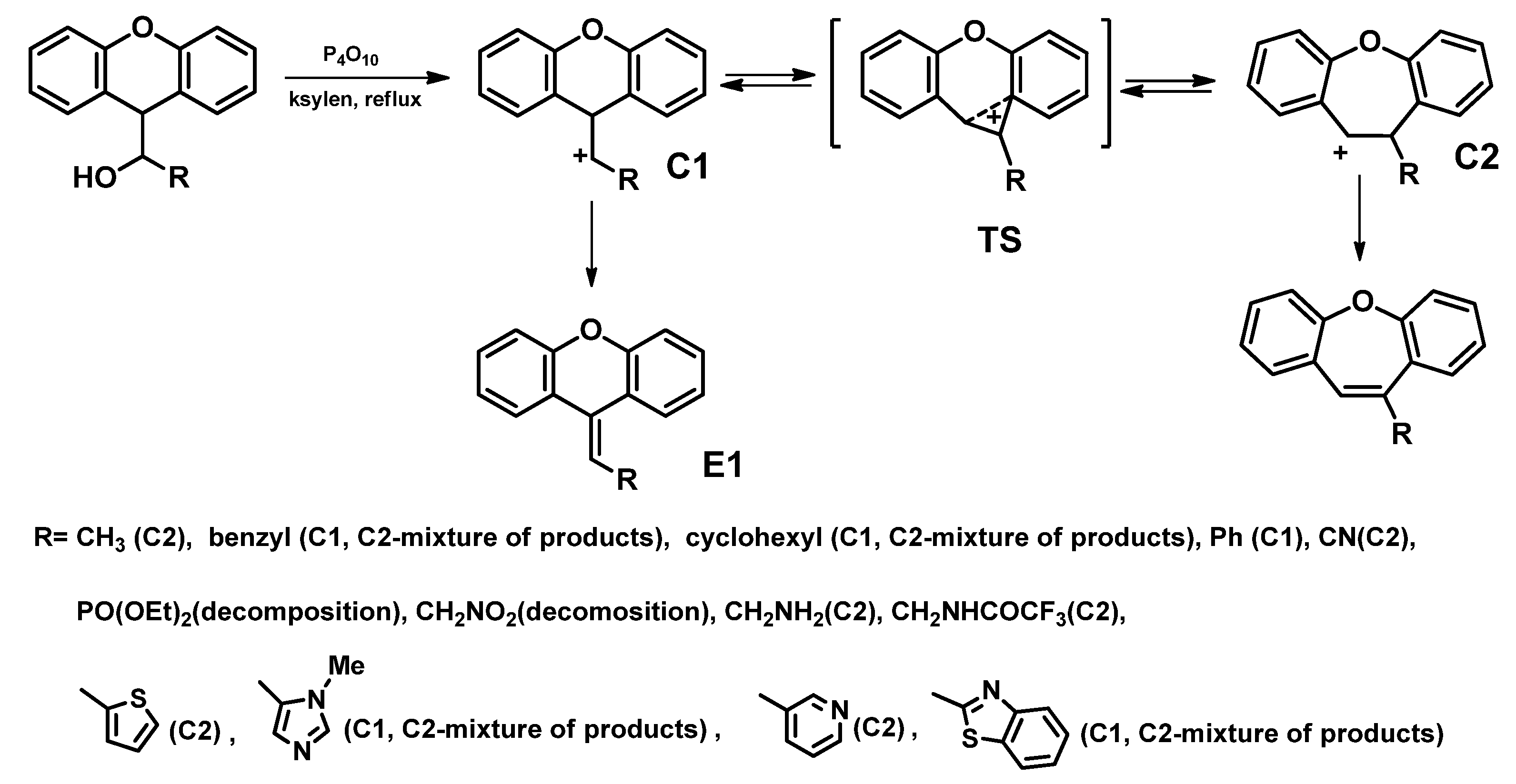
Dibenzo[b,f]oxepines can be obtained from 9-hydroxyalkylxanthene via Wagner-Meerwein rearrangement (Scheme 2) [66]. The solvent used is xylene. This reaction involves phosphorus (V) oxide as a generating substance carbocation (C1). This carbocation can transform into a transition state (TS), which then converts to the second carbocation (C2), which in turn converts to the desired product. C1 can also undergo β-elimination to the xanthenylid-9-ene derivative (E1). The nature of the R substituent determines which of the two reactions is favored. Depending on it, the C1 or C2 carbocation is more energetically stable.
1.
Ullman coupling with a subsequent Friedel–Crafts reaction [67,68]
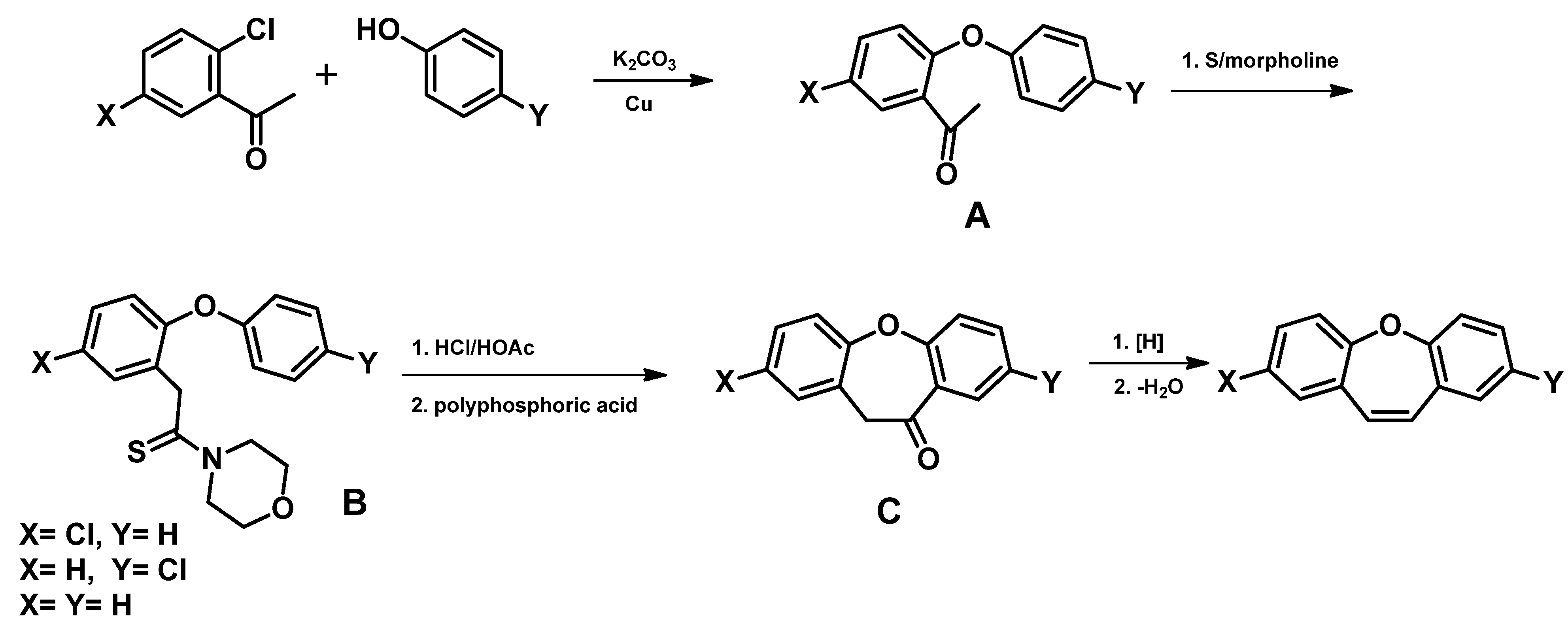
In this method, the appropriate acetophenone halogen derivative is reacted with a phenol derivative (the so-called Ullman diaryl ether synthesis, Scheme 3) [67,68], which leads to the preparation of a diphenyl ether derivative (A). Conversion of the ketone to acid or its derivative (B) then takes place, followed by a Friedel–Crafts reaction to give the compound (C). In the last step, the reduction of the carbonyl group is carried out with subsequent dehydration, which leads to the preparation of dibenzo[b,f]oxepine.
2.
Ullmann coupling and ring-closing metathesis reaction [69,70]
The substrates of the Ullmann coupling reaction are the halogen derivative and hydroxy derivative of styrene. The compounds undergo the following reactions: metathesis with the closure of the ring and conversion to the dibenzo[b,f]oxepine derivative (see Scheme 4) [69,70].
3.
Preparation from 2-halogenobenzaldehydes [71]
This is a one-step method that produces dibenzo[b,f]oxepine. The reaction of 2-halogenobenzaldehyde with a derivative of 2-(2-hydroxyphenyl) acetonitrile runs through sequential aldol condensation and an intramolecular ether formation reaction in the presence of Cs2CO3, molecular sieves, and in toluene as a solvent (Scheme 5) [71].
4.
Intramolecular aromatic nucleophilic substitution SNAr [58]
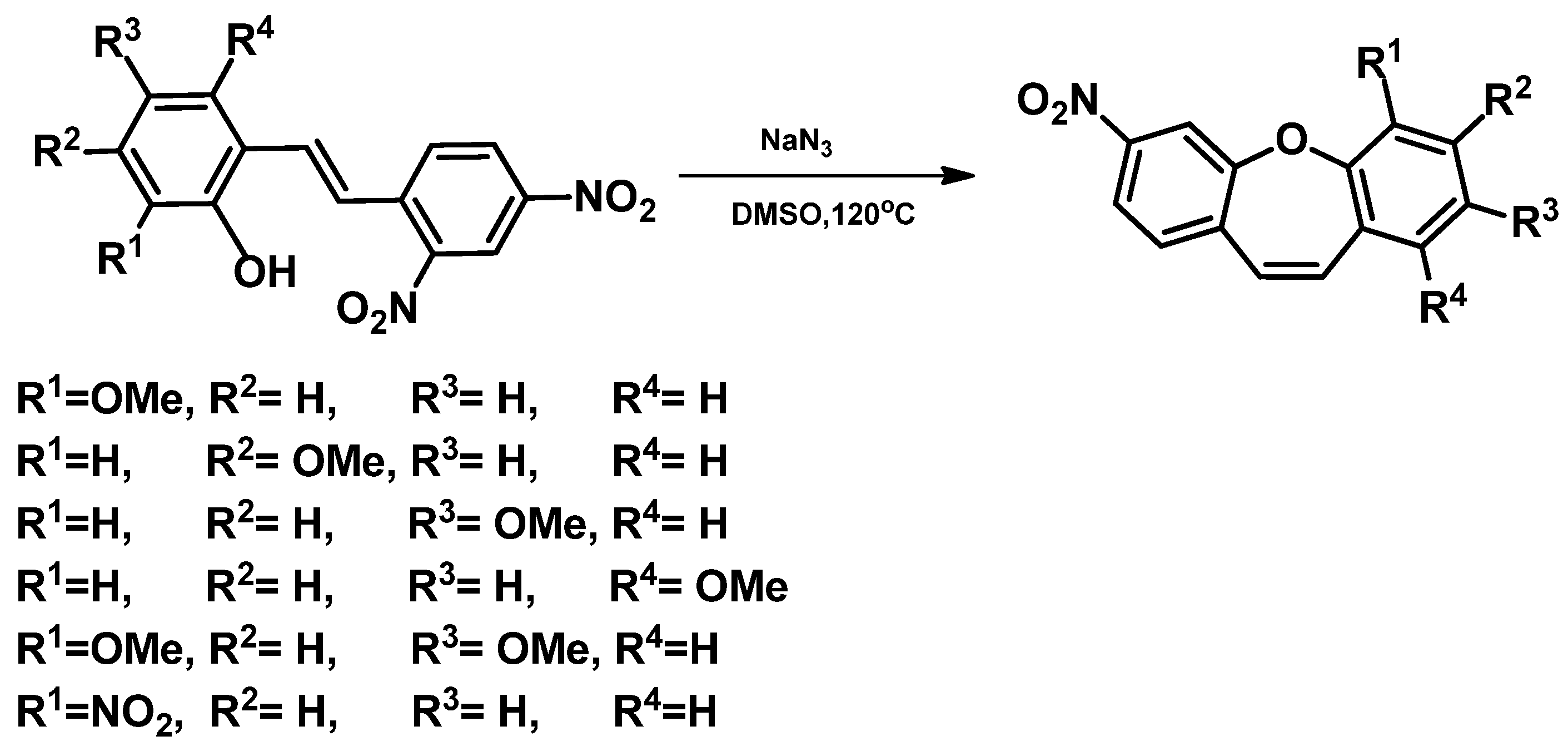
The above method of dibenzo[b,f]oxepine synthesis was developed by the research team of Krawczyk et al. [58] (Scheme 6). Intramolecular aromatic nucleophilic substitution is conducted in a sodium azide environment, which increases the yield of the reaction by about 50%. Two strongly electron-withdrawing nitro groups are attached to one of the stilbene rings. The donating hydroxyl group is located on the second ring. The substitution of one of the nitro groups by the hydroxyl group gives dibenzo[b,f]oxepine. The yield of the reaction is estimated from 88 to even 95% (Scheme 6).
5.
Knoevenagel condensation [72]
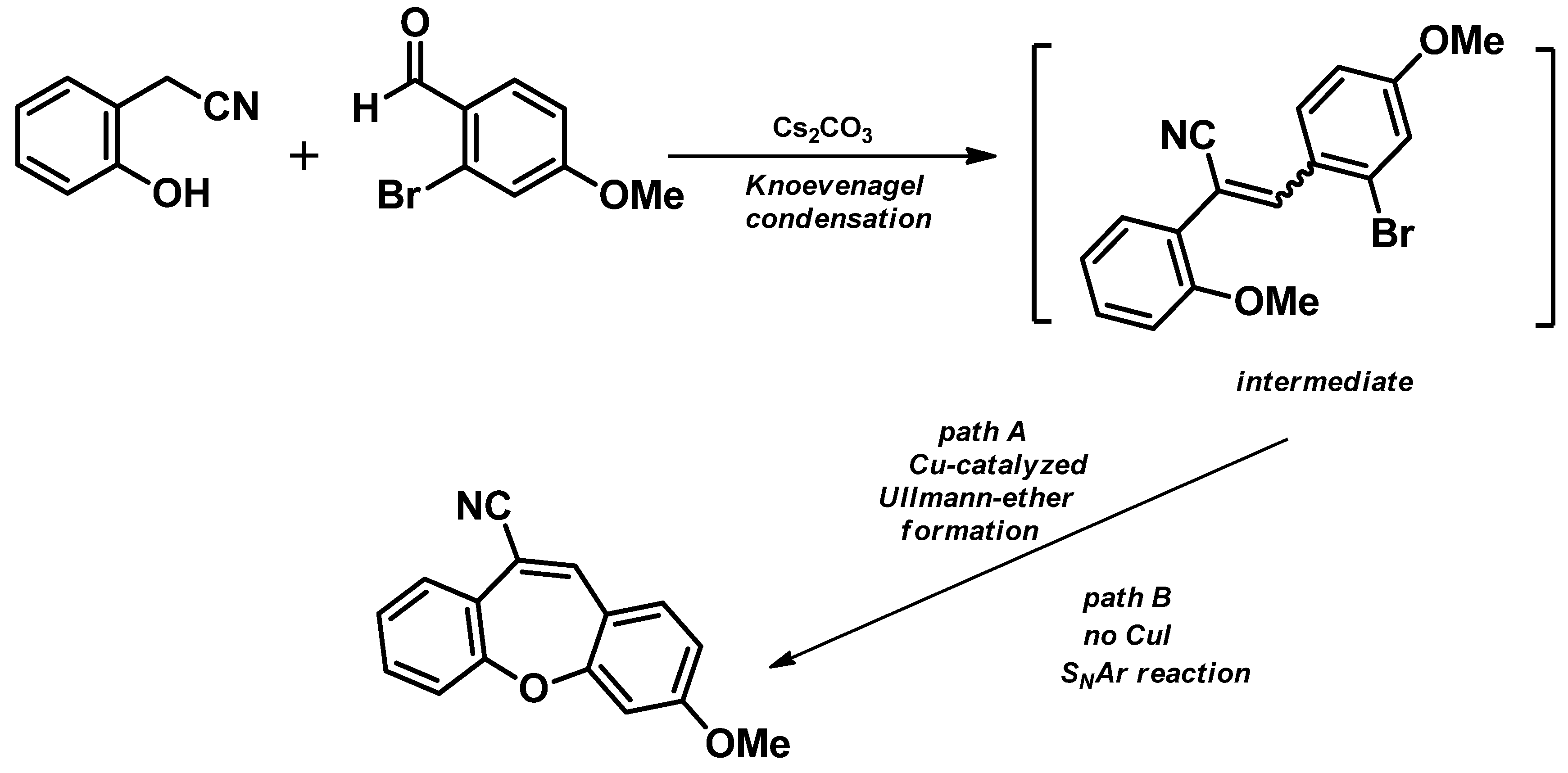
The authors propose a two-step course of reaction: the first step is a Knoevenagel condensation, and the next is the Ullmann ether formation (Scheme 7) [72]. The intermediate (Scheme 7) after Knoevenagel condensation is converted to dibenzo[b,f]oxepine via cyclization. The reaction from the intermediate can run with CuI (path A) or without CuI (path B). The yield of this reaction is high at 92%.
An interesting method is a recently developed simple one-pot synthesis of substituted dibenzo[b,f]oxepines under transition-metal-free conditions (Scheme 8) [73]. This cascade process involves nucleophilic aromatic substitution followed by Knoevenagel condensation. The yield of this process is 77%.
6.
Intramolecular McMurry reaction [74]
Dibenzo[b,f]oxepines can also be obtained as a result of an intramolecular McMurry reaction. In such, the diaryl ethers are used as the substrates. They can be obtained by the reaction of salicylaldehydes with fluorobenzaldehydes exposed to microwaves. The McMurry reaction for synthesized dibenzo[b,f]oxepine using TiCl4/Zn catalyst in THF has a yield of 53–55%. The synthesis mechanism is still under discussion, but the reaction possibly passes through the intermediate state of metallopinacol formed by the dimerization of ketyl radicals (Scheme 9).
7.
Mn(III)-based oxidative radical rearrangement [75]
In 2009, Cong et al. [75] performed a Mn III-based oxidative 1,2-radical rearrangement in order to obtain dibenzo[b,f]oxepines (Scheme 10). They used a large excess of Mn(OAc)3 (4 mol equiv.) in boiling glacial AcOH. The yield of this reaction was in the range of 63%–85% depending on the reaction time, substituents, and additives.
8.
Sequential Mizoroki–Heck reaction and Pd-catalyzed etherification [76]
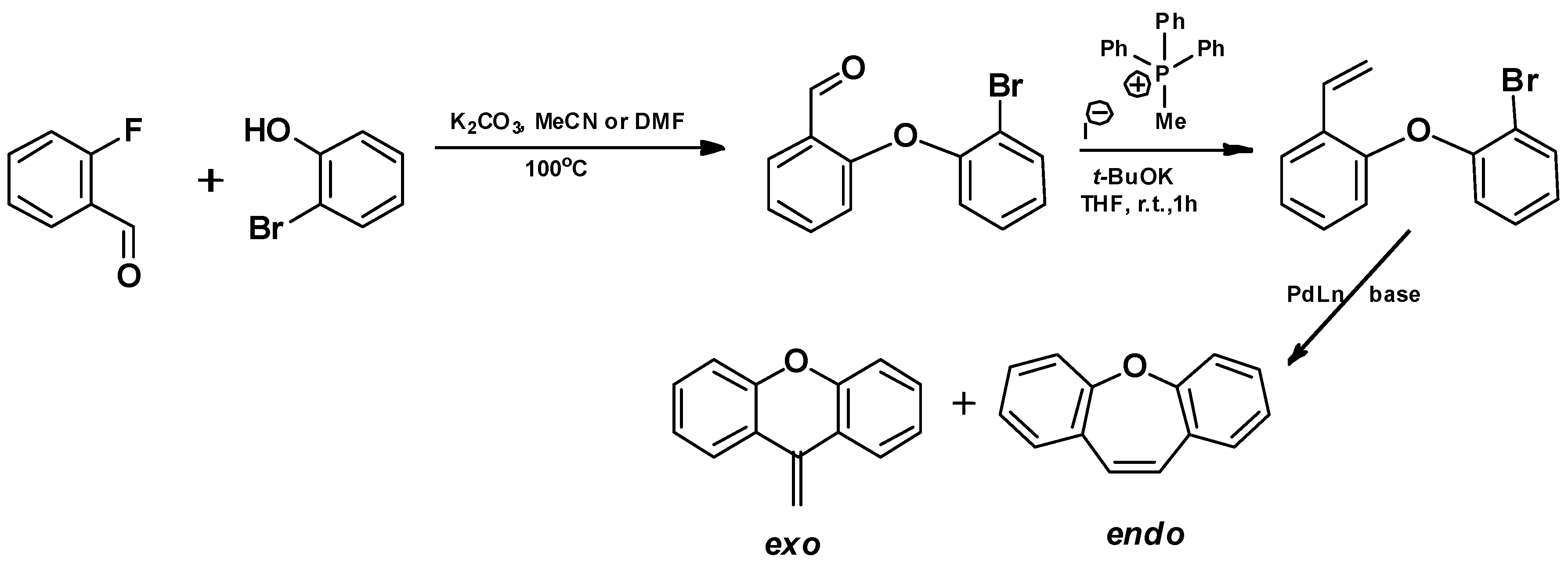
The protocol was based on a palladium-catalyzed intramolecular Mizoroki–Heck reaction of diaryl ethers (Scheme 11). The first step is a nucleophilic aromatic substitution reaction of 2-bromophenol and 2-fluorobenzaldehyde in the presence of potassium carbonate. Next, the obtained product reacts in Wittig olefination using methyl triphenylphosphonium iodide and potassium tert-butoxide. Closure of the bromoolefin diaryl ether in the presence of Pd2(dba)3 tert-butoxide and various phosphine ligands leads to dibenzo[b,f]oxepine (endo product) and to an exoproduct in the Mizoroki–Heck reaction. The yield of dibenzo[b,f]oxepine is 59% [76].
9.
Oxidative CH bond functionalization and ring expansion with TMSCHN2 [77]
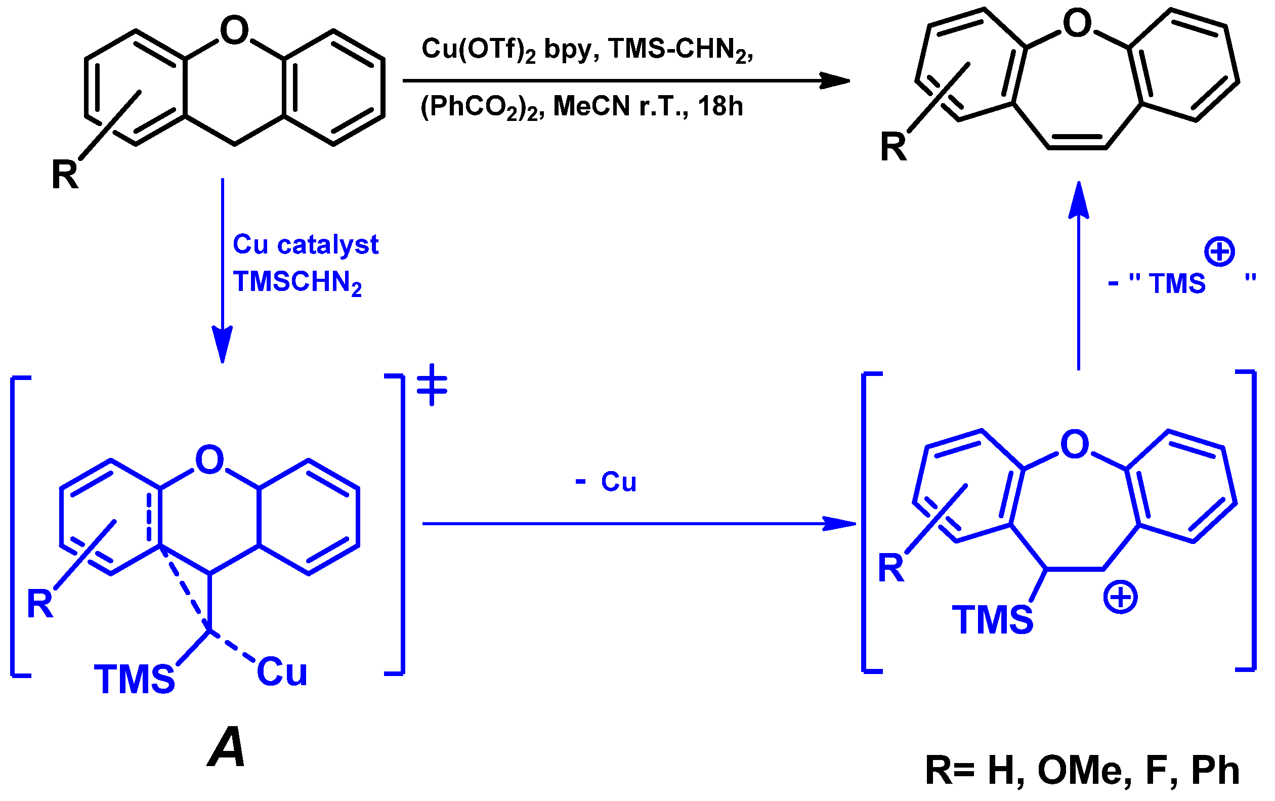
The authors [77] proposed a direct C-H functionalization/rearrangement sequence approach (Scheme 12) to obtain dibenzo[b,f]oxepine. They chose trimethylsilyl diazomethane (TMS-CHN2), which might also facilitate or promote the in situ rearrangement to the tricyclic backbone, nonprotic peroxide (PhCO2)2, a Cu(OTf)2 copper catalyst, which is reduced in situ to active CuI and different ligands such as 2,2′-bipyridine (to improve the properties and stability of the copper catalyst). In summary, the C-H functionalization, insertion, and rearrangement sequence provided the dibenz[b,f]oxepine scaffold with a moderate 55% yield.

5. Active Synthetic Dibenzo[b,f]oxepin Derivatives

Using the methods presented in the previous paragraph, many dibenzo[b,f]oxepin derivatives were synthesized and showed biological activity. The antipsychotic properties of these products were studied by the Smith group [78]. They found that among the tested compounds, dibenz[b,f]oxepine (1, Figure 4) has the highest dopamine D-4 receptor activity and is twice as active as clozapine (clozapine is an antipsychotic drug used in the clinic) in blocking the D-4 dopamine receptor. The affinity of 10-(4-methylpiperazino)dibenz[b,f]oxepine for clozapine binding sites in rat brains was studied [79]. [3H] Clozapine binding in the presence of atropine represents non-muscarinic binding, while binding in the absence of atropine represents muscarinic (cholinergic) binding. It was found that the ratios of the IC50 values for dopaminergic to non-muscarinic clozapine binding for (2ac) are 9.4 ± 2.0, 7.3 + 2.7, and 4.0 ± 1.3, respectively. In turn, the ratios of the IC50 values for dopaminergic to non-muscarinic clozapine binding for clozapine and (2c) are the same. Schindler and Blattner [80] studied 10-(dimethylaminomethyl)dibenzo[b,f]oxepine (3a, Figure 4) and its acid addition salts. The research confirmed its adrenolytic and depressant effect on the central nervous system (CNS). It is therefore useful as a sedative, anticonvulsant, and anesthesia enhancer. In 1980, the Kruse group [80,81] detected similar properties for [(alkylamino)ethyl]thio]dibenz[b,f]oxepines (3b3d, Figure 4). Trabanco and Megen’s group [82,83,84] synthesized series trans and cis tetrahydrodibenzo[b,f]furo [2,3-d]oxepin derivatives and studied their anxiolytic properties (4, Figure 4). The products were evaluated for in vitro affinities for the norepinephrine transporter 5-HT2A and 5-HT2C receptors. This action was also foreseen based on the ED50 values obtained in some in vivo assays. The dibenzo[b,f]oxepine imidazole derivatives (5a, 5b, Figure 4) were recognized as a novel class of tetracyclic compounds with anti-inflammatory activity through the specific inhibition of TNF-a secretion [85]. One of the most interesting examples of the use of dibenzo[b,f]oxepine is a compound that is an AII (angiotensin II) receptor antagonist (6, Figure 4) [86]. Angiotensin II causes an increase in blood pressure, which in turn causes damage to the blood vessels and the heart. AII antagonists bind to angiotensin receptors, thereby preventing angiotensin II itself from binding to its receptor. As a result, this compound may have electrolyte and blood-pressure-regulating properties and antihypertensive properties. The treatment of progressive neurodegenerative diseases such as Parkinson’s and Alzheimer’s diseases [87] with synthetic dibenzo[b,f]oxepine derivatives is of particular interest. Synthetic dibenzo[b,f]oxepines as antiestrogenic agents have also been studied [88]. The O-bridged compounds (7, Figure 4) were moderately active, and there are no new reports of research in this direction. A very promising derivative with a dibenzo[b,f]oxepine skeleton is omigapil (TCH346 or CGP3466, 8, Figure 4), first synthesized at Ciba-Geigy, Basel, Switzerland at the beginning of the 21st century. Santhera Pharmaceuticals has since taken over the production of omigapil and preclinical trials for congenital muscular dystrophy [89]. Notably, omigapil has shown safety in Phase 1 clinical trials for patients with pediatric and adolescent congenital muscular dystrophy (CMD) [89,90,91]. Recent studies with omigapil in rats with traumatic brain injury (TBI), which is the largest non-genetic-, non-aging-related risk factor for Alzheimer’s disease (AD), have shown that TCH346 reduces the level of high-level Tau protein and protects mice from neurodegeneration and AD [92]. Moreover, the derivatives containing the azepane ring presented in Figure 4 (9, Figure 4) show a strong inhibitory effect on the proliferation of breast cancer cells of the MDA-MB-231 line [93]. In addition, analgesic [94,95] and antidiabetic [96] properties have been found for dibenzo[b,f]oxepine derivatives, as well as insecticidal activities [97].

6. Potential of Dibenzo[b,f]oxepines as Microtubule Inhibitors

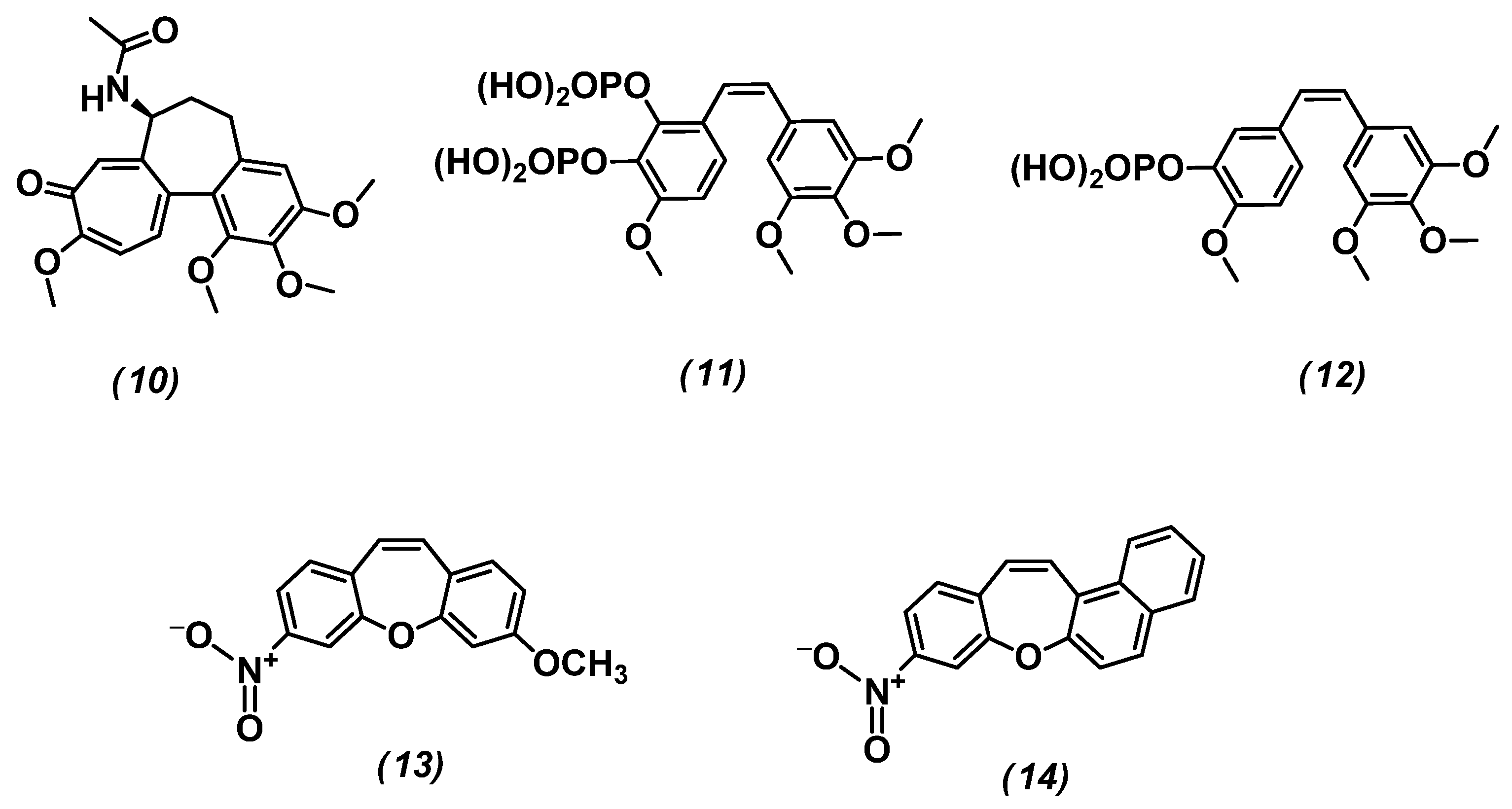
Notably, a very important new application of dibenzo[b,f]oxepines is targeting the polymerization/depolymerization of microtubules. Microtubules are fibers that are part of the cytoskeleton. They are composed of α and β-tubulin heterodimers. They form important structures from the point of view of cell division—such as karyokinetic spindles [98]. They are responsible for the alignment of the chromosome lines on the metaphase plate and their precise segregation to the opposite poles during the anaphase of cell division. Thanks to this, when mitosis occurs correctly, the same amount of genetic material goes to the daughter cells. There are chemical compounds that bind to the structural unit of the protein—such as tubulin. Some of them are active substances in anticancer drugs that disrupt the dynamics of microtubules (the balance of their shortening/extension). This phenomenon causes cancer cells to undergo apoptosis [99]. The chemical molecules interact through at least six binding sites: the laulimalide/peloruside binding site, taxane (including paclitaxel, docetaxel, and epothilones), vinca (vinblastine, vincristine, vinorelbine), pironetin, maytansine, and colchicine [100]. Of particular interest from the point of view of anticancer therapies is the colchicine (10, Figure 5) binding site. This substance effectively inhibits mitosis by inducing the depolymerization of microtubules, and cancer cells, due to the faster rate of cell division, are more susceptible to its poisoning. However, it is a substance with a low therapeutic index and is also very toxic to healthy cells. It causes, e.g., neutropenia (decrease in the number of neutrophils), gastrointestinal disorders, bone marrow damage, and anemia [101,102]. For this reason, structurally similar substances interacting with the colchicine binding site are sought. Stilbenoids (combretastatins CA1P-11 and CA4P-12, Figure 5) and dibenzo[b,f]oxepines, which contain the (Z)-stilbene motif in their skeleton, show great potential [103]. Several derivatives of these compounds have been studied in the literature, e.g., 13 and 14 (Figure 5). Using molecular modeling, their ability to interact with tubulin was analyzed. The analysis shows that both compounds bind to the colchicine binding site through hydrophobic interactions stabilized by hydrogen bonds. On the other hand, in vitro studies on cell cultures led to the conclusion that 9-nitrobenzo[b]naphtho [1,2-f]oxepine (14) has strong cytotoxic properties, as it does not affect cancer cells selectively, but it also induces apoptosis of “healthy” cells. For this reason, methods are sought to enable “targeted therapies” that selectively affect cancer cells.
Such a new, but very promising, therapy is photopharmacology (Figure 6) [104,105,106,107,108].
The scope of photopharmacology is the design, synthesis, research, and use of drugs whose activity can be controlled by light. Photopharmacology is a way to solve the activity of a therapeutic off-target (introduction of the drug to the target only after light activation) and a way to avoid side effects. This is possible by controlling the activity of the therapeutic agent from outside the body (Figure 6). Using this therapy, it will be possible to conduct a selective photo-controlled interaction of the drug with a molecular target located in the human body. Molecular switches are molecules that can be reversibly switched between at least two thermodynamically stable states. The pharmacological agents used in photopharmacology are bioactive molecules capable of changing their state, and, thus, their structures, under the influence of light-photochromic molecular switches. Azo compounds are one of the most numerous and most widely studied classes of photochromic molecular switches. They can exist in two isomeric states—E/Z—with E being on average about 10 kcal/mol more stable than Z. Derivatives of azobenzene are the most popular and most typically used photochromic molecular switches in biomedical applications. This is due, among other reasons, to their easy synthesis, relatively high photostationary states and quantum efficiency, fast photoisomerization, and low photo-quenching coefficient. It should be emphasized that photochromic switches with an azo group absorb light in the range from 650 nm to 900 nm (visible light range 380–750 nm—harmless), which means that light is not absorbed by water and hemoglobin and can penetrate deeper tissue layers. The combination of the aspects of microtubule polymerization inhibition and electromagnetic radiation activation of compounds containing azo bonds and a dibenzo[b,f]oxepine backbone with methoxy groups was the subject of research by Borys et al. [109,110]. Firstly, their studies focused on the synthesis of bioactive compounds interacting with tubulin (protein-building microtubules performing the function of the cytoskeleton) and assessing their usefulness in anticancer therapy. Secondly, their research aimed to determine the ability of the newly obtained azo derivatives of methoxydibenzo[b,f]oxepine to act as molecular switches in the system with tubulin and the way to activate them under the influence of harmless visible light (Figure 7).
In silico studies showed that after combining dibenzo[b,f]oxepines with an azo switch, the molecules interact with the colchicine binding site in tubulin with a part of the dibenzo[b,f]oxepine, in a part of the azo switch, or both at the same time. Based on the UV-VIS spectra, it was found that compounds switch in the visible part of the spectrum and, therefore, these derivatives have the potential to be used in photopharmacology.
To summarize, in Table 2, synthesized dibenzo[b,f]oxepine derivatives that exhibit biological activity or that have such potential are presented.

7. The Perspective for Applications of Dibenzo[b,f]oxepine Derivatives

In conclusion, in this short review, we discussed the use of synthetic and natural derivatives of dibenzo[b,f]oxepin. We have shown the use of these compounds mainly in medicine. Several directions can be listed for further research on dibenzo[b,f]oxepines, searching for new, not-yet-isolated compounds (naturally occurring dibenzo[b,f]oxepins) from plants that have a healing effect. Omigapil, which exhibits dose-dependent inhibition of HIV, dengue virus, and Zika virus [111], appears to have a high potential for medical use and, as previously mentioned, may alleviate symptoms of congenital muscular dystrophy. Dibenzo[b,f]oxepines also have the potential as microtubule polymerization inhibitors. The use of combinations of dibenzo[b,f]oxepines with molecular switches for applications in photopharmacology is another way of using these compounds. The activity of dibenzo[b,f]oxepines should also be checked, as they are small molecules binding to RNAs implicated in infectious disease (for PreQ(1) riboswitches), whose structure is very similar to the structure of 2-(dibenzo[b,d]furan-2-yloxy)ethanamine and which have recently been discovered [112,113].

Funding

The research was funded by the Warsaw University of Technology within the Excellence Initiative: Research University (IDUB) program.

Institutional Review Board Statement

Not applicable.

Informed Consent Statement

Not applicable.

Data Availability Statement

Not applicable.

Conflicts of Interest

The author declares no conflict of interest.

References

  1. Ram, V.J.; Sethi, A.; Nath, M.; Pratap, R. Chapter 3—Seven-Membered Heterocycles. In The Chemistry of Heterocycles Chemistry of Six-to-Eight-Membered N, O, S, P and Se Heterocycles; Elsevier: Amsterdam, The Netherlands, 2019; Volume 3, pp. 393–425. [Google Scholar]
  2. Rosowsky, A. (Ed.) The Chemistry of Heterocyclic Compounds; Seven-Membered Heterocyclic Compounds Containing Oxygen and Sulfur; John Wiley & Sons Inc.: Hoboken, NJ, USA, 1972; Volume XXVI. [Google Scholar]
  3. Beleńki, L.I. Comprehensive Heterocyclic Chemistry III. 13.02—Oxepanes and Oxepines; Elsevier: Amsterdam, The Netherlands, 2008; Volume 13, pp. 45–95. [Google Scholar]
  4. Rutz, A.; Sorokina, M.; Galgonek, J.; Mietchen, D.; Willighagen, E.; Gaudry, A.; Graham, J.G.; Stephan, R.; Page, R.; Vondrášek, J.; et al. The LOTUS initiative for open knowledge management in natural products research. eLife 2022, 11, e70780. [Google Scholar] [CrossRef] [PubMed]
  5. Shen, T.; Wang, X.-N.; Lou, H.-X. Natural stilbenes: An overview. Nat. Prod. Rep. 2009, 26, 916–935. [Google Scholar] [CrossRef] [PubMed]
  6. Zhang, Q.-W.; Lin, L.-G.; Ye, W.-C. Techniques for extraction and isolation of natural products: A comprehensive review. Chin. Med. 2018, 13, 20. [Google Scholar] [CrossRef] [PubMed] [Green Version]
  7. De Sousa, L.M.; de Carvalho, J.L.; da Silva, H.C.; Lemos, T.L.G.; Arriaga, A.M.C.; Braz-Filho, R.; Militao, G.C.G.; Silva, T.D.S.; Ribeiro, P.R.V.; Santiago, G.M.P. New Cytotoxic Bibenzyl and Other Constituents from Bauhinia ungulata L. (Fabaceae). Chem. Biodivers. 2016, 13, 1630–1635. [Google Scholar] [CrossRef]
  8. Pettit, G.R.; Numata, A.; Iwamoto, C.; Usami, Y.; Yamada, T.; Ohishi, H.; Cragg, G.M. Antineoplastic Agents. 551. Isolation and Structures of Bauhiniastatins 1–4 from Bauhinia purpurea. J. Nat. Prod. 2006, 69, 323–327. [Google Scholar] [CrossRef]
  9. Boonphong, S.; Puangsombat, P.; Baramee, A.; Mahidol, C.; Ruchirawat, S.; Kittakoop, P. Bioactive Compounds from Bauhinia purpurea Possessing Antimalarial, Antimycobacterial, Antifungal, Anti-inflammatory, and Cytotoxic Activities. J. Nat. Prod. 2007, 70, 795–801. [Google Scholar] [CrossRef]
  10. Anjaneyulu, A.S.R.; Reddy, A.V.R.; Reddy, D.S.K.; Ward, R.S.; Adhikesavalu, D.; Cameron, T.S. Pacharin: A new dibenzo(2,3-6,7)oxepin derivative from Bauhinia racemosa Lamk. Tetrahedron 1984, 40, 4245–4252. [Google Scholar] [CrossRef]
  11. Tanjung, M.; Saputri, R.D.; Sri Tjahjandarie, T. Antioxidant activity of two isomeric benzoxepin derivatives from the stembark of Bauhinia aculeata L. J. Chem. Pharm. Res. 2014, 6, 705–708. [Google Scholar]
  12. Góis, R.W.S.; de Sousa, L.M.; da Silva, H.C.; da Silva, F.E.F.; Pimenta, A.T.A.; Lima, M.A.S.; Arriaga, A.M.C.; Lemos, T.L.G.; Braz-Filho, R.; Militãod, G.C.G.; et al. Chemical constituents from Bauhinia acuruana and their cytotoxicity. Rev. Bras. Farmacogn. Braz. J. Pharmacogn. 2017, 27, 711–715. [Google Scholar] [CrossRef]
  13. De Maria Souza, S.; Santos de Souza, L.; Silva, V.R.; Pereira Soares, M.B.; Bezerra, D.P.; Wagner da Silva Gois, R.; da Silva, H.C.; Pinheiro Santiago, G.M.; Militao, G.C.G. Natural Dibenzo[b,f]oxepines, Pacharin and Bauhiniastatin-1, Isolated from Bauhinia acuruana Induce Apoptosis on Breast Cancer Cells via MCL-1 Protein Reduction. Planta Med. 2023, 89, 286–294. [Google Scholar] [CrossRef]
  14. Chen, K.; Tang, H.; Zheng, L.; Wang, L.; Xue, L.; Peng, A.; Tang, M.; Long, C.; Chen, X.; Yeb, H.; et al. Identification of compounds with cytotoxic activity from Millettia dorwardi Coll. Et. Hemsl. Phytochem. Lett. 2018, 25, 60–64. [Google Scholar] [CrossRef]
  15. Bindu Mekala, A.; Satyal, P.; Setzer, W.N. Phytochemicals from the Bark of Rhamnus caroliniana. Nat. Prod. Commun. 2017, 12, 403–406. [Google Scholar]
  16. Mu, L.; Li, J.; Yang, J.; Zhang, D. New dibenz[b,f]oxepins from Cercis chinensis Bunge. J. Asian Nat. Prod. Res. 2007, 9, 649–653. [Google Scholar] [CrossRef]
  17. Gois, R.W.S.; De Sousa, L.M.; Lemos, T.L.G.; Arriaga, A.M.C.; Andrade-Neto, M.; Santi-ago, G.M.P.; Ferreira, Y.S.; Alves, P.B.; De Jesus, H.C.R. Chemical composition and larvicidal effects of essential oil from Bauhinia acuruana (Moric) against Aedes aegypti. J. Essent. Oil Res. 2011, 23, 59–62. [Google Scholar] [CrossRef]
  18. Góis, R.W.S.; De Sousa, L.M.; Santiago, G.M.P.; Romero, N.R.; Lemos, T.L.G.; Arriaga, A.M.C.; Braz-Filho, R. Larvicidal activity against Aedes aegypti of pacharin from Bauhinia acuruana. Parasitol. Res. 2013, 112, 2753–2757. [Google Scholar] [CrossRef]
  19. Narita, K.; Nakamura, K.; Abe, Y.; Katoh, T. Total Synthesis of Bauhinoxepin J: A Biologically Active Dibenzo[b,f]oxepin Isolated from Bauhinia purpurea. Eur. J. Org. Chem. 2011, 26, 4985–4988. [Google Scholar] [CrossRef]
  20. Prudhomme, V.; Cucca, M.; Nauton, L.; Andrieu, E.; Fereyrolles, M.; Lamoine, S.; Michelin, C.; Bennis, K.; Collin, A.; De Ceuninck, F.; et al. Design, synthesis and biological evaluation of conformationnally-restricted analogues of E7010 as inhibitors of tubulin assembly (ITA) and vascular disrupting agents (VDA). Eur. J. Med. Chem. 2022, 244, 114809. [Google Scholar] [CrossRef]
  21. Yang, W.J.; Zhou, P.; Liang, L.; Cao, Y.; Qiao, J.; Li, X.; Teng, Z.; Wang, L. Nanogel-Incorporated Injectable Hydrogel for Synergistic Therapy Based on Sequential Local Delivery of Combretastatin-A4 Phosphate (CA4P) and Doxorubicin (DOX). ACS Appl. Mater. Interfaces 2018, 10, 18560–18573. [Google Scholar] [CrossRef]
  22. Kittakoop, P.; Nopichai, S.; Thongon, N.; Charoenchai, P.; Thebtaranonth, Y. Bauhinoxepins A and B: New Antimycobacterial Dibenzo[b,f]oxepins from Bauhinia saccocalyx. Helv. Chim. Acta 2004, 87, 175–179. [Google Scholar] [CrossRef]
  23. De Lera, A.R.; Suau, R.; Castedo, L. Synthetic approaches to cularines. II. Ullmann condensation. J. Heterocycl. Chem. 1987, 24, 313–319. [Google Scholar] [CrossRef]
  24. Kim, Y.H.; Kim, M.; Yoo, M.; Kim, J.E.; Lee, H.K.; Heo, J.-N.; Lee, C.O.; Yoo, M.; Junga, K.-Y.; Yun, C.-S.; et al. A natural compound, aristoyagonine, is identified as a potent bromodomain inhibitor by mid-throughput screening. Biochem. Biophys. Res. Commun. 2018, 503, 882–887. [Google Scholar] [CrossRef] [PubMed]
  25. Boente, J.M.; Castedo, L.; Domínguez, D.; Fariña, A.; Rodriguez de Lera, A.; Villaverde, C. Secocularines: A new group of isoquinoline related alkaloids present in the fumariaceae. Tetrahedron Lett. 1984, 25, 889–892. [Google Scholar] [CrossRef]
  26. Castedo, L.; Dominguez, D.; Lopez, S.; Tojo, E.; Villaverde, C. Five new secocularine alkaloids have been isolated from Sarcocapnos species and their structures elucidated by spectroscopic studies and chemical correlations. Heterocycles 1987, 26, 591–594. [Google Scholar] [CrossRef]
  27. Castedo, L.; López, S.; Rodríguez De Lera, A.; Villaverde, C. Alkaloids of Sarcocapnos crassifolia subsp. speciosa. Phytochemistry 1989, 28, 251–257. [Google Scholar] [CrossRef]
  28. Frotáis, P.; Arbaoui, J.; Bakkali, E.-H. Effects of various isoquinoline alkaloids on in vitro 3H-dopamine uptake by rat striatal synaptosomes. J. Nat. Prod. 1995, 58, 1475–1484. [Google Scholar] [CrossRef]
  29. Bentley, K.W. β-Phenylethylamines and the lsoquinoline Alkaloids. Nat. Prod. Rep. 1989, 6, 405–432. [Google Scholar] [CrossRef]
  30. Tojo, E.; Dominguez, D.; Castedo, L. Alkaloids from Sarcocapnos enneaphylla. Phytochemistry 1991, 30, 1005–1010. [Google Scholar] [CrossRef]
  31. Phillipson, D.; Roberts, M.F.; Zenk, M.H. (Eds.) The Chemistry and Biology of Iso Quinoline Alkaloids; Springer: Berlin/Heidelberg, Germany; New York, NY, USA; Tokyo, Japan, 1985; ISBN 978-3-642-70130-6. [Google Scholar]
  32. Kumari, S.; Sharma, A.; Sharma, U. A cularine-type isoquinoline alkaloid from the root part of Cissampelos pareira. Nat. Prod. Res. 2022; in press. [Google Scholar] [CrossRef]
  33. Chung, M.-I.; Ko, H.-H.; Yen, M.-H.; Lin, C.-N.; Yang, S.-Z.; Tsao, L.-T.; Wang, J.-P. Artocarpol A, A Novel Constituent with Potent Anti-Inflammatory Effect, Isolated from Artocarpus rigida. Helv. Chim. Acta 2000, 83, 1200–1204. [Google Scholar] [CrossRef]
  34. Ko, H.H.; Lin, C.N.; Yang, S.Z. New Constituents of Artocarpus rigida. Helv. Chim. Acta 2000, 83, 3000–3005. [Google Scholar] [CrossRef]
  35. Ko, H.-H.; Yang, S.-Z.; Lin, C.-N. Artocarpol F, A Phenolic Compound with A Novel Skeleton, Isolated from Artocarpus rigida. Tetrahedron Lett. 2001, 42, 5269–5270. [Google Scholar] [CrossRef]
  36. Lu, Y.H.; Lin, C.-N.; Ko, H.-H.; Yang, S.-Z.; Tsao, L.T.; Wang, J.-P. Two Novel and Anti-Inflammatory Constituents of Artocarpus rigida. Helv. Chim. Acta 2002, 85, 1626–1632. [Google Scholar] [CrossRef]
  37. Lu, Y.H.; Lin, C.-N.; Ko, H.-H.; Yang, S.-Z.; Tsao, L.T.; Wang, J.-P. Novel Anti-Inflammatory Constituents of Artocarpus rigida. Helv. Chim. Acta 2003, 86, 2566–2572. [Google Scholar] [CrossRef]
  38. Kuan, Y.-H.; Lin, R.-H.; Tsao, L.-T.; Lin, C.-N.; Wang, J.-P. Artocarpol A stimulation of superoxide anion generation in neutrophils involved the activation of PLC, PKC and p38 mitogen-activated PK signaling pathways. Br. J. Pharm. 2005, 145, 460–468. [Google Scholar] [CrossRef]
  39. Lin, Y.-L.; Chang, Y.-Y.; Kuo, Y.-H.; Shiao, M.-S. Anti-Lipid-Peroxidative Principles from Tournefortia sarmentosa. J. Nat. Prod. 2002, 65, 745–747. [Google Scholar] [CrossRef]
  40. Chen, P.-C.; Tsai, W.-J.; Ueng, Y.-F.; Tzeng, T.-T.; Chen, H.-L.; Zhu, P.-R.; Huang, C.-H.; Shiao, Y.-J.; Li, W.-T. Neuroprotective and Antineuroinflammatory Effects of Hydroxyl-Functionalized Stilbenes and 2-Arylbenzo[b]furans. J. Med. Chem. 2017, 60, 4062–4073. [Google Scholar] [CrossRef]
  41. Wang, C.N.; Pan, H.C.; Lin, Y.L.; Chi, C.W.; Shiao, Y.J. Ester derivatives of tournefolic acid B attenuate N methyl-D-aspartatemediated excitotoxicity in rat cortical neurons. Mol. Pharmacol. 2006, 69, 950–959. [Google Scholar] [CrossRef] [Green Version]
  42. Chi, C.W.; Wang, C.N.; Lin, Y.L.; Chen, C.F.; Shiao, Y.J. Tournefolic acid B methyl ester attenuates glutamate-induced toxicity by blockade of ROS accumulation and abrogating the activation of caspases and JNK in rat cortical neurons. J. Neurochem. 2005, 92, 692–700. [Google Scholar] [CrossRef]
  43. Chi, C.W.; Lin, Y.L.; Wang, Y.H.; Chen, C.F.; Wang, C.N.; Shiao, Y.J. Tournefolic acid B attenuates amyloid beta proteinmediated toxicity by abrogating the calcium overload in mitochondria and retarding the caspase 8-truncated Bid-cytochrome c pathway in rat cortical neurons. Eur. J. Pharmacol. 2008, 586, 35–44. [Google Scholar] [CrossRef]
  44. Lin, Y.L.; Tsay, H.J.; Lai, T.H.; Tzeng, T.T.; Shiao, Y.J. Lithospermic acid attenuates 1-methyl-4-phenylpyridine-induced neurotoxicity by blocking neuronal apoptotic and neuroinflammatory pathways. J. Biomed. Sci. 2015, 22, 37. [Google Scholar] [CrossRef] [Green Version]
  45. Lipinski, C.A.; Lombardo, F.; Dominy, B.W.; Feeney, P.J. Experimental and computational approaches to estimate solubility and permeability in drug discovery and development settings. Adv. Drug Deliv. Rev. 2001, 46, 3–26. [Google Scholar] [CrossRef] [PubMed]
  46. Lipinski, C.A. Lead- and drug-like compounds: The rule-of-five revolution. Drug Discov. Today. Technol. 2004, 1, 337341. [Google Scholar] [CrossRef] [PubMed]
  47. Daniel, F.V.; Stephen, R.J.; Cheng, H.; Smith, B.R.; Keith, W.W.; Kenneth, D.K.; Veber, D.F.; Johnson, S.R.; Cheng, H.; Smith, B.R.; et al. Molecular Properties That Influence the Oral Bioavailability of Drug Candidates. J. Med. Chem. 2002, 45, 2615–2623. [Google Scholar]
  48. Kenny, P.W. Hydrogen-Bond Donors in Drug Design. J. Med. Chem. 2022, 65, 14261–14275. [Google Scholar] [CrossRef] [PubMed]
  49. Wells, T.N.C.; Fersht, A.R. Hydrogen bonding in enzymatic catalysis analysed by protein engineering. Nature 1985, 316, 656–657. [Google Scholar] [CrossRef]
  50. Tromans, R.A.; Carter, T.S.; Chabanne, L.; Crump, M.P.; Li, H.; Matlock, J.V.; Orchard, M.G.; Davis, A.P. A biomimetic receptorfor glucose. Nat. Chem. 2019, 11, 52–56. [Google Scholar] [CrossRef]
  51. Desiraju, G.R. Crystal engineering: A holistic view. Angew. Chem. Int. Ed. 2007, 46, 8342–8356. [Google Scholar] [CrossRef]
  52. Etter, M.C. Encoding and decoding hydrogen-bond patterns of organic compounds. Acc. Chem. Res. 1990, 23, 120–126. [Google Scholar] [CrossRef]
  53. Bonnet, J.; Colotte, M.; Coudy, D.; Couallier, V.; Portier, J.; Morin, B.; Tuffet, S. Chain and conformation stability of solid-state DNA: Implications for room temperature storage. Nucleic Acids Res. 2010, 38, 1531–1546. [Google Scholar] [CrossRef] [Green Version]
  54. Campello, M.J.; Castedo, L.; Dominguez, D.; Rodriguez de Lera, A.; Saá, J.M.; Suau, R.; Tojo, E.; Vidal, M.C. New oxidized isocularine alkaloids from sarcocapnos plants. Tetrahedron Lett. 1984, 25, 5933–5936. [Google Scholar] [CrossRef]
  55. Castedo, L.; Suau, R.; Mouriño, A. A revised structure for pontevedrine. Tetrahedron Lett. 1976, 17, 501–502. [Google Scholar] [CrossRef]
  56. Drake, J.A.G.; Jones, D.W. The structure of dibenz[b,f]oxepin. Acta Crystallogr. B Struct. Cryst. Cryst. Chem. 1982, 38, 200–203. [Google Scholar] [CrossRef] [Green Version]
  57. Shukla, D.; Wan, P. Evidence for a planar cyclically conjugated 8π system in the excited state: Large Stokes shift observed for dibenz[b,f]oxepin fluorescence. J. Am. Chem. Soc. 1993, 115, 2990–2991. [Google Scholar] [CrossRef]
  58. Wrzesiński, M.; Mielecki, D.; Szczeciński, P.; Grzesiuk, E.; Krawczyk, H. Synthesis of derivatives of methoxydibenzo[b,f]oxepine in the presence of sodium azide. Tetrahedron 2016, 72, 3877–3884. [Google Scholar]
  59. Toldo, J.; El Bakouri, O.; Sola, M.; Norrby, P.-O.; Ottosson, H. Is Excited-State Aromaticity a Driving Force for Planarization of Dibenzannelated 8π-Electron Heterocycles? ChemPlusChem 2019, 84, 712–721. [Google Scholar] [CrossRef]
  60. Ottosson, H. Organic photochemistry: Exciting excited-state aromaticity. Nat. Chem. 2012, 4, 969–971. [Google Scholar] [CrossRef]
  61. Kotani, R.; Liu, L.; Kumar, P.; Kuramochi, H.; Tahara, T.; Liu, P.; Osuka, A.; Karadakov, P.B.; Saito, S. Controlling the S1 Energy Profile by Tuning Excited-State Aromaticity. J. Am. Chem. Soc. 2020, 142, 14985–14992. [Google Scholar] [CrossRef]
  62. Pschorr, R.; Knöffler, C. Űber die Konstitution des Morphothebains II. Synthese des durch Abbau von Morphothebain erhaltenen Tetramethoxyphenanthrens. Liebigs Ann. Chem. 1911, 382, 50–61. [Google Scholar] [CrossRef]
  63. Gironés-Vilaplana, A.; Moreno, D.A.; García-Viguera, C. Maqui berry vs. Sloe berry–liquor-based beverage for new development. Nat. Prod. Commun. 2015, 10, 81–82. [Google Scholar] [CrossRef]
  64. Ugwu, D.I.; Eze, F.U.; Ezugwu, J.A.; Attah, S.I.; Ugwu, M.C.; Ekoh, O.C.; Nyoyoko, I.F. Chemotherapeutic Importance of Oxepines. Int. J. Chem. Sci. 2021, 19, 401. [Google Scholar]
  65. Yadav, S.; Ramarao, J.; Suresh, S. Recent Advances on the Development of Synthetic Strategies to Access Dibenzoxepine Derivatives. Synthesis 2023, 55, 369–399. [Google Scholar] [CrossRef]
  66. Storz, T.; Vangrevelinghe, E.; Dittmar, P. Synthesis and Wagner-Meerwein Rearrangement of 9-(α-Hydroxyalkyl)xanthenes to 10-Substituted Dibenz[b,f]oxepins: Scope, Limitations and ab initio Calculations. Synthesis 2005, 15, 2562–2570. [Google Scholar] [CrossRef]
  67. Olivera, R.; San Martin, R.; Churruca, F.; Dominguez, E. Revisiting the Ullmann—Ether Reaction: A Concise and Amenable Synthesis of Novel Dibenzoxepino [4,5-d]pyrazoles by Intramolecular Etheration of 4,5-(o,o′-Halohydroxy)arylpyrazoles. J. Org. Chem. 2002, 67, 7215–7225. [Google Scholar] [CrossRef] [PubMed]
  68. Nagai, Y.; Irie, A.; Nakamura, H.; Hino, K.; Uno, H.; Nishimura, H. Nonsteroidal antiinflammatory agents. 1. 10,11-Dihydro-11-oxodibenz[b,f] oxepinacetic acids and related compounds. J. Med. Chem. 1982, 25, 1065–1070. [Google Scholar] [CrossRef] [PubMed]
  69. Bharath, Y.; Thirupathi, B.; Ranjit, G.; Mohapatra, D.K. An Efficient Synthesis of Dibenzo[b,f]oxepins by Ring-Closing Metathesis. Asian J. Org. Chem. 2013, 2, 848–851. [Google Scholar] [CrossRef]
  70. Matsuda, T.; Sato, S. Synthesis of Dibenzoheteropines of Group 13–16 Elements via Ring-Closing Metathesis. J. Org. Chem. 2013, 78, 3329–3335. [Google Scholar] [CrossRef]
  71. Choi, Y.L.; Lim, H.S.; Lim, H.J.; Heo, J.-N. One-Pot Transition-Metal-Free Synthesis of Dibenzo[b,f]oxepins from 2-Halobenzaldehydes. Org. Lett. 2012, 14, 5102–5105. [Google Scholar] [CrossRef]
  72. Wang, Y.; Chen, Y.; He, Q.; Xie, Y.; Yang, C. Copper-Assisted/Copper-Free Synthesis of Functionalized Dibenzo[b,f]oxepins and Their Analogs via a One-Pot Tandem Reaction. Helv. Chim. Acta 2013, 96, 296–308. [Google Scholar] [CrossRef]
  73. Taweesak, P.; Thongaram, P.; Kraikruan, P.; Thanetchaiyakup, A.; Chuanopparat, N.; Hsieh, H.-P.; Uang, B.-J.; Ngernmeesri, P. One-Pot Synthesis of Dibenzo[b,f]oxepines and Total Synthesis of Bauhinoxepin C. J. Org. Chem. 2021, 86, 1955–1963. [Google Scholar] [CrossRef]
  74. Moreno, D.R.R.; Giorgi, G.; Salas, C.O.; Tapia, R.A. New Short Strategy for the Synthesis of the Dibenz[b,f]oxepin Scaffold. Molecules 2013, 18, 14797–14806. [Google Scholar] [CrossRef] [Green Version]
  75. Cong, Z.; Miki, T.; Urakawa, O.; Nishino, H. Synthesis of Dibenz[b,f]oxepins via Manganese(III)-Based Oxidative 1,2-Radical Rearrangement. J. Org. Chem. 2009, 74, 3978–3981. [Google Scholar] [CrossRef]
  76. Jepsen, T.H.; Larsen, M.; Jørgensen, M.; Nielsen, M.B. Three-Step Synthesis of (Thio)xanthene and Dibenzothiepine/Dibenzoxepine by an Intramolecular Mizoroki-Heck Reaction of Diaryl (Thio)Ethers. Synlett 2012, 23, 418–422. [Google Scholar]
  77. Stopka, T.; Marzo, L.; Zurro, M.; Janich, S.; Wrthwein, E.-U.; Daniliuc, C.G.; Aleman, J.; Mancheno, O.G. Oxidative CH Bond Functionalization and Ring Expansion with TMSCHN2: A Copper(I)-Catalyzed Approach to Dibenzoxepines and Dibenzoazepines. Angew. Chem. Int. Ed. 2015, 54, 5049–5053. [Google Scholar] [CrossRef]
  78. Phillips, S.T.; de Paulis, T.; Neergaard, J.R.; Baron, B.M.; Siegel, B.W.; Seeman, P.; Van Tol, H.H.M.; Guan, H.-C.; Smith, H.E. Binding of 5H-Dibenzo[a,d]cycloheptene and Dibenz[b,f]oxepin Analogs of Clozapine to Dopamine and Serotonin Receptors. J. Med. Chem. 1995, 38, 708–714. [Google Scholar] [CrossRef]
  79. Harris, T.W.; Smith, H.E.; Mobley, P.L.; Manier, D.H.; Sulser, F. Affinity of 10-(4-Methylpiperazino)dibenz[b,f]oxepins for Clozapine and Spiroperidol Binding Sites in Rat Brain. J. Med. Chem. 1982, 25, 855–858. [Google Scholar] [CrossRef]
  80. Schindler, W.; Blattner, H. 10-(Aminomethyl)- and (Aminoethyl)-dibenz(b,f)oxepins. U.S. Patent 3,641,056, 8 February 1972. [Google Scholar]
  81. Ong, H.H.; Profitt, J.A.; Anderson, V.B.; Spaulding, T.C.; Wilker, J.C.; Geyer, H.M.; Kruse, H. Tricyclics with Analgesic and Antidepressant Activity. 1. [(Alkylamino) ethyl]thio] dibenz[b,f]-oxepins and 10,11-Dihydro Derivatives. J. Med. Chem. 1980, 23, 494–501. [Google Scholar] [CrossRef]
  82. Trabanco, A.A.; Alonso, J.M.; Andrés, J.I.; Cid, J.M.; Fernández, J.; Iturrino, L.; Megens, A. Synthesis of 2-N,N-Dimethylaminomethyl-2,3,3a,12b-tetrahydrodibenzo-[b,f] furo [2,3-d]-oxepin Derivatives as Potential Anxiolytic Agents. Chem. Pharm. Bull. 2004, 52, 262–265. [Google Scholar] [CrossRef] [Green Version]
  83. Trabanco, A.A.; Alonso, J.M.; Cid, J.M.; Font, L.M.; Megens, A. Synthesis of 2-N,N-dimethylaminomethyl-2,3,3a,12b-tetrahydro dibenzo[b,f]furo [2,3-d]oxepine derivatives as potential anxiolytic agents. Part 2: Substitutions by methyl groups on the tetrahydrofuran ring. Farmaco 2005, 60, 241–248. [Google Scholar] [CrossRef]
  84. Bartolomé, J.M.; Alcudia, A.; Andrés, J.I.; Cid, J.M.; García, M.; Megens, A.; Toledo, M.A.; Trabanco, A.A. Novel 2-N,N-dimethylaminomethyl-2,3,3a,12b-tetrahydrodibenzo [b,f]furo [2,3-d]oxepin derivatives displaying combined norepinephrine reuptake inhibition and 5-HT2A/2C receptor antagonism. Bioorg. Med. Lett. 2005, 15, 2898–2901. [Google Scholar] [CrossRef]
  85. Rupčić, R.; Modrić, M.; Hutinec, A.; Čikoš, A.; Stanić, B.; Mesić, M.; Pešić, D.; Merćep, M. Novel Tetracyclic Imidazole Derivatives: Synthesis, Dynamic NMR Study, and Anti-Inflammatory Evaluation. J. Heterocycl. Chem. 2010, 47, 640–656. [Google Scholar] [CrossRef]
  86. Kiyama, R.; Honma, T.; Hayashi, K.; Ogawa, M.; Hara, M.; Fujimoto, M.; Fujishita, T. Novel Angiotensin II Receptor Antagonists. Design, Synthesis, and in Vitro Evaluation of Dibenzo-[a,d]cycloheptene and Dibenzo[b,f]oxepin Derivatives. Searching for Bioisosteres of Biphenyltetrazole Using a Three-Dimensional Search Technique. J. Med. Chem. 1995, 38, 2728–2741. [Google Scholar] [CrossRef] [PubMed]
  87. Cruz, A. Combination Treatments for Alzheimer’s Disease and Similar Diseases. U.S. Patent S 2010/0292157 A1, 18 November 2010. [Google Scholar]
  88. Acton, D.; Hill, G.; Tait, B.S. Tricyclic Triarylethylene Antiestrogens: Dibenz[b,f] oxepins, Dibenzo[b,f ]thiepins, Dibenzo-[a,e]cyclooctenes, and Dibenzo[b,f]thiocins. J. Med. Chem. 1983, 26, 1131–1137. [Google Scholar] [CrossRef] [PubMed]
  89. Identifier: NCT01805024 “Congenital Muscular Dystrophy Ascending Multiple Dose Cohort Study Analyzing Pharmacokinetics at Three Dose Levels in Children and Adolescents with Assessment of Safety and Tolerability of Omigapil (CALLISTO) (CALLISTO)”. Available online: https://ClinicalTrials.gov (accessed on 22 June 2023).
  90. Zimmermann, K.; Waldmeier, P.C.; Tatton, W.G. Dibenzoxepines as treatments for neurodegenerative diseases. Pure Appl. Chem. 1999, 71, 2039–2046. [Google Scholar] [CrossRef] [Green Version]
  91. Sagot, Y.; Toni, N.; Perrelet, D.; Lurot, S.; King, B.; Rixner, H.; Mattenberger, L.; Waldmeier, P.C.; Kato, A.C. An orally active anti-apoptotic molecule (CGP 3466B) preservesmitochondria and enhances survival in an animal model ofmotoneuron disease. Br. J. Pharmacol. 2000, 131, 721–728. [Google Scholar] [CrossRef] [PubMed] [Green Version]
  92. Shin, M.-K.; Vázquez-Rosa, E.; Koh, Y.; Dhar, M.; Chaubey, K.; Cintrón-Pérez, C.J.; Barker, S.; Miller, E.; Franke, K.; Noterman, M.F.; et al. Reducing acetylated tau is neuroprotective in brain injury. Cell 2021, 184, 2715–2732. [Google Scholar] [CrossRef]
  93. Ansari, M.; Hussain, M.; Arun, A.; Chakravarti, B.; Konwar, R.; Hajela, K. Synthesis of targeted dibenzo[b,f]thiepines and dibenzo[b,f]oxepines as potential lead molecules with promising anti-breast cancer activity. Eur. J. Med. Chem. 2015, 99, 113–124. [Google Scholar] [CrossRef]
  94. Agnew, M.N.; Rizwaniuk, A.; Ong, H.H.; Wichmann, J.K. Nuclear hydroxylated metabolite of fluradoline (2-fluoro-11-[(β-methylamino)ethylthio]dibenz[b,f]oxepin hydrochloride). Identification and synthesis. J. Heterocycl. Chem. 1986, 23, 265–269. [Google Scholar] [CrossRef]
  95. Jinno, S.; Okita, T. Synthesis of an Antioxidant Having a Dibenz[b,f]oxepine Skeleton. Heterocycles 1999, 51, 303–314. [Google Scholar]
  96. Cloos, P.A.C.; Jensen, F.R.; Boissy, P.; Stahlhut, M. Compounds Modulating the Activity of Gapdh and/or Iamt. Patent WO2004039773A8, 13 May 2004. [Google Scholar]
  97. Roeder, T.; Nathanson, J.A. Characterization of Insect Neuronal Octopamine Receptors (OA3 Receptors). Neurochem. Res. 1993, 18, 921–925. [Google Scholar] [CrossRef]
  98. Hammond, J.; Cai, D.; Verhey, K. Tubulin modifications and their cellular functions. Curr. Opin. Cell Biol. 2008, 20, 71–76. [Google Scholar] [CrossRef] [Green Version]
  99. Borys, F.; Joachimiak, E.; Krawczyk, H.; Fabczak, H. Intrinsic and Extrinsic Factors Affecting Microtubule Dynamics in Cancer. Molecules 2020, 25, 3705. [Google Scholar] [CrossRef]
  100. Steinmetz, M.; Prota, A. Microtubule-Targeting Agents: Strategies To Hijack the Cytoskeleton. Trends Cell Biol. 2018, 28, 776–792. [Google Scholar] [CrossRef]
  101. Lu, Y.; Chen, J.; Xiao, M.; Li, W.; Miller, D. An overview of tubulin inhibitors that interact with the colchicine binding site. Pharm. Res. 2012, 29, 2943–2971. [Google Scholar] [CrossRef] [Green Version]
  102. Lowe, S.; Lin, A. Apoptosis in cancer. Carcinogenesis 2000, 21, 485–495. [Google Scholar] [CrossRef] [Green Version]
  103. Garbicz, D.; Tobiasz, P.; Borys, F.; Poterała, M.; Grzesiuk, E.; Krawczyk, H. The stilbene and dibenzo[b,f]oxepine derivatives as anticancer compounds. Biomed. Pharmacother. 2020, 123, 109781. [Google Scholar] [CrossRef]
  104. Hüll, K.; Morstein, J.; Trauner, D. In Vivo Photopharmacology. Chem. Rev. 2018, 118, 10710–10747. [Google Scholar] [CrossRef]
  105. Fuchter, M.J. On the Promise of Photopharmacology Using Photoswitches: A Medicinal Chemist’s Perspective. J. Med. Chem. 2020, 63, 11436–11447. [Google Scholar] [CrossRef]
  106. Kobauri, P.; Galenkamp, N.S.; Schulte, A.M.; de Vries, J.; Simeth, N.A.; Maglia, G.; Thallmair, S.; Kolarski, D.A.; Szymanski, W.; Feringa, B.L. Hypothesis-Driven, Structure-Based Design in Photopharmacology: The Case of eDHFR Inhibitors. J. Med. Chem. 2022, 65, 4798–4817. [Google Scholar] [CrossRef]
  107. Pianowski, Z.L. (Ed.) Molecular Photoswitches: Chemistry, Properties, and Applications; John Wiley & Sons: Hoboken, NJ, USA, 2022. [Google Scholar] [CrossRef]
  108. Kirchner, S.; Pianowski, Z. Photopharmacology of Antimitotic Agents. Int. J. Mol. Sci. 2022, 23, 5657. [Google Scholar] [CrossRef]
  109. Tobiasz, P.; Borys, F.; Borecka, M.; Krawczyk, H. Synthesis of building blocks with dibenzo[b,f] oxepin for use in photopharmacology. Int. J. Mol. Sci. 2021, 22, 11033. [Google Scholar] [CrossRef]
  110. Borys, F.; Tobiasz, P.; Sobel, J.; Krawczyk, H. Synthesis and study of dibenzo[b,f] oxepine combined with fluoroazobenzenes—New photoswitches for application in biological systems. Molecules 2022, 27, 5836. [Google Scholar] [CrossRef] [PubMed]
  111. Gale, T.V.; Horton, T.M.; Hoffmann, A.R.; Branco, L.M.; Garry, R.F. Host proteins identified in extracellular viral particles as targets for broad-spectrum antiviral inhibitors. J. Proteome Res. 2019, 18, 7–17. [Google Scholar] [CrossRef] [PubMed]
  112. Childs-Disney, J.L.; Yang, X.; Gibaut, M.R.Q.; Tong, Y.; Batey, R.T.; Disney, M.D. Targeting RNA structures with small molecules. Nat. Rev. Drug Discov. 2022, 21, 736–762. [Google Scholar] [CrossRef] [PubMed]
  113. Connelly, C.M.; Numata, T.; Boer, R.E.; Moon, M.H.; Sinniah, R.S.; Barchi, J.J.; Ferré-D’Amaré, A.R.; Schneekloth, J.S., Jr. Synthetic ligands for PreQ1 riboswitches provide structural and mechanistic insights into targeting RNA tertiary structure. Nat. Commun. 2019, 10, 1501. [Google Scholar] [CrossRef] [Green Version]
Figure 1. Structure of (a) azepine, thiepine, phosphepine, selenepine, and oxepine (A); (b) dibenzo[b,f]oxepine (B) (with the designation of the bonds) and stilbene with the Z configuration (C).
Ijms 24 12066 g001
Scheme 1. Possible biosynthetic pathways of Yagonine and Aristoyagonine.
Ijms 24 12066 sch001
Scheme 2. Wagner–Meerwein rearrangement on the example of 9-hydroxyalkylxanthene. Preferred carbocation in brackets.
Ijms 24 12066 sch002
Scheme 3. Synthesis of dibenzo[b,f]oxepines via Ullman coupling reaction followed by Friedel–Crafts reaction.
Ijms 24 12066 sch003
Scheme 4. Reaction for the preparation of dibenzo[b,f]oxepines on the example of the Ullmann coupling reaction, and then ring-closing metathesis reaction.
Ijms 24 12066 sch004
Scheme 5. Synthesis of dibenzo[b,f]oxepines from 2-halogenobenzaldehyde and 2-(2-hydroxyphenyl) acetonitrile derivatives.
Ijms 24 12066 sch005
Scheme 6. Synthesis of dibenzo[b,f]oxepines via intramolecular aromatic nucleophilic substitution SNAr.
Ijms 24 12066 sch006
Scheme 7. Proposed reaction mechanism of Knoevenagel condensation and, next, the Ullmann ether formation in the synthesis of dibenzo[b,f]oxepine.
Ijms 24 12066 sch007
Scheme 8. The process involves nucleophilic aromatic substitution followed by Knoevenagel condensation.
Ijms 24 12066 sch008
Scheme 9. Proposed McMurry reaction course.
Ijms 24 12066 sch009
Scheme 10. Synthesis of dibenzo[b,f]oxepines via Mn(III)-based oxidative radical rearrangement.
Ijms 24 12066 sch010
Scheme 11. Synthesis dibenzo[b,f]oxepine in intramolecular Mizoroki–Heck reaction of diaryl ether.
Ijms 24 12066 sch011
Scheme 12. The synthesis of dibenzo[b,f]oxepines using oxidative CH bond functionalization and ring expansion with the TMS-CHN2 method.
Ijms 24 12066 sch012
Figure 4. Structure of dibenzo[b,f]oxepine derivatives (1), (2a2c), (3a3d) type, (4), (5a,5b), (6), (7), (8), and (9).
Ijms 24 12066 g004
Figure 5. Structue of colchicine (10), combretastatin CA1P (11), combretastatin CA4P (12), and dibenzo[b,f]oxepines (13,14).
Ijms 24 12066 g005
Figure 6. Idea of photopharmacology.
Ijms 24 12066 g006
Figure 7. Structure of hybrid dibenzo[b,f]oxepines with azofluorostilbenes (15a15h) and (16a16h).
Ijms 24 12066 g007
Table 1. The XLogP3-AA, TPSA, and hydrogen bond donor/acceptor count parameters for extracted dibenzo[b,f]oxepines from plants. The data were taken from [45,46]. Norsecocularidine, 4-hydroxysecosarcocapnine, Artocarpol A, Artocarpol G, and Artocarpol I are derived from plants, but there is no information about the parameters listed below.
EntryName of CompoundXLogP3-AATPSA
[Ų]
Hydrogen Bond Donor CountHydrogen Bond
Acceptor Count
1Pacharin3.658.924
2Bauhiniastatin 12.572.815
3Bauhiniastatin 23.568.225
4Bauhiniastatin 33.568.225
5Bauhiniastatin 43.658.924
6Bauhinoxepin A4.558.924
7Bauhinoxepin B5.558.924
8Yagonine2.674.306
9Aristoyagonine2.757.205
10Secocularidine3.851.215
11Secocularine4.240.205
12Secosarcocapnine4.240.205
13Norsecosarcocapnine3.74915
14Artocarpol D5.458.924
15Artocarpol E7.969.934
16Artocarpol F5.177.426
17Tournefolic acid B2.810746
18Tournefolic acid B methyl ester3.296.236
19Tournefolic acid B ethyl ester3.596.236
20Combretastatin A-43.757.215
Table 2. Synthesized compounds exhibiting biological activity.
CompoundPharmacological ActivitiesFigure on Which the Structure Was Placed
1antipsychotic propertiesFigure 4
2acantipsychotic propertiesFigure 4
3adsedative, anticonvulsant, and anesthetic-enhancing propertiesFigure 4
4anxiolytic propertiesFigure 4
5a,banti-inflammatory activityFigure 4
6AII (angiotensin II) receptor antagonistFigure 4
7antiestrogenic agent (moderately active)Figure 4
8phase 1 clinical trials with pediatric and adolescent congenital muscular dystrophy; reduce the level of high-level Tau proteinFigure 4
9strong inhibitory effect on the proliferation of breast cancer cells of the MDA-MB-231 lineFigure 4
13the strongest cytotoxic effect against HeLa and U87 cancerous cells linesFigure 5
14the strongest cytotoxic effect against HeLa and U87 cancerous cells linesFigure 5
15ahIn silico studies showed that compounds 15ah and 16ah interact with the colchicine binding site in tubulin with a part of the dibenzo[b,f]oxepine, in a part of the azo switch, or both at the same time.Figure 7
16ahFigure 7
Disclaimer/Publisher’s Note: The statements, opinions and data contained in all publications are solely those of the individual author(s) and contributor(s) and not of MDPI and/or the editor(s). MDPI and/or the editor(s) disclaim responsibility for any injury to people or property resulting from any ideas, methods, instructions or products referred to in the content.