Article

Secretory Proteomic Responses of Endometrial Epithelial Cells to Trophoblast-Derived Extracellular Vesicles

by
Subhashini Muhandiram
1,
Keerthie Dissanayake
1,2,3,
Toomos Orro
1,
Kasun Godakumara
1,
Suranga Kodithuwakku
1,4 and
Alireza Fazeli
1,2,5,*
1
Institute of Veterinary Medicine and Animal Sciences, Estonian University of Life Sciences, Kreutzwaldi 62, 51006 Tartu, Estonia
2
Department of Pathophysiology, Institute of Biomedicine and Translational Medicine, University of Tartu, Ravila St. 14B, 50411 Tartu, Estonia
3
Department of Anatomy, Faculty of Medicine, University of Peradeniya, Kandy 20400, Sri Lanka
4
Department of Animal Science, Faculty of Agriculture, University of Peradeniya, Kandy 20400, Sri Lanka
5
Academic Unit of Reproductive and Developmental Medicine, Department of Oncology and Metabolism, Medical School, University of Sheffield, Sheffield S10 2TN, UK
*
Author to whom correspondence should be addressed.
Int. J. Mol. Sci. 2023, 24(15), 11924; https://doi.org/10.3390/ijms241511924
Submission received: 21 June 2023 / Revised: 14 July 2023 / Accepted: 18 July 2023 / Published: 25 July 2023

Abstract

:
Synchronized crosstalk between the embryo and endometrium during the periconception period is integral to pregnancy establishment. Increasing evidence suggests that the exchange of extracellular vesicles (EVs) of both embryonic and endometrial origin is a critical component of embryo–maternal communication during peri-implantation. Here, we investigated whether embryonic signals in the form of EVs can modulate the endometrial epithelial cell secretome. Receptive endometrial analog RL95-2 cells were supplemented with trophoblast analog JAr cell-derived EVs, and the secretory protein changes occurring in the RL95-2 cells were analyzed using mass spectrometry. EVs of non-trophoblastic origin (HEK 293 cells) were used as the control EV source to supplement endometrial cells. Trophoblast cell-derived EVs enriched endometrial epithelial cell secretions with proteins that support embryo development, attachment, or implantation, whereas control EVs were unable to induce the same effect. The present study suggests that embryonic signals in the form of EVs may prime receptive endometrial epithelial cells to enrich their secretory proteome with critical proteomic molecules with functional importance for periconception milieu formation.

1. Introduction

Advances in assisted reproduction technologies (ARTs) and infertility treatments have shown limited progress in improving human fertility, and embryo implantation failure remains a major cause of pregnancy failure in humans [1,2]. Synchronized crosstalk between the embryo and receptive endometrium during peri-implantation is required for a successful pregnancy. Embryo implantation is a highly regulated process wherein the competent blastocyst interacts with the receptive endometrium (apposition), attaches to the endometrium (attachment), and then invades the endometrium and maternal circulation to form the placenta (invasion) [3]. Numerous endocrine, paracrine, and autocrine modulators govern the initial communication between the outer layer of the embryo, known as the trophectoderm, and the endometrium when in close proximity [4]. During this time, the embryo undergoes the intrinsic molecular reprogramming of cellular growth and differentiation, whereas the endometrium undergoes temporal and spatial differentiation to achieve endometrial receptivity [5]. Impairment of reciprocal molecular communication between the embryo and the endometrium during this time interrupts uterine receptivity and impedes embryo development to implantation competency, which subsequently may lead to implantation failure or embryo rejection even after breaching in to the decidualized stroma [6].
A variety of cytokines, growth factors, transcription factors, extracellular matrix-related enzymes or inhibitors, and adhesion molecules of both embryonic and uterine origin are involved in the structural and functional remodeling of endometrial cells for embryo reception [7]. However, the exact molecular and hormonal pathways involved remain elusive. Extracellular vesicles (EVs) are a heterogeneous group of secretory messengers that mediate the novel mechanism of intercellular communication by delivering their bioactive cargo, including proteins, RNA, lipids, and DNA, to adjacent and distant recipient cells. The production of EVs from pre-implantation embryos in vitro has been reported in several species, including humans [8,9,10]. EVs from embryonic or trophoblast cells are then uptaken in an autocrine [11] and/or paracrine manner [8]. Embryo-derived EVs have the ability to alter the gene expression profile of oviductal epithelial cells, and this effect is dependent on embryo quality [9]. These findings suggest that EVs secreted by the embryo may carry a specific message to the maternal tract, providing information about the presence and quality of the embryo. Increasing evidence suggests that embryonic-derived EVs may also deliver their bioactive cargo to the endometrium, potentially impacting the phenotype and activity of endometrial cells in the early phases of embryo implantation [8,12,13,14]. In our previous reports, we demonstrated that trophoblast cell-derived EVs can specifically reprogram the endometrial epithelial cell transcriptome to support early embryo–maternal communication. Our results suggested that the gene expression in endometrial epithelial cells could be affected by the specific miRNA cargo content of trophoblastic EVs- [13]. Accumulating evidence also suggests that trophectoderm-derived EVs can modulate the proteomic repertoire of endometrial epithelial cells to acquire the receptive phenotype [14]. Trophoblast-derived EVs not only have a specific miRNA profile but also possess a distinctive protein cargo [14,15]. However, the exact mechanisms of how trophoblast EV miRNA or protein cargo induce favorable changes in endometrial cells are not completely understood. Recent studies have shown that human blastocysts are capable of taking up EVs from endometrial epithelial cells, carrying miRNA cargo with functional importance for embryo development and implantation [16,17,18]. Another study showed that EVs derived from endometrial cells play a crucial role in modulating the trophectoderm cell secretome, thereby promoting embryo implantation [19]. According to the literature, cargo in EVs derived from endometrial cells carry messages about endometrial receptivity status. Specifically, EVs obtained from receptive endometrial cells were capable of enhancing embryo potency, while those derived from non-receptive endometrial cells did not exert the same effect [18]. This evidence depicts the potential role of EV exchange as an emerging bidirectional mode of communication at the embryo–maternal interface during the periconception period. However, how trophoblast EVs alter the endometrial epithelial cell secretome and what properties of EVs can be attributed to such changes are understudied facets.
The endometrial microenvironment consists of a mixture of secretions from both the embryo and endometrium that change consistently during the pre-conception period. These changes support the growth and development of blastocysts to establish a pregnancy [20,21,22]. The role of estrogen and progesterone signaling in the endometrium in secreting a supporting medium-targeting embryo has been demonstrated in multiple studies [23,24]. A study by Haart et al. reported that endometrial-cell-derived EVs released during the receptive period can contain proteins that support embryo implantation [22]. Notably, EVs and the secretome of hormone-primed receptive endometrium have proven capable of inducing cellular adhesion and outgrowth in trophectoderm cell spheroids, increasing implantation rates [14,25,26]. Hence, various signaling molecules have the potential to modulate the secretome and the EV cargo composition of endometrial epithelial cells. This modulation is crucial in preparing an embryo for successful implantation.
Recent studies have clearly demonstrated the significance of estrogen and progesterone hormones in controlling the secretome and EV cargo of endometrial epithelial cells. However, whether the EVs from the embryo, a novel form of signaling, can prime endometrial epithelial cell secretions and embryo implantation remains unknown. In the present study, we used an in vitro cell culture model consisting of JAr cells (to mimic pre-implantation trophoblast cells) and RL95-2 cells (to mimic receptive endometrial epithelial cells) to simulate the embryo–maternal interface during the periconception period [12,13]. Using this in vitro model, we demonstrated that trophoblast-derived EVs have the ability to alter the secretory proteome of endometrial cells to ensure that they contain key molecules that may facilitate embryo development and implantation. Moreover, the identification of the secretory protein responses of endometrial cells to embryonic signals will enable a better understanding of the exact signaling pathways and networks that orchestrate the implantation process.

2. Results

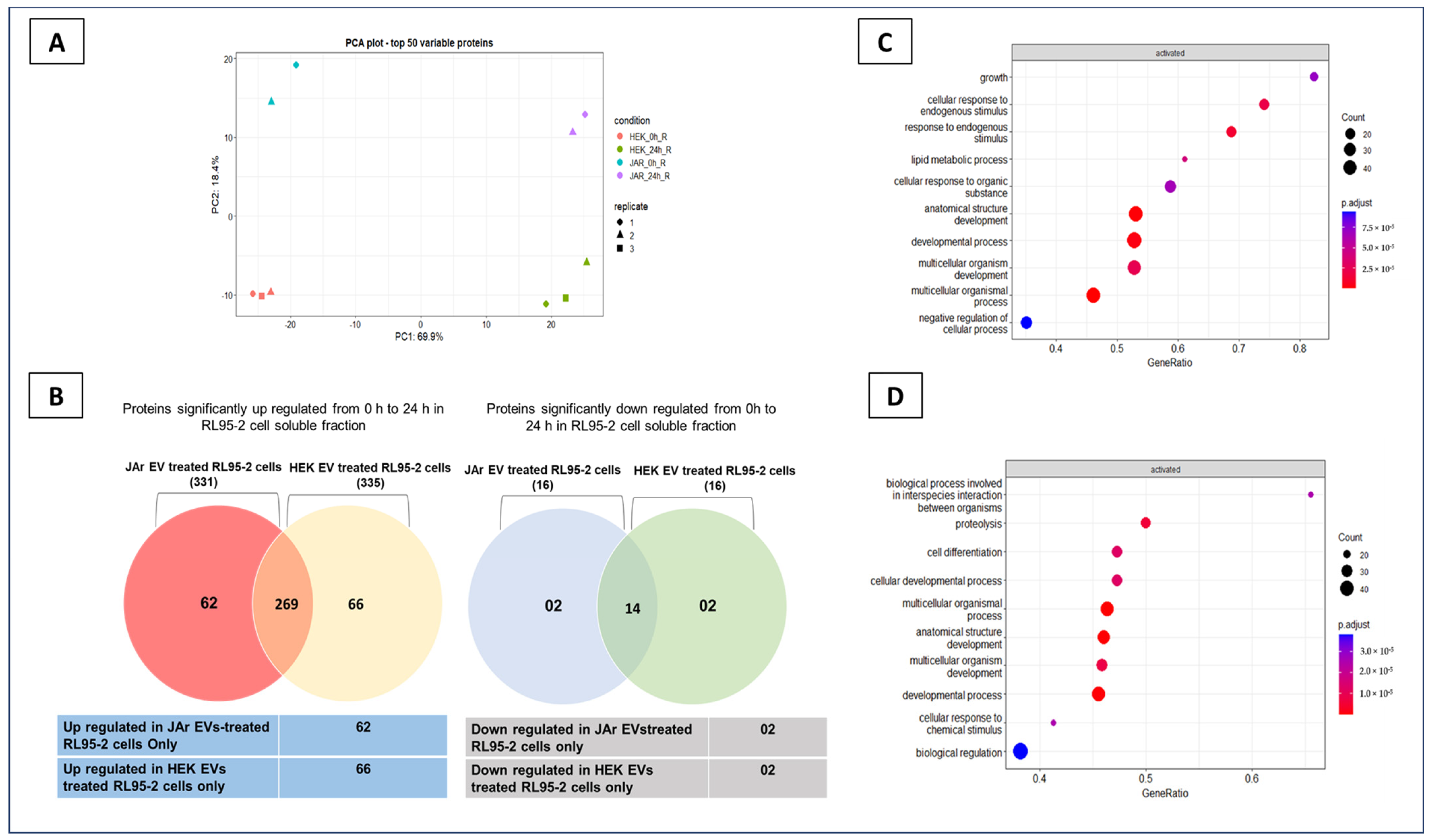
2.1. Trophoblast Cell-Derived EVs Induce Specific Secretory Protein Response in Endometrial Epithelial Cells Compared to Non-Trophoblast Cell-Derived EVs

JAr and HEK 293 cell-derived EVs were isolated, and RL952 cells were treated with each EV type at a concentration of 1 × 10⁹ nanoparticles/mL [12,13]. Secretory proteomic alterations in RL95-2 cells in response to JAr and HEK EVs were identified. The proteomic profiles of cell culture supernatants of RL95-2 cells at 0 h and 24 h after treatment with EVs were analyzed to identify differential enrichment of proteins. In total, 1023 proteins were identified (including 142 explicitly identified protein isoforms and 22 uncharacterized proteins) in RL95-2 cell culture-conditioned media. The total number of proteins identified in the 24 h post-treatment samples was much higher than that in the 0 h samples (Figure S3). Therefore, it was apparent that, in the presence of EVs, the expression of multiple proteins in the RL95-2 cell secretome changed between 0 h and 24 h. The protein expression profiles of the top 50 variable proteins in principal component analysis (PCA) are shown in Figure 1A. One replicate of the JAr EV-treated RL95-2 cells, which was identified as an outlier in the PCA, was excluded from the analysis (Figure S3). The secretory protein changes occurring in the JAr EV-treated RL95-2 cells were distinguishable from the HEK EV-treated RL95-2 cells after 24 h of incubation (Figure 1A,B). The fold change regarding proteins identified only in the JAr EV-treated RL95-2 cell secretome was plotted against the average protein abundance of the proteins across all samples (Figure S4). Proteins such as PRDX2, MIF, LDHA, PGK1, etc., were highly abundant in the cell culture-conditioned media and changed in the JAr EV-treated RL95-2 secretome. Interestingly, these proteins are known to have roles in the process of embryo implantation [14,27,28,29].
Gene set enrichment analysis of all of the proteins that were significantly altered (from 0 h to 24 h) in the JAr EV-treated group revealed “growth” and “cellular response to endogenous stimulus” as the most enriched pathways (Figure 1C). On the other hand, in the HEK EVs-treated group, “biological process involved in interspecies interaction between organisms” and “proteolysis” were identified as the most enriched pathways (Figure 1D). Overall, JAr EV treatment induced specific secretory protein changes in the RL95-2 cell secretome that were clearly distinguishable from the control EV (HEK EVs)-treated RL95-2 cell secretome.

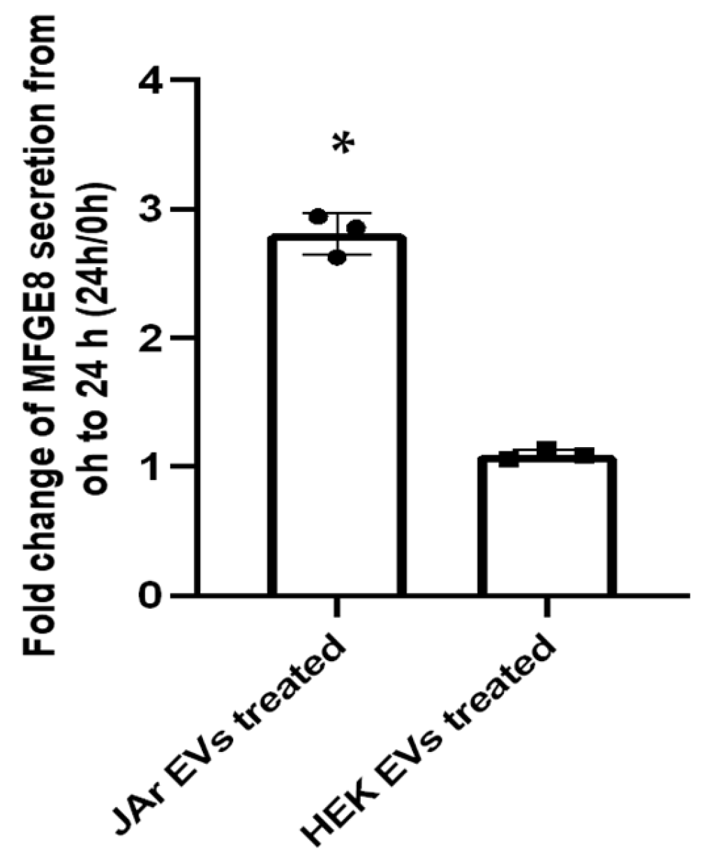
2.2. Specific Secretory Protein Changes in RL95-2 Cells in Response to JAr Cell-Derived EVs Reveal Potential Players of Embryo Implantation

The specific secretory protein alterations in RL95-2 cells attributed to JAr and HEK EVs treatment were sorted, and functional annotation and KEGG pathway analysis were performed using the Database for Annotation, Visualization, and Integrated Discovery (DAVID 6.8) online tools (version DAVID 6.8) (Figure 1B). JAr EVs induced specific secretory proteins in RL95-2 cells, showing enrichment in pathways such as “glutathione metabolism” (a key antioxidant response pathway), “amino acid metabolism”, “glycolysis”, “tight junctions”, and “protein processing in endoplasmic reticulum” (p ≤ 0.05). These pathways have been previously implicated in the processes of embryo development and implantation [14,30,31] (Table 1). Proteins expressed specifically in RL95-2 cells in response to HEK EVs revealed no such significant pathways in the context of embryo implantation (Supplementary Table S1).
The proteins specifically altered in the JAr EV-treated group from 0 h to 24 h were subjected to functional annotation and GO enrichment pathway analysis. In the results, proteins involved in multiple cellular metabolic pathways such as “cellular responses to oxidative stress”, “redox homeostasis”, and “glutathione metabolism” were significantly enriched (Supplementary Figure S3A). The major proteins related to these GO enrichment pathways were GSR, PRDX2, PRDX6, SOD1, PRDX1, and GSTP1, and most of these antioxidant enzymes have been previously reported to change inside endometrial epithelial cells in response to trophectoderm-derived EVs [14]. Our GO enrichment analysis of the proteins specifically altered in the HEK EV-treated group from 0 h to 24 h varied completely from the JAr EV-treated group and did not indicate any significant pathways related to embryo implantation (Figure S5).
Manual top-down data mining of differentially expressed proteins enabled the identification of 24 more proteins that are often involved in the process of embryo implantation, endometrial receptivity, or embryo development (Table 2).
Orthogonal validation of the mass spectrometry data was performed on the MFGE8 protein using ELISA (Figure 2).
Here, the data indicate that trophoblast-derived EVs can enrich the endometrial cell secretory proteome with known players of embryo implantation.

3. Discussion

Successful embryo implantation is attributed to the synchronized development and differentiation of both the endometrium and the embryo [33]. The endometrium becomes receptive to the embryo only during the short window of implantation, characterized by the optimal cellular and molecular transformation of the endometrium and reciprocal interactions between the embryo and endometrium through a range of signaling molecules [32]. Previously, multiple studies have shown that trophoblast or trophectoderm-derived EVs can signal endometrial epithelial cells to acquire a receptive phenotype [12,13,14]. EVs generated by embryonic [13,14] or endometrial cells [19,22] have distinct protein [14,22] and RNA [13] cargos which can be internalized by the endometrium or embryo [8,11]. Embryonic signals in the form of EVs have been shown to alter the transcriptomic [13] and proteomic [14] landscapes of endometrial cells. These changes potentially contribute to critical components of embryo–maternal communication during peri-implantation. Nevertheless, whether embryonic cell-derived EVs can modulate the endometrial cell secretory proteome is not yet known.
In the current study, we demonstrated that trophoblast-derived EVs can prime endometrial epithelial cell secretions to ensure they contain the critical proteins involved in the process of embryo implantation. Importantly, EVs derived from non-trophoblastic cells (HEK 293 cells) did not induce the same effect in the endometrial cells, which is in line with our previous report on the effects of trophoblast-derived EVs on the endometrial cell transcriptome [13]. The proteomic profile of JAr EV-treated RL95-2 cell culture supernatant (JAR_0h_R) was distinguishable from HEK 293 EV-treated cell culture supernatant (HEK_0h_R) in the PCA plot, even at 0 h (Figure 1A). This suggests inherent differences in the molecular cargos of JAr and HEK EVs. However, we assume that, after 24 h, the supplemented EVs are either internalized or degraded, leading to changes in endometrial epithelial cell secretome. Each EV type can have a unique molecular signature depending on its cell of origin, which can mediate specific biological functions in the recipient cells [70,71,72]. To account for the unique molecular signature of JAr and HEK EVs, we used the 0 h samples of JAr and HEK EVs containing cell culture supernatants as the baseline samples. However, the exact mechanism by which the protein signatures of EVs contribute to specific biological functions in recipient cells is not fully understood. The enrichment pattern of our proteomic results was further validated by a significantly high level of MFGE8 protein secretion in the JAr EV-treated RL95-2 cells secretome compared to the control EV-treated RL95-2 cell secretome.
Bioinformatics analysis revealed that glutathione metabolism (a key antioxidant response pathway) [30,69,73], amino acid metabolism [74,75], glycolysis [69,76], tight junctions [14,77,78], and protein processing in the endoplasmic reticulum [79,80] were highly upregulated in the endometrial epithelial cells in response to the trophoblast-derived EVs. Interestingly, all these proteins play critical roles in embryo implantation. Moreover, the major subset of proteins uniquely identified in the JAr EV-treated RL95-2 cell secretome consisted of antioxidant enzymes, including PRDX1/2/6, SOD1, GSR, and GST. The optimum balance of reactive oxygen species (ROS) in the uterine microenvironment is vital for early embryonic development and the regulation of innate and acquired immunity in embryonic cells [59,81]. Glutathione is a major cellular antioxidant that regulates ROS production. Glutathione and other antioxidative enzymes such as superoxide dismutase (SODs) can remove ROS and create a favorable microenvironment for embryo development [82,83]. Antioxidant activity in the vicinity of the embryo can improve in vitro fertilization (IVF) efficiency and subsequent embryo development [73]. SOD activity is elevated during the window of implantation and early pregnancy in the endometrium, suggesting its potential role in endometrial receptivity and embryo implantation [57]. Furthermore, JAr EV treatment caused secretory protein changes related to glycolysis or gluconeogenesis in RL95-2 cells, which are critical for maintaining antioxidant defense systems [84]. The proteins found in this study—GPI, LDHA, and PGK1—are known to be abundant in avian uterine fluid [85]. The same proteins are upregulated in uterine fluid during the maternal recognition of pregnancy in mares [60]. Furthermore, proteins related to cellular tight junction formation and the maintenance signaling pathway were increased in endometrial epithelial cells upon exposure to trophoblastic EVs. The same observation was reported recently, where an increase in transepithelial resistance (TER) was observed in Ishikawa cells after treatment with trophectoderm-derived EVs. Notably, all these pathways and most of the protein changes were reportedly highly upregulated in the intracellular proteome of the Ishikawa cells primed with trophectoderm-derived EVs in the same study, further validating the results of the present study [14].
The transformation of the endometrium, which ensures they acquire receptive features, occurs in the mid-secretory phase (day 20–24) of the human menstrual cycle [86]. Major changes in the endometrium during this period have been linked to extracellular matrix remodeling [87,88] and epithelial cell membrane or cytoplasmic rearrangement (to acquire apical cell polarity) [89,90]. Once the embryo reaches the receptive endometrium, it undergoes cellular growth and differentiation to facilitate implantation [91]. Thus, the peri-conception and implantation microenvironment is highly enriched in adhesion molecules, growth factors, and immune-related proteins such as cytokines [92]. Manual data mining of our dataset also revealed additional protein markers involved in embryo implantation, endometrial receptivity, and embryo development in the endometrial secretome after exposure to trophoblastic EVs. Through proteomic profiling, we identified that trophoblast-derived EVs can prime endometrial cells to secrete the critical proteomic factors of embryo development (PCNA [34], GSR [35,36], MAT2A [38], UBE2L3 [44], PRDX2 [14,27], PRDX6 [14,52], STIP1 [56], SOD1 [14,57], GSTP1 [14,60], and MYH9 [62]), endometrial differentiation (COL5A2 [33], XPO [39], LGALS3 [14,42,43], PGK1 [53]), and embryo attachment or implantation (MFGE8 [14], CCT8 [46], LDHA [14,31]). The factors that change in the embryonic vicinity can potentially alter the epigenome of the peri-implanting embryo, subsequently affecting embryonic gene expression, metabolism, and developmental capacity [37]. The changes in proteins associated with epigenetic modifications such as DNA methylation (e.g., MAT2A) in the JAr EV-treated group suggest that these changes in the feto–maternal interface can have long-term effects on the future development of the fetus, and this can even continue into adult life and affect future generations.
Hence, it can be postulated that the secretory protein changes that occur in endometrial epithelial cells in response to trophoblast cells can modulate the immediate and long-term effects that function both in an autocrine and/or paracrine manner in the embryo–maternal interface during the peri-implantation period (Figure 3).
Major protein changes were associated with glutathione metabolism, cellular responses to oxidative stress, cellular redox homeostasis, gluconeogenesis, tight junctions, and protein processing in the endoplasmic reticulum (ER). All these pathways may have critical roles in the embryo–maternal interface during pre-implantation. In addition to antioxidant activity-related proteins, several other known proteins that play a role in endometrial receptivity, embryo implantation, and early embryo development were also identified.
The in vitro model we used consisted of JAr and RL95-2 cells and is a well-established cell culture model that has been frequently used in our laboratory for many other studies to simulate embryo–endometrial interactions [12,13,93,94]. However, there is a need in the field for more advanced models to simulate the embryo–maternal interface during the peri-conception period. There are serious ethical concerns in utilizing real human embryos for experimentation. Although fixed human tissues can be obtained and provide information about the cellular location of molecules, they are not suitable for functional studies. Hence, in vitro models utilizing cell lines have facilitated the study of embryo–maternal interactions on a functional level. In this study, JAr cells derived from first-trimester choriocarcinoma cells (which possess trophoblastic cell characteristics) were used as a renewable source of cells for EV isolation. This approach was chosen due to the difficulty in obtaining large amounts of freshly isolated trophoblast cells for the continuous production of EVs. We used RL95-2 cells to simulate a receptive endometrial cell line that facilitates the initial communication between the embryo and endometrium during peri-implantation. RL95-2 cells are generally used as a model of receptive endometrial epithelial cells. While there are drawbacks in in vitro systems, primarily due to the fact that these cells originate in cancer, many other models of human embryo implantation also have their own limitations. For example, studying embryo implantation using mouse models is deemed suitable for simulating the in utero environment; however, it is limited by the fact that the embryo implantation process is species-specific. Even differentiated embryonic stem cells have been shown to exhibit different adhesion capacities based on the epithelial cell lines used in the receptive end (Ishikawa or primary cells) [93].
It is likely that the endometrial epithelial cell secretome changes reported in this study are due to either EV uptake or EV signaling by binding to the endometrial epithelial cell membrane at ligand–receptor interaction-level (upstream). The effects of EVs can also be driven by cargo release in the epithelial cell membrane or inside the cell by binding to the ER. It is known that miRNAs secreted by blastocysts modulate the different cellular processes related to implantation [95]. Previously, we showed that trophoblast-derived miRNAs can be packaged in EVs and partly regulate gene expression in endometrial epithelial cells using the same in vitro model [13]. Emerging evidence also suggests that trophoblast-derived EVs can carry a unique protein cargo [14]. Hence, secretory protein changes in endometrial epithelial cells can be attributed to the unique molecular cargo content of trophoblastic EVs. Nevertheless, further studies are required to dissect different cargo molecules in trophoblastic EVs. Furthermore, comprehensive investigations are required to detect and understand the EV signaling and cargo release mechanisms inside endometrial epithelial cells. These studies will pave the way to better understand how specific EV messages are decoded by selected recipient cells to induce desired molecular changes.

4. Materials and Methods

4.1. Cell Culture

The human endometrial adenocarcinoma cell line, RL95-2, was obtained from American Type culture Collection (ATCC CRL-1671, Teddington, UK). The human choriocarcinoma cell line, JAr (HTB-144™, Teddington, UK), and human embryonic kidney cells, HEK 293 (ATCC®, CRL-3216™, Teddington, UK), were also purchased from ATCC. RL95-2 cells were routinely maintained in Dulbecco’s Modified Eagles medium F12 (DMEM 12-604F, Lonza, Verviers, Belgium) supplemented with 10% Fetal Bovine Serum (FBS) (Gibco™, 10500064), 1% penicillin streptomycin (P/S) (Gibco™, 15140122, Bleiswijk, The Netherlands), and 5 μg/mL insulin (human recombinant insulin, Gibco™, Invitrogen, Denmark) in 5% CO2 at 37 °C. The JAr cells were grown in T75 flasks containing RPMI 1640 medium (Gibco™, Inchinnan, UK) supplemented with 10% FBS, 1% P/S, and 1% L-glutamine in 5% CO2 at 37 °C. The HEK 293 T cell line was used as a source of control EVs. Briefly, the HEK 293 cells were grown in T75 flasks in DMEM F12 medium supplemented with 10% FBS, 1% P/S, and 1% L-glutamine in 5% CO2 at 37 °C. The media were changed every second day until the cells reached 80% confluency.

4.2. Preparation of EV-Depleted Media

EV-depleted medium was prepared as previously described [12,13,96]. In summary, FBS was filtered using Amicon® Ultra-15 centrifugal filters with a 100 kDa cut-off (Merk KGaA, Darmstadt, Germany) at 3000× g for 55 min. The EV-depleted FBS was 90% depleted of nanoparticles and used at a 10% concentration to supplement all complete culture media specific to each cell type mentioned above.

4.3. EV Isolation and Characterization

At 80% confluency, the JAr and HEK 293 cell culture-conditioned media were replaced with EV-depleted culture media. After 24 h, the cell culture supernatants were collected and centrifuged at 400× g for 10 min to remove contaminating cells. The resulting supernatant was centrifuged again at 4000× g for 10 min, followed by centrifugation at 10,000× g for 10 min to remove cellular debris. The collected media were concentrated to a final volume of 500 µL using Amicon® Ultra-15 centrifugal filters with a 10 kDa cut-off. Next, the EVs were purified using size-exclusion chromatography (SEC). A gel filtration medium consisting of 4–6% agarose matrix was used in columns that were 15 cm in length to separate the EV fractions from contaminating proteins. Fractions 7–10 were collected (each fraction was 500 µL in volume) and concentrated again to a total volume of 500 µL using an Amicon® Ultra centrifugal filter device with a 10 kDa cut-off. Isolation and characterization of the isolated EVs was carried out using methods described in detail elsewhere and according to ISEV 2018 guidelines [12,13,97]. In summary, both the size and concentration of nanoparticles in the EV fractions were measured using Nano Particle Tracking Analyser (Particle Metrix GmbH, Inning am Ammersee, Germany). Transmission electron microscopy was used for the physical characterization of the EVs. The enrichment of EV surface protein markers CD 9, CD 63, and CD 81 was confirmed via western blotting. The methods used for JAr and HEK 293 cell-derived EV characterization have been fully described in our previous publications [12,13,97]. EVs derived from JAr cells and HEK 293 cells were referred to as JAr EVs and HEK EVs, respectively. JAr and HEK 293 cell-derived EVs with a high purity were obtained using size-exclusion chromatography with a nanoparticle distribution within a similar size range. The original nanoparticle concentrations and size profiles of JAr and HEK EVs are shown in Figure S1.

4.4. Collection of Cell Culture Supernatants for Secretory Proteome Analysis and Enzyme-Linked Immunosorbent Assay

Cell culture supernatants (1 mL of media) were collected from RL95-2 cells seeded in 12-well plates (seeding density of 1 × 10⁶ cells per well) and centrifuged at 400× g for 10 min to remove any contaminating cells; this was followed by centrifugation at 4000× g for 10 min and 10,000× g for 10 min to remove other cellular debris and apoptotic bodies.

4.5. Protein Quantification and Identification with Liquid Chromatography and Tandem Mass Spectroscopy (LC-MS/MS)

The total protein precipitation of cell culture supernatant samples was performed overnight with trichloroacetic acid deoxycholate (TCA-DOC). Approximate protein quantities were estimated based on the size of the pellets. The pellets were then solubilized in 7 M urea, 2 M thiourea, 100 mM ammonium bicarbonate (ABC), and 20 mM methylamine buffer. Protein reduction was performed via incubation for 1 h at room temperature using 5 mM dithiothreitol (DTT). Protein alkylation was performed via incubation for 1 h at room temperature in the dark using 10 mM chloroacetamide. Next, protease LysC (Wako, Monza, Italy) was added to an enzyme/substrate ratio (E:S) of 1:50, and the samples were incubated for 1 h at room temperature. The samples were then diluted five times with 100 mM ABC, and trypsin (Sigma Aldrich, St. Louis, MO, USA) was added at a 1:50 E:S ratio and incubated overnight at room temperature. After digestion, the samples were acidified with trifluoroacetic acid (TFA) at a concentration of 1%; subsequently, the samples were desalted on in-house made C18 StageTips. Then, the samples were reconstituted in 0.5% TFA, and peptide concentrations were determined using the Pierce colorimetric peptide assay (Thermo Fisher Scientific, Waltham, MA, USA). Next, 1 µg of peptide was injected into an Easy-nLC 1000 system® (Thermo Scientific) and eluted at 250 nL/min from the trap to a 75 µm ID × 50 cm emitter column (New Objective) packed with C18 material (3 µm, 300 Å particles, Dr Maisch). The separation gradient was 2–35% B for 60 min and 40–100% B for 5 min (A: 0.1% formic acid (FA), B: 80% ACN + 0.1% FA). The eluted peptides were sprayed into a Q Exactive Plus® (Thermo Fisher Scientific) quadrupole-orbitrap mass spectrometer (MS) using nano-electrospray ionization at 2.4 kV (applied through liquid junction). The MS was operated with a top 5 data-dependent acquisition strategy. Briefly, one 350–1400 m/z MS scan at a resolution setting of R = 70,000 at 200 m/z was followed by high-energy collisional dissociation fragmentation (normalized collision energy of 26) of the five most intense ions (z: +2 to +6) at R = 17,500. The MS and MS/MS ion target values were 3 × 10⁶ and 5 × 10⁴ (with an injection time of 50 ms). Dynamic exclusion was limited to 40 s. Mass spectrometric raw files were processed using the MaxQuant software package (versions 1.6.15.0 and 2.0.3.0). Methionine oxidation, asparagine and glutamine deamidation, and protein N-terminal acetylation were set as variable modifications, whereas cysteine carbamidomethylation was defined as a fixed modification. Label-free protein quantification (LFQ) was enabled using LFQ mode, and the protein minimum ratio count was set to 1. A search was performed against Homo sapiens and Bos taurus reference proteomes using the tryptic digestion rule. The peptide–spectrum match and protein false discovery rate (FDR) were kept below 1% using a target-decoy approach. All other parameters were set to default values. Mass spectrometry data are available in ProteomeXchange Consortium via the PRIDE with the dataset identifier PXD040311.

4.6. Differential Protein Expression and Bioinformatics Analysis

Differential protein expression between the treated and control groups was determined using R software version 4.2.1. using the DEP package (used for the analysis of mass spectrometry proteomics data for differential protein expression or differential enrichment). The DEP package for label-free proteomic data is extensively described in https://bioconductor.org/packages/devel/bioc/vignettes/DEP/inst/doc/DEP.html (accessed on 23 November 2022) [97]. Potential contaminating proteins and those originating from reverse sequencing were filtered out. Proteins with ≥2 valid values in at least one sample group were used for the analysis. Data were normalized using Variance Stabilizing Normalization (VSN). Missing values were imputed under low-intensity assumption, and statistical comparisons among the samples were performed using protein-wise linear models combined with empirical Bayesian statistics. Multiple testing was corrected using the Benjamini–Hochberg FDR (BH-FDR) [98]. Proteins were considered significantly differentially expressed if the fold change in protein expression was (FC) log2 > 1 or log2 < −1 and FDR < 0.05 between the 0 h and 24 h samples. Lists of the differentially expressed proteins between 0 h and 24 h samples were made for both the JAr and HEK EV-treated groups and then compared. The average protein abundances of the proteins across all samples were calculated, and the first and third quartiles of the data distribution were used to identify the most and least abundant protein groups.
Functional annotation and KEGG pathway analysis were performed using the DAVID Bioinformatics platform (https://david.ncifcrf.gov/, accessed on 23 November 2022). Functional annotation, GO pathway analysis, and gene set enrichment analysis were performed using the clusterProfiler package in R (https://bioconductor.org/packages/release/bioc/html/clusterProfiler.html, accessed on 23 November 2022). All relevant differentially enriched gene IDs were submitted to the DAVID Bioinformatics platform for KEGG pathway enrichment analysis. A complete protein list with log2 fold change and FDR was submitted to clusterProfiler to perform pathway overrepresentation analysis and visualization.

4.7. Verification of Proteomic Data Using Enzyme-Linked Immunosorbent Assay (ELISA)

To validate the mass spectrometry-based protein expression data, Milk fat globule-EGF factor 8 protein (MFGE-8) concentration was measured in the EV-treated cell culture supernatants using a commercially available ELISA kit (Human MFG-E8 Quantikine ELISA Kit, R&D systems) according to the manufacturer’s instructions. Briefly, 150 µL of the cell culture supernatant from each sample was diluted twice with the sample diluent provided with the kit. Then, the assay diluent was added to the precoated 96-well plate, followed by 100 µL of the sample, standard, or control. After two hours of incubation, the wells were washed and incubated with the MFGE-8 conjugate for 2 h. Next, the wells were washed, filled with 200 µL of substrate solution, and incubated for 30 min. Finally, stop solution was added, and the optical density of each well was measured using a microplate reader set to 450 nm (Multiskan FC microplate photometer, Life Technologies, Chongqing, China). The MFGE8 protein concentration changes in the JAr and HEK EV-treated groups from 0 h to 24 h were shown as mean ± SD (fold change expression). Statistical significance was determined using Student’s t-test, and results were considered significant at a p value of <0.05.

4.8. Experimental Design

The secretory protein changes in the RL95-2 cells, which occurred in response to trophoblastic cell-derived EVs (JAr cell EVs) vs. non trophoblastic cell-derived EVs (HEK 293 cell EV), were determined as follows:
The RL95-2 cells were seeded in 12-well plates and grown until they reached 85% confluency, as described above. After reaching the desired confluency, the cells were washed with Dulbecco’s phosphate-buffered saline without Ca2+ and Mg2+ (DPBS, Verviers, Belgium). The RL95-2 cells were then supplemented with EVs derived from trophoblast analog JAr cells at a concentration of 1 × 10⁹ nanoparticles/mL in EV-depleted medium. The nanoparticle concentrations were measured with a nanoparticle tracking analyzer, and similar concentrations of nanoparticle/mL from each EV source were used to treat the RL95-2 cells. As a control, the RL95-2 cells were treated with non-trophoblastic cell-derived EVs at similar concentration (HEK 293 cell EVs). Immediately after adding fresh media containing EVs to the cells, the cell culture supernatants were collected back into Eppendorf tubes and sequential centrifugation was performed. (0 h samples). Next, the RL95-2 cells were incubated with JAr and HEK cell-derived EVs for 24 h, and afterwards, the cell culture supernatants were collected (Figure S2). The proteomic profiles of the cell culture supernatants were analyzed using LC-MS/MS. The experiment was performed in triplicate on three different days. The protein fold change in the 0 h and 24 h samples was analyzed and used to identify proteins that were significantly changed in the RL95-2 cell secretome in response to JAr and HEK cell-derived EVs after 24 h. Proteins that uniquely changed in the JAr and HEK EV-treated RL95-2 cell secretome were sorted, and functional annotation and pathway overrepresentation analysis were performed. One differentially expressed protein marker, identified by mass spectrometry analysis, was validated using ELISA.

5. Conclusions

Trophoblast-derived EVs can change the endometrial cell secretory protein repertoire by harboring critical players of implantation, which is consistent with the findings of previous reports. These results suggest that embryonic signals can potentially regulate endometrial secretory protein responses, enhancing endometrial receptivity for the embryo implantation process. However, the exact mechanism by which embryonic EVs regulate these signaling pathways and their impact on structural and functional development, the attachment of the embryo, or the receptivity of the endometrium requires further study. Functional analyses of the effect of embryonic EVs on the endometrium may provide molecular insights into the process of embryo implantation, potentially identifying modulatory targets to treat implantation failure in humans and other mammalian species.

Supplementary Materials

The following supporting information can be downloaded at: https://www.mdpi.com/article/10.3390/ijms241511924/s1.

Author Contributions

Conceptualization, S.M., K.D., K.G. and A.F.; Formal analysis, S.M., K.D., T.O., K.G., S.K. and A.F.; Funding acquisition, A.F.; Investigation, S.M., K.D., K.G. and A.F.; Methodology, S.M., K.D., K.G. and A.F.; Project administration, A.F.; Resources, T.O. and A.F.; Supervision, T.O., S.K. and A.F.; Visualization, S.M. and A.F.; Writing—original draft, S.M. and A.F.; Writing—review and editing, S.M., K.D., T.O., K.G., S.K. and A.F. All authors have read and agreed to the published version of the manuscript.

Funding

This research was funded by The European Union’s Horizon 2020 research and innovation programme under grant agreement No. 857418 COMBIVET and The European Union through Horizon coordination and support actions under grant agreement No. 101079349 OH-Boost.

Institutional Review Board Statement

Not applicable.

Informed Consent Statement

Not applicable.

Data Availability Statement

The mass spectrometry proteomics data have been deposited to the ProteomeXchange Consortium via the PRIDE [1] partner repository with the dataset identifier PXD040311.

Acknowledgments

We thank Amber Hart (University of Sheffield) for her invaluable help with English editing, which greatly improved the clarity and coherence of this paper.

Conflicts of Interest

The authors declare no conflict of interests.

References

  1. Busnelli, A.; Somigliana, E.; Cirillo, F.; Baggiani, A.; Levi-Setti, P.E. Efficacy of Therapies and Interventions for Repeated Embryo Implantation Failure: A Systematic Review and Meta-Analysis. Sci. Rep. 2021, 11, 1747. [Google Scholar] [CrossRef] [PubMed]
  2. Timeva, T.; Shterev, A.; Kyurkchiev, S. Recurrent Implantation Failure: The Role of the Endometrium. J. Reprod. Infertil. 2014, 15, 173–183. [Google Scholar] [PubMed]
  3. Kim, S.-M.; Kim, J.-S. A Review of Mechanisms of Implantation. Dev. Reprod. 2017, 21, 351–359. [Google Scholar] [CrossRef] [PubMed] [Green Version]
  4. Fritz, R.; Jain, C.; Randall Armant, D. Cell Signaling in Trophoblast-Uterine Communication. Int. J. Dev. Biol. 2014, 58, 261–271. [Google Scholar] [CrossRef] [Green Version]
  5. Singh, M.; Chaudhry, P.; Asselin, E. Bridging Endometrial Receptivity and Implantation: Network of Hormones, Cytokines, and Growth Factors. J. Endocrinol. 2011, 210, 5–14. [Google Scholar] [CrossRef] [PubMed] [Green Version]
  6. Zhang, S.; Lin, H.; Kong, S.; Wang, S.; Wang, H.; Wang, H.; Armant, D.R. Physiological and Molecular Determinants of Embryo Implantation. Mol. Asp. Med. 2014, 34, 939–980. [Google Scholar] [CrossRef] [Green Version]
  7. Dey, S.K.; Lim, H.; Das, S.K.; Reese, J.; Paria, B.C.; Daikoku, T.; Wang, H. Molecular Cues to Implantation. Endocr. Rev. 2004, 25, 341–373. [Google Scholar] [CrossRef] [PubMed] [Green Version]
  8. Giacomini, E.; Vago, R.; Sanchez, A.M.; Podini, P.; Zarovni, N.; Murdica, V.; Rizzo, R.; Bortolotti, D.; Candiani, M.; Viganò, P. Secretome of in Vitro Cultured Human Embryos Contains Extracellular Vesicles That Are Uptaken by the Maternal Side. Sci. Rep. 2017, 7, 5210. [Google Scholar] [CrossRef]
  9. Dissanayake, K.; Nõmm, M.; Lättekivi, F.; Ressaissi, Y.; Godakumara, K.; Lavrits, A.; Midekessa, G.; Viil, J.; Bæk, R.; Jørgensen, M.M.; et al. Individually Cultured Bovine Embryos Produce Extracellular Vesicles That Have the Potential to Be Used as Non-Invasive Embryo Quality Markers. Theriogenology 2020, 149, 104–116. [Google Scholar] [CrossRef]
  10. Saadeldin, I.M.; Kim, S.J.; Choi, Y.B.; Lee, B.C. Improvement of Cloned Embryos Development by Co-Culturing with Parthenotes: A Possible Role of Exosomes/Microvesicles for Embryos Paracrine Communication. Cell. Reprogram. 2014, 16, 223–234. [Google Scholar] [CrossRef] [Green Version]
  11. Pavani, K.C.; Hendrix, A.; Van Den Broeck, W.; Couck, L.; Szymanska, K.; Lin, X.; De Koster, J.; Van Soom, A.; Leemans, B. Isolation and Characterization of Functionally Active Extracellular Vesicles from Culture Medium Conditioned by Bovine Embryos in Vitro. Int. J. Mol. Sci. 2019, 20, 38. [Google Scholar] [CrossRef] [Green Version]
  12. Es-Haghi, M.; Godakumara, K.; Häling, A.; Lättekivi, F.; Lavrits, A.; Viil, J.; Andronowska, A.; Nafee, T.; James, V.; Jaakma, Ü.; et al. Specific Trophoblast Transcripts Transferred by Extracellular Vesicles Affect Gene Expression in Endometrial Epithelial Cells and May Have a Role in Embryo-Maternal Crosstalk. Cell Commun. Signal. 2019, 17, 146. [Google Scholar] [CrossRef] [Green Version]
  13. Godakumara, K.; Ord, J.; Lättekivi, F.; Dissanayake, K.; Viil, J.; Boggavarapu, N.R.; Faridani, O.R.; Jääger, K.; Meikas, A.V.; Jaakma, Ü. Trophoblast Derived Extracellular Vesicles Specifically Alter the Transcriptome of Endometrial Cells and May Constitute a Critical Component of Embryo-Maternal Communication. Reprod. Biol. Endocrinol. 2021, 19, 115. [Google Scholar] [CrossRef]
  14. Poh, Q.H.; Rai, A.; Carmichael, I.I.; Salamonsen, L.A.; Greening, D.W. Proteome Reprogramming of Endometrial Epithelial Cells by Human Trophectodermal Small Extracellular Vesicles Reveals Key Insights into Embryo Implantation. Proteomics 2021, 21, 13–14. [Google Scholar] [CrossRef]
  15. Ouyang, Y.; Bayer, A.; Chu, T.; Tyurin, V.A.; Kagan, V.E.; Morelli, A.E.; Coyne, C.B.; Sadovsky, Y. Isolation of Human Trophoblastic Extracellular Vesicles and Characterization of Their Cargo and Antiviral Activity. Physiol. Behav. 2017, 176, 139–148. [Google Scholar] [CrossRef] [PubMed] [Green Version]
  16. Segura-Benítez, M.; Bas-Rivas, A.; Juárez-Barber, E.; Carbajo-Garcia, M.C.; Faus, A.; Pellicer, A.; Ferrero, H. Human Blastocysts Uptake Extracellular Vesicles Secreted By Primary Endometrial Epithelial Cells Containing miRNAs Related To Implantation and Early Embryo Development. Fertil. Steril. 2022, 118, e73–e74. [Google Scholar] [CrossRef]
  17. Hua, R.; Liu, Q.; Lian, W.; Kang, T.; Gao, D.; Huang, C.; Wang, Y.; Lei, M. Extracellular Vesicles Derived from Endometrial Epithelial Cells Deliver Exogenous miR-92b-3p to Affect the Function of Embryonic Trophoblast Cells via Targeting TSC1 and DKK3. Reprod. Biol. Endocrinol. 2022, 20, 152. [Google Scholar] [CrossRef] [PubMed]
  18. Tan, Q.; Shi, S.; Liang, J.; Cao, D.; Wang, S.; Wang, Z. Endometrial Cell-Derived Small Extracellular Vesicle miR-100-5p Promotes Functions of Trophoblast during Embryo Implantation. Mol. Ther. Nucleic Acids. 2021, 23, 217–231. [Google Scholar] [CrossRef]
  19. Evans, J.; Rai, A.; Nguyen, H.P.T.; Poh, Q.H.; Elglass, K.; Simpson, R.J.; Salamonsen, L.A.; Greening, D.W. Human Endometrial Extracellular Vesicles Functionally Prepare Human Trophectoderm Model for Implantation: Understanding Bidirectional Maternal-Embryo Communication. Proteomics 2019, 19, 1800423. [Google Scholar] [CrossRef]
  20. Salamonsen, L.A.; Evans, J.; Nguyen, H.P.T.; Edgell, T.A. The Microenvironment of Human Implantation: Determinant of Reproductive Success. Am. J. Reprod. Immunol. 2016, 75, 218–225. [Google Scholar] [CrossRef]
  21. van der Weijden, V.A.; Bick, J.T.; Bauersachs, S.; Arnold, G.J.; Fröhlich, T.; Drews, B.; Ulbrich, S.E. Uterine Fluid Proteome Changes during Diapause and Resumption of Embryo Development in Roe Deer (Capreolus capreolus). Reproduction 2019, 158, 13–24. [Google Scholar] [CrossRef] [PubMed]
  22. Hart, A.R.; Liaqat, N.; Khan, A.; Dissanayake, K.; Godakumara, K.; Andronowska, A.; Eapen, S.; Heath, P.R.; Fazeli, A. The Extracellular Vesicles Proteome of Endometrial Cells Simulating the Receptive Menstrual Phase Differs from That of Endometrial Cells Simulating the Non-Receptive Menstrual Phase. Biomolecules 2023, 13, 279. [Google Scholar] [CrossRef]
  23. Salleh, N.; Baines, D.L.; Naftalin, R.J.; Milligan, S.R. The Hormonal Control of Uterine Luminal Fluid Secretion and Absorption. J. Membr. Biol. 2005, 206, 17–28. [Google Scholar] [CrossRef]
  24. Chattopadhyay, R.; Juneau, C.R.; Landis, J.; Morin, S.J.; Neal, S.A.; Scott, R.T. Persistent Fluid in the Endometrial Cavity That Resolves after Progesterone Administration Prior to Transfer Does Impact Live Birth Rate. Fertil. Steril. 2017, 107, e11. [Google Scholar] [CrossRef]
  25. Gurung, S.; Greening, D.W.; Catt, S.; Salamonsen, L.; Evans, J. Exosomes and Soluble Secretome from Hormone-Treated Endometrial Epithelial Cells Direct Embryo Implantation. Mol. Hum. Reprod. 2020, 26, 510–520. [Google Scholar] [CrossRef]
  26. Ruiz-González, I.; Xu, J.; Wang, X.; Burghardt, R.C.; Dunlap, K.A.; Bazer, F.W. Exosomes, Endogenous Retroviruses and Toll-like Receptors: Pregnancy Recognition in Ewes. Reproduction 2015, 149, 281–291. [Google Scholar] [CrossRef] [PubMed]
  27. Wu, F.; Tian, F.; Zeng, W.; Liu, X.; Fan, J.; Lin, Y.; Zhang, Y. Role of Peroxiredoxin 2 Downregulation in Recurrent Miscarriage through Regulation of Trophoblast Proliferation and Apoptosis. Cell Death Dis. 2017, 8, e2908. [Google Scholar] [CrossRef] [PubMed] [Green Version]
  28. Arcuri, F.; Ricci, C.; Ietta, F.; Cintorino, M.; Tripodi, S.A.; Cetin, I.; Garzia, E.; Schatz, F.; Klemi, P.; Santopietro, R.; et al. Macrophage Migration Inhibitory Factor in the Human Endometrium: Expression and Localization during the Menstrual Cycle and Early Pregnancy. Biol. Reprod. 2001, 64, 1200–1205. [Google Scholar] [CrossRef] [Green Version]
  29. Liang, X.; Tang, S.; Li, D.; Song, Y.; He, M.; Duan, Y.; Du, H. Shoutai Wan Improves Embryo Survival by Regulating Aerobic Glycolysis of Trophoblast Cells in a Mouse Model of Recurrent Spontaneous Abortion. Evid.-Based Complement. Altern. Med. 2022, 2022, 8251503. [Google Scholar] [CrossRef]
  30. Guérin, P.; El Mouatassim, S.; Ménézo, Y. Oxidative Stress and Protection against Reactive Oxygen Species in the Pre-Implantation Embryo and Its Surroundings. Hum. Reprod. Update 2001, 7, 175–189. [Google Scholar] [CrossRef]
  31. Gardner, D.K. Lactate Production by the Mammalian Blastocyst: Manipulating the Microenvironment for Uterine Implantation and Invasion? BioEssays 2015, 37, 364–371. [Google Scholar] [CrossRef] [Green Version]
  32. Haouzi, D.; Dechaud, H.; Assou, S.; Monzo, C.; De Vos, J.; Hamamah, S. Transcriptome Analysis Reveals Dialogues between Human Trophectoderm and Endometrial Cells during the Implantation Period. Hum. Reprod. 2011, 26, 1440–1449. [Google Scholar] [CrossRef] [PubMed] [Green Version]
  33. Liu, J.L.; Zhao, M.; Peng, Y.; Fu, Y.S. Identification of Gene Expression Changes in Rabbit Uterus during Embryo Implantation. Genomics 2016, 107, 216–221. [Google Scholar] [CrossRef] [PubMed]
  34. Öner, H.; Öner, J.; Demir, R. Distributions of PCNA and Cas-3 in Rat Uterus during Early Pregnancy. Folia Histochem. Cytobiol. 2010, 48, 71–77. [Google Scholar] [CrossRef] [PubMed] [Green Version]
  35. Wathlet, S.; Adriaenssens, T.; Segers, I.; Verheyen, G.; van Landuyt, L.; Coucke, W.; Devroey, P.; Smitz, J. Pregnancy Prediction in Single Embryo Transfer Cycles after ICSI Using QPCR: Validation in Oocytes from the Same Cohort. PLoS ONE 2013, 8, e54226. [Google Scholar] [CrossRef]
  36. Zarbakhsh, S. Effect of Antioxidants on Preimplantation Embryo Development in Vitro: A Review. Zygote 2021, 29, 179–193. [Google Scholar] [CrossRef]
  37. Ikeda, S.; Kawahara-Miki, R.; Iwata, H.; Sugimoto, M.; Kume, S. Role of Methionine Adenosyltransferase 2A in Bovine Preimplantation Development and Its Associated Genomic Regions. Sci. Rep. 2017, 7, 3800. [Google Scholar] [CrossRef] [Green Version]
  38. Cai, S.; Ye, Q.; Zeng, X.; Yang, G.; Ye, C.; Chen, M.; Yu, H.; Wang, Y.; Wang, G.; Huang, S.; et al. CBS and MAT2A Improve Methionine-Mediated DNA Synthesis through SAMTOR/MTORC1/S6K1/CAD Pathway during Embryo Implantation. Cell Prolif. 2021, 54, e12950. [Google Scholar] [CrossRef]
  39. Wang, F.; Liu, Y. Identification of Key Genes, Regulatory Factors, and Drug Target Genes of Recurrent Implantation Failure (RIF). Gynecol. Endocrinol. 2020, 36, 448–455. [Google Scholar] [CrossRef]
  40. Nangia-Makker, P.; Honjo, Y.; Sarvis, R.; Akahani, S.; Hogan, V.; Pienta, K.J.; Raz, A. Galectin-3 Induces Endothelial Cell Morphogenesis and Angiogenesis. Am. J. Pathol. 2000, 156, 899–909. [Google Scholar] [CrossRef] [Green Version]
  41. Blidner, A.G.; Rabinovich, G.A. “Sweetening” Pregnancy: Galectins at the Fetomaternal Interface. Am. J. Reprod. Immunol. 2013, 69, 369–382. [Google Scholar] [CrossRef] [PubMed]
  42. Yang, H.; Lei, C.; Zhang, W. Expression of Galectin-3 in Mouse Endometrium and Its Effect during Embryo Implantation. Reprod. Biomed. Online 2012, 24, 116–122. [Google Scholar] [CrossRef] [PubMed] [Green Version]
  43. Lei, C.X.; Zhang, W.; Zhou, J.P.; Liu, Y.K. Interactions between Galectin-3 and Integrinβ3 in Regulating Endometrial Cell Proliferation and Adhesion. Hum. Reprod. 2009, 24, 2879–2889. [Google Scholar] [CrossRef] [PubMed] [Green Version]
  44. Grzmil, P.; Altmann, M.E.; Adham, I.M.; Engel, U.; Jarry, H.; Schweyer, S.; Wolf, S.; Mänz, J.; Engel, W. Embryo Implantation Failure and Other Reproductive Defects in Ube2q1-Deficient Female Mice. Reproduction 2013, 145, 45–56. [Google Scholar] [CrossRef]
  45. Altmäe, S.; Reimand, J.R.; Hovatta, O.; Zhang, P.; Kere, J.; Laisk, T.; Saare, M.; Peters, M.; Vilo, J.; Stavreus-Evers, A.; et al. Implantation: Identification of Gene Expression Pathways, Regulation, and Integrated Regulatory Networks. Mol. Endocrinol. 2012, 26, 203–217. [Google Scholar] [CrossRef] [PubMed]
  46. Prašnikar, E.; Kunej, T.; Knez, J.; Repnik, K.; Potočnik, U.; Kovačič, B. Determining the Molecular Background of Endometrial Receptivity in Adenomyosis. Biomolecules 2020, 10, 1311. [Google Scholar] [CrossRef]
  47. Franchi, A.; Bocca, S.; Anderson, S.; Riggs, R.; Oehninger, S. Expression of Milk Fat Globule EGFfactor 8 (MFG-E8) mRNA and Protein in the Human Endometrium and Its Regulation by Prolactin. Mol. Hum. Reprod. 2011, 17, 360–371. [Google Scholar] [CrossRef] [Green Version]
  48. Schmitz, C.; Yu, L.; Bocca, S.; Anderson, S.; Cunha-Filho, J.S.; Rhavi, B.S.; Oehninger, S. Role for the Endometrial Epithelial Protein MFG-E8 and Its Receptor Integrin Avβ3 in Human Implantation: Results of an in Vitro Trophoblast Attachment Study Using Established Human Cell Lines. Fertil. Steril. 2014, 101, 874–882. [Google Scholar] [CrossRef]
  49. Bocca, S.M.; Anderson, S.; Amaker, B.; Swanson, R.J.; Franchi, A.; Lattanzio, F.; Oehninger, S. Milk Fat Globule Epidermal Growth Factor 8 (MFG-E8): A Novel Protein in the Mammalian Endometrium with Putative Roles in Implantation and Placentation. Placenta 2012, 33, 795–802. [Google Scholar] [CrossRef]
  50. Barua, S.; Macedo, A.; Kolb, D.S.; Wynne-Edwards, K.E.; Klein, C. Milk-Fat Globule Epidermal Growth Factor 8 (MFGE8) Is Expressed at the Embryo- and Fetal-Maternal Interface in Equine Pregnancy. Reprod. Fertil. Dev. 2018, 30, 585–590. [Google Scholar] [CrossRef]
  51. Yu, L.; Hu, R.; Sullivan, C.; Swanson, R.J.; Oehninger, S.; Sun, Y.P.; Bocca, S. MFGE8 Regulates TGF-β-Induced Epithelial Mesenchymal Transition in Endometrial Epithelial Cells in Vitro. Reproduction 2016, 152, 225–233. [Google Scholar] [CrossRef] [PubMed] [Green Version]
  52. Popli, P.; Shukla, V.; Kaushal, J.B.; Kumar, R.; Gupta, K.; Dwivedi, A. Peroxiredoxin 6 Plays Essential Role in Mediating Fertilization and Early Embryonic Development in Rabbit Oviduct. Reprod. Sci. 2022, 29, 1560–1576. [Google Scholar] [CrossRef] [PubMed]
  53. Tong, J.; Yang, J.; Lv, H.; Lv, S.; Zhang, C.; Chen, Z.J. Dysfunction of Pseudogene PGK1P2 is Involved in Preeclampsia by Acting as a Competing Endogenous RNA of PGK1. Pregnancy Hypertens. 2018, 13, 37–45. [Google Scholar] [CrossRef] [PubMed]
  54. Killeen, A.P.; Morris, D.G.; Kenny, D.A.; Mullen, M.P.; Diskin, M.G.; Waters, S.M. Global Gene Expression in Endometrium of High and Low Fertility Heifers during the Mid-Luteal Phase of the Estrous Cycle. BMC Genom. 2014, 15, 234. [Google Scholar] [CrossRef] [Green Version]
  55. Munch, E.M.; Sparks, A.E.; Gonzalez Bosquet, J.; Christenson, L.K.; Devor, E.J.; Van Voorhis, B.J. Differentially Expressed Genes in Preimplantation Human Embryos: Potential Candidate Genes for Blastocyst Formation and Implantation. J. Assist. Reprod. Genet. 2016, 33, 1017–1025. [Google Scholar] [CrossRef] [Green Version]
  56. Beraldo, F.H.; Soares, I.N.; Goncalves, D.F.; Fan, J.; Thomas, A.A.; Santos, T.G.; Mohammad, A.H.; Roffé, M.; Calder, M.D.; Nikolova, S.; et al. Stress-Inducible Phosphoprotein 1 Has Unique Cochaperone Activity during Development and Regulates Cellular Response to Ischemia via the Prion Protein. FASEB J. 2013, 27, 3594–3607. [Google Scholar] [CrossRef] [Green Version]
  57. Sugino, N.; Shimamura, K.; Takiguchi, S.; Ono, M.; Nakata, M.; Nakamura, Y.; Uda, T.; Kato, H. Changes in Activity of Superoxide Dismutase in the Human Endometrium throughout the Menstrual Cycle and in Early Pregnancy. Hum Reprod. 1996, 11, 1073–1078. [Google Scholar] [CrossRef] [Green Version]
  58. Combelles, C.M.H.; Holick, E.A.; Racowsky, C. Release of Superoxide Dismutase-1 by Day 3 Embryos of Varying Quality and Implantation Potential. J. Assist. Reprod. Genet. 2012, 29, 305–311. [Google Scholar] [CrossRef] [Green Version]
  59. Ribeiro, J.C.; Braga, P.C.; Martins, A.D.; Silva, B.M.; Alves, M.G.; Oliveira, P.F. Antioxidants Present in Reproductive Tract Fluids and Their Relevance for Fertility. Antioxidants 2021, 10, 1441. [Google Scholar] [CrossRef]
  60. Smits, K.; Willems, S.; Van Steendam, K.; Van De Velde, M.; De Lange, V.; Ververs, C.; Roels, K.; Govaere, J.; Van Nieuwerburgh, F.; Peelman, L.; et al. Proteins Involved in Embryo- Maternal Interaction around the Signalling of Maternal Recognition of Pregnancy in the Horse. Sci. Rep. 2018, 8, 5249. [Google Scholar] [CrossRef] [Green Version]
  61. Pierzchała, D.; Liput, K.; Korwin-Kossakowska, A.; Ogłuszka, M.; Poławska, E.; Nawrocka, A.; Urbański, P.; Ciepłoch, A.; Juszczuk-Kubiak, E.; Lepczyński, A.; et al. Molecular Characterisation of Uterine Endometrial Proteins during Early Stages of Pregnancy in Pigs by Maldi Tof/Tof. Int. J. Mol. Sci. 2021, 22, 6720. [Google Scholar] [CrossRef] [PubMed]
  62. Schliffka, M.F.; Tortorelli, A.F.; Özgüç, Ö.; de Plater, L.; Polzer, O.; Pelzer, D.; Maître, J.L. Multiscale Analysis of Single and Double Maternal-Zygotic Myh9 and Myh10 Mutants during Mouse Preimplantation Development. Elife 2021, 10, e68536. [Google Scholar] [CrossRef] [PubMed]
  63. Schulz, L.C.; Bahr, J.M. Glucose-6-Phosphate Isomerase is Necessary for Embryo Implantation in the Domestic Ferret. Proc. Natl. Acad. Sci. USA 2003, 100, 8561–8566. [Google Scholar] [CrossRef]
  64. Bondza, P.K.; Metz, C.N.; Akoum, A. Postgestational Effects of Macrophage Migration Inhibitory Factor on Embryonic Implantation in Mice. Fertil. Steril. 2008, 90, 1433–1443. [Google Scholar] [CrossRef]
  65. Loones, M.T.; Rallu, M.; Mezger, V.; Morange, M. HSP Gene Expression and HSF2 in Mouse Development. Cell. Mol. Life Sci. 1997, 53, 179–190. [Google Scholar] [CrossRef]
  66. Chin, P.Y.; MacPherson, A.M.; Thompson, J.G.; Lane, M.; Robertson, S.A. Stress Response Genes are Suppressed in Mouse Preimplantation Embryos by Granulocyte-Macrophage Colony-Stimulating Factor (GM-CSF). Hum. Reprod. 2009, 24, 2997–3009. [Google Scholar] [CrossRef] [PubMed] [Green Version]
  67. Samakovli, D.; Tichá, T.; Vavrdová, T.; Závorková, N.; Pecinka, A.; Ovecka, M.; Samaj, J. HEAT SHOCK PROTEIN 90 Proteins and YODA Regulate Main Body Axis Formation during Early Embryogenesis. Plant Physiol. 2021, 186, 1526–1544. [Google Scholar] [CrossRef]
  68. Chen, X.; Chen, X. PRM-Based Quantitative Proteomics Analysis of Altered HSPs Expression in Villi and Decidua of Early Missed Abortion Patients. Res. Square 2022, preprint. [Google Scholar] [CrossRef]
  69. Yang, Q.; Liu, J.; Wang, Y.; Zhao, W.; Wang, W.; Cui, J.; Yang, J.; Yue, Y.; Zhang, S.; Chu, M.; et al. A Proteomic Atlas of Ligand-Receptor Interactions at the Ovine Maternal-Fetal Interface Reveals the Role of Histone Lactylation in Uterine Remodeling. J. Biol. Chem. 2022, 298, 101456. [Google Scholar] [CrossRef]
  70. Fisher, S.J.; Francisco, S.; Francisco, S.; Francisco, S.; Francisco, S.; Francisco, S.; Francisco, S.; Francisco, S.; Francisco, S. Tumour Exosome Integrins Determine Organotropic Metastasis. Nature 2016, 213, 329–335. [Google Scholar] [CrossRef] [Green Version]
  71. Rontogianni, S.; Synadaki, E.; Li, B.; Liefaard, M.C.; Lips, E.H.; Wesseling, J.; Wu, W.; Altelaar, M. Proteomic Profiling of Extracellular Vesicles Allows for Human Breast Cancer Subtyping. Commun. Biol. 2019, 2, 325. [Google Scholar] [CrossRef] [Green Version]
  72. Segura-Benítez, M.; Carbajo-García, M.C.; Corachán, A.; Faus, A.; Pellicer, A.; Ferrero, H. Proteomic Analysis of Extracellular Vesicles Secreted by Primary Human Epithelial Endometrial Cells Reveals Key Proteins Related to Embryo Implantation. Reprod. Biol. Endocrinol. 2022, 20, 3. [Google Scholar] [CrossRef]
  73. Truong, T.; Gardner, D.K. Antioxidants Improve IVF Outcome and Subsequent Embryo Development in the Mouse. Hum. Reprod. 2017, 32, 2404–2413. [Google Scholar] [CrossRef] [Green Version]
  74. Van Winkle, L.J. Amino Acid Transport and Metabolism Regulate Early Embryo Development: Species Differences, Clinical Significance, and Evolutionary Implications. Cells 2021, 10, 3154. [Google Scholar] [CrossRef]
  75. Huo, P.; Zhu, Y.; Liang, C.; Yao, J.; Le, J.; Qin, L.; Lei, X.; Zhang, S. Non-Invasive Amino Acid Profiling of Embryo Culture Medium Using HPLC Correlates with Embryo Implantation Potential in Women Undergoing in Vitro Fertilization. Front. Physiol. 2020, 11, 405. [Google Scholar] [CrossRef]
  76. Rebecca, L.; Krisher, R.S.P. A Role for the Warburg Effect in Preimplantation Embryo Development: Metabolic Modification to Support Rapid Cell Proliferation. Mol. Repro. Dev. 2012, 23, 311–320. [Google Scholar] [CrossRef] [Green Version]
  77. Wang, X.; Matsumoto, H.; Zhao, X.; Das, S.K.; Paria, B.C. Embryonic Signals Direct the Formation of Tight Junctional Permeability Barrier in the Decidualizing Stroma during Embryo Implantation. J. Cell Sci. 2004, 117, 53–62. [Google Scholar] [CrossRef] [PubMed] [Green Version]
  78. Whitby, S.; Zhou, W.; Dimitriadis, E. Alterations in Epithelial Cell Polarity during Endometrial Receptivity: A Systematic Review. Front. Endocrinol. 2020, 11, 596324. [Google Scholar] [CrossRef] [PubMed]
  79. Jee, B.; Dhar, R.; Singh, S.; Karmakar, S. Heat Shock Proteins and Their Role in Pregnancy: Redefining the Function of “Old Rum in a New Bottle”. Front. Cell Dev. Biol. 2021, 9, 648463. [Google Scholar] [CrossRef] [PubMed]
  80. Okan, A.; Demir, N.; Sozen, B. Unfolded Protein Response Triggers Differential Apoptotic Mechanisms in Ovaries and Early Embryos Exposed to Maternal Type 1 Diabetes. Sci. Rep. 2021, 11, 12759. [Google Scholar] [CrossRef] [PubMed]
  81. Ufer, C.; Wang, C.C. The Roles of Glutathione Peroxidases during Embryo Development. Front. Mol. Neurosci. 2011, 4, 11531. [Google Scholar] [CrossRef] [PubMed] [Green Version]
  82. Bedaiwy, M.; Agarwal, A.; Said, T.M.; Goldberg, J.M.; Sharma, R.K.; Worley, S.; Falcone, T. Role of Total Antioxidant Capacity in the Differential Growth of Human Embryos in Vitro. Fertil. Steril. 2006, 86, 304–309. [Google Scholar] [CrossRef] [PubMed]
  83. Paszkowski, T.; Clarke, R.N. Antioxidative Capacity of Preimplantation Embryo Culture Medium Declines Following the Incubation of Poor Quality Embryos. Hum. Reprod. 1996, 11, 2493–2495. [Google Scholar] [CrossRef] [PubMed] [Green Version]
  84. Orsi, N.M.; Leese, H.J. Protection against Reactive Oxygen Species during Mouse Preimplantation Embryo Development: Role of EDTA, Oxygen Tension, Catalase, Superoxide Dismutase and Pyruvate. Mol. Reprod. Dev. 2001, 53, 44–53. [Google Scholar] [CrossRef] [PubMed]
  85. Riou, C.; Brionne, A.; Cordeiro, L.; Harichaux, G.; Gargaros-ratajczak, A.; Labas, V.; Gautron, J.; Gérard, N.; Riou, C.; Brionne, A.; et al. Avian Uterine Fluid Proteome: Exosomes and Biological Processes Potentially Involved in Sperm Survival. Mol. Reprod. Dev. 2020, 87, 454–470. [Google Scholar] [CrossRef]
  86. Achache, H.; Revel, A. Endometrial Receptivity Markers, the Journey to Successful Embryo Implantation. Hum. Reprod. 2006, 12, 731–746. [Google Scholar] [CrossRef] [Green Version]
  87. O’Connor, B.B.; Pope, B.D.; Peters, M.M.; Ris-Stalpers, C.; Parker, K.K. The Role of Extracellular Matrix in Normal and Pathological Pregnancy: Future Applications of Microphysiological Systems in Reproductive Medicine. Exp. Biol. Med. 2020, 245, 1163–1174. [Google Scholar] [CrossRef]
  88. Kaloglu, C.; Onarlioglu, B. Extracellular Matrix Remodelling in Rat Endometrium during Early Pregnancy: The Role of Fibronectin and Laminin. Tissue Cell 2010, 42, 301–306. [Google Scholar] [CrossRef]
  89. Whitby, S.; Salamonsen, L.A.; Evans, J. The Endometrial Polarity Paradox: Differential Regulation of Polarity within Secretory-Phase Human Endometrium. Endocrinology 2018, 159, 506–518. [Google Scholar] [CrossRef] [Green Version]
  90. Yuan, J.; Cha, J.; Deng, W.; Bartos, A.; Sun, X.; Ho, H.Y.H.; Borg, J.P.; Yamaguchi, T.P.; Yang, Y.; Dey, S.K.; et al. Planar Cell Polarity Signaling in the Uterus Directs Appropriate Positioning of the Crypt for Embryo Implantation. Proc. Natl. Acad. Sci. USA 2016, 113, E8079–E8088. [Google Scholar] [CrossRef]
  91. Teh, W.T.; McBain, J.; Rogers, P. What is the Contribution of Embryo-Endometrial Asynchrony to Implantation Failure? J. Assist. Reprod. Genet. 2016, 33, 1419–1430. [Google Scholar] [CrossRef] [Green Version]
  92. Greening, D.W.; Nguyen, H.P.T.; Elgass, K.; Simpson, R.J.; Salamonsen, L.A. Human Endometrial Exosomes Contain Hormone-Specific Cargo Modulating Trophoblast Adhesive Capacity: Insights into Endometrial-Embryo Interactions. Biol. Reprod. 2016, 94, 38–51. [Google Scholar] [CrossRef] [PubMed]
  93. Evans, J.; Walker, K.J.; Bilandzic, M.; Kinnear, S.; Salamonsen, L.A. A Novel “Embryo-Endometrial” Adhesion Model Can Potentially Predict “Receptive” or “Non-Receptive” Endometrium. J. Assist. Reprod. Genet. 2020, 37, 5–16. [Google Scholar] [CrossRef]
  94. Bhagwat, S.R.; Redij, T.; Phalnikar, K.; Nayak, S.; Iyer, S.; Gadkar, S. Cell Surfactomes of Two Endometrial Epithelial Cell Lines That Differ in Their Adhesiveness to Embryonic Cells. Mol. Reprod. Dev. 2014, 340, 326–340. [Google Scholar] [CrossRef] [PubMed]
  95. Azizi, E.; Mofarahe, Z.S.; Naji, M. MicroRNAs, Small Regulatory Elements with Significant Effects on Human Implantation: A Review. J. Assist. Reprod. Genet. 2023, 40, 697–717. [Google Scholar] [CrossRef]
  96. Midekessa, G.; Godakumara, K.; Ord, J.; Viil, J.; Lättekivi, F.; Dissanayake, K.; Kopanchuk, S.; Rinken, A.; Andronowska, A.; Bhattacharjee, S.; et al. Zeta Potential of Extracellular Vesicles: Toward Understanding the Attributes That Determine Colloidal Stability. ACS Omega 2020, 5, 16701–16710. [Google Scholar] [CrossRef] [PubMed]
  97. Sticker, A.; Goeminne, L.; Martens, L.; Clement, L. Robust Summarization and Inference in Proteome-Wide Label-Free Quantification. Mol. Cell. Proteom. 2020, 19, 1209–1219. [Google Scholar] [CrossRef] [Green Version]
  98. Goeminne, L.J.E.; Sticker, A.; Martens, L.; Gevaert, K.; Clement, L. MSqRob Takes the Missing Hurdle: Uniting Intensity and Count-Based Proteomics. Anal. Chem. 2020, 92, 6278–6287. [Google Scholar] [CrossRef]
Figure 1. The secretory proteomic profiles of the RL952 cells responding to JAr EVs were distinguishable from RL95-2 cells treated with HEK EVs after 24 h of treatment. (A) Variation in the protein expression profiles of secretory components of RL 95-2 cells after treatment with JAr and HEK EVs at 0 h and 24 h. Two principal components of the first 50 variable proteins are shown. JAr EV-treated RL95-2 secretory protein profile at 0 h; JAR_0h_R, JAr EV-treated RL 95-2 secretory protein profile at 24 h; JAR_24h_R, HEK EV-treated RL 95-2 secretory protein profile at 0 h; HEK_0h_R, HEK EV-treated RL 95-2 secretory protein profile at 24 h; HEK_24h_R. (B) Venn diagram of proteins identified in RL95-2 cell-conditioned media significantly changed from 0 h to 24 h in response to JAr and HEK cell-derived EVs (proteins with fold change log2 > 1, FDR < 0.05 were considered upregulated proteins from 0 h to 24 h, and proteins with fold change log2 < −1, FDR < 0.05 were considered downregulated proteins). Up- and downregulated proteins from 0 h to 24 h in JAr and HEK EV-treated groups were determined separately, and common and unique protein changes in each treatment group are shown in the Venn diagram. (C) Gene set enrichment analysis of all proteins significantly altered in the JAr EV-treated RL95-2 cell secretome from 0 h to 24 h. (D) Gene set enrichment analysis of all the proteins significantly altered in the HEK 293 EV-treated RL95-2 cell secretome from 0 h to 24 h.
Ijms 24 11924 g001
Figure 2. Verification of mass spectrometry data for the MFGE8 protein. JAr EV-treated RL95-2 cells significantly increased the secretion of MFGE8 proteins between 0 h and 24 h, while HEK EV-treated RL95-2 cells were unable to induce the same effect. The asterisk indicates p < 0.05.
Ijms 24 11924 g002
Figure 3. Trophoblast cell-derived EVs can modulate the endometrial epithelial cell secretory protein repertoire to facilitate embryo implantation.
Ijms 24 11924 g003
Table 1. KEGG pathway enrichment analysis of differentially expressed proteins in JAr EV-treated RL95-2 cells.
KEGG PathwayFDRFold EnrichmentGene Names
Glutathione metabolism *4.0 × 10−16.6GSTP1, GSR, IDH1, PRDX6
Biosynthesis of amino acids *4.40 × 10−16.6GOT1, IDH1, MAT2A, PGK1
Metabolic pathways *6.90 × 10−124.6CNDP2, GPI, GOT1, GSTP1, GSR, HPRT1, ISYNA1, IDH1, LDHA, MIF, MAT2A, NIT2, PRDX6,PGK1, GALNT6
Carbon metabolism *6.90 × 10−16.6GPI, GOT1, IDH1, PGK1,
Cysteine and methionine metabolism *7.40 × 10−14.9GOT1, LDHA, MAT2A
Glycolysis/Gluconeogenesis *9.4 × 10−14.9GPI, LDHA, PGK1
Tight junction *9.4 × 10−16.6ACTB, MYH9, PCNA, PPP2R1A
Protein processing in endoplasmic reticulum * 9.4 × 10−16.6RAD23B, CAPN1, HSP90AB1, LMAN2,
Peroxisome9.7 × 10−14.9IDH1, PRDX1, SOD1
Phenylalanine metabolism1.0 × 103.3GOT1, MIF
2-Oxocarboxylic acid metabolism1.0 × 103.3GOT1, IDH1
* Asterix indicates p ≤ 0.05.
Table 2. Differentially expressed proteins in the JAr EV-treated RL95-2 cell secretome with a potential role in the process of embryo implantation (evidence related to the embryo implantation process, including endometrial receptivity, embryo implantation, and early embryo development, is provided).
UniProt
Accession
Gene NameProtein DescriptionProtein Fold ChangeFunctionReferences
P05997COL5A2Collagen alpha-2(V) chain 5.28A main protein component of the extracellular matrix and upregulated in receptive endometrium and implantation sites.[32,33]
P12004PCNAProliferating cell nuclear agent 4.32Increased the expression of PCNA in stromal cells and myometrium with progressing gestation in rats and has a role in stromal cell proliferation. [34]
P00390GSRGlutathione reductase4.07Reported as a potential human cumulus cell quality marker for pregnancy prediction. Known to regulate glutathione (a major antioxidant) production in cells; hence, it has a role in embryo development.[35,36]
P31153MAT2AMethionine Adenosyltransferase 2A3.91Improves methionine-mediated DNA synthesis through the SAMTOR/mTORC1/S6K1/CAD pathway during human embryo implantation. Supports peri-conception embryo development in bovine.[37,38]
O14980XPO1Exportin-13.86Linked with repeated implantation failure by affecting the proliferation and differentiation of endometrial stromal cells in humans.[39]
P17931LGALS3Galectin-33.95Amplifies the inflammatory response; hence, it might play a role in early embryo implantation.
Increased in the embryo implantation site and required for embryo implantation in mice.
Increased LGALS3 induces endothelial cells morphogenesis and angiogenesis. Previously reported as being upregulated in Ishikawa cells treated with trophectoderm EVs (intracellularly).
[14,40,41,42,43]
P68036UBE2L3Ubiquitin-conjugating enzyme E2 L33.12Only expressed in the uterus during pregnancy, supports embryo survival, and increases implantation potential in mice.[44]
Q13753LAMC2Laminin subunit gamma-22.82Interacting molecule in the embryo–endometrial interface.[45]
P32119PRDX2Peroxiredoxin-22.82Regulates trophoblast cell proliferation and apoptosis during early pregnancy and is mediated by c-Myc. Downregulation is linked with recurrent miscarriages.
Previously reported as being upregulated in Ishikawa cells treated with trophectoderm EVs (intracellularly).
[14,27]
P50990CCT8T-complex protein 1 subunit theta3.87Altered in females with endometriosis during the window of implantation in humans.[46]
Q08431MFGE8Lactadherin2.75Expressed in embryo–maternal interface in humans and equine. Known to play a role in embryo attachment to the endometrial epithelial cells. Increased secretion has been linked with stimulation by hCG. Previously identified as being significantly upregulated in Ishikawa cells treated with trophectoderm EVs (intracellularly).[14,47,48,49,50,51]
P30041PRDX6Peroxiredoxin 62.78Mediates antioxidant activity; hence, it is important for embryo development. Previously identified as changing intracellularly in Ishikawa cells treated with trophectoderm.[14,52]
P00558PGK1Phosphoglycerate kinase 12.69Increased in in vitro decidualization in endometrial stromal cells by regulating angiogenesis and glycolysis. Deficiency leads to impaired decidualization.[53]
P52434POLR2HRNA polymerase II, I and III subunit H2.66Increased in endometrium in high fertility heifers in the midluteal phase of the estrous cycle.[54]
Q8NCL4GALNT6Polypeptide N-acetylgalactosaminyltransferase 62.54Upregulated in human blastocyst-stage embryos; potentially involved in the synthesis of oncofetal fibronectin, thus facilitating embryo attachment to endometrium.[55]
P00338LDHALactate dehydrogenase A2.35Lactate dehydrogenase (LDH) isoform, LDHB (which favors pyruvate formation) is transformed to LDHA (which favors lactate formation) during the early phase of embryo implantation in blastocysts that can potentially support tissue invasion. Previously reported as being upregulated in Ishikawa cells treated with trophectoderm-derived EVs (intracellularly).[14,31]
P31948STIP1Stress-inducible phosphoprotein 12.28Lack of STIP1 causes embryonic lethality in mice.[56]
P00441SOD1Superoxide dismutase 12.27Activity peaks in the midluteal phase of the menstrual cycle in humans. Released by human embryos and found in IVF-spent media, but its relation to implantation potential is not clear. Has been linked with fertility capacity in mice. Previously reported as being upregulated in Ishikawa cells treated with trophectoderm-derived EVs (intracellularly).[14,57,58,59]
P09211GSTP1Glutathione S-transferase Pi2.11Increased in the uterine fluid of pregnant mares compared to cyclin mares. Reduce inflammation by reducing cyclooxygenase-2 (COX-2). Previously reported as being upregulated in Ishikawa cells treated with trophectoderm-derived EVs (intracellularly).[14,60]
P17987TCP1T-complex 12.1Increased during pregnancy in horses and potentially play a role in the maternal recognition of pregnancy. Downregulated in pregnancy loss compared to healthy pregnancies. Necessary for folding newly synthesized proteins such as actin and tubulin. Previously reported as being upregulated in Ishikawa cells treated with trophectoderm-derived EVs (intracellularly).[14,61]
P35579MYH9Myosin heavy chain 92.02Loss of MYH9 is lethal to embryos and plays a key role in cytokinesis in mice.[62]
P06744GPIGlucose-6-phosphate isomerase2.53Plays a role in glycolysis and is needed for embryo implantation in ferrets.[63]
P14174MIFMacrophage migration inhibitory factor2.43A pro-inflammatory cytokine that showed a slight increase in the secretory phase of the menstrual cycle in humans.[28,64]
P08238 HSP90AB1Heat shock protein HSP 90-beta2.18Downregulated in human villi and decidua of early missed abortion patients. Play roles in placental development and cell proliferation in early mouse embryo development. Previously reported as being upregulated in Ishikawa cells treated with trophectoderm-derived EVs (intracellularly).[14,65,66,67,68,69]
Disclaimer/Publisher’s Note: The statements, opinions and data contained in all publications are solely those of the individual author(s) and contributor(s) and not of MDPI and/or the editor(s). MDPI and/or the editor(s) disclaim responsibility for any injury to people or property resulting from any ideas, methods, instructions or products referred to in the content.

