Review

Microtubule-Associated Serine/Threonine (MAST) Kinases in Development and Disease

1
Department of Developmental Genetics, Institute of Biology, University of Kassel, 34321 Kassel, Germany
2
Department of Biochemistry, Institute of Biology, University of Kassel, 34321 Kassel, Germany
*
Author to whom correspondence should be addressed.
Int. J. Mol. Sci. 2023, 24(15), 11913; https://doi.org/10.3390/ijms241511913
Submission received: 28 June 2023 / Revised: 17 July 2023 / Accepted: 19 July 2023 / Published: 25 July 2023

Abstract

:
Microtubule-Associated Serine/Threonine (MAST) kinases represent an evolutionary conserved branch of the AGC protein kinase superfamily in the kinome. Since the discovery of the founding member, MAST2, in 1993, three additional family members have been identified in mammals and found to be broadly expressed across various tissues, including the brain, heart, lung, liver, intestine and kidney. The study of MAST kinases is highly relevant for unraveling the molecular basis of a wide range of different human diseases, including breast and liver cancer, myeloma, inflammatory bowel disease, cystic fibrosis and various neuronal disorders. Despite several reports on potential substrates and binding partners of MAST kinases, the molecular mechanisms that would explain their involvement in human diseases remain rather obscure. This review will summarize data on the structure, biochemistry and cell and molecular biology of MAST kinases in the context of biomedical research as well as organismal model systems in order to provide a current profile of this field.

1. Introduction

Protein kinases belong to the second-largest protein superfamilies encoded by eukaryotic genomes. The human genome encodes about 518 kinases, roughly half of which are mapped to loci associated with genetically predisposed diseases and over 200 of which are identified as mutated in cancer by the ‘Mutations of Kinases in Cancer’ (MoKCa) database [1,2]. Protein kinases are key mediators in cellular signaling pathways and many other cellular processes. Their function relies upon the transfer of a phosphoryl group onto proteins to influence the structure, activity, stability, and/or localization of the respective substrates. The catalytic activity of protein kinases resides within a highly conserved kinase domain, which in eukaryotes ranges between 250 and 300 amino acids. The kinase domain catalyzes a phosphorylation reaction by binding a substrate to its active center, which transfers the phosphate in the gamma position of adenosine triphosphate (ATP) to the hydroxyl group of serine/threonine (serine/threonine kinases) or tyrosine (tyrosine kinases) side chains.
Most eukaryotic protein kinases can be grouped into seven superfamilies based on the conservation of functional amino acid motifs and substrate specificities [1,3,4]. The AGC protein kinase superfamily was first defined by Hanks and Hunter in 1995 as proteins containing kinase domains most similar to PKA, PKG and PKC [4,5]. The superfamily contains more than 10% of over 500 human kinases. A total of 42 of the AGC kinases are modular proteins containing additional protein domains that are involved in regulating the activity and localization of the kinase. The catalytic Ser/Thr kinase domain of AGC kinases is composed of two lobes, which are linked through a hinge region [3,6,7]. All 14 AGC kinases whose structure has been solved adopt this bi-lobal kinase fold, where the beta-strand-based N-lobe and the alpha-helical C-lobe sandwich one molecule of ATP. Interactions between a phosphorylated Ser in the C-terminal tail and positively charged residues in the N-lobe stabilize the active conformation of the kinase domain [7,8]. The N-lobe and the C-lobe surround the active site cleft, at which substrates interact with the kinase domain [9,10].
Phospho-regulation through AGC kinases is vital, as exemplified by many disease-associated mutations in genes encoding AGC kinases, pointing to the deregulation of these kinases in various types of cancer and diabetes, as well as other inherited disorders [5]. Members of the AGC kinase family respond to various extracellular signals and are involved in a range of well-characterized signaling pathways in which they phosphorylate distinct protein substrates. Although many AGC kinases have been the focus of molecular cell biology research to reveal their molecular mechanisms in diverse biological processes, some groups within the AGC kinase superfamily, including the microtubule-associated serine/threonine (MAST) kinases, remain poorly understood.
The first member of the MAST kinase family to be identified was MAST2 (originally named MAST205). MAST kinases are modular proteins containing three evolutionary conserved protein domains (Figure 1). The founding member, MAST2, was discovered as a protein associated with the spermatid manchette in mouse testes and has been suggested to play a role in sperm maturation [11,12]. Although the name suggests that MAST kinases are directly associated with microtubules, a direct interaction of MAST kinases with microtubules has not yet been demonstrated. In the case of the mouse spermatid, the interaction of MAST2 with microtubules depended on microtubule-associated proteins (MAPs) and required the kinase domain of MAST2, as well as a region comprising the PDZ domain [11]. In this article, we will review the current literature on this family of protein kinases with a special focus on structure and function, interacting proteins, involvement in human diseases and emerging developmental model systems.

2. Domain Composition of MAST Kinases

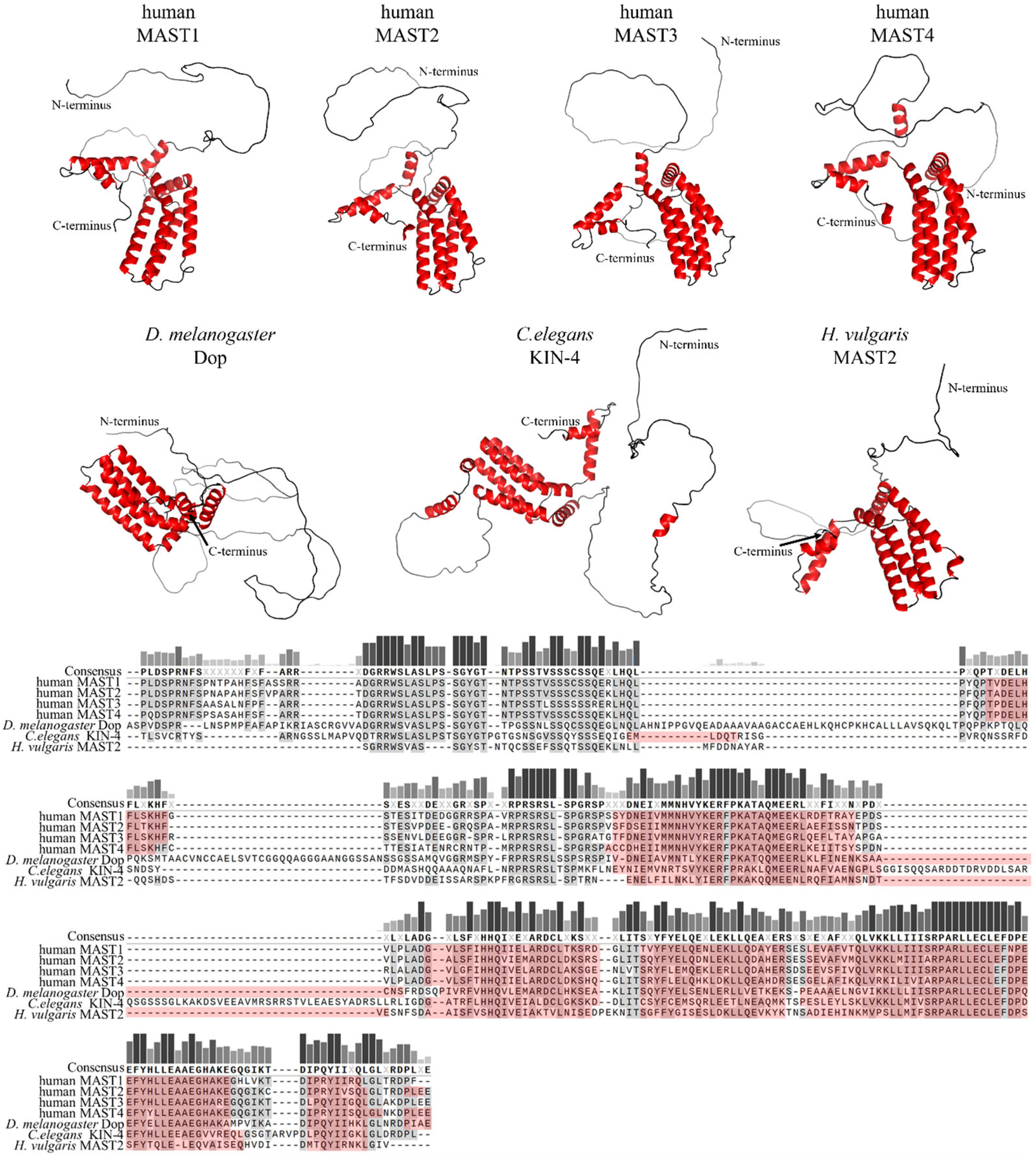
Members of the MAST kinase family are modular proteins characterized by a conserved domain composition and arrangement. The central Ser/Thr kinase domain is flanked towards the C-terminal end by a PDZ domain (post-synaptic density protein, disc large and Zonula occludens-1) and towards the N-terminus by a DUF (domain of unknown function) 1908 domain (Figure 1). The DUF1908 domain is a common feature of all MAST kinases; however, the function of this domain with respect to the activity of MAST kinases remains poorly understood. The DUF1908 domain (PFAM ID PF0826) is about 275 amino acids long, and within the human MAST kinases, this size differs only slightly. The predicted structure of the DUF1908 domain can be divided into an unstructured N-terminal half and a structured C-terminal half containing 8 alpha-helixes (Figure 2). A striking feature of the N-terminal domain of MAST kinases is the fact that their overall sequence is highly enriched serine, tyrosine and threonine residues, which are potential targets for posttranslational modifications including phosphorylation.
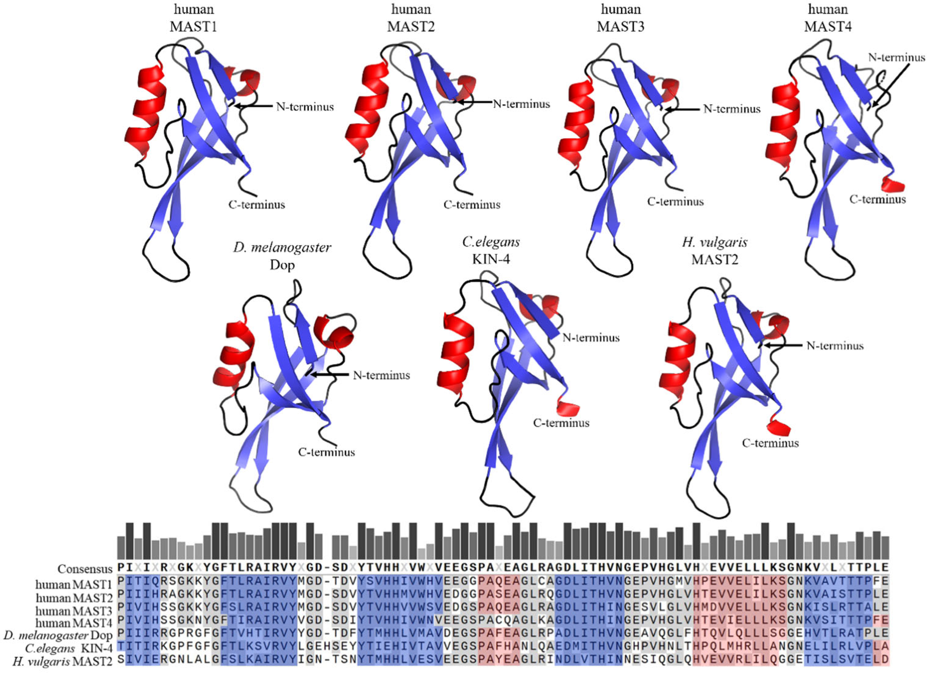
The PDZ domains, in contrast to DUF1908, are well studied and occur in proteins of a variety of organisms such as bacteria, yeast, plants, and metazoans, including Caenorhabditis elegans, Drosophila melanogaster and Homo sapiens [17]. PDZ domains are often found in multidomain proteins and contain 80–100 amino acids that are arranged in five beta strands and two alpha helices [18,19,20]. Within a given protein, single or several copies of PDZ domains can occur; MAST kinases contain only one PDZ domain in the carboxy-terminal half of the protein (Figure 3). PDZ domains are major hubs for protein-protein interactions, and their binding specificity is often based upon conserved amino acid motifs at the very carboxy-terminus of ligands [19]. PDZ domains can be divided into three classes based on their binding specificities. Class 1 PDZ domains recognize the binding motif X-S/T-X-V/I/L (where X represents any amino acid). MAST2-PDZ is an example of this class, as it recognizes this motif in its ligand PTEN [21]. Class 2 PDZ domains recognize a motif of four amino acids, alternating between a hydrophobic and a variable amino acid, and the third class recognizes the motif X-D/E-X-⌽ [22,23]. Another mode of binding allows the complexation of PDZ domains with each other, thus causing PDZ-based dimerization, which occurs mainly in proteins that have more than one PDZ domain [24,25]. Homodimer formation was also observed for proteins with only one PDZ domain; notably, this mode of binding was reported for MAST2-PDZ [21,22]. This self-association due to PDZ binding suggests an influence of this domain on the regulation of MAST kinases through dimerization.
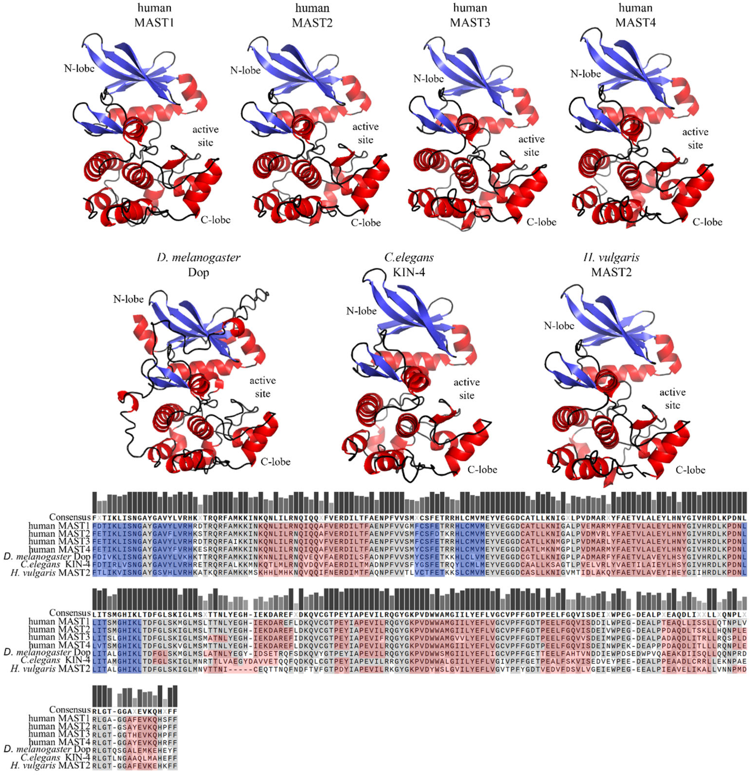
The core domain of the MAST kinase family is a Ser/Thr kinase domain divided into two subdomains. Since the MAST kinases belong to the AGC kinase family, the amino acid sequences of the human MAST kinase domains show a high degree of conservation compared to AGC kinases, for example, from 34% to 36% sequence identity and 54% to 59% amino acid sequence similarity when comparing PKA, PKC and PKG (Figure 4). Moreover, the catalytic domain of the MAST kinases shares with the AGC kinases all highly conserved motifs such as DFG, APE and HRD, which are important for ATP binding and magnesium transfer and are therefore essential for kinase activity and activation [3,4,5]. However, there is one exception, which is not found in other AGC kinases: the first glycine within the conserved glycine-rich loop (GXGXXG), is replaced by a serine in MAST kinases [11]. This exchange of glycine for a serine allows speculation about possible phosphorylation at this site, which could influence the regulation of the kinase activity. The structure of the modulatory C-terminal tail region of MAST kinases is also well conserved (Figure 5). The comparison of the sequence of the C-terminal tail of the human MAST kinases with PKA and PKG revealed a 42–57% amino acid similarity and 29–43% identity. In summary, the overall structure of the MAST kinase domains exhibits a high degree of structural and sequence conservation with other members of the AGC kinase family.

3. Substrates and Interactors of MAST Kinases

A range of different MAST kinase-interacting proteins have been reported, some of which represent potential substrates of MAST kinases (Table 1). MAST1, 2 and 3 were all shown to bind to the lipid phosphatase PTEN (Phosphatase and Tensin homolog) [26,27]. PTEN is a key regulator of cell growth and cell survival, and as such, represents an important tumor suppressor in humans [28]. The interaction of PTEN with MAST2 was shown to facilitate the phosphorylation of PTEN in vitro [26]. Phosphorylation of the carboxy-terminal domain of PTEN reduces its activity and prevents PTEN from degradation, suggesting that MAST2 regulates PTEN activity and stability in vivo [29]. This MAST kinase-dependent inhibition of PTEN activity was also observed in multiple myeloma cell lines, where knockdown of MAST4 leads to reduced PTEN activity and increased activity of proteins involved in the mTOR signaling pathway [30]. The PTEN/MAST2 interaction might also play an important role in neuronal homeostasis. In human neuroblastoma cells, complex formation of PTEN and MAST2 was reported to suppress neurite outgrowth [31,32]. This mechanism also plays an important role during a rabies virus infection, where the glycoprotein (G protein) of the rabies virus (RABV) competes with PTEN to interact with MAST2 through its class 1 PDZ binding site [21,33].
Another MAST kinase binding partner and potential substrate is the Na+/H+ exchanger NHE3 [34]. NHE3 is a major Na+/H+ exchanger that functions in the apical membrane domain of renal and intestinal epithelial cells, in particular in the renal proximal tubules [35]. The interaction of MAST2 with NHE3 requires the PDZ domain of MAST2. Co-expression of MAST2 and NHE3 in opossum kidney cells caused an inhibition of pH recovery, suggesting that MAST2 inhibits NHE3’s ion transport function. The mechanism of this regulation might involve the phosphorylation of NHE3 by MAST2 [34].
The earliest studies on MAST kinases suggested that these proteins are part of large multi-protein complexes and that MAST2 presumably binds to MAPs [11]. In addition to PTEN and NHE3, many other proteins have been reported to interact with MAST kinases (Table 1). MAST1 has been found to bind β2-syntrophin, a component of the dystrophin/utrophin network within the plasma membrane cortex at neuromuscular synapses [36]. The interaction of MAST1 and β2-syntrophin involves the PDZ domains of both proteins, and it was proposed that MAST kinases might link the dystrophin complex via β2-syntrophin with the microtubule network. Additionally, protocadherin LKC (expressed in liver, kidney and colon tissues) (PCLKC) protein was reported to bind MAST2 through its PDZ domain [37]. The interaction of MAST2 with PCLKC resembles a canonical interaction between the PDZ domain of MAST2 and a carboxy-terminal PDZ-binding motif in PCLKC. Interestingly, this member of the atypical cadherin superfamily acts as a tumor suppressor by inducing contact inhibition in epithelial cells and is frequently lost in cancer cells [37].
Proteins involved in immune responses have been found to interact with MAST2, including TRAF6, a component of the NF-κB pathway [38]. The interaction with TRAF6 is dependent on the amino-terminal region of MAST2 and is presumed to function in the regulation of immune responses. In this context, it is interesting that the induction of proinflammatory cytokines, including interleukin 12 (Il12) by LPS involves MAST2 [38,39]. Xiong and colleagues (2004) proposed that the association of MAST2 with TRAF6 might result in the inhibition of TRAF6-dependent NF-κB activation. Signaling through NF-κB controls gene transcription in response to extracellular signals, which in turn affects important cellular functions, in particular during immune responses and inflammation, and its incorrect regulation has been linked to many human diseases (see below). A potential role for MAST3 in inflammatory bowel disease may be explained by the misregulation of the same pathway [40]. However, the details of how MAST2 affects the activity of TRAF6, or PCLKC function, remain to be resolved.
Analyses of the phosphoproteome of 14-3-3 revealed that MAST2 directly interacts with 14-3-3 proteins in a phosphorylation-dependent manner [41]. 14-3-3 proteins are ubiquitously expressed in eukaryotic organisms and display a plethora of interacting proteins that are involved in numerous biological processes [42]. There are 7 isoforms found in humans: β, γ, ε, ζ, η, θ and σ [43]. However, direct interaction between MAST2 and 14-3-3 has been demonstrated by Western assays using an antibody binding to the N terminus of 14-3-3 β, and it remains currently unclear whether the other isoforms also interact directly with MAST2.
Table 1. MAST kinase interacting proteins *.
MASTKInteractorPhosphosite 1Interaction Domain 2References
MAST1USP1-PDZ[44]
MAST1Cdh1-PDZ[44]
MAST1CHIP-K317, K545[45]
MAST1MEKS221-[46]
MAST1c-Raf--[46]
MAST1HSP90--[45]
MAST1
MAST2
SNTB2-PDZ[36]
MAST1
MAST2
MAST3
PTENC-term.PDZ[26]
MAST1
MAST2
MAPs
MAPs
-KD + aa 948–1212[11,47]
MAST2TRAF6-N-term.[38]
MAST2RABV-G-PDZ[21]
MAST2CFTR-PDZ[48]
MAST2NHE3n.d.PDZ[34]
MAST2PCLKC-PDZ[37]
MAST214-3-3--[41]
MAST3ARPP-16S46-[49]
MAST4Sox9S494-[50]
* The table summarizes published interactions of MAST kinases with other proteins. Where known, the phosphorylation site (Phosphosite) 1, if the interactor was identified as a substrate, and the interaction site 2 within MAST kinases are listed. An empty field (“-”) indicates that a phosphorylation site or interaction domain has not been described. MASTK = MAST kinase; KD = kinase domain; n.d. = not determined.
A well-confirmed substrate of MAST3 is ARPP-16 (cAMP-regulated phosphoprotein of molecular weight 16 kDa), an alternatively spliced variant of ARPP-19 [49]. ARPP-16 and ARPP-19 proteins represent cAMP-regulated phosphoproteins, as they are substrates of PKA and highly expressed in the medium spiny neurons in the striatum of the brain [51,52,53]. MAST3 is part of a complex in which it competes with PKA for the ARPP16-dependent regulation of protein phosphatase 2A (PP2A) in the striatum. PP2A activity is inhibited by the phosphorylation of serine 46 of ARPP-16 by MAST3 [49,54]. In contrast, the phosphorylation of serine 88 in ARPP-16 by PKA leads to a decreased phosphorylation of serine 46 and prevents the inhibition of PP2A [52]. Moreover, MAST3 is regulated by PKA through phosphorylation at threonine 389 [54]. In summary, these results demonstrate the important role of MAST3 in the regulation of PP2A in neuronal cells.
MAST kinases have also been reported to interfere with transcriptional regulation. Sox9 belongs to the family of high-mobility group domain transcription factors and is an important regulator in the differentiation of mesenchymal stromal cells into chondrocytes [55,56,57]. In this system, MAST4 acts as a negative regulator, as by phosphorylating Sox9 at serine 494, Sox9 is targeted for degradation through the proteasome. This MAST4-dependent degradation of Sox9 suggests an essential role for MAST4 in the differentiation of mesenchymal stromal cells [50]. The ETS transcription factor family member ERM (Ets-related molecule) is one essential component of the gene regulatory network in spermatogonial stem cell self-renewal, and it is controlled by the fibroblast growth factor (FGF) 2. Phosphorylation of ERM by MAST4 at serine 367 enhances the transcription of ERM-target genes, suggesting that MAST4, together with the FGF2 signaling pathway, is involved in spermatogonial stem cell self-renewal by regulating the transcription factor ERM [58].

4. MAST Kinases in Human Disease

As discussed in the previous section, members of the MAST kinase family interact with a large variety of distinct binding partners, which may act as potential regulators or substrates. In mice, the genes encoding MAST kinases are widely expressed across various tissues such as the brain, heart, spleen, lung, liver, skeletal muscle, kidney and testis, suggesting a large diversity of functions [59]. Consistent with their broad expression patterns, malfunctions of MAST kinases are implicated in a wide range of human diseases affecting distinct tissues and organs, including breast cancer, inflammatory bowel disease, neuronal disorders and cystic fibrosis (Table 2) [21,31,33,34,40,48,60,61,62,63].

4.1. Association of MAST Kinase Mutations in Cancer

Somatic alterations in the genes for MAST1 and MAST2 were discovered and characterized in material obtained from breast cancer samples [60,64]. An increased risk for breast cancer was also reported to be associated with nine SNPs (single nucleotide polymorphism) found in MAST2 [61]. On the molecular level, three types of gene fusions were found for the MAST1 gene (with ZNF700, with taDA2A, and with NFIX) and two types were discovered in the case of the MAST2 gene (with ARIDA1a and GPBP1L1) in different transcriptomes from breast cancer samples and breast cancer-derived cell lines [60]. Overexpression of these fused gene products lead to enhanced proliferation in a benign breast cell line and RNAi-mediated knockdown of MAST2 in a cancer cell line with MAST2 gene fusions led to reduced growth and reduced tumor formation in mice xenografts. Together, these studies implicate the MAST1 and MAST2 gene fusions in the development of invasive breast carcinomas.
Alterations in MAST kinase gene expression have been revealed as a major feature of the relationship of this family of protein kinases with cancer. MAST2 was identified in a screen for pro-survival factors in a cDNA library of a glioblastoma cell line. Further studies showed that MAST2 overexpression promotes cell survival and cell growth, and the knock-down of MAST2 led to reduced tumor growth in a xenograft tumor mouse model, supporting a role of MAST2 in tumor progression that is characteristic of an oncogene [65]. Altered RNA levels of MAST kinases are associated with liver cancers and circular MAST1 RNA is upregulated in hepatocellular carcinoma and may serve as a diagnostic marker [66]. MAST2 mRNA is upregulated in liver cancer and expands the diagnostic spectrum of the disease [67]. MAST1, MAST2 and MAST3 were all found to bind to the crucial tumor suppressor Adenomatous polyposis coli (APC); however, the functional significance of this interaction has yet to be investigated [68].
MAST4 was identified as an estrogen response gene that was upregulated in female patients with multiple myeloma (MM) associated with low osteolytic lesions [69]. A recent investigation confirmed this association and revealed a mechanistic insight into the role of MAST4 in MM [30]. MM can be accompanied by characteristic lytic bone lesions derived from malignant plasma cells accumulating in the bone marrow, a syndrome called MM bone disease (MMBD). Estrogen levels are correlated with the extent of the disease, raising the hypothesis that estrogen may enhance the severity of myeloma. The search for estrogen response genes revealed, among others, MAST4. The study further showed that MAST4 is a negative regulator of MMBD and that the levels of MAST4 in MM cells negatively correlate with the severity of MMBD; high estrogen pathway activation levels correlated with high levels of MAST4 [30]. Pathway analyses suggested that MAST4 regulates the PI3-Kinase and mTOR pathways and that MAST4 colocalizes and co-immunoprecipitates with PTEN in MM cells. MAST4 knockdown resulted in an upregulation of Pi3-Kinase and mTOR signaling. Furthermore, the MAST4 gene harbors an estrogen receptor-responsive element in the enhancer of the gene, providing further evidence that MAST4 is indeed a target of estrogen receptor transcriptional activity.
Since the MAST kinases are known to be associated with different cancer types, further studies addressed the biological role of these kinases and identified MAST1 as a key regulator for cisplatin resistance in human cancer cells. Cisplatin is one of the most commonly used chemotherapeutic agents, as it blocks cell proliferation by inhibiting DNA replication [70]. A major complication of cisplatin chemotherapy is the upregulation of the ERK mitogen-activated protein kinase (MAPK) cascade, which contributes to cisplatin resistance by inhibiting cisplatin-induced apoptosis [71]. MAST1 was found to form a complex with components of the ERK pathway, including MEK1 and its upstream kinase, cRaf. Cisplatin interferes with this complex formation and causes the dissociation of cRaf but not MAST1, which then activates MEK1 by phosphorylation at serine 221 [46,72]. The regulation of MAST1 is based on the heat shock protein 90 (hsp90) and the E3 ubiquitin ligase CHIP. The direct binding between MAST1 and hsp90 increases the stabilization of MAST1, as the ubiquitination of lysines 317 and 545 of MAST1 by CHIP is inhibited [45]. A recent study revealed that the stability of MAST1 is also enhanced by the deubiquitinase USP1 in cisplatin-resistant cancer cells [44]. Together, these findings suggest that Hsp90, MAST1 and USP1 may be excellent targets for cisplatin-resistant cancer cells, as their inhibition will enhance cisplatin sensitivity and result in increased cell death. The negative side effects of cisplatin-based chemotherapy are also associated with increased inflammatory responses, which can be ameliorated by glucosteroid receptor (GR) agonists. A recent study found that MAST1 transcription is upregulated through GR activation and thus mediates resistance through the aforementioned reprogramming of the ERK pathway [73].

4.2. MAST Kinases in Neuronal Diseases

Rabies infections are deleterious viral infections of the brain. MAST1 and MAST2 were shown to interact with the glycoprotein of the virulent rabies virus in infected cells, and the binding was shown to inhibit normal MAST2 localization to apical membranes [33,63]. An interaction between the PDZ domain of MAST2 and the C-terminal residues of the glycoprotein of the rabies virus was required for the survival of infected neuronal cells, a signature of virulence [33,63]. Rabies virus glycoprotein was reported to disrupt interaction between PTEN and MAST2 [21]. PTEN was shown to bind to the PDZ domains of MAST1, MAST2 and MAST3, and phosphorylation of PTEN in vitro by MAST2 indicates that PTEN may be a substrate of MAST kinases [26]. The PDZ domain of MAST2 binds both PTEN and glycoprotein with similar affinity, and the viral glycoprotein disrupts the interaction of MAST2 with PTEN. The presence of glycoprotein decreased the nuclear localization of PTEN, which results in increased neuronal survival after infection with the rabies virus [21]. Therefore, the MAST kinases may be an important regulator of PTEN localization and activity in neuronal cells.
The aforementioned interaction between MAST2 and PTEN inhibits PTEN function by preventing neuronal outgrowth and regeneration. This mechanism might explain the enhanced neuronal survival following rabies virus infection. In support of this idea, an RNAi screen designed to identify protein kinases that regulate growth cone collapse, neurite retraction and neurite outgrowth showed that RNAi knockdown of MAST2 promotes neurite outgrowth and inhibits (lipopolysaccharide (LPS)-induced) neurite retraction in rat primary midbrain neurons [31]. Therefore, an overall function of MAST2 might be to control PTEN localization and activity in the nervous system, suggesting that the MAST kinases are involved in regulating processes that are often disrupted in neuronal disorders.
Both MAST1 and MAST2 have been found to interact with β2-syntrophin, coupling MAST2 via its PDZ domain to the dystrophin/utrophin network at the neuromuscular junction (NMJ) [36]. The NMJ and neuronal postsynaptic densities are composed of receptors, receptor clustering elements, cytoskeletal components and signal transduction molecules. The interaction between β2-syntrophin and MAST2 may function to link the dystrophin and utrophin networks to cellular signaling pathways.

4.3. MAST Kinases in Cystic Fibrosis and Diarrhea

MAST kinases are also linked to cystic fibrosis and secretory diarrhea. MAST2 was shown to form a complex with the key factor associated with cystic fibrosis, the cystic fibrosis transmembrane conductance regulator (CFTR) [48]. CFTR represents the arguably best-known anion channel specific for the transport of chloride (Cl) and bicarbonate (HCO3) through the apical membranes in most, if not all, epithelia-producing mucus, including the lung and intestine. CFTR interacts with several proteins, including PDZ domain proteins, which CFTR binds through its conserved C-terminal PDZ-binding motif [74,75,76,77,78]. The PDZ-based interactions of CFTR with PDZ-domain proteins play important roles in the biosynthesis and membrane transport of the protein [79,80]. The Golgi-associated PDZ protein CFTR-associated ligand (CAL) regulates the cell surface levels of CFTR and can promote lysosomal degradation of CFTR [81,82]. The binding between CFTR and CAL is sensitive to competitive binding to other PDZ domain proteins, including MAST2; indeed, high levels of MAST2 expression have been shown to increase CFTR surface levels, while knock-down of MAST2 inhibits CFTR function [48].
Besides its functional importance in cystic fibrosis, CFTR also represents a major chloride and hydrogen carbonate ion channel in the intestinal mucosa to control ion and pH homeostasis in the gut [78]. In addition, MAST2 also binds to and phosphorylates another important intestinal ion channel, the Na+/H+ exchanger NHE3 [34]. The phosphorylation by MAST2 was shown to inhibit NHE3 activity. NHE3 is important to maintain normal gastrointestinal physiology and its malfunction leads to impaired absorption and can increase the fluidity of diarrhea [48]. The results, which show that MAST2 regulates NHE3 as well as CFTR function suggest MAST2 as a potential cross-regulator that can assemble both channels and maybe other proteins into a macromolecular complex, thereby increasing its impact on intestinal physiology.

4.4. MAST Kinases in Male Fertility

Since MAST2 was the first of the MAST kinases to be discovered in the testis in mice, it was not surprising that MAST2 was reported to be associated with infertility in humans [11,12]. A study that suggested recurrent copy number variations as the cause of idiopathic nonobstructive azoospermia showed that, among other copy number variations, the presence of duplication of MAST2 is a risk factor for this severe form of male infertility [83]. Besides MAST2, MAST4 is associated with spermatogenesis in mice as it controls the self-renewal of spermatogonial stem cells by phosphorylating the transcription factor ERM. Knockout of MAST4 results in a reduced sperm number and the typical phenotype of SCO (Sertoli cell-only syndrome), which represents a form of male infertility [58].

4.5. MAST Kinases in Inflammation

Both MAST2 and MAST3 were associated with mechanisms that are required for NF-κB activity. MAST3 was found to be a factor in inflammatory bowel disease (IBD) tissues to increase Toll-like receptor (TLR) 4-dependent NF-κB activity and knock-down of MAST3 resulted in reduced NF-κB activity [40]. A follow-up study reported that MAST3 acts on the NF-κB pathway by changing the expression of several genes in the gut of IBD patients and might thereby trigger immune reactions [62]. However, the direct target of MAST3 in this process remains to be determined. Nevertheless, these studies highlight the potential involvement of MAST3 in the pathogenesis of IBD.
In B-cells, the expression of MAST2 was shown to be under the control of the class II MHCII transactivator, a key regulator of the MHCII response [84]. Furthermore, MAST2 regulates LPS-induced NF-κB regulation by forming a complex with TRAF6 (TNF receptor-associated factor 6), which results in NF-κB inhibition and a reduction of inflammatory responses [38,39]. Thus, MAST2 and MAST3 might have opposite effects on NF-κB regulation depending on different stimuli: MAST2 exhibits an inhibitory influence in LPS-dependent NF-κB regulation, while MAST3 may be enhancing NF-κB target activation in TLR4-dependent NF-κB regulation.
Table 2. Overview of MAST Kinase-associated human diseases **.
Disease Subgroup 1MASTKDisease 2Cause 3References
InfertilityMAST2Nonobstructive azoospermiaGene Duplication[83]
CancerMAST4Multiple myeloma bone diseaseOverexpressed[30]
Acral melanomaVarious deleterious mutations[85]
Ductal carcinoma
in-situ
Invasive breast cancer
Upregulated[86]
MAST3Prostate cancerGene conversion[87]
MAST2Cutaneous melanomaTranslocation [88]
MAST2Esophageal cancer
Pancreatic cancer
Sarcomas
Overexpressed[65]
MAST2Liver cancerOverexpression[67]
MAST2Chronic myeloid
leukemia
Insertion of exon 8 in a BCR-ABL1
fusion gene
[89]
MAST2Breast cancerTranslocation[64]
MAST2Breast cancerGene fusion[60]
MAST1
MAST1Breast cancerHigh levels of DNA methylation[90]
MAST1Non-small-cell lung cancerUpregulated[91]
MAST1Pheochromocytoma, paragangliomaOverexpression by hypomethylation [92]
MAST1Hepatocellular
carcinoma
Upregulated circRNA[66]
MAST1Uterine corpus endometrial carcinomaUpregulated[93]
MAST1Lung cancerS81Y
C291F
V316E
[94]
Cardiovascular
Diseases
MAST2Venous thrombosisR89Q[95]
Neuronal diseasesMAST3Developmental and epileptic encephalopathyS101F
S104L
G515S
L516P
[96]
MAST3Developmental and epileptic encephalopathy G510S
G515S
[97]
MAST4Juvenile myoclonic epilepsy (JME)T347M [98]
Childhood absence epilepsyP1201R
MAST1Intellectual disability, speech delay, hypotonia, facial dysmorphism, autismS93L[99]
MAST1Cerebral palsy, intellectual disabilityP500L[100]
MAST1Intellectual disabilityP1177R[101]
MAST1Neurologic abnormalities, developmental disability, mental retardationDeletion[102]
MAST1Intellectual disabilityL1180R[103]
MAST1Congenital bilateral Perisylvian syndromeDeletion of Q223 to D230[104]
MAST1Mega-corpus-callosum syndrome with cortical malformations without cerebellar
Hypoplasia
G522E[105]
MAST1Mega-corpus-callosum syndrome with cerebellar hypoplasia and cortical malformationsL278del
E194del
K276del
G517S
E697del
[47]
Inflammatory bowel diseaseMAST3Crohn’s disease (CD) and ulcerative colitis (UC)S861G[40]
OthersMAST4Asthma (horses)Overexpressed[106]
MAST3Hepatic steatosisIntronic variant[107]
MAST3Rheumatoid arthritisOverexpressed in fibroblast-like synovial cells[108]
MAST2Type 2 diabetes
mellitus
A1463T[39]
MAST2Rabies infectionViral glycoprotein prevents complex formation (MAST2-PDZ and PTEN) and promotes neuronal survival [21,33,63,109,110]
** The table summarizes human diseases associated with MAST kinases (MASTK). Diseases were assembled into subgroups 1 and the exact disease 2 name and its cause 3 are indicated.

5. MAST Kinases in Model Organisms

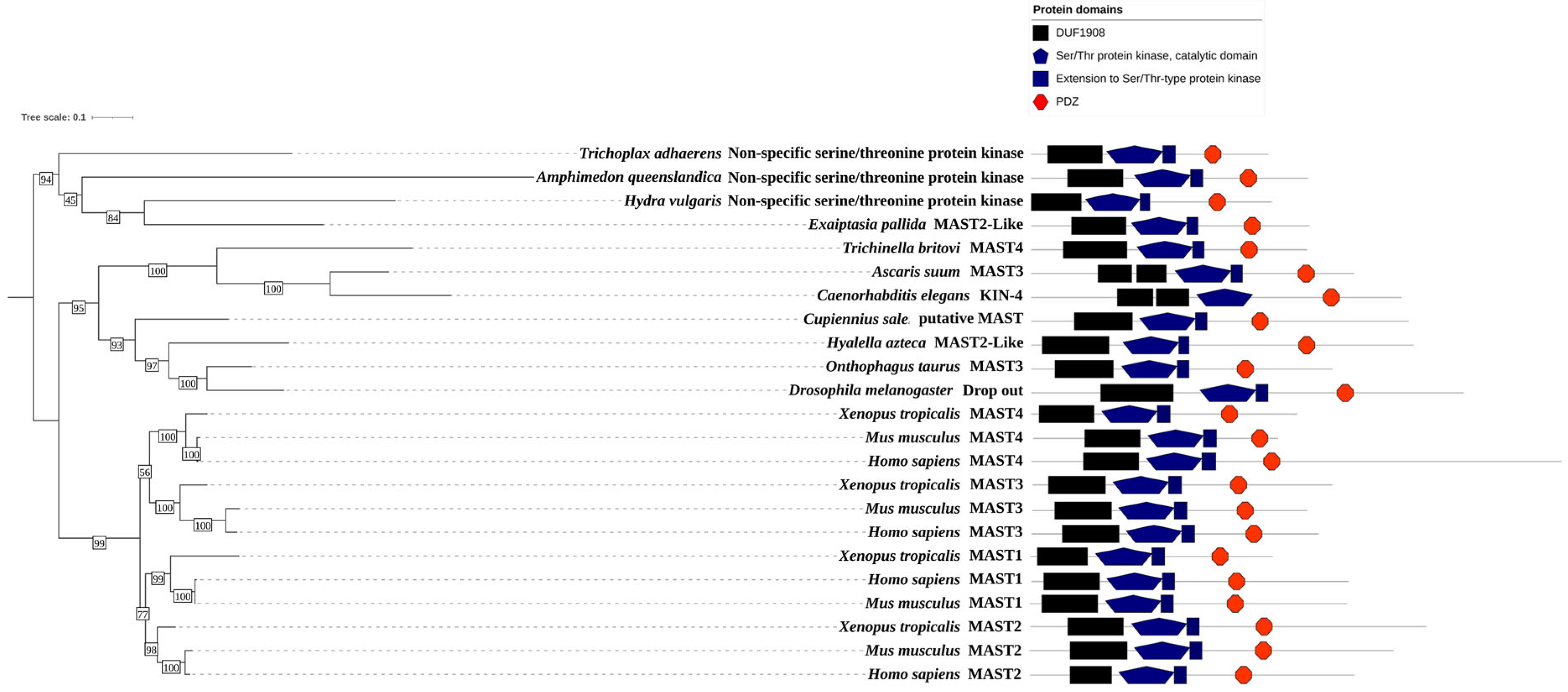
The human MAST kinases exhibit a remarkably conserved modular domain composition and arrangement (Figure 1). Using BLAST search analyses, homologs of MAST kinases were found in organisms of the metazoan kingdom. BLAST searches against annotated proteins demonstrate a high degree of amino acid sequence conservation of the MAST kinase domain in vertebrates, insects, nematodes and simple metazoans (Figure 6). All MAST kinase homologs identified in this survey encode proteins with one DUF1908, one Ser/Thr kinase domain and one PDZ domain as identified using the domain detection tool SMART [111,112]. Interestingly, in both exemplar nematodes (Caenorhabditis elegans, Ascaris suum) the DUF1908 domain is split into two subdomains. This split of the DUF1908 domain was not found in any other class than nematodes. It is also noticeable that due to evolutionary gene duplication, higher-evolved species such as humans, mice, and frogs possess four MAST kinases, while simpler species such as insects, nematodes, and simple metazoans comprise only one MAST kinase. Since the overall domain composition and structure are highly conserved and the human genome encodes four MAST kinases, model organisms provide simple and genetically tractable experimental systems to investigate the molecular mechanisms of MAST kinase function in vivo.
Mice serve as excellent model systems to examine the effect of human disease-related mutations within genes encoding human MAST kinases and to analyze their impact on the activity, regulation and localization of the kinase. MAST kinases are broadly expressed in the central nervous system [59]. Mutations in human MAST1 are associated with the mega-corpus-callosum syndrome, which includes cerebellar hypoplasia and cortical malformations in humans. One of these mutations, deletion of Lysine 278 (L278del), was introduced into a transgenic mouse line [47]. In heterozygous animals, an increase in the apoptosis of neurons and an enlarged corpus callosum were observed. The L278del in MAST1 was associated with reduced protein levels of the MAST2 and MAST3 kinases, suggesting that the regulation of MAST kinases may be regulated by interdependent mechanisms. Interestingly, the deletion of the MAST1 gene in mice is viable, suggesting that MAST1 either is not essential or the other members of the MAST kinases may compensate for the lack of MAST1 [47]. In support of the latter idea, it was reported that the individual deletion of either MAST kinase gene is not lethal but rather leads to distinct phenotypes. For example, the knockdown of MAST1 results in a decreased heart weight; the knockout of MAST2 causes an increased bone density and bone mineral content, and mice where MAST3 is deleted display behavioral problems resulting in hyperactivity as well as developing a cataract and increased circulating alkaline phosphatase level [116]. Mice with a truncated MAST4 kinase (E726stop) show a craniofacial phenotype caused by dental malocclusions [117] and complete knockdown of MAST 4 is associated with an osteoporosis phenotype [50]. In addition, the knockdown of MAST4 reveals its role in spermatogenesis as it leads to decreased testes size and sperm number and the typical phenotype of Sertoli cell-only syndrome (SCO) [58].
In simpler model organisms such as nematodes (Caenorhabditis elegans) or insects (Drosophila melanogaster), the MAST kinases are expressed by a single gene. This is an advantage, as mutations cannot be compensated for by other MAST kinase family members in the genome. In C. elegans, the MAST kinase homolog KIN-4 is involved in the control of thermotaxis in conjunction with the stomatin homolog MEC-2 [118] and the diacylglycerol kinase DGK-1 [119,120]. In this system, the roles of KIN-4, DGK-1 and MEC-2 regulate presynaptic mechanisms, which control the release of neurotransmitters at the synapse of thermosensory neurons [121]. The C. elegans Kin-4 has also been shown to be required for longevity through a mechanism involving binding of PTEN and its contribution to the insulin/IGF-1 signaling pathway in the regulation of lifespan [122]. It is interesting to note that PTEN is also a known interaction partner of human MAST kinases; however, an association between human MAST kinases and the regulation of lifespan is yet unknown [26].
In the genetic model organism Drosophila melanogaster, a single MAST kinase homolog was identified as a recessive female sterile mutation called drop out (dop) [123,124]. Females homozygous for the mutant dop1 allele lay eggs, but the embryos exhibit severe morphogenetic defects early in embryogenesis. The developmental stage at which dop mutant embryos become abnormal is called cellularization, a process that transforms the syncytial blastoderm into a polarized blastoderm epithelium [125,126]. The phenotype of dop mutant embryos includes severely reduced membrane growth and a dropping of the nuclei out of the cortical cytoplasm, and most embryos fail to undergo gastrulation and further development [124]. The analyses of the cell biological role of dop in cellularization revealed that the membrane growth defect is associated with the mislocalization of several membrane proteins that are essential for the assembly of membrane cortex domains during cellularization [126,127]. These proteins include the apical scaffold protein and Par-3 homolog Bazooka (Baz) and the adherens junction protein E-cadherin [123]. Another membrane subdomain, the furrow channel marked by Slam, the small GTPase Rho1 and the scaffold protein dPatJ exhibited an overlap with a lateral membrane marker Discs large (Dlg) suggesting a requirement of Dop for compartmentalization of the plasma-membrane associated cortex [123]. Interestingly, all known phenotypes of dop mutants share the common feature that the defects can be linked to impaired dynein-dependent transport along microtubules. This includes the apical transport of lipid droplets in the embryo, the apical transport of ribonucleoprotein particles, the apical transport of Baz and the apical transport of Golgi-derived membrane reservoirs that fuel membrane growth in cellularization [123,128,129,130]. Genetic and biochemical studies revealed that Dop possibly regulates the dynein complex by phosphorylating the dynein intermediate chain (Dic) [123]. A domain function analysis in flies suggests that the DUF1908 and the PDZ domains might have regulatory functions, and further studies should reveal how the kinase activity of the fly MAST kinase homolog is controlled in embryogenesis, which may reveal important general properties of MAST kinase regulation in general.

6. Conclusions and Outlook

MAST kinases are involved in an increasing number of diverse biological processes ranging from acute and chronic human diseases to stem cell maintenance and longevity. The modular structure of MAST kinases enables interaction with many distinct proteins, facilitating substrate recognition and binding and bearing the potential for regulatory inputs. In several instances, competitive interactions appear as a theme in the pathophysiological aspects of MAST kinases. Within the past 10 years, the first defined molecular mechanisms that link MAST kinase function to a particular protein substrate phosphorylation event that causes specific biological responses have emerged. More detailed mechanistic studies in human diseases, including the use of model organisms, are likely to reveal MAST kinase interactors and substrates that play crucial roles in various signaling pathways regulating distinct cellular functionalities.

Author Contributions

Conceptualization, M.R., S.P. and H.-A.J.M.; formal analysis, M.R., S.P. and B.D.; investigation, M.R. and B.D.; writing—original draft preparation, H.-A.J.M.; writing—review and editing, M.R., S.P., B.D., F.W.H. and H.-A.J.M.; visualization, M.R. and B.D.; supervision, H.-A.J.M.; project administration, H.-A.J.M.; funding acquisition, F.W.H. and H.-A.J.M. All authors have read and agreed to the published version of the manuscript.

Funding

This research was funded by the University of Kassel, Research Consortium ‘Future’ to FWH and HAJM and the research training grant ‘Multiscale Clocks’ (DFG GRK 2749-1).

Institutional Review Board Statement

Not applicable.

Informed Consent Statement

Not applicable.

Data Availability Statement

Not applicable.

Acknowledgments

The authors acknowledge initial support by the University of Kassel through their ‘Profilbildungsinitiative/Future’ for funding of the PhosMOrg research consortium.

Conflicts of Interest

The authors declare no conflict of interest.

References

  1. Manning, G.; Whyte, D.B.; Martinez, R.; Hunter, T.; Sudarsanam, S. The protein kinase complement of the human genome. Science 2002, 298, 1912–1934. [Google Scholar] [CrossRef] [Green Version]
  2. Richardson, C.J.; Gao, Q.; Mitsopoulous, C.; Zvelebil, M.; Pearl, L.H.; Pearl, F.M. MoKCa database--mutations of kinases in cancer. Nucleic Acids Res. 2009, 37, D824–D831. [Google Scholar] [CrossRef] [Green Version]
  3. Hanks, S.K.; Quinn, A.M.; Hunter, T. The protein kinase family: Conserved features and deduced phylogeny of the catalytic domains. Science 1988, 241, 42–52. [Google Scholar] [CrossRef]
  4. Hanks, S.K.; Hunter, T. Protein kinases 6. The eukaryotic protein kinase superfamily: Kinase (catalytic) domain structure and classification. FASEB J. 1995, 9, 576–596. [Google Scholar] [CrossRef]
  5. Pearce, L.R.; Komander, D.; Alessi, D.R. The nuts and bolts of AGC protein kinases. Nat. Rev. Mol. Cell Biol. 2010, 11, 9–22. [Google Scholar] [CrossRef]
  6. Hanks, S.K. Genomic analysis of the eukaryotic protein kinase superfamily: A perspective. Genome Biol. 2003, 4, 111. [Google Scholar] [CrossRef] [PubMed] [Green Version]
  7. Johnson, D.A.; Akamine, P.; Radzio-Andzelm, E.; Madhusudan, M.; Taylor, S.S. Dynamics of cAMP-dependent protein kinase. Chem. Rev. 2001, 101, 2243–2270. [Google Scholar] [CrossRef] [PubMed]
  8. Kannan, N.; Haste, N.; Taylor, S.S.; Neuwald, A.F. The hallmark of AGC kinase functional divergence is its C-terminal tail, a cis-acting regulatory module. Proc. Natl. Acad. Sci. USA 2007, 104, 1272–1277. [Google Scholar] [CrossRef] [PubMed]
  9. Knighton, D.R.; Zheng, J.H.; Ten Eyck, L.F.; Xuong, N.H.; Taylor, S.S.; Sowadski, J.M. Structure of a peptide inhibitor bound to the catalytic subunit of cyclic adenosine monophosphate-dependent protein kinase. Science 1991, 253, 414–420. [Google Scholar] [CrossRef]
  10. Taylor, S.S.; Soberg, K.; Kobori, E.; Wu, J.; Pautz, S.; Herberg, F.W.; Skalhegg, B.S. The Tails of Protein Kinase A. Mol. Pharmacol. 2022, 101, 219–225. [Google Scholar] [CrossRef]
  11. Walden, P.D.; Cowan, N.J. A novel 205-kilodalton testis-specific serine/threonine protein kinase associated with microtubules of the spermatid manchette. Mol. Cell Biol. 1993, 13, 7625–7635. [Google Scholar] [PubMed] [Green Version]
  12. Walden, P.D.; Millette, C.F. Increased activity associated with the MAST205 protein kinase complex during mammalian spermiogenesis. Biol. Reprod. 1996, 55, 1039–1044. [Google Scholar] [PubMed] [Green Version]
  13. Jumper, J.; Evans, R.; Pritzel, A.; Green, T.; Figurnov, M.; Ronneberger, O.; Tunyasuvunakool, K.; Bates, R.; Zidek, A.; Potapenko, A.; et al. Highly accurate protein structure prediction with AlphaFold. Nature 2021, 596, 583–589. [Google Scholar] [CrossRef] [PubMed]
  14. Monzon, V.; Paysan-Lafosse, T.; Wood, V.; Bateman, A. Reciprocal best structure hits: Using AlphaFold models to discover distant homologues. Bioinform. Adv. 2022, 2, vbac072. [Google Scholar]
  15. Varadi, M.; Anyango, S.; Deshpande, M.; Nair, S.; Natassia, C.; Yordanova, G.; Yuan, D.; Stroe, O.; Wood, G.; Laydon, A.; et al. AlphaFold Protein Structure Database: Massively expanding the structural coverage of protein-sequence space with high-accuracy models. Nucleic Acids Res. 2022, 50, D439–D444. [Google Scholar]
  16. Paysan-Lafosse, T.; Blum, M.; Chuguransky, S.; Grego, T.; Pinto, B.L.; Salazar, G.A.; Bileschi, M.L.; Bork, P.; Bridge, A.; Colwell, L.; et al. InterPro in 2022. Nucleic Acids Res 2023, 51, D418–D427. [Google Scholar] [CrossRef]
  17. Liu, X.; Fuentes, E.J. Emerging Themes in PDZ Domain Signaling: Structure, Function, and Inhibition. Int. Rev. Cell Mol. Biol. 2019, 343, 129–218. [Google Scholar]
  18. Ponting, C.P.; Phillips, C.; Davies, K.E.; Blake, D.J. PDZ domains: Targeting signalling molecules to sub-membranous sites. Bioessays 1997, 19, 469–479. [Google Scholar] [CrossRef]
  19. Songyang, Z.; Fanning, A.S.; Fu, C.; Xu, J.; Marfatia, S.M.; Chishti, A.H.; Crompton, A.; Chan, A.C.; Anderson, J.M.; Cantley, L.C. Recognition of unique carboxyl-terminal motifs by distinct PDZ domains. Science 1997, 275, 73–77. [Google Scholar]
  20. Nourry, C.; Grant, S.G.; Borg, J.P. PDZ domain proteins: Plug and play! Sci. STKE 2003, 2003, RE7. [Google Scholar] [CrossRef]
  21. Terrien, E.; Chaffotte, A.; Lafage, M.; Khan, Z.; Prehaud, C.; Cordier, F.; Simenel, C.; Delepierre, M.; Buc, H.; Lafon, M.; et al. Interference with the PTEN-MAST2 interaction by a viral protein leads to cellular relocalization of PTEN. Sci. Signal 2012, 5, ra58. [Google Scholar] [CrossRef] [PubMed]
  22. Lee, H.J.; Zheng, J.J. PDZ domains and their binding partners: Structure, specificity, and modification. Cell Commun. Signal 2010, 8, 8. [Google Scholar] [CrossRef] [PubMed] [Green Version]
  23. Hung, A.Y.; Sheng, M. PDZ domains: Structural modules for protein complex assembly. J. Biol. Chem. 2002, 277, 5699–5702. [Google Scholar] [CrossRef] [Green Version]
  24. Brenman, J.E.; Chao, D.S.; Gee, S.H.; McGee, A.W.; Craven, S.E.; Santillano, D.R.; Wu, Z.; Huang, F.; Xia, H.; Peters, M.F.; et al. Interaction of nitric oxide synthase with the postsynaptic density protein PSD-95 and alpha1-syntrophin mediated by PDZ domains. Cell 1996, 84, 757–767. [Google Scholar] [CrossRef] [PubMed] [Green Version]
  25. Chang, B.H.; Gujral, T.S.; Karp, E.S.; BuKhalid, R.; Grantcharova, V.P.; MacBeath, G. A systematic family-wide investigation reveals that ~30% of mammalian PDZ domains engage in PDZ-PDZ interactions. Chem. Biol. 2011, 18, 1143–1152. [Google Scholar] [CrossRef] [PubMed] [Green Version]
  26. Valiente, M.; Andres-Pons, A.; Gomar, B.; Torres, J.; Gil, A.; Tapparel, C.; Antonarakis, S.E.; Pulido, R. Binding of PTEN to specific PDZ domains contributes to PTEN protein stability and phosphorylation by microtubule-associated serine/threonine kinases. J. Biol. Chem. 2005, 280, 28936–28943. [Google Scholar] [CrossRef]
  27. Adey, N.B.; Huang, L.; Ormonde, P.A.; Baumgard, M.L.; Pero, R.; Byreddy, D.V.; Tavtigian, S.V.; Bartel, P.L. Threonine phosphorylation of the MMAC1/PTEN PDZ binding domain both inhibits and stimulates PDZ binding. Cancer Res. 2000, 60, 35–37. [Google Scholar]
  28. Lee, Y.R.; Chen, M.; Pandolfi, P.P. The functions and regulation of the PTEN tumour suppressor: New modes and prospects. Nat. Rev. Mol. Cell Biol. 2018, 19, 547–562. [Google Scholar] [CrossRef]
  29. Vazquez, F.; Ramaswamy, S.; Nakamura, N.; Sellers, W.R. Phosphorylation of the PTEN tail regulates protein stability and function. Mol. Cell Biol. 2000, 20, 5010–5018. [Google Scholar] [CrossRef] [Green Version]
  30. Cui, Y.; Wang, F.; Zhang, D.; Huang, J.; Yang, Y.; Xu, J.; Gao, Y.; Ding, H.; Qu, Y.; Zhang, W.; et al. Estrogen-Responsive Gene MAST4 Regulates Myeloma Bone Disease. J. Bone Miner. Res. 2022, 37, 711–723. [Google Scholar] [CrossRef]
  31. Loh, S.H.; Francescut, L.; Lingor, P.; Bahr, M.; Nicotera, P. Identification of new kinase clusters required for neurite outgrowth and retraction by a loss-of-function RNA interference screen. Cell Death Differ. 2008, 15, 283–298. [Google Scholar] [CrossRef] [PubMed] [Green Version]
  32. Gregorian, C.; Nakashima, J.; Dry, S.M.; Nghiemphu, P.L.; Smith, K.B.; Ao, Y.; Dang, J.; Lawson, G.; Mellinghoff, I.K.; Mischel, P.S.; et al. PTEN dosage is essential for neurofibroma development and malignant transformation. Proc. Natl. Acad. Sci. USA 2009, 106, 19479–19484. [Google Scholar] [CrossRef] [PubMed]
  33. Prehaud, C.; Wolff, N.; Terrien, E.; Lafage, M.; Megret, F.; Babault, N.; Cordier, F.; Tan, G.S.; Maitrepierre, E.; Menager, P.; et al. Attenuation of rabies virulence: Takeover by the cytoplasmic domain of its envelope protein. Sci. Signal 2010, 3, ra5. [Google Scholar] [CrossRef] [PubMed]
  34. Wang, D.; Lee, H.J.; Cooper, D.S.; Cebotaro, L.; Walden, P.D.; Choi, I.; Yun, C.C. Coexpression of MAST205 inhibits the activity of Na+/H+ exchanger NHE3. Am. J. Physiol. Renal Physiol. 2006, 290, F428–F437. [Google Scholar] [CrossRef]
  35. Alexander, R.T.; Grinstein, S. Tethering, recycling and activation of the epithelial sodium-proton exchanger, NHE3. J. Exp. Biol. 2009, 212 Pt 11, 1630–1637. [Google Scholar] [CrossRef] [Green Version]
  36. Lumeng, C.; Phelps, S.; Crawford, G.E.; Walden, P.D.; Barald, K.; Chamberlain, J.S. Interactions between beta 2-syntrophin and a family of microtubule-associated serine/threonine kinases. Nat. Neurosci. 1999, 2, 611–617. [Google Scholar] [CrossRef]
  37. Okazaki, N.; Takahashi, N.; Kojima, S.; Masuho, Y.; Koga, H. Protocadherin LKC, a new candidate for a tumor suppressor of colon and liver cancers, its association with contact inhibition of cell proliferation. Carcinogenesis 2002, 23, 1139–1148. [Google Scholar] [CrossRef]
  38. Xiong, H.; Li, H.; Chen, Y.; Zhao, J.; Unkeless, J.C. Interaction of TRAF6 with MAST205 regulates NF-kappaB activation and MAST205 stability. J. Biol. Chem. 2004, 279, 43675–43683. [Google Scholar] [CrossRef] [Green Version]
  39. Zhou, H.; Xiong, H.; Li, H.; Plevy, S.E.; Walden, P.D.; Sassaroli, M.; Prestwich, G.D.; Unkeless, J.C. Microtubule-associated serine/threonine kinase-205 kDa and Fc gamma receptor control IL-12 p40 synthesis and NF-kappa B activation. J. Immunol. 2004, 172, 2559–2568. [Google Scholar] [CrossRef]
  40. Labbe, C.; Goyette, P.; Lefebvre, C.; Stevens, C.; Green, T.; Tello-Ruiz, M.K.; Cao, Z.; Landry, A.L.; Stempak, J.; Annese, V.; et al. MAST3: A novel IBD risk factor that modulates TLR4 signaling. Genes. Immun. 2008, 9, 602–612. [Google Scholar] [CrossRef]
  41. Johnson, C.; Tinti, M.; Wood, N.T.; Campbell, D.G.; Toth, R.; Dubois, F.; Geraghty, K.M.; Wong, B.H.; Brown, L.J.; Tyler, J.; et al. Visualization and biochemical analyses of the emerging mammalian 14-3-3-phosphoproteome. Mol. Cell Proteom. 2011, 10, M110.005751. [Google Scholar] [CrossRef] [Green Version]
  42. Mackintosh, C. Dynamic interactions between 14-3-3 proteins and phosphoproteins regulate diverse cellular processes. Biochem. J. 2004, 381 Pt 2, 329–342. [Google Scholar] [CrossRef] [Green Version]
  43. Fu, H.; Subramanian, R.R.; Masters, S.C. 14-3-3 proteins: Structure, function, and regulation. Annu. Rev. Pharmacol. Toxicol. 2000, 40, 617–647. [Google Scholar] [CrossRef]
  44. Tyagi, A.; Kaushal, K.; Chandrasekaran, A.P.; Sarodaya, N.; Das, S.; Park, C.H.; Hong, S.H.; Kim, K.S.; Ramakrishna, S. CRISPR/Cas9-based genome-wide screening for deubiquitinase subfamily identifies USP1 regulating MAST1-driven cisplatin-resistance in cancer cells. Theranostics 2022, 12, 5949–5970. [Google Scholar] [CrossRef]
  45. Pan, C.; Chun, J.; Li, D.; Boese, A.C.; Li, J.; Kang, J.; Umano, A.; Jiang, Y.; Song, L.; Magliocca, K.R.; et al. Hsp90B enhances MAST1-mediated cisplatin resistance by protecting MAST1 from proteosomal degradation. J. Clin. Investig. 2019, 129, 4110–4123. [Google Scholar] [CrossRef] [Green Version]
  46. Jin, L.; Chun, J.; Pan, C.; Li, D.; Lin, R.; Alesi, G.N.; Wang, X.; Kang, H.B.; Song, L.; Wang, D.; et al. MAST1 Drives Cisplatin Resistance in Human Cancers by Rewiring cRaf-Independent MEK Activation. Cancer Cell 2018, 34, 315–330.e7. [Google Scholar] [CrossRef] [Green Version]
  47. Tripathy, R.; Leca, I.; van Dijk, T.; Weiss, J.; van Bon, B.W.; Sergaki, M.C.; Gstrein, T.; Breuss, M.; Tian, G.; Bahi-Buisson, N.; et al. Mutations in MAST1 Cause Mega-Corpus-Callosum Syndrome with Cerebellar Hypoplasia and Cortical Malformations. Neuron 2018, 100, 1354–1368.e5. [Google Scholar] [CrossRef]
  48. Ren, A.; Zhang, W.; Yarlagadda, S.; Sinha, C.; Arora, K.; Moon, C.S.; Naren, A.P. MAST205 competes with cystic fibrosis transmembrane conductance regulator (CFTR)-associated ligand for binding to CFTR to regulate CFTR-mediated fluid transport. J. Biol. Chem. 2013, 288, 12325–12334. [Google Scholar] [CrossRef] [Green Version]
  49. Andrade, E.C.; Musante, V.; Horiuchi, A.; Matsuzaki, H.; Brody, A.H.; Wu, T.; Greengard, P.; Taylor, J.R.; Nairn, A.C. ARPP-16 Is a Striatal-Enriched Inhibitor of Protein Phosphatase 2A Regulated by Microtubule-Associated Serine/Threonine Kinase 3 (Mast 3 Kinase). J. Neurosci. 2017, 37, 2709–2722. [Google Scholar] [CrossRef]
  50. Kim, P.; Park, J.; Lee, D.J.; Mizuno, S.; Shinohara, M.; Hong, C.P.; Jeong, Y.; Yun, R.; Park, H.; Park, S.; et al. Mast4 determines the cell fate of MSCs for bone and cartilage development. Nat. Commun. 2022, 13, 3960. [Google Scholar] [CrossRef]
  51. Girault, J.A.; Walaas, S.I.; Hemmings, H.C., Jr.; Greengard, P. ARPP-21, a cAMP-regulated phosphoprotein enriched in dopamine-innervated brain regions: Tissue distribution and regulation of phosphorylation in rat brain. Neuroscience 1990, 37, 317–325. [Google Scholar] [CrossRef]
  52. Horiuchi, A.; Williams, K.R.; Kurihara, T.; Nairn, A.C.; Greengard, P. Purification and cDNA cloning of ARPP-16, a cAMP-regulated phosphoprotein enriched in basal ganglia, and of a related phosphoprotein, ARPP-19. J. Biol. Chem. 1990, 265, 9476–9484. [Google Scholar] [CrossRef]
  53. Brene, S.; Lindefors, N.; Ehrlich, M.; Taubes, T.; Horiuchi, A.; Kopp, J.; Hall, H.; Sedvall, G.; Greengard, P.; Persson, H. Expression of mRNAs encoding ARPP-16/19, ARPP-21, and DARPP-32 in human brain tissue. J. Neurosci. 1994, 14 Pt 1, 985–998. [Google Scholar] [CrossRef]
  54. Musante, V.; Li, L.; Kanyo, J.; Lam, T.T.; Colangelo, C.M.; Cheng, S.K.; Brody, A.H.; Greengard, P.; Le Novere, N.; Nairn, A.C. Reciprocal regulation of ARPP-16 by PKA and MAST3 kinases provides a cAMP-regulated switch in protein phosphatase 2A inhibition. Elife 2017, 6, e24998. [Google Scholar] [CrossRef]
  55. Bell, D.M.; Leung, K.K.; Wheatley, S.C.; Ng, L.J.; Zhou, S.; Ling, K.W.; Sham, M.H.; Koopman, P.; Tam, P.P.; Cheah, K.S. SOX9 directly regulates the type-II collagen gene. Nat. Genet. 1997, 16, 174–178. [Google Scholar] [CrossRef]
  56. Bi, W.; Deng, J.M.; Zhang, Z.; Behringer, R.R.; de Crombrugghe, B. Sox9 is required for cartilage formation. Nat. Genet. 1999, 22, 85–89. [Google Scholar] [CrossRef]
  57. Lefebvre, V.; Huang, W.; Harley, V.R.; Goodfellow, P.N.; de Crombrugghe, B. SOX9 is a potent activator of the chondrocyte-specific enhancer of the pro alpha1(II) collagen gene. Mol. Cell Biol. 1997, 17, 2336–2346. [Google Scholar] [CrossRef] [Green Version]
  58. Lee, S.J.; Park, J.; Lee, D.J.; Otsu, K.; Kim, P.; Mizuno, S.; Lee, M.J.; Kim, H.Y.; Harada, H.; Takahashi, S.; et al. Mast4 knockout shows the regulation of spermatogonial stem cell self-renewal via the FGF2/ERM pathway. Cell Death Differ. 2021, 28, 1441–1454. [Google Scholar] [CrossRef]
  59. Garland, P.; Quraishe, S.; French, P.; O’Connor, V. Expression of the MAST family of serine/threonine kinases. Brain Res. 2008, 1195, 12–19. [Google Scholar] [CrossRef]
  60. Robinson, D.R.; Kalyana-Sundaram, S.; Wu, Y.M.; Shankar, S.; Cao, X.; Ateeq, B.; Asangani, I.A.; Iyer, M.; Maher, C.A.; Grasso, C.S.; et al. Functionally recurrent rearrangements of the MAST kinase and Notch gene families in breast cancer. Nat. Med. 2011, 17, 1646–1651. [Google Scholar] [CrossRef] [Green Version]
  61. Wang, X.; Fredericksen, Z.S.; Vierkant, R.A.; Kosel, M.L.; Pankratz, V.S.; Cerhan, J.R.; Justenhoven, C.; Brauch, H.; Olson, J.E.; Couch, F.J. Association of genetic variation in mitotic kinases with breast cancer risk. Breast Cancer Res. Treat. 2010, 119, 453–462. [Google Scholar] [CrossRef] [Green Version]
  62. Labbe, C.; Boucher, G.; Foisy, S.; Alikashani, A.; Nkwimi, H.; David, G.; Beaudoin, M.; Goyette, P.; Charron, G.; Xavier, R.J.; et al. Genome-wide expression profiling implicates a MAST3-regulated gene set in colonic mucosal inflammation of ulcerative colitis patients. Inflamm. Bowel Dis. 2012, 18, 1072–1080. [Google Scholar] [CrossRef]
  63. Terrien, E.; Simenel, C.; Prehaud, C.; Buc, H.; Delepierre, M.; Lafon, M.; Wolff, N. 1H, 13C and 15N resonance assignments of the PDZ of microtubule-associated serine/threonine kinase 205 (MAST205) in complex with the C-terminal motif from the rabies virus glycoprotein. Biomol. NMR Assign. 2009, 3, 45–48. [Google Scholar] [CrossRef]
  64. Clay, M.R.; Varma, S.; West, R.B. MAST2 and NOTCH1 translocations in breast carcinoma and associated pre-invasive lesions. Hum. Pathol. 2013, 44, 2837–2844. [Google Scholar] [CrossRef]
  65. Eissmann, M.; Schwamb, B.; Melzer, I.M.; Moser, J.; Siele, D.; Kohl, U.; Rieker, R.J.; Wachter, D.L.; Agaimy, A.; Herpel, E.; et al. A functional yeast survival screen of tumor-derived cDNA libraries designed to identify anti-apoptotic mammalian oncogenes. PLoS ONE 2013, 8, e64873. [Google Scholar] [CrossRef] [Green Version]
  66. Yu, X.; Sheng, P.; Sun, J.; Zhao, X.; Zhang, J.; Li, Y.; Zhang, Y.; Zhang, W.; Wang, J.; Liu, K.; et al. The circular RNA circMAST1 promotes hepatocellular carcinoma cell proliferation and migration by sponging miR-1299 and regulating CTNND1 expression. Cell Death Dis. 2020, 11, 340. [Google Scholar] [CrossRef]
  67. Jiao, Y.; Li, Y.; Jiang, P.; Fu, Z.; Liu, Y. High MAST2 mRNA expression and its role in diagnosis and prognosis of liver cancer. Sci. Rep. 2019, 9, 19865. [Google Scholar] [CrossRef] [Green Version]
  68. Sotelo, N.S.; Valiente, M.; Gil, A.; Pulido, R. A functional network of the tumor suppressors APC, hDlg, and PTEN, that relies on recognition of specific PDZ-domains. J. Cell Biochem. 2012, 113, 2661–2670. [Google Scholar] [CrossRef]
  69. Zhang, D.; Huang, J.; Zhang, W.; Pan, L.; Zhang, D.; Zhao, P.; Wang, F.; Luo, H.; He, J.; Qin, Y.; et al. Young female patients with multiple myeloma have low occurrence of osteolytic lesion. Bone 2018, 110, 21–28. [Google Scholar] [CrossRef]
  70. Fuertes, M.A.; Castilla, J.; Alonso, C.; Perez, J.M. Cisplatin biochemical mechanism of action: From cytotoxicity to induction of cell death through interconnections between apoptotic and necrotic pathways. Curr. Med. Chem. 2003, 10, 257–266. [Google Scholar] [CrossRef]
  71. Kong, L.R.; Chua, K.N.; Sim, W.J.; Ng, H.C.; Bi, C.; Ho, J.; Nga, M.E.; Pang, Y.H.; Ong, W.R.; Soo, R.A.; et al. MEK Inhibition Overcomes Cisplatin Resistance Conferred by SOS/MAPK Pathway Activation in Squamous Cell Carcinoma. Mol. Cancer Ther. 2015, 14, 1750–1760. [Google Scholar] [CrossRef] [Green Version]
  72. Jaykumar, A.B.; Karra, A.S.; Cobb, M.H. Pulling a MAST1 on Cisplatin Resistance. Cancer Cell 2018, 34, 183–185. [Google Scholar] [CrossRef] [Green Version]
  73. Pan, C.; Kang, J.; Hwang, J.S.; Li, J.; Boese, A.C.; Wang, X.; Yang, L.; Boggon, T.J.; Chen, G.Z.; Saba, N.F.; et al. Cisplatin-mediated activation of glucocorticoid receptor induces platinum resistance via MAST1. Nat. Commun. 2021, 12, 4960. [Google Scholar] [CrossRef]
  74. Guggino, W.B.; Stanton, B.A. New insights into cystic fibrosis: Molecular switches that regulate CFTR. Nat. Rev. Mol. Cell Biol. 2006, 7, 426–436. [Google Scholar] [CrossRef]
  75. Guggino, W.B. The cystic fibrosis transmembrane regulator forms macromolecular complexes with PDZ domain scaffold proteins. Proc. Am. Thorac. Soc. 2004, 1, 28–32. [Google Scholar] [CrossRef]
  76. Broadbent, D.; Ahmadzai, M.M.; Kammala, A.K.; Yang, C.; Occhiuto, C.; Das, R.; Subramanian, H. Roles of NHERF Family of PDZ-Binding Proteins in Regulating GPCR Functions. Adv. Immunol. 2017, 136, 353–385. [Google Scholar]
  77. Li, C.; Naren, A.P. CFTR chloride channel in the apical compartments: Spatiotemporal coupling to its interacting partners. Integr. Biol. 2010, 2, 161–177. [Google Scholar] [CrossRef] [Green Version]
  78. Lamprecht, G.; Seidler, U. The emerging role of PDZ adapter proteins for regulation of intestinal ion transport. Am. J. Physiol. Gastrointest. Liver Physiol. 2006, 291, G766–G777. [Google Scholar] [CrossRef]
  79. Cheng, J.; Cebotaru, V.; Cebotaru, L.; Guggino, W.B. Syntaxin 6 and CAL mediate the degradation of the cystic fibrosis transmembrane conductance regulator. Mol. Biol. Cell 2010, 21, 1178–1187. [Google Scholar] [CrossRef]
  80. Farinha, C.M.; Matos, P.; Amaral, M.D. Control of cystic fibrosis transmembrane conductance regulator membrane trafficking: Not just from the endoplasmic reticulum to the Golgi. FEBS J. 2013, 280, 4396–4406. [Google Scholar] [CrossRef]
  81. Cheng, J.; Moyer, B.D.; Milewski, M.; Loffing, J.; Ikeda, M.; Mickle, J.E.; Cutting, G.R.; Li, M.; Stanton, B.A.; Guggino, W.B. A Golgi-associated PDZ domain protein modulates cystic fibrosis transmembrane regulator plasma membrane expression. J. Biol. Chem. 2002, 277, 3520–3529. [Google Scholar] [CrossRef] [Green Version]
  82. Cheng, J.; Wang, H.; Guggino, W.B. Modulation of mature cystic fibrosis transmembrane regulator protein by the PDZ domain protein CAL. J. Biol. Chem. 2004, 279, 1892–1898. [Google Scholar] [CrossRef] [Green Version]
  83. Huang, N.; Wen, Y.; Guo, X.; Li, Z.; Dai, J.; Ni, B.; Yu, J.; Lin, Y.; Zhou, W.; Yao, B.; et al. A Screen for Genomic Disorders of Infertility Identifies MAST2 Duplications Associated with Nonobstructive Azoospermia in Humans. Biol. Reprod. 2015, 93, 61. [Google Scholar] [CrossRef] [Green Version]
  84. Nagarajan, U.M.; Bushey, A.; Boss, J.M. Modulation of gene expression by the MHC class II transactivator. J. Immunol. 2002, 169, 5078–5088. [Google Scholar] [CrossRef] [Green Version]
  85. Lim, Y.; Lee, J.; Lee, D.Y. Is the survival rate for acral melanoma actually worse than other cutaneous melanomas? J. Dermatol. 2020, 47, 251–256. [Google Scholar] [CrossRef]
  86. Beretov, J.; Wasinger, V.C.; Millar, E.K.; Schwartz, P.; Graham, P.H.; Li, Y. Proteomic Analysis of Urine to Identify Breast Cancer Biomarker Candidates Using a Label-Free LC-MS/MS Approach. PLoS ONE 2015, 10, e0141876. [Google Scholar] [CrossRef]
  87. Jiang, Y.; Meyers, T.J.; Emeka, A.A.; Cooley, L.F.; Cooper, P.R.; Lancki, N.; Helenowski, I.; Kachuri, L.; Lin, D.W.; Stanford, J.L.; et al. Genetic Factors Associated with Prostate Cancer Conversion from Active Surveillance to Treatment. HGG Adv. 2022, 3, 100070. [Google Scholar] [CrossRef]
  88. Moran, J.M.T.; Le, L.P.; Nardi, V.; Golas, J.; Farahani, A.A.; Signorelli, S.; Onozato, M.L.; Foreman, R.K.; Duncan, L.M.; Lawrence, D.P.; et al. Identification of fusions with potential clinical significance in melanoma. Mod. Pathol. 2022, 35, 1837–1847. [Google Scholar] [CrossRef]
  89. Riva, E.; Manrique Arechavaleta, G.; De Almeida, C.; Costa, V.; Fernandez Del Campo, M.; Ifran Gonzalez, S.; Uriarte, R. A novel e8a2 BCR-ABL1 fusion with insertion of MAST2 exon 2 in a four-way translocation t (1;17;9;22) (p35;q24;q44;q11) in a patient with chronic myeloid leukemia. Leuk. Lymphoma 2016, 57, 203–205. [Google Scholar] [CrossRef]
  90. Mao, X.H.; Ye, Q.; Zhang, G.B.; Jiang, J.Y.; Zhao, H.Y.; Shao, Y.F.; Ye, Z.Q.; Xuan, Z.X.; Huang, P. Identification of differentially methylated genes as diagnostic and prognostic biomarkers of breast cancer. World J. Surg. Oncol. 2021, 19, 29. [Google Scholar] [CrossRef]
  91. Tan, B.; Zhang, J.; Wang, W.; Ma, H.; Yang, Y. E3 Ubiquitin Ligase CHIP Inhibits the Interaction between Hsp90beta and MAST1 to Repress Radiation Resistance in Non-Small-Cell Lung Cancer Stem Cells. Stem Cells Int. 2022, 2022, 2760899. [Google Scholar] [CrossRef]
  92. Oishi, T.; Iino, K.; Okawa, Y.; Kakizawa, K.; Matsunari, S.; Yamashita, M.; Taniguchi, T.; Maekawa, M.; Suda, T.; Oki, Y. DNA methylation analysis in malignant pheochromocytoma and paraganglioma. J. Clin. Transl. Endocrinol. 2017, 7, 12–20. [Google Scholar] [CrossRef] [PubMed]
  93. Wei, S.; Zhang, J.; Shi, R.; Yu, Z.; Chen, X.; Wang, H. Identification of an integrated kinase-related prognostic gene signature associated with tumor immune microenvironment in human uterine corpus endometrial carcinoma. Front. Oncol. 2022, 12, 944000. [Google Scholar] [CrossRef] [PubMed]
  94. Tomoshige, K.; Matsumoto, K.; Tsuchiya, T.; Oikawa, M.; Miyazaki, T.; Yamasaki, N.; Mishima, H.; Kinoshita, A.; Kubo, T.; Fukushima, K.; et al. Germline mutations causing familial lung cancer. J. Hum. Genet. 2015, 60, 597–603. [Google Scholar] [CrossRef] [PubMed]
  95. Morange, P.E.; Peiretti, F.; Gourhant, L.; Proust, C.; Soukarieh, O.; Pulcrano-Nicolas, A.S.; Saripella, G.V.; Stefanucci, L.; Lacroix, R.; Ibrahim-Kosta, M.; et al. A rare coding mutation in the MAST2 gene causes venous thrombosis in a French family with unexplained thrombophilia: The Breizh MAST2 Arg89Gln variant. PLoS Genet. 2021, 17, e1009284. [Google Scholar] [CrossRef]
  96. Shu, L.; Xiao, N.; Qin, J.; Tian, Q.; Zhang, Y.; Li, H.; Liu, J.; Li, Q.; Gu, W.; Wang, P.; et al. The Role of Microtubule Associated Serine/Threonine Kinase 3 Variants in Neurodevelopmental Diseases: Genotype-Phenotype Association. Front. Mol. Neurosci. 2021, 14, 775479. [Google Scholar] [CrossRef] [PubMed]
  97. Spinelli, E.; Christensen, K.R.; Bryant, E.; Schneider, A.; Rakotomamonjy, J.; Muir, A.M.; Giannelli, J.; Littlejohn, R.O.; Roeder, E.R.; Schmidt, B.; et al. Pathogenic MAST3 Variants in the STK Domain Are Associated with Epilepsy. Ann. Neurol. 2021, 90, 274–284. [Google Scholar] [CrossRef]
  98. Landoulsi, Z.; Laatar, F.; Noe, E.; Mrabet, S.; Ben Djebara, M.; Achaz, G.; Nava, C.; Baulac, S.; Kacem, I.; Gargouri-Berrechid, A.; et al. Clinical and genetic study of Tunisian families with genetic generalized epilepsy: Contribution of CACNA1H and MAST4 genes. Neurogenetics 2018, 19, 165–178. [Google Scholar] [CrossRef] [PubMed]
  99. Bowling, K.M.; Thompson, M.L.; Amaral, M.D.; Finnila, C.R.; Hiatt, S.M.; Engel, K.L.; Cochran, J.N.; Brothers, K.B.; East, K.M.; Gray, D.E.; et al. Genomic diagnosis for children with intellectual disability and/or developmental delay. Genome Med. 2017, 9, 43. [Google Scholar] [CrossRef] [Green Version]
  100. McMichael, G.; Bainbridge, M.N.; Haan, E.; Corbett, M.; Gardner, A.; Thompson, S.; van Bon, B.W.; van Eyk, C.L.; Broadbent, J.; Reynolds, C.; et al. Whole-exome sequencing points to considerable genetic heterogeneity of cerebral palsy. Mol. Psychiatry 2015, 20, 176–182. [Google Scholar] [CrossRef]
  101. Gilissen, C.; Hehir-Kwa, J.Y.; Thung, D.T.; van de Vorst, M.; van Bon, B.W.; Willemsen, M.H.; Kwint, M.; Janssen, I.M.; Hoischen, A.; Schenck, A.; et al. Genome sequencing identifies major causes of severe intellectual disability. Nature 2014, 511, 344–347. [Google Scholar] [CrossRef]
  102. Dolan, M.; Mendelsohn, N.J.; Pierpont, M.E.; Schimmenti, L.A.; Berry, S.A.; Hirsch, B. A novel microdeletion/microduplication syndrome of 19p13.13. Genet. Med. 2010, 12, 503–511. [Google Scholar] [CrossRef] [Green Version]
  103. Ben-Mahmoud, A.; Al-Shamsi, A.M.; Ali, B.R.; Al-Gazali, L. Evaluating the Role of MAST1 as an Intellectual Disability Disease Gene: Identification of a Novel De Novo Variant in a Patient with Developmental Disabilities. J. Mol. Neurosci. 2020, 70, 320–327. [Google Scholar] [CrossRef] [PubMed]
  104. Hecher, L.; Johannsen, J.; Bierhals, T.; Buhk, J.H.; Hempel, M.; Denecke, J. The Clinical Picture of a Bilateral Perisylvian Syndrome as the Initial Symptom of Mega-Corpus-Callosum Syndrome due to a MAST1-Gene Mutation. Neuropediatrics 2020, 51, 435–439. [Google Scholar] [CrossRef] [PubMed]
  105. Rodriguez-Garcia, M.E.; Cotrina-Vinagre, F.J.; Gomez-Cano, M.L.A.; Martinez de Aragon, A.; Martin-Hernandez, E.; Martinez-Azorin, F. MAST1 variant causes mega-corpus-callosum syndrome with cortical malformations but without cerebellar hypoplasia. Am. J. Med. Genet. A 2020, 182, 1483–1490. [Google Scholar] [CrossRef] [PubMed]
  106. Mainguy-Seers, S.; Beaudry, F.; Fernandez-Prada, C.; Martin, J.G.; Lavoie, J.P. Neutrophil Extracellular Vesicles and Airway Smooth Muscle Proliferation in the Natural Model of Severe Asthma in Horses. Cells 2022, 11, 3347. [Google Scholar] [CrossRef]
  107. Haas, M.E.; Pirruccello, J.P.; Friedman, S.N.; Wang, M.; Emdin, C.A.; Ajmera, V.H.; Simon, T.G.; Homburger, J.R.; Guo, X.; Budoff, M.; et al. Machine learning enables new insights into genetic contributions to liver fat accumulation. Cell Genom. 2021, 1, 100066. [Google Scholar] [CrossRef]
  108. Xu, Q.; Yin, S.; Yao, Y.; Li, X.; Song, B.; Yang, Y.; Liu, Y.; Chen, R.; Li, J.; Ma, T.; et al. MAST3 modulates the inflammatory response and proliferation of fibroblast-like synoviocytes in rheumatoid arthritis. Int. Immunopharmacol. 2019, 77, 105900. [Google Scholar] [CrossRef]
  109. Khan, Z.; Terrien, E.; Delhommel, F.; Lefebvre-Omar, C.; Bohl, D.; Vitry, S.; Bernard, C.; Ramirez, J.; Chaffotte, A.; Ricquier, K.; et al. Structure-based optimization of a PDZ-binding motif within a viral peptide stimulates neurite outgrowth. J. Biol. Chem. 2019, 294, 13755–13768. [Google Scholar] [CrossRef]
  110. Delhommel, F.; Chaffotte, A.; Terrien, E.; Raynal, B.; Buc, H.; Delepierre, M.; Cordier, F.; Wolff, N. Deciphering the unconventional peptide binding to the PDZ domain of MAST2. Biochem. J. 2015, 469, 159–168. [Google Scholar] [CrossRef]
  111. Letunic, I.; Bork, P. Interactive Tree Of Life (iTOL): An online tool for phylogenetic tree display and annotation. Bioinformatics 2007, 23, 127–128. [Google Scholar] [CrossRef] [PubMed] [Green Version]
  112. Letunic, I.; Bork, P. Interactive Tree Of Life (iTOL) v5: An online tool for phylogenetic tree display and annotation. Nucleic Acids Res. 2021, 49, W293–W296. [Google Scholar] [CrossRef] [PubMed]
  113. Letunic, I.; Khedkar, S.; Bork, P. SMART: Recent updates, new developments and status in 2020. Nucleic Acids Res 2021, 49, D458–D460. [Google Scholar] [CrossRef] [PubMed]
  114. Katoh, K.; Rozewicki, J.; Yamada, K.D. MAFFT online service: Multiple sequence alignment, interactive sequence choice and visualization. Brief. Bioinform. 2019, 20, 1160–1166. [Google Scholar] [CrossRef] [Green Version]
  115. Stamatakis, A. RAxML version 8: A tool for phylogenetic analysis and post-analysis of large phylogenies. Bioinformatics 2014, 30, 1312–1313. [Google Scholar] [CrossRef] [Green Version]
  116. Groza, T.; Gomez, F.L.; Mashhadi, H.H.; Munoz-Fuentes, V.; Gunes, O.; Wilson, R.; Cacheiro, P.; Frost, A.; Keskivali-Bond, P.; Vardal, B.; et al. The International Mouse Phenotyping Consortium: Comprehensive knockout phenotyping underpinning the study of human disease. Nucleic Acids Res. 2023, 51, D1038–D1045. [Google Scholar] [CrossRef]
  117. Baldarelli, R.M.; Smith, C.M.; Finger, J.H.; Hayamizu, T.F.; McCright, I.J.; Xu, J.; Shaw, D.R.; Beal, J.S.; Blodgett, O.; Campbell, J.; et al. The mouse Gene Expression Database (GXD): 2021 update. Nucleic Acids Res. 2021, 49, D924–D931. [Google Scholar] [CrossRef]
  118. Mannsfeldt, A.G.; Carroll, P.; Stucky, C.L.; Lewin, G.R. Stomatin, a MEC-2 like protein, is expressed by mammalian sensory neurons. Mol. Cell Neurosci. 1999, 13, 391–404. [Google Scholar] [CrossRef]
  119. Nurrish, S.; Segalat, L.; Kaplan, J.M. Serotonin inhibition of synaptic transmission: Galpha(0) decreases the abundance of UNC-13 at release sites. Neuron 1999, 24, 231–242. [Google Scholar] [CrossRef] [Green Version]
  120. Miller, K.G.; Emerson, M.D.; Rand, J.B. Goalpha and diacylglycerol kinase negatively regulate the Gqalpha pathway in C. elegans. Neuron 1999, 24, 323–333. [Google Scholar] [CrossRef] [Green Version]
  121. Nakano, S.; Ikeda, M.; Tsukada, Y.; Fei, X.; Suzuki, T.; Niino, Y.; Ahluwalia, R.; Sano, A.; Kondo, R.; Ihara, K.; et al. Presynaptic MAST kinase controls opposing postsynaptic responses to convey stimulus valence in Caenorhabditis elegans. Proc. Natl. Acad. Sci. USA 2020, 117, 1638–1647. [Google Scholar] [CrossRef] [PubMed] [Green Version]
  122. An, S.W.A.; Choi, E.S.; Hwang, W.; Son, H.G.; Yang, J.S.; Seo, K.; Nam, H.J.; Nguyen, N.T.H.; Kim, E.J.E.; Suh, B.K.; et al. KIN-4/MAST kinase promotes PTEN-mediated longevity of Caenorhabditis elegans via binding through a PDZ domain. Aging Cell 2019, 18, e12906. [Google Scholar] [CrossRef] [PubMed] [Green Version]
  123. Hain, D.; Langlands, A.; Sonnenberg, H.C.; Bailey, C.; Bullock, S.L.; Muller, H.A. The Drosophila MAST kinase Drop out is required to initiate membrane compartmentalisation during cellularisation and regulates dynein-based transport. Development 2014, 141, 2119–2130. [Google Scholar] [CrossRef]
  124. Galewsky, S.; Schulz, R.A. Drop out: A third chromosome maternal-effect locus required for formation of the Drosophila cellular blastoderm. Mol. Reprod. Dev. 1992, 32, 331–338. [Google Scholar] [CrossRef] [PubMed]
  125. Mazumdar, A.; Mazumdar, M. How one becomes many: Blastoderm cellularization in Drosophila melanogaster. Bioessays 2002, 24, 1012–1022. [Google Scholar] [CrossRef]
  126. Sokac, A.M.; Biel, N.; De Renzis, S. Membrane-actin interactions in morphogenesis: Lessons learned from Drosophila cellularization. Semin. Cell Dev. Biol. 2023, 133, 107–122. [Google Scholar] [CrossRef]
  127. Schmidt, A.; Grosshans, J. Dynamics of cortical domains in early Drosophila development. J. Cell Sci. 2018, 131, jcs212795. [Google Scholar] [CrossRef] [Green Version]
  128. Gross, S.P.; Welte, M.A.; Block, S.M.; Wieschaus, E.F. Dynein-mediated cargo transport in vivo. A switch controls travel distance. J. Cell Biol. 2000, 148, 945–956. [Google Scholar] [CrossRef]
  129. Meyer, W.J.; Schreiber, S.; Guo, Y.; Volkmann, T.; Welte, M.A.; Muller, H.A. Overlapping functions of argonaute proteins in patterning and morphogenesis of Drosophila embryos. PLoS Genet. 2006, 2, e134. [Google Scholar] [CrossRef]
  130. Welte, M.A.; Gross, S.P.; Postner, M.; Block, S.M.; Wieschaus, E.F. Developmental regulation of vesicle transport in Drosophila embryos: Forces and kinetics. Cell 1998, 92, 547–557. [Google Scholar] [CrossRef] [Green Version]
Figure 1. Domain arrangement of human MAST kinases. This cartoon demonstrates the domain composition and arrangement of the four family members of MAST kinases in humans. From N- to C-terminus, the DUF 1908 domain is followed by the serine/threonine kinase domain (drawn without its C-terminal extension) and the PDZ domain in the C-terminal half of the protein.
Ijms 24 11913 g001
Figure 2. Primary sequence comparison and structural comparison of DUF1908 domains in selected MAST kinases. Top panel: AlphaFold predicted 3D models of DUF1908 of MAST kinases in humans, Drosophila (D.) melanogaster, Caenorhabditis (C.) elegans and Hydra (H.) vulgaris [13,14,15]. Alpha helices are shown in red. The 3D structures were oriented and labeled using the PyMol software (Schrödinger, L.; DeLano, W. PyMOL [Internet]. 2020. Available online: http://www.pymol.org/pymol; accessed on 5 December 2022). Lower panel: Multiple amino acid sequence alignment of DUF1908 of MAST kinases in different species using MAFFT (Snapgen; GSL Biotec, San Diego, CA, USA). The helices are marked in red. Conserved amino acids are highlighted in gray and dark red, respectively. A threshold of 55% was chosen for consensus. The bars above the consensus sequence indicate the degree of conservation; the darker and higher the bar, the higher the conservation. The sequences and domain annotations of the proteins were obtained from InterPro [16].
Ijms 24 11913 g002
Figure 3. Primary sequence comparison and structural comparison of PDZ domains in selected MAST kinases. AlphaFold-predicted 3D models of PDZ domains of MAST kinases in humans, Drosophila (D.) melanogaster, Caenorhabditis (C.) elegans, and Hydra (H.) vulgaris [13,14,15]. Alpha helices are shown in red, and beta sheets in blue color. The 3D structures were oriented and labeled using the PyMol software (Schrödinger, LLC, New York, NY, USA). The structure of all four human MAST kinases was also solved experimentally (MAST1: https://doi.org/10.2210/pdb3ps4/pdb; MAST2: https://doi.org/10.2210/pdb2kyl/pdb; MAST3: https://doi.org/10.2210/pdb3khf/pdb; MAST4: https://doi.org/10.2210/pdb2w7r/pdb) (accessed on 14 July 2023). The structural models based on AlphaFold and based on experimental data do not exhibit appreciable differences. Lower panel: Multiple protein amino acid sequence alignment of PDZ domains of MAST kinases in different species using MAFFT (Snapgen; GSL Biotec). For further detail see the legend of Figure 2.
Ijms 24 11913 g003
Figure 4. Structure models and sequence comparison of the MAST kinase domains in different species. Top panels: AlphaFold-prediction models of the kinase domain structure in humans, Drosophila (D.) melanogaster, Caenorhabditis (C.) elegans, and Hydra (H.) vulgaris [13,14,15]. The alpha helices are shown in red, and beta sheets in blue. The 3D structures were oriented and labeled using PyMol (Schrödinger, LLC). Lower panel: Multiple protein amino acid sequence alignment of MAST kinase domains of different species. Alignment was performed using MAFFT of the Snapgen software (GSL Biotec). For further detail see the legend of Figure 2.
Ijms 24 11913 g004
Figure 5. Sequence comparison of the AGC-kinase C-terminal region in MAST kinases of different species. Multiple protein amino acid sequence alignments of AGC-kinase C-terminal regions of MAST kinases in humans, Drosophila (D.) melanogaster and Hydra (H.) vulgaris. Alignment was performed using MAFFT (Snapgene; GSL Biotec). Predicted alpha-helical domains are marked in red and conserved amino acids are marked in grey or dark red.
Ijms 24 11913 g005
Figure 6. Phylogenetic analyses of MAST kinases. Maximum likelihood phylogenetic tree showing the relation of MAST, MAST-like and putative MAST kinases with similar protein domain architecture from simple organisms like Trichoplax adhaerens, Hydra vulgaris to higher organisms such as Homo sapiens and Mus musculus. The protein domain annotations were identified by SMART [113] and the alignment was performed by using MAFFT version 7 [114] and RAxML was used for phylogenetic analysis [115]. All bootstrap values are shown, and the tree image was drawn using the iTOL web server [111,112].
Ijms 24 11913 g006
Disclaimer/Publisher’s Note: The statements, opinions and data contained in all publications are solely those of the individual author(s) and contributor(s) and not of MDPI and/or the editor(s). MDPI and/or the editor(s) disclaim responsibility for any injury to people or property resulting from any ideas, methods, instructions or products referred to in the content.