Article

Enzymatic Characterization of the Isocitrate Dehydrogenase with Dual Coenzyme Specificity from the Marine Bacterium Umbonibacter marinipuiceus

Anhui Provincial Key Laboratory of Molecular Enzymology and Mechanism of Major Diseases, Key Laboratory of Biomedicine in Gene Diseases and Health of Anhui Higher Education Institutes, Anhui Normal University, Wuhu 241000, China
*
Authors to whom correspondence should be addressed.
These authors contributed equally to this work.
Int. J. Mol. Sci. 2023, 24(14), 11428; https://doi.org/10.3390/ijms241411428
Submission received: 29 June 2023 / Revised: 7 July 2023 / Accepted: 11 July 2023 / Published: 13 July 2023
(This article belongs to the Section Molecular Microbiology)

Abstract

:
Isocitrate dehydrogenase (IDH) can be divided into NAD+-dependent and NADP+-dependent types based on the coenzyme specificity. It is worth noting that some IDHs exhibit dual coenzyme specificity characteristics. Herein, a dual coenzyme-dependent IDH from Umbonibacter Marinipuiceus (UmIDH) was expressed, purified, and identified in detail for the first time. SDS-PAGE and Gel filtration chromatography analyses showed that UmIDH is an 84.7 kDa homodimer in solution. The Km values for NAD+ and NADP+ are 1800.0 ± 64.4 μM and 1167.7 ± 113.0 μM in the presence of Mn2+, respectively. Meanwhile, the catalytic efficiency (kcat/Km) of UmIDH is only 2.3-fold greater for NADP+ than NAD+. The maximal activity for UmIDH occurred at pH 8.5 (with Mn2+) or pH 8.7 (with Mg2+) and at 35 °C (with Mn2+ or Mg2+). Heat inactivation assay revealed that UmIDH sustained 50% of maximal activity after incubation at 57 °C for 20 min with either Mn2+ or Mg2+. Moreover, three putative core coenzyme binding residues (R345, L346, and V352) of UmIDH were evaluated by site-directed mutagenesis. This recent work identified a unique dual coenzyme-dependent IDH and achieved the groundbreaking bidirectional modification of this specific IDH’s coenzyme dependence for the first time. This provides not only a reference for the study of dual coenzyme-dependent IDH, but also a basis for the investigation of the coenzyme-specific evolutionary mechanisms of IDH.

1. Introduction

Isocitrate dehydrogenase (IDH) is a key limiting enzyme in the tricarboxylic acid cycle (TCA cycle), which catalyzes the oxidation and decarboxylation of isocitrate (ICT) to alpha-ketoglutarate (α-KG), NAD(P)H, and CO2 with the participation of the coenzymes NAD+/NADP+ and the activator Mn2+ or Mg2+ [1]. NAD+-dependent IDH (EC 1.1.1.41, NAD-IDH) produces NADH, which participates in energy metabolism. It is mainly distributed in the mitochondria of eukaryotic cells, as well as in a few bacteria and archaea [2]. NADP+-dependent IDH (EC 1.1.1.42, NADP-IDH) is widely distributed in various organisms, and can generate NADPH through catalytic action, which provides reducing power for biosynthesis [3]. Mutations in the relevant sites of cytoplasmic and mitochondrial NADP-IDH in human cells can lead to an increased occurrence rate of cancer [4,5,6].
The IDH family belongs to the β-decarboxylating dehydrogenase superfamily, can be divided into three subfamilies (Type I, Type II, and Type III) based on phylogenetic analysis [7,8,9]. Type I IDHs comprise the vast majority of bacterial and all archaeal homodimeric NAD(P)-IDHs, mitochondrial hetero-oligomeric NAD-IDHs, and bacterial homotetrameric NAD-IDHs. Type II IDHs contain bacterial homodimeric NADP-IDHs and homohexameric NAD-IDHs and eukaryotic homodimeric NAD(P)-IDHs. Type III IDHs are mainly composed of bacterial monomeric NAD(P)-IDHs and a few marine archaeon monomeric NADP-IDHs. A group of structurally unique homodimeric or homotetrameric IDHs with monomer-liked subunits are also clustered into the type III subfamily. Notably, phylogenetic analysis revealed that three IDH subfamilies evolved independently.
IDH plays a crucial role in studying molecular evolution and it has been a significant topic for research and analysis. NADP-IDH predominates in the IDH family, and research on Type I IDHs suggested that NAD+-dependency is an ancestral trait of IDH, while NADP+-dependency is an adaptation phenotype [10,11,12]. The evolution from NAD-IDH to NADP-IDH occurred approximately 3.5 billion years ago, and allowed bacteria to overcome the stress of carbon limitation [13]. Coenzyme-specific modification experiments based on site-directed mutagenesis had simulated this evolutionary mechanism, and to some extent, validated this hypothesis. Therefore, as the transition state in the evolution of coenzyme specificity of IDH, dual coenzyme-dependent IDH may have phylogenetic implications.
However, there is limited research on dual coenzyme IDH. Among the reported dual coenzyme IDHs, there are substantial variations in preference for either of the two coenzymes, with one of the coenzymes lacking physiological significance due to its extremely low catalytic efficiency [14,15,16]. In comparison, the Methylobacillus flagellatus IDH (MfIDH), belonging to the Type I subfamily, exhibits relatively comparable affinity for and catalytic efficiency with either coenzyme. The Km values of MfIDH for NAD+ and NADP+ were 113 μM and 184 μM, respectively, while the specificity (kcat/Km) was only five times higher for NAD+ than for NADP+ [17]. Another pyridine nucleotide-dependent oxidase, an oxidase from Lactobacillus reuteri (LreNox) [18], also exhibits unique dual substrate specificity, showing equal catalytic efficiency with NADH or NADPH.
Umbonibacterium marinipuiceus is a gram-negative bacterium that was originally isolated from marine mollusc costellae collected in the Sea of Japan [19]. It exhibits an aerobic lifestyle, appears as rod-shaped cells, lacks motility, and displays sensitivity to slight changes in salinity. Moreover, it demonstrates resilience to cold temperatures but is incapable of degrading most of the complex polysaccharides and polar lipids that were tested. The growth of U. marinipuiceus necessitates the presence of sodium chloride (NaCl). It thrives within a range of 2% to 5% NaCl, with the most favorable concentration being approximately 2.5% to 3%. In this study, we conducted overexpression, purification, and detailed biochemical characterization of a type I homodimeric IDH from U. marinipuiceus (UmIDH), which will provide reference for future research on dual coenzyme-dependent IDHs.

2. Results

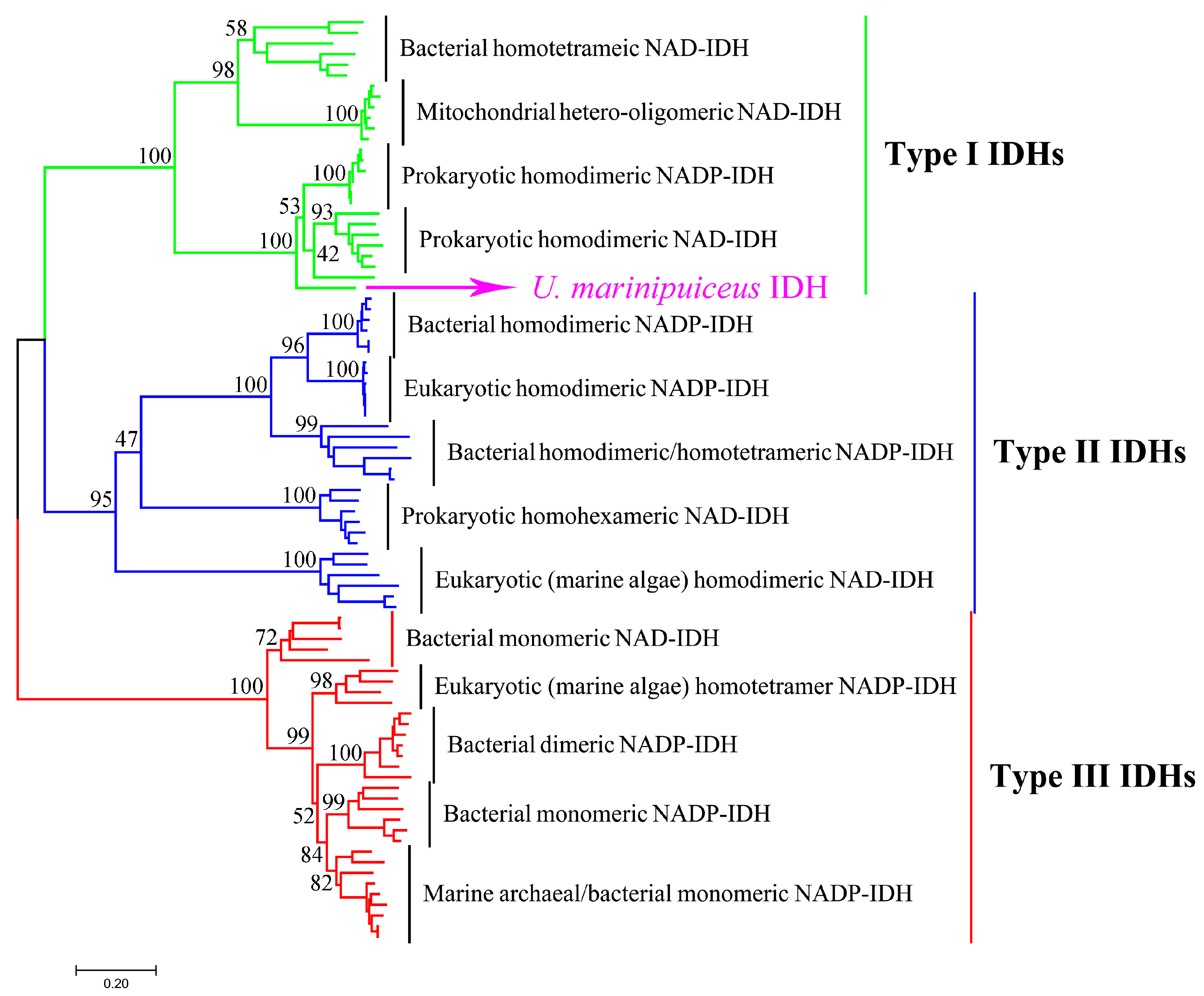
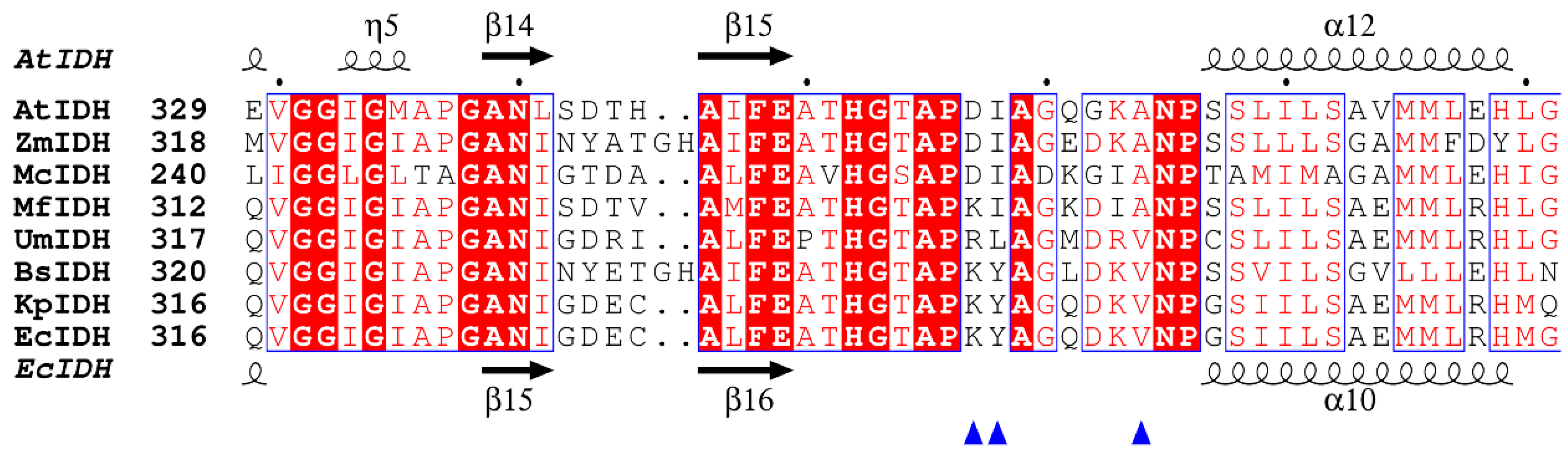
2.1. Bioinformatics Analysis

The UmIDH gene has a total length of 1272 base pairs (bp) and encodes a protein consisting of 420 amino acids, which conforms to the characteristics of Type I IDH. The sequence identities of UmIDH with typical Type I subfamily members NADP+-IDH from E. coli (EcIDH), NAD+-IDH from A. thiooxidans (AtIDH), and dual coenzyme-dependent MfIDH are 63.6%, 54.6%, and 61.0%, respectively. In this study, we performed a detailed phylogenetic analysis to reconstruct the evolutionary tree of the IDH family (Figure 1). It is evident that UmIDH belongs to the Type I subfamily, and shows a closer phylogenetic relationship to the prokaryotic homodimeric NAD(P)-IDH, while distinguishing itself from them. This implies that UmIDH is not a typical NAD-IDH or NADP-IDH.
A secondary structure-based amino acid sequence alignment was performed to speculate the residues of Type I homodimeric IDHs involved in coenzyme binding (Figure 2), the result indicated that the putative critical coenzyme binding sites of UmIDH differ significantly from its homologue. Among the Type I homodimeric IDHs analyzed, it has been consistently observed that NAD-IDHs utilize Asp, Ile, and Ala residues for binding to NAD+ and NADP-IDHs utilize Lys, Tyr, and Val residues for binding to NADP+. Dual coenzyme-dependent MfIDH’s corresponding homologous sites are Lys, Ile, and Ala, and binding sites for UmIDH are Arg, Leu, and Val.

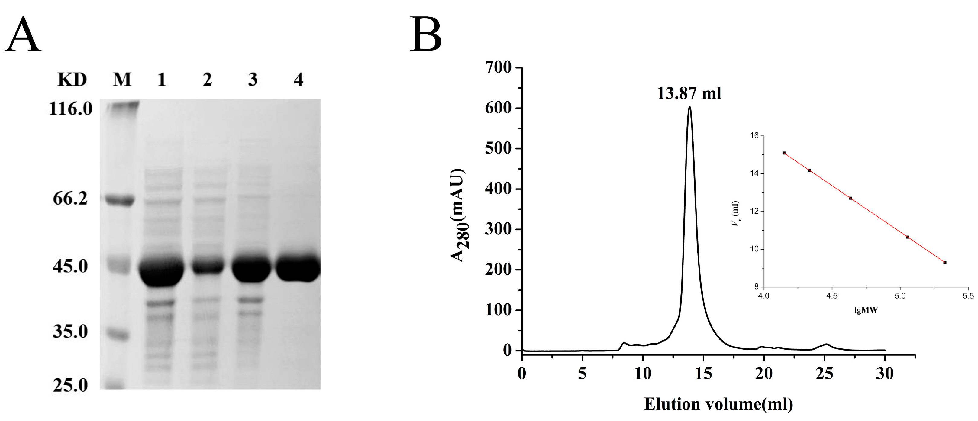
2.2. Expression and Purification

The recombinant UmIDH with a 6 × His-tag was effectively expressed in E. coli Rosetta (DE3) and subsequently purified using Co2+ affinity chromatography. SDS-PAGE analysis revealed that the molecular mass of UmIDH was approximately 45 kDa (Figure 3A), which closely matched the theoretical value of 46.7 kDa. Gel filtration chromatography analysis was used to determine the oligomeric status of UmIDH in solution, and a single symmetric elution peak with a molecular mass of 84.7 kDa was observed, indicating that the UmIDH exists as a homodimer in solution (Figure 3B).

2.3. The Effects of pH, Temperature, and Metal Ions

The effects of pH and temperature on the activity of UmIDH were evaluated using NADP+ as a coenzyme. The optimum pH of UmIDH was approximately 8.5 with Mn2+ and 8.7 with Mg2+ (Figure 4A). This is similar to the optimum pH of NAD-ZmIDH (pH 8.0 with Mn2+ and pH 8.5 with Mg2+) [12], lower than that of NAD-H. thermophilus IDH (pH 10.5 with Mg2+) [20], but higher than that of NADP-MfIDH (pH 6.0 with Mn2+) [17]. Recombinant UmIDH exhibited excellent stability in alkaline conditions, maintaining over 60% of its maximum activity across a broad pH range of 7.5 to 9.0, and the alkaline nature of UmIDH may be related to the marine living environment of U. marinipuiceus.
The influence of temperature on enzyme activity is manifested in two characteristics: firstly, enzymes have an optimal temperature, and secondly, enzymes have a range of temperature tolerance. UmIDH exhibited maximal activity at approximately 35 °C in the presence of Mn2+ or Mg2+ (Figure 4B), similar to that of marine bacterial IDHs from Congregibacter litoralis (35 °C with Mn2+ or Mg2+) [21] and Psychromonas marina (35 °C with Mn2+) [22], but lower than that of ZmIDH (55 °C with Mn2+ or Mg2+) [12] and SsIDH (50 °C with Mg2+) [11]. The heat-inactivation profiles suggested that UmIDH can maintain over 60% of its activity before 50 °C (Figure 4C), while most marine bacterial IDHs have already completely lost their activity at this temperature after a 20 min incubation [21,22,23].
The impact of nine different metal ions on the activity of UmIDH were assessed in the NADP+-linked reaction (Table 1). The results indicated that UmIDH, like other previously studied IDHs, relied entirely on the presence of a divalent cation. Among the activators tested for UmIDH catalysis, Mn2+ emerged as the most effective, while Mg2+ was almost as effective, capable of partially substituting for Mn2+ activation (19.51%). In the presence of Mn2+ or Mg2+, three divalent metal cations (Cu2+, Co2+, and Ca2+) exhibited inhibitory effects on the activity of UmIDH.

2.4. Kinetics Analysis

The recombinant UmIDH demonstrated a highly similar affinity for and catalytic efficiency with either coenzyme, indicating UmIDH is a dual coenzyme-dependent IDH, although it slightly prefers NADP+. The Km values of UmIDH for NAD+ and NADP+ were 1800.0 ± 64.4 μM and 1167.7 ± 113.0 μM, respectively, while the catalytic efficiency (kcat/Km) was only 2.3-times higher for NADP+ than for NAD+ (Table 2). UmIDH exhibits lower enzymatic activity compared to other type I homodimeric IDHs, for example, NAD-AtIDH (kcat/Km = 0.25 μM−1s−1) [24], NAD-S. mutans IDH (kcat/Km = 0.36 μM−1s−1) [25], NAD-P. furiosus IDH (kcat/Km = 0.88 μM−1s−1) [26], NADP-EcIDH (kcat/Km = 4.7 μM−1s−1) [27], and NADP-M. aeruginosa IDH (kcat/Km = 0.8 μM−1s−1) [28].
By aligning UmIDH with typical Type I homodimeric NAD-IDHs and NADP-IDHs, three residues (Arg345, Leu346, and Val352) were identified as putative coenzyme binding sites for UmIDH (Figure 2). In this study, two mutants (a double mutant R345K/L346Y and a triple mutant R345D/L346I/V352A) were constructed for bidirectional modification of the coenzyme specificity of UmIDH. Circular dichroism spectra (Figure 4D) revealed that the mutant enzymes retained their secondary structure after mutagenesis.
The kinetic parameters of the UmIDH mutants are presented in Table 2. The data suggested that, compared to the wild-type UmIDH, R345K/L346Y had a Km value that was approximately 2-times lower and a catalytic efficiency towards NADP+ that was 2.6-times higher. In contrast, R345D/L346I/V352A showed a decrease (approximately 4.9-fold) in the Km value and an increase (about 11.4-fold) in the catalytic efficiency towards NAD+, when compared to the wild-type UmIDH.

3. Discussion

The IDH family, as housekeeping proteins within organisms, has ancient origins, widespread distribution, and species-diversity, making it a valuable basis for studying protein molecular evolution. In this study, the Type I homodimeric UmIDH was characterized. From the phylogenetic tree (Figure 1), it can be observed that although UmIDH clusters with the prokaryotic homodimeric IDH, it is distinct from the NAD-IDH and NADP-IDH branches, indicating its dual coenzyme specificity. The determination of kinetic parameters confirmed that UmIDH is indeed a dual coenzyme-dependent IDH.
The concept of dual coenzyme-dependent IDH has been proposed in the past; however, these IDHs exhibit a significant difference in their preference for one of the two coenzymes [14,15,16]. Therefore, strictly speaking, these IDHs could not be referred to as dual coenzyme-dependent IDHs. Subsequent studies have discovered some IDHs that exhibit the ability to utilize both coenzymes; however, they similarly cannot be called dual coenzyme-dependent IDHs [7,29]. MfIDH is a recently reported dual coenzyme-dependent IDH that shows similar affinity for both NAD+ and NADP+. Compared to MfIDH, the UmIDH reported in this study better fits the characteristics of dual coenzyme dependency. This is evident from two findings: first, UmIDH exhibits more similar catalytic efficiency between the two coenzymes (the ratio of catalytic efficiency between the two coenzymes is 1:2 for UmIDH, while for MfIDH, this ratio is 1:5), and second, the conservation of the coenzyme binding sites in UmIDH is lower.
Type I IDH, Type II IDH, and Type III IDH have a monophyletic origin and encompass both NAD-IDH and NADP-IDH [7,21]. The results of molecular phylogenetic studies indicate that approximately 3.5 billion years ago, bacterial IDH solely relied on NAD+ as its coenzyme. Subsequently, certain bacteria encountered environmental stress due to carbon source limitations, leading to a coenzyme specificity shift in IDH from NAD+ to NADP+, and this transition provided the bacteria with the necessary reducing power, NADPH. The coenzyme adaptive evolution mechanism of IDH has been reproduced and validated in E. coli [10,13,30,31], and numerous experiments have demonstrated its applicability across different subfamilies [21,32,33,34,35]. The majority of the existing NAD-IDHs are found in the Type I subfamily, while a small number are also present in the Type II and Type III subfamilies. For example, marine bacterial homohexameric NAD-IDH and eukaryotic homodimeric NAD-IDH are considered the ancestors of Type II IDHs [21,29,32], while Campylobacter NAD-IDHs are considered the ancestors of Type III IDHs [7,29].
Each subfamily of IDH has two sets of amino acid residues for binding different coenzymes, and these residues are highly conserved across IDHs of different subfamilies. In Type I subfamily, different oligomeric forms of IDHs utilize Asp, Ile, and Ala in the coenzyme binding pocket to bind NAD+, while Lys, Tyr, and Val are utilized for NADP+ binding. In UmIDH, the corresponding homologous site is replaced with Arg, Leu, and Val, which differs from the typical coenzyme binding sites of Type I IDHs and results in the dual coenzyme specificity of UmIDH. We speculate that the coenzyme determinant Asp, Ile, and Ala represents the ancestral phenotype, Lys, Tyr, and Val represents the evolved phenotype, and Arg, Leu, and Val represents an intermediate state between the two. It can be anticipated that the intermediate state phenotype cannot be well stabilized due to its lower catalytic activity; therefore, UmIDH is a survivor.
Early studies have shown that EcIDH is regulated by phosphorylation through the IDH kinase/phosphatase (IDH K/P) system, resulting in a loss of 75–80% of its activity in carbon-limited conditions. This allows the glyoxylate shunt, facilitated by the enzyme isocitrate lyase (ICL), to competitively bind and utilize more substrates [36,37]. In the E. coli genome, the gene aceK, which encodes EcIDH K/P, is part of the aceBAK operon along with the genes aceA and aceB, which encode ICL and malate synthase (MS), respectively [38,39]. Therefore, the expression of IDH K/P is coupled with the expression of ICL and MS, both of which are key enzymes in the glyoxylate bypass pathway. Through bioinformatic analysis, we discovered that the M. flagellatus genome lacks the aceBAK operon, while the U. marinipuiceus genome possesses a complete aceBAK operon, which is consistent with E. coli, this indicates that UmIDH has a physiological basis for phosphorylation, and the potential phosphorylation site is Ser114 (Figure S1). We also analyzed the potential acetylation and succinylation sites of UmIDH and found that it has fewer relevant sites compared to EcIDH and MfIDH (Figure S1) [17,40,41].
Bioinformatics analysis methods and protein engineering approaches play a crucial role in the field of molecular evolution. The former involves comparing enzyme sequences with sequence information in databases, enabling the prediction of enzyme structure and function. Through sequence conservation analysis, specific sequence patterns related to substrate specificity can be identified. The latter allows for the alteration of substrate specificity through methods such as site-directed mutagenesis, insertion, deletion, or recombination. Additionally, protein engineering methods utilize directed evolution, enzyme library screening, and computational simulations to design and optimize enzyme catalytic performance and substrate specificity [42,43]. One method to validate an evolutionary pathway is reverse engineering. In this study, two mutants were constructed, and only two to three sites mutations are required to achieve bidirectional modification of the coenzyme specificity of UmIDH, which indicates that our speculation is reasonable. This current work provide compelling evidence for the evolutionary mechanism of coenzyme specificity of IDH.

4. Materials and Methods

4.1. Strains, Plasmids and Reagents

E. coli DH5α and E. coli Rosetta (DE3) were stored at an ultra-low temperature freezer for preservation. Full-length gene encoding UmIDH was synthesized by Generay Biotech Co., Ltd. (Shanghai, China), and the coding sequence was codon optimized according to the Escherichia coli bias. The plasmid used was pET28b, and the restriction enzyme sites at both ends of the gene were NdeI and XhoI. PrimeSTAR Max DNA Polymerase was purchased from TaKaRa (Dalin, China). Restriction enzymes (NdeI, XhoI, and DpnI), T4 DNA ligase, and Protein Molecular Weight Standards were purchased from Thermo Fisher Scientific (Shanghai, China). The TALON Metal Affinity Resin used for protein purification was obtained from TaKaRa. All other biochemical reagents were sourced from Sangon Biotech and BBI (Shanghai, China).

4.2. Site-Directed Mutagenesis

UmIDH mutants (R345K/L346Y and R345D/L346I/V352A) were constructed through site-directed mutagenesis by using UmIDH-pET28b as the template. The primers that were used to generate the R345K/L346Y mutant were as follows: forward, 5′-CATGGTACTGCCCCTAAATATGCAGGTATGGATC-3′, and reverse, 5′-ATATTTAGGGGCAGTACCATGGGTCGGTTCA-3′. The R345D/L346I/V352A was constructed by two rounds of site-directed mutagenesis. The first round of mutation introduced the R345D/L346I change, using the following primers: forward, 5′-CATGGTACTGCCCCTGATATTGCAGGTATGGAT-3′, and reverse, 5′-AATATCAGGGGCAGTACCATGGGTCGGTTC-3′. The second round of mutation introduced the V352A change, using the following primers: forward, 5′-GCAGGTATGGATCGTGCAAATCCGTGTAGC-3′, and reverse, 5′-TGCACGATCCATACCTGCAATATCAGGGG-3′. The underlined codons are mutated sequences. After PCR, the template sequences were eliminated using the methylation-specific DpnI enzyme. Then, the products were transformed into competent bacterial strains.

4.3. Recombinant Protein Expression and Purification

Escherichia coli Rosetta (DE3) strains harboring the recombinant plasmid were pre-cultured overnight in Luria-Bertani (LB) medium supplemented with 30 μg/mL kanamycin and 30 μg/mL chloramphenicol at 37 °C with agitation at 225 rpm. Subsequently, the cells were inoculated at a 1:100 dilution into 200 mL fresh LB medium containing the same antibiotics and incubated at 37 °C and 225 rpm. The expression of the recombinant protein was induced by adding 0.5 mM IPTG when the optical density of the cultures reached an OD600 of 0.4–0.6. The cultivation was continued for 20 h at 20 °C with agitation at 180 rpm. The cells were harvested by centrifugation at 5000 rpm for 5 min at 4 °C. The resulting pellets were then resuspended in lysis buffer containing 20 mM Tris-HCl, 300 mM NaCl (pH 7.5). After sonication, the cell debris was eliminated by centrifugation at 10,000 rpm for 20 min at 4 °C. The supernatant containing the 6×His-tagged recombinant UmIDH was purified Co2+ affinity resin (Cat. #635502, Clontech, TaKaRa, Dalin, China).

4.4. SDS-PAGE and Gel Filtration Chromatography

In order to determine the molecular mass of the subunit(s), the purified proteins were subjected to denaturing electrophoresis using a discontinuous SDS-PAGE system with a 5% stacking gel and a 12% separating gel. The molecular mass of the active UmIDH was estimated using gel filtration chromatography on the Superdex 200 10/300 increase column (GE Healthcare Life Sciences, Pittsburgh, PA, USA), which was equilibrated with a 50 mM potassium phosphate buffer (pH 7.0) containing 150 mM NaCl and 0.01% sodium azide. Protein standards such as Ovalbumin (44 kDa), Conalbumin (75 kDa), Aldolase (158 kDa), Ferritin (440 kDa), and Thyroglobulin (669 kDa) were used for calibration of the gels.

4.5. Circular Dichroism Spectroscopy

Circular dichroism (CD) spectra were acquired using a Jasco model J-810 spectropolarimeter. UmIDH and mutants concentrations were diluted to 0.2 mg/mL using a CD buffer containing 20 mM NaH2PO4 and 75 mM Na2SO4 at pH 7.5. Then, 200 μL of the protein was loaded into a microcuvette and transferred into the instrument at a wavelength range of 190 nm to 280 nm. The mean residue ellipticity ([θ], deg·cm2·dmole−1) of the protein was calculated using the following formula [θ] = θ/10·(n − 1)·C·l, where θ is the ellipticity measured in mdeg; n is the number of protein amino acid residues; C is the concentration of the protein in mol/L; and l is the light path of the cuvette (0.1 cm). Three scans were performed for each sample, and the average value was recorded. The protein secondary structure was estimated following the method described by Raussens et al. [44].

4.6. Enzyme Characterization

The pH-dependent activity of the recombinant UmIDH was investigated in 50 mM Tris-HCl buffer ranging from pH 7.7 to 9.5 at 25 °C. The temperature dependence of the recombinant UmIDH activity was assessed across the temperature range of 25–60 °C at pH 8.5. The thermostability of the recombinant UmIDH was evaluated by subjecting enzyme aliquots to heat inactivation at temperatures ranging from 25 °C to 65 °C at pH 8.5 for 20 min. Following incubation, the aliquots were rapidly cooled on ice, and the residual enzyme activity was determined using the standard enzyme assay. The effects of various metal ions (Mn2+, Mg2+, Ca2+, Co2+, Cu2+, Ni2+, K+, Na+, and Li+) on MmIDH were assessed using the standard assay method. All data for enzyme activity were tested in at least three independent experiments.

4.7. Enzyme Assay

Enzyme assays were performed in a 1 mL reaction volume consisting of 50 mM Tris-HCl (pH 8.5), 2 mM MnCl2 or MgCl2, 1 mM trisodium DL-isocitrate, and 0.5 mM NAD(P)+ at a temperature of 25 °C. The increase in NAD(P)H absorbance was monitored at 340 nm using a Cary 300 UV-Vis spectrophotometer (Varian, Polo Alto, CA, USA) with an extinction coefficient (ε340) of 6.22 mM−1·cm−1. One unit (U) of enzyme activity was defined as the generation of 1 μmol of NADPH per minute. Protein concentrations were determined using a Bio-Rad protein assay kit (Bio-Rad, Hercules, CA, USA). The enzyme’s kinetic parameters were determined by measuring its activity at various concentrations of NADP+ or NAD+. The apparent kinetic parameters were calculated using nonlinear regression analysis with Prism 5.0 software (GraphPad Software, San Diego, CA, USA). All experiments were conducted at least three times to ensure reproducibility.

Supplementary Materials

The following supporting information can be downloaded at: https://www.mdpi.com/article/10.3390/ijms241411428/s1.

Author Contributions

G.Z. and P.W. conceived and designed the experiments; M.B. and J.Z. conducted the bioinformatics analysis; M.B. and W.X. performed experiments related to wild-type UmIDH; J.Z. and X.H. performed experiments related to mutant UmIDH; M.B. and X.C. analyzed the data; M.B. wrote the draft; G.Z. and P.W. reviewed and edited the paper. All authors have read and agreed to the published version of the manuscript.

Funding

This work was supported by the National Natural Science Foundation of China (32071270), Outstanding Innovative Research Team for Molecular Enzymology and Detection in Anhui Provincial Universities (2022AH010012), the Major Science and Technology Projects in Anhui Province (202003a06020009), Anhui Natural Science Foundation (2208085MC48), and Auhui Provincial Engineering Research Centre for Molecular Detection and Diagnostics.

Institutional Review Board Statement

This study did not involve humans or animals.

Informed Consent Statement

This study did not involve humans.

Data Availability Statement

The original contributions presented in the study are included in the article and its Supplementary Material. Further inquiries can be directed to the corresponding authors.

Conflicts of Interest

The authors declare no conflict of interest.

References

  1. Chen, R.; Jeong, S.S. Functional prediction: Identification of protein orthologs and paralogs. Protein Sci. 2000, 9, 2344–2353. [Google Scholar] [CrossRef] [PubMed] [Green Version]
  2. Sun, P.; Liu, Y.; Ma, T.; Ding, J. Structure and allosteric regulation of human NAD+-dependent isocitrate dehydrogenase. Cell Discov. 2020, 6, 94. [Google Scholar] [CrossRef] [PubMed]
  3. Spaans, S.K.; Weusthuis, R.A.; van der Oost, J.; Kengen, S.W. NADPH-generating systems in bacteria and archaea. Front. Microbiol. 2015, 6, 742. [Google Scholar] [CrossRef] [PubMed]
  4. Parsons, D.W.; Jones, S.; Zhang, X.; Lin, J.C.-H.; Leary, R.J.; Angenendt, P.; Mankoo, P.; Carter, H.; Siu, I.M.; Gallia, G.L.; et al. An integrated genomic analysis of human glioblastoma multiforme. Science 2008, 321, 1807–1812. [Google Scholar] [CrossRef] [Green Version]
  5. Dang, L.; Yen, K.; Attar, E.C. IDH mutations in cancer and progress toward development of targeted therapeutics. Ann. Oncol. 2016, 27, 599–608. [Google Scholar] [CrossRef] [Green Version]
  6. Eniafe, J.; Jiang, S. The functional roles of TCA cycle metabolites in cancer. Oncogene 2021, 40, 3351–3363. [Google Scholar] [CrossRef]
  7. Wang, P.; Lv, C.; Zhu, G. Novel type II and monomeric NAD+-specific isocitrate dehydrogenases: Phylogenetic affinity, enzymatic characterization, and evolutionary implication. Sci. Rep. 2015, 5, 9150. [Google Scholar] [CrossRef] [Green Version]
  8. Wang, P.; Wu, Y.; Liu, J.; Song, P.; Li, S.; Zhou, X.; Zhu, G. Crystal structure of the isocitrate dehydrogenase 2 from Acinetobacter baumannii (AbIDH2) reveals a novel dimeric structure with two monomeric IDH-like subunits. Int. J. Mol. Sci. 2018, 19, 1131. [Google Scholar] [CrossRef] [Green Version]
  9. Huang, S.; Zhao, J.; Li, W.; Wang, P.; Xue, Z.; Zhu, G. Biochemical and Phylogenetic Characterization of a Novel NADP+-Specific Isocitrate Dehydrogenase from the Marine Microalga Iphaeodactylum trcornutum. Front. Mol. Biosci. 2021, 8, 702083. [Google Scholar] [CrossRef]
  10. Zhu, G.; Golding, G.B.; Dean, A.M. The selective cause of an ancient adaptation. Science 2005, 307, 1279–1282. [Google Scholar] [CrossRef]
  11. Wang, P.; Jin, M.; Su, R.; Song, P.; Wang, M.; Zhu, G. Enzymatic characterization of isocitrate dehydrogenase from an emerging zoonotic pathogen Streptococcus suis. Biochimie 2011, 93, 1470–1475. [Google Scholar] [CrossRef]
  12. Wang, P.; Jin, M.; Zhu, G. Biochemical and molecular characterization of NAD+-dependent isocitrate dehydrogenase from the ethanologenic bacterium Zymomonas mobilis. FEMS Microbiol. Lett. 2012, 327, 134–141. [Google Scholar] [CrossRef] [Green Version]
  13. Dean, A.M.; Golding, G.B. Protein engineering reveals ancient adaptive replacements in isocitrate dehydrogenase. Proc. Natl. Acad. Sci. USA 1997, 94, 3104–3109. [Google Scholar] [CrossRef] [PubMed]
  14. Leyland, M.L.; Kelly, D.J. Purification and characterization of a monomeric isocitrate dehydrogenase with dual coenzyme specificity from the photosynthetic bacterium Rhodomicrobium vannielii. Eur. J. Biochem. 1991, 202, 85–93. [Google Scholar] [CrossRef] [PubMed]
  15. Potter, S. Evidence for a dual-specificity isocitrate dehydrogenase in the euryarchaeotan Thermoplasma acidophilum. Can. J. Microbiol. 1993, 39, 262–264. [Google Scholar] [CrossRef]
  16. Kim, H.; Mozaffar, Z.; Weete, J.D. A dual cofactor-specific isocitrate dehydrogenase from Pythium ultimum. Can J. Microbiol. 1996, 42, 1241–1247. [Google Scholar] [CrossRef]
  17. Romkina, A.Y.; Kiriukhin, M.Y. Biochemical and molecular characterization of the isocitrate dehydrogenase with dual coenzyme specificity from the obligate methylotroph Methylobacillus Flagellatus. PLoS ONE 2017, 12, e0176056. [Google Scholar] [CrossRef] [Green Version]
  18. Gao, H.; Li, J.; Sivakumar, D.; Kim, T.S.; Patel, S.K.S.; Kalia, V.C.; Kim, I.W.; Zhang, Y.W.; Lee, J.K. NADH oxidase from Lactobacillus reuteri: A versatile enzyme for oxidized cofactor regeneration. Int. J. Biol. Macromol. 2019, 123, 629–636. [Google Scholar] [CrossRef]
  19. Romanenko, L.A.; Tanaka, N.; Frolova, G.M. Umboniibacter marinipuniceus gen. nov., sp. nov., a marine gammaproteobacterium isolated from the mollusc Umbonium costatum from the Sea of Japan. Int. J. Syst. Evol. Microbiol. 2010, 60, 603–609. [Google Scholar] [CrossRef]
  20. Aoshima, M.; Ishii, M.; Igarashi, Y. A novel biotin protein required for reductive carboxylation of 2-oxoglutarate by isocitrate dehydrogenase in Hydrogenobacter thermophilus TK-6. Mol. Microbiol. 2004, 51, 791–798. [Google Scholar] [CrossRef]
  21. Wu, M.C.; Tian, C.Q.; Cheng, H.M.; Xu, L.; Wang, P.; Zhu, G.P. A novel type II NAD+-specific isocitrate dehydrogenase from the marine bacterium Congregibacter litoralis KT71. PLoS ONE 2015, 10, e0125229. [Google Scholar] [CrossRef]
  22. Hirota, R.; Tsubouchi, K.; Takada, Y. NADP+-dependent isocitrate dehydrogenase from a psychrophilic bacterium, Psychromonas marina. Extremophiles 2017, 21, 711–721. [Google Scholar] [CrossRef] [Green Version]
  23. Yasuda, W.; Kobayashi, M.; Takada, Y. Analysis of amino acid residues involved in cold activity of monomeric isocitrate dehydrogenase from psychrophilic bacteria, Colwellia maris and Colwellia psychrerythraea. J. Biosci. Bioeng. 2013, 116, 567–572. [Google Scholar] [CrossRef] [Green Version]
  24. Inoue, H.; Tamura, T.; Ehara, N.; Nishito, A.; Nakayama, Y.; Maekawa, M.; Imada, K.; Tanaka, H.; Inagaki, K. Biochemical and molecular characterization of the NAD(+)-dependent isocitrate dehydrogenase from the chemolithotroph Acidithiobacillus thiooxidans. FEMS Microbiol. Lett. 2002, 214, 127–132. [Google Scholar] [CrossRef] [PubMed]
  25. Wang, P.; Song, P.; Jin, M.; Zhu, G. Isocitrate dehydrogenase from Streptococcus mutans: Biochemical properties and evaluation of a putative phosphorylation site at Ser102. PLoS ONE 2013, 8, e58918. [Google Scholar] [CrossRef] [PubMed] [Green Version]
  26. Steen, I.H.; Madern, D.; Karlstrom, M.; Lien, T.; Ladenstein, R.; Birkeland, N.K. Comparison of isocitrate dehydrogenase from three hyperthermophiles reveals differences in thermostability, cofactor specificity, oligomeric state, and phylogenetic affiliation. J. Biol. Chem. 2001, 276, 43924–43931. [Google Scholar] [CrossRef] [PubMed] [Green Version]
  27. Chen, R.; Greer, A.; Dean, A.M. A highly active decarboxylating dehydrogenase with rationally inverted coenzyme specificity. Proc Natl Acad Sci. USA 1995, 92, 11666–11670. [Google Scholar] [CrossRef]
  28. Jin, M.M.; Wang, P.; Li, X.; Zhao, X.Y.; Xu, L.; Song, P.; Zhu, G.P. Biochemical characterization of NADP(+)-dependent isocitrate dehydrogenase from Microcystis aeruginosa PCC7806. Mol. Biol. Rep. 2013, 40, 2995–3002. [Google Scholar] [CrossRef]
  29. Huang, S.P.; Zhou, L.C.; Wen, B.; Wang, P.; Zhu, G.P. Biochemical Characterization and Crystal Structure of a Novel NAD+-Dependent Isocitrate Dehydrogenase from Phaeodactylum tricornutum. Int. J. Mol. Sci. 2020, 21, 5915. [Google Scholar] [CrossRef]
  30. Ellington, A.D.; Bull, J.J. Evolution. Changing the cofactor diet of an enzyme. Science 2005, 310, 454–455. [Google Scholar] [CrossRef]
  31. Golding, G.B.; Dean, A.M. The structural basis of molecular adaptation. Mol. Biol. Evol. 1998, 15, 355–369. [Google Scholar] [CrossRef] [Green Version]
  32. Tang, W.G.; Song, P.; Cao, Z.Y.; Wang, P.; Zhu, G.P. A unique homodimeric NAD⁺-linked isocitrate dehydrogenase from the smallest autotrophic eukaryote Ostreococcus tauri. FASEB J. 2015, 29, 2462–2472. [Google Scholar] [CrossRef] [Green Version]
  33. Lv, C.; Wang, P.; Wang, W.; Su, R.; Ge, Y.; Zhu, Y.; Zhu, G. Two isocitrate dehydrogenases from a plant pathogen Xanthomonas campestris pv. campestris 8004. Bioinformatic analysis, enzymatic characterization, and implication in virulence. J. Basic Microbiol. 2016, 56, 975–985. [Google Scholar] [CrossRef]
  34. Huang, S.P.; Cheng, H.M.; Wang, P.; Zhu, G.P. Biochemical Characterization and Complete Conversion of Coenzyme Specificity of Isocitrate Dehydrogenase from Bifidobacterium longum. Int. J. Mol. Sci. 2016, 17, 296. [Google Scholar] [CrossRef] [PubMed] [Green Version]
  35. Wang, P.; Wang, Y.; Guo, X.; Huang, S.; Zhu, G. Biochemical and phylogenetic characterization of a monomeric isocitrate dehydrogenase from a marine methanogenic archaeon Methanococcoides methylutens. Extremophiles 2020, 24, 319–328. [Google Scholar] [CrossRef] [PubMed]
  36. Edlin, J.D.; Sundaram, T.K. Regulation of isocitrate dehydrogenase by phosphorylation in Escherichia coli K-12 and a simple method for determining the amount of inactive phosphoenzyme. J Bacteriol. 1989, 171, 2634–2638. [Google Scholar] [CrossRef] [PubMed] [Green Version]
  37. Hurley, J.H.; Thorsness, P.E.; Ramalingam, V.; Helmers, N.H.; Koshland, D.E.; Stroud, R.M. Structure of a bacterial enzyme regulated by phosphorylation, isocitrate dehydrogenase. Proc. Natl. Acad. Sci. USA 1989, 86, 8635–8639. [Google Scholar] [CrossRef]
  38. Cortay, J.C.; Bleicher, F.; Rieul, C.; Reeves, H.C.; Cozzone, A.J. Nucleotide sequence and expression of the aceK gene coding for isocitrate dehydrogenase kinase/phosphatase in Escherichia coli. J. Bacteriol. 1988, 170, 89–97. [Google Scholar] [CrossRef] [Green Version]
  39. Cozzone, A.J.; El-Mansi, M. Control of isocitrate dehydrogenase catalytic activity by protein phosphorylation in Escherichia coli. J. Mol. Microbiol. Biotechnol. 2005, 9, 132–146. [Google Scholar] [CrossRef]
  40. Yu, B.J.; Kim, J.A.; Moon, J.H.; Ryu, S.E.; Pan, J.G. The diversity of lysine-acetylated proteins in Escherichia coli. J. Microbiol. Biotechnol. 2008, 18, 1529–1536. [Google Scholar]
  41. Zhang, Z.; Tan, M.; Xie, Z.; Dai, L.; Chen, Y.; Zhao, Y. Identification of lysine succinylation as a new post-translational modification. Nat. Chem. Biol. 2011, 7, 58–63. [Google Scholar] [CrossRef] [PubMed]
  42. Packer, M.S.; Liu, D.R. Methods for the directed evolution of proteins. Nat. Rev. Genet. 2015, 16, 379–394. [Google Scholar] [CrossRef] [PubMed]
  43. Kim, J.S.; Patel, S.K.S.; Tiwari, M.K.; Lai, C.; Kumar, A.; Kim, Y.S.; Kalia, V.C.; Lee, J.K. Phe-140 Determines the catalytic efficiency of arylacetonitrilase from Alcaligenes faecalis. Int. J. Mol. Sci. 2020, 21, 7859. [Google Scholar] [CrossRef] [PubMed]
  44. Raussens, V.; Ruysschaert, J.M.; Goormaghtigh, E. Protein concentration is not an absolute prerequisite for the determination of secondary structure from circular dichroism spectra: A new scaling method. Anal. Biochem. 2003, 319, 114–121. [Google Scholar] [CrossRef]
Figure 1. The phylogenetic tree of IDH family. The evolutionary tree was constructed by MEGA 7.0 using the neighbor-joining method with 1000 bootstrap replicates, which involved 87 IDH sequences. The UmIDH was targeted by pink arrow.
Ijms 24 11428 g001
Figure 2. Structure-based amino acid sequence alignment. The comparison of UmIDH with three typical Type I homodimeric NAD-IDHs from Acidithiobacillus thiooxidans (AtIDH, NCBI Reference Sequence: WP_193650900.1), Zymomonas mobilis (ZmIDH, NCBI Reference Sequence: WP_014500849.1) and Methylococcus capsulatus (McIDH, NCBI Reference Sequence: WP_010962255.1), and three typical Type I homodimeric NADP-IDHs from Escherichia coli (EcIDH, NCBI Reference Sequence: WP_096963047.1), Bacillus subtilis (BsIDH, NCBI Reference Sequence: WP_003152454.1), and Klebsiella pneumoniae (KpIDH, NCBI Reference Sequence: WP_129543013.1), and dual coenzyme-dependent MfIDH (NCBI Reference Sequence: WP_011480360.1). The structure of AtIDH (PDB ID: 2D4V) and EcIDH (PDB ID: 3ICD) were downloaded from the PDB database. The critical coenzyme binding sites were indicated by blue triangles. The figure was created by ESPript 3.0.
Ijms 24 11428 g002
Figure 3. Overexpression and purification of UmIDH. (A) SDS-PAGE analysis. M, molecular mass marker; lane 1, crude extract; lane 2, supernatant of crude extract; lane 3, precipitation of crude extract; lane 4, purified UmIDH. (B) Gel filtration chromatography analysis. Ve of the UmIDH was 13.87 mL.
Ijms 24 11428 g003
Figure 4. (A) Effects of pH on the activity of UmIDH in the presence of Mn2+ or Mg2+. (B) Effects of temperature on the activity of UmIDH in the presence of Mn2+ or Mg2+. (C) Heat-inactivation profiles of UmIDH in the presence of Mn2+ or Mg2+. (D) CD spectra of the wild-type UmIDH and its mutants.
Ijms 24 11428 g004
Table 1. Effect of different metal ions on the activity of UmIDH.
Metal IonsRelative
Activity (%)
Metal IonsRelative
Activity (%)
Metal IonsRelative
Activity (%)
None0
Mn2+100 ± 0Mn2+100 ± 0
Mg2+19.51 ± 4.66Mn2+ + Mg2+114.97 ± 5.13Mg2+100 ± 0
Ca2+15.90 ± 1.32Mn2+ + Ca2+67.10 ± 4.27Mg2+ + Ca2+20.75 ± 1.34
Co2+0.66 ± 1.29Mn2+ + Co2+20.04 ± 2.80Mg2+ + Co2+7.68 ± 0.74
Cu2+0.31 ± 0.27Mn2+ + Cu2+4.04 ± 0.33Mg2+ + Cu2+6.97 ± 0.61
Ni2+2.29 ± 0.64Mn2+ + Ni2+103.51 ± 3.42Mg2+ + Ni2+102.07 ± 0.59
K+8.30 ± 0.43Mn2+ + K+106.13 ± 4.37Mg2+ + K+110.91 ± 1.10
Na+2.31 ± 0.75Mn2+ + Na+104.88 ± 3.92Mg2+ + Na+101.03 ± 2.11
Li+7.69 ± 1.07Mn2+ + Li+103.53 ± 0.98Mg2+ + Li+107.04 ± 1.20
The relative activity was assessed in a standard reaction mixture containing 2 mM of the indicated metal ion(s). Data are the mean ± SD of at least three independent measurements.
Table 2. The kinetic parameters of the wild-type UmIDH and its mutants.
Enzyme NAD+ NADP+
Km
(μM)
kcat
(s−1)
kcat/Km
(μM−1s−1)
Km
(μM)
kcat
(s−1)
kcat/Km
(μM−1s−1)
UmIDH1800.0 ± 64.47.5 ± 0.30.00421167.7 ± 113.011.1 ± 0.40.0095
R345K/L346Y---605.9 ± 38.215.3 ± 0.20.025
R345D/L346I/V352A369.7 ± 11.8417.7 ± 0.90.048---
“-” not detected. The values indicate the means of at least three independent measurements.
Disclaimer/Publisher’s Note: The statements, opinions and data contained in all publications are solely those of the individual author(s) and contributor(s) and not of MDPI and/or the editor(s). MDPI and/or the editor(s) disclaim responsibility for any injury to people or property resulting from any ideas, methods, instructions or products referred to in the content.

