Review

Unlocking Neural Function with 3D In Vitro Models: A Technical Review of Self-Assembled, Guided, and Bioprinted Brain Organoids and Their Applications in the Study of Neurodevelopmental and Neurodegenerative Disorders

by
Chiara D’Antoni
1,2,†,
Lorenza Mautone
1,2,†,
Caterina Sanchini
2,
Lucrezia Tondo
1,2,
Greta Grassmann
2,3,
Gianluca Cidonio
2,
Paola Bezzi
1,4,
Federica Cordella
1,2,* and
Silvia Di Angelantonio
1,2,5,*
1
Department of Physiology and Pharmacology, Sapienza University of Rome, 00185 Rome, Italy
2
Center for Life Nano- and Neuro-Science of Istituto Italiano di Tecnologia (IIT), 00161 Rome, Italy
3
Department of Biochemical Sciences “Alessandro Rossi Fanelli”, Sapienza University of Rome, 00185 Rome, Italy
4
Department of Fundamental Neurosciences, University of Lausanne, 1011 Lausanne, Switzerland
5
D-Tails s.r.l., 00165 Rome, Italy
*
Authors to whom correspondence should be addressed.
These authors contributed equally to this work.
Int. J. Mol. Sci. 2023, 24(13), 10762; https://doi.org/10.3390/ijms241310762
Submission received: 29 May 2023 / Revised: 18 June 2023 / Accepted: 21 June 2023 / Published: 28 June 2023
(This article belongs to the Special Issue Recent Advance in 3D Cultures)

Abstract

:
Understanding the complexities of the human brain and its associated disorders poses a significant challenge in neuroscience. Traditional research methods have limitations in replicating its intricacies, necessitating the development of in vitro models that can simulate its structure and function. Three-dimensional in vitro models, including organoids, cerebral organoids, bioprinted brain models, and functionalized brain organoids, offer promising platforms for studying human brain development, physiology, and disease. These models accurately replicate key aspects of human brain anatomy, gene expression, and cellular behavior, enabling drug discovery and toxicology studies while providing insights into human-specific phenomena not easily studied in animal models. The use of human-induced pluripotent stem cells has revolutionized the generation of 3D brain structures, with various techniques developed to generate specific brain regions. These advancements facilitate the study of brain structure development and function, overcoming previous limitations due to the scarcity of human brain samples. This technical review provides an overview of current 3D in vitro models of the human cortex, their development, characterization, and limitations, and explores the state of the art and future directions in the field, with a specific focus on their applications in studying neurodevelopmental and neurodegenerative disorders.

1. Introduction

The human brain is one of the most complex organs in the body and is the center of the central nervous system. Understanding its function and disease processes, especially neurodevelopmental and neurodegenerative disorders, is a major challenge in modern neuroscience. While traditional methods such as animal models and post-mortem studies have contributed greatly to our knowledge, they have limitations and may not fully reflect the human condition. As a result, there is a growing need for in vitro models that can simulate the complex structure and function of the human brain.
Three-dimensional (3D) in vitro models of the human brain, such as organoids, cerebral organoids, three-dimensional bioprinted brain models, and functionalized brain organoids, have the potential to provide a platform for the study of human brain development, physiology, and disease. These models can recapitulate various aspects of human brain anatomy, gene expression, and cellular behavior, making them useful tools for drug discovery and toxicology studies [1]. Moreover, 3D in vitro models allow for the exploration of human-specific phenomena that cannot be studied in animal models. The use of 3D in vitro models for the study of neurodevelopmental and neurodegenerative disorders has gained significant attention in recent years thanks to the availability of different types of stem cells.
Human pluripotent stem cells (hPSCs), which include embryonic stem cells (ESCs) and induced pluripotent stem cells (iPSCs), are known for their remarkable self-organization and ability to form neural cells as a default pathway when spontaneously aggregated into 3D spheres [2]. Yoshiki Sasai’s lab made a breakthrough by demonstrating how ESCs cultured in 3D aggregates could spontaneously generate highly organized brain structures [3,4]. Furthermore, by utilizing the extracellular matrix support of Matrigel, Lancaster and colleagues created a more complex 3D tissue from hPSCs, called a cerebral organoid, that closely resembles a growing human brain [5]. These developments in stem cell research and our understanding of developmental biology have led to the creation of various techniques for generating brain regions over the past decade. These techniques have been extensively reviewed [6,7,8,9,10,11] and are generally categorized into two groups based on patterning approaches. Unguided protocols rely on the intrinsic differentiation of PSCs without growth factor patterning, resulting in brain organoids that contain a diverse mixture of neurons, glia, photoreceptor cells, and even cells of non-ectodermal origin [5]. However, this method is hampered by variability in the emergence and size of different brain regions, making it difficult to study the development and function of specific brain structures. On the other hand, guided protocols use small molecules and patterning growth factors to direct cells towards a specific fate, resulting in the enrichment of a region of interest [4,12]. A wide range of region-specific brain organoids have been generated using guided protocols, including the cortex [12,13], cerebellum [14], hippocampus [15], pituitary gland [16], hypothalamus [17,18], spinal cord [19], thalamus [20,21], choroid plexus [22], striatum [23], and optic cup [24]. The choice of protocol for generating cerebral or region-specific brain organoids depends on the scientific questions being addressed. Nonetheless, the ability to generate different brain regions with distinct cellular identities from PSCs in vitro provides access to human-specific brain development and pathophysiology, which was previously limited by the scarcity of embryonic human brain samples.
In this technical review, we aim to provide an overview of current 3D in vitro models of the human cortex, including organoids, cerebral organoids, 3D bioprinted brain models, and functionalized brain organoids, and their applications in the study of neurodevelopmental and neurodegenerative disorders. The review will cover various aspects of these models, including their development, characterization, and limitations, and will discuss the current state of the art and future directions in this rapidly evolving field.

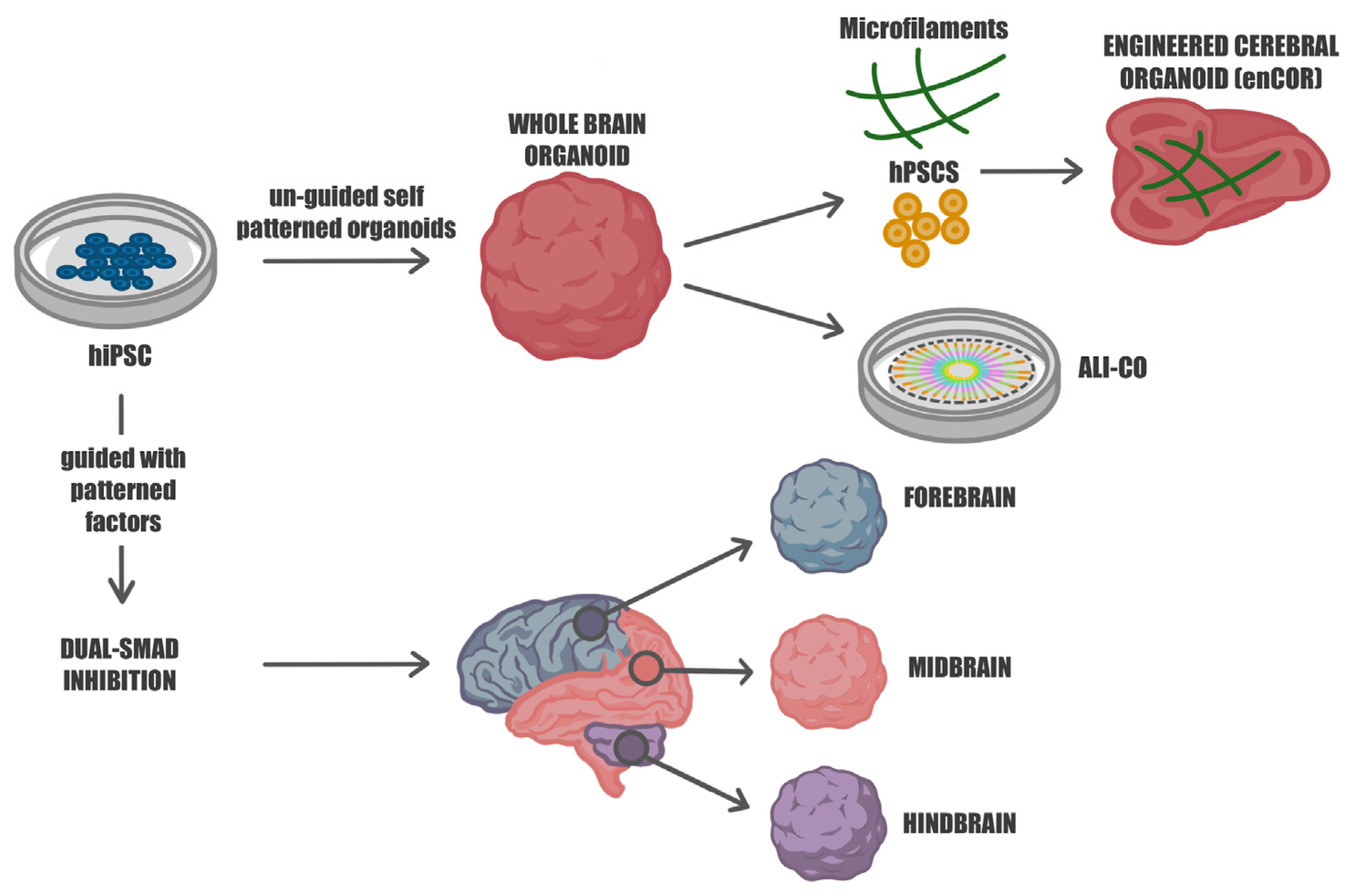
2. From 2D to 3D: The Rise of Cerebral Organoids for Studying Human Brain Development

The development and maturation of the human brain are complex processes that rely on the intricate interactions between neuronal and glial cells and the establishment of connections with other brain regions. Two-dimensional (2D) stem-cell-based models have contributed to our understanding of different molecular processes involved in human brain development, function, and neurodegeneration [25,26,27,28,29], but these models lack the complex cytoarchitecture of the cerebral cortex, limiting their ability to study the 3D interaction of brain cells during development. To address this limitation, human-derived brain organoids, which are self-assembled, organized structures composed of neuronal and glial cells, have emerged as valuable tools to study the developing brain under both physiological and pathological conditions, enabling the study of previously inaccessible aspects in a controlled laboratory setting (for a systematic review, see [30]).
Since the early 1990s, researchers have been actively pursuing methodologies to induce neural differentiation from pluripotent stem cells (PSCs) in three-dimensional (3D) environments [31]. However, it is important to note that not all 3D neural culture systems can be classified as brain organoids. Neurospheres, for instance, are 3D aggregations of various cell types derived from neural progenitor cells within the central nervous system (CNS) [31]. The neurosphere culture system has been extensively utilized for comprehensive investigations into the proliferation, self-renewal capabilities, and differentiation potential of neural stem cells [32,33]. Neurospheres represent an in vitro model system consisting of clusters of neural progenitor cells (NPCs), including neural stem cells [34]. These neurospheres are derived from isolated primary tissue or low-passaged NPCs obtained from induced pluripotent stem cells (iPSCs) and are cultured for limited periods before being utilized in various research applications. Neurospheres have been widely employed as the conventional method for NPC culture, with over 2000 publications since the original report by Reynolds [31]. They are particularly favored for obtaining NPCs from neurogenic areas in animal and human tissues [35]. Typically cultured in serum-free medium supplemented with fibroblast growth factor 2 (FGF-2) and epidermal growth factor (EGF), neurospheres do not require an adherent substrate that supports NPC expansion. This unique culture system provides an unparalleled platform to assess the stem-cell-like behavior of neurogenic tissue and enables investigations into the molecular and cellular characteristics of NPCs for in vivo transplantation in mice and humans [36,37,38]. However, despite their widespread use, maintaining neurosphere cultures can be challenging due to their rapid growth and substantial apoptosis. NPC cultures necessitate frequent dissociation and passaging every 7–10 days to control the neurosphere size and prevent excessive cell death. It is important to note that neurosphere cultures are most useful within the first 5–10 passages, as prolonged passaging can lead to aneuploidy and the selection of clones that may alter the intrinsic properties of NPCs [35]. Furthermore, neurospheres are limited in their ability to determine the self-renewal capacity of NPCs due to the fusion of neurospheres in vitro, making them non-clonal [34]. Criticism has been directed towards the use of neurospheres in vitro, suggesting that neurosphere formation may be an artifact of any cultured cell in the absence of a substrate and under the influence of selected growth factors, thus lacking intrinsic biological significance.
However, the emergence of cerebral organoid technology as a model system for human brain development provides new evidence to support the contention that neurosphere 3D aggregates may indeed convey biological relevance (see Table 1). Overall, while neurospheres have been widely utilized and serve as a valuable tool for NPC culture and exploration, their maintenance can be challenging, and there are limitations regarding their clonality and biological significance. The advent of cerebral organoids offers an exciting avenue for further understanding the biological implications of neurosphere 3D aggregates.
Recent advances have led to the development of 3D in vitro models that recapitulate the hallmarks of the developing cerebral cortex [39] (Figure 1). Human pluripotent stem cells (hPSCs) have an inherent tendency to differentiate into the neuroectodermal lineage and generate neural tissue even in the absence of external patterning factors, thanks to their “neural default pathway” [9,40]. Lancaster and Knoblich [40] described the development of cerebral organoids from hPSCs by utilizing the self-organization and self-patterning ability of hPSCs, along with minimalistic media and matrix-embedding techniques. Their protocol led to the generation of neuroectodermal tissue, primarily in the form of neural rosettes, which recapitulated the tissue architecture of the germinal zones of neural stem and progenitor cells, with differentiated neurons migrating outward. This technique, which produced organoids with broad regional identities, was thus named cerebral organoids. These 3D structures, thanks to Matrigel droplet embedding, are able to perform apicobasal expansion of neuroepithelial buds through the basement scaffold of the extracellular membrane provided by Matrigel. After the subsequent transfer to a rotating bioreactor for greater nutritional absorption, the neuroepithelial regions began to generate fluid-filled cavities similar to ventricles. Over time, these structures became increasingly complex generating populations of cortical progenitors. For terminal differentiation, retinoic acid was added, and several populations of neural progenitors appeared, including radial glia cells, the progenitor cells responsible for the formation and the correct alignment of cortical neurons in space. At this point, in some regions of the organoid, the ventricular (VZ) and subventricular zones (SVZ) were formed and, similar to neurogenesis in vivo, the neural progenitors began to migrate and differentiate by forming the layers of the cortical plate [8]. As well as the neuronal layers of the cerebral cortex and because of the lack of external inductive signals, a variety of regional identities are also present in whole-brain organoids. These structures spatially and functionally resemble different regions of the developing brain including the hindbrain, midbrain, forebrain, and retinal tissues.
Interestingly, the different brain identities present inside the cerebral organoids are not randomly dispersed; in fact, some of the neighboring regions have boundaries that resemble borders found in vivo. During brain development, the presence of multiple organizing centers near the neuroepithelial sheet has a crucial role in releasing different morphogen gradients responsible for brain patterning. The different combinations of several signaling factors will affect the cellular-specific regional identities. Among the organizing centers, hem and antihem are responsible for telencephalic signaling. The former is located at the midline adjacent to the choroid plexus and dorsal telencephalon and releases bone morphogenetic proteins (BMP) and Wnts involved in dorsal identities. The latter instead sits opposite the hem and separates dorsal and ventral telencephalic regions through the expression of various morphogens including Wnt antagonists. In cerebral organoids, the VZ-like structure has abrupt borders between the dorsal (TBR2+) and ventral (GSX2+) forebrain identities as would be found at the antihem, as well as being tissue-positive for Wnt2b and BMP6, molecules produced by the hem in vivo, observed adjacent to choroid plexus, which was immediately followed by the presence of dorsal telencephalic tissue (TBR2+) [41]. These suggest that self-patterned organoids are able to develop into complex brain architectures without any cues or a body axis for reference. Neuroepithelial tissue is also capable of spontaneously setting up signaling centers and developing local tissue patterning.
A major limitation of cerebral organoids is their high batch-to-batch variability due to the stochastic nature of the spontaneous differentiation of human pluripotent stem cells (hPSCs) [42]. This unpredictability and inconsistency in development can be seen even when comparing brain organoids from the same differentiation batch, affecting the model’s repeatability and applicability [5,43,44]. To mitigate this issue, researchers have developed various methods to decrease or eliminate batch-to-batch variability in cerebral organoids. For example, Lancaster and colleagues successfully improved and standardized the workflow of cerebral organoid generation using poly(lactide-co-glycolide) copolymer fiber microfilaments to generate elongated embryoid bodies (EBs) [45]. They discovered that organoids at the embryoid body stage were relatively homogeneous, whereas variability, especially between batches, increased during neural induction. This increase in variability may be due to the low surface-area-to-volume ratio, which can influence neuroectoderm development on the exterior of the embryoid body.
Taking advantage of recent findings on micropatterned substrates, Lancaster and colleagues engineered microfilament-based cerebral organoids (enCORs) with increased surface area, dense cell composition, and polarized neural ectoderm formation. Notably, the efficiency of neuroectoderm formation was improved, and the amounts of endoderm and mesoderm identities decreased, in contrast to whole-brain organoids, which typically develop non-ectodermal identities. The quantification of these identities revealed the reproducible production of neuroectoderm in enCORs with minimal non-neural tissues, whereas spheroids had highly variable levels of all germ layer identities [45].
Another important limitation of cerebral organoids is the lack of a homogeneous and deep oxygen and nutrient supply, leading to the necrosis of cells deep within the organoid. To overcome this limitation, Lancaster and colleagues developed a novel method based on the organotypic slice culture technique, generating air–liquid interface cerebral organoids (ALI-COs). They demonstrate that ALI-COs improve neuronal survival and morphology compared to whole brain organoids when kept in culture for several years. In fact, they notice that whole-brain organoids after months of growth show a significant loss of neurons and start to accumulate reactive astrocytes on the edge of the organoid. In contrast, ALI-COs show an extensive axon outgrowth reminiscent of in vivo nerve tracts. Additionally, astrocytes grown inside ALI-COs displayed healthier morphology, with numerous fine processes. These findings suggest that ALI-COs could be a useful in vitro model for investigating the later stages of neuronal maturation and neurological diseases [44,46].

3. Beyond Batch Syndrome: Guiding Cellular Fate in Brain Organoid Development

Self-assembled organoids were found to be limited in their usefulness for modeling specific brain regions and therefore for studying the regional mechanisms of brain development, due to their lack of spatial organization. To overcome these limitations, researchers have developed novel 3D brain models modulating specific morphogen-related signaling to commit iPSCs towards a specific neuronal fate. These organoids can be directed towards the development and maturation of specific brain regions and are named patterned organoids [12,17] (Figure 1).
To produce patterned organoids, single pluripotent stem cells, neuronal progenitors, or neuroepithelial stem cells are seeded in microwells, such as Aggrewell, U-bottom wells, or 3D-printed supports, to form embryoid bodies (EBs). The concurrent use of inhibitors of the BMP, TGF-ß, and WNT signaling pathways, respectively, accelerates the induction of the differentiation into forebrain lineage cells [21,47]. Indeed, SB-431542, LDN-193189, and XAV939 prompt cells to activate a differentiation program that directs their development and maturation into neurons or glial cells of the cerebral cortex; additionally, the addition of SHH and CHIR-99021 commits cells to differentiate in midbrain cells, thereby avoiding the “batch syndrome” that characterizes the generation of unguided brain organoids. Specifically, by inhibiting TGF-β signaling using SB-431542, BMP signaling with LDN-193189, and Wnt signaling with XAV939, it is possible to promote the generation of specific cortical neuronal populations, regulating the balance between stem cell self-renewal and differentiation and thus the formation of cortical layers within the organoids. Pasca and colleagues in 2015 generated dorsal forebrain-like structures from pluripotent stem cells, called human-cortical spheroids (hCSs) via the addition of small molecules such as dorsomorphin and SB431542, which inhibit bone morphogenic proteins (BMPs) and TGF-β signaling, enhancing the neuroectoderm fate. It has been demonstrated through the analysis of hCSs’ transcriptional profiles that they exhibit developmental maturity and regional identity during their maturation into two distinct time points such as day 52 and day 76 of hCS development. In particular, they found a strong overlap between hCSs and cortical developmental stages up to late–mid-fetal periods (19–24 PCW). Qian and colleagues performed a large-scale comparison of transcriptome datasets between organoids and 16 different human brain areas at different developmental stages, revealing a temporal correlation between hCSs and fetal human brain development, particularly in the prefrontal cortex. In this regard, organoids at day 50 showed a transcriptional profile closely related to the prefrontal cortex at post-conception week (PCW) 8–9, while organoids at day 100 were more similar to brain regions during the 35th PCW. During hCSs maturation, there is an up-regulation of synaptic transmission genes and a down-regulation of genes involved in the cell cycle and cell division [12,17]. Similarly to in vivo corticogenesis, day 14 forebrain organoids show both proliferative zones containing neural progenitors organized inside VZ-like structures as well as GFAP (glial fibrillary acidic protein)-positive extensions resembling radial glia [48]. Moreover, it has been shown that in this time window, hCSs also express forebrain-specific progenitor markers, including PAX6, OTX2, and FOXG1, with minimal expression of markers for other brain regions, while at day 28, there is a consistent increase in TUJ1/CTIP2-positive cells [17]. With further development of the hCSs, the separation of early-born CTIP2 (which labels neurons of layer V)-positive neurons and late-born SATB2+ (which labels neurons of layers II–IV) neurons becomes evident, indicating the specification of deep and upper cortical layers. Immunostaining analysis performed at days 56 and 70 demonstrated that SVZ-containing neurons express low amounts of CTIP2, a transcription factor which is related to migrating neurons, while at day 84, late-born SATB2+ neurons formed a layer partially separated from the early-born CTIP2+ layer, suggesting the specification of upper and deep cortical layers [17].
It has also been demonstrated that glial cells, including astrocytes and oligodendrocytes, are present within cortical organoids. Astrocytes have been recently demonstrated to be crucial in the regulation and modulation of synaptogenesis [49,50]. Astrogenesis, as well as the maturation of astrocytes, is indeed another key point for proper corticogenesis [51,52]. Several studies revealed the presence of astroglial cells, positive for GFAP and S100 calcium-binding protein-β (S100b) [12,53], thus suggesting that cerebral organoids are valuable tools for studying astrogenesis and the role of astrocytes in corticogenesis. Finally, electrophysiological recordings on both whole and sliced organoids at day 130 have shown that cells exhibit spontaneous synaptic activity and are capable of generating repetitive action potentials when depolarized, confirming the presence of a proper neuronal network and synaptic functionality [54].

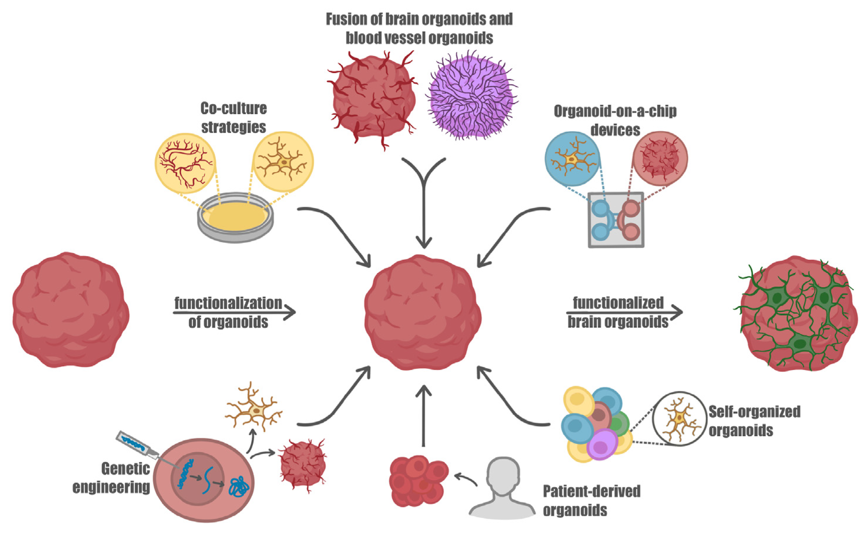
4. Improving Brain Organoids by Introducing Microglia and Vascularization

Brain organoids face a significant drawback due to their absence of vascularization and microglia, which are crucial components in the development and functioning of the brain. Microglia are essential for maintaining homeostasis and the immune response in the brain [55,56,57,58,59,60], and the absence of microglia in brain organoids limits not only their ability to accurately model all brain diseases characterized by the presence of a neuroinflammatory state but also the correct brain development and function in physiological conditions.
Microglia, the primary neuroimmune cells in the brain, originate from erythro-myeloid progenitors in the embryonic yolk sac and differentiate into microglia after migrating into the developing brain. Microglia are sustained through self-renewal processes dependent on cytokines like IL-34 and CSF-1, as well as transcription factors such as PU.1 and interferon-regulatory factor 8. Their primary role is immune surveillance, where resting microglia constantly monitor the brain environment, while activated microglia respond to pathological insults and eliminate harmful species through various mechanisms of phagocytosis. Furthermore, microglia contribute to neural development, synaptic formation and plasticity, and neural network maturation. They regulate neural development by colonizing cortical proliferative zones and phagocytosing neural precursor cells. During postnatal brain development, microglia actively prune weak synapses, thereby shaping neuronal circuits [58,60,61].
Neuronal lineage cells originate from the ectoderm, while differentiation into endoderm and mesoderm lineages is typically suppressed in the formation of human brain organoids. Consequently, microglia, which arise from non-neuroectodermal origins, are usually absent in brain organoids. However, recent advancements have led to the development of strategies to generate microglia-containing brain organoids, offering valuable insights into microglial functions and their relevance to brain disorders. Several strategies have been developed to address this limitation, such as incorporating microglia into organoids through co-culture, genetic modification, or separate culture insertion. One study conducted by Ormel et al. in 2018 reports the possibility to generate human iPSC-derived brain organoids containing innately developed microglia (oMG), using a protocol that, instead of adding extra BDNF to the differentiation cocktail, like Quadrato et al., reduced the levels of the neuroectoderm stimulant heparin and delayed Matrigel embedment of the organoids [10,62]. This approach resulted in the development of organoids with microglia cells characterized by ramified morphology, microglia-specific marker expression, and a more realistic immune response to inflammation. Another study conducted by Abud et al. in 2017 introduced microglia into cerebral organoids derived from iPSCs through a co-culture system. The resulting organoids exhibited a more mature and functional microglial phenotype validated using a range of methods. Firstly, flow cytometry analysis was employed to assess their phenotype. In addition, the secretion of cytokines/chemokines, specifically IL-1b and IFNγ, was examined subsequent to stimulation with lipopolysaccharide (LPS). Furthermore, to investigate the synaptic pruning capabilities, synaptosome phagocytosis assays were established. These findings provide compelling evidence that co-culture strategies hold significant potential for recapitulating the essential factors involved in the development of tissue-resident microglia in the brain [63]. In 2019, Song et al. used a guided protocol of 30 days (utilizing a differentiation medium containing BMP-4, Activin-A, SCF, and VEGF) to obtain microglial cells derived from iPSCs, which were then introduced into cerebral organoids. To assess the functional activity of these microglia-like cells, the researchers examined their phagocytosis capability, increased expression of TNF-α, and secretion of cytokines in response to pro-inflammatory Aβ42 oligomers, which are known to modulate microglia responses through TREM2 binding. The dorsal or ventral organoids displayed synaptic activities and action potentials, whereas the microglia-like cells exhibited differential migration ability and immune response, with higher TNF-α expression observed in ventral-MG co-cultures. This co-cultured strategy elucidated the impact of microglia on brain organoids, revealing their ability to stimulate cell proliferation and effectively reduce ROS expression, thus closely resembling the microenvironment found in specific brain tissues [64].
An innovative approach was developed by Ao et al. in 2021, where a tubular organoid device was integrated with isogenic microglia to enable hypoxia-free brain organoid culture with continuous medium and oxygen perfusion. The protocol commences by carefully loading human embryonic stem cells (hESCs) into the designated basket within a tubular device. Neural fate induction is accomplished by administering dual-SMAD inhibitors dorsomorphin and A83. In order to direct cellular differentiation towards a forebrain identity, the neural induction medium is subsequently replaced with a composition containing CHIR-99021 and SB-431542. To establish inner lumen fluid flow, the loaded device is placed onto a rocking platform, supplemented with N2 and B27 components. Neuronal characterization involved the assessment of marker expression, specifically, the dorsal forebrain neural progenitor cell markers PAX6 and SOX2, as well as the deep cortical neuron marker TBR1. In contrast, for the characterization of induced microglia, a double staining approach employing Iba1 and CD68 was conducted. Upon LPS/ATP treatment, activated induced microglia also exhibited elevated secretion levels of IL-1β and IL-18, indicating functional inflammasome activation. Additionally, a noteworthy increase in TNF-α secretion was also observed. The present device effectively emulated microglial responses in an environment that closely replicates the inherent microenvironment of the brain. In fact, it was observed that tubular organoids demonstrate a substantial decrease in hypoxia compared to conventional organoids. This finding is particularly intriguing considering that hypoxia is a recognized challenge commonly associated with organoid culture [65].
Until now, a significant limitation in the integration of microglia cells into organoids has been their notable heterogeneity, which impairs reproducibility due to the acquisition of highly divergent cell populations. However, in 2022, Cakir et al. achieved a substantial advancement by inducing the expression of PU.1 in cells, thereby generating microglia-like cells that could be successfully introduced into human cortical organoids. This pioneering approach represents noteworthy progress in addressing the challenge of heterogeneity, ultimately enhancing the reproducibility and consistency of microglia integration within organoid models [66] (Figure 2).
Another weakness of conventional brain organoids is the lack of vascularization, which limits growth, functionality, and immune response. Indeed, without the presence of blood vessels, organoids have a limited capacity for growth and functionality. Indeed, the lack of oxygen and nutrients due to the absence of blood vessels can limit the lifespan of organoids, leading to cell death over time and making it difficult to use organoids for long-term studies [40].
Innovative approaches have successfully developed vascularized organoids capable of performing more complex functions, revolutionizing the field of organoid research. By integrating functional blood vessels within the organoid structures, these advancements have facilitated improved nutrient delivery, waste removal, and oxygenation, closely mimicking the physiological conditions found in real tissues. This vascularization has enabled enhanced cellular interactions, organoid maturation, and the emergence of more intricate multicellular systems, thereby expanding the potential applications and utility of organoids in various biomedical research areas, such as drug discovery, disease modeling, and regenerative medicine. To overcome this limitation, a number of different solutions have been implemented, such as co-culture strategies, 3D-printed microfluidic chips, genome editing, and the fusion of blood vessel organoids with brain organoids. In this section, we will examine the current understanding of the lack of vascularization in organoids, its consequences, and the current progress towards creating more vascularized organoid models.
Pioneering work in the field involved the generation of vascularized organoids by introducing iPSC-derived endothelial cells. This was accomplished through a three-stage process utilizing FGF2, CHIR99012, BMP4, and VEGF to promote proper endothelial differentiation [67]. More recent studies have proposed alternative methods to generate vascularized organoids. One approach involves co-culturing hESCs or iPSCs with umbilical vein endothelial cells, resulting in a high success rate of vascularization exceeding 95%. The vascularized organoids (vOrganoids) exhibit mesh-like and tube-like structures formed by HUVECs, which are identified using markers such as laminin and isolectin I-B4 (IB4) that specifically label blood vessels and endothelial cells [68]. Another effective method involves the integration of differentiated vessels into brain organoids. Ahn et al. developed separate brain organoids and blood vessel organoids and subsequently co-cultured the obtained blood vessel cells with cortical organoids. These vascularized human cortical organoids demonstrate the presence of a functional vascular network, which is absent in regular hCOs [69]. In an alternative approach for generating brain-specific vascular organoids, a guided protocol was utilized to differentiate H9 hESCs into human blood vessel organoids. These blood vessel organoids were then fused with cerebral organoids to create brain-specific vascular organoids. Notably, this technique successfully formed a tightly sealed blood–brain barrier (BBB), consisting of brain microvascular endothelial cells. This closely mimics the BBB function in the selective regulation of the transport of substances to and from the brain and in the protection against harmful agents. The expression of key tight junction proteins, including Claudin5 (CLDN5) and ZO-1, as well as the efflux transporter p-glycoprotein, was examined to evaluate the BBB-like characteristics. These proteins facilitate the recycling of small lipophilic molecules that diffuse into endothelial cells back into the bloodstream [70]. Several additional approaches have been proposed to enhance vascularization in organoids. One strategy involves utilizing 3D-printed microfluidic chips where pericytes and endothelial cells derived from hPSCs spontaneously form well-structured vascular networks. These vascular cells establish physical interactions with cerebral organoids, resulting in the formation of integrated neurovascular organoids on the chip [71]. Another approach involves reprogramming human dermal fibroblasts into endothelial cells using the transcription factor human ETS variant 2 (hETV2). Bilal Cakir et al. (2022) introduced engineered hESCs expressing hETV2 into hCOs, leading to the development of vascularized hCOs with a functional, perfusable vascular-like network. These vascularized organoids exhibit BBB characteristics, such as increased expression of tight junctions, nutrient transporters, and transendothelial electrical resistance [66,72].
Although there are still limitations in the current state of 3D organoids for brain research, the ongoing efforts to improve vascularization hold great promise for the field. These advancements in vascularized organoid development provide opportunities for gaining deeper insights into brain function [68,69,70]. However, it is important to note that in addition to the lack of vascularization, another limitation of organoids is the absence of microglia, which hinders their ability to fully mimic the brain’s cellular composition and interactions [40] (Figure 2).

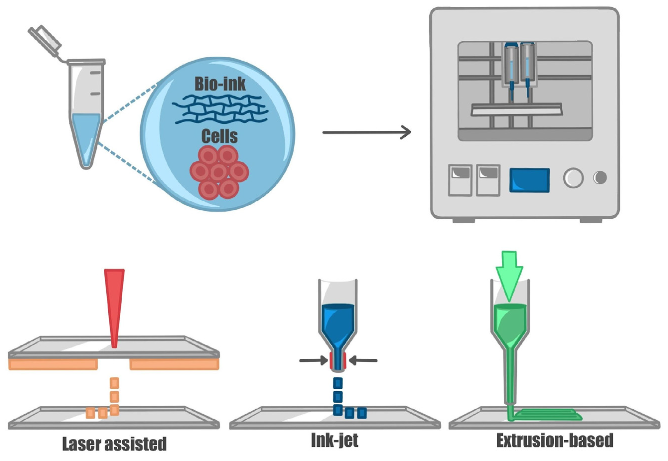
5. Assembling Neural Tissue: Hydrogels and Techniques Used in 3D Bioprinting and Organ-on-a-Chip Technologies

To date, brain organoid techniques still present many challenges: therefore, the convergence between organoid technology and 3D bioprinting could pave the way for optimizing functional 3D in vitro brain models.
Bioprinting is an automated, layer-by-layer deposition of cells embedded in biocompatible materials or bio-inks to fabricate 3D constructs. The development of this technology can also allow for the repair or regeneration of tissues in patients by performing pharmacokinetic studies in vitro [73]. Three-dimensional bioprinting techniques allow for the generation of organized structures, which increase reproducibility. Numerous studies have utilized 3D bioprinting technology to print stem cells, as demonstrated by Reid et al. [74], Gu et al. [75], Nguyen et al. [76], De La Vega [77], and Koch et al. [78]. This approach offers numerous advantages, as embedding ESC and hPSC in a 3D construct maintains their multilineage potential, enabling differentiation and maturation directly within the scaffold. Alternatively, another strategy involves differentiating cells from hPSCs into specific cell types before printing, as described by Faulkner-Jones et al. [79], Ma et al. [80], Yu et al. [81], Ong et al. [82], Moldovan et al. [83], Sorkio et al. [84], Joung et al. [85], and De la Vega et al. [86].
The precise assembly and guidance offered by 3D bioprinting enable the generation of organized structures, improving reproducibility. However, there are significant challenges to overcome, including controlling the relative distribution of cells and the impact of printing on cell viability. In particular, one of the most significant challenges in the bio-fabrication of 3D neural structures is developing bio-ink formulations that enable neuronal survival, differentiation, and maturation while modeling physiological extracellular matrix components and maintaining suitable mechanical properties for printing. In the context of 3D bioprinting, hydrogels are a popular choice for scaffolds due to their ability to retain large amounts of water, biocompatibility, and ability to form networks via the crosslinking of hydrophilic chains. These properties allow for the culturing of neural cells in 3D, which requires a material that can support cell bodies while allowing for process extension and connection. Hydrogels that have been used to print neural cells include alginate, agarose, chitosan ([75,87]), gellan gum-RGD [88], collagen [89], modified gelatin GelMa [90], and Matrigel [85,91] (see Table 2). Matrigel is a solubilized basement membrane matrix that contains many extracellular matrix (ECM) components and is secreted by Engelbreth-Holm-Swarm mouse sarcoma cells. However, this material’s batch-to-batch variability can impact the reproducibility of printing.
There are three main printing methods: laser-assisted, inkjet, and extrusion-based techniques (Figure 3). Laser-assisted bioprinting utilizes two co-planar slides, where a laser is used to transfer bio-ink droplets from the donor to the collector slide. This process is mediated by the formation and expansion of microbubbles on the surface of the donor slide. This nozzle-free technique allows for greater freedom in the choice of bio-ink and results in lower damage to the cells during printing. However, it is also associated with high costs, and slide preparation is complex and time-consuming. The inkjet technique utilizes a drop-on-demand method where micrometric droplets of bio-ink are ejected from the dispenser tip by applying a thermal or mechanical force [93,94,95]. Inkjet was one of the earliest approaches to bioprinting and enables the precise placement of cells with droplets in the picoliter range. However, this technique is only suitable for low-viscosity materials, despite being low-cost and straightforward [17,87]. Extrusion-based bioprinting involves physically extruding the material through the dispenser tip using pneumatic or piston-driven actuators in a continuous manner [96]. This technique is one of the most widely used, but it allows for lower resolution and is particularly suitable for viscous gels. After deposition, materials require a sol-gel transition, from liquid- to solid-like behavior, which can be achieved via direct printing into a cross-linking solution, such as calcium chloride for alginate [97], or using temperature changes or light for photo-cross-linked materials [98]. Coaxial needle printing, which simultaneously extrudes bio-ink and coagulation solution, is an approach that uses ionic crosslinking [99,100].
Hsieh et al. [101] demonstrated that murine NSCs embedded in thermoresponsive water-based polyurethane dispersions could be printed and implanted into a zebrafish brain injury model, leading to the successful rescue of functions of the impaired CNS. Joung et al. [85] combined extrusion-based bioprinting with 3D-printed scaffolds to model the spinal cord. They used an alginate and methylcellulose blend scaffold, and simultaneously printed neuronal progenitor cell (NPC) or oligodendrocyte progenitor cell (OPC) bio-ink into the scaffold, resulting in the development of functional neurons with extensive axon propagation. Gu et al. [75] used an extrusion-based technique to print neuronal constructs. They printed human iPSCs with a bio-ink composed of alginate, chitosan, and agarose, cross-linked in calcium chloride, which allowed in situ proliferation and differentiation into neuroglia and neurons. These cells displayed functional features 30–40 days post-printing. Recent implementations of extrusion 3D bioprinting techniques include their integration with microfluidic devices [102]. Microfluidic-based devices are made of microchannels containing microliter to picoliter volumes of fluids that interconnect chambers where different types of cells can be placed, closely modeling compartmentalized microenvironments. Microfluidic chips have also been demonstrated to be optimal platforms to model endothelial layers and therefore vascularized systems [103,104]. Yu et al. [81] demonstrated that functional tissue structures can be directly printed on microfluidic devices. The integration of 3D bioprinters with microfluidic-based heads allows the precise control of bio-ink volume, the printing of higher cell concentrations [105], the simultaneous extrusion of different materials [100], and the possibility to mimic complex or graded patterns and tissue compositions [106]. De la Vega et al. [77] printed hiPSC-derived NPCs into cylindrical constructs with an extrusion-based microfluidic printer using a fibrin, chitosan, and alginate bio-ink containing purmorphamine- and retinoic acid-loaded microspheres. This allowed for the continuous release of these drugs, leading to differentiation and maturation into spinal motor neurons.
iPSC-derived cortical neurons and glial cells were successfully printed by Salaris et al. in 2019 [91] using a custom extrusion-based bioprinter implemented with co-axial wet-spinning microfluidic devices to print Matrigel/alginate-embedded cells and crosslinking solution simultaneously. The cells could further differentiate within the construct, expressing both neuronal and astrocytic markers, with good long-term cell survival (up to 40 days post-printing). Moreover, functional analysis revealed properties typical of immature neuronal networks.
In conclusion, the recent technology of 3D bioprinting still faces numerous challenges: on the one hand, the development of ink formulations and materials that guarantee the reproduction of complex tissue composition with specific mechanical and physical properties (such as porosity, or the coexistence of organic and inorganic building blocks necessary to reproduce the bone structures, see Refs. [73,107,108,109]). On the other hand, there is an increasing need for sophisticated printing technologies to process the needed material to model human tissues correctly. In the case of neuronal 3D structures [110,111], this technology is additionally challenged by the physiological properties of neurons, such as building a three-dimensional, often patterned network. Overall, 3D bioprinting represents a promising technology to model organized neuronal structures reproducibly. Finally, recently, 3D printing has also been employed to standardize the production of organoids [111,112,113,114,115]. Despite still being at the beginning stages, and no attempts having yet been made with neural tissues, the convergence between 3D bioprinting technology and organoids is an appealing strategy in brain research.
Organ-on-a-chip is a transformative approach that harnesses the versatility of microfluidic platforms for the engineering of new functional tissues. Spanning from this, organoids-on-a-chip are currently attracting attention, due to the optimal results obtained from the synergistic combination between organoids and microfluidic technology. Comprehensive reviews have recently detailed the latest results in brain organoid-on-chip technology elsewhere [116,117]. Among the most relevant advancements in brain organoid-on-a-chip research, the work from Karzbrun and colleagues has revealed the underlying mechanisms of folding involved in neurodevelopmental disorders [118]. The authors reported the formation of surface wrinkles during the in vitro self-organization of human brain organoids pre-assembled in microfabricated compartments. The microchip platform was engineered to host and facilitate brain organoid assembly and monitoring with in situ imaging technology during weeks of development. The folding wavelength was found to scale linearly with the thickness of the forming tissue, indicating a balance between energies associated with bending and stretching. Moving beyond the study of brain development, Wang and co-workers [119] reported the use of a novel brain organoid-on-a-chip approach to study prenatal nicotine exposure. A multi-inlet device housing a number of brain organoids was engineered to offer the possibility to screen for a single drug on multiple organoids at the same time. The overall development of the brain organoids was guided and ultimately satisfactory, resulting in the ability to recapitulate defined neural differentiation and regionalization. Nicotine exposure was found to induce premature and abnormal differentiation with the overexpression of TUJ1 and the disruption of neurodevelopment with the expression of PAX2, PAX6, FOXG1, and KROX20, among other markers indicating pathological alteration. Brain organoid-on-a-chip platforms have demonstrated unparalleled potential in generating functional models for the screening of new therapeutics. Nevertheless, the lack of available microfabrication facilities, the difficulties associated with engineering methodologies to replicate microfluidic architecture, as well as the limited number of organoids that can be cultivated at the same time are some of the pivotal factors that are currently limiting incremental research associated with brain organoid-on-a-chip technology.

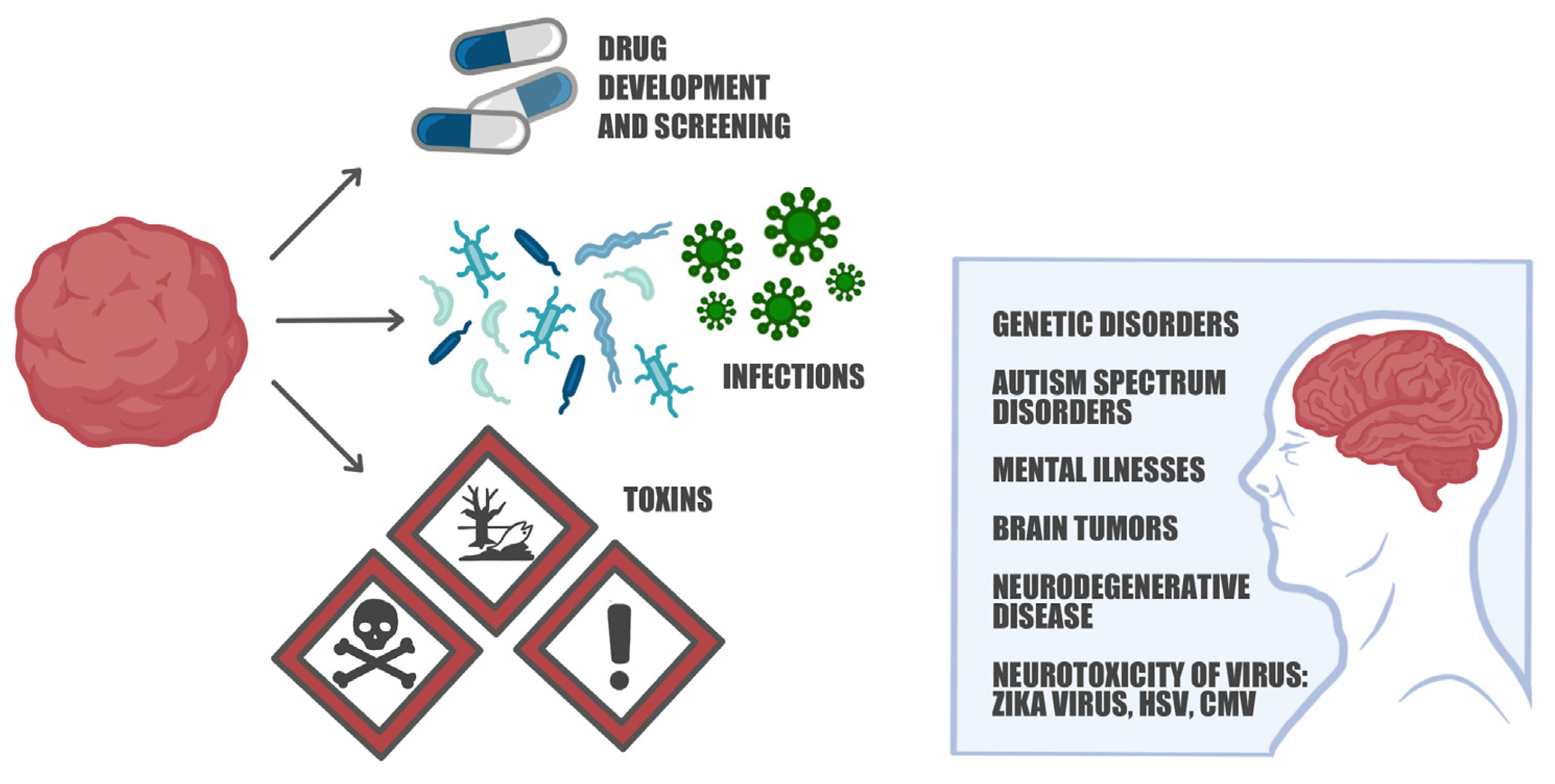
6. The Versatility of 3D Brain Organoids: Modeling Neurodevelopmental and Neurodegenerative Disorders Unraveling Pathophysiological Mechanisms

As we reported so far, 3D brain organoids have emerged as a potent tool for modeling human neurological disorders, including neurodevelopmental and neurodegenerative disorders, thus offering a physiologically relevant and personalized approach over 2D cell cultures [120,121]. Organoids can be derived from patient-specific iPSCs offering an opportunity to model diseases using cells from affected individuals. Moreover, organoids can recapitulate aspects of organ development, allowing researchers to study disease processes during embryonic development. In addition, organoids can be generated in large numbers, facilitating the high-throughput screening of drugs and therapeutic compounds, potentially accelerating the drug discovery process (Figure 4).
Patient-derived stem cells can be utilized to generate brain organoids of a specific pathology, considering not only the disease but also the genetic background of the patient. These advancements in organoid technology open new horizons to better decipher mechanisms involved in brain development and maturation, as well as pathologies affecting neurons and glial cells. They hold value in drug development and screening and the exploration of personalized therapies [122,123,124]. It has to be noticed that the absence of microglia and proper vascularization, including the presence of the blood–brain barrier (BBB), has been recognized as a significant limitation in brain organoids when it comes to modeling brain functions in pathological conditions, particularly in drug testing experiments [125]. The role of microglia and the immune system cannot be overlooked, as they play crucial roles in various neurological conditions. Additionally, vascularization is essential for drug distribution within the organoid, and disruptions in BBB integrity are commonly observed in neurodegenerative disorders, brain cancers, and traumatic brain injuries. These limitations underscore the need for further advancements in brain organoid technology to mimic the complex cellular interactions and pathological features of the brain more accurately [125].
Brain organoids have emerged as a valuable tool for modeling neurodevelopmental genetic disorders, allowing researchers to recreate pathologies, such as Tourette’s Syndrome [126], microcephaly, and macrocephaly, and investigate the impact of pathogens on brain development [127,128,129]. For instance, studies using induced pluripotent stem cells (iPSCs) from microcephaly patients have demonstrated smaller organoids and premature differentiation in the neural progenitor regions [128], while the deletion of the tumor-suppressor gene PTEN in human iPSCs results in the formation of abnormally large organoids with the over-proliferation and delayed neurogenesis of neural progenitor cells [129]. Brain organoids have also proved to be valuable for investigating viral infections, including the Zika virus, the herpes simplex virus (HSV), and the cytomegalovirus. In brain organoids, the Zika virus specifically targets SOX2-positive neural progenitor cells, leading to suppressed proliferation, increased cell death, and a significant reduction in organoid size [17,129,130]. Similarly, early-stage brain organoids composed of human iPSC-derived neural rosettes infected with HSV-1 exhibit a loss of structural integrity and neuronal alterations [131]. These studies highlight the utility of brain organoids in studying the effects of viral infections on brain development and function [131,132,133,134,135,136].
Brain organoids have become a valuable tool for studying neurodevelopmental disorders such as schizophrenia (SZ), and fragile X syndrome (FXS). SZ is a complex and severe neuropsychiatric disorder associated with a wide range of debilitating symptoms. Many aspects of its multifactorial complexity are still unknown, and some are accepted to be an early developmental deficiency with a more specifically neurodevelopmental origin. The neurodevelopmental hypothesis suggests that the interactions of multiple genes trigger a cascade of neuropathological events during the embryonic and post-natal development of the brain that may be initiated by environmental factors such as maternal infections or infectious agents associated with the onset of inflammatory responses [137] that trigger symptoms in early adolescence [138] and lead to the emergence of psychosis at the time of the transition from late adolescence to young adulthood. The disorder can be heritable and polygenic, with risk alleles distributed widely across the genome [139] and people with SZ are enriched by rare copy variants (CVs) in genes associated with neurodevelopmental disorders, particularly autism spectrum disorders and intellectual disability [140]. CVs can disrupt gene function by increasing or decreasing gene dosage, and one recurrent CV is a deletion in the 22q11.2 region that typically encompasses around 50 protein-coding genes and is the most common CV in humans [141]. This deletion causes the 22q11.2 deletion syndrome (DS), a neurodevelopmental disorder that is one of the most frequent genetic risk factors for SZ [142,143], and, like SZ, the 22q11.2 DS patients display neurodevelopmental delays and cognitive dysfunctions [144]. One challenge facing researchers trying to elucidate the mechanisms underlying SZ and 22q11.2 DS is that the molecular and cellular processes governing embryonic and early postnatal brain maturation are still unclear.
Recent studies on mice models of 22q11.2 DS have provided important insights [143,145,146,147] including a description of circuit dysfunctions [148,149,150] and the implication of mitochondrial dysfunctions in the onset of cognitive phenotypes [151,152,153]. However, the molecular and cellular mechanisms leading to human neuronal phenotypes remain poorly understood. In recent years, some studies have begun to explore transcriptional changes and the implication of mitochondrial dysfunctions in neural cells derived from 22q11DS patients [154,155,156], but the functional defects in human 22q11.2 DS neurons and the underlying mechanisms have not been investigated. Therefore, understanding the timepoints of neuronal and, eventually, glial cell phenotypes during cell differentiation and maturation processes could lead to an insight into the development of the cellular phenotypes associated with the disorder.
Cerebral organoids provide a promising tool to investigate the molecular and cellular alterations involved in the onset of 22q11.2 DS, exploiting the possibility to directly use patient-derived IPSCs which consider not only the footprint of the disease but also the genetic background of the patient, enabling the evaluation of specific early pathological targets for drug development. Khan and colleagues successfully generated 3D cerebral cortical organoids from 22q11.2 DS-derived IPSCs. They analyzed the transcriptional profile along 100 days of differentiation and found an altered expression of several genes, related to RNA modification and RNA silencing, mitochondria functions, and neuronal excitability, including those coding for calcium transport. Electrophysiological and calcium imaging experiments confirmed changes in spontaneous firing and depolarization-induced calcium signaling in 22q11.2 DS cortical neurons due to abnormalities in the resting membrane potential and defects in the function of the voltage-gated L-type calcium channels. Interestingly, the voltage-gated L-type calcium channels have been identified as common risk genes for SZ [157], thus suggesting that alterations of cytoplasmic calcium signaling may be one of the key targets for drug development.
FXS, the most common single-gene cause of autism spectrum disorder (ASD), is characterized by an expansion of the CGG triplet in the FMR1 gene, leading to the loss of the FMR1 protein [158]). The FMRP protein, which is involved in RNA regulation, plays a crucial role in neuronal development, synaptic plasticity, and dendritic spine architecture [159], and recent studies on FXS-patient-derived neural progenitor cells (NPCs) have indeed revealed abnormal gene expressions related to protein synthesis, neural development, and migration [160,161,162,163]. Differential gene expression analysis revealed 218 differentially expressed genes involved in cell fate commitment and differentiation, with down-regulated genes related to fate specification, migration, differentiation, and maturation, and up-regulated genes associated with cell proliferation [164]. A recent study by Kang et al. [165] observed alterations in neural proliferation and differentiation in brain organoids derived from FXS patients’ iPSCs during a developmental period corresponding to mid-fetal human brain development. In FXS, alterations in gene expressions related to synapse development, axon targeting, and cytoskeleton organization [166] result in an imbalance between excitatory and inhibitory signaling [28,167,168,169,170,171].
FMRP knockout brain organoids have shown significant phenotypes of immature astroglial cells by revealing an increased number of GFAP-positive cells [28], a situation similar to the FXS postmortem brain tissues [172,173]. Interestingly, the gene of FMR1 is also part of group I of metabotropic glutamate receptor (mGlu) signaling, and FMR1 together with the mGlu type 5 and the scaffold proteins Homer1b and Shank3 are highly expressed in immature astrocytes [174,175], but their expression decreases over the differentiation and maturation of the cells, thus suggesting that they can play crucial roles during the acquisition of the astrocytic phenotype [176]. Indeed, accumulating evidence indicates that mGluR5 signaling in immature astrocytes controls astrocyte morphogenesis [177], thus suggesting that abnormal astrocyte maturation may also occur in the absence of FMR1 in the brain organoids [28]. These findings highlight the potential of brain organoids in unraveling the underlying mechanisms of neurodevelopmental disorders, particularly providing insights into the pathophysiology of FXS.
The use of “fused” dorsal–ventral forebrain organoids, also known as assembloids, generated from iPSCs, has provided valuable insights into the cellular and molecular mechanisms underlying Timothy syndrome (TS) [54,178]. TS is a rare and severe neurodevelopmental disorder caused by a mutation in an L-type calcium channel subunit. Calcium influx through voltage-gated calcium channels is involved in regulating processes such as cytoskeletal dynamics, cell adhesion, and signaling pathways crucial for neuronal migration [179], and in TS-patient-derived brain organoids, Birey et al. [180] showed the inefficient migration of cortical interneurons and increased calcium signaling following depolarization. These findings shed light on the role of calcium signaling in interneuron migration and the establishment of neural circuits. To gain a more comprehensive understanding of TS pathophysiology, Revah et al. [181] transplanted TS brain organoids into the cerebral cortex of newborn athymic rats. This transplantation approach enabled the investigation of organoid maturation and integration with a functional neural circuit, leading to the manifestation of disease-related phenotypes. These models offer valuable insights into the pathophysiology of TS and have the potential to identify novel therapeutic targets for this neurodevelopmental disorder.
Another promising tool for the study of Tourette’s syndrome is represented by basal ganglia organoids [182]. Tourette’s syndrome is a neuropsychiatric disorder characterized by uncontrollable motor or vocal tics that manifest in childhood and that very often co-occur with obsessive-compulsive disorder (OCD). Tourette’s syndrome and OCD are compulsive repetitive behaviors that are considered to be neurodevelopmental in origin, but their etiology and pathophysiology are unknown. Convergent neuroimaging and neurophysiological findings support a model in which abnormal cortico-striato-thalamo-cortical (CSTC) loops [183,184] and abnormalities in the basal ganglia may play a crucial role in the onset of symptoms [185]. Supporting the CSTC loop hypothesis, it is known that the optogenetic activation of the prefrontal cortex (PFC) induces excessive grooming in rodents [184] and that dopamine, one important modulator in CSTC circuits, plays a role in the onset of OCD symptoms in patients [186,187] and of stereotypical behavior in rodents [188,189]. There is also strong evidence that the dopaminergic system plays a key role in Tourette’s syndrome [190] and there might be some therapeutic role for dopamine D2 receptor blockers in OCD [191]. The current CSTC model of OC-like behavior and the hypothesis of dopaminergic involvement are both entirely neurocentric, but recent studies have identified a role for astrocytes in the modulation of dopamine homeostasis in the PFC [176] and revealed the importance of these cells in a number of behaviors, including repetitive behaviors [189,192], thus indicating that animal behavior is not a result of neuronal activity alone but requires the coordinated activity of neurons and astrocytes. Overall, these studies on rodent models of OC-like disorders have provided important insights, but human neuronal and astroglial phenotypes remain poorly described.
To better understand the neuronal pathways leading to OC-like disorders, recent studies have explored the early developmental pathophysiology of Tourette’s syndrome by using iPSC-derived basal ganglia organoids [182]. Repetitive behaviors and tic release elicit prominent activity in the basal ganglia [193], and neuroimaging data suggest decreases in the striatal volume in Tourette’s syndrome patients [193]. The basal ganglia organoids exhibit impaired development of medial ganglionic eminence and reduced differentiation of cholinergic and GABAergic interneurons. The transcriptome analysis revealed the mis-patterning of the ventral telencephalon with a relative lack of ventromedial progenitors accompanied by enhanced dorsolateral fates. This results in the developmental loss of interneurons, suggesting that the interneuron loss noted in the postmortem basal ganglia of patients with Tourette’s syndrome is a potential consequence of an inherent tendency of the basal ganglia to undergo different regional specifications. These findings contribute to our understanding of Tourette’s syndrome etiology and offer a developmental lens to the pathologies currently associated with Tourette’s syndrome such as OC-like disorders.
Brain organoids offer researchers a valuable platform to study the molecular mechanisms underlying neurodegenerative diseases such as Alzheimer’s (AD) and Parkinson’s (PD), enabling the observation of changes in gene expression, protein function, and cellular interactions. It is noteworthy that neurodegenerative diseases are commonly associated with late-onset symptoms and are linked to the aging process. However, emerging evidence suggests that pathological mechanisms, such as amyloid-β accumulation in AD or changes in brain structure and function in PD, can initiate decades before symptom onset [194,195,196,197]. Moreover, genetic predisposition can lead to the development of neuronal and glial dysfunctions at an earlier age. By utilizing organoid technology, researchers have successfully recapitulated neuropathological hallmarks associated with AD, including amyloid-β deposition, the hyperphosphorylation of tau protein, continuous aggregation in 3D models, neuroinflammation, and gliosis [25,194,198,199]. Most of the contributions come from brain organoids modeling familial AD, which represents a small fraction (<5%) of AD cases caused by genetic variants in the amyloid precursor protein (APP) gene or presenilin genes (PSEN1, PSEN2), while the majority of cases (sporadic AD) result from a combination of multiple factors [200,201]. These organoids can also be gene-edited to express specific genetic variations or those derived from patient-derived iPSCs. Abnormalities in tau protein, synaptic dysfunction, disturbance in the balance of excitatory and inhibitory neuronal circuits, and the presence of amyloid-β plaques contribute to enhanced neuronal hyperactivity observed in AD organoids compared to wild type [202,203]. Co-cultures of microglia and brain organoids have been utilized to study the interaction between microglia, neural cells, and pathological features of Alzheimer’s disease. Specifically, the role of APOE4, the strongest genetic risk factor for late-onset sporadic AD, has been investigated in co-culture models, revealing a reduced Aβ plaque clearance ability compared to APOE3 [204]. AD brain organoids have also been employed as a drug platform, demonstrating a reduction in amyloid-β aggregates through the use of β- or γ-secretase modulators, along with mitigating the effect of tau pathology [194].
Human-derived 3D models of Parkinson’s disease (PD) offer a close resemblance to the disease phenotype, exhibiting characteristic features such as the shortened neurite length of dopaminergic neurons and α-synuclein aggregation, surpassing the limitations of 2D and animal models [205]. Brain organoids derived from PD patients carrying missense mutations in the LRRK2 gene, associated with late-onset disease, demonstrate impaired dopaminergic neuron development and reduced astrocytic activity [206,207]. Additionally, brain organoids have been employed to investigate the molecular mechanisms underlying α-synuclein deposition, revealing a correlation with LRRK2 mutation [205]. Furthermore, PD organoids have been instrumental in studying the juvenile form of PD by introducing mutations into the DNAJ6 gene. Mutant organoids exhibit impaired neurodevelopment, reduced dopamine release, increased oxidative impairment, and dysfunctions in mitochondria and lysosomes [208].
Over the past decade, a number of studies have highlighted the crucial roles of inflammatory processes in many brain diseases, highlighting a possible role of reactive glial cells in the pathophysiology of neurodegenerative disorders. Astrocytes are indeed involved in all forms of brain disease and lesions, to which they respond by undergoing a series of cellular, molecular, and functional changes known as “reactive astrogliosis” [209]. These alterations may be transient or long-lasting, and interdisciplinary approaches combining omics with physiology and genetic manipulations have shown that they can have both harmful and beneficial effects, thus indicating that astrocytes have multiple states of reactivity and functions depending on the myriad of intrinsic and extrinsic cues governing their post-injury gene expression and function [209]. Many aspects of astrocyte functioning have been unveiled from studies conducted in murine models; however, growing evidence shows many differences between mouse and human astrocytes starting from their development and encompassing morphological, transcriptomic, and physiological variations when they achieve complete maturation. The study of human reactive astrocytes has, therefore, been limited by the availability of resources and, more importantly, because there are important transcriptional and functional differences between rodent and human astrocytes [127,210]. In a recent study, Cvetkovic et al. address this technical shortcoming by developing bio-engineered neural organoid cultures containing mature astrocytes which allow for the investigation of the dynamics of astrocyte reactivity and its downstream effects on neuronal activity [211,212]. The authors successfully generated multicellular organoid systems containing astrocytes that exhibited key features of mature cells. After extensively validating the morphology and gene expression of astrocytes, Cvetkovic et al. investigated the effects of over-activating calcium signaling in astrocytes on neuronal activity. They successfully found that the chronic activation of astrocytes resulted in changes in gene expression like those observed in reactive astrocytes, suggesting aberrant calcium signaling in inducing a reactive phenotype. However, the observed reactivity did not display neurotoxicity as seen in other studies, possibly due to differences in experimental conditions or the requirement of additional disease-associated stressors [211]. Overall, this new in vitro model is promising for studying the dynamics of human reactive astrocytes and their impact on neuronal function [212]. Future research should explore more complex models that incorporate other cell types involved in neuroinflammatory processes to better understand the contribution of reactive astrocytes to disease progression and test potential therapies [212].

7. Conclusions

Over the last decade, 3D brain cultures, such as brain organoids, have emerged as powerful tools for studying neural function and modeling neurological diseases. Brain organoids are self-organized 3D structures that recapitulate key aspects of human brain development and pathology [5]. One of the key advantages of brain organoids is that they can be generated from patient-specific pluripotent stem cells, providing a personalized approach to studying disease mechanisms and developing new therapies [213].
There are several methods for generating brain organoids, including self-assembly, guided differentiation, and bioprinting. Self-assembled organoids are formed by allowing cells to self-organize into 3D structures without external manipulation. In contrast, guided differentiation involves the use of growth factors and other signaling molecules to drive cell differentiation and tissue patterning. Bioprinting allows precise control over the spatial arrangement of cells and extracellular matrix components, enabling the generation of complex, multi-layered structures [17].
While each method has its advantages and limitations, the choice of method should be tailored to the specific research question. For example, self-assembled organoids may better recapitulate the complex interactions between different cell types and signaling pathways that occur during brain development, whereas bioprinted organoids may provide more precise control over tissue structure and composition [62].
Incorporating microglia and vascularization into brain organoids can further enhance their relevance as disease models and improve drug development outcomes. Microglia are the resident immune cells of the central nervous system, and they play a critical role in neuroinflammation and neurodegeneration. Recent studies have shown that microglia can innately develop within cerebral organoids, providing a new platform for studying their function in the context of neurodegenerative diseases [62]. In addition, incorporating a vascular network into brain organoids can improve nutrient and oxygen delivery, allowing for longer-term culture and more accurate modeling of disease progression and drug response [213].
We also need to consider some ethical issues surrounding advancements in brain organoid research, particularly as it moves towards creating more complex models of mature human cortical regions and their interconnectedness. One significant concern revolves around the potential emergence of consciousness in brain organoids or the complex brain assembloids that combine organoids from multiple cell lineages [214].
While these concerns exist, several factors mitigate them. It is difficult to compare organoid brain waves with those of developing human brains. The neural correlates of consciousness are complex and involve diverse brain regions. The term “consciousness” has different meanings, and the ethical implications depend on the specific definition. Organoids lack the necessary structures for complex consciousness. The moral concerns may be influenced by the human origin of organoids. However, the absence of social interaction and language acquisition makes conscious self-awareness unlikely. Thus, the ethical challenges related to consciousness in organoids are not significant in current research.
In conclusion, 3D brain cultures offer a promising platform for studying neural function and developing new therapeutic strategies for neurodevelopmental and neurodegenerative disorders. Self-assembled, guided, and bioprinted organoids each have unique advantages and limitations, and the choice of method should be tailored to the specific research question. Incorporating microglia and vascularization into brain organoids can enhance their relevance as disease models and improve drug development outcomes. As the field continues to advance, it is likely that 3D brain cultures will play an increasingly important role in unlocking the secrets of neural function and developing effective treatments for neurological disorders.

Author Contributions

Conceptualization, C.D., L.M., F.C. and S.D.A.; writing—original draft preparation, C.D., L.M., C.S., L.T., P.B., F.C., G.C. and S.D.A.; images preparation G.G.; writing—review and editing, all authors; funding acquisition, P.B., C.D. and S.D.A. All authors have read and agreed to the published version of the manuscript.

Funding

This research was funded by a Fulbright Award for S.D.A (FSP-P005556), the D-Tails-IIT JointLab (to S.D.A.), Rome Technopole PNRR, FP7 (to S.D.A.), the Regione Lazio FSE 2014–2020 (19036AP000000019 and A0112E0073) grants (to S.D.A.), and Fondazione Instituto Italiano di Tecnologia (to L.M. and C.S.). C.D.A. and L.M. were also supported by the Ph.D. program in Life Science at Sapienza University in Rome and C.S. by Project “National Center for Gene Therapy and Drugs based on RNA Technology” (CN00000041) financed by NextGeneration EU PNRR MUR—M4C2—Action 1.4—Call “Potenziamento Strutture di Ricerca e Creazione di “Campioni Nazionali di R&S” (CUP J33C22001130001). This research was funded by the Swiss National Science Foundation SNSF (310030_185363) (to P.B.); Telethon Italy (GGP20037) (to P.B.); ERANET-Neuron SNSF (31NE30_204022) (to P.B.). This research was partially funded by grants from Sapienza University (to C.D.A. and S.D.A.).

Institutional Review Board Statement

Not applicable.

Informed Consent Statement

Not applicable.

Data Availability Statement

Not applicable.

Acknowledgments

The authors wish to thank Ylenia Gigante for the continuous support in the Lab.

Conflicts of Interest

S.D.A. is a member of the scientific advisory board of D-Tails s.r.l. The funders had no role in the design of the study; in the collection, analyses, or interpretation of data; in the writing of the manuscript; or in the decision to publish the results.

References

  1. Steimberg, N.; Chiono, V.; Era, P.D.; Di Angelantonio, S. iPS, Organoids and 3D Models as Advanced Tools for In Vitro Toxicology. ALTEX Altern. Anim. Exp. 2020, 37, 136–140. [Google Scholar] [CrossRef] [Green Version]
  2. Muñoz-sanjuán, I.; Brivanlou, A.H. Neural Induction, The Default Model and Embryonic Stem Cells. Nat. Rev. Neurosci. 2002, 3, 271–280. [Google Scholar] [CrossRef] [PubMed]
  3. Eiraku, M.; Takata, N.; Ishibashi, H.; Kawada, M.; Sakakura, E.; Okuda, S. Self-Organizing Optic-Cup Morphogenesis in Three-Dimensional Culture. Nature 2011, 472, 51–56. [Google Scholar] [CrossRef] [PubMed]
  4. Eiraku, M.; Watanabe, K.; Matsuo-takasaki, M.; Kawada, M.; Yonemura, S.; Matsumura, M.; Wataya, T.; Nishiyama, A.; Muguruma, K.; Sasai, Y. Article Self-Organized Formation of Polarized Cortical Tissues from ESCs and Its Active Manipulation by Extrinsic Signals. Stem Cell 2008, 3, 519–532. [Google Scholar] [CrossRef] [Green Version]
  5. Lancaster, M.A.; Renner, M.; Martin, C.A.; Wenzel, D.; Bicknell, L.S.; Hurles, M.E.; Homfray, T.; Penninger, J.M.; Jackson, A.P.; Knoblich, J.A. Cerebral Organoids Model Human Brain Development and Microcephaly. Nature 2013, 501, 373–379. [Google Scholar] [CrossRef] [Green Version]
  6. Del Dosso, A.; Urenda, J.; Nguyen, T.; Quadrato, G. Primer Upgrading the Physiological Relevance of Human Brain Organoids. Neuron 2020, 107, 1014–1028. [Google Scholar] [CrossRef]
  7. Di Lullo, E.; Kriegstein, A.R. The Use of Brain Organoids To investigate neural development and disease. Nat. Rev. Neurosci. 2017, 18, 573–584. [Google Scholar] [CrossRef] [Green Version]
  8. Kelava, I.; Lancaster, M.A. Protocol Review Stem Cell Models of Human Brain Development. Stem Cell 2016, 18, 736–748. [Google Scholar] [CrossRef] [Green Version]
  9. Qian, X.; Song, H.; Ming, G.L. Brain Organoids: Advances, Applications and Challenges. Development 2019, 146, dev166074. [Google Scholar] [CrossRef] [Green Version]
  10. Quadrato, G.; Brown, J.; Arlotta, P. The Promises and Challenges of Human Brain Organoids as Models of Neuropsychiatric Disease. Nat. Med. 2016, 22, 1220–1228. [Google Scholar] [CrossRef]
  11. Xiang, Y.; Cakir, B.; Park, I. Seminars in Cell and Developmental Biology Deconstructing and Reconstructing the Human Brain with Regionally Specified Brain Organoids. Semin. Cell Dev. Biol. 2021, 111, 40–51. [Google Scholar] [CrossRef]
  12. Pasca, A.M.; Sloan, S.A.; Clarke, L.E.; Tian, Y.; Makinson, C.D.; Huber, N.; Kim, C.H.; Park, J.Y.; O’Rourke, N.A.; Nguyen, K.D.; et al. Functional Cortical Neurons and Astrocytes from Human Pluripotent Stem Cells in 3D Culture. Nat. Methods 2015, 12, 671–678. [Google Scholar] [CrossRef] [Green Version]
  13. Kadoshima, T.; Sakaguchi, H.; Nakano, T.; Soen, M.; Ando, S.; Eiraku, M.; Sasai, Y. Self-Organization of Axial Polarity, inside-out Layer Pattern, and Species-Specific Progenitor Dynamics in Human ES Cell-Derived Neocortex. Proc. Natl. Acad. Sci. USA 2013, 110, 20284–20289. [Google Scholar] [CrossRef] [Green Version]
  14. Muguruma, K.; Nishiyama, A.; Kawakami, H.; Hashimoto, K.; Sasai, Y. Self-Organization of Polarized Cerebellar Tissue in 3D Culture of Human Pluripotent Stem Cells. Cell Rep. 2015, 10, 537–550. [Google Scholar] [CrossRef] [Green Version]
  15. Sakaguchi, H.; Kadoshima, T.; Soen, M.; Narii, N.; Ishida, Y.; Ohgushi, M.; Takahashi, J.; Eiraku, M.; Sasai, Y. Generation of Functional Hippocampal Neurons from Self-Organizing Human Embryonic Stem Cell-Derived Dorsomedial Telencephalic Tissue. Nat. Commun. 2015, 6, 8896. [Google Scholar] [CrossRef] [Green Version]
  16. Ozone, C.; Suga, H.; Eiraku, M.; Kadoshima, T.; Yonemura, S.; Takata, N.; Oiso, Y.; Tsuji, T.; Sasai, Y. Functional Anterior Pituitary Generated in Self-Organizing Culture of Human Embryonic Stem Cells. Nat. Commun. 2016, 7, 1–10. [Google Scholar] [CrossRef] [Green Version]
  17. Qian, X.; Nguyen, H.N.; Song, M.M.; Hadiono, C.; Ogden, S.C.; Hammack, C.; Yao, B.; Hamersky, G.R.; Jacob, F.; Zhong, C.; et al. Brain-Region-Specific Organoids Using Mini-Bioreactors for Modeling ZIKV Exposure. Cell 2016, 165, 1238–1254. [Google Scholar] [CrossRef] [Green Version]
  18. Wataya, T.; Ando, S.; Muguruma, K.; Ikeda, H.; Watanabe, K.; Eiraku, M.; Kawada, M.; Takahashi, J.; Hashimoto, N.; Sasai, Y. Minimization of Exogenous Signals in ES Cell Culture Induces Rostral Hypothalamic Differentiation. Proc. Natl. Acad. Sci. USA 2008, 105, 11796–11801. [Google Scholar] [CrossRef] [Green Version]
  19. Ogura, T.; Sakaguchi, H.; Miyamoto, S.; Takahashi, J. Three-Dimensional Induction of Dorsal, Intermediate and Ventral Spinal Cord Tissues from Human Pluripotent Stem Cells. Development 2018, 145, dev162214. [Google Scholar] [CrossRef] [Green Version]
  20. Xiang, Y.; Cakir, B.; Park, I.H. Generation of Regionally Specified Human Brain Organoids Resembling Thalamus Development. STAR Protoc. 2020, 1, 100001. [Google Scholar] [CrossRef]
  21. Cakir, B.; Xiang, Y.; Tanaka, Y.; Kural, M.H.; Parent, M.; Kang, Y.J.; Chapeton, K.; Patterson, B.; Yuan, Y.; He, C.S.; et al. Engineering of Human Brain Organoids with a Functional Vascular-like System. Nat. Methods 2019, 16, 1169–1175. [Google Scholar] [CrossRef] [PubMed]
  22. Pellegrini, L.; Bonfio, C.; Chadwick, J.; Begum, F.; Skehel, M.; Lancaster, M.A. Human CNS Barrier-Forming Organoids with Cerebrospinal Fluid Production. Science 2020, 369, eaaz5626. [Google Scholar] [CrossRef] [PubMed]
  23. Miura, Y.; Li, M.Y.; Birey, F.; Ikeda, K.; Revah, O.; Thete, M.V.; Park, J.Y.; Puno, A.; Lee, S.H.; Porteus, M.H.; et al. Generation of Human Striatal Organoids and Cortico-Striatal Assembloids from Human Pluripotent Stem Cells. Nat. Biotechnol. 2020, 38, 1421–1430. [Google Scholar] [CrossRef] [PubMed]
  24. Nakano, T.; Ando, S.; Takata, N.; Kawada, M.; Muguruma, K.; Sekiguchi, K.; Saito, K.; Yonemura, S.; Eiraku, M.; Sasai, Y. Self-Formation of Optic Cups and Storable Stratified Neural Retina from Human ESCs. Cell Stem Cell 2012, 10, 771–785. [Google Scholar] [CrossRef] [PubMed] [Green Version]
  25. Cordella, F.; Ferrucci, L.; D’Antoni, C.; Ghirga, S.; Brighi, C.; Soloperto, A.; Gigante, Y.; Ragozzino, D.; Bezzi, P.; DiAngelantonio, S. Human iPSC-Derived Cortical Neurons Display Homeostatic Plasticity. Life 2022, 12, 1884. [Google Scholar] [CrossRef]
  26. Garone, M.G.; Birsa, N.; Rosito, M.; Salaris, F.; Mochi, M.; de Turris, V.; Nair, R.R.; Cunningham, T.J.; Fisher, E.M.C.; Morlando, M.; et al. ALS-Related FUS Mutations Alter Axon Growth in Motoneurons and Affect HuD/ELAVL4 and FMRP Activity. Commun. Biol. 2021, 4, 1025. [Google Scholar] [CrossRef]
  27. Garone, M.G.; D’Antoni, C.; Rosa, A. Culture of Human IPSC-Derived Motoneurons in Compartmentalized Microfluidic Devices and Quantitative Assays for Studying Axonal Phenotypes BT—Stem Cell Assays: Methods and Protocols. In Stem Cell Assays; Kannan, N., Beer, P., Eds.; Methods in Molecular Biology; Springer: New York, NY, USA, 2022; pp. 189–199. ISBN 978-1-0716-1979-7. [Google Scholar]
  28. Brighi, C.; Salaris, F.; Soloperto, A.; Cordella, F.; Ghirga, S.; de Turris, V.; Rosito, M.; Porceddu, P.F.; D’Antoni, C.; Reggiani, A.; et al. Novel Fragile X Syndrome 2D and 3D Brain Models Based on Human Isogenic FMRP-KO IPSCs. Cell Death Dis. 2021, 12. [Google Scholar] [CrossRef]
  29. Shi, Y.; Inoue, H.; Wu, J.C.; Yamanaka, S. Induced Pluripotent Stem Cell Technology: A Decade of Progress. Nat. Rev. Drug Discov. 2017, 16, 115–130. [Google Scholar] [CrossRef]
  30. Mulder, L.A.; Depla, J.A.; Sridhar, A.; Wolthers, K.; Pajkrt, D.; Vieira de Sá, R. A Beginner’s Guide on the Use of Brain Organoids for Neuroscientists: A Systematic Review. Stem Cell Res. Ther. 2023, 14, 87. [Google Scholar] [CrossRef]
  31. Reynolds, B.A.; Weiss, S. Generation of Neurons and Astrocytes from Isolated Cells of the Adult Mammalian Central Nervous System. Science 1992, 255, 1707–1710. [Google Scholar] [CrossRef] [Green Version]
  32. Jensen, J.B.; Parmar, M. Strengths and Limitations of the Neurosphere Culture System. Mol. Neurobiol. 2006, 34, 153–161. [Google Scholar] [CrossRef]
  33. Soares, R.; Ribeiro, F.; Lourenço, D.M.; Rodrigues, R.; Moreira, J.; Sebastião, A.; Morais, V.; Xapelli, S. The Neurosphere Assay: An Effective in Vitro Technique to Study Neural Stem Cells. Neural Regen. Res. 2021, 16, 2229–2231. [Google Scholar] [CrossRef]
  34. Singec, I.; Knoth, R.; Meyer, R.P.; Maciaczyk, J.; Volk, B.; Nikkhah, G.; Frotscher, M.; Snyder, E.Y. Defining the Actual Sensitivity and Specificity of the Neurosphere Assay in Stem Cell Biology. Nat. Methods 2006, 3, 801–806. [Google Scholar] [CrossRef]
  35. Gil-Perotín, S.; Duran-Moreno, M.; Cebrián-Silla, A.; Ramírez, M.; García-Belda, P.; García-Verdugo, J.M. Adult Neural Stem Cells from the Subventricular Zone: A Review of the Neurosphere Assay. Anat. Rec. 2013, 296, 1435–1452. [Google Scholar] [CrossRef]
  36. Merzaban, J.S.; Imitola, J.; Starossom, S.C.; Zhu, B.; Wang, Y.; Lee, J.; Ali, A.J.; Olah, M.; Abuelela, A.F.; Khoury, S.J.; et al. Cell Surface Glycan Engineering of Neural Stem Cells Augments Neurotropism and Improves Recovery in a Murine Model of Multiple Sclerosis. Glycobiology 2015, 25, 1392–1409. [Google Scholar] [CrossRef] [Green Version]
  37. Rasmussen, S.; Imitola, J.; Ayuso-Sacido, A.; Wang, Y.; Starossom, S.C.; Kivisäkk, P.; Zhu, B.; Meyer, M.; Bronson, R.T.; Garcia-Verdugo, J.M.; et al. Reversible Neural Stem Cell Niche Dysfunction in a Model of Multiple Sclerosis. Ann. Neurol. 2011, 69, 878–891. [Google Scholar] [CrossRef] [Green Version]
  38. Sheen, V.L.; Ganesh, V.S.; Topcu, M.; Sebire, G.; Bodell, A.; Hill, R.S.; Grant, P.E.; Shugart, Y.Y.; Imitola, J.; Khoury, S.J.; et al. Mutations in ARFGEF2 Implicate Vesicle Trafficking in Neural Progenitor Proliferation and Migration in the Human Cerebral Cortex. Nat. Genet. 2004, 36, 69–76. [Google Scholar] [CrossRef] [Green Version]
  39. Ganapathee, D.S.; Gunz, P. Insights into Brain Evolution through the Genotype-Phenotype Connection. Prog. Brain Res. 2023, 275, 73–92. [Google Scholar] [CrossRef]
  40. Lancaster, M.A.; Knoblich, J.A. Generation of Cerebral Organoids from Human Pluripotent Stem Cells. Nat. Protoc. 2014, 9, 2329–2340. [Google Scholar] [CrossRef] [Green Version]
  41. Renner, M.; Lancaster, M.A.; Bian, S.; Choi, H.; Ku, T.; Peer, A.; Chung, K.; Knoblich, J.A. Self-organized Developmental Patterning and Differentiation in Cerebral Organoids. EMBO J. 2017, 36, 1316–1329. [Google Scholar] [CrossRef]
  42. Camp, J.G.; Badsha, F.; Florio, M.; Kanton, S.; Gerber, T.; Wilsch-Bräuninger, M.; Lewitus, E.; Sykes, A.; Hevers, W.; Lancaster, M.; et al. Human Cerebral Organoids Recapitulate Gene Expression Programs of Fetal Neocortex Development. Proc. Natl. Acad. Sci. USA 2015, 112, 15672–15677. [Google Scholar] [CrossRef] [PubMed] [Green Version]
  43. Quadrato, G.; Nguyen, T.; Macosko, E.Z.; Sherwood, J.L.; Yang, S.M.; Berger, D.R.; Maria, N.; Scholvin, J.; Goldman, M.; Kinney, J.P.; et al. Cell Diversity and Network Dynamics in Photosensitive Human Brain Organoids. Nature 2017, 545, 48–53. [Google Scholar] [CrossRef] [Green Version]
  44. Giandomenico, S.L.; Mierau, S.B.; Gibbons, G.M.; Wenger, L.M.D.; Masullo, L.; Sit, T.; Sutcliffe, M.; Boulanger, J.; Tripodi, M.; Derivery, E.; et al. Cerebral Organoids at the Air–Liquid Interface Generate Diverse Nerve Tracts with Functional Output. Nat. Neurosci. 2019, 22, 669–679. [Google Scholar] [CrossRef] [PubMed]
  45. Lancaster, M.A.; Corsini, N.S.; Wolfinger, S.; Gustafson, E.H.; Phillips, A.W.; Burkard, T.R.; Otani, T.; Livesey, F.J.; Knoblich, J.A. Guided Self-Organization and Cortical Plate Formation in Human Brain Organoids. Nat. Biotechnol. 2017, 35, 659–666. [Google Scholar] [CrossRef] [PubMed] [Green Version]
  46. Giandomenico, S.L.; Sutcliffe, M.; Lancaster, M.A. Generation and Long-Term Culture of Advanced Cerebral Organoids for Studying Later Stages of Neural Development. Nat. Protoc. 2021, 16, 579–602. [Google Scholar] [CrossRef]
  47. Tchieu, J.; Zimmer, B.; Fattahi, F.; Amin, S.; Zeltner, N.; Chen, S.; Studer, L. A Modular Platform for Differentiation of Human PSCs into All Major Ectodermal Lineages. Cell Stem Cell 2017, 21, 399–410.e7. [Google Scholar] [CrossRef] [Green Version]
  48. Xiang, Y.; Tanaka, Y.; Patterson, B.; Kang, Y.J.; Govindaiah, G.; Roselaar, N.; Cakir, B.; Kim, K.Y.; Lombroso, A.P.; Hwang, S.M.; et al. Fusion of Regionally Specified HPSC-Derived Organoids Models Human Brain Development and Interneuron Migration. Cell Stem Cell 2017, 21, 383–398.e7. [Google Scholar] [CrossRef] [Green Version]
  49. Allen, N.J.; Eroglu, C. Cell Biology of Astrocyte-Synapse Interactions. Neuron 2017, 96, 697–708. [Google Scholar] [CrossRef]
  50. Tan, C.X.; Burrus Lane, C.J.; Eroglu, C. Role of Astrocytes in Synapse Formation and Maturation. In Molecular Mechanisms of Neural Development and Insights into Disease, 1st ed.; Elsevier: Amsterdam, The Netherlands, 2021; Volume 142, pp. 371–407. [Google Scholar]
  51. Zehnder, T.; Petrelli, F.; Romanos, J.; Polleux, F.; Santello, M.; Bezzi, P.; Zehnder, T.; Petrelli, F.; Romanos, J.; Figueiredo, E.C.D.O.; et al. Article Mitochondrial Biogenesis in Developing Astrocytes Regulates Astrocyte Maturation and Synapse Formation Graphical Abstract Ll Mitochondrial Biogenesis in Developing Astrocytes Regulates Astrocyte Maturation and Synapse Formation. Cell Rep. 2021, 35. [Google Scholar] [CrossRef]
  52. Stogsdill, J.A.; Ramirez, J.; Liu, D.; Kim, Y.H.; Baldwin, K.T.; Enustun, E.; Ejikeme, T.; Ji, R.R.; Eroglu, C. Astrocytic Neuroligins Control Astrocyte Morphogenesis and Synaptogenesis. Nature 2017, 551, 192–197. [Google Scholar] [CrossRef] [Green Version]
  53. Yakoub, A.M.; Sadek, M. Development and Characterization of Human Cerebral Organoids: An Optimized Protocol. Cell Transplant. 2018, 27, 393–406. [Google Scholar] [CrossRef] [Green Version]
  54. Pasca, S.P. Assembling Human Brain Organoids. Science 2019, 363, 126–127. [Google Scholar] [CrossRef]
  55. Basilico, B.; Ferrucci, L.; Ratano, P.; Golia, M.T.; Grimaldi, A.; Rosito, M.; Ferretti, V.; Reverte, I.; Sanchini, C.; Marrone, M.C.; et al. Microglia Control Glutamatergic Synapses in the Adult Mouse Hippocampus. Glia 2022, 70, 173–195. [Google Scholar] [CrossRef] [PubMed]
  56. Rosito, M.; Sanchini, C.; Gosti, G.; Moreno, M.; De Panfilis, S.; Giubettini, M.; Debellis, D.; Catalano, F.; Peruzzi, G.; Marotta, R.; et al. Microglia Reactivity Entails Microtubule Remodeling from Acentrosomal to Centrosomal Arrays. Cell Rep. 2023, 42, 112104. [Google Scholar] [CrossRef]
  57. Cordella, F.; Sanchini, C.; Rosito, M.; Ferrucci, L.; Pediconi, N.; Cortese, B.; Guerrieri, F.; Pascucci, G.R.; Antonangeli, F.; Peruzzi, G.; et al. Antibiotics Treatment Modulates Microglia–Synapses Interaction. Cells 2021, 10, 2648. [Google Scholar] [CrossRef]
  58. Basilico, B.; Pagani, F.; Grimaldi, A.; Cortese, B.; Di Angelantonio, S.; Weinhard, L.; Gross, C.; Limatola, C.; Maggi, L.; Ragozzino, D. Microglia Shape Presynaptic Properties at Developing Glutamatergic Synapses. Glia 2019, 67, 53–67. [Google Scholar] [CrossRef] [Green Version]
  59. Pagani, F.; Paolicelli, R.C.; Murana, E.; Cortese, B.; Di Angelantonio, S.; Zurolo, E.; Guiducci, E.; Ferreira, T.A.; Garofalo, S.; Catalano, M.; et al. Defective Microglial Development in the Hippocampus of Cx3cr1 Deficient Mice. Front. Cell. Neurosci. 2015, 9, 111. [Google Scholar] [CrossRef] [Green Version]
  60. Paolicelli, R.C.; Bolasco, G.; Pagani, F.; Maggi, L.; Scianni, M.; Panzanelli, P.; Giustetto, M.; Ferreira, T.A.; Guiducci, E.; Dumas, L.; et al. Synaptic Pruning by Microglia Is Necessary for Normal Brain Development. Science 2011, 333, 1456–1458. [Google Scholar] [CrossRef] [Green Version]
  61. Salter, M.W.; Stevens, B. Microglia Emerge as Central Players in Brain Disease. Nat. Med. 2017, 23, 1018–1027. [Google Scholar] [CrossRef]
  62. Ormel, P.R.; Vieira de Sá, R.; van Bodegraven, E.J.; Karst, H.; Harschnitz, O.; Sneeboer, M.A.M.; Johansen, L.E.; van Dijk, R.E.; Scheefhals, N.; Berdenis van Berlekom, A.; et al. Microglia Innately Develop within Cerebral Organoids. Nat. Commun. 2018, 9, 4167. [Google Scholar] [CrossRef] [Green Version]
  63. Abud, E.M.; Ramirez, R.N.; Martinez, E.S.; Healy, L.M.; Nguyen, C.H.H.; Newman, S.A.; Yeromin, A.V.; Scarfone, V.M.; Marsh, S.E.; Fimbres, C.; et al. IPSC-Derived Human Microglia-like Cells to Study Neurological Diseases. Neuron 2017, 94, 278–293.e9. [Google Scholar] [CrossRef] [Green Version]
  64. Song, L.; Yuan, X.; Jones, Z.; Vied, C.; Miao, Y.; Marzano, M.; Hua, T.; Sang, Q.X.A.; Guan, J.; Ma, T.; et al. Functionalization of Brain Region-Specific Spheroids with Isogenic Microglia-like Cells. Sci. Rep. 2019, 9, 11055. [Google Scholar] [CrossRef] [PubMed] [Green Version]
  65. Ao, Z.; Cai, H.; Wu, Z.; Song, S.; Karahan, H.; Kim, B.; Lu, H.C.; Kim, J.; Mackie, K.; Guo, F. Tubular Human Brain Organoids to Model Microglia-Mediated Neuroinflammation. Lab Chip 2021, 21, 2751–2762. [Google Scholar] [CrossRef] [PubMed]
  66. Cakir, B.; Tanaka, Y.; Kiral, F.R.; Xiang, Y.; Dagliyan, O.; Wang, J.; Lee, M.; Greaney, A.M.; Yang, W.S.; duBoulay, C.; et al. Expression of the Transcription Factor PU.1 Induces the Generation of Microglia-like Cells in Human Cortical Organoids. Nat. Commun. 2022, 13, 430. [Google Scholar] [CrossRef]
  67. Pham, M.T.; Pollock, K.M.; Rose, M.D.; Cary, W.A.; Stewart, H.R.; Zhou, P.; Nolta, J.A.; Waldau, B. Generation of Human Vascularized Brain Organoids. Neuroreport 2018, 29, 588–593. [Google Scholar] [CrossRef]
  68. Shi, Y.; Sun, L.; Wang, M.; Liu, J.; Zhong, S.; Li, R.; Li, P.; Guo, L.; Fang, A.; Chen, R.; et al. Vascularized Human Cortical Organoids (VOrganoids) Model Cortical Development In Vivo. PLoS Biol. 2020, 18, e3000705. [Google Scholar] [CrossRef]
  69. Ahn, Y.; An, J.H.; Yang, H.J.; Lee, D.G.; Kim, J.; Koh, H.; Park, Y.H.; Song, B.S.; Sim, B.W.; Lee, H.J.; et al. Human Blood Vessel Organoids Penetrate Human Cerebral Organoids and Form a Vessel-like System. Cells 2021, 10, 2036. [Google Scholar] [CrossRef]
  70. Sun, X.Y.; Ju, X.C.; Li, Y.; Zeng, P.M.; Wu, J.; Zhou, Y.Y.; Shen, L.B.; Dong, J.; Chen, Y.J.; Luo, Z.G. Generation of Vascularized Brain Organoids to Study Neurovascular Interactions. eLife 2022, 11, e76707. [Google Scholar] [CrossRef]
  71. Salmon, I.; Grebenyuk, S.; Abdel Fattah, A.R.; Rustandi, G.; Pilkington, T.; Verfaillie, C.; Ranga, A. Engineering Neurovascular Organoids with 3D Printed Microfluidic Chips. Lab Chip 2022, 22, 1615–1629. [Google Scholar] [CrossRef]
  72. Lee, S.; Park, C.; Han, J.W.; Kim, J.Y.; Cho, K.; Kim, E.J.; Kim, S.; Lee, S.J.; Oh, S.Y.; Tanaka, Y.; et al. Direct Reprogramming of Human Dermal Fibroblasts into Endothelial Cells Using ER71/ETV2. Circ. Res. 2017, 120, 848–861. [Google Scholar] [CrossRef] [Green Version]
  73. Iafrate, L.; Benedetti, M.C.; Donsante, S.; Rosa, A.; Corsi, A.; Oreffo, R.O.C.; Riminucci, M.; Ruocco, G.; Scognamiglio, C.; Cidonio, G. Modelling Skeletal Pain Harnessing Tissue Engineering. Vitr. Model. 2022, 1, 289–307. [Google Scholar] [CrossRef]
  74. Reid, J.A.; Mollica, P.A.; Johnson, G.D.; Ogle, R.C.; Bruno, R.D.; Sachs, P.C. Accessible Bioprinting: Adaptation of a Low-Cost 3D-Printer for Precise Cell Placement and Stem Cell Differentiation. Biofabrication 2016, 8, 025017. [Google Scholar] [CrossRef] [PubMed] [Green Version]
  75. Gu, Q.; Tomaskovic-Crook, E.; Lozano, R.; Chen, Y.; Kapsa, R.M.; Zhou, Q.; Wallace, G.G.; Crook, J.M. Functional 3D Neural Mini-Tissues from Printed Gel-Based Bioink and Human Neural Stem Cells. Adv. Healthc. Mater. 2016, 5, 1429–1438. [Google Scholar] [CrossRef] [Green Version]
  76. Nguyen, D.; Hgg, D.A.; Forsman, A.; Ekholm, J.; Nimkingratana, P.; Brantsing, C.; Kalogeropoulos, T.; Zaunz, S.; Concaro, S.; Brittberg, M.; et al. Cartilage Tissue Engineering by the 3D Bioprinting of IPS Cells in a Nanocellulose/Alginate Bioink. Sci. Rep. 2017, 7, 658. [Google Scholar] [CrossRef] [PubMed] [Green Version]
  77. De la Vega, L.; Abelseth, L.; Sharma, R.; Triviño-Paredes, J.; Restan, M.; Willerth, S.M. 3D Bioprinting Human-Induced Pluripotent Stem Cells and Drug-Releasing Microspheres to Produce Responsive Neural Tissues. Adv. NanoBiomed Res. 2021, 1, 2000077. [Google Scholar] [CrossRef]
  78. Koch, L.; Deiwick, A.; Franke, A.; Schwanke, K.; Haverich, A.; Zweigerdt, R.; Chichkov, B. Laser Bioprinting of Human Induced Pluripotent Stem Cells—The Effect of Printing and Biomaterials on Cell Survival, Pluripotency, and Differentiation. Biofabrication 2018, 10, 035005. [Google Scholar] [CrossRef]
  79. Faulkner-Jones, A.; Fyfe, C.; Cornelissen, D.J.; Gardner, J.; King, J.; Courtney, A.; Shu, W. Bioprinting of Human Pluripotent Stem Cells and Their Directed Differentiation into Hepatocyte-like Cells for the Generation of Mini-Livers in 3D. Biofabrication 2015, 7, 044102. [Google Scholar] [CrossRef]
  80. Ma, X.; Qu, X.; Zhu, W.; Li, Y.S.; Yuan, S.; Zhang, H.; Liu, J.; Wang, P.; Lai, C.S.E.; Zanella, F.; et al. Deterministically Patterned Biomimetic Human IPSC-Derived Hepatic Model via Rapid 3D Bioprinting. Proc. Natl. Acad. Sci. USA 2016, 113, 2206–2211. [Google Scholar] [CrossRef] [Green Version]
  81. Yu, C.; Ma, X.; Zhu, W.; Wang, P.; Miller, K.L.; Stupin, J.; Koroleva-Maharajh, A.; Hairabedian, A.; Chen, S. Scanningless and Continuous 3D Bioprinting of Human Tissues with Decellularized Extracellular Matrix. Biomaterials 2019, 194, 1–13. [Google Scholar] [CrossRef]
  82. Ong, C.S.; Fukunishi, T.; Zhang, H.; Huang, C.Y.; Nashed, A.; Blazeski, A.; Disilvestre, D.; Vricella, L.; Conte, J.; Tung, L.; et al. Biomaterial-Free Three-Dimensional Bioprinting of Cardiac Tissue Using Human Induced Pluripotent Stem Cell Derived Cardiomyocytes. Sci. Rep. 2017, 7, 2–12. [Google Scholar] [CrossRef] [Green Version]
  83. Moldovan, L.; Barnard, A.; Gil, C.H.; Lin, Y.; Grant, M.B.; Yoder, M.C.; Prasain, N.; Moldovan, N.I. IPSC-Derived Vascular Cell Spheroids as Building Blocks for Scaffold-Free Biofabrication. Biotechnol. J. 2017, 12, 1700444. [Google Scholar] [CrossRef]
  84. Sorkio, A.; Koch, L.; Koivusalo, L.; Deiwick, A.; Miettinen, S.; Chichkov, B.; Skottman, H. Human Stem Cell Based Corneal Tissue Mimicking Structures Using Laser-Assisted 3D Bioprinting and Functional Bioinks. Biomaterials 2018, 171, 57–71. [Google Scholar] [CrossRef]
  85. Joung, D.; Truong, V.; Neitzke, C.C.; Guo, S.Z.; Walsh, P.J.; Monat, J.R.; Meng, F.; Park, S.H.; Dutton, J.R.; Parr, A.M.; et al. 3D Printed Stem-Cell Derived Neural Progenitors Generate Spinal Cord Scaffolds. Adv. Funct. Mater. 2018, 28, 1801850. [Google Scholar] [CrossRef] [PubMed]
  86. De la Vega, L.; Gómez, D.A.R.; Abelseth, E.; Abelseth, L.; da Silva, V.A.; Willerth, S.M. 3D Bioprinting Human Induced Pluripotent Stem Cell-Derived Neural Tissues Using a Novel Lab-on-a-Printer Technology. Appl. Sci. 2018, 8, 2414. [Google Scholar] [CrossRef] [Green Version]
  87. Kador, K.E.; Grogan, S.P.; Dorthé, E.W.; Venugopalan, P.; Malek, M.F.; Goldberg, J.L.; D’Lima, D.D. Control of Retinal Ganglion Cell Positioning and Neurite Growth: Combining 3D Printing with Radial Electrospun Scaffolds. Tissue Eng. Part A 2016, 22, 286–294. [Google Scholar] [CrossRef] [PubMed] [Green Version]
  88. Lozano, R.; Stevens, L.; Thompson, B.C.; Gilmore, K.J.; Gorkin, R.; Stewart, E.M.; in het Panhuis, M.; Romero-Ortega, M.; Wallace, G.G. 3D Printing of Layered Brain-like Structures Using Peptide Modified Gellan Gum Substrates. Biomaterials 2015, 67, 264–273. [Google Scholar] [CrossRef]
  89. Lee, W.; Pinckney, J.; Lee, V.; Lee, J.H.; Fischer, K.; Polio, S.; Park, J.K.; Yoo, S.S. Three-Dimensional Bioprinting of Rat Embryonic Neural Cells. Neuroreport 2009, 20, 798–803. [Google Scholar] [CrossRef]
  90. Zhu, W.; Harris, B.T.; Zhang, L.G. Gelatin Methacrylamide Hydrogel with Graphene Nanoplatelets for Neural Cell-Laden 3D Bioprinting. In Proceedings of the 2016 38th Annual International Conference of the IEEE Engineering in Medicine and Biology Society (EMBC), Orlando, FL, USA, 16–20 August 2016; pp. 4185–4188. [Google Scholar] [CrossRef]
  91. Salaris, F.; Colosi, C.; Brighi, C.; Soloperto, A.; de Turris, V.; Benedetti, M.C.; Ghirga, S.; Rosito, M.; Di Angelantonio, S.; Rosa, A. 3D Bioprinted Human Cortical Neural Constructs Derived from Induced Pluripotent Stem Cells. J. Clin. Med. 2019, 8, 1595. [Google Scholar] [CrossRef] [Green Version]
  92. Dao, D.T.; Mahon, P.B.; Cai, X.; Kovacsics, C.E.; Blackwell, R.A.; Arad, M.; Shi, J.; Zandi, P.P.; Donnell, P.O.; Genome, B.; et al. Mood Disorder Susceptibility Gene CACNA1C Modifies Mood-Related Behaviors in Mice and Interacts with Sex to Influence Behavior in Mice and Diagnosis in Humans. Biol. Psychiatry 2021, 68, 801–810. [Google Scholar] [CrossRef] [Green Version]
  93. Nishiyama, Y.; Nakamura, M.; Henmi, C.; Yamaguchi, K.; Mochizuki, S.; Nakagawa, H.; Takiura, K. Development of a Three-Dimensional Bioprinter: Construction of Cell Supporting Structures Using Hydrogel and State-of-the-Art Inkjet Technology. J. Biomech. Eng. 2009, 131, 1–6. [Google Scholar] [CrossRef]
  94. Hewes, S.; Wong, A.D.; Searson, P.C. Bioprinting Microvessels Using an Inkjet Printer. Bioprinting 2017, 7, 14–18. [Google Scholar] [CrossRef]
  95. Solis, L.H.; Ayala, Y.; Portillo, S.; Varela-Ramirez, A.; Aguilera, R.; Boland, T. Thermal Inkjet Bioprinting Triggers the Activation of the VEGF Pathway in Human Microvascular Endothelial Cells In Vitro. Biofabrication 2019, 11, 045005. [Google Scholar] [CrossRef]
  96. Liu, W.; Zhang, Y.S.; Heinrich, M.A.; De Ferrari, F.; Jang, H.L.; Bakht, S.M.; Alvarez, M.M.; Yang, J.; Li, Y.C.; Trujillo-de Santiago, G.; et al. Rapid Continuous Multimaterial Extrusion Bioprinting. Adv. Mater. 2017, 29, 1604630. [Google Scholar] [CrossRef] [Green Version]
  97. Andersen, T.; Auk-Emblem, P.; Dornish, M. 3D Cell Culture in Alginate Hydrogels. Microarrays 2015, 4, 133–161. [Google Scholar] [CrossRef]
  98. Cidonio, G.; Glinka, M.; Dawson, J.I.; Oreffo, R.O.C. The Cell in the Ink: Improving Biofabrication by Printing Stem Cells for Skeletal Regenerative Medicine. Biomaterials 2019, 209, 10–24. [Google Scholar] [CrossRef]
  99. Colosi, C.; Shin, S.R.; Manoharan, V.; Massa, S.; Costantini, M.; Barbetta, A.; Dokmeci, M.R.; Dentini, M.; Khademhosseini, A. Microfluidic Bioprinting of Heterogeneous 3D Tissue Constructs Using Low-Viscosity Bioink. Adv. Mater. 2016, 28, 677–684a. [Google Scholar] [CrossRef] [Green Version]
  100. Costantini, M.; Colosi, C.; Świȩszkowski, W.; Barbetta, A. Co-Axial Wet-Spinning in 3D Bioprinting: State of the Art and Future Perspective of Microfluidic Integration. Biofabrication 2019, 11, 012001. [Google Scholar] [CrossRef]
  101. Hsieh, F.Y.; Lin, H.H.; Hsu, S. hui 3D Bioprinting of Neural Stem Cell-Laden Thermoresponsive Biodegradable Polyurethane Hydrogel and Potential in Central Nervous System Repair. Biomaterials 2015, 71, 48–57. [Google Scholar] [CrossRef]
  102. Davoodi, E.; Sarikhani, E.; Montazerian, H.; Ahadian, S.; Costantini, M.; Swieszkowski, W.; Willerth, S.M.; Walus, K.; Mofidfar, M.; Toyserkani, E.; et al. Extrusion and Microfluidic-Based Bioprinting to Fabricate Biomimetic Tissues and Organs. Adv. Mater. Technol. 2020, 5, 1901044. [Google Scholar] [CrossRef]
  103. Silvani, G.; Scognamiglio, C.; Caprini, D.; Marino, L.; Chinappi, M.; Sinibaldi, G.; Peruzzi, G.; Kiani, M.F.; Casciola, C.M. Reversible Cavitation-Induced Junctional Opening in an Artificial Endothelial Layer. Small 2019, 15, 1905375. [Google Scholar] [CrossRef]
  104. Grisanti, G.; Caprini, D.; Sinibaldi, G.; Scognamiglio, C.; Silvani, G.; Peruzzi, G.; Casciola, C.M. A Microfluidic Platform for Cavitation-Enhanced Drug Delivery. Micromachines 2021, 12, 658. [Google Scholar] [CrossRef] [PubMed]
  105. Serex, L.; Sharma, K.; Rizov, V.; Bertsch, A.; McKinney, J.D.; Renaud, P. Microfluidic-Assisted Bioprinting of Tissues and Organoids at High Cell Concentrations. Biofabrication 2021, 13, 025006. [Google Scholar] [CrossRef] [PubMed]
  106. Marcotulli, M.; Tirelli, M.C.; Volpi, M.; Jaroszewicz, J.; Scognamiglio, C.; Kasprzycki, P.; Karnowski, K.; Święszkowski, W.; Ruocco, G.; Costantini, M.; et al. Microfluidic 3D Printing of Emulsion Ink for Engineering Porous Functionally Graded Materials. Adv. Mater. Technol. 2023, 8, 2201244. [Google Scholar] [CrossRef]
  107. Van der Heide, D.; Cidonio, G.; Stoddart, M.J.; D’Este, M. 3D Printing of Inorganic-Biopolymer Composites for Bone Regeneration. Biofabrication 2022, 14, 042003. [Google Scholar] [CrossRef]
  108. Okesola, B.O.; Mendoza-Martinez, A.K.; Cidonio, G.; Derkus, B.; Boccorh, D.K.; Osuna De La Peña, D.; Elsharkawy, S.; Wu, Y.; Dawson, J.I.; Wark, A.W.; et al. De Novo Design of Functional Coassembling Organic-Inorganic Hydrogels for Hierarchical Mineralization and Neovascularization. ACS Nano 2021, 15, 11202–11217. [Google Scholar] [CrossRef]
  109. Cidonio, G.; Costantini, M.; Pierini, F.; Scognamiglio, C.; Agarwal, T.; Barbetta, A. 3D Printing of Biphasic Inks: Beyond Single-Scale Architectural Control. J. Mater. Chem. C 2021, 9, 12489–12508. [Google Scholar] [CrossRef]
  110. Antill-O’Brien, N.; Bourke, J.; O’Connell, C.D. Layer-by-Layer: The Case for 3D Bioprinting Neurons to Create Patient-Specific Epilepsy Models. Materials 2019, 12, 3218. [Google Scholar] [CrossRef]
  111. Qiu, B.; Bessler, N.; Figler, K.; Buchholz, M.B.; Rios, A.C.; Malda, J.; Levato, R.; Caiazzo, M. Bioprinting Neural Systems to Model Central Nervous System Diseases. Adv. Funct. Mater. 2020, 30, 1910250. [Google Scholar] [CrossRef] [Green Version]
  112. Brassard, J.A.; Nikolaev, M.; Hübscher, T.; Hofer, M.; Lutolf, M.P. Recapitulating Macro-Scale Tissue Self-Organization through Organoid Bioprinting. Nat. Mater. 2021, 20, 22–29. [Google Scholar] [CrossRef]
  113. Lawlor, K.T.; Vanslambrouck, J.M.; Higgins, J.W.; Chambon, A.; Bishard, K.; Arndt, D.; Er, P.X.; Wilson, S.B.; Howden, S.E.; Tan, K.S.; et al. Cellular Extrusion Bioprinting Improves Kidney Organoid Reproducibility and Conformation. Nat. Mater. 2021, 20, 260–271. [Google Scholar] [CrossRef]
  114. Clark, C.C.; Yoo, K.M.; Sivakumar, H.; Strumpf, K.; Laxton, A.W.; Tatter, S.B.; Strowd, R.E.; Skardal, A. Immersion Bioprinting of Hyaluronan and Collagen Bioink-Supported 3D Patient-Derived Brain Tumor Organoids. Biomed. Mater. 2023, 18, 015014. [Google Scholar] [CrossRef]
  115. Layrolle, P.; Payoux, P.; Chavanas, S. Message in a Scaffold: Natural Biomaterials for Three-Dimensional (3D) Bioprinting of Human Brain Organoids. Biomolecules 2023, 13, 25. [Google Scholar] [CrossRef]
  116. Song, J.; Bang, S.; Choi, N.; Kim, H.N. Brain Organoid-on-a-Chip: A next-Generation Human Brain Avatar for Recapitulating Human Brain Physiology and Pathology. Biomicrofluidics 2022, 16, 061301. [Google Scholar] [CrossRef] [PubMed]
  117. Castiglione, H.; Vigneron, P.A.; Baquerre, C.; Yates, F.; Rontard, J.; Honegger, T. Human Brain Organoids-on-Chip: Advances, Challenges, and Perspectives for Preclinical Applications. Pharmaceutics 2022, 14, 2301. [Google Scholar] [CrossRef] [PubMed]
  118. Karzbrun, E.; Kshirsagar, A.; Cohen, S.R.; Hanna, J.H.; Reiner, O. Human Brain Organoids on a Chip Reveal the Physics of Folding. Nat. Phys. 2018, 14, 515–522. [Google Scholar] [CrossRef] [PubMed]
  119. Wang, Y.; Wang, L.; Zhu, Y.; Qin, J. Human Brain Organoid-on-a-Chip to Model Prenatal Nicotine Exposure. Lab Chip 2018, 18, 851–860. [Google Scholar] [CrossRef]
  120. Chen, H.I.; Song, H.; Ming, G. li Applications of Human Brain Organoids to Clinical Problems. Dev. Dyn. 2019, 248, 53–64. [Google Scholar] [CrossRef] [Green Version]
  121. Eichmüller, O.L.; Knoblich, J.A. Human Cerebral Organoids—A New Tool for Clinical Neurology Research. Nat. Rev. Neurol. 2022, 18, 661–680. [Google Scholar] [CrossRef]
  122. Zhou, T.; Tan, L.; Cederquist, G.Y.; Fan, Y.; Hartley, B.J.; Mukherjee, S.; Tomishima, M.; Brennand, K.J.; Zhang, Q.; Schwartz, R.E.; et al. High-Content Screening in HPSC-Neural Progenitors Identifies Drug Candidates That Inhibit Zika Virus Infection in Fetal-like Organoids and Adult Brain. Cell Stem Cell 2017, 21, 274–283.e5. [Google Scholar] [CrossRef]
  123. Shou, Y.; Liang, F.; Xu, S.; Li, X. The Application of Brain Organoids: From Neuronal Development to Neurological Diseases. Front. Cell Dev. Biol. 2020, 8, 579659. [Google Scholar] [CrossRef]
  124. Tang, X.Y.; Wu, S.; Wang, D.; Chu, C.; Hong, Y.; Tao, M.; Hu, H.; Xu, M.; Guo, X.; Liu, Y. Human Organoids in Basic Research and Clinical Applications. Signal Transduct. Target. Ther. 2022, 7, 168. [Google Scholar] [CrossRef]
  125. Ravi, K.; Franson, A.; Homan, M.J.; Roberts, H.; Pai, M.P.; Miklja, Z.; He, M.; Wen, B.; Benitez, L.L.; Perissinotti, A.J.; et al. Comparative Pharmacokinetic Analysis of the Blood-Brain Barrier Penetration of Dasatinib and Ponatinib in Mice. Leuk. Lymphoma 2021, 62, 1990–1994. [Google Scholar] [CrossRef]
  126. Eichmüller, O.L.; Corsini, N.S.; Vértesy, Á.; Morassut, I.; Scholl, T.; Gruber, V.E.; Peer, A.M.; Chu, J.; Novatchkova, M.; Hainfellner, J.A.; et al. Amplification of Human Interneuron Progenitors Promotes Brain Tumors and Neurological Defects. Science 2022, 375, eabf5546. [Google Scholar] [CrossRef]
  127. Li, R.; Sun, L.; Fang, A.; Li, P.; Wu, Q.; Wang, X. Recapitulating Cortical Development with Organoid Culture In Vitro and Modeling Abnormal Spindle-like (ASPM Related Primary) Microcephaly Disease. Protein Cell 2017, 8, 823–833. [Google Scholar] [CrossRef] [Green Version]
  128. Hernández, D.; Rooney, L.A.; Daniszewski, M.; Gulluyan, L.; Liang, H.H.; Cook, A.L.; Hewitt, A.W.; Pébay, A. Culture Variabilities of Human IPSC-Derived Cerebral Organoids Are a Major Issue for the Modelling of Phenotypes Observed in Alzheimer’s Disease. Stem Cell Rev. Rep. 2022, 718–731. [Google Scholar] [CrossRef]
  129. Li, Y.; Muffat, J.; Omer, A.; Bosch, I.; Lancaster, M.A.; Sur, M.; Gehrke, L.; Knoblich, J.A.; Jaenisch, R. Induction of Expansion and Folding in Human Cerebral Organoids. Cell Stem Cell 2017, 20, 385–396.e3. [Google Scholar] [CrossRef] [Green Version]
  130. Garcez, P.P.; Loiola, E.C.; Madeiro da Costa, R.; Higa, L.M.; Trindade, P.; Delvecchio, R.; Nascimento, J.M.; Brindeiro, R.; Tanuri, A.; Rehen, S.K. Zika Virus Impairs Growth in Human Neurospheres and Brain Organoids. Science 2016, 352, 816–818. [Google Scholar] [CrossRef] [Green Version]
  131. D’Aiuto, L.; Bloom, D.C.; Naciri, J.N.; Smith, A.; Edwards, T.G.; McClain, L.; Callio, J.A.; Jessup, M.; Wood, J.; Chowdari, K.; et al. Modeling Herpes Simplex Virus 1 Infections in Human Central Nervous System Neuronal Cells Using Two- and Three-Dimensional Cultures Derived from Induced Pluripotent Stem Cells. J. Virol. 2019, 93, e00111-19. [Google Scholar] [CrossRef] [Green Version]
  132. Krenn, V.; Bosone, C.; Burkard, T.R.; Spanier, J.; Kalinke, U.; Calistri, A.; Salata, C.; Rilo Christoff, R.; Pestana Garcez, P.; Mirazimi, A.; et al. Organoid Modeling of Zika and Herpes Simplex Virus 1 Infections Reveals Virus-Specific Responses Leading to Microcephaly. Cell Stem Cell 2021, 28, 1362–1379.e7. [Google Scholar] [CrossRef]
  133. Qiao, H.; Guo, M.; Shang, J.; Zhao, W.; Wang, Z.; Liu, N.; Li, B.; Zhou, Y.; Wu, Y.; Chen, P. Herpes Simplex Virus Type 1 Infection Leads to Neurodevelopmental Disorder-Associated Neuropathological Changes. PLoS Pathog. 2020, 16, e1008899. [Google Scholar] [CrossRef]
  134. Qiao, H.; Zhao, W.; Guo, M.; Zhu, L.; Chen, T.; Wang, J.; Xu, X.; Zhang, Z.; Wu, Y.; Chen, P. Cerebral Organoids for Modeling of HSV-1-Induced-Amyloid β Associated Neuropathology and Phenotypic Rescue. Int. J. Mol. Sci. 2022, 23, 5981. [Google Scholar] [CrossRef]
  135. Sun, G.; Chiuppesi, F.; Chen, X.; Wang, C.; Tian, E.; Nguyen, J.; Kha, M.; Trinh, D.; Zhang, H.; Marchetto, M.C.; et al. Modeling Human Cytomegalovirus-Induced Microcephaly in Human IPSC-Derived Brain Organoids. Cell Rep. Med. 2020, 1, 100002. [Google Scholar] [CrossRef] [PubMed]
  136. Brown, R.M.; Rana, P.S.J.B.; Jaeger, H.K.; O’Dowd, J.M.; Balemba, O.B.; Fortunato, E.A. Human Cytomegalovirus Compromises Development of Cerebral Organoids. J. Virol. 2019, 93, e00957-19. [Google Scholar] [CrossRef] [Green Version]
  137. Russo, M.; Levine, S.Z.; Demjaha, A.; Di Forti, M.; Bonaccorso, S.; Fearon, P.; Dazzan, P.; Pariante, C.M.; David, A.S.; Morgan, C.; et al. Association Between Symptom Dimensions and Categorical Diagnoses of Psychosis: A Cross-Sectional and Longitudinal Investigation. Schizophr. Bull. 2014, 40, 111–119. [Google Scholar] [CrossRef] [PubMed] [Green Version]
  138. Davis, J.; Eyre, H.; Jacka, F.N.; Dodd, S.; Dean, O.; McEwen, S.; Debnath, M.; McGrath, J.; Maes, M.; Amminger, P.; et al. A Review of Vulnerability and Risks for Schizophrenia: Beyond the Two Hit Hypothesis. Neurosci. Biobehav. Rev. 2016, 65, 185–194. [Google Scholar] [CrossRef] [PubMed] [Green Version]
  139. Sullivan, P.F. Puzzling over Schizophrenia: Schizophrenia as a Pathway Disease. Nat. Med. 2012, 18, 210–211. [Google Scholar] [CrossRef] [Green Version]
  140. Rees, E.; Creeth, H.D.J.; Hwu, H.; Chen, W.J.; Tsuang, M.; Glatt, S.J.; Rey, R.; Kirov, G.; Walters, J.T.R.; Holmans, P.; et al. Schizophrenia, Autism Spectrum Disorders and Developmental Disorders Share Specific Disruptive Coding Mutations. Nat. Commun. 2021, 12, 5353. [Google Scholar] [CrossRef] [PubMed]
  141. Schmock, H.; Vangkilde, A.; Larsen, K.M.; Fischer, E.; Birknow, M.R.; Richardt, J.; Jepsen, M.; Olesen, C.; Skovby, F.; Plessen, K.J.; et al. The Danish 22q11 Research Initiative. BMC Psychiatry 2015, 15, 1–12. [Google Scholar] [CrossRef] [Green Version]
  142. Karayiorgou, M.; Morris, M.A.; Morrow, B.; Shprintzeni, R.J.; Goldberg, R.; Borrowii, J.; Gost, A.; Nestadt, G.; Wolyniec, P.S.; Lasseter, V.K.; et al. Schizophrenia Susceptibility Associated with Interstitial Deletions of Chromosome 22qll. Proc. Natl. Acad. Sci. USA 1995, 92, 7612–7616. [Google Scholar] [CrossRef] [Green Version]
  143. Karayiorgou, M.; Simon, T.J.; Gogos, J.A. 22q11.2 microdeletions: Linking DNA structural variation to brain dysfunction and schizophrenia. Nat. Rev. Neurosci. 2010, 11, 402–416. [Google Scholar] [CrossRef]
  144. Mcdonald, T.V.; Choi, C.H.; Schoenfeld, B.P.; Bell, A.J.; Hinchey, J.; Hinchey, P.; Kollaros, M.; Ferrick, N.J.; Chambers, D.B.; Langer, S. Multiple Drug Treatments That Increase CAMP Signaling Restore Long-Term Memory and Aberrant Signaling in Fragile X Syndrome Models. Front. Behav. Neurosci. 2016, 10, 136. [Google Scholar] [CrossRef] [Green Version]
  145. De Oliveira Figueiredo, E.C.; Bondiolotti, B.M.; Laugeray, A.; Bezzi, P. Synaptic Plasticity Dysfunctions in the Pathophysiology of 22q11 Deletion Syndrome: Is There a Role for Astrocytes? Int. J. Mol. Sci. 2022, 23, 4412. [Google Scholar] [CrossRef]
  146. Zinkstok, J.R.; Boot, E.; Bassett, A.S.; Hiroi, N.; Butcher, N.J.; Vingerhoets, C.; Vorstman, J.A.S.; van Amelsvoort, T.A.M.J. Neurobiological Perspective of 22q11.2 Deletion Syndrome. Lancet. Psychiatry 2019, 6, 951–960. [Google Scholar] [CrossRef] [PubMed]
  147. De Oliveira Figueiredo, E.C.; Calì, C.; Petrelli, F.; Bezzi, P. Emerging Evidence for Astrocyte Dysfunction in Schizophrenia. Glia 2022, 70, 1585–1604. [Google Scholar] [CrossRef]
  148. Chun, S.; Westmoreland, J.J.; Bayazitov, I.T.; Eddins, D.; Pani, A.K.; Smeyne, R.J.; Yu, J.; Blundon, J.A.; Zakharenko, S.S. Specific Disruption of Thalamic Inputs to the Auditory Cortex in Schizophrenia Models. Science 2014, 344, 1178–1182. [Google Scholar] [CrossRef] [Green Version]
  149. Sigurdsson, T.; Stark, K.L.; Karayiorgou, M.; Gogos, J.A.; Gordon, J.A. Impaired Hippocampal–Prefrontal Synchrony in a Genetic Mouse Model of Schizophrenia. Nature 2010, 464, 763–767. [Google Scholar] [CrossRef] [Green Version]
  150. Mukherjee, A.; Carvalho, F.; Eliez, S.; Caroni, P.; Mukherjee, A.; Carvalho, F.; Eliez, S.; Caroni, P. Long-Lasting Rescue of Network and Cognitive Dysfunction in a Genetic Schizophrenia Model Article Long-Lasting Rescue of Network and Cognitive Dysfunction. Cell 2019, 178, 1387–1402.e14. [Google Scholar] [CrossRef]
  151. Fernandez, A.; Meechan, D.W.; Karpinski, B.A.; Elizabeth, M.; Bryan, C.A.; Rutz, H.L.; Radin, E.A.; Lubin, N.; Erin, R.; Popratiloff, A.; et al. Mitochondrial Dysfunction Leads to Cortical Under-Connectivity and Cognitive Impairment. Neuron 2020, 102, 1127–1142.E3. [Google Scholar] [CrossRef]
  152. Devaraju, P.; Zakharenko, S.S. Mitochondria in Complex Psychiatric Disorders: Lessons from Mouse Models of 22q11.2 Deletion Syndrome. Bioessays 2016, 39, 1600177. [Google Scholar] [CrossRef] [Green Version]
  153. Steullet, P.; Cabungcal, J.; Coyle, J.; Didriksen, M.; Gill, K.; Grace, A.A.; Hensch, T.K.; Lamantia, A.; Lindemann, L.; Maynard, T.M. Oxidative Stress-Driven Parvalbumin Interneuron Impairment as a Common Mechanism in Models of Schizophrenia. Mol. Psychiatry 2017, 22, 936–943. [Google Scholar] [CrossRef]
  154. Zhao, D.; Lin, M.; Chen, J.; Pedrosa, E.; Hrabovsky, A.; Fourcade, H.M.; Zheng, D.; Lachman, H.M. MicroRNA Profiling of Neurons Generated Using Induced Pluripotent Stem Cells Derived from Patients with Schizophrenia and Schizoaffective Disorder, and 22q11.2 Del. PLoS ONE 2015, 10, e0132387. [Google Scholar] [CrossRef] [PubMed]
  155. Toyoshima, M.; Akamatsu, W.; Okada, Y.; Ohnishi, T.; Balan, S.; Hisano, Y.; Iwayama, Y.; Toyota, T.; Matsumoto, T.; Itasaka, N. Analysis of Induced Pluripotent Stem Cells Carrying 22q11. 2 Deletion. Transl. Psychiatry 2016, 6, e934. [Google Scholar] [CrossRef] [Green Version]
  156. Lin, M.; Pedrosa, E.; Hrabovsky, A.; Chen, J.; Puliafito, B.R.; Gilbert, S.R.; Zheng, D.; Lachman, H.M. Integrative Transcriptome Network Analysis of IPSC-Derived Neurons from Schizophrenia and Schizoaffective Disorder Patients with 22q11.2 Deletion. BMC Syst. Biol. 2016, 10, 105. [Google Scholar] [CrossRef] [PubMed] [Green Version]
  157. Moskvina, V.; Craddock, N.; Holmans, P.; Nikolov, I.; Pahwa, J.S.; Green, E.; Owen, M.J.; O’Donovan, M.C.; Consortium, W.T.C.C. Gene-Wide Analyses of Genome-Wide Association Data Sets: Evidence for Multiple Common Risk Alleles for Schizophrenia and Bipolar Disorder and for Overlap in Genetic Risk. Mol. Psychiatry 2009, 14, 252–260. [Google Scholar] [CrossRef] [PubMed] [Green Version]
  158. Liu, X.S.; Wu, H.; Krzisch, M.; Wu, X.; Graef, J.; Muffat, J.; Hnisz, D.; Li, C.H.; Yuan, B.; Xu, C.; et al. Rescue of Fragile X Syndrome Neurons by DNA Methylation Editing of the FMR1 Gene. Cell 2018, 172, 979–992.e6. [Google Scholar] [CrossRef] [Green Version]
  159. Bagni, C.; Zukin, R.S. A Synaptic Perspective of Fragile X Syndrome and Autism Spectrum Disorders. Neuron 2022, 101, 1070–1088. [Google Scholar] [CrossRef] [Green Version]
  160. Boland, M.J.; Nazor, K.L.; Tran, H.T.; Szücs, A.; Lynch, C.L.; Paredes, R.; Tassone, F.; Sanna, P.P.; Hagerman, R.J.; Loring, J.F. Molecular Analyses of Neurogenic Defects in a Human Pluripotent Stem Cell Model of Fragile X Syndrome. Brain 2017, 140, 582–598. [Google Scholar] [CrossRef] [PubMed]
  161. Halevy, T.; Czech, C.; Benvenisty, N. Molecular Mechanisms Regulating the Defects in Fragile X Syndrome Neurons Derived from Human Pluripotent Stem Cells. Stem Cell Rep. 2015, 4, 37–46. [Google Scholar] [CrossRef] [Green Version]
  162. Sunamura, N.; Iwashita, S.; Enomoto, K.; Kadoshima, T.; Isono, F. Loss of the Fragile X Mental Retardation Protein Causes Aberrant Differentiation in Human Neural Progenitor Cells. Sci. Rep. 2018, 8, 11585. [Google Scholar] [CrossRef] [Green Version]
  163. Utami, K.H.; Skotte, N.H.; Colaço, A.R.; Amirah, N.; Mohammad, B.; Sim, B.; Yeo, X.Y.; Bae, H.; Garcia-miralles, M.; Radulescu, C.I.; et al. Integrative Analysis Identi Fi Es Key Molecular Signatures Underlying Neurodevelopmental De Fi Cits in Fragile X Syndrome. Biol. Psychiatry 2020, 88, 500–511. [Google Scholar] [CrossRef]
  164. Raj, N.; McEachin, Z.T.; Harousseau, W.; Zhou, Y.; Zhang, F.; Merritt-Garza, M.E.; Taliaferro, J.M.; Kalinowska, M.; Marro, S.G.; Hales, C.M.; et al. Cell-Type-Specific Profiling of Human Cellular Models of Fragile X Syndrome Reveal PI3K-Dependent Defects in Translation and Neurogenesis. Cell Rep. 2021, 35, 108991. [Google Scholar] [CrossRef] [PubMed]
  165. Kang, Y.; Zhou, Y.; Li, Y.; Han, Y.; Xu, J.; Niu, W.; Li, Z.; Liu, S.; Feng, H.; Huang, W.; et al. A Human Forebrain Organoid Model of Fragile X Syndrome Exhibits Altered Neurogenesis and Highlights New Treatment Strategies. Nat. Neurosci. 2021, 24, 1377–1391. [Google Scholar] [CrossRef] [PubMed]
  166. Bassell, G.J.; Gross, C. Reducing Glutamate Signaling Pays off in Fragile X. Nat. Med. 2008, 14, 249–250. [Google Scholar] [CrossRef] [PubMed]
  167. Lozano, R.; Rosero, C.A.; Hagerman, R.J. Fragile X Spectrum Disorders. Intractable Rare Dis. Res. 2014, 3, 134–146. [Google Scholar] [CrossRef] [Green Version]
  168. Choi, H.; Song, J.; Park, G.; Kim, J. Modeling of Autism Using Organoid Technology. Mol. Neurobiol. 2017, 54, 7789–7795. [Google Scholar] [CrossRef]
  169. Mariani, J.; Coppola, G.; Zhang, P.; Abyzov, A.; Provini, L.; Tomasini, L.; Amenduni, M.; Szekely, A.; Palejev, D.; Wilson, M.; et al. FOXG1-Dependent Dysregulation of GABA/Glutamate Neuron Differentiation in Autism Spectrum Disorders. Cell 2015, 162, 375–390. [Google Scholar] [CrossRef] [Green Version]
  170. Wang, P.; Mokhtari, R.; Pedrosa, E.; Kirschenbaum, M.; Bayrak, C.; Zheng, D.; Lachman, H.M. CRISPR/Cas9-Mediated Heterozygous Knockout of the Autism Gene CHD8 and Characterization of Its Transcriptional Networks in Cerebral Organoids Derived from IPS Cells. Mol. Autism 2017, 8, 1–17. [Google Scholar] [CrossRef] [Green Version]
  171. Schafer, S.T.; Paquola, A.C.M.; Stern, S.; Gosselin, D.; Ku, M.; Pena, M.; Kuret, T.J.M.; Liyanage, M.; Mansour, A.A.F.; Jaeger, B.N.; et al. Pathological Priming Causes Developmental Gene Network Heterochronicity in Autistic Subject-Derived Neurons. Nat. Neurosci. 2019, 22, 243–255. [Google Scholar] [CrossRef]
  172. Wang, L.; Wang, Y.; Zhou, S.; Yang, L.; Shi, Q.; Li, Y.; Zhang, K.; Yang, L.; Zhao, M.; Yang, Q. Imbalance between Glutamate and GABA in Fmr1 Knockout Astrocytes Influences Neuronal Development. Genes 2016, 7, 45. [Google Scholar] [CrossRef] [Green Version]
  173. Hagerman, R.J. Lessons from Fragile X Regarding Neurobiology, Autism, and Neurodegeneration. J. Dev. Behav. Pediatr. 2006, 27, 63–74. [Google Scholar] [CrossRef] [Green Version]
  174. Buscemi, L.; Ginet, V.; Lopatar, J.; Montana, V.; Pucci, L.; Spagnuolo, P.; Zehnder, T.; Grubi, V.; Truttman, A.; Sala, C.; et al. Homer1 Scaffold Proteins Govern Ca2+ Dynamics in Normal and Reactive Astrocytes. Cereb. Cortex 2017, 23, 2365–2384. [Google Scholar] [CrossRef] [Green Version]
  175. Han, Q.; Kim, Y.H.; Wang, X.; Liu, D.; Zhang, Z.-J.; Bey, A.L.; Lay, M.; Chang, W.; Berta, T.; Zhang, Y.; et al. SHANK3 Deficiency Impairs Heat Hyperalgesia and TRPV1 Signaling in Primary Sensory Neurons. Neuron 2016, 92, 1279–1293. [Google Scholar] [CrossRef] [PubMed] [Green Version]
  176. Petrelli, F.; Bezzi, P. ScienceDirect MGlu5-Mediated Signalling in Developing Astrocyte and the Pathogenesis of Autism Spectrum Disorders. Curr. Opin. Neurobiol. 2018, 48, 139–145. [Google Scholar] [CrossRef] [PubMed]
  177. Morel, L.; Higashimori, H.; Tolman, M.; Yang, Y. VGluT1+ Neuronal Glutamatergic Signaling Regulates Postnatal Developmental Maturation of Cortical Protoplasmic Astroglia. J. Neurosci. 2014, 34, 10950–10962. [Google Scholar] [CrossRef] [PubMed]
  178. Bajaj, S.; Bagley, J.A.; Sommer, C.; Vertesy, A.; Knoblich, J.A.; Wong, S.N.; Krenn, V.; Julie, L. Neurotransmitter Signaling Regulates Distinct Phases of Multimodal Human Interneuron Migration. EMBO J. 2021, 40, e108714. [Google Scholar] [CrossRef]
  179. Splawski, I.; Timothy, K.W.; Sharpe, L.M.; Decher, N.; Kumar, P.; Bloise, R.; Napolitano, C.; Schwartz, P.J.; Joseph, R.M.; Condouris, K.; et al. CaV1.2 Calcium Channel Dysfunction Causes a Multisystem Disorder Including Arrhythmia and Autism. Cell 2004, 119, 19–31. [Google Scholar] [CrossRef] [Green Version]
  180. Birey, F.; Andersen, J.; Makinson, C.D.; Islam, S.; Wei, W.; Huber, N.; Fan, H.C.; Metzler, K.R.C.; Panagiotakos, G.; Thom, N.; et al. Assembly of Functionally Integrated Human Forebrain Spheroids. Nature 2017, 545, 54–59. [Google Scholar] [CrossRef] [Green Version]
  181. Revah, O.; Gore, F.; Kelley, K.W.; Andersen, J.; Sakai, N.; Chen, X.; Li, M.Y.; Birey, F.; Yang, X.; Saw, N.L.; et al. Maturation and Circuit Integration of Transplanted Human Cortical Organoids. Nature 2022, 610, 319–326. [Google Scholar] [CrossRef]
  182. Brady, M.V.; Mariani, J.; Koca, Y.; Szekely, A.; King, R.A.; Bloch, M.H.; Landeros-Weisenberger, A.; Leckman, J.F.; Vaccarino, F.M. Mispatterning and Interneuron Deficit in Tourette Syndrome Basal Ganglia Organoids. Mol. Psychiatry 2022, 27, 5007–5019. [Google Scholar] [CrossRef]
  183. Welch, J.M.; Lu, J.; Rodriguiz, R.M.; Trotta, N.C.; Peca, J.; Ding, J.-D.; Feliciano, C.; Chen, M.; Adams, J.P.; Luo, J.; et al. Cortico-Striatal Synaptic Defects and OCD-like Behaviours in Sapap3-Mutant Mice. Nature 2007, 448, 894–900. [Google Scholar] [CrossRef] [Green Version]
  184. Ahmari, S.E.; Spellman, T.; Douglass, N.L.; Kheirbek, M.A.; Simpson, H.B.; Deisseroth, K.; Gordon, J.A.; Hen, R. Repeated Cortico-Striatal Stimulation Generates Persistent OCD-Like Behavior. Science 2013, 340, 1234–1239. [Google Scholar] [CrossRef] [Green Version]
  185. Wang, Z.; Maia, T.V.; Marsh, R.; Colibazzi, T.; Gerber, A.; Peterson, B.S. The Neural Circuits That Generate Tics in Tourette’s Syndrome. Am. J. Psychiatry 2011, 168, 1326–1337. [Google Scholar] [CrossRef]
  186. Nikolaus, S.; Antke, C.; Beu, M.; Müller, H.W. Cortical GABA, Striatal Dopamine and Midbrain Serotonin as the Key Players in Compulsive and Anxiety Disorders—Results from In Vivo Imaging Studies. Rev. Neurosci. 2010, 21, 119–140. [Google Scholar] [CrossRef]
  187. Olver, J.S.; O’Keefe, G.; Jones, G.R.; Burrows, G.D.; Tochon-Danguy, H.J.; Ackermann, U.; Scott, A.M.; Norman, T.R. Dopamine D1 Receptor Binding in the Anterior Cingulate Cortex of Patients with Obsessive–Compulsive Disorder. Psychiatry Res. Neuroimaging 2010, 183, 85–88. [Google Scholar] [CrossRef]
  188. Kalueff, A.V.; Stewart, A.M.; Song, C.; Berridge, K.C.; Graybiel, A.M.; Fentress, J.C. Neurobiology of Rodent Self-Grooming and Its Value for Translational Neuroscience. Nat. Rev. Neurosci. 2016, 17, 45–59. [Google Scholar] [CrossRef] [Green Version]
  189. Petrelli, F.; Zehnder, T.; Laugeray, A.; Mondoloni, S.; Calì, C.; Pucci, L.; Molinero Perez, A.; Bondiolotti, B.M.; De Oliveira Figueiredo, E.; Dallerac, G.; et al. Disruption of Astrocyte-Dependent Dopamine Control in the Developing Medial Prefrontal Cortex Leads to Excessive Grooming in Mice. Biol. Psychiatry 2023, 93, 966–975. [Google Scholar] [CrossRef]
  190. Goodman, W.K.; Price, L.H.; Delgado, P.L.; Palumbo, J.; Krystal, J.H.; Nagy, L.M.; Rasmussen, S.A.; Heninger, G.R.; Charney, D.S. Specificity of Serotonin Reuptake Inhibitors in the Treatment of Obsessive-Compulsive Disorder: Comparison of Fluvoxamine and Desipramine. Arch. Gen. Psychiatry 1990, 47, 577–585. [Google Scholar] [CrossRef]
  191. Ducasse, D.; Boyer, L.; Michel, P.; Loundou, A.; Macgregor, A.; Micoulaud-Franchi, J.-A.; Courtet, P.; Abbar, M.; Leboyer, M.; Fond, G. D2 and D3 Dopamine Receptor Affinity Predicts Effectiveness of Antipsychotic Drugs in Obsessive-Compulsive Disorders: A Metaregression Analysis. Psychopharmacology 2014, 231, 3765–3770. [Google Scholar] [CrossRef]
  192. Kofuji, P.; Araque, A. Astrocytes and Behavior. Annu. Rev. Neurosci. 2021, 44, 49–67. [Google Scholar] [CrossRef]
  193. Peterson, B.; Riddle, M.A.; Cohen, D.J.; Katz, L.D.; Smith, J.C.; Hardin, M.T.; Leckman, J.F. Reduced Basal Ganglia Volumes in Tourette’s Syndrome Using Three-dimensional Reconstruction Techniques from Magnetic Resonance Images. Neurology 1993, 43, 941. [Google Scholar] [CrossRef] [PubMed]
  194. Choi, S.H.; Kim, Y.H.; Hebisch, M.; Sliwinski, C.; Lee, S.; Avanzo, C.D.; Chen, H.; Hooli, B.; Asselin, C.; Muffat, J.; et al. Model of Alzheimer’s Disease. Nature 2014, 515, 274–278. [Google Scholar] [CrossRef] [PubMed] [Green Version]
  195. Jorfi, M.; Avanzo, C.D.; Tanzi, R.E.; Kim, D.Y.; Irimia, D. Human Neurospheroid Arrays for In Vitro Studies of Alzheimer’s Disease. Sci. Rep. 2018, 8, 2450. [Google Scholar] [CrossRef] [PubMed] [Green Version]
  196. Garcia-reitboeck, P.; Anichtchik, O.; Dalley, J.W.; Ninkina, N.; Tofaris, G.K.; Buchman, V.L.; Grazia, M. Endogenous Alpha-Synuclein in Fl Uences the Number of Dopaminergic Neurons in Mouse Substantia Nigra. Exp. Neurol. 2013, 248, 541–545. [Google Scholar] [CrossRef] [PubMed] [Green Version]
  197. Le Grand, J.N.; Gonzalez-Cano, L.; Pavlou, M.A.; Schwamborn, J.C. Neural Stem Cells in Parkinson’s Disease: A Role for Neurogenesis Defects in Onset and Progression. Cell. Mol. Life Sci. 2015, 72, 773–797. [Google Scholar] [CrossRef] [PubMed]
  198. Kim, Y.H.; Choi, S.H.; D’Avanzo, C.; Hebisch, M.; Sliwinski, C.; Bylykbashi, E.; Washicosky, K.J.; Klee, J.B.; Brüstle, O.; Tanzi, R.E.; et al. A 3D Human Neural Cell Culture System for Modeling Alzheimer’s Disease. Nat. Protoc. 2015, 10, 985–1006. [Google Scholar] [CrossRef] [PubMed] [Green Version]
  199. Cairns, D.M.; Rouleau, N.; Parker, R.N.; Walsh, K.G.; Gehrke, L.; Kaplan, D.L. A 3D Human Brain—Like Tissue Model of Herpes-Induced Alzheimer’s Disease. Sci. Adv. 2020, 6, eaay8828. [Google Scholar] [CrossRef]
  200. Gonzalez, C.; Armijo, E.; Bravo-alegria, J.; Becerra-Calixto, A.; Mays, C.E.; Soto, C. Modeling Amyloid Beta and Tau Pathology in Human Cerebral Organoids. Mol. Psychiatry 2018, 23, 2363–2374. [Google Scholar] [CrossRef]
  201. Alić, I.; Goh, P.A.; Murray, A.; Portelius, E.; Gkanatsiou, E.; Gough, G.; Mok, K.Y.; Koschut, D.; Brunmeir, R.; Yeap, Y.J.; et al. Patient-Specific Alzheimer-like Pathology in Trisomy 21 Cerebral Organoids Reveals BACE2 as a Gene Dose-Sensitive AD Suppressor in Human Brain. Mol. Psychiatry 2021, 26, 5766–5788. [Google Scholar] [CrossRef]
  202. Park, J.; Wetzel, I.; Marriott, I.; Dréau, D.; D’Avanzo, C.; Kim, D.Y.; Tanzi, R.E.; Cho, H. A 3D Human Triculture System Modeling Neurodegeneration and Neuroinflammation in Alzheimer’s Disease. Nat. Neurosci. 2018, 21, 941–951. [Google Scholar] [CrossRef]
  203. Yin, J.; Vandongen, A.M. Enhanced Neuronal Activity and Asynchronous Calcium Transients Revealed in a 3D Organoid Model of Alzheimer’s Disease. ACS Biomater. Sci. Eng. 2021, 7, 254–264. [Google Scholar] [CrossRef]
  204. Lin, Y.T.; Seo, J.; Gao, F.; Feldman, H.M.; Wen, H.L.; Penney, J.; Cam, H.P.; Gjoneska, E.; Raja, W.K.; Cheng, J.; et al. APOE4 Causes Widespread Molecular and Cellular Alterations Associated with Alzheimer’s Disease Phenotypes in Human IPSC-Derived Brain Cell Types. Neuron 2018, 98, 1141–1154.e7. [Google Scholar] [CrossRef] [Green Version]
  205. Kim, S.; Kwon, S.-H.; Kam, T.-I.; Panicker, N.; Karuppagounder, S.S.; Lee, S.; Lee, J.H.; Kim, W.R.; Kook, M.; Foss, C.A.; et al. Transneuronal Propagation of Pathologic α-Synuclein from the Gut to the Brain Models Parkinson’s Disease. Neuron 2019, 103, 627–641.e7. [Google Scholar] [CrossRef]
  206. Smits, L.M.; Reinhardt, L.; Reinhardt, P.; Glatza, M.; Monzel, A.S.; Stanslowsky, N.; Rosato-Siri, M.D.; Zanon, A.; Antony, P.M.; Bellmann, J.; et al. Modeling Parkinson’s Disease in Midbrain-like Organoids. npj Park. Dis. 2019, 5, 5. [Google Scholar] [CrossRef] [Green Version]
  207. Kano, M.; Takanashi, M.; Oyama, G.; Yoritaka, A.; Hatano, T.; Shiba-Fukushima, K.; Nagai, M.; Nishiyama, K.; Hasegawa, K.; Inoshita, T.; et al. Reduced Astrocytic Reactivity in Human Brains and Midbrain Organoids with PRKN Mutations. npj Park. Dis. 2020, 6, 33. [Google Scholar] [CrossRef]
  208. Wulansari, N.; Darsono, W.H.W.; Woo, H.J.; Chang, M.Y.; Kim, J.; Bae, E.J.; Sun, W.; Lee, J.H.; Cho, I.J.; Shin, H.; et al. Neurodevelopmental Defects and Neurodegenerative Phenotypes in Human Brain Organoids Carrying Parkinson’s Disease-Linked DNAJC6 Mutations. Sci. Adv. 2021, 7, eabb1540. [Google Scholar] [CrossRef]
  209. Escartin, C.; Galea, E.; Lakatos, A.; O’Callaghan, J.P.; Petzold, G.C.; Serrano-Pozo, A.; Steinhäuser, C.; Volterra, A.; Carmignoto, G.; Agarwal, A.; et al. Reactive Astrocyte Nomenclature, Definitions, and Future Directions. Nat. Neurosci. 2021, 24, 312–325. [Google Scholar] [CrossRef]
  210. Kelly, Á.M. Exercise-Induced Modulation of Neuroinflammation in Models of Alzheimer’s Disease. Brain Plast. 2018, 4, 81–94. [Google Scholar] [CrossRef] [Green Version]
  211. Cvetkovic, C.; Patel, R.; Shetty, A.; Hogan, M.K.; Anderson, M.; Basu, N.; Aghlara-Fotovat, S.; Ramesh, S.; Sardar, D.; Veiseh, O.; et al. Assessing Gq-GPCR–Induced Human Astrocyte Reactivity Using Bioengineered Neural Organoids. J. Cell Biol. 2022, 221, e202107135. [Google Scholar] [CrossRef]
  212. Bezzi, P. Tweaking Neural Organoids to Model Human Reactive Astrocytes. J. Cell Biol. 2022, 221, e202202026. [Google Scholar] [CrossRef]
  213. Mansour, A.A.; Gonçalves, J.T.; Bloyd, C.W.; Li, H.; Fernandes, S.; Quang, D.; Johnston, S.; Parylak, S.L.; Jin, X.; Gage, F.H. An In Vivo Model of Functional and Vascularized Human Brain Organoids. Nat. Biotechnol. 2018, 36, 432–441. [Google Scholar] [CrossRef]
  214. Lavazza, A.; Massimini, M. Cerebral Organoids: Ethical Issues and Consciousness Assessment. J. Med. Ethics 2018, 44, 606–610. [Google Scholar] [CrossRef] [PubMed] [Green Version]
Figure 1. Guided and unguided approaches for differentiating human iPSCs into diverse neural organoids. Human hiPSCs can be cultivated in self-organizing 3D cultures to yield either unguided neural organoids, also known as whole-brain organoids, or regionalized neural organoids through guided approaches. The unguided methods (top) rely on the intrinsic signaling and self-organization capacities of hiPSCs, leading to their spontaneous differentiation into organoids that closely resemble developing brain tissues. These organoids exhibit heterogeneity, representing various brain regions. To mitigate this variability and enhance cell survival, microfilament-engineered cerebral organoids (enCORs) and air–liquid interface cerebral organoids (ALI-COs) have been developed. The guided approaches (bottom) utilize specific small molecules and growth factors to generate spheroids that predominantly represent a particular tissue type, reducing heterogeneity and promoting standardization.
Ijms 24 10762 g001
Figure 2. Advancement in brain organoids functionalization. IPS-derived brain organoids represent valuable in vitro models to study relevant physiological mechanisms underlying brain development and functions as well as to investigate pathologies which affect the nervous system. Advances in culturing and differentiating induced pluripotent stem cells (iPSCs) led to the possibility of integrating brain organoids with different cell types, including those that do not originate from the neuroectodermal line, such as microglia, providing a valuable platform to investigate the neuro-immune crosstalk. Moreover, the integration of a vascular system within the organoid structures improves nutrient delivery, waste removal, and oxygenation, closely mimicking the physiological conditions found in real tissues. In addition, patient-derived stem cells have been used to generate brain organoids with a specific pathology, thus paving the way for personalized therapies. These advancements in organoid technology open new horizons to better decipher processes involved in brain development and maturation, as well as pathologies affecting neurons and glial cells.
Ijms 24 10762 g002
Figure 3. Three-dimensional bioprinting techniques. The figure represents the main printing techniques used for printing cells. Cells are encapsulated in a bio-compatible material (bio-ink) and printed. Through laser-assisted printing, the material is transferred from a donor to a receiver slide through a laser; in ink-jet printers, the material is dispensed drop-wise applying a force (thermal, acoustic, or mechanical) at the dispenser tip; extrusion-based techniques allow for the continuous extrusion of the material through the nozzle.
Ijms 24 10762 g003
Figure 4. Brain organoids: modeling brain disorders, drug screening, and immune response investigations. Brain organoids serve as sophisticated models that replicate the intricate cellular interactions and developmental processes of the brain. They have proven invaluable in unraveling the underlying mechanisms of various conditions such as Fragile X syndrome, 22q11.2 deletion syndrome, Tourette’s syndrome, Alzheimer’s disease, and Parkinson’s disease. Moreover, these organoids provide a valuable platform for screening potential drug candidates and identifying therapeutic agents. Researchers can also infect organoids with viruses or expose them to toxins to study the impact on neural cells and investigate the subsequent immune response. These capabilities make brain organoids a versatile tool for advancing our understanding of brain disorders and exploring novel treatment strategies.
Ijms 24 10762 g004
Table 1. Comparison of neurospheres and brain organoids in 3D neural cell culture.
Pros of NeurospheresPros of Brain Organoids
-
Three-dimensional aggregates of differentiated neural cells or neural progenitor cells derived from isolated primary tissue or iPSCs
-
Organized 3D structures derived from embryonic stem cells or induced pluripotent stem cells (iPSC)
-
Homogeneous population of neural cells
-
Heterogeneous population of neural cells that closely resemble the composition of the developing brain
-
Simple and rudimentary structures
-
Complex structures
-
Rapid growth for a limited period of time
-
Maturation over an extended period of time
-
Convenient system for studying early neural development and basic cellular and molecular processes (e.g., effects on gene expression, signaling pathways, drug treatment, proliferation, and differentiation potential)
-
Platform for more sophisticated analysis (e.g., electrophysiological activity, synaptic connections, disease modeling, drug screening, and testing)
Table 2. Comparison of hydrogels in bioprinting neural tissue. Table comparing different hydrogel precursors and bioink compositions used for the printing of neural tissues. In addition to laser-assisted, inkjet, and extrusion-based techniques, other types of printing techniques cited in this table include: microvalve bioprinting, a drop-on-demand technique based on a robotic platform capable of moving along three axes, accompanied by an array of multiple electromechanical microvalve print-heads; stereolithography bioprinting, a technology that utilizes light to precisely create three-dimensional structures by layering and polymerizing light-sensitive materials.
CellsHydrogel PrecursorBioprinting TechnologyFeaturesRef.
Human neural stem cells
  • Agarose
  • Alginate
  • Carboxymethyl chitosan
Extrusion
  • In vitro poor stability
  • Spontaneously active neurons
[75]
iPSC derived NPC
  • Fibrin
  • Chitosan
  • Alginate
Microfluidic-assisted extrusion
  • Neuronal maturation with microsphere-released morphogens
[77]
Primary rat cortical neurons
  • Gellan-gum RGD
Extrusion
  • Stability in vitro
  • Dendrite extension
  • Ability to layer neurons in hierarchical constructs
[88]
Rat embryonic neurons and astrocytes
  • Type I collagen
Microvalve
  • Multilayered scaffold
  • Neurite extension
[89]
Neural stem cells
  • GelMA + graphene
Stereolithography
  • Limited promotion of neuronal differentiation
[90]
sNPC and OPC
  • Matrigel
  • Gelatin/fibrin blend
  • GelMA
  • PEGDA + AG/MC
Extrusion
  • Axon propagation
  • NPC maturation
  • No OPC maturation or axon myelination
  • Neuronal spontaneous and induced activity
[85]
iPSC-derived cortical neurons and glial cells.
  • Matrigel/Alginate
Extrusion
  • Cell viability up to 70 days post-printing
  • Functional neural network
[92]
iPSC = induced pluripotent stem cell; NPC = neural precursor cell; RGD = arginine-glycine-aspartic acid; GelMA = gelatin methacrylamide; sNPC = spinal NPC; OPC = oligodendrocyte progenitor cell; PEGDA = poly(ethylene glycol) diacrylate; AG/MC = alginate mixed with methylcellulose.
Disclaimer/Publisher’s Note: The statements, opinions and data contained in all publications are solely those of the individual author(s) and contributor(s) and not of MDPI and/or the editor(s). MDPI and/or the editor(s) disclaim responsibility for any injury to people or property resulting from any ideas, methods, instructions or products referred to in the content.

