Communication

A Transcriptome Array-Based Approach Links Proteinuria and Distinct Molecular Signatures to Intrarenal Expression of Type I Interferon IFNA5 in Lupus Nephritis

Department of Nephrology and Rheumatology, University Medical Center Göttingen, 37075 Göttingen, Germany
*
Author to whom correspondence should be addressed.
Int. J. Mol. Sci. 2023, 24(13), 10636; https://doi.org/10.3390/ijms241310636
Submission received: 13 May 2023 / Revised: 15 June 2023 / Accepted: 23 June 2023 / Published: 25 June 2023

Abstract

:
In systemic lupus erythematosus (SLE), the relevance of non-hematopoietic sources of type I interferon in human autoimmunity has recently been recognized. Particularly, type I interferon production precedes autoimmunity in early skin lesions related to SLE. However, the relevance of intrarenal type I interferon expression has not been shown in lupus nephritis. From transcriptome array datasets, median-centered log2 mRNA expression levels of IFNα (IFNA1, IFNA2, IFNA4, IFNA5, IFNA6, IFNA7, IFNA8, IFNA10, IFNA13, IFNA14, IFNA16, IFNA17, and IFNA21), IFNω (IFNW1), and IFNβ (IFNB1) in lupus nephritis were extracted specifically from microdissected tubulointerstitial (n = 32) and glomerular compartments (n = 32). We found an association between proteinuria and tubulointerstitial expression of type I interferon IFNA5 (p = 0.0142), while all others were not significantly associated. By contrast, no such correlation was observed between proteinuria and any type I interferon expression in the glomerular compartment in lupus nephritis. Interestingly, there was no difference between female and male patients (p = 0.8237) and no association between type I interferon IFNA5 expression and kidney function or lupus nephritis progression. Finally, we identified distinct molecular signatures involved in transcriptional regulation (GLI protein-regulated transcription, IRF7 activation, and HSF1-dependent transactivation) and receptor signaling (BMP signaling and GPCR ligand binding) in association with tubulointerstitial expression of type I interferon IFNA5 in the kidney. In summary, this transcriptome array-based approach links proteinuria to the tubulointerstitial expression of type I interferon IFNA5 in lupus nephritis. Because type I interferon receptor subunit I antagonism has recently been investigated in active SLE, the current study further emphasizes the role of type I interferons in lupus nephritis and might also be of relevance for mechanistic studies.

1. Introduction

Systemic lupus erythematosus (SLE) is a chronic, multisystem autoimmune disease characterized by reoccurring flares and remissions [1]. Clinical manifestations of SLE vary in severity and presentation, including life-threatening renal and neuropsychiatric morbidity in addition to pathogenic secondary complications [2]. Multiple genetic and environmental factors trigger SLE in predisposed individuals, contributing to the complex epidemiology of affected populations [3]. An important serological hallmark of SLE is the presence of autoantibodies that are capable to bind to circulating nuclear antigens. A strong association also exists between the presence of anti-double-stranded DNA (anti-dsDNA), anti-Ro and anti-ribonucleoprotein, and type I interferon activity in SLE [4,5]. A growing body of evidence supports the roles of type I interferons in the immunopathogenesis of SLE and other interferonopathies [6,7,8]. Given the key role of type I interferons in the initiation and perpetuation of autoimmunity in SLE, many efforts have been made to obtain an in-depth understanding of type I interferons and the development of targeted SLE therapies through intervening in IFN signaling pathways [9].
The family of type I interferons includes 13 interferon (IFN)-α subtypes, IFN-β, IFN-δ, IFN-ε, IFN-κ, and IFN-ω. Type I interferons and the IFN-α receptors 1 (IFNAR1) and 2 (IFNAR2) form a functional IFNAR complex, leading to tyrosine phosphorylation of signal transducers and activation of transcription 1 (STAT1) and 2 (STAT2) [10,11]. Phosphorylated STAT1 and STAT2 translocate with interferon regulatory factor 9 (IRF9) to the nucleus and drive IFN-stimulated response element (ISRE) activation. ISRE promotes transcription of multiple interferon-responsive genes, leading to the production of multiple proinflammatory and immunomodulatory factors involved in the host innate immunity in response to viral infection [12,13]. IFNAR activation also mediates cell-intrinsic induction of ISRE to produce type I interferons, thereby amplifying the interferon-driven response [14,15,16]. Thus, immune cells can rapidly produce large amounts of IFN at localized sites that ultimately can lead to tissue injury and disease exacerbation [17]. Multiple genetic polymorphisms increase type I interferon signaling and are associated with increased susceptibility to SLE [18]. Increased type I interferon expression and downstream type I interferon-induced molecular signatures correlate with SLE severity, and therapeutic use of type I interferons for patients with viral hepatitis can induce a lupus-like syndrome [19,20,21]. The primary source of type I interferons appears to be plasmacytoid dendritic cells (pDCs) that are capable to incorporate immune complexes containing DNA/RNA through Fcγ receptor (FcγR) IIA (CD32A). In turn, DNA/RNA uptake activates intracellular nucleic acid-sensing toll-like receptors (TLRs) that trigger the induction of type I interferons [22,23].
Recently, the relevance of non-hematopoietic sources of type I interferons in human autoimmunity has also been recognized in SLE [24]. Particularly, type I interferon production precedes autoimmunity in early skin lesions related to systemic lupus erythematosus [24]. Furthermore, it was recently hypothesized that this local type I interferon synthesis is also relevant for lupus nephritis [25]. Autoimmune connective tissue diseases are recognized to arise from asymptomatic preclinical autoimmunity. Autoantibodies precede symptoms by years and are far more common than clinical autoimmune diseases [26,27,28,29]. Hence, autoantibody-positive individuals constitute a population at risk of whom only a minority will develop clinical autoimmunity. A key determinant of progression to overt autoimmune disease is the level of interferon activity [26]. In humans, type I interferons have been described predominantly in active class IV lupus nephritis [30]. Moreover, expression of type I interferon has been observed in renal proximal tubular epithelial cells (RPTECs) and tubulointerstitial synthesis in human class IV lupus nephritis [30].
However, the relevance of local synthesis and intrarenal expression of type I interferons regarding distinct molecular signatures has not been shown in lupus nephritis. Therefore, we here pursued a transcriptome array-based approach to dissect the relevance of intrarenal type I interferon expression in lupus nephritis.

2. Results

Because proteinuria is most sensitive for early renal involvement and outcomes in SLE, we first compared tubulointerstitial (n = 32) and glomerular (n = 32) type I interferon mRNA expression levels available from transcriptome array datasets (IFNA1, IFNA2, IFNA4, IFNA5, IFNA6, IFNA7, IFNA8, IFNA10, IFNA13, IFNA14, IFNA16, IFNA17, and IFNA21), IFN-ω (IFNW1), and IFN-β (IFNB1) in lupus nephritis with proteinuria in lupus nephritis (Supplementary Tables S1–S3) [31,32,33,34]. Specifically, we identified a positive association between proteinuria and tubulointerstitial expression of type I interferon IFNA5 (p = 0.0142), while all other type I interferons were not significantly associated (Figure 1). However, there was no correlation between proteinuria and any type I interferon expression in the glomerular compartment in lupus nephritis (Figure 1). In summary, proteinuria was associated with intrarenal expression of type I interferon IFNA5 in lupus nephritis.
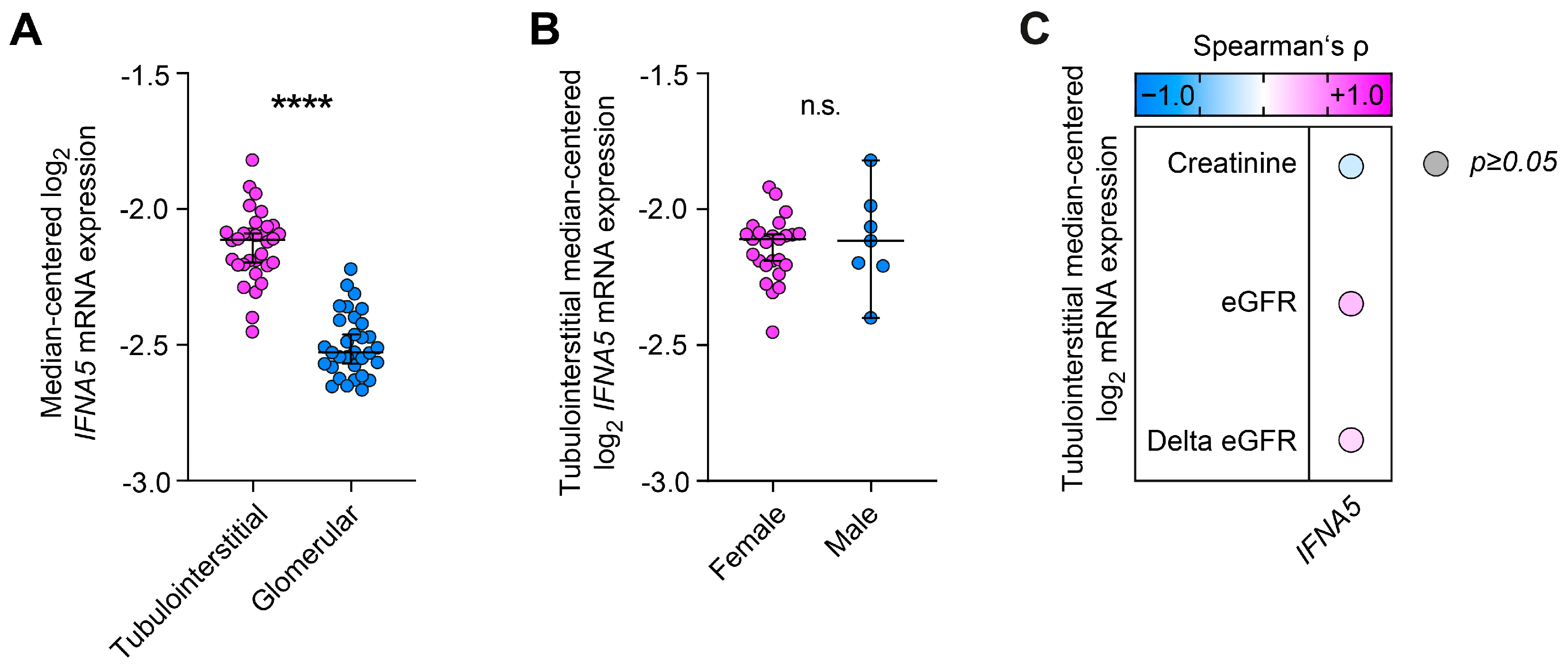
Next, we characterized tubulointerstitial and glomerular type I interferon IFNA5 expression with regard to clinical and laboratory parameters in lupus nephritis. We identified a predominant tubulointerstitial expression of IFNA5 compared to the glomerular compartment (p < 0.0001, Figure 2A). Moreover, there was no difference between female and male patients (p = 0.8237, Figure 2B) or any association between type I interferon IFNA5 expression, kidney function reflected by serum creatinine, and estimated glomerular filtration rate (eGFR) at the time of biopsy or thereafter (delta eGFR) in lupus nephritis (Figure 2C). In summary, this transcriptome array-based approach links proteinuria to the tubulointerstitial expression of type I interferon IFNA5 in lupus nephritis.
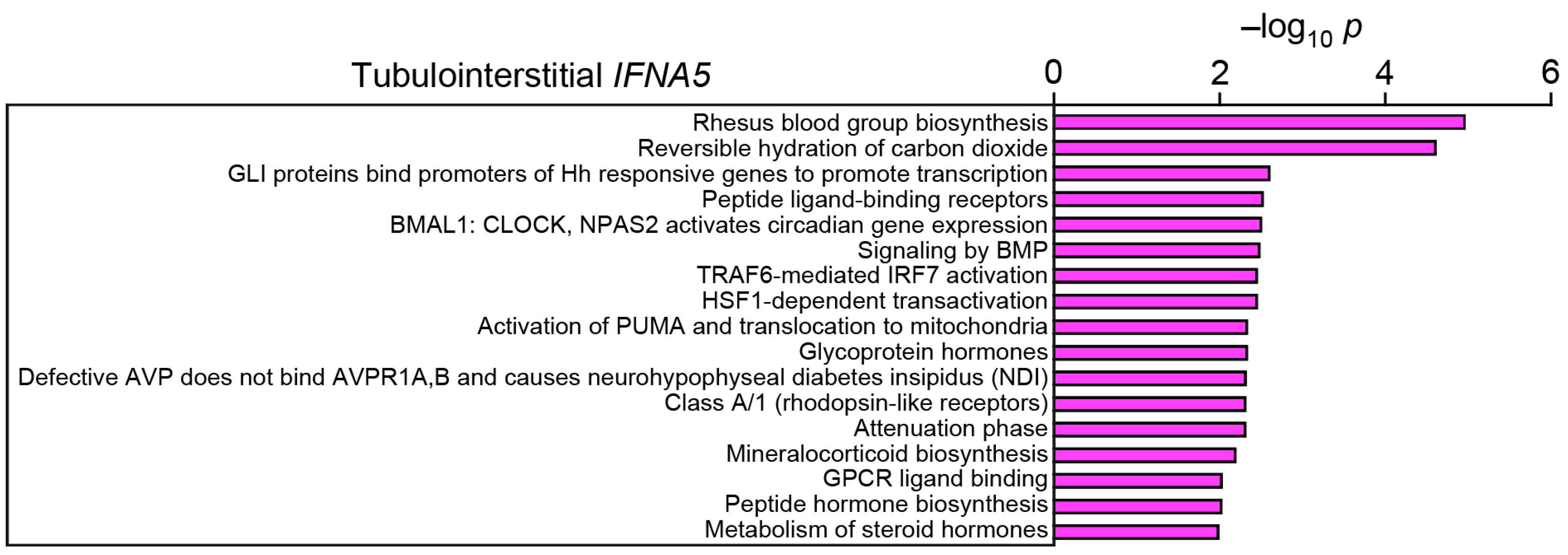
Finally, we aimed to identify molecular signatures associated with tubulointerstitial expression of type I interferon IFNA5. Tubulointerstitial IFNA5 expression was associated with enrichment of distinct molecular signatures. Particularly, pathways were enriched that are involved in transcriptional regulation: glioma-associated oncogene transcription factor (GLI) protein-regulated transcription, interferon regulatory factor 7 (IRF7) activation, and heat shock factor 1 (HSF1)-dependent transactivation (Figure 3 and Supplementary Table S4). Moreover, several receptor signaling pathways were associated with intrarenal IFN5A expression: bone morphogenic protein (BMP) signaling and G-protein coupled receptor (GPCR) ligand binding (Figure 3 and Supplementary Table S4). In summary, we here identified novel molecular signatures associated with intrarenal expression of type I interferon IFNA5.

3. Discussion

Interferons are potent stimulators of autoimmunity in a wide range of tissues affected by SLE, including the vasculature, and all nucleated cells express type I interferon receptors. Most tissues affected by SLE exhibit higher expression of interferon-stimulated genes than healthy individuals. Accumulating evidence indicates that tissue sources of type I interferons in response to environmental or other local stimuli are relevant in disease pathogenesis and clinical presentation rather than circulating sources [24,25]. Here we pursued a transcriptome array-based approach and linked proteinuria to the intrarenal expression of type I interferon IFNA5 in lupus nephritis.
Genetic studies have established a positive association between IFNA5 and SLE [35]. Regarding the kidney, it has already been shown that type I interferons are highly expressed in tubular epithelial cells [30]. In lupus nephritis, it has been demonstrated that local production of IFN-α was associated with a type I interferon signature in renal proximal tubular epithelium [30]. Therefore, local synthesis of type I interferon may act with an autocrine effect on RPTECs, leading to amplification of the tubulointerstitial injury in lupus nephritis. This aligns with our observation that IFNA5 is predominantly expressed in the tubulointerstitial compartment in lupus nephritis. Stimulation of tubular epithelial cells with type I interferon induces the expression of the immunoproteasome subunit LMP-7 that might shape the repertoire of antigens presented via MHC class I molecules [30]. Similarly, upregulation of LMP-7 is found in lupus nephritis along with transcriptional induction of gene sets involved in antigen processing and presentation, response to hypoxia, and SLE pathogenesis [30]. Consistently, results from single-cell transcriptomic studies in lupus nephritis demonstrated that tubular epithelial cell interferon scores are associated with proliferative histology, markers of fibrosis, a keratinocyte interferon score, and non-response to therapy [36]. By contrast, interferon scores in B, T, and NK cells and in monocytes are not associated with clinical response. In addition, the number of dendritic cells is low in lupus nephritis and is not associated with interferon-stimulated genes [36]. These observations suggest non-hematopoietic local sources of type I interferon in human SLE autoimmunity [24,25].
We here provide further insights into the intrarenal expression of type I interferons and show that tubulointerstitial IFNA5 expression might be relevant specifically in lupus nephritis autoimmunity. Furthermore, there was no association between type I interferon IFNA5 expression and kidney function or progressive disease. These observations imply that type I interferon IFNA5 expression might be of relevance, particularly in early stages of renal involvement in SLE, which is also of relevance for outcome [31,32]. Finally, we identified distinct molecular signatures associated with intrarenal expression of type I interferon IFNA5. Among them, these were involved in transcriptional regulation (GLI protein-regulated transcription, IRF7 activation, and HSF1-dependent transactivation) and receptor signaling (BMP signaling and GPCR ligand binding). Interestingly, a functional variant of IRF7 with consecutive activation has already been shown to predispose to the development of SLE [37]. Furthermore, we here identified an association between intrarenal IFNA5 expression and GPCR ligand binding. As part of the GPCR protein family, the complement receptor C5aR (synonym CD88) has already been described in human lupus nephritis [38,39]. Moreover, enhanced C5a/C5aR signaling has been shown to promote development of experimental lupus nephritis [40]. These observations support our finding that identified molecular signatures associated with intrarenal type I interferon could be of relevance, requiring further investigation especially in the context of current therapeutic strategies targeting interferon signaling in lupus nephritis.
Among emerging therapeutic approaches targeting interferon signaling, type I interferon receptor subunit I antagonism by anifrolumab has recently been investigated in active SLE [41]. Anifrolumab (also known as MEDI546) is a fully human, effector-null, Ig G1 κ monoclonal antibody that binds to IFNAR1 and thereby blocks type I interferon signaling [41]. Anifrolumab is currently in clinical development for treatment of SLE and lupus nephritis. Its crystal structure confirmed that anifrolumab sterically inhibits type I interferons by binding to IFNAR1 [42]. Anifrolumab was engineered with a triple mutation L234F/L235E/P331S in the heavy chain to reduce engagement with the cell surface receptor FcγR and potential Fc-mediated effector functions (e.g., effector-null), such as antibody-dependent cell-mediated cytotoxicity and complement-dependent cytotoxicity. Therefore, the current study further emphasizes the role of type I interferons in lupus nephritis and might also be of relevance for mechanistic studies. We also identified that proteinuria correlated with intrarenal synthesis of complement C5. Moreover, this is of particular interest since ongoing trials are currently testing efficacy and safety of anti-C5 antibodies (clinical trial: NCT04564339) and C5a receptor (C5aR) antagonists (clinical trial: NCT02151409) in patients with lupus nephritis.
Although we are aware that these observations require further validation in experimental models, we here provide the first evidence to link proteinuria and intrarenal expression of type I interferon IFNA5 in lupus nephritis.

4. Materials and Methods

4.1. Analyses of Publicly Available Array Datasets

Publicly available datasets were extracted from the Nephroseq database (www.nephroseq.org, 30 April 2023, University of Michigan, Ann Arbor, MI, USA) [34]. Particularly, median-centered log2 mRNA expression levels of IFNA1 (reporter ID: 208375_at), IFNA2 (211338_at), IFNA4 (207964_x_at), IFNA5 (214569_at), IFNA6 (208548_at), IFNA7 (208259_x_at), IFNA8 (207932_at), IFNA10 (208261_x_at), IFNA13 (208344_x_at), IFNA14 (208182_x_at), IFNA16 (208448_x_at), IFNA17 (211405_x_at), IFNA21 (211145_x_at), IFNW1 (207817_at), and IFNB1 (208173_at) were extracted specifically from microdissected tubulointerstitial (n = 32) and glomerular compartments (n = 32, platform: Affymetrix Human Genome U133 Plus 2.0 Array, altCDF v10).

4.2. Gene Set Enrichment

For gene set enrichment analysis, genes coexpressed with IFNA5 were extracted from the whole dataset (including 199 glomerular and 201 tubulointerstitial compartments) from the Nephroseq database (www.nephroseq.org, 30 April 2023, University of Michigan, Ann Arbor, MI, USA). Pathway analysis was performed for gene enrichment associated with log2 IFNA5 mRNA expression with a correlation threshold of ≥0.4 by using reactome (http://reactome.org, 30 April 2023); pathways with a predefined entities value of p ≤ 0.01 were included and shown in Supplementary Table S4.

4.3. Statistical Analysis

Spearman’s correlation was used to assess the correlation between continuous variables, and the Mann–Whitney U-test was used to determine differences between medians. Data analyses were performed with GraphPad Prism (version 9.3.1 for macOS, GraphPad Software, San Diego, CA, USA). A value of p < 0.05 was considered statistically significant.

Supplementary Materials

The supporting information can be downloaded at: https://www.mdpi.com/article/10.3390/ijms241310636/s1.

Author Contributions

Conceptualization, B.T.; methodology, B.T.; software, B.T.; validation, B.T.; formal analysis, B.T.; investigation, B.T.; resources, B.T.; data curation, B.T.; writing—original draft preparation, B.T.; writing—review and editing, P.K.; visualization, B.T.; supervision, B.T.; project administration, B.T.; funding acquisition, P.K. and B.T. All authors have read and agreed to the published version of the manuscript.

Funding

We acknowledge support by the Open Access Publication Funds of the Göttingen University.

Institutional Review Board Statement

Data were extracted from Nephroseq Research Edition (Ann Arbor, MI: University of Michigan; available from: www.nephroseq.org, 30 April 2023).

Informed Consent Statement

Patient consent for participation was not required for this study because data were extracted from Nephroseq Research Edition (Ann Arbor, MI: University of Michigan; available from: www.nephroseq.org, 30 April 2023).

Data Availability Statement

The original contributions presented in the study are included in the article’s Supplementary Materials; further data and material are available from the corresponding author upon reasonable request.

Conflicts of Interest

P.K. has received research grants from GlaxoSmithKline and Diamed Medizintechnik GmbH, all unrelated to this paper. P.K. has received honoraria or travel support from Abbvie, Amgen, AstraZeneca, Boehringer Ingelheim, Bristol-Myers-Squibb, Chugai, Galapagos, GlaxoSmithKline, Janssen-Cilag, Lilly, Novartis, and Pfizer, all unrelated to this paper. B.T. has received research grants from Evotec SE and CSL Vifor, all unrelated to this paper. B.T. has received honoraria or travel support from CSL Vifor, all unrelated to this paper.

References

  1. Tsokos, G.C. Systemic Lupus Erythematosus. N. Engl. J. Med. 2011, 365, 2110–2121. [Google Scholar] [CrossRef] [PubMed] [Green Version]
  2. Lam, N.C.; Ghetu, M.V.; Bieniek, M.L. Systemic Lupus Erythematosus: Primary Care Approach to Diagnosis and Management. Am. Fam. Physician 2016, 94, 284–294. [Google Scholar] [PubMed]
  3. Lisnevskaia, L.; Murphy, G.; Isenberg, D. Systemic Lupus Erythematosus. Lancet 2014, 384, 1878–1888. [Google Scholar] [CrossRef]
  4. Weckerle, C.E.; Franek, B.S.; Kelly, J.A.; Kumabe, M.; Mikolaitis, R.A.; Green, S.L.; Utset, T.O.; Jolly, M.; James, J.A.; Harley, J.B.; et al. Network Analysis of Associations between Serum Interferon-Alpha Activity, Autoantibodies, and Clinical Features in Systemic Lupus Erythematosus. Arthritis Rheum. 2011, 63, 1044–1053. [Google Scholar] [CrossRef]
  5. Kirou, K.A.; Lee, C.; George, S.; Louca, K.; Peterson, M.G.; Crow, M.K. Activation of the Interferon-Alpha Pathway Identifies a Subgroup of Systemic Lupus Erythematosus Patients with Distinct Serologic Features and Active Disease. Arthritis Rheum. 2005, 52, 1491–1503. [Google Scholar] [CrossRef] [PubMed]
  6. Hall, J.C.; Rosen, A. Type I Interferons: Crucial Participants in Disease Amplification in Autoimmunity. Nat. Rev. Rheumatol. 2010, 6, 40–49. [Google Scholar] [CrossRef] [Green Version]
  7. Ivashkiv, L.B.; Donlin, L.T. Regulation of Type I Interferon Responses. Nat. Rev. Immunol. 2014, 14, 36–49. [Google Scholar] [CrossRef] [Green Version]
  8. Hooks, J.J.; Moutsopoulos, H.M.; Geis, S.A.; Stahl, N.I.; Decker, J.L.; Notkins, A.L. Immune Interferon in the Circulation of Patients with Autoimmune Disease. N. Engl. J. Med. 1979, 301, 5–8. [Google Scholar] [CrossRef] [PubMed]
  9. Chyuan, I.T.; Tzeng, H.T.; Chen, J.Y. Signaling Pathways of Type I and Type Iii Interferons and Targeted Therapies in Systemic Lupus Erythematosus. Cells 2019, 8, 963. [Google Scholar] [CrossRef] [Green Version]
  10. Kim, J.M.; Park, S.H.; Kim, H.Y.; Kwok, S.K. A Plasmacytoid Dendritic Cells-Type I Interferon Axis Is Critically Implicated in the Pathogenesis of Systemic Lupus Erythematosus. Int. J. Mol. Sci. 2015, 16, 14158–14170. [Google Scholar] [CrossRef]
  11. Goropevsek, A.; Holcar, M.; Avcin, T. The Role of Stat Signaling Pathways in the Pathogenesis of Systemic Lupus Erythematosus. Clin. Rev. Allergy Immunol. 2017, 52, 164–181. [Google Scholar] [CrossRef]
  12. Sozzani, S.; Bosisio, D.; Scarsi, M.; Tincani, A. Type I Interferons in Systemic Autoimmunity. Autoimmunity 2010, 43, 196–203. [Google Scholar] [CrossRef]
  13. Crow, M.K.; Olferiev, M.; Kirou, K.A. Targeting of Type I Interferon in Systemic Autoimmune Diseases. Transl. Res. 2015, 165, 296–305. [Google Scholar] [CrossRef] [PubMed] [Green Version]
  14. Kessler, D.S.; Levy, D.E.; Darnell, J.E., Jr. Two Interferon-Induced Nuclear Factors Bind a Single Promoter Element in Interferon-Stimulated Genes. Proc. Natl. Acad. Sci. USA 1988, 85, 8521–8525. [Google Scholar] [CrossRef] [PubMed] [Green Version]
  15. Levy, D.E.; Kessler, D.S.; Pine, R.; Reich, N.; Darnell, J.E., Jr. Interferon-Induced Nuclear Factors That Bind a Shared Promoter Element Correlate with Positive and Negative Transcriptional Control. Genes Dev. 1988, 2, 383–393. [Google Scholar] [CrossRef] [PubMed] [Green Version]
  16. Kim, S.; Kaiser, V.; Beier, E.; Bechheim, M.; Guenthner-Biller, M.; Ablasser, A.; Berger, M.; Endres, S.; Hartmann, G.; Hornung, V. Self-Priming Determines High Type I Ifn Production by Plasmacytoid Dendritic Cells. Eur. J. Immunol. 2014, 44, 807–818. [Google Scholar] [CrossRef] [Green Version]
  17. Liao, A.P.; Salajegheh, M.; Morehouse, C.; Nazareno, R.; Jubin, R.G.; Jallal, B.; Yao, Y.; Greenberg, S.A. Human Plasmacytoid Dendritic Cell Accumulation Amplifies Their Type 1 Interferon Production. Clin. Immunol. 2010, 136, 130–138. [Google Scholar] [CrossRef] [Green Version]
  18. Deng, Y.; Tsao, B.P. Advances in Lupus Genetics and Epigenetics. Curr. Opin. Rheumatol. 2014, 26, 482–492. [Google Scholar] [CrossRef]
  19. Postal, M.; Sinicato, N.A.; Pelicari, K.O.; Marini, R.; Costallat, L.T.L.; Appenzeller, S. Clinical and Serological Manifestations Associated with Interferon-Alpha Levels in Childhood-Onset Systemic Lupus Erythematosus. Clinics 2012, 67, 157–162. [Google Scholar] [CrossRef]
  20. Dall’era, M.C.; Cardarelli, P.M.; Preston, B.T.; Witte, A.; Davis, J.C., Jr. Type I Interferon Correlates with Serological and Clinical Manifestations of Sle. Ann. Rheum. Dis. 2005, 64, 1692–1697. [Google Scholar] [CrossRef]
  21. Ioannou, Y.; Isenberg, D.A. Current Evidence for the Induction of Autoimmune Rheumatic Manifestations by Cytokine Therapy. Arthritis Rheum. 2000, 43, 1431–1442. [Google Scholar] [CrossRef]
  22. Panda, S.K.; Kolbeck, R.; Sanjuan, M.A. Plasmacytoid Dendritic Cells in Autoimmunity. Curr. Opin. Immunol. 2017, 44, 20–25. [Google Scholar] [CrossRef] [PubMed]
  23. Lovgren, T.; Eloranta, M.L.; Bave, U.; Alm, G.V.; Ronnblom, L. Induction of Interferon-Alpha Production in Plasmacytoid Dendritic Cells by Immune Complexes Containing Nucleic Acid Released by Necrotic or Late Apoptotic Cells and Lupus Igg. Arthritis Rheum. 2004, 50, 1861–1872. [Google Scholar] [CrossRef] [PubMed]
  24. Psarras, A.; Alase, A.; Antanaviciute, A.; Carr, I.M.; Yusof, M.Y.M.; Wittmann, M.; Emery, P.; Tsokos, G.C.; Vital, E.M. Functionally Impaired Plasmacytoid Dendritic Cells and Non-Haematopoietic Sources of Type I Interferon Characterize Human Autoimmunity. Nat. Commun. 2020, 11, 6149. [Google Scholar] [CrossRef]
  25. Psarras, A.; Wittmann, M.; Vital, E.M. Emerging Concepts of Type I Interferons in Sle Pathogenesis and Therapy. Nat. Rev. Rheumatol. 2022, 18, 575–590. [Google Scholar] [CrossRef] [PubMed]
  26. Md Yusof, M.Y.; Psarras, A.; El-Sherbiny, Y.M.; Hensor, E.M.A.; Dutton, K.; Ul-Hassan, S.; Zayat, A.S.; Shalbaf, M.; Alase, A.; Wittmann, M.; et al. Prediction of Autoimmune Connective Tissue Disease in an at-Risk Cohort: Prognostic Value of a Novel Two-Score System for Interferon Status. Ann. Rheum. Dis. 2018, 77, 1432–1439. [Google Scholar] [CrossRef] [PubMed] [Green Version]
  27. Cherian, T.S.; Kariuki, S.N.; Franek, B.S.; Buyon, J.P.; Clancy, R.M.; Niewold, T.B. Brief Report: Irf5 Systemic Lupus Erythematosus Risk Haplotype Is Associated with Asymptomatic Serologic Autoimmunity and Progression to Clinical Autoimmunity in Mothers of Children with Neonatal Lupus. Arthritis Rheum. 2012, 64, 3383–3387. [Google Scholar] [CrossRef] [PubMed] [Green Version]
  28. Niewold, T.B.; Hua, J.; Lehman, T.J.; Harley, J.B.; Crow, M.K. High Serum Ifn-Alpha Activity Is a Heritable Risk Factor for Systemic Lupus Erythematosus. Genes Immun. 2007, 8, 492–502. [Google Scholar] [CrossRef]
  29. Lu, R.; Munroe, M.E.; Guthridge, J.M.; Bean, K.M.; Fife, D.A.; Chen, H.; Slight-Webb, S.R.; Keith, M.P.; Harley, J.B.; James, J.A. Dysregulation of Innate and Adaptive Serum Mediators Precedes Systemic Lupus Erythematosus Classification and Improves Prognostic Accuracy of Autoantibodies. J. Autoimmun. 2016, 74, 182–193. [Google Scholar] [CrossRef] [Green Version]
  30. Castellano, G.; Cafiero, C.; Divella, C.; Sallustio, F.; Gigante, M.; Pontrelli, P.; De Palma, G.; Rossini, M.; Grandaliano, G.; Gesualdo, L. Local Synthesis of Interferon-Alpha in Lupus Nephritis Is Associated with Type I Interferons Signature and Lmp7 Induction in Renal Tubular Epithelial Cells. Arthritis Res. Ther. 2015, 17, 72. [Google Scholar] [CrossRef] [Green Version]
  31. Dall’Era, M.; Cisternas, M.G.; Smilek, D.E.; Straub, L.; Houssiau, F.A.; Cervera, R.; Rovin, B.H.; Mackay, M. Predictors of Long-Term Renal Outcome in Lupus Nephritis Trials: Lessons Learned from the Euro-Lupus Nephritis Cohort. Arthritis Rheumatol. 2015, 67, 1305–1313. [Google Scholar] [CrossRef] [PubMed]
  32. Tamirou, F.; Lauwerys, B.R.; Dall’Era, M.; Mackay, M.; Rovin, B.; Cervera, R.; Houssiau, F.A.; Maintain Nephritis Trial Investigators. A Proteinuria Cut-Off Level of 0.7 G/Day after 12 Months of Treatment Best Predicts Long-Term Renal Outcome in Lupus Nephritis: Data from the Maintain Nephritis Trial. Lupus Sci. Med. 2015, 2, e000123. [Google Scholar] [CrossRef] [Green Version]
  33. Razaghi, A.; Brusselaers, N.; Bjornstedt, M.; Durand-Dubief, M. Copy Number Alteration of the Interferon Gene Cluster in Cancer: Individual Patient Data Meta-Analysis Prospects to Personalized Immunotherapy. Neoplasia 2021, 23, 1059–1068. [Google Scholar] [CrossRef] [PubMed]
  34. Berthier, C.C.; Bethunaickan, R.; Gonzalez-Rivera, T.; Nair, V.; Ramanujam, M.; Zhang, W.; Bottinger, E.P.; Segerer, S.; Lindenmeyer, M.; Cohen, C.D.; et al. Cross-Species Transcriptional Network Analysis Defines Shared Inflammatory Responses in Murine and Human Lupus Nephritis. J. Immunol. 2012, 189, 988–1001. [Google Scholar] [CrossRef] [PubMed] [Green Version]
  35. Hirankarn, N.; Tangwattanachuleeporn, M.; Wongpiyabovorn, J.; Wongchinsri, J.; Avihingsanon, Y. Genetic Association of Interferon-Alpha Subtypes 1, 2 and 5 in Systemic Lupus Erythematosus. Tissue Antigens 2008, 72, 588–592. [Google Scholar] [CrossRef]
  36. Der, E.; Ranabothu, S.; Suryawanshi, H.; Akat, K.M.; Clancy, R.; Morozov, P.; Kustagi, M.; Czuppa, M.; Izmirly, P.; Belmont, H.M.; et al. Single Cell Rna Sequencing to Dissect the Molecular Heterogeneity in Lupus Nephritis. JCI Insight 2017, 2, e93009. [Google Scholar] [CrossRef] [Green Version]
  37. Fu, Q.; Zhao, J.; Qian, X.; Wong, J.L.; Kaufman, K.M.; Yu, C.Y.; Siew, H.H.; Group Tan Tock Seng Hospital Lupus Study; Mok, M.Y.; Harley, J.B.; et al. Association of a Functional Irf7 Variant with Systemic Lupus Erythematosus. Arthritis Rheum. 2011, 63, 749–754. [Google Scholar] [CrossRef] [Green Version]
  38. Tampe, D.; Hakroush, S.; Tampe, B. Dissecting Signalling Pathways Associated with Intrarenal Synthesis of Complement Components in Lupus Nephritis. RMD Open. 2022, 8, e002517. [Google Scholar] [CrossRef]
  39. Tampe, D.; Hakroush, S.; Tampe, B. Molecular Signatures of Intrarenal Complement Receptors C3ar1 and C5ar1 Correlate with Renal Outcome in Human Lupus Nephritis. Lupus Sci. Med. 2022, 9, e000831. [Google Scholar] [CrossRef]
  40. Bao, L.; Osawe, I.; Puri, T.; Lambris, J.D.; Haas, M.; Quigg, R.J. C5a Promotes Development of Experimental Lupus Nephritis Which Can Be Blocked with a Specific Receptor Antagonist. Eur. J. Immunol. 2005, 35, 2496–2506. [Google Scholar] [CrossRef]
  41. Morand, E.F.; Furie, R.; Tanaka, Y.; Bruce, I.N.; Askanase, A.D.; Richez, C.; Bae, S.C.; Brohawn, P.Z.; Pineda, L.; Berglind, A.; et al. Trial of Anifrolumab in Active Systemic Lupus Erythematosus. N. Engl. J. Med. 2020, 382, 211–221. [Google Scholar] [CrossRef] [PubMed]
  42. Peng, L.; Oganesyan, V.; Wu, H.; Dall’Acqua, W.F.; Damschroder, M.M. Molecular Basis for Antagonistic Activity of Anifrolumab, an Anti-Interferon-Alpha Receptor 1 Antibody. MAbs 2015, 7, 428–439. [Google Scholar] [CrossRef] [PubMed] [Green Version]
Figure 1. Proteinuria is associated with intrarenal expression of type I interferon IFNA2 in lupus nephritis. Median-centered log2 mRNA expression levels of IFNα (IFNA1, IFNA2, IFNA4, IFNA5, IFNA6, IFNA7, IFNA8, IFNA10, IFNA13, IFNA14, IFNA16, IFNA17, and IFNA21), IFNω (IFNW1), and IFNβ (IFNB1) in microdissected tubulointerstitial (n = 32) and glomerular compartments (n = 32) are shown in correlation with proteinuria in lupus nephritis. The heatmap reflects the mean values of Spearman’s ρ; circle size represents the significance level.
Ijms 24 10636 g001
Figure 2. Predominant tubulointerstitial type I interferon IFNA5 expression is independent of sex, kidney function at time of kidney biopsy, or kidney function thereafter (delta eGFR) in lupus nephritis. (A) Median-centered log2 IFNA5 mRNA expression levels in lupus nephritis are shown by group separation for tubulointerstitial and glomerular mRNA expression levels. Group comparisons were performed using the Mann–Whitney U-test to determine differences in medians (**** p < 0.0001). (B) Tubulointerstitial median-centered log2 IFNA5 mRNA expression levels in lupus nephritis are shown by group separation for female and male sex. Comparisons of groups were performed using the Mann–Whitney U-test to determine differences in medians (n.s. not significant). (C) Correlations between tubulointerstitial IFNA5 mRNA expression levels and laboratory markers of kidney function in lupus nephritis are shown by heatmap reflecting the mean values of Spearman’s ρ; the circle size represents the significance level.
Ijms 24 10636 g002
Figure 3. Tubulointerstitial type I interferon IFNA5 expression is associated with distinct molecular signatures in the kidney: −log10 p values of signaling pathways associated with tubulointerstitial IFNA5 mRNA expression in the kidney are shown.
Ijms 24 10636 g003
Disclaimer/Publisher’s Note: The statements, opinions and data contained in all publications are solely those of the individual author(s) and contributor(s) and not of MDPI and/or the editor(s). MDPI and/or the editor(s) disclaim responsibility for any injury to people or property resulting from any ideas, methods, instructions or products referred to in the content.