
Therapeutic Candidates for Alzheimer’s Disease: Saponins

 ,
,
Abstract
1. Introduction
2. Mechanism of Saponins in Treating Alzheimer’s Disease
2.1. Inhibition of Aβ Deposition and Neurotoxicity
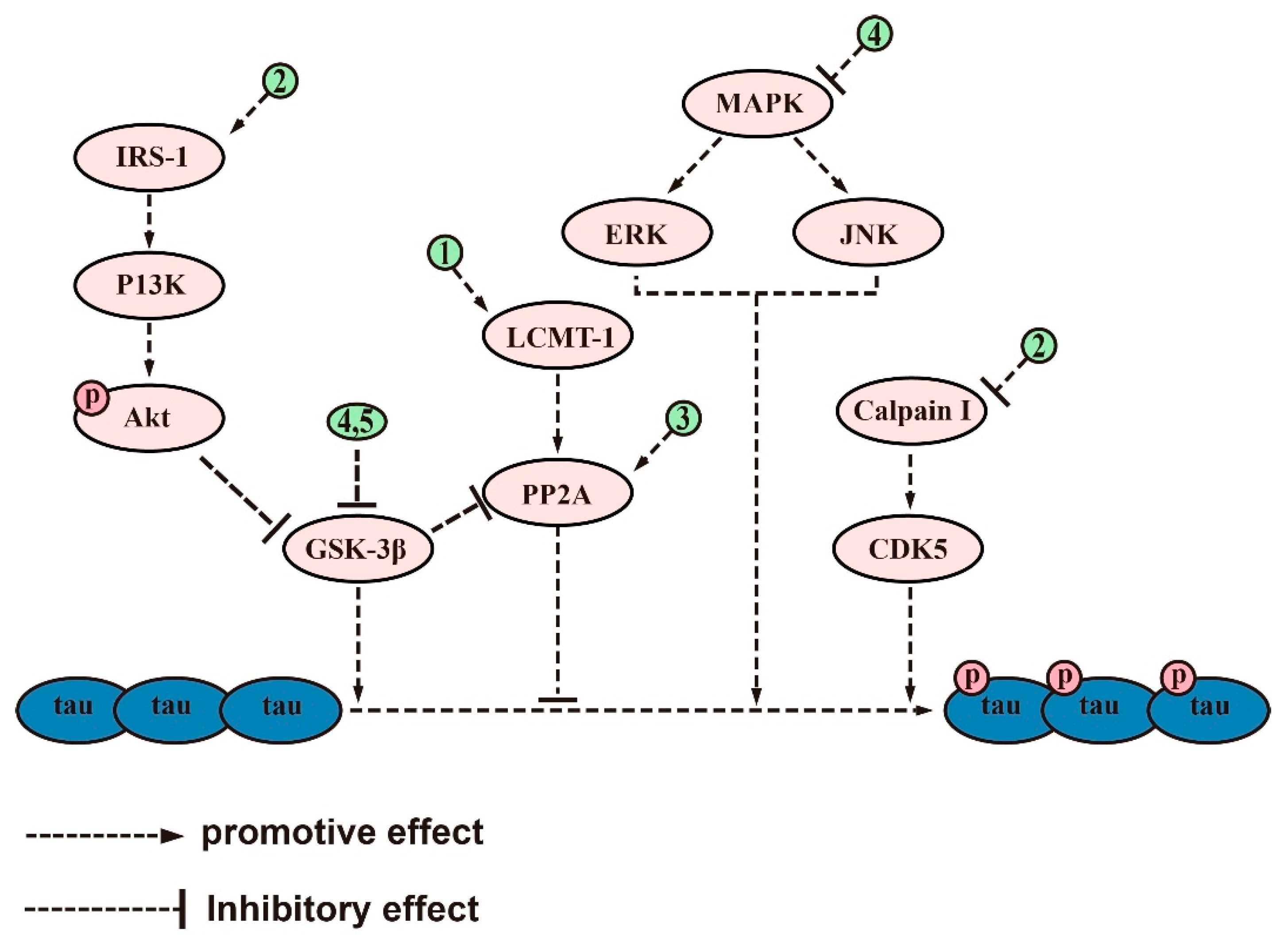
2.2. Inhibiting Aberrant Tau Protein Phosphorylation
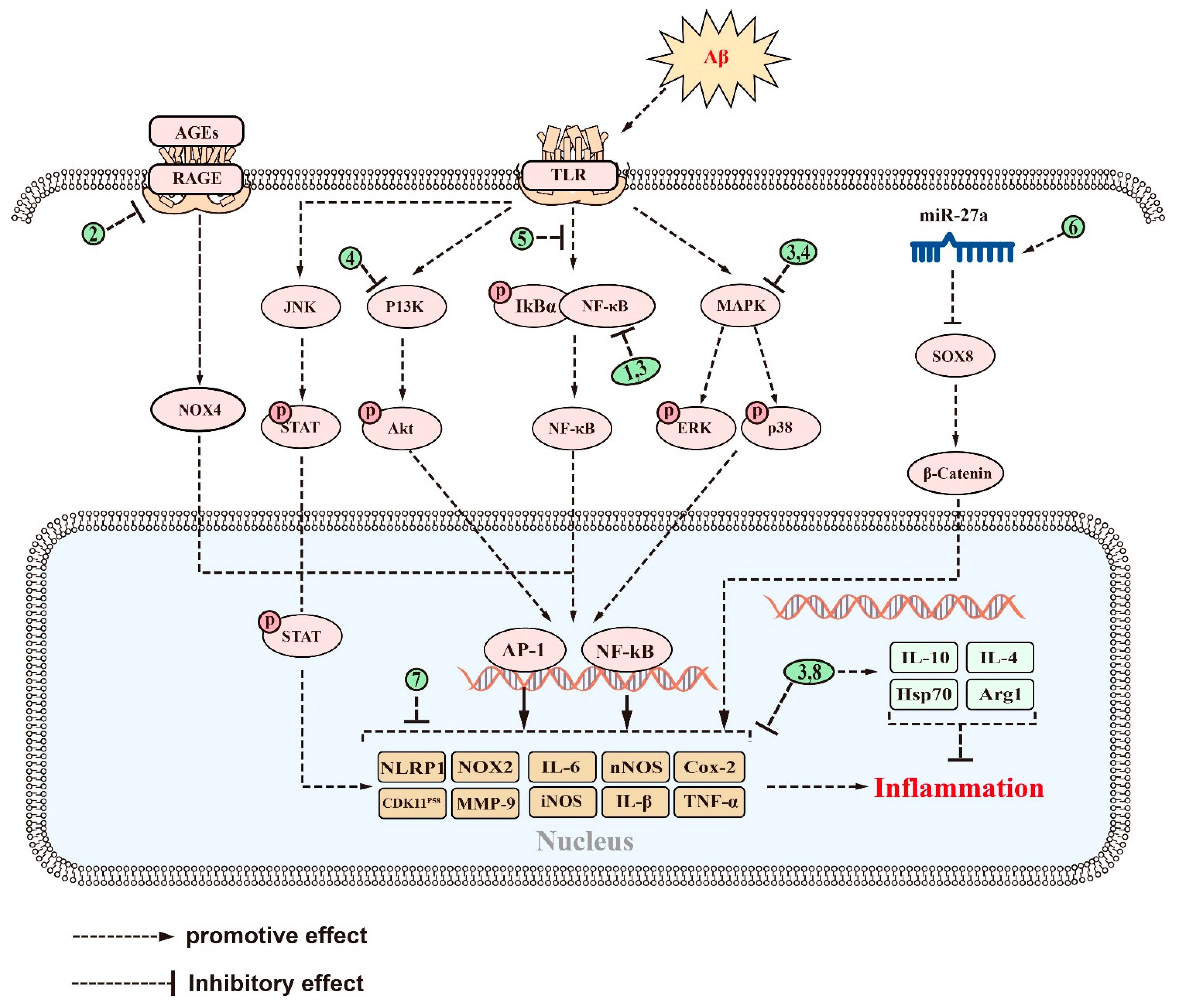
2.3. Anti-Inflammatory Effect
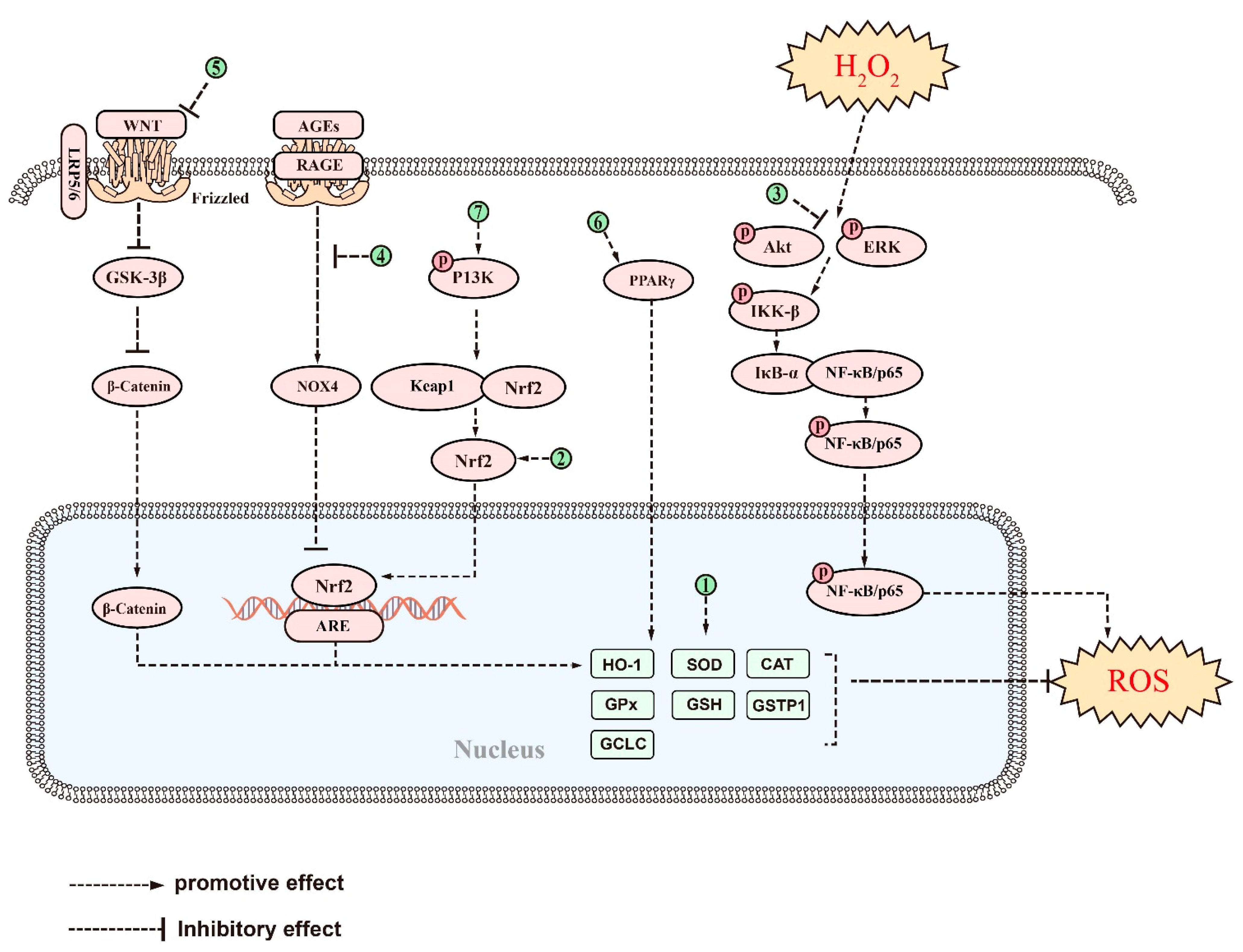
2.4. Improvement in Mitochondrial Function and Antioxidative Stress
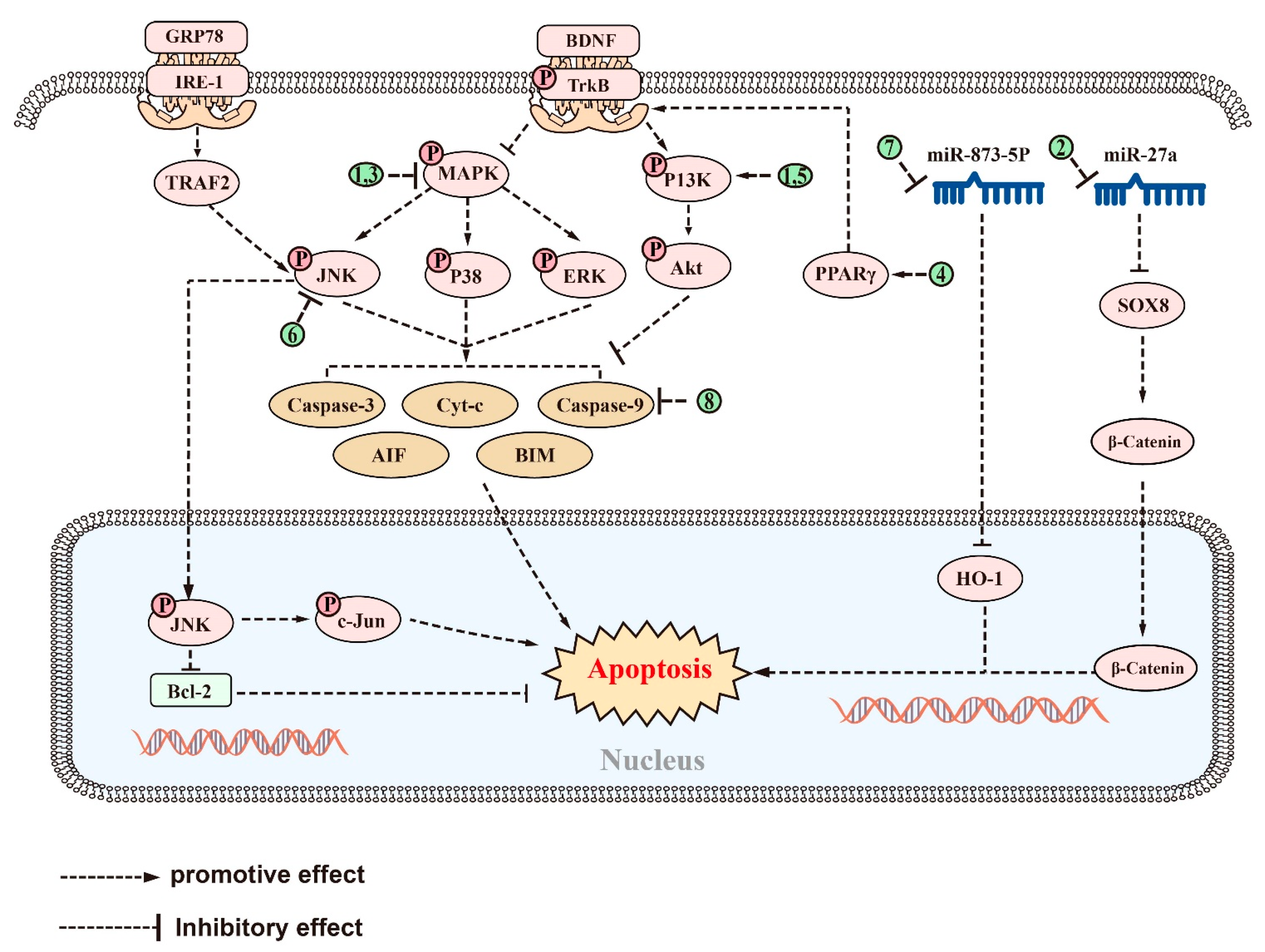
2.5. Antiapoptotic Effect
3. Discussion
4. Conclusions and Perspective
5. Limitations and Future Directions
Author Contributions
Funding
Conflicts of Interest
References
- Vossel, K.; Ranasinghe, K.G.; Beagle, A.J.; La, A.; Ah, P.K.; Castro, M.; Mizuiri, D.; Honma, S.M.; Venkateswaran, N.; Koestler, M.; et al. Effect of Levetiracetam on Cognition in Patients with Alzheimer Disease With and Without Epileptiform Activity: A Randomized Clinical Trial. JAMA Neurol. 2021, 78, 1345–1354. [Google Scholar] [CrossRef] [PubMed]
- Balupuri, A.; Choi, K.E.; Kang, N.S. Aggregation Mechanism of Alzheimer’s Amyloid β-Peptide Mediated by α-Strand/α-Sheet Structure. Int. J. Mol. Sci. 2020, 21, 1094. [Google Scholar] [CrossRef] [PubMed]
- Grimm, A.; Biliouris, E.E.; Lang, U.E.; Götz, J.; Mensah-Nyagan, A.G.; Eckert, A. Sex hormone-related neurosteroids differentially rescue bioenergetic deficits induced by amyloid-β or hyperphosphorylated tau protein. Cell. Mol. Life Sci. 2016, 73, 201–215. [Google Scholar] [CrossRef]
- Zheng, H.; Fridkin, M.; Youdim, M. From single target to multitarget/network therapeutics in Alzheimer’s therapy. Pharmaceuticals 2014, 7, 113–135. [Google Scholar] [CrossRef]
- Tamayev, R.; Matsuda, S.; Arancio, O.; D’Adamio, L. β- but not γ-secretase proteolysis of APP causes synaptic and memory deficits in a mouse model of dementia. EMBO Mol. Med. 2012, 4, 171–179. [Google Scholar] [CrossRef]
- Chandra, S.; Roy, A.; Jana, M.; Pahan, K. Cinnamic acid activates PPARα to stimulate Lysosomal biogenesis and lower Amyloid plaque pathology in an Alzheimer’s disease mouse model. Neurobiol. Dis. 2019, 124, 379–395. [Google Scholar] [CrossRef]
- Polis, B.; Srikanth, K.D.; Elliott, E.; Gil-Henn, H.; Samson, A.O. L-Norvaline Reverses Cognitive Decline and Synaptic Loss in a Murine Model of Alzheimer’s Disease. Neurotherapeutics 2018, 15, 1036–1054. [Google Scholar] [CrossRef]
- Steffen, J.; Krohn, M.; Schwitlick, C.; Brüning, T.; Paarmann, K.; Pietrzik, C.U.; Biverstål, H.; Jansone, B.; Langer, O.; Pahnke, J. Expression of endogenous mouse APP modulates β-amyloid deposition in hAPP-transgenic mice. Acta Neuropathol. Commun. 2017, 20, 49. [Google Scholar] [CrossRef] [PubMed]
- Govindpani, K.; Turner, C.; Waldvogel, H.J.; Faull, R.L.M.; Kwakowsky, A. Impaired Expression of GABA Signaling Components in the Alzheimer’s Disease Middle Temporal Gyrus. Int. J. Mol. Sci. 2020, 21, 8704. [Google Scholar] [CrossRef]
- Ruthirakuhan, M.; Herrmann, N.; Vieira, D.; Gallagher, D.; Lanctot, K.L. The Roles of Apathy and Depression in Predicting Alzheimer Disease: A Longitudinal Analysis in Older Adults With Mild Cognitive Impairment. Am. J. Geriatr. Psychiatry 2019, 27, 873–882. [Google Scholar] [CrossRef]
- Mphahlele, M.J.; Gildenhuys, S.; Agbo, E.N. In Vitro Evaluation and Docking Studies of 5-oxo-5H-furo [3,2-g] chromene-6-carbaldehyde Derivatives as Potential Anti-Alzheimer’s Agents. Int. J. Mol. Sci. 2019, 20, 5451. [Google Scholar] [CrossRef] [PubMed]
- Saleem, F.; Mehmood, R.; Mehar, S.; Khan, M.T.J.; Khan, Z.U.; Ashraf, M.; Ali, M.S.; Abdullah, I.; Froeyen, M.; Mirza, M.U.; et al. Bioassay Directed Isolation, Biological Evaluation and in Silico Studies of New Isolates from Pteris cretica L. Antioxidants 2019, 8, 231. [Google Scholar] [CrossRef] [PubMed]
- Lee, B.D.; Yoo, J.M.; Baek, S.Y.; Li, F.Y.; Sok, D.E.; Kim, M.R. 3,3’-Diindolylmethane Promotes BDNF and Antioxidant Enzyme Formation via TrkB/Akt Pathway Activation for Neuroprotection against Oxidative Stress-Induced Apoptosis in Hippocampal Neuronal Cells. Antioxidants 2019, 9, 3. [Google Scholar] [CrossRef]
- Chen, C.; Zhu, H.; Kang, J.; Warusawitharana, H.K.; Chen, S.; Wang, K.; Yu, F.; Wu, Y.; He, P.; Tu, Y.; et al. Comparative Transcriptome and Phytochemical Analysis Provides Insight into Triterpene Saponin Biosynthesis in Seeds and Flowers of the Tea Plant (Camellia sinensis). Metabolites 2022, 12, 204. [Google Scholar] [CrossRef] [PubMed]
- Yoon, S.; Iqbal, H.; Kim, S.M.; Jin, M. Phytochemicals That Act on Synaptic Plasticity as Potential Prophylaxis against Stress-Induced Depressive Disorder. Biomol. Ther. 2023, 31, 148–160. [Google Scholar] [CrossRef]
- Guclu-Ustundag, O.; Mazza, G. Saponins: Properties, applications and processing. Crit. Rev. Food Sci. Nutr. 2007, 47, 231–258. [Google Scholar] [CrossRef] [PubMed]
- Ramos-Morales, E.; de la Fuente, G.; Duval, S.; Wehrli, C.; Bouillon, M.; Lahmann, M.; Preskett, D.; Braganca, R.; Newbold, C.J. Antiprotozoal Effect of Saponins in the Rumen Can Be Enhanced by Chemical Modifications in Their Structure. Front. Microbiol. 2017, 8, 399. [Google Scholar] [CrossRef]
- Ghiulai, R.; Roşca, O.J.; Antal, D.S.; Mioc, M.; Mioc, A.; Racoviceanu, R.; Macaşoi, I.; Olariu, T.; Dehelean, C.; Creţu, O.M.; et al. Tetracyclic and Pentacyclic Triterpenes with High Therapeutic Efficiency in Wound Healing Approaches. Molecules. 2020, 25, 5557. [Google Scholar] [CrossRef]
- Dashbaldan, S.; Pączkowski, C.; Szakiel, A. Variations in Triterpenoid Deposition in Cuticular Waxes during Development and Maturation of Selected Fruits of Rosaceae Family. Int. J. Mol. Sci. 2020, 21, 9762. [Google Scholar] [CrossRef]
- Massana-Codina, J.; Schnee, S.; Allard, P.M.; Rutz, A.; Boccard, J.; Michellod, E.; Cleroux, M.; Schurch, S.; Gindro, K.; Wolfender, J.L. Insights on the Structural and Metabolic Resistance of Potato (Solanum tuberosum) Cultivars to Tuber Black Dot (Colletotrichum coccodes). Front. Plant Sci. 2020, 11, 1287. [Google Scholar] [CrossRef]
- Sy, L.K.; Lok, C.N.; Wang, J.Y.; Liu, Y.; Cheng, L.; Wan, P.K.; Leung, C.T.; Cao, B.; Kwong, W.L.; Chang, R.C.; et al. Identification of “sarsasapogenin-aglyconed” timosaponins as novel Aβ-lowering modulators of amyloid precursor protein processing. Chem. Sci. 2016, 7, 3206–3214. [Google Scholar] [CrossRef]
- Liu, Y.W.; Zhu, X.; Lu, Q.; Wang, J.Y.; Li, W.; Wei, Y.Q.; Yin, X.X. Total saponins from Rhizoma Anemarrhenae ameliorate diabetes-associated cognitive decline in rats: Involvement of amyloid-beta decrease in brain. J. Ethnopharmacol. 2012, 139, 194–200. [Google Scholar] [CrossRef]
- Abduljawad, A.A.; Elawad, M.A.; Elkhalifa, M.; Ahmed, A.; Hamdoon, A.; Salim, L.; Ashraf, M.; Ayaz, M.; Hassan, S.; Bungau, S. Alzheimer’s Disease as a Major Public Health Concern: Role of Dietary Saponins in Mitigating Neurodegenerative Disorders and Their Underlying Mechanisms. Molecules 2022, 27, 6804. [Google Scholar] [CrossRef] [PubMed]
- Rao, A.V.; Gurfinkel, D.M. The bioactivity of saponins: Triterpenoid and steroidal glycosides. Drug Metabol. Drug Interact. 2000, 17, 211–235. [Google Scholar] [CrossRef] [PubMed]
- Esmeeta, A.; Adhikary, S.; Dharshnaa, V.; Swarnamughi, P.; Ummul Maqsummiya, Z.; Banerjee, A.; Pathak, S.; Duttaroy, A.K. Plant-derived bioactive compounds in colon cancer treatment: An updated review. Biomed. Pharmacother. 2022, 153, 113384. [Google Scholar] [CrossRef]
- Dandawate, P.R.; Subramaniam, D.; Padhye, S.B.; Anant, S. Bitter melon: A panacea for inflammation and cancer. Chin. J. Nat. Med. 2016, 14, 81–100. [Google Scholar] [CrossRef]
- Shalini, V.T.; Neelakanta, S.J.; Sriranjini, J.S. Neuroprotection with Bacopa monnieri—A review of experimental evidence. Mol. Biol. Rep. 2021, 48, 2653–2668. [Google Scholar] [CrossRef]
- Bahrami, Y.; Zhang, W. MMFC Distribution of Saponins in the Sea Cucumber Holothuria lessoni; the Body Wall Versus the Viscera, and Their Biological Activities. Mar. Drugs 2018, 16, 423. [Google Scholar] [CrossRef] [PubMed]
- Fujihara, K.; Shimoyama, T.; Kawazu, R.; Sasaki, H.; Koyama, K.; Takahashi, K.; Kinoshita, K. Amyloid β aggregation inhibitory activity of triterpene saponins from the cactus Stenocereus pruinosus. J. Nat. Med. 2021, 75, 284–298. [Google Scholar] [CrossRef]
- Shin, S.J.; Park, Y.H.; Jeon, S.G.; Kim, S.; Nam, Y.; Oh, S.M.; Lee, Y.Y.; Moon, M. Red Ginseng Inhibits Tau Aggregation and Promotes Tau Dissociation In Vitro. Oxid. Med. Cell. Longev. 2020, 2020, 7829842. [Google Scholar] [CrossRef]
- Khan, M.I.; Karima, G.; Khan, M.Z.; Shin, J.H.; Kim, J.D. Therapeutic Effects of Saponins for the Prevention and Treatment of Cancer by Ameliorating Inflammation and Angiogenesis and Inducing Antioxidant and Apoptotic Effects in Human Cells. Int. J. Mol. Sci. 2022, 23, 10065. [Google Scholar] [CrossRef]
- Zeng, M.; Zhang, R.; Yang, Q.; Guo, L.; Zhang, X.; Yu, B.; Gan, J.; Yang, Z.; Li, H.; Wang, Y.; et al. Pharmacological therapy to cerebral ischemia-reperfusion injury: Focus on saponins. Biomed. Pharmacother. 2022, 155, 113696. [Google Scholar] [CrossRef]
- Rudakewich, M.; Ba, F.; Benishin, C.G. Neurotrophic and neuroprotective actions of ginsenosides Rb (1) and Rg (1). Planta Med. 2001, 67, 533–537. [Google Scholar] [CrossRef]
- Morroni, F.; Sita, G.; Graziosi, A.; Turrini, E.; Fimognari, C.; Tarozzi, A.; Hrelia, P. Protective Effects of 6-(Methylsulfinyl) hexyl Isothiocyanate on Abeta (1-42)-Induced Cognitive Deficit, Oxidative Stress, Inflammation, and Apoptosis in Mice. Int. J. Mol. Sci. 2018, 19, 2083. [Google Scholar] [CrossRef]
- Mucke, L.; Selkoe, D.J. Neurotoxicity of amyloid β-protein: Synaptic and network dysfunction. Cold Spring Harb Perspect Med. 2012, 2, a006338. [Google Scholar] [CrossRef] [PubMed]
- Lesne, S.; Koh, M.T.; Kotilinek, L.; Kayed, R.; Glabe, C.G.; Yang, A.; Gallagher, M.; Ashe, K.H. A specific amyloid-β protein assembly in the brain impairs memory. Nature 2006, 440, 352–357. [Google Scholar] [CrossRef] [PubMed]
- Mumford, P.; Tosh, J.; Anderle, S.; Gkanatsiou, W.E.; Lau, G.; Noy, S.; Cleverley, K.; Saito, T.; Saido, T.C.; Yu, E.; et al. Genetic Mapping of APP and Amyloid-beta Biology Modulation by Trisomy 21. J. Neurosci. 2022, 42, 6453–6468. [Google Scholar] [CrossRef]
- Zhao, P.; Xu, Y.; Jiang, L.L.; Fan, X.; Ku, Z.; Li, L.; Liu, X.; Deng, M.; Arase, H.; Zhu, J.J.; et al. LILRB2-mediated TREM2 signaling inhibition suppresses microglia functions. Mol. Neurodegener. 2022, 17, 44. [Google Scholar] [CrossRef]
- Lee, E.K.; Kim, H.H.; Kuwano, Y.; Abdelmohsen, K.; Srikantan, S.; Subaran, S.S.; Gleichmann, M.; Mughal, M.R.; Martindale, J.L.; Yang, X.; et al. hnRNP C promotes APP translation by competing with FMRP for APP mRNA recruitment to P bodies. Nat. Struct. Mol. Biol. 2010, 17, 732–739. [Google Scholar] [CrossRef]
- Sachdev, R.; Kappes-Horn, K.; Paulsen, L.; Duernberger, Y.; Pleschka, C.; Denner, P.; Kundu, B.; Reimann, J.; Vorberg, I. Endoplasmic Reticulum Stress Induces Myostatin High Molecular Weight Aggregates and Impairs Mature Myostatin Secretion. Mol. Neurobiol. 2018, 55, 8355–8373. [Google Scholar] [CrossRef] [PubMed]
- Mockett, B.G.; Guévremont, D.; Elder, M.K.; Parfitt, K.D.; Peppercorn, K.; Morrissey, J.; Singh, A.; Hintz, T.J.; Kochen, L.; Tom Dieck, S.; et al. Glutamate Receptor Trafficking and Protein Synthesis Mediate the Facilitation of LTP by Secreted Amyloid Precursor Protein-α. J. Neurosci. 2019, 39, 3188–3203. [Google Scholar] [CrossRef]
- Cox, P.A.; Davis, D.A.; Mash, D.C.; Metcalf, J.S.; Banack, S.A. Dietary exposure to an environmental toxin triggers neurofibrillary tangles and amyloid deposits in the brain. Proc. R. Soc. B. Biol. Sci. 2016, 283, 20152317. [Google Scholar] [CrossRef] [PubMed]
- Samaey, C.; Schreurs, A.; Stroobants, S.; Balschun, D. Early Cognitive and Behavioral Deficits in Mouse Models for Tauopathy and Alzheimer’s Disease. Front. Aging Neurosci. 2019, 11, 335. [Google Scholar] [CrossRef] [PubMed]
- Koon, N.A.; Itokazu, Y.; Yu, R.K. Ganglioside-Dependent Neural Stem Cell Proliferation in Alzheimer’s Disease Model Mice. ASN Neuro. 2015, 7, 1759091415618916. [Google Scholar] [CrossRef]
- Hongo, N.; Takamura, Y.; Nishimaru, H.; Matsumoto, J.; Tobe, K.; Saito, T.; Saido, T.C.; Nishijo, H. Astaxanthin Ameliorated Parvalbumin-Positive Neuron Deficits and Alzheimer’s Disease-Related Pathological Progression in the Hippocampus of App (NL-G-F/NL-G-F) Mice. Front. Pharmacol. 2020, 11, 307. [Google Scholar] [CrossRef] [PubMed]
- Sasaguri, H.; Hashimoto, S.; Watamura, N.; Sato, K.; Takamura, R.; Nagata, K.; Tsubuki, S.; Ohshima, T.; Yoshiki, A.; Sato, K.; et al. Recent Advances in the Modeling of Alzheimer’s Disease. Front. Neurosci. 2022, 16, 807473. [Google Scholar] [CrossRef]
- Lu, X.Y.; Huang, S.; Chen, Q.B.; Zhang, D.; Li, W.; Ao, R.; Leung, F.C.; Zhang, Z.; Huang, J.; Tang, Y.; et al. Metformin Ameliorates Aβ Pathology by Insulin-Degrading Enzyme in a Transgenic Mouse Model of Alzheimer’s Disease. Oxid. Med. Cell. Longev. 2020, 2020, 2315106. [Google Scholar] [CrossRef]
- Moosecker, S.; Gomes, P.; Dioli, C.; Yu, S.; Sotiropoulos, I.; Almeida, O. Activated PPARγ Abrogates Misprocessing of Amyloid Precursor Protein, Tau Missorting and Synaptotoxicity. Front. Cell. Neurosci. 2019, 13, 239. [Google Scholar] [CrossRef]
- Medrano-Jiménez, E.; Jiménez-Ferrer Carrillo, I.; Pedraza-Escalona, M.; Ramírez-Serrano, C.E.; Álvarez-Arellano, L.; Cortés-Mendoza, J.; Herrera-Ruiz, M.; Jiménez-Ferrer, E.; Zamilpa, A.; Tortoriello, J.; et al. Malva parviflora extract ameliorates the deleterious effects of a high fat diet on the cognitive deficit in a mouse model of Alzheimer’s disease by restoring microglial function via a PPAR-γ-dependent mechanism. J. Neuroinflammation 2019, 16, 143. [Google Scholar] [CrossRef]
- Dinda, B.; Dinda, M.; Kulsi, G.; Chakraborty, A.; Dinda, S. Therapeutic potentials of plant iridoids in Alzheimer’s and Parkinson’s diseases: A review. Eur. J. Med. Chem. 2019, 169, 185–199. [Google Scholar] [CrossRef]
- Kumar, A.P.; Prabhakaran, P.; Kumar, B.R.P.; Jeyarani, V.; Dhanabal, S.P.; Justin, A. Glitazones, PPAR-γ and Neuroprotection. Mini Rev. Med. Chem. 2021, 21, 1457–1464. [Google Scholar] [CrossRef]
- Mohamed, L.A.; Qosa, H.; Kaddoumi, A. Age-Related Decline in Brain and Hepatic Clearance of Amyloid-β is Rectified by the Cholinesterase Inhibitors Donepezil and Rivastigmine in Rats. ACS Chem. Neurosci. 2015, 6, 725–736. [Google Scholar] [CrossRef] [PubMed]
- Selkoe, D.J.; Hardy, J. The amyloid hypothesis of Alzheimer’s disease at 25 years. EMBO Mol. Med. 2016, 8, 595–608. [Google Scholar] [CrossRef] [PubMed]
- Pagano, K.; Tomaselli, S.; Molinari, H.; Ragona, L. Natural Compounds as Inhibitors of Aβ Peptide Aggregation: Chemical Requirements and Molecular Mechanisms. Front. Neurosci. 2020, 14, 619667. [Google Scholar] [CrossRef] [PubMed]
- Alolga, R.N.; Nuer-Allornuvor, G.F.; Kuugbee, E.D.; Yin, X.; Ma, G. Ginsenoside Rg1 and the control of inflammation implications for the therapy of type 2 diabetes: A review of scientific findings and call for further research. Pharmacol. Res. 2020, 152, 104630. [Google Scholar] [CrossRef]
- Li, F.; Wu, X.; Li, J.; Niu, Q. Ginsenoside Rg1 ameliorates hippocampal long-term potentiation and memory in an Alzheimer’s disease model. Mol. Med. Rep. 2016, 13, 4904–4910. [Google Scholar] [CrossRef]
- Xue, W.; Liu, Y.; Qi, W.Y.; Gao, Y.; Li, M.; Shi, A.X.; Li, K.X. Pharmacokinetics of ginsenoside Rg1 in rat medial prefrontal cortex, hippocampus, and lateral ventricle after subcutaneous administration. J. Asian Nat. Prod. Res. 2016, 18, 587–595. [Google Scholar] [CrossRef]
- Kunisawa, K.; Shan, J.; Lu, Q.; Yang, Y.; Kosuge, A.; Kurahashi, H.; Saito, K.; Zou, L.; Nabeshima, T.; Mouri, A. Loureirin C and Xanthoceraside Attenuate Depression-Like Behaviors and Expression of Interleukin-17 in the Prefrontal Cortex Induced by Chronic Unpredictable Mild Stress in Mice. Neurochem. Res. 2022, 47, 2880–2889. [Google Scholar] [CrossRef]
- Jin, G.; Wang, L.H.; Ji, X.F.; Chi, T.Y.; Qi, Y.; Jiao, Q.; Xu, Q.; Zhou, X.Y.; Zhang, R.; Zou, L.B. Xanthoceraside rescues learning and memory deficits through attenuating β-amyloid deposition and tau hyperphosphorylation in APP mice. Neurosci. Lett. 2014, 573, 58–63. [Google Scholar] [CrossRef]
- Qiu, J.; Li, W.; Feng, S.H.; Wang, M.; He, Z.Y. Ginsenoside Rh2 promotes nonamyloidgenic cleavage of amyloid precursor protein via a cholesterol-dependent pathway. Genet. Mol. Res. 2014, 13, 3586–3598. [Google Scholar] [CrossRef]
- Fang, F.; Chen, X.; Huang, T.; Lue, L.F.; Luddy, J.S.; Yan, S.S. Multi-faced neuroprotective effects of Ginsenoside Rg1 in an Alzheimer mouse model. Biochim. Biophys. Acta 2012, 1822, 286–292. [Google Scholar] [CrossRef]
- Li, X.; Cui, J.; Yu, Y.; Li, W.; Hou, Y.; Wang, X.; Qin, D.; Zhao, C.; Yao, X.; Zhao, J.; et al. Traditional Chinese Nootropic Medicine Radix Polygalae and Its Active Constituent Onjisaponin B Reduce β-Amyloid Production and Improve Cognitive Impairments. PLoS ONE 2016, 11, e151147. [Google Scholar] [CrossRef]
- Zhang, Z.; Yang, J.; Liu, C.; Xie, J.; Qiu, S.; Yang, X.; Wu, C. Pseudoginsenoside-F11 alleviates cognitive deficits and Alzheimer’s disease-type pathologies in SAMP8 mice. Pharmacol. Res. 2019, 139, 512–523. [Google Scholar] [CrossRef]
- Khan, M.I.; Shin, J.H.; Kim, M.Y.; Shin, T.S.; Kim, J.D. Green Tea Seed Isolated Theasaponin E1 Ameliorates AD Promoting Neurotoxic Pathogenesis by Attenuating Aβ Peptide Levels in SweAPP N2a Cells. Molecules 2020, 25, 2334. [Google Scholar] [CrossRef] [PubMed]
- Kang, M.S.; Baek, S.H.; Chun, Y.S.; Moore, A.Z.; Landman, N.; Berman, D.; Yang, H.O.; Morishima-Kawashima, M.; Osawa, S.; Funamoto, S.; et al. Modulation of lipid kinase PI4KIIα activity and lipid raft association of presenilin 1 underlies γ-secretase inhibition by ginsenoside (20S)-Rg3. J. Biol. Chem. 2013, 288, 20868–20882. [Google Scholar] [CrossRef] [PubMed]
- Yang, Q.; Lin, J.; Zhang, H.; Liu, Y.; Kan, M.; Xiu, Z.; Chen, X.; Lan, X.; Li, X.; Shi, X.; et al. Ginsenoside Compound K Regulates Amyloid β via the Nrf2/Keap1 Signaling Pathway in Mice with Scopolamine Hydrobromide-Induced Memory Impairments. J. Mol. Neurosci. 2019, 67, 62–71. [Google Scholar] [CrossRef]
- Zhang, X.; Wang, J.; Xing, Y.; Gong, L.; Li, H.; Wu, Z.; Li, Y.; Wang, J.; Wang, Y.; Dong, L.; et al. Effects of ginsenoside Rg1 or 17β-estradiol on a cognitively impaired, ovariectomized rat model of Alzheimer’s disease. Neuroscience 2012, 220, 191–200. [Google Scholar] [CrossRef]
- Shi, C.; Zheng, D.D.; Fang, L.; Wu, F.; Kwong, W.H.; Xu, J. Ginsenoside Rg1 promotes nonamyloidgenic cleavage of APP via estrogen receptor signaling to MAPK/ERK and PI3K/Akt. Biochim. Biophys. Acta 2012, 1820, 453–460. [Google Scholar] [CrossRef] [PubMed]
- Lin, Z.; Gu, J.; Xiu, J.; Mi, T.; Dong, J.; Tiwari, J.K. Traditional Chinese medicine for senile dementia. Evid. Based Complement. Altern. Med. 2012, 2012, 692621. [Google Scholar] [CrossRef] [PubMed]
- Zhu, L.; Hou, X.J.; Che, X.H.; Zhou, T.S.; Liu, X.Q.; Wu, C.F.; Yang, J.Y. Pseudoginsenoside-F11 attenuates cognitive dysfunction and tau phosphorylation in sporadic Alzheimer’s disease rat model. Acta Pharmacol. Sin. 2021, 42, 1401–1408. [Google Scholar] [CrossRef]
- Kim, J.D.; Chaudhary, N.; Seo, H.J.; Kim, M.Y.; Shin, T.S. Theasaponin E1 as an effective ingredient for anti-angiogenesis and anti-obesity effects. Biosci. Biotechnol. Biochem. 2014, 78, 279–287. [Google Scholar] [CrossRef]
- Oh, J.; Kim, J.S. Compound K derived from ginseng: Neuroprotection and cognitive improvement. Food Funct. 2016, 7, 4506–4515. [Google Scholar] [CrossRef] [PubMed]
- Lee, S.; Kwon, M.C.; Jang, J.P.; Sohng, J.K.; Jung, H.J. The ginsenoside metabolite compound K inhibits growth, migration and stemness of glioblastoma cells. Int. J. Oncol. 2017, 51, 414–424. [Google Scholar] [CrossRef] [PubMed]
- Kim, J.K.; Cui, C.H.; Yoon, M.H.; Kim, S.C.; Im, W.T. Bioconversion of major ginsenosides Rg1 to minor ginsenoside F1 using novel recombinant ginsenoside hydrolyzing glycosidase cloned from Sanguibacter keddieii and enzyme characterization. J. Biotechnol. 2012, 161, 294–301. [Google Scholar] [CrossRef] [PubMed]
- Han, J.; Oh, J.P.; Yoo, M.; Cui, C.H.; Jeon, B.M.; Kim, S.C.; Han, J.H. Minor ginsenoside F1 improves memory in APP/PS1 mice. Mol. Brain 2019, 12, 77. [Google Scholar] [CrossRef] [PubMed]
- Chakravarty, A.K.; Sarkar, T.; Masuda, K.; Shiojima, K.; Nakane, T.; Kawahara, N. Bacopaside I and II: Two pseudojujubogenin glycosides from Bacopa monniera. Phytochemistry 2001, 58, 553–556. [Google Scholar] [CrossRef]
- Pham, H.T.N.; Tran, H.N.; Nguyen, P.T.; Le, X.T.; Nguyen, K.M.; Phan, S.V.; Yoneyama, M.; Ogita, K.; Yamaguchi, T.; Folk, W.R.; et al. Bacopa monnieri (L.) Wettst. Extract Improves Memory Performance via Promotion of Neurogenesis in the Hippocampal Dentate Gyrus of Adolescent Mice. Int. J. Mol. Sci. 2020, 21, 3365. [Google Scholar] [CrossRef]
- Ramasamy, S.; Chin, S.P.; Sukumaran, S.D.; Buckle, M.J.; Kiew, L.V.; Chung, L.Y. In Silico and In Vitro Analysis of Bacoside A Aglycones and Its Derivatives as the Constituents Responsible for the Cognitive Effects of Bacopa monnieri. PLoS ONE 2015, 10, e0126565. [Google Scholar] [CrossRef] [PubMed]
- Li, Y.; Yuan, X.; Shen, Y.; Zhao, J.; Yue, R.; Liu, F.; He, W.; Wang, R.; Shan, L.; Zhang, W. Bacopaside I ameliorates cognitive impairment in APP/PS1 mice via immune-mediated clearance of β-amyloid. Aging 2016, 8, 521–533. [Google Scholar] [CrossRef] [PubMed]
- Seyfried, N.T.; Dammer, E.B.; Swarup, V.; Nandakumar, D.; Duong, D.M.; Yin, L.; Deng, Q.; Nguyen, T.; Hales, C.M.; Wingo, T.; et al. A Multi-network Approach Identifies Protein-Specific Co-expression in Asymptomatic and Symptomatic Alzheimer’s Disease. Cell Syst. 2017, 4, 60–72. [Google Scholar] [CrossRef]
- Brown, R.A.; Voit, A.; Srikanth, M.P.; Thayer, J.A.; Kingsbury, T.J.; Jacobson, M.A.; Lipinski, M.M.; Feldman, R.A.; Awad, O. mTOR hyperactivity mediates lysosomal dysfunction in Gaucher’s disease iPSC-neuronal cells. Dis. Model. Mech. 2019, 12, dmm038596. [Google Scholar] [CrossRef] [PubMed]
- Yao, X.C.; Xue, X.; Zhang, H.T.; Zhu, M.M.; Yang, X.W.; Wu, C.F.; Yang, J.Y. Pseudoginsenoside-F11 alleviates oligomeric β-amyloid-induced endosome-lysosome defects in microglia. Traffic 2019, 20, 61–70. [Google Scholar] [CrossRef] [PubMed]
- Villarroel-Vicente, C.; Gutierrez-Palomo, S.; Ferri, J.; Cortes, D.; Cabedo, N. Natural products and analogs as preventive agents for metabolic syndrome via peroxisome proliferator-activated receptors: An overview. Eur. J. Med. Chem. 2021, 221, 113535. [Google Scholar] [CrossRef] [PubMed]
- Kim, S.; Oh, M.H.; Kim, B.S.; Kim, W.I.; Cho, H.S.; Park, B.Y.; Park, C.; Shin, G.W.; Kwon, J. Upregulation of heme oxygenase-1 by ginsenoside Ro attenuates lipopolysaccharide-induced inflammation in macrophage cells. J. Ginseng Res. 2015, 39, 365–370. [Google Scholar] [CrossRef]
- Cao, G.; Su, P.; Zhang, S.; Guo, L.; Zhang, H.; Liang, Y.; Qin, C.; Zhang, W. Ginsenoside Re reduces Aβ production by activating PPARγ to inhibit BACE1 in N2a/APP695 cells. Eur. J. Pharmacol. 2016, 793, 101–108. [Google Scholar] [CrossRef]
- Limsuwanchote, S.; Wungsintaweekul, J.; Yusakul, G.; Han, J.Y.; Sasaki-Tabata, K.; Tanaka, H.; Shoyama, Y.; Morimoto, S. Preparation of a monoclonal antibody against notoginsenoside R1, a distinctive saponin from Panax notoginseng, and its application to indirect competitive ELISA. Planta Med. 2014, 80, 337–342. [Google Scholar] [CrossRef] [PubMed]
- Li, Z.; Li, H.; Zhao, C.; Lv, C.; Zhong, C.; Xin, W.; Zhang, W. Protective Effect of Notoginsenoside R1 on an APP/PS1 Mouse Model of Alzheimer’s Disease by Up-Regulating Insulin Degrading Enzyme and Inhibiting Aβ Accumulation. CNS Neurol. Disord. Drug Targets 2015, 14, 360–369. [Google Scholar] [CrossRef] [PubMed]
- Shao, L.; Sun, C.; Lu, W.; Chen, J.; Su, D.; Gao, S.; Chen, S.; Fang, W.; Liu, Y.; Wang, B.; et al. Effects of Borneol on the Release of Compound Danshen Colon-Specific Osmotic Pump Capsule In Vitro and Pharmacokinetics Study in Beagle Dogs. AAPS PharmSciTech 2020, 21, 316. [Google Scholar] [CrossRef]
- Kasetti, R.B.; Maddineni, P.; Kodati, B.; Nagarajan, B.; Yacoub, S. Astragaloside IV Attenuates Ocular Hypertension in a Mouse Model of TGFβ2 Induced Primary Open Angle Glaucoma. Int. J. Mol. Sci. 2021, 22, 12508. [Google Scholar] [CrossRef]
- Tohda, C.; Tamura, T.; Matsuyama, S.; Komatsu, K. Promotion of axonal maturation and prevention of memory loss in mice by extracts of Astragalus mongholicus. Br. J. Pharmacol. 2006, 149, 532–541. [Google Scholar] [CrossRef]
- Zhang, W.D.; Zhang, C.; Liu, R.H.; Li, H.L.; Zhang, J.T.; Mao, C.; Moran, S.; Chen, C.L. Preclinical pharmacokinetics and tissue distribution of a natural cardioprotective agent astragaloside IV in rats and dogs. Life Sci. 2006, 79, 808–815. [Google Scholar] [CrossRef] [PubMed]
- Wang, X.; Wang, Y.; Hu, J.P.; Yu, S.; Li, B.K.; Cui, Y.; Ren, L.; Zhang, L.D. Astragaloside IV, a Natural PPARγ Agonist, Reduces Aβ Production in Alzheimer’s Disease Through Inhibition of BACE1. Mol. Neurobiol. 2017, 54, 2939–2949. [Google Scholar] [CrossRef] [PubMed]
- Oda, K.; Matsuda, H.; Murakami, T.; Katayama, S.; Ohgitani, T.; Yoshikawa, M. Adjuvant and haemolytic activities of 47 saponins derived from medicinal and food plants. Biol. Chem. 2000, 381, 67–74. [Google Scholar] [CrossRef] [PubMed]
- Tabassum, S.; Misrani, A.; Tang, B.L.; Chen, J.; Yang, L.; Long, C. Jujuboside A prevents sleep loss-induced disturbance of hippocampal neuronal excitability and memory impairment in young APP/PS1 mice. Sci. Rep. 2019, 9, 4512. [Google Scholar] [CrossRef]
- Zhang, M.; Qian, C.; Zheng, Z.G.; Qian, F.; Wang, Y.; Thu, P.M.; Zhang, X.; Zhou, Y.; Tu, L.; Liu, Q.; et al. Jujuboside A promotes Aβ clearance and ameliorates cognitive deficiency in Alzheimer’s disease through activating Axl/HSP90/PPARγ pathway. Theranostics 2018, 8, 4262–4278. [Google Scholar] [CrossRef]
- Quan, Q.; Li, X.; Feng, J.; Hou, J.; Li, M.; Zhang, B. Ginsenoside Rg1 reduces &β-amyloid levels by inhibiting CDΚ5-induced PPAR&γ; phosphorylation in a neuron model of Alzheimer’s disease. Mol. Med. Rep. 2020, 22, 3277–3288. [Google Scholar] [CrossRef]
- Lee, W.; Choi, H.J.; Sim, H.; Choo, S.; Song, G.Y.; Bae, J.S. Barrier protective functions of hederacolchiside-E against HMGB1-mediated septic responses. Pharmacol. Res. 2021, 163, 105318. [Google Scholar] [CrossRef]
- Seo, J.S.; Kim, T.K.; Leem, Y.H.; Lee, K.W.; Park, S.K.; Baek, I.S.; Kim, K.S.; Im, G.J.; Lee, S.M.; Park, Y.H.; et al. SK-PC-B70M confers anti-oxidant activity and reduces Aβ levels in the brain of Tg2576 mice. Brain Res. 2009, 1261, 100–108. [Google Scholar] [CrossRef]
- Han, C.K.; Choi, W.R.; Oh, K.B. Cognition-enhancing and neuroprotective effects of hederacolchiside-E from Pulsatilla koreana. Planta Med. 2007, 73, 665–669. [Google Scholar] [CrossRef]
- Fujihara, K.; Koike, S.; Ogasawara, Y.; Takahashi, K.; Koyama, K.; Kinoshita, K. Inhibition of amyloid β aggregation and protective effect on SH-SY5Y cells by triterpenoid saponins from the cactus Polaskia chichipe. Bioorg. Med. Chem. 2017, 25, 3377–3383. [Google Scholar] [CrossRef]
- Yan, S.; Li, Z.; Li, H.; Arancio, O.; Zhang, W. Notoginsenoside R1 increases neuronal excitability and ameliorates synaptic and memory dysfunction following amyloid elevation. Sci. Rep. 2014, 4, 6352. [Google Scholar] [CrossRef] [PubMed]
- Lee, S.G.; Lee, E.; Chae, J.; Kim, J.S.; Lee, H.S.; Lim, Y.M.; So, J.H.; Hahn, D.; Nam, J.O. Bioconverted Fruit Extract of Akebia Quinata Exhibits Anti-Obesity Effects in High-Fat Diet-Induced Obese Rats. Nutrients 2022, 14, 4683. [Google Scholar] [CrossRef]
- Chowdhury, M.A.; Ko, H.J.; Lee, H.; Aminul, H.M.; Park, I.S.; Lee, D.S.; Woo, E.R. Oleanane triterpenoids from Akebiae Caulis exhibit inhibitory effects on Aβ42 induced fibrillogenesis. Arch. Pharm. Res. 2017, 40, 318–327. [Google Scholar] [CrossRef] [PubMed]
- Usenovic, M.; Niroomand, S.; Drolet, R.E.; Yao, L.; Gaspar, R.C.; Hatcher, N.G.; Schachter, J.; Renger, J.J.; Parmentier-Batteur, S. Internalized Tau Oligomers Cause Neurodegeneration by Inducing Accumulation of Pathogenic Tau in Human Neurons Derived from Induced Pluripotent Stem Cells. J. Neurosci. 2015, 35, 14234–14250. [Google Scholar] [CrossRef] [PubMed]
- Rubenstein, R.; Chang, B.; Grinkina, N.; Drummond, E.; Davies, P.; Ruditzky, M.; Sharma, D.; Wang, K.; Wisniewski, T. Tau phosphorylation induced by severe closed head traumatic brain injury is linked to the cellular prion protein. Acta Neuropathol. Commun. 2017, 5, 30. [Google Scholar] [CrossRef]
- Papanikolopoulou, K.; Roussou, I.G.; Gouzi, J.Y.; Samiotaki, M.; Panayotou, G.; Turin, L.; Skoulakis, E.M.C. Drosophila Tau Negatively Regulates Translation and Olfactory Long-Term Memory, but Facilitates Footshock Habituation and Cytoskeletal Homeostasis. J. Neurosci. 2019, 39, 8315–8329. [Google Scholar] [CrossRef]
- Lacovich, V.; Espindola, S.L.; Alloatti, M.; Pozo, D.V.; Cromberg, L.E.; Carna, M.E.; Forte, G.; Gallo, J.M.; Bruno, L.; Stokin, G.B.; et al. Tau Isoforms Imbalance Impairs the Axonal Transport of the Amyloid Precursor Protein in Human Neurons. J. Neurosci. 2017, 37, 58–69. [Google Scholar] [CrossRef] [PubMed]
- Barini, E.; Antico, O.; Zhao, Y.; Asta, F.; Tucci, V.; Catelani, T.; Marotta, R.; Xu, H.; Gasparini, L. Metformin promotes tau aggregation and exacerbates abnormal behavior in a mouse model of tauopathy. Mol. Neurodegener. 2016, 11, 16. [Google Scholar] [CrossRef]
- Candia, R.F.; Cohen, L.S.; Morozova, V.; Corbo, C.; Alonso, A.D. Importin-Mediated Pathological Tau Nuclear Translocation Causes Disruption of the Nuclear Lamina, TDP-43 Mislocalization and Cell Death. Front. Molec. Neurosci. 2022, 15, 888420. [Google Scholar] [CrossRef]
- Noel, A.; Foveau, B.; LeBlanc, A.C. Caspase-6-cleaved Tau fails to induce Tau hyperphosphorylation and aggregation, neurodegeneration, glial inflammation, and cognitive deficits. Cell Death Dis. 2021, 12, 227. [Google Scholar] [CrossRef]
- van der Kant, R.; Goldstein, L.S.B.; Ossenkoppele, R. Amyloid-β-independent regulators of tau pathology in Alzheimer disease. Nat. Rev. Neurosci. 2020, 21, 21–35. [Google Scholar] [CrossRef] [PubMed]
- Pîrşcoveanu, D.F.V.; Pirici, I.; Tudorică, V.; Bălşeanu, T.A.; Albu, V.C.; Bondari, S.; Bumbea, A.M.; Pîrşcoveanu, M. Tau protein in neurodegenerative diseases—A review. Rom. J. Morphol. Embryol. 2017, 58, 1141–1150. [Google Scholar] [PubMed]
- Voss, K.; Gamblin, T.C. GSK-3β phosphorylation of functionally distinct tau isoforms has differential, but mild effects. Mol. Neurodegener 2009, 4, 18. [Google Scholar] [CrossRef] [PubMed]
- Mahmoudi, N.; Kiasalari, Z.; Rahmani, T.; Sanaierad, A.; Afshin-Majd, S.; Naderi, G.; Baluchnejadmojarad, T.; Roghani, M. Diosgenin Attenuates Cognitive Impairment in Streptozotocin-Induced Diabetic Rats: Underlying Mechanisms. Neuropsychobiology 2021, 80, 25–35. [Google Scholar] [CrossRef]
- Ahmed, T.; Raza, S.H.; Maryam, A.; Setzer, W.N.; Braidy, N.; Nabavi, S.F.; de Oliveira, M.R.; Nabavi, S.M. Ginsenoside Rb1 as a neuroprotective agent: A review. Brain Res. Bull. 2016, 125, 30–43. [Google Scholar] [CrossRef]
- Shalaby, A.M.; Alnasser, S.M.; Ahmed Khairy, D.; Alabiad, M.A.; Alorini, M.; Jaber, F.A.; Tawfeek, S.E. The neuroprotective effect of ginsenoside Rb1 on the cerebral cortex changes induced by aluminium chloride in a mouse model of Alzheimer’s disease: A histological, immunohistochemical, and biochemical study. J. Chem. Neuroanat. 2023, 129, 102248. [Google Scholar] [CrossRef]
- Zhao, H.H.; Di, J.; Liu, W.S.; Liu, H.L.; Lai, H.; Lu, Y.L. Involvement of GSK3 and PP2A in ginsenoside Rb1’s attenuation of aluminum-induced tau hyperphosphorylation. Behav. Brain Res. 2013, 241, 228–234. [Google Scholar] [CrossRef]
- Nabavi, S.F.; Sureda, A.; Habtemariam, S.; Nabavi, S.M. Ginsenoside Rd and ischemic stroke; a short review of literatures. J. Ginseng Res. 2015, 39, 299–303. [Google Scholar] [CrossRef]
- Adams, M.E.; Li, D.K.; McConkey, J.P.; Davidson, R.G.; Day, B.; Duncan, C.P.; Tron, V. Evaluation of cartilage lesions by magnetic resonance imaging at 0.15 T: Comparison with anatomy and concordance with arthroscopy. J. Rheumatol. 1991, 18, 1573–1580. [Google Scholar]
- Li, L.; Liu, J.; Yan, X.; Qin, K.; Shi, M.; Lin, T.; Zhu, Y.; Kang, T.; Zhao, G. Protective effects of ginsenoside Rd against okadaic acid-induced neurotoxicity in vivo and in vitro. J. Ethnopharmacol. 2011, 138, 135–141. [Google Scholar] [CrossRef]
- Khan, M.I.; Khan, M.Z.; Shin, J.H.; Shin, T.S.; Lee, Y.B.; Kim, M.Y.; Kim, J.D. Neuroprotective Effects of Green Tea Seed Isolated Saponin Due to the Amelioration of Tauopathy and Alleviation of Neuroinflammation: A Therapeutic Approach to Alzheimer’s Disease. Molecules 2022, 27, 2079. [Google Scholar] [CrossRef] [PubMed]
- Zhou, H.; Tai, J.; Xu, H.; Lu, X.; Meng, D. Xanthoceraside Could Ameliorate Alzheimer’s Disease Symptoms of Rats by Affecting the Gut Microbiota Composition and Modulating the Endogenous Metabolite Levels. Front. Pharmacol. 2019, 10, 1035. [Google Scholar] [CrossRef]
- Li, F.; Eteleeb, A.M.; Buchser, W.; Sohn, C.; Wang, G.; Xiong, C.; Payne, P.R.; McDade, E.; Karch, C.M.; Harari, O.; et al. Weakly activated core neuroinflammation pathways were identified as a central signaling mechanism contributing to the chronic neurodegeneration in Alzheimer’s disease. Front. Aging Neurosci. 2022, 14, 935279. [Google Scholar] [CrossRef]
- Howe, A.M.; Burke, S.; O’Reilly, M.E.; McGillicuddy, F.C.; Costello, D.A. Palmitic Acid and Oleic Acid Differently Modulate TLR2-Mediated Inflammatory Responses in Microglia and Macrophages. Mol. Neurobiol. 2022, 59, 2348–2362. [Google Scholar] [CrossRef]
- Kurtys, E.; Casteels, C.; Real, C.C.; Eisel, U.; Verkuyl, J.M.; Broersen, L.M.; Klein, H.C.; Dierckx, R.; Doorduin, J.; de Vries, E. Therapeutic effects of dietary intervention on neuroinflammation and brain metabolism in a rat model of photothrombotic stroke. CNS Neurosci. Ther. 2019, 25, 36–46. [Google Scholar] [CrossRef]
- Alkhatip, A.A.A.M.M.; Kamel, M.G.; Hamza, M.K.; Farag, E.M.; Yassin, H.M.; Elayashy, M.; Naguib, A.A.; Wagih, M.; Abd-Elhay, F.A.; Algameel, H.Z.; et al. The diagnostic and prognostic role of neutrophil-to-lymphocyte ratio in COVID-19: A systematic review and meta-analysis. Expert Rev. Mol. Diagn. 2021, 21, 505–514. [Google Scholar] [CrossRef]
- Kumar, P.; Mishra, J.; Kumar, N. Mechanistic Role of Jak3 in Obesity-Associated Cognitive Impairments. Nutrients. 2022, 14, 3715. [Google Scholar] [CrossRef] [PubMed]
- Merighi, S.; Nigro, M.; Travagli, A.; Gessi, S. Microglia and Alzheimer’s Disease. Int. J. Mol. Sci. 2022, 23, 12990. [Google Scholar] [CrossRef]
- Tiwari, S.; Atluri, V.; Kaushik, A.; Yndart, A.; Nair, M. Alzheimer’s disease: Pathogenesis, diagnostics, and therapeutics. Int. J. Nanomed. 2019, 14, 5541–5554. [Google Scholar] [CrossRef]
- Yu, Y.; Ye, R.D. Microglial Aβ receptors in Alzheimer’s disease. Cell. Mol. Neurobiol. 2015, 35, 71–83. [Google Scholar] [CrossRef] [PubMed]
- Gambuzza, M.E.; Sofo, V.; Salmeri, F.M.; Soraci, L.; Marino, S.; Bramanti, P. Toll-like receptors in Alzheimer’s disease: A therapeutic perspective. CNS Neurol. Disord. Drug Targets 2014, 13, 1542–1558. [Google Scholar] [CrossRef]
- Abubakar, M.B.; Sanusi, K.O.; Ugusman, A.; Mohamed, W.; Kamal, H.; Ibrahim, N.H.; Khoo, C.S.; Kumar, J. Alzheimer’s Disease: An Update and Insights Into Pathophysiology. Front. Aging Neurosci. 2022, 14, 742408. [Google Scholar] [CrossRef]
- Subedi, L.; Venkatesan, R.; Kim, S.Y. Neuroprotective and anti-inflammatory activities of allyl isothiocyanate through attenuation of JNK/NF-κB/TNF-α signaling. Int. J. Mol. Sci. 2017, 18, 1423. [Google Scholar] [CrossRef]
- Radenovic, L.; Nenadic, M.; Ulamek-Koziol, M.; Januszewski, S.; Czuczwar, S.J.; Andjus, P.R.; Pluta, R. Heterogeneity in brain distribution of activated microglia and astrocytes in a rat ischemic model of Alzheimer’s disease after 2 years of survival. Aging 2020, 12, 12251–12267. [Google Scholar] [CrossRef]
- Kaur, D.; Sharma, V.; Deshmukh, R. Activation of microglia and astrocytes: A roadway to neuroinflammation and Alzheimer’s disease. Inflammopharmacology 2019, 27, 663–677. [Google Scholar] [CrossRef]
- Lawrence, T. The nuclear factor NF-κB pathway in inflammation. Cold Spring Harb. Perspect. Biol. 2009, 1, a001651. [Google Scholar] [CrossRef] [PubMed]
- Kim, Y.; Lee, H.; Kim, S.Y.; Lim, Y. Effects of Lespedeza Bicolor Extract on Regulation of AMPK Associated Hepatic Lipid Metabolism in Type 2 Diabetic Mice. Antioxidants 2019, 8, 599. [Google Scholar] [CrossRef] [PubMed]
- Urasaki, Y.; Beaumont, C.; Talbot, J.N.; Hill, D.K.; Le, T.T. Akt3 Regulates the Tissue-Specific Response to Copaiba Essential Oil. Int. J. Mol. Sci. 2020, 21, 2851. [Google Scholar] [CrossRef]
- Tyagi, A.; Kamal, M.A.; Poddar, N.K. Integrated Pathways of COX-2 and mTOR: Roles in Cell Sensing and Alzheimer’s Disease. Front. Neurosci. 2020, 14, 693. [Google Scholar] [CrossRef]
- Dhapola, R.; Hota, S.S.; Sarma, P.; Bhattacharyya, A.; Medhi, B.; Reddy, D.H. Recent advances in molecular pathways and therapeutic implications targeting neuroinflammation for Alzheimer’s disease. Inflammopharmacology 2021, 29, 1669–1681. [Google Scholar] [CrossRef] [PubMed]
- Falcicchia, C.; Tozzi, F.; Arancio, O.; Watterson, D.M.; Origlia, N. Involvement of p38 MAPK in Synaptic Function and Dysfunction. Int. J. Mol. Sci. 2020, 21, 5624. [Google Scholar] [CrossRef] [PubMed]
- Jain, M.; Singh, M.K.; Shyam, H.; Mishra, A.; Kumar, S.; Kumar, A.; Kushwaha, J. Role of JAK/STAT in the Neuroinflammation and its Association with Neurological Disorders. Ann. Neurosci. 2021, 28, 191–200. [Google Scholar] [CrossRef]
- Azam, S.; Haque, M.E.; Cho, D.Y.; Kim, J.S.; Jakaria, M.; Kim, I.S.; Choi, D.K. Dioscin-Mediated Autophagy Alleviates MPP+-Induced Neuronal Degeneration: An In Vitro Parkinson’s Disease Model. Molecules 2022, 27, 2827. [Google Scholar] [CrossRef] [PubMed]
- Guan, L.; Mao, Z.; Yang, S.; Wu, G.; Chen, Y.; Yin, L.; Qi, Y.; Han, L.; Xu, L. Dioscin alleviates Alzheimer’s disease through regulating RAGE/NOX4 mediated oxidative stress and inflammation. Biomed. Pharmacother. 2022, 152, 113248. [Google Scholar] [CrossRef] [PubMed]
- Liu, Y.; Tian, S.; Yi, B.; Feng, Z.; Chu, T.; Liu, J.; Zhang, C.; Zhang, S.; Wang, Y. Platycodin D sensitizes KRAS-mutant colorectal cancer cells to cetuximab by inhibiting the PI3K/Akt signaling pathway. Front. Oncol. 2022, 12, 1046143. [Google Scholar] [CrossRef]
- Son, I.H.; Park, Y.H.; Lee, S.I.; Yang, H.D.; Moon, H.I. Neuroprotective activity of triterpenoid saponins from Platycodi radix against glutamate-induced toxicity in primary cultured rat cortical cells. Molecules 2007, 12, 1147–1152. [Google Scholar] [CrossRef]
- Yang, Z.; Liu, B.; Yang, L.E.; Zhang, C. Platycodigenin as Potential Drug Candidate for Alzheimer’s Disease via Modulating Microglial Polarization and Neurite Regeneration. Molecules 2019, 24, 3207. [Google Scholar] [CrossRef]
- He, L.; Sun, J.; Miao, Z.; Chen, S.; Yang, G. Astragaloside IV attenuates neuroinflammation and ameliorates cognitive impairment in Alzheimer’s disease via inhibiting NF-κB signaling pathway. Heliyon 2023, 9, e13411. [Google Scholar] [CrossRef]
- Kim, S.N.; Ha, Y.W.; Shin, H.; Son, S.H.; Wu, S.J.; Kim, Y.S. Simultaneous quantification of 14 ginsenosides in Panax ginseng C.A. Meyer (Korean red ginseng) by HPLC-ELSD and its application to quality control. J. Pharm. Biomed. Anal. 2007, 45, 164–170. [Google Scholar] [CrossRef]
- Lee, Y.Y.; Park, J.S.; Jung, J.S.; Kim, D.H.; Kim, H.S. Anti-inflammatory effect of ginsenoside Rg5 in lipopolysaccharide-stimulated BV2 microglial cells. Int. J. Mol. Sci. 2013, 14, 9820–9833. [Google Scholar] [CrossRef]
- Jiao, H.; Jia, J. Ginsenoside compound K acts via LRP1 to alleviate Amyloid β (42)-induced neuroinflammation in microglia by suppressing NF-κB. Biochem. Biophys. Res. Commun. 2022, 590, 14–19. [Google Scholar] [CrossRef]
- Nyakudya, E.; Jeong, J.H.; Lee, N.K.; Jeong, Y.S. Platycosides from the Roots of Platycodon grandiflorum and Their Health Benefits. Prev. Nutr. Food Sci. 2014, 19, 59–68. [Google Scholar] [CrossRef] [PubMed]
- Ji, Y.J.; Kim, S.; Kim, J.J.; Jang, G.Y.; Moon, M.; Kim, H.D. Crude Saponin from Platycodon grandiflorum Attenuates Aβ-Induced Neurotoxicity via Antioxidant, Anti-Inflammatory and Anti-Apoptotic Signaling Pathways. Antioxidants 2021, 10, 1968. [Google Scholar] [CrossRef] [PubMed]
- Luo, Q.; Wei, G.; Wu, X.; Tang, K.; Xu, M.; Wu, Y.; Liu, Y.; Li, X.; Sun, Z.; Ju, W.; et al. Platycodin D inhibits platelet function and thrombus formation through inducing internalization of platelet glycoprotein receptors. J. Transl. Med. 2018, 16, 311. [Google Scholar] [CrossRef]
- Zhang, J.; Song, N.; Liu, Y.; Guo, J. Platycodin D Inhibits β-Amyloid-Induced Inflammation and Oxidative Stress in BV-2 Cells Via Suppressing TLR4/NF-κB Signaling Pathway and Activating Nrf2/HO-1 Signaling Pathway. Neurochem. Res. 2021, 46, 638–647. [Google Scholar] [CrossRef]
- Stępnik, K. Biomimetic Chromatographic Studies Combined with the Computational Approach to Investigate the Ability of Triterpenoid Saponins of Plant Origin to Cross the Blood-Brain Barrier. Int. J. Mol. Sci. 2021, 22, 3573. [Google Scholar] [CrossRef]
- Fu, Y.; Xin, Z.; Liu, B.; Wang, J.; Wang, J.; Zhang, X.; Wang, Y.; Li, F. Platycodin D Inhibits Inflammatory Response in LPS-Stimulated Primary Rat Microglia Cells through Activating LXRα-ABCA1 Signaling Pathway. Front. Immunol. 2018, 8, 1929. [Google Scholar] [CrossRef]
- González-Burgos, E.; Fernandez-Moriano, C.; Gómez-Serranillos, M.P. Potential neuroprotective activity of Ginseng in Parkinson’s disease: A review. J. Neuroimmune Pharmacol. 2015, 10, 14–29. [Google Scholar] [CrossRef]
- Hu, Y.; Wu, L.; Jiang, L.; Liang, N.; Zhu, X.; He, Q.; Qin, H.; Chen, W. Notoginsenoside R2 reduces Aβ25-35-induced neuronal apoptosis and inflammation via miR-27a/SOX8/β-catenin axis. Hum. Exp. Toxicol. 2021, 40, S347–S358. [Google Scholar] [CrossRef]
- Jeyasri, R.; Muthuramalingam, P.; Suba, V.; Ramesh, M.; Chen, J.T. Bacopa monnieri and Their Bioactive Compounds Inferred Multi-Target Treatment Strategy for Neurological Diseases: A Cheminformatics and System Pharmacology Approach. Biomolecules 2020, 10, 536. [Google Scholar] [CrossRef] [PubMed]
- Oyeleke, M.B.; Owoyele, B.V. Saponins and flavonoids from Bacopa floribunda plant extract exhibit antioxidant and anti-inflammatory effects on amyloid β 1-42-induced Alzheimer’s disease in BALB/c mice. J. Ethnopharmacol. 2022, 288, 114997. [Google Scholar] [CrossRef] [PubMed]
- Chen, F.; Yang, D.; Cheng, X.Y.; Yang, H.; Yang, X.H.; Liu, H.T.; Wang, R.; Zheng, P.; Yao, Y.; Li, J. Astragaloside IV Ameliorates Cognitive Impairment and Neuroinflammation in an Oligomeric Aβ Induced Alzheimer’s Disease Mouse Model via Inhibition of Microglial Activation and NADPH Oxidase Expression. Biol. Pharm. Bull. 2021, 44, 1688–1696. [Google Scholar] [CrossRef] [PubMed]
- Xu, T.Z.; Shen, X.Y.; Sun, L.L.; Chen, Y.L.; Zhang, B.Q.; Huang, D.K.; Li, W.Z. Ginsenoside Rg1 protects against H2O2-induced neuronal damage due to inhibition of the NLRP1 inflammasome signalling pathway in hippocampal neurons in vitro. Int. J. Mol. Med. 2019, 43, 717–726. [Google Scholar] [CrossRef] [PubMed]
- Li, X.; Huang, L.; Kong, L.; Su, Y.; Zhou, H.; Ji, P.; Sun, R.; Wang, C.; Li, W.; Li, W. Ginsenoside Rg1 alleviates learning and memory impairments and Aβ disposition through inhibiting NLRP1 inflammasome and autophagy dysfunction in APP/PS1 mice. Mol. Med. Rep. 2023, 27, 6. [Google Scholar] [CrossRef] [PubMed]
- Passos, F.R.S.; Araújo-Filho, H.G.; Monteiro, B.S.; Shanmugam, S.; Araújo, A.A.S.; Almeida, J.R.G.D.S.; Thangaraj, P.; Júnior, L.J.Q.; Quintans, J.S.S. Anti-inflammatory and modulatory effects of steroidal saponins and sapogenins on cytokines: A review of pre-clinical research. Phytomedicine. 2022, 96, 153842. [Google Scholar] [CrossRef] [PubMed]
- Chen, W.; Li, R.; Zhu, S.; Ma, J.; Pang, L.; Ma, B.; Du, L.; Jin, Y. Nasal timosaponin BII dually sensitive in situ hydrogels for the prevention of Alzheimer’s disease induced by lipopolysaccharides. Int. J. Pharm. 2020, 578, 119115. [Google Scholar] [CrossRef]
- Wang, Y.; Liu, J.; Zhang, Z.; Bi, P.; Qi, Z.; Zhang, C. Anti-neuroinflammation effect of ginsenoside Rbl in a rat model of Alzheimer disease. Neurosci. Lett. 2011, 487, 70–72. [Google Scholar] [CrossRef]
- Chu, J.; Wang, J.; Cui, L.; Liu, S.; An, N.; Han, J.; Che, X.; Wu, C.; Yang, J. Pseudoginsenoside-F11 ameliorates okadiac acid-induced learning and memory impairment in rats via modulating protein phosphatase 2A. Mech. Ageing Dev. 2021, 197, 111496. [Google Scholar] [CrossRef]
- Ikram, M.; Jo, M.H.; Choe, K.; Khan, A.; Ahmad, S.; Saeed, K.; Kim, M.W.; Kim, M.O. Cycloastragenol, a Triterpenoid Saponin, Regulates Oxidative Stress, Neurotrophic Dysfunctions, Neuroinflammation and Apoptotic Cell Death in Neurodegenerative Conditions. Cells 2021, 10, 2719. [Google Scholar] [CrossRef]
- Liu, J.; Yan, X.; Li, L.; Zhu, Y.; Qin, K.; Zhou, L.; Sun, D.; Zhang, X.; Ye, R.; Zhao, G. Ginsennoside rd attenuates cognitive dysfunction in a rat model of Alzheimer’s disease. Neurochem. Res. 2012, 37, 2738–2747. [Google Scholar] [CrossRef]
- Begum, S.; Saxena, B.; Goyal, M.; Ranjan, R.; Joshi, V.B.; Rao, C.; Krishnamurthy, S.; Sahai, M. Study of anti-inflammatory, analgesic and antipyretic activities of seeds of Hyoscyamus niger and isolation of a new coumarinolignan. Fitoterapia 2010, 81, 178–184. [Google Scholar] [CrossRef]
- Banjari, I.; Marcek, T.; Tomic, S.; Waisundara, V.Y. Forestalling the Epidemics of Parkinson’s Disease Through Plant-Based Remedies. Front. Nutr. 2018, 5, 95. [Google Scholar] [CrossRef] [PubMed]
- Soureshjani, F.H.; Kheirollahi, M.; Yaghmaei, P.; Fattahjadnematalahi, S. Possible Preventive Effect of Donepezil and Hyoscyamoside by Reduction of Plaque Formation and Neuroinflammation in Alzheimer’s Disease. Int. J. Prevent. Med. 2021, 12, 66. [Google Scholar] [CrossRef]
- Ayalon, I.; de Barros Marangoni, L.F.; Benichou, J.I.C.; Avisar, D.; Levy, O. Red Sea corals under Artificial Light Pollution at Night (ALAN) undergo oxidative stress and photosynthetic impairment. Glob. Chang. Biol. 2019, 25, 4194–4207. [Google Scholar] [CrossRef] [PubMed]
- Gargouri, B.; Yousif, N.M.; Bouchard, M.; Fetoui, H.; Fiebich, B.L. Inflammatory and cytotoxic effects of bifenthrin in primary microglia and organotypic hippocampal slice cultures. J. Neuroinflamm. 2018, 15, 159. [Google Scholar] [CrossRef]
- Pousa, P.A.; Souza, R.M.; Melo, P.; Correa, B.; Mendonca, T.; Simoes-E-Silva, A.C.; Miranda, D.M. Telomere Shortening and Psychiatric Disorders: A Systematic Review. Cells 2021, 10, 1423. [Google Scholar] [CrossRef] [PubMed]
- Muke, S.; Kaikini, A.; Peshattiwar, V.; Bagle, S.; Dighe, V.; Sathaye, S. Neuroprotective Effect of Coumarin Nasal Formulation: Kindling Model Assessment of Epilepsy. Front. Pharmacol. 2018, 9, 992. [Google Scholar] [CrossRef]
- Ward, S.M.; Himmelstein, D.S.; Ren, Y.; Fu, Y.; Yu, X.W.; Roberts, K.; Binder, L.I.; Sahara, N. TOC1: A valuable tool in assessing disease progression in the rTg4510 mouse model of tauopathy. Neurobiol. Dis. 2014, 67, 37–48. [Google Scholar] [CrossRef]
- Ohashi, H.; Tsuji, M.; Oguchi, T.; Momma, Y.; Nohara, T.; Ito, N.; Yamamoto, K.; Nagata, M.; Kimura, A.M.; Kiuchi, Y.; et al. Combined Treatment with Curcumin and Ferulic Acid Suppressed the Aβ-Induced Neurotoxicity More than Curcumin and Ferulic Acid Alone. Int. J. Mol. Sci. 2022, 23, 9685. [Google Scholar] [CrossRef]
- Kim, J.I.; Lee, S.Y.; Park, M.; Kim, S.Y.; Kim, J.W.; Kim, S.A.; Kim, B.N. Peripheral Mitochondrial DNA Copy Number is Increased in Korean Attention-Deficit Hyperactivity Disorder Patients. Front. Psychiatry 2019, 10, 506. [Google Scholar] [CrossRef]
- Polyák, H.; Galla, Z.; Nánási, N.; Cseh, E.K.; Rajda, C.; Veres, G.; Spekker, E.; Szabó, Á.; Klivényi, P.; Tanaka, M.; et al. The Tryptophan-Kynurenine Metabolic System Is Suppressed in Cuprizone-Induced Model of Demyelination Simulating Progressive Multiple Sclerosis. Biomedicines 2023, 11, 945. [Google Scholar] [CrossRef] [PubMed]
- Hayes, H.M.; Angerosa, J.; Piers, A.T.; White, J.D.; Koleff, J.; Thurgood, M.; Moody, J.; Cheung, M.M.; Pepe, S. Preserved Left Ventricular Function despite Myocardial Fibrosis and Myopathy in the Dystrophin-Deficient D2.B10-Dmd mdx/J Mouse. Oxid. Med. Cell. Longev. 2022, 2022, 5362115. [Google Scholar] [CrossRef]
- Rezaei, T.; Rezaei, M.; Karimifard, S.; Mahmoudi, B.F.; Dakkali, M.S.; Heydari, M.; Afshari-Behbahanizadeh, S.; Mostafavi, E.; Bokov, D.O.; Ansari, M.J.; et al. Folic Acid-Decorated pH-Responsive Nanoniosomes With Enhanced Endocytosis for Breast Cancer Therapy: In Vitro Studies. Front. Pharmacol. 2022, 13, 851242. [Google Scholar] [CrossRef] [PubMed]
- Nataraj, A.; Govindan, S.; Ramani, P.; Subbaiah, K.A.; Sathianarayanan, S.; Venkidasamy, B.; Thiruvengadam, M.; Rebezov, M.; Shariati, M.A.; Lorenzo, J.M.; et al. Antioxidant, Anti-Tumour, and Anticoagulant Activities of Polysaccharide from Calocybe indica (APK2). Antioxidants 2022, 11, 1694. [Google Scholar] [CrossRef] [PubMed]
- Abbasi-Oshaghi, E.; Mirzaei, F.; Pourjafar, M. NLRP3 inflammasome, oxidative stress, and apoptosis induced in the intestine and liver of rats treated with titanium dioxide nanoparticles: In vivo and in vitro study. Int. J. Nanomed. 2019, 14, 1919–1936. [Google Scholar] [CrossRef] [PubMed]
- Robinson, R.; Srinivasan, M.; Shanmugam, A.; Ward, A.; Ganapathy, V.; Bloom, J.; Sharma, A.; Sharma, S. Interleukin-6 trans-signaling inhibition prevents oxidative stress in a mouse model of early diabetic retinopathy. Redox Biol. 2020, 34, 101574. [Google Scholar] [CrossRef]
- Valeri, A.; Chiricosta, L.; Calcaterra, V.; Biasin, M.; Cappelletti, G.; Carelli, S.; Zuccotti, G.V.; Bramanti, P.; Pelizzo, G.; Mazzon, E.; et al. Transcriptomic Analysis of HCN-2 Cells Suggests Connection among Oxidative Stress, Senescence, and Neuron Death after SARS-CoV-2 Infection. Cells 2021, 10, 2189. [Google Scholar] [CrossRef]
- Tönnies, E.; Trushina, E. Oxidative Stress, Synaptic Dysfunction, and Alzheimer’s Disease. J. Alzheimer’s Dis. 2017, 57, 1105–1121. [Google Scholar] [CrossRef]
- Alavi Naini, S.M.; Soussi-Yanicostas, N. Tau Hyperphosphorylation and Oxidative Stress, a Critical Vicious Circle in Neurodegenerative Tauopathies? Oxid. Med. Cell. Longev. 2015, 2015, 151979. [Google Scholar] [CrossRef] [PubMed]
- Merelli, A.; Repetto, M.; Lazarowski, A.; Auzmendi, J. Hypoxia, Oxidative Stress, and Inflammation: Three Faces of Neurodegenerative Diseases. J. Alzheimer’s Dis. 2021, 82, S109–S126. [Google Scholar] [CrossRef]
- Radbruch, H.; Bremer, D.; Guenther, R.; Cseresnyes, Z.; Lindquist, R.; Hauser, A.E.; Niesner, R. Ongoing Oxidative Stress Causes Subclinical Neuronal Dysfunction in the Recovery Phase of EAE. Front. Immunol. 2016, 7, 92. [Google Scholar] [CrossRef] [PubMed]
- Cui, Y.; Li, F.; Zhu, X.; Xu, J.; Muhammad, A.; Chen, Y.; Li, D.; Liu, B.; Wang, C.; Wang, Z.; et al. Alfalfa saponins inhibit oxidative stress-induced cell apoptosis through the MAPK signaling pathway. Redox Rep. 2022, 27, 1–8. [Google Scholar] [CrossRef] [PubMed]
- Yoo, K.Y.; Park, S.Y. Terpenoids as potential anti-Alzheimer’s disease therapeutics. Molecules 2012, 17, 3524–3538. [Google Scholar] [CrossRef]
- Hsieh, Y.H.; Deng, J.S.; Chang, Y.S.; Huang, G.J. Ginsenoside Rh2 Ameliorates Lipopolysaccharide-Induced Acute Lung Injury by Regulating the TLR4/PI3K/Akt/mTOR, Raf-1/MEK/ERK, and Keap1/Nrf2/HO-1 Signaling Pathways in Mice. Nutrients 2018, 10, 1208. [Google Scholar] [CrossRef] [PubMed]
- Lv, J.; Lu, C.; Jiang, N.; Wang, H.; Huang, H.; Chen, Y.; Li, Y.; Liu, X. Protective effect of ginsenoside Rh2 on scopolamine-induced memory deficits through regulation of cholinergic transmission, oxidative stress and the ERK-CREB-BDNF signaling pathway. Phytother. Res. 2021, 35, 337–345. [Google Scholar] [CrossRef]
- Lee, S.J.; Lee, W.J.; Chang, S.E.; Lee, G.Y. Antimelanogenic effect of ginsenoside Rg3 through extracellular signal-regulated kinase-mediated inhibition of microphthalmia-associated transcription factor. J. Ginseng Res. 2015, 39, 238–242. [Google Scholar] [CrossRef]
- Zhang, Y.; Yang, X.; Wang, S.; Song, S. Ginsenoside Rg3 Prevents Cognitive Impairment by Improving Mitochondrial Dysfunction in the Rat Model of Alzheimer’s Disease. J. Agric. Food Chem. 2019, 67, 10048–10058. [Google Scholar] [CrossRef] [PubMed]
- Wang, C.M.; Liu, M.Y.; Wang, F.; Wei, M.J.; Wang, S.; Wu, C.F.; Yang, J.Y. Anti-amnesic effect of pseudoginsenoside-F11 in two mouse models of Alzheimer’s disease. Pharmacol. Biochem. Behav. 2013, 106, 57–67. [Google Scholar] [CrossRef]
- Huang, T.; Fang, F.; Chen, L.; Zhu, Y.; Zhang, J.; Chen, X.; Yan, S.S. Ginsenoside Rg1 attenuates oligomeric Aβ (1-42)-induced mitochondrial dysfunction. Curr. Alzheimer Res. 2012, 9, 388–395. [Google Scholar] [CrossRef]
- Huang, L.; Wu, Y.; Yin, H.; Yang, X.; Yuan, M.; Ning, H. Two new compounds from the stewed Polygonatum cyrtonema Hua and their protective effect against Aβ (25–35) induced cytotoxicity and oxidative stress damage. Nat. Prod. Res. 2021, 35, 4945–4952. [Google Scholar] [CrossRef]
- Elekofehinti, O.O.; Kamdem, J.P.; Meinerz, D.F.; Kade, I.J.; Adanlawo, I.G.; Rocha, J.B. Saponin from the fruit of Solanum anguivi protects against oxidative damage mediated by Fe (2+) and sodium nitroprusside in rat brain synaptosome P2 fraction. Arch. Pharm. Res. 2015, 10. [Google Scholar] [CrossRef]
- Ma, B.; Meng, X.; Wang, J.; Sun, J.; Ren, X.; Qin, M.; Sun, J.; Sun, G.; Sun, X. Notoginsenoside R1 attenuates amyloid-beta-induced damage in neurons by inhibiting reactive oxygen species and modulating MAPK activation. Int. Immunopharmacol. 2014, 22, 151–159. [Google Scholar] [CrossRef] [PubMed]
- Zhou, N.; Tang, Y.; Keep, R.F.; Ma, X.; Xiang, J. Antioxidative effects of Panax notoginseng saponins in brain cells. Phytomedicine 2014, 21, 1189–1195. [Google Scholar] [CrossRef] [PubMed]
- Liu, Q.; Kou, J.P.; Yu, B.Y. Ginsenoside Rg1 protects against hydrogen peroxide-induced cell death in PC12 cells via inhibiting NF-κB activation. Neurochem. Int. 2011, 58, 119–125. [Google Scholar] [CrossRef]
- Yang, Y.; Wang, L.; Zhang, C.; Guo, Y.; Li, J.; Wu, C.; Jiao, J.; Zheng, H. Ginsenoside Rg1 improves Alzheimer’s disease by regulating oxidative stress, apoptosis, and neuroinflammation through Wnt/GSK-3β/β-catenin signaling pathway. Chem. Biol. Drug Des. 2022, 99, 884–896. [Google Scholar] [CrossRef] [PubMed]
- Momenah, M.A.; Almutairi, L.A.; Alqhtani, H.A.; Al-Saeed, F.A.; Syaad, K.M.A.; Alhag, S.K.; Al-Qahtani, M.A.; Hakami, Z.H.; Mallick, J.; Ahmed, A.E. Esculentoside A Inhibits Proliferation, Colony Formation, Migration, and Invasion of Human Colorectal Cancer Cells. Evid. Based Complement. Alternat. Med. 2023, 2023, 7530725. [Google Scholar] [CrossRef] [PubMed]
- He, Z.; Li, X.; Wang, Z.; Tu, S.; Feng, J.; Du, X.; Ni, J.; Li, N.; Liu, Q. Esculentoside A alleviates cognitive deficits and amyloid pathology through peroxisome proliferator-activated receptor γ-dependent mechanism in an Alzheimer’s disease model. Phytomedicine 2022, 98, 153956. [Google Scholar] [CrossRef]
- Changhong, K.; Peng, Y.; Yuan, Z.; Cai, J. Ginsenoside Rb1 protected PC12 cells from Aβ25-35-induced cytotoxicity via PPARγ activation and cholesterol reduction. Eur. J. Pharmacol. 2021, 893, 173835. [Google Scholar] [CrossRef]
- Liu, M.; Bai, X.; Yu, S.; Zhao, W.; Qiao, J.; Liu, Y.; Zhao, D.; Wang, J.; Wang, S. Ginsenoside Re Inhibits ROS/ASK-1 Dependent Mitochondrial Apoptosis Pathway and Activation of Nrf2-Antioxidant Response in β-Amyloid-Challenged SH-SY5Y Cells. Molecules 2019, 24, 2687. [Google Scholar] [CrossRef]
- Manoharan, S.; Deivendran, B.; Perumal, E. Chemotherapeutic Potential of Saikosaponin D: Experimental Evidence. J. Xenobiot. 2022, 12, 378–405. [Google Scholar] [CrossRef]
- Gao, X.; Li, Y.; Meng, M.; Wang, P.; Feng, Y.; Jia, J.; Qin, X. Exploration of chemical composition and absorption characteristics of Chaigui granules based on UHPLC-Q-orbitrap-MS/MS. J. Pharm. Biomed. Anal. 2020, 187, 113293. [Google Scholar] [CrossRef] [PubMed]
- Du, J.; Song, D.; Li, Y.; Liu, J.; Huang, X.; Li, B.; Li, L. Saikosaponin-D Mitigates Oxidation in SH-SY5Y Cells Stimulated by Glutamate Through Activation of Nrf2 Pathway: Involvement of PI3K. Neurotox. Res. 2022, 40, 230–240. [Google Scholar] [CrossRef] [PubMed]
- Okouchi, M.; Ekshyyan, O.; Maracine, M.; Aw, T.Y. Neuronal apoptosis in neurodegeneration. Antioxid. Redox Signal. 2007, 9, 1059–1096. [Google Scholar] [CrossRef] [PubMed]
- Sharma, V.K.; Singh, T.G.; Singh, S.; Garg, N.; Dhiman, S. Apoptotic Pathways and Alzheimer’s Disease: Probing Therapeutic Potential. Neurochem. Res. 2021, 46, 3103–3122. [Google Scholar] [CrossRef] [PubMed]
- Mufson, E.J.; He, B.; Nadeem, M.; Perez, S.E.; Counts, S.E.; Leurgans, S.; Fritz, J.; Lah, J.; Ginsberg, S.D.; Wuu, J.; et al. Hippocampal proNGF signaling pathways and β-amyloid levels in mild cognitive impairment and Alzheimer disease. J. Neuropathol. Exp. Neurol. 2012, 71, 1018–1029. [Google Scholar] [CrossRef] [PubMed]
- Roth, K.A. Caspases, apoptosis, and Alzheimer disease: Causation, correlation, and confusion. J. Neuropathol. Exp. Neurol. 2001, 60, 829–838. [Google Scholar] [CrossRef] [PubMed]
- Castro, M.A.; Dalmolin, R.J.; Moreira, J.C.; Mombach, J.C.; de Almeida, R.M. Evolutionary origins of human apoptosis and genome-stability gene networks. Nucleic Acids Res. 2008, 36, 6269–6283. [Google Scholar] [CrossRef]
- Park, K.R.; Lee, H.; Cho, M.; Yun, H.M. A Phytochemical Constituent, (E)-Methyl-Cinnamate Isolated from Alpinia katsumadai Hayata Suppresses Cell Survival, Migration, and Differentiation in Pre-Osteoblasts. Int. J. Mol. Sci. 2020, 21, 3700. [Google Scholar] [CrossRef]
- Karousi, P.; Artemaki, P.I.; Sotiropoulou, C.D.; Christodoulou, S.; Scorilas, A.; Kontos, C.K. Identification of Two Novel Circular RNAs Deriving from BCL2L12 and Investigation of Their Potential Value as a Molecular Signature in Colorectal Cancer. Int. J. Mol. Sci. 2020, 21, 8867. [Google Scholar] [CrossRef]
- Asara, Y.; Marchal, J.A.; Carrasco, E.; Boulaiz, H.; Solinas, G.; Bandiera, P.; Garcia, M.A.; Farace, C.; Montella, A.; Madeddu, R. Cadmium modifies the cell cycle and apoptotic profiles of human breast cancer cells treated with 5-fluorouracil. Int. J. Mol. Sci. 2013, 14, 16600–16616. [Google Scholar] [CrossRef]
- Lanning, M.E.; Yu, W.; Yap, J.L.; Chauhan, J.; Chen, L.; Whiting, E.; Pidugu, L.S.; Atkinson, T.; Bailey, H.; Li, W.; et al. Structure-based design of N-substituted 1-hydroxy-4-sulfamoyl-2-naphthoates as selective inhibitors of the Mcl-1 oncoprotein. Eur. J. Med. Chem. 2016, 113, 273–292. [Google Scholar] [CrossRef]
- Valentin, R.; Grabow, S.; Davids, M.S. The rise of apoptosis: Targeting apoptosis in hematologic malignancies. Blood 2018, 132, 1248–1264. [Google Scholar] [CrossRef] [PubMed]
- Öztaş, E.; Kara, M.; Boran, T.; Bişirir, E.; Karaman, E.F.; Kaptan, E.; Özhan, G. Cellular Stress Pathways Are Linked to Acetamiprid-Induced Apoptosis in SH-SY5Y Neural Cells. Biology 2021, 10, 820. [Google Scholar] [CrossRef]
- Nagy, Z.S.; Esiri, M.M. Apoptosis-related protein expression in the hippocampus in Alzheimer’s disease. Neurobiol. Aging 1997, 18, 565–571. [Google Scholar] [CrossRef] [PubMed]
- MacGibbon, G.A.; Lawlor, P.A.; Sirimanne, E.S.; Walton, M.R.; Connor, B.; Young, D.; Williams, C.; Gluckman, P.; Faull, R.L.; Hughes, P.; et al. Bax expression in mammalian neurons undergoing apoptosis, and in Alzheimer’s disease hippocampus. Brain Res. 1997, 750, 223–234. [Google Scholar] [CrossRef] [PubMed]
- Costa, I.M.; Lima, F.O.V.; Fernandes, L.C.B.; Norrara, B.; Neta, F.I.; Alves, R.D.; Cavalcanti, J.R.L.P.; Lucena, E.E.S.; Cavalcante, J.S.; Rego, A.C.M.; et al. Astragaloside IV Supplementation Promotes A Neuroprotective Effect in Experimental Models of Neurological Disorders: A Systematic Review. Curr. Neuropharmacol. 2019, 17, 648–665. [Google Scholar] [CrossRef] [PubMed]
- Chang, C.P.; Liu, Y.F.; Lin, H.J.; Hsu, C.C.; Cheng, B.C.; Liu, W.P.; Lin, M.T.; Hsu, S.F.; Chang, L.S.; Lin, K.C. Beneficial Effect of Astragaloside on Alzheimer’s Disease Condition Using Cultured Primary Cortical Cells Under β-amyloid Exposure. Mol. Neurobiol. 2016, 53, 7329–7340. [Google Scholar] [CrossRef] [PubMed]
- Wang, X.; Xu, W.; Chen, H.; Li, W.; Li, W.; Zhu, G. Astragaloside IV prevents Aβ (1-42) oligomers-induced memory impairment and hippocampal cell apoptosis by promoting PPARγ/BDNF signaling pathway. Brain Res. 2020, 1747, 147041. [Google Scholar] [CrossRef]
- Cui, J.; Shan, R.; Cao, Y.; Zhou, Y.; Liu, C.; Fan, Y. Protective effects of ginsenoside Rg2 against memory impairment and neuronal death induced by Aβ25-35 in rats. J. Ethnopharmacol. 2021, 266, 113466. [Google Scholar] [CrossRef]
- Mu, J.S.; Lin, H.; Ye, J.X.; Lin, M.; Cui, X.P. Rg1 exhibits neuroprotective effects by inhibiting the endoplasmic reticulum stress-mediated c-Jun N-terminal protein kinase apoptotic pathway in a rat model of Alzheimer’s disease. Mol. Med. Rep. 2015, 12, 3862–3868. [Google Scholar] [CrossRef]
- Nho, J.H.; Kim, A.H.; Jung, H.K.; Lee, M.J.; Jang, J.H.; Yang, B.D.; Lee, H.J.; Lee, K.H.; Woo, K.W.; Cho, H.W. Water Extract of Acori Graminei Rhizoma Attenuates Features of Rheumatoid Arthritis in DBA/1 Mice. Evid. Based Complement. Alternat. Med. 2019, 2019, 3637453. [Google Scholar] [CrossRef] [PubMed]
- Shi, R.; Zhang, S.; Cheng, G.; Yang, X.; Zhao, N.; Chen, C. Ginsenoside Rg1 and Acori Graminei Rhizoma Attenuates Neuron Cell Apoptosis by Promoting the Expression of miR-873-5p in Alzheimer’s Disease. Neurochem. Res. 2018, 43, 1529–1538. [Google Scholar] [CrossRef] [PubMed]
- Contreras-Castro, A.I.; Oidor-Chan, V.H.; Bustamante-Camilo, P.; Pelayo-Zaldívar, C.; Díaz de León-Sánchez, F.; Mendoza-Espinoza, J.A. Chemical Characterization and Evaluation of the Antihyperglycemic Effect of Lychee (Litchi chinensis Sonn.) cv. Brewster. J. Med. Food 2021, 25, 61–69. [Google Scholar] [CrossRef] [PubMed]
- Wang, X.; Wu, J.; Yu, C.; Tang, Y.; Liu, J.; Chen, H.; Jin, B.; Mei, Q.; Cao, S.; Qin, D. Lychee Seed Saponins Improve Cognitive Function and Prevent Neuronal Injury via Inhibiting Neuronal Apoptosis in a Rat Model of Alzheimer’s Disease. Nutrients 2017, 9, 105. [Google Scholar] [CrossRef]
- Shin, J.W.; Park, H.; Cho, Y.; Lee, S.; Yoon, J.; Maeng, S. Reduced Consolidation, Reinstatement, and Renewal of Conditioned Fear Memory by Repetitive Treatment of Radix Polygalae in Mice. Front. Psychiatry 2017, 8, 97. [Google Scholar] [CrossRef]
- Sun, C.; Cao, X.C.; Liu, Z.Y.; Ma, C.L.; Li, B.M. Polygalasaponin F protects hippocampal neurons against glutamate-induced cytotoxicity. Neural Regen. Res. 2022, 17, 178–184. [Google Scholar] [CrossRef]
- Battaglia, S.; Nazzi, C.; Thayer, J.F. Fear-induced bradycardia in mental disorders: Foundations, current advances, future perspectives. Neurosci. Biobehav. Rev. 2023, 149, 105163. [Google Scholar] [CrossRef]
- Battaglia, S.; Di Fazio, C.; Vicario, C.M.; Avenanti, A. Neuropharmacological Modulation of N-methyl-D-aspartate, Noradrenaline and Endocannabinoid Receptors in Fear Extinction Learning: Synaptic Transmission and Plasticity. Int. J. Mol. Sci. 2023, 24, 5926. [Google Scholar] [CrossRef]
- Tanaka, M.; Diano, M.; Battaglia, S. Editorial: Insights into structural and functional organization of the brain: Evidence from neuroimaging and non-invasive brain stimulation techniques. Front. Psychiatry. 2023, 2023, 1068. [Google Scholar] [CrossRef]
- Chen, Z.; Duan, H.; Tong, X.; Hsu, P.; Han, L.; Morris-Natschke, S.L.; Yang, S.; Liu, W.; Lee, K.H. Cytotoxicity, Hemolytic Toxicity, and Mechanism of Action of Pulsatilla Saponin D and Its Synthetic Derivatives. J. Nat. Prod. 2018, 81, 465–474. [Google Scholar] [CrossRef]
- El Hazzam, K.; Hafsa, J.; Sobeh, M.; Mhada, M.; Taourirte, M.; El Kacimi, K.; Yasri, A. An Insight into Saponins from Quinoa (Chenopodium quinoa Willd): A Review. Molecules 2020, 25, 1059. [Google Scholar] [CrossRef] [PubMed]
- Greenspan, E.M. Ginseng and vaginal bleeding. JAMA 1983, 249, 2018. [Google Scholar] [CrossRef] [PubMed]
- Siegel, R.K. Ginseng and high blood pressure. JAMA 1980, 243, 32. [Google Scholar] [CrossRef] [PubMed]
- Coon, J.T.; Ernst, E. Panax ginseng: A systematic review of adverse effects and drug interactions. Drug Saf. 2002, 25, 323–344. [Google Scholar] [CrossRef]
- Zhou, P.; Shi, W.; He, X.Y.; Du, Q.Y.; Wang, F.; Guo, J. Saikosaponin D: Review on the antitumour effects, toxicity and pharmacokinetics. Pharm. Biol. 2021, 59, 1480–1489. [Google Scholar] [CrossRef]
- Lee, W.S.; Lee, H.J.; Yang, J.Y.; Shin, H.L.; Choi, S.W.; Kim, J.K.; Seo, W.D.; Kim, E.H. The Potential Neuroprotective Effects of Extracts from Oat Seedlings against Alzheimer’s Disease. Nutrients 2022, 14, 4103. [Google Scholar] [CrossRef]
- Juang, Y.P.; Liang, P.H. Biological and Pharmacological Effects of Synthetic Saponins. Molecules 2020, 25, 4974. [Google Scholar] [CrossRef]


| Compound and Original Source | Model | Dose | Drug Administration Time | Mechanism | Target | Signaling Pathway | Refs. |
|---|---|---|---|---|---|---|---|
| Ginsenoside Rg1, ginseng | Male APP/PS1 mice | 0.1, 1, 10 mg/kg | 30 days | Inhibition of Aβ deposition | APP, Aβ↓ | Unknown | [56] |
| Xanthoperidinol, husks of xanthoceras sorbifolia bunge | Male APP transgenic mice | 0.02, 0.08 or 0.32 mg/kg | 6 months | Inhibition of Aβ deposition | APP, Aβ↓ | Unknown | [59] |
| Ginsenoside Rh2, ginseng | Tg2576 mice | 10 mg/kg | 8 weeks | Inhibition of Aβ deposition | APP, Aβ↓ sAPPα↑ | Unknown | [60] |
| Ginsenoside Rg1, ginseng | Male Tg mapp mice | 10 mg/kg | 3 months | Inhibition of Aβ deposition | Aβ, γ-secretase↓ | Unknown | [61] |
| Ginsenoside Rg1, ginseng | OVX and d-gal-induced female Wistar rats | 5, 10, 20 mg/kg | 6 weeks | Inhibition of Aβ deposition | Aβ, BACE1↓ ADAM10↑ | Unknown | [67] |
| Ginsenoside Rg1, ginseng | HT22 cells and SH-SY5Y cells stably expressing the Swedish mutant APP Female Wistar rats | In vitro: 2–10 μM In vivo: 10 mg/kg | In vitro: 30 min In vivo: 8 weeks | Inhibition of Aβ deposition | Aβ, sappα↓ α-secretase↑ | ERK/MAPK PI3K/Akt | [68] |
| RAPO-1-3 and onjisaponin B, radix polygalae | Male APP/PS1 mice | In vivo: 200 μL of Onjisaponin B (1 mg/mL), RAPO-1-3 (15 mg/mL) | 3 months | Inhibition of Aβ deposition | Aβ, APP↓ Interfered with the interaction between PS1 and BACE1 | Unknown | [62] |
| Pseudoginsenoside-F11, ginseng | Male SAMP8 mice | 2, 8, 32 mg/kg | 3 months | Inhibition of Aβ deposition | Aβ, BACE1↓ | Unknown | [63] |
| Theasaponin E1, green tea seeds | Sweapp N2a cells | 5, 10, 15, 20, 25, and 30 μg/mL | 24 h | Inhibition of Aβ deposition | Aβ, γ-secretase, BACE1, APP↓ Neprilysin, ADAM10↑ | Unknown | [64] |
| Ginsenoside compound K Protopanaxadiol saponin, panax notoginseng | Scopolamine-induced male ICR mice | 20, 40 mg/kg | 14 days | Inhibition of Aβ deposition | BACE1, PS1, Aβ↓ IDE↑ | Unknown | [66] |
| ginsenoside (20S)-Rg3, ginseng | Primary neuron APP/PS1 mice | In vitro: 10, 30, 50 μM In vivo: 20 mg/kg | In vitro: 6 h In vivo: 10 weeks | Inhibition of Aβ deposition | Aβ, γ-secretase↓ PI4KIIα↑ | Unknown | [65] |
| Minor ginsenoside F1, ginseng | Male APP/PS1 mice | 20 mg/kg | 8 weeks | Inhibition of Aβ deposition | Aβ↓ | Unknown | [75] |
| Bacopaside I, bacopa monniera | Male APP/PS1 mice | 15, 50 mg/kg | 2 months | Inhibition of Aβ deposition | Aβ↓ Innate immune stimulation and phagocytosis | Unknown | [79] |
| Pseudoginsenoside-F11, ginseng | Primary rat microglial cells | 10, 30, and 100 μM | 6 h | Inhibition of Aβ deposition | Aβ↓ Cate, LAMP2, V-ATP, Rab5 and Rab7↑ | Mtor/TFEB | [82] |
| Ginsenoside Re, ginseng | The n2a/APP695 cell line that stably expressed Swedish mutant human APP695 | 0, 25, 50, 100, 150, and 200 μM | 24 h | Inhibition of Aβ deposition | Sappβ, BACE1, Aβ↓ PPARγ↑ | PPARγ | [84] |
| Notoginsenoside R1, panax notoginseng | N2a-APP695sw cells Male APP/PS1 mice | In vitro: 1, 10, 100 μM In vivo: 5, 25 mg/kg | In vitro: 48 h In vivo: 3 months | Inhibition of Aβ deposition | Aβ↓ PPARγ, IDE↑ | PPARγ | [84] |
| Astragaloside IV, astmgali Radix | SH-SY5Y cells transfected with BACE1 Male APP/PS1 mice | In vitro: 250 μg/mL In vivo: 1, 10 mg/kg | In vitro: 24 h In vivo: 3 months | Inhibition of Aβ deposition | BACE1, Aβ↓ PPARγ↑ | PPARγ | [92] |
| Jujuboside A, semen ziziphi spinosae | Aβ-induced BV2 cells Male APP/PS1 mice | In vitro: 1, 5, 25 μM In vivo: 0.5, 1.5, or 5 mg/kg | In vitro: 18 h In vivo: 7 days | Inhibition of Aβ deposition | Aβ↓ PPARγ, Axl, HSP90β↑ | Axl/HSP90/PPARγ | [95] |
| Ginsenoside Rg1, ginseng | Aβ-induced rat hippocampal neurons | 60 µM | 1 h | Inhibition of Aβ deposition | CDK5, BACE1, APP and Aβ↓ IDE↑ | PPARγ | [96] |
| Hederacochiside-E, pulsatilla koreana | Aβ-induced human neuroblastoma SK-N-SH cells Scopolamine-induced rats | In vitro: 1, 10, 30, 100 μM In vivo: 30, 60 mg/kg | In vitro: 48 h In vivo: Unknown | Inhibition of Aβ neurotoxicity | Neurotoxicity of Aβ↓ Neuronal cell activity↑ | Unknown | [99] |
| Polanoside A and chikusetsusaponinv, polaskia chichipe backbg | SH-SY5Y cells | 25, 50 μM | 24 h | Inhibition of Aβ neurotoxicity | Neurotoxicity of Aβ↓ | Unknown | [100] |
| Notoginsenoside R1, panax notoginseng | C57BL/6J mice and APP/PS1 mice | 5 mg kg | 4 months | Inhibition of Aβ neurotoxicity | Neuronal excitability↑ | Unknown | [101] |
| Akequintoside F, akebia quinata | Aβ-induced E. Coli | 40 μM | 6 h | Inhibition of Aβ neurotoxicity | Inhibited Aβ-induced fibrosis | Unknown | [103] |
| Pseudoginsenoside-F11, ginseng | Male SAMP8 mice | 2, 8, 32 mg/kg | 3 months | Inhibiting aberrant tau-like protein phosphorylation | Tau hyperphosphorylation in serine 396 and tyrosine 205↓ m-PP2A, LCMT-1↑ | Unknown | [63] |
| Pseudoginsenoside-F11, ginseng | STZ-induced male Wistar rats | 2, 4, 8 mg/kg | 4 weeks | Inhibiting aberrant tau-like protein phosphorylation | The autolytic form of calpain, p35, p85, p25, CDK5, Hyperphosphorylation of tau protein at the specific sites Ser396 and Ser199/202 ↓ P-GSK-3β, phosphorylated AKT↑ | Calpaini/CDK5 | [70] |
| Ginsenoside Rb1, ginseng | Alcl3-induced 32 female ICR mice | 20 mg/kg | 4 months | Inhibiting aberrant tau-like protein phosphorylation | Hyperphosphorylation of tau protein at the specific sites Ser396, p-GSK3 ↓ PP2A↑ | Unknown | [117] |
| Ginsenoside Rd, ginseng | Cortical neurons cells OA-induced Sprague Dawley rats | In vitro: 2.5, 5 μM In vivo: 10 mg/kg | In vitro: 12 h In vivo: 7 days | Inhibiting aberrant tau-like protein phosphorylation | Hyperphosphorylation of tau protein at ps199/S202 and ps396↓ PP2A↑ | Unknown | [120] |
| Theasaponin E1, green tea seeds | SH-SY5Y human neuroblastoma cells and U-87 MG glioblastoma cells | 5, 10, 15, 20 μg/mL | 24 h | Inhibiting aberrant tau-like protein phosphorylation | GSK-3β, CDK5, JNK, MAPK, ERK1/MARK camkiiα, p-tau, APP, PS1, presenilin-2, apolipoprotein E4, and phosphatidylinositol binding clathrin assembly protein↓ TREM2 and IDE↑ | Unknown | [121] |
| Xanthoceraside, husks of xanthoceras sorbifolia bunge | Male APP transgenic mice | 0.02, 0.08, 0.32 mg/kg | 6 months | Inhibiting aberrant tau-like protein phosphorylation | Tau protein at Ser396 and Ser404, pgsk-3β↓ | Unknown | [59] |
| Theasaponin E1, green tea seeds | SH-SY5Y human neuroblastoma cells and U-87 MG glioblastoma cells | 5, 10, 15, 20 μg/mL | 24 h | Anti-inflammatory effect | IL-1β, IL-6, and TNF-α↓ | NF-κB | [121] |
| Dioscin, dioscoreae nipponicae rhizomes | H2O2-induced SH-SY5Y cells Alcl3 combined with D-galactose-induced male C57BL/6 mice | In vitro: 2.5, 5 μM In vivo: 20, 40, 80 mg/kg | In vitro: 12 h In vivo: 4 weeks | Anti-inflammatory effect | RAGE, NOX4, IL-1β, IL-6, TNF-α, p-NF-κB(p-p65/NF-κB(p65), and AP-1↓ | RAGE/NOX4 | [144] |
| Platycodigenin, platycodon grandifloras | Aβ-induced BV2 microglia and primary cortical microglia | 0.01–10 μM | 4 days | Anti-inflammatory effect | Inos, TNF-α, Cox2, IL-1β phosphorylated p65, and p38↓ IL-10, IL-4, mannose receptor, arg 1, and chitinase-like proteins↑ | PPARγ | [147] |
| Astragaloside IV, astmgali Radix, | LPS-induced BV2 microglial cells Male 5xFAD mice | In vitro: 10, 25, 50 μM In vivo: 10, 20 mg/kg | In vitro: 6 h In vivo: 3 months | Anti-inflammatory effect | IL-1β, COX-2, iNOS, and TNF-α↓ | Unknown | [148] |
| Ginsenoside Rg5, ginseng | LPS-induced BV2 microglial cells | 10, 30, 50 μM | 1 h | Anti-inflammatory effect | Inos, IL-1β, COX-2, MMP-9 NO, TNF-α, NF-κB, and AP-1↓ | MAPK and PI3K/Akt | [150] |
| Ginsenoside compound K, Panax notoginseng | LPS-induced BV2 microglial cells | 0, 1, 2, 4, 6, 8, and 10 μM | 2 h | Anti-inflammatory effect | TNF-α, IL-6, IL-1β↓ LRP1, NF-κB (p65)↑ | NF-κB | [151] |
| P. Grandiflorum crude saponin, platycodon grandiflorum | Aβ-induced HT22 Hippocampal-derived neurons cells 5XFAD mouse | In vitro: 5, 10, and 20 μg/mL In vivo: 50 mg/kg | In vitro: 24 h In vivo: 3 weeks | Anti-inflammatory effect | P-NF-κB/NF-κB, COX-2↓ P-IκBα↑ | NF-κB | [153] |
| Platycodin D, platycodon grandiflorum | Aβ-induced BV2 cells | 0, 5, 10, 20, and 40 μM | 24 h | Anti-inflammatory effect | TNF-α, IL-1β, and IL-6↓ | TLR4/NF-κB | [155] |
| Notoginsenoside R2, Panax ginseng | Aβ-induced primary rat cortical neurons cells Male SAMP/8 mice Male SD rats | In vitro: 30 μM In vivo: 250 mg/kg | In vitro: Unknown In vivo: 8 weeks | Anti-inflammatory effect | COX-2↓ | Mir-27a/SOX8/β-catenin | [159] |
| Saponins from bacopa floribunda, bacopa floribunda | Aβ-induced Eighty healthy BALB/c mice | 50, 100, and 200 mg/kg | 21 days | Anti-inflammatory effect | IL1β, TNF-α↓ | Unknown | [161] |
| Astragaloside IV, astmgali Radix | Aβ-induced male ICR mice | 20, 40, and 80 mg/kg | 3 weeks | Anti-inflammatory effect | TNF-α, IL-1β, and IL-6↓ | Unknown | [162] |
| Ginsenoside Rg1, ginseng | H2O2-inducedhippocampal neuron cells | 1, 5, and 10 µM | 24 h | Anti-inflammatory effect | NOX2, NLRP1↓ | Unknown | [163] |
| Ginsenoside Rg1, ginseng | Male APP/PS1 mice | 5, 10 mg/kg | 12 weeks | Anti-inflammatory effect | NLRP1, IL-1β, and TNF-α↓ | Unknown | [164] |
| Timosaponin BII, rhizoma anemarrhenae | LPS-induced PC12 cells LPS-induced C57BL/6J mice | In vitro: 0.313, 0.625, 1.25, 2.50, and 5.00 mg/mL In vivo: 20 mg/kg | In vitro: 12 h In vivo: 38 days | Anti-inflammatory effect | TNF-α, IL-1β↓ | Unknown | [166] |
| Ginsenoside Rbl, ginseng | Aβ-induced male Wistar rats | 10 mg/kg | 4 weeks | Anti-inflammatory effect | COX-2, Ikb-α, Nnos↓ | Unknown | [167] |
| Pseudoginsenoside-F11, ginseng | OA-induced male SD rats | 2, 4, 8 mg/kg | 4 weeks | Anti-inflammatory effect | TNF-α, IL-1β↓ | Unknown | [168] |
| Cycloastragenol, astragalus radix | Aβ-induced male C57BL/6N mice | 20 mg/kg/d | 6 weeks | Anti-inflammatory effect | TNF-α and IL -1β↓ | MAPK | [169] |
| Ginsenoside Rd, ginseng | Aβ-induced Sprague Dawley rat | 10, 30 mg/kg | 30 days | Anti-inflammatory effect | IL-1β, IL-6, TNF-α, and S100β↓ IL-10 and HSP70↑ | Unknown | [170] |
| Hyoscyamoside, hyoscymus niger | Aβ-induced male Wistar rats | 10 mg/kg | 28 days | Anti-inflammatory effect | CDK11-P58, IL-6↓ IL-4↑ | Unknown | [173] |
| Ginsenoside Rh2, ginseng | Scop-induced male ICR mice | 12.5, 25 mg/kg | 14 days | Antioxidative stress | MDA↓ GSH, SOD↑ | Unknown | [195] |
| Saponins from bacopa floribunda, bacopa floribunda | Aβ-induced Eighty healthy BALB/c mice | 50, 100, and 200 mg/kg | 21 days | Antioxidative stress | ROS, MDA↓ GSH-Px, GSH, and CAT↑ | Unknown | [161] |
| Ginsenoside Rd, ginseng | Aβ-induced Sprague Dawley rat | 10, 30 mg/kg | 30 days | Antioxidative stress | GSSG/GSH, HNE↓ | Unknown | [170] |
| Ginsenoside Rg3, ginseng | D-gal-induced male wistar rats | 20 mg/kg/d | 60 days | Antioxidative stress | ROS↓ SOD, CAT, and GSH-Px↑ | Unknown | [197] |
| Pseudoginsenoside-F11, ginseng | Aβ-induced male KM mice and APP/PS1 mice | 0.32, 1.6, 8 mg/kg | 4 weeks | Antioxidative stress | MDA↓ SOD, GSH-Px↑ | Unknown | [198] |
| Ginsenoside Rg1, ginseng | Aβ-induced cortical neuron cells | 2.5–10 μM | 24 h | Antioxidative stress | ROS, H2O2↓ Cyt-c oxidase activity↑ | Unknown | [199] |
| Huangjingsterol B, Stewed polygonatum cyrtonema hua. | Aβ-induced PC 12 cells | 0, 1.25, 12.5, 25, 50, and 100 mm | 2 h | Antioxidative stress | GSH-Px and SOD↑ | Unknown | [200] |
| Solanum anguivi, solanum anguivi lam | Fe [2]+ and SNP-induced Rat brain synaptosome P2 fractions | 10-200μg/mL | 30 min | Antioxidative stress | ROS↓ Total thiol↑ | Unknown | [201] |
| P. Grandiflorum crude saponin, platycodon grandiflorum | Aβ-induced HT22 hippocampal-derived neurons cells 5XFAD mouse | In vitro: 5, 10 and 20 μg/mL In vivo: 50 mg/kg | In vitro: 24 h In vivo: 3 weeks | Antioxidative stress | ROS↓ HO-1, SOD, CAT, and GSH-Px↑ | Unknown | [153] |
| Notoginsenoside R1, panax notoginseng | Aβ-induced PC12 cells | 1, 5, 10, 50, 100 μM | 24 h | Antioxidative stress | ROS↓ | Unknown | [202] |
| Protopanoxadiol saponins, leaves of p. Notoginseng | H2O2 or OGD-induced astrocytes and SH-SY5Ycells | 2.5, 5, 10 μg/mL | 24 h | Antioxidative stress | ROS Nrf2, HO-1, glutathione S-transferase pi 1↑ | Unknown | [203] |
| Cycloastragenol, astragalus radix | Aβ-induced male C57BL/6N mice | 20 mg/kg/d | 6 weeks | Antioxidative stress | ROS, LPO↓ Nrf2, HO-1↑ | Nrf2/HO-1 | [169] |
| Ginsenoside Rg1, ginseng | H2O2-induced PC12 cells | 0.1–10 μM | 24 h | Antioxidative stress | Phosphorylation of NF-κB/p65 and IκB-α↓ IκB-α↑ | NF-κB | [204] |
| Dioscin, dioscoreae nipponicae rhizomes | H2O2-induced SH-SY5Y cells Alcl3 combined with D-galactose-induced male C57BL/6 mice | In vitro: 2.5, 5 μM In vivo: 20, 40, 80 mg/kg | In vitro: 12 h In vivo: 4 weeks | Antioxidative stress | MDA, ROS↓ Nrf2, HO-1, SOD↑ | RAGE/NOX4 | [144] |
| Ginsenoside Rg1, ginseng | Aβ and D-gal-induced male tree shrews | 7.5, 15, 30 mg/kg | 8 weeks | Antioxidative stress | Nitrotyrosine, 8-OHG, and MDA↓ SOD, CAT, and GSH-Px↑ | Wnt/GSK-3β/β-catenin | [205] |
| Esculentoside A, phytolaca esculenta | Triple transgenic AD mice | 5, 10 mg/kg | 8 weeks | Antioxidative stress | MDA, ROS↓ GSH, SOD↑ | PPARγ | [207] |
| Ginsenoside Rb1, ginseng | Aβ-induced PC12 cells | 0, 10, 20, 30, 40, and 50 μM | 24 h | Antioxidative stress | MDA, ROS↓ cholesterol efflux↑ | PPARγ | [208] |
| Platycodin D, platycodon grandiflorum | Aβ-induced BV2 cells | 0, 5, 10, 20, and 40 μM | 24 h | Antioxidative stress | ROS, MDA↓ SOD, NQO1↑ | Nrf2/HO-1 | [155] |
| Ginsenoside Re, ginseng | Aβ-induced SH-SY5Y cells | 0, 5, 10, 20, 25, 30, 50, 75 μM | 24 h | Antioxidative stress | ROS↓ NAD(P)H dehydrogenase 1, glutamate-cysteine, GSH, SOD, GSH-Px, GCLC↑ | Nrf2/HO-1 | [209] |
| Saikosaponin-D, Bupleurum falcatum | Glutamate-induced SH-SY5Y cells | 0.5, 1, 5, 10 μM | 6 h | Antioxidative stress | ROS, MDA↓ SO, gpx, CAT, Nrf2, and HO-1↑ | PI3K | [212] |
| Astragaloside, Astragalus membranaceus | Aβ-induced primary cortical neuron Aβ-induced male Sprague Dawley rats | In vitro: 0, 50, 100, 200, 400 μg/mL In vivo: 40 mg/kg | In vitro: 24 h In vivo: 14 days | Antiapoptotic | Caspase-3↓ | PI3K/AKT and MAPK (or ERK) | [227] |
| Notoginsenoside R2, Panax ginseng Male SAMP/8 mice Male SD rats | Aβ-induced primary rat cortical neurons cells | In vitro: 30 μM In vivo: 250 mg/kg | In vitro: Unknown In vivo: 8 weeks | Antiapoptotic | Caspase-3↓ SOX8, β-catenin↑ | Mir-27a/SOX8/β-catenin | [159] |
| P. Grandiflorum crude saponin, Platycodon grandiflorum | Aβ-induced HT22 hippocampal-derived neurons cells 5XFAD mouse | In vitro: 5, 10 and 20 μg/mL In vivo: 50 mg/kg | In vitro: 24 h In vivo: 3 weeks | Antiapoptotic | Cyt-c release and caspase-9 and -3↓ Bcl-2 and Bcl-xl↑ | MAPK | [153] |
| Astragaloside IV, Astmgali Radix | AβO-induced ht-22 cells AβO-induced Male C57BL/6 mice | In vitro: 0.1 μM In vivo: 10, 20, 40 mg/kg | In vitro: 24, 48 or 72 h In vivo: 1 week | Antiapoptotic | Caspase-3↓ PPARγ, BDNF↑ | PPARγ/BDNF | [228] |
| Notoginsenoside R1, Panax notoginseng | Aβ-induced PC12 cells | 1, 5, 10, 50, 100 μM | 24 h | Antiapoptotic | Caspase-3, Bax, Bax/Bcl-2↓ | MAPK | [202] |
| Ginsenoside Rg2, Ginseng | Aβ-induced SD rats | 25, 50, 100 mg/kg | 15 days | Antiapoptotic | Caspase-3↓ Bcl-2/Bax↑ | PI3K/Akt | [229] |
| Ginsenoside Rg1, Ginseng | A double transgenic APP/PS1 rat model | AD rats were fed with 0.5% Rg1-enriched food | 2 months | Antiapoptotic | Caspase-3, Grp78, p-JNK↓ | JNK apoptotic pathway | [230] |
| Ginsenoside Rg1 and Acori graminei Rhizoma, Ginseng and Acori graminei Rhizoma | Aβ-induced primary hippocampal neurons Male SAMP8 mice | In vitro: 10 µmol/L In vivo: Ginsenoside Rg1 (7.5 mg/kg/day) and 0.1 g AGR/10 g of body weight | In vitro: 48 h In vivo: 3 weeks | Antiapoptotic | HO-1↓ Mir-873-5p↑ | Unknown | [232] |
| Ginsenoside Rd, Ginseng | Aβ-induced Sprague Dawley rat | 10, 30 mg/kg | 30 days | Antiapoptotic | Caspase-3↓ | Unknown | [170] |
| Ginsenoside Rg3, Ginseng | D-gal-induced male wistar rats | 20 mg/kg/d | 60 days | Antiapoptotic | Caspase-3, caspase-9, Bax, AIF, and Cyt C↓ Bcl-2↑ | Unknown | [197] |
| Pseudoginsenoside-F11, Ginseng | Aβ-induced male KM mice and APP/PS1 mice | 0.32, 1.6, 8 mg/kg | 4 weeks | Antiapoptotic | JNK 2, p53, and cleaved caspase-3↓ | Unknown | [198] |
| Cycloastragenol, Astragalus radix | Aβ-induced male C57BL/6N mice | 20 mg/kg/d | 6 weeks | Antiapoptotic | Bax, caspase-3, BIM↓ Bcl-2↑ | Unknown | [169] |
| Lychee Seed Saponins, Lychee seed | Aβ-induced PC12 cells Aβ-induced male SD rats | In vitro: 0.95, 1.90, 3.80, 7.60 mg/L In vivo: 120, 240, 480 mg/kg | In vitro: 12 h In vivo: 28 days | Antiapoptotic | Bax, caspase-3↓ Bcl-2, Bcl-2/Bax↑ | Unknown | [234] |
| Polygalasaponin F, Polygala japonica | Glutamate-induced primary culture of hippocampal neurons | 2, 4, 6, 8, 10 μM | 30 min | Antiapoptotic | Caspase-3↓ | Unknown | [236] |
Disclaimer/Publisher’s Note: The statements, opinions and data contained in all publications are solely those of the individual author(s) and contributor(s) and not of MDPI and/or the editor(s). MDPI and/or the editor(s) disclaim responsibility for any injury to people or property resulting from any ideas, methods, instructions or products referred to in the content. |
© 2023 by the authors. Licensee MDPI, Basel, Switzerland. This article is an open access article distributed under the terms and conditions of the Creative Commons Attribution (CC BY) license (https://creativecommons.org/licenses/by/4.0/).
Share and Cite
Zhang, R.; Zeng, M.; Zhang, X.; Zheng, Y.; Lv, N.; Wang, L.; Gan, J.; Li, Y.; Jiang, X.; Yang, L. Therapeutic Candidates for Alzheimer’s Disease: Saponins. Int. J. Mol. Sci. 2023, 24, 10505. https://doi.org/10.3390/ijms241310505
Zhang R, Zeng M, Zhang X, Zheng Y, Lv N, Wang L, Gan J, Li Y, Jiang X, Yang L. Therapeutic Candidates for Alzheimer’s Disease: Saponins. International Journal of Molecular Sciences. 2023; 24(13):10505. https://doi.org/10.3390/ijms241310505
Chicago/Turabian StyleZhang, Ruifeng, Miao Zeng, Xiaolu Zhang, Yujia Zheng, Nuan Lv, Luming Wang, Jiali Gan, Yawen Li, Xijuan Jiang, and Lin Yang. 2023. "Therapeutic Candidates for Alzheimer’s Disease: Saponins" International Journal of Molecular Sciences 24, no. 13: 10505. https://doi.org/10.3390/ijms241310505
APA StyleZhang, R., Zeng, M., Zhang, X., Zheng, Y., Lv, N., Wang, L., Gan, J., Li, Y., Jiang, X., & Yang, L. (2023). Therapeutic Candidates for Alzheimer’s Disease: Saponins. International Journal of Molecular Sciences, 24(13), 10505. https://doi.org/10.3390/ijms241310505