Article

The Sigma Factor AsbI Is Required for the Expression of Acinetobactin Siderophore Transport Genes in Aeromonas salmonicida

Departmento de Microbiología y Parasitología, Instituto de Acuicultura, Universidade de Santiago de Compostela, 15782 Santiago de Compostela, Spain
*
Author to whom correspondence should be addressed.
Int. J. Mol. Sci. 2023, 24(11), 9672; https://doi.org/10.3390/ijms24119672
Submission received: 28 April 2023 / Revised: 30 May 2023 / Accepted: 31 May 2023 / Published: 2 June 2023
(This article belongs to the Special Issue Bacterial Regulatory Proteins 2.0)

Abstract

:
Aeromonas salmonicida subsp. salmonicida (A. salmonicida), a Gram-negative bacterium causing furunculosis in fish, produces the siderophores acinetobactin and amonabactins in order to extract iron from its hosts. While the synthesis and transport of both systems is well understood, the regulation pathways and conditions necessary for the production of each one of these siderophores are not clear. The acinetobactin gene cluster carries a gene (asbI) encoding a putative sigma factor belonging to group 4 σ factors, or, the ExtraCytoplasmic Function (ECF) group. By generating a null asbI mutant, we demonstrate that AsbI is a key regulator that controls acinetobactin acquisition in A. salmonicida, since it directly regulates the expression of the outer membrane transporter gene and other genes necessary for Fe-acinetobactin transport. Furthermore, AsbI regulatory functions are interconnected with other iron-dependent regulators, such as the Fur protein, as well as with other sigma factors in a complex regulatory network.

1. Introduction

Iron is an essential nutrient for all living beings, including bacteria, but its solubility in physiological conditions is extremely low [1]. Therefore, in order to extract the necessary iron from their hosts, most pathogenic bacteria produce small metallophores, known as siderophores [2,3]. Aeromonas salmonicida subsp. salmonicida (hereafter A. salmonicida) is a Gram-negative γ-proteobacteria which is the causative agent of furunculosis, a devastating disease with high morbidity and mortality affecting salmonids and many other fish species of high commercial value, causing great economic losses in the aquaculture industry worldwide [4]. A. salmonicida possesses two siderophore systems that produce the catechol siderophores acinetobactin and four different amonabactins [5,6,7,8]. Acinetobactin was firstly described in the human pathogen Acinetobacter baumannii (A. baumannii) [9], while amonabactins were first identified in Aeromonas hydrophila (A. hydrophila) [10]. The main genes involved in synthesis and transport of both siderophores and the biosynthetic and uptake pathways were described by our group in previous studies [7,11,12].
The amonabactin genes are conserved in most Aeromonas species and, thus, are part of the Aeromonas core genome. By contrast, the acinetobactin siderophore system is present only in A. salmonicida, and the most feasible hypothesis is that it was acquired through horizontal gene transfer [7]. This hypothesis is supported by the fact that this gene cluster is homologous to the clusters of the siderophore pseudomonine of Pseudomonas entomophila [13] and to the acinetobactin cluster of A. baumannii [14]. These two siderophores differ only in the presence of a salicylic moiety in the catechol group of pseudomonine [7]. A. salmonicida clearly produces acinetobactin, as has been demonstrated by chemical characterization [7]. Thus, although most A. salmonicida strains could simultaneously produce both acinetobactin and amonabactins, some strains isolated from fish raised in seawater produce only acinetobactin due to a deletion present in the amonabactin biosynthetic key gene amoG [7]. Once synthetized, the siderophores are exported through bacterial membranes to the external media where they bind Fe+3, thus forming the ferric-siderophore complex, which is then internalized trough specific TonB-dependent transporters (TBDTs) [3]. Previous studies have demonstrated that A. salmonicida uses TBDTs for the internalization of siderophores, specifically the outer membrane transporter FstB for acinetobactin uptake [11] and FstC for amonabactins uptake [12].
Although iron is an essential nutrient, an excess of free forms of this metal can result in the formation of hydroxyl radicals that are toxic for bacterial cells. Thus, iron assimilation and storage systems are regulated by the intracellular concentration of Fe+2 [1,3]. In most bacteria, this regulation is mainly carried out through the Fur protein (Ferric uptake regulator), which is an iron-dependent transcriptional repressor that blocks the expression of genes related to iron uptake when internal levels of Fe+2 reach the required levels [15,16,17]. However, Fur is not the only regulator and most bacteria use other regulatory mechanisms, such as σ factors, to maintain the internal iron homeostasis [18].
The acinetobactin gene cluster of A. salmonicida encodes a putative RNA polymerase sigma-70 factor named AsbI, which belongs to the ECF subfamily, which has not been studied to date. These sigma factors are multi-domain subunits of bacterial RNA polymerase (RNAP) that play critical roles in transcription initiation, modulating the gene expression at the initiation of the transcription by modifying the affinity of the RNAP for DNA [19]. Therefore, in this work, we studied the role of RNAP sigma factor AsbI in the expression modulation of acinetobactin genes in A. salmonicida, analyzing the effect of asbI mutation in the activity of the promoters that control the expression of the acinetobactin siderophore system.

2. Results

2.1. Inactivation of asbI Reduces the Ability to Grow under Iron-Restricted Conditions

The putative ECF sigma factor AsbI (acc. No. ABO92290.1) is encoded within A. salmonicida amonabactin gene clusters (Figure 1A) and has a predicted structure composed by sigma domains 2 and 4 connected through a linker sequence (Figure 1B). This domain structure is typically found in ECF sigma factors, and is the minimum requirement for binding to its cognate promoter and RNA polymerase [20]. In addition, close homologues of AsbI (with more than 62% similarity) can be found in numerous Aeromonas and Pseudomonas species as part of other siderophore systems (Figure 1C).
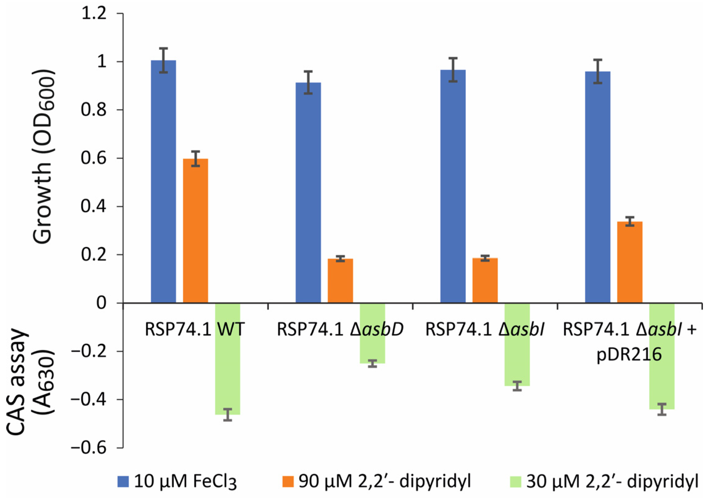
To study the role of AsbI, a deletion mutant of this gene was constructed in the A. salmonicida RSP74.1 background. This strain was isolated from a furunculosis outbreak in turbot and produces only acinetobactin, as it harbors a natural inactivation of the amonabactin biosynthesis genes [7]. Then, the ability of the RSP74.1 ΔasbI mutant to grow under low iron availability, and to produce acinetobactin, was compared to that of an RSP74.1 ΔasbD mutant, which is unable to produce any siderophore. All the strains assayed grew at the same level under iron-rich conditions (CM9 supplemented with FeCl3 10 µM), achieving an OD600 of approximately 1 after 12 h of incubation (Figure 2). In contrast, since OD600 values ≤ 0.2 are attributed to the initial inoculum, mutant strains RSP74.1 ΔasbI and RSP74.1 ΔasbD were unable to grow in low iron availability (CM9 supplemented with the iron chelator 2,2′-dipyridyl at 90 µM). The parental strain RSP74.1 reached an OD600 value of 0.6 under this condition. The reintroduction by gene complementation of a functional copy of asbI in the mutant strain RSP74.1 ΔasbI partially restored its growth ability under low-iron conditions (Figure 2). This result strongly suggests that the inactivation of asbI reduces the ability of A. salmonicida to grow under low iron availability.
To study whether asbI is required to produce the siderophore acinetobactin, the strains were grown under weak iron-limiting conditions (CM9 with 2,2′-dipyridyl 30 µM) that do not limit the growth of any strain, and cell free supernatants were evaluated by the CAS assay in order to determine siderophore content. The CAS value found in the ΔasbI mutant supernatants was significantly higher than that found by the ΔasbD mutant (Figure 2), which suggest that the ΔasbI mutant still produces acinetobactin. Thus, the reduced ability to grow under low iron availability of this ΔasbI mutant would not be due to a lack of siderophore production.

2.2. AsbI Regulates the Expression of Acinetobactin Transport Functions, but Not Its Synthesis

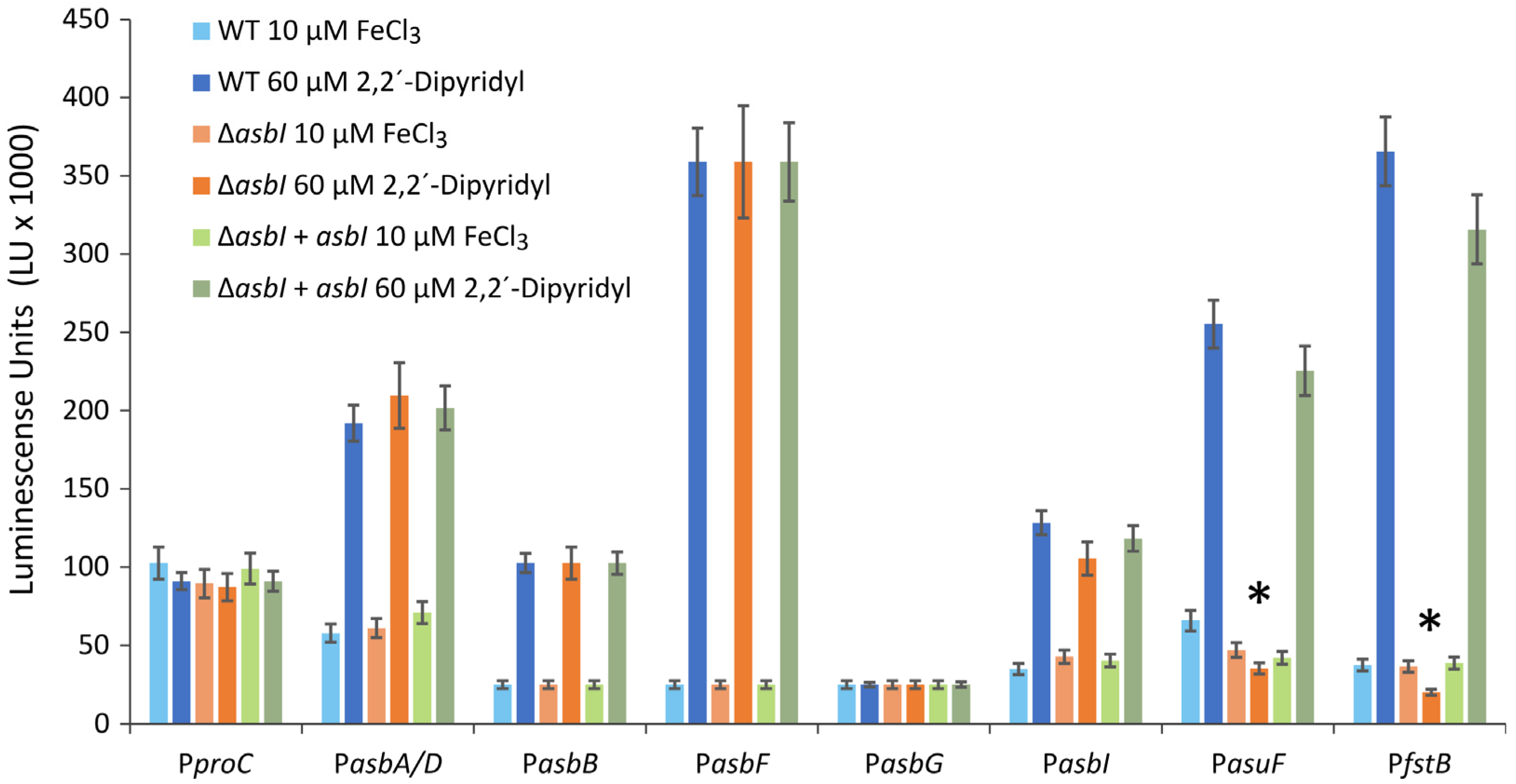
Since asbI would encode a putative transcriptional ECF sigma factor, we wanted to study whether it had a role in the modulation of acinetobactin gene expression. The transcriptional association of acinetobactin biosynthetic genes was previously studied, showing that asbC and asbA/D are cotranscribed in a polycistronic mRNA from a promoter upstream of asbD, and that asbFG could be expressed from a promoter located upstream of asbG (Figure 1A) [5]. In the present work, DNA regions immediately upstream of asbG, asbF, asbD and asbB (Figure 1A) were cloned into plasmid pSEVA236 upstream of a promoterless lux operon. Thus, the transcriptional activity of the promoters can be quantified as the luminescence produced by expression of the lux genes [21]. Resultant lux transcriptional fusions were named PasbG, PasbF, PasbD, and PasbB, respectively. Then, transcriptional fusions were conjugated into A. salmonicida, and luminescence was measured when the bacteria was cultured under high or low iron availability. The promoter of proC (PproC fusion) was used as a constitutive expression control [22,23]. The results of the assay are shown in Figure 3.
All acinetobactin promoters significantly increase their transcriptional activity under low iron availability. By contrast, lux genes were constitutively expressed when transcribed under control of the proC promoter (PproC). The lowest transcription level was observed for promoter PasbG, and it did not show any response to iron concentration variations (Figure 3). Thus, DNA region upstream of asbG likely is not a real promoter and asbFG genes would be expressed together under control of the asbF promoter (PasbF). Under iron-rich conditions all acinetobactin promoters showed low activity (luminescence) and no differences were found between luminescence emitted by the fusions in the wild type and in asbI mutant (Figure 3). Interestingly, PasuF or PfstB promoters were almost non-luminescent under low-iron conditions in the ΔasbI mutant strain background. The mutant strain RSP74.1 ΔasbI complemented in trans with wild-type asbI (pDR216) showed the same expression pattern than the wild-type strain (Figure 3). These results clearly show that AsbI is required to express acinetobactin transport functions from promoters PasuF and PfstB. In addition, AsbI would not regulate its own expression.
The acinetobactin transport genes are expressed as a polycistronic mRNA including asuIFJDCEB-fstB genes under the control of a promoter upstream of the asbI gene (PasbI) (Figure 1A) (Rey-Varela, Balado & Lemos, manuscript in preparation). Notably, Figure 3 shows that expression from other promoters upstream of fstB (PfstB) and asuF (PasuF) is preponderant, as they show higher transcriptional activity than PasbI under low-iron conditions.
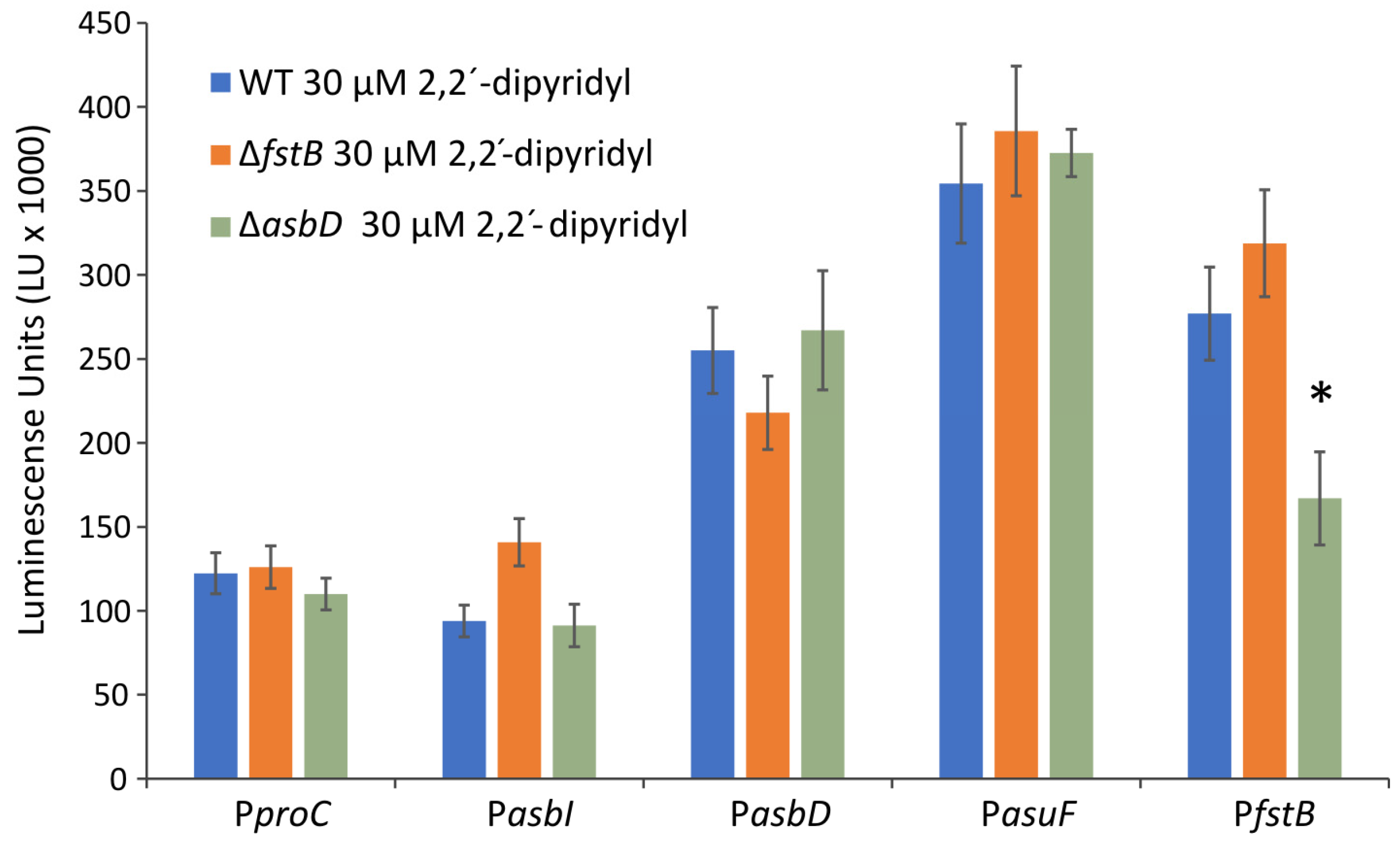
In other siderophore systems, the genes encoding transport functions increase their expression when the ferri-siderophore complexes are internalized [24]. To evaluate whether expression of the acinetobactin uptake genes in A. salmonicida respond to a ferri-acinetobactin presence, fusions with PasbI, PasbD, PasuF, and PfstB were introduced into the mutants RSP74.1 ΔasbD and RSP74.1 ΔfstB. The mutant ΔfstB produces acinetobactin but lacks the ferri-acinetobactin outer membrane transporter FstB, and it is therefore unable to use it as an iron source [11], while RSP74.1 ΔasbD does not produce acinetobactin. RSP74.1 ΔfstB showed expression patterns indistinguishable from the wild-type strain (Figure 4). This result strongly suggests that acinetobactin does not modulate the expression of its own transport genes. Nevertheless, RSP74.1 ΔasbD showed a significant lower expression for the promoter PfstB than for the wild-type strain, suggesting that this promoter requires the presence of acinetobactin for its expression.

2.3. Presence of Fur-Boxes in Acinetobactin Promoters

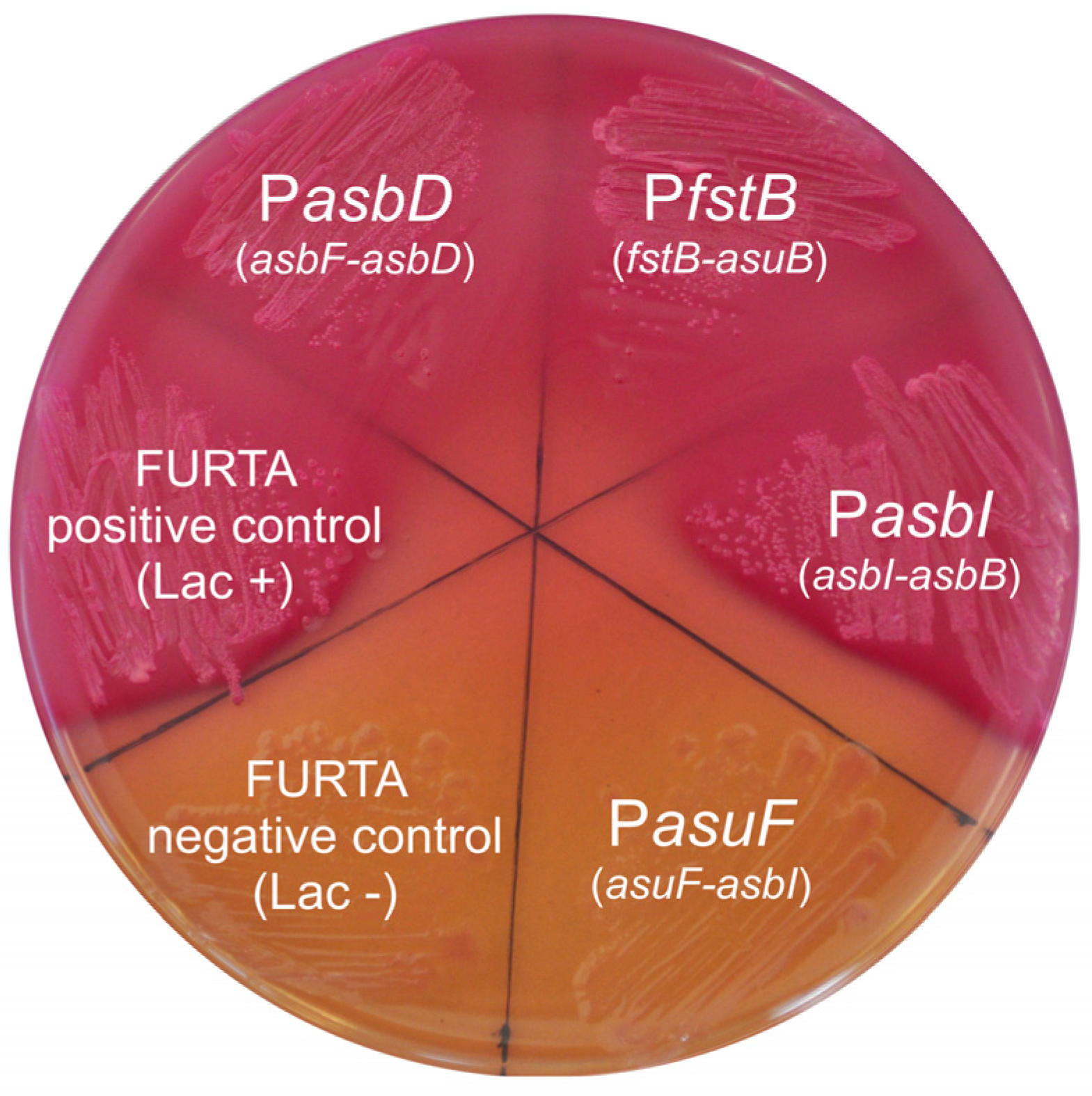
Acinetobactin promoter regions contain a probable Fur box motif [5], which would mediate the strong iron response found in the expression pattern. To study whether acinetobactin gene promoters contain Fur box motifs, DNA regions of approximately 1 kb flanking fstB-asuB, asuF-asbI, asbI-asbB, and asbF-asbD intergenic sequences were tested in a Fur titration assay (FURTA) by using an E. coli H1717 indicator strain (Figure 5).
E. coli H1717 harboring either fstB-asuB, asbI-asbB, or asbF-asbD sequences showed a FURTA positive phenotype (Lac+), which strongly suggests that Fur efficiently binds to them. By contrast, a FURTA-negative phenotype was observed when the asuF-asbI intergenic region was cloned in E. coli H1717 (Figure 5). This result suggests that PasuF transcriptional activity is independent of Fur. Thus, while PfstB is under the control of Fur and AsbI, PasuF would not be iron-regulated through the Fur repressor.

2.4. In Silico Prediction of AsbI Interactions with Other Proteins

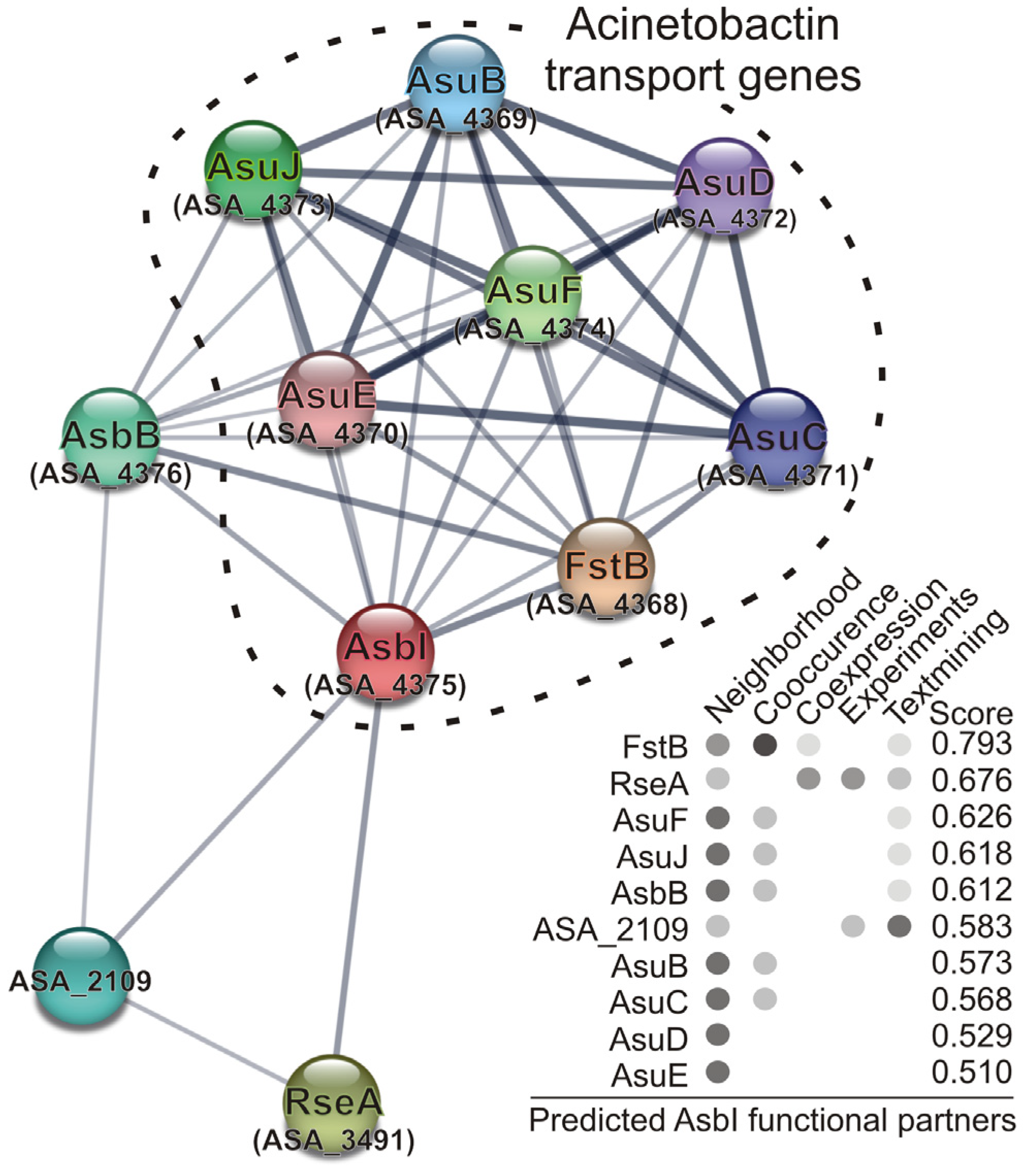
Finally, prediction of protein–protein interactions based on the STRING Database (Figure 6) showed a strong association of AsbI with the acinetobactin transport functions, as well as strong interactions among them, which are mainly derived from their genomic context. This means that these proteins have more interactions among themselves than what would be expected for a random set of proteins of the same size and degree distribution drawn from the genome. Such an enrichment indicates that the proteins are at least partially biologically connected, as a group. It is noteworthy that there are also interactions with anti-sigma RseA (ABO91459.1) and locus ASA_2109 (ABO90175.1), encoding a putative two-component system hybrid sensor histidine kinase/response regulator. The role of these genes in the modulation of acinetobactin gene expression must be further studied.

3. Discussion

In this work, we describe the role of the putative sigma-factor AsbI in the expression of acinetobactin siderophore genes in the Gram-negative fish pathogen A. salmonicida. Usually, expression of most iron-acquisition-related genes is controlled by iron levels through the ferric-uptake regulator (Fur) [15,16,17]. However, additional control mechanisms have been described in many bacteria. One of these mechanisms involves the transcriptional regulators belonging to the Sigma (σ) factors family [26].
A. salmonicida AsbI belongs to group 4 σ factors, also known as the ExtraCytoplasmic Function (ECF) group, with roles in sensing and response to specific conditions that are generated outside of the cell or in the cell membrane [27]. The ECF group, with at least 43 distinct subgroups, is numerically the largest and most diverse group of sigma factors. Their roles include general stress response (Methylobacterium extorquens EcfG1), envelope stress response (E. coli σE or Bacillus subtilis σW), oxidative stress (Streptomyces coelicolor σR and Rhodobacter sphaeroides σE), or iron starvation [26]. The first described iron-related sigma factor was FecI from E. coli, which regulates the transcription initiation of the iron-citrate transporter genes fecABCDE [28]. Several studies have demonstrated the effects of these types of sigma factors on the iron metabolism, e.g., that pyoverdine biosynthetic genes in pseudomonads are under the control of the sigma factor PvdS [29]. More specifically, ECF sigma factors are usually involved in the regulation of the transport of siderophores, such as BupI and EcfI of Bordetella bronchiseptica, that control the expression of the siderophore receptors BfrZ and BfrH, respectively [30,31,32]. In Pseudomonas aeruginosa, FpvI mediates expression of fpvA, the pyoverdine transporter gene [33]. This is also the case for A. salmonicida AsbI reported here, which is required for the expression of genes that encode acinetobactin transporters located upstream of fstB and asuF genes. In addition, the FURTA assay results suggest that the promoter that controls expression of fstB, the acinetobactin TBDT transporter, is dually regulated through Fur repressors and AsbI, whereas the promoter of asuF, which controls the expression of the probable ABC efflux system, would be exclusively regulated by AsbI. This double regulation by Fur and ECF factors is present in other siderophore receptors. For instance, expression of BfrH, a putative siderophore receptor of Bordetella bronchiseptica, is regulated by Fur and by sigma factor EcfI [32].
The expression and activity of ECF factors can be controlled at several levels, but it is particularly prevalent the post-translational control by anti-factors that sequester these proteins, thus preventing their interaction with RNAP [26]. However, none of the genes adjacent to asbI or within the acinetobactin cluster of A. salmonicida are predicted to encode an anti-σ factor. Nevertheless, the STRING database showed an association of AsbI with anti-sigma RseA and a putative two-component hybrid sensor histidine kinase/response regulator of A. salmonicida. The relationship of these genes with the modulation of acinetobactin genes requires further investigation.
Some groups of ECF σ factors appear not to be controlled by any specific anti-σ factor and are directly regulated at the level of transcription [34]. The presence of a Fur-box upstream of A. salmonicida asbI indicates that, probably, it is directly controlled at the transcriptional level by the iron-dependent Fur protein. When iron is present, Fe(II) reversibly binds Fur, which facilitates the binding of Fur to its cognate sites (Fur-boxes), thus inactivating the expression of Fur-regulated genes [15,16,17]. In Burkholderia cepacia transcription of the OrbS ornibactin sigma regulator is controlled by Fur [35]. The transcription of biosynthetic genes of malleobactin in Burkholderia pseudomallei requires the sigma factor MbaS, and the mbaS promoter contains a Fur-binding site [36]. In Pseudomonas aeruginosa, a Fur-box was identified upstream of sigma-factor gene fpvI, consistent with the iron-regulated expression of this gene and its cognate pyoverdine transporter fpvA [33].
ECF sigma factors are often co-expressed with a second regulator which, in turn, transfers the extracellular signal to the cognate ECF sigma factor. For instance, E. coli FecR protein transmits the signal across the cytoplasmic membrane, through its transmembrane segment, into the cytoplasm, where it interacts through its short cytoplasmic portion with the FecI sigma factor which binds the promoter upstream of fecA gene [28,37]. In Pseudomonas putida, PupR is involved in the signal transduction for activation of PupI that regulates the synthesis of the outer membrane protein PupB [38]. Bordetella bronchiseptica HurR positively modulates HurI in the presence of heme to mediate the heme-dependent expression of BhuR, the outer membrane heme receptor [39]. Finally, in Pseudomonas aeruginosa, ferric pyoverdine induces expression of the outer membrane protein FpvA, the cytoplasmic membrane protein FpvR, and the two FecI-type sigma factors PvdS and FpvI in order to activate pyoverdine synthesis and uptake [24]. Bacterial sigma factors can be activated by their own siderophores produced by a bacterium or by xenosiderophores, iron-citrate, or heme groups [18,39]. In A. salmonicida, it seems likely that AsbI is regulated only by Fur, since our results demonstrate that neither acinetobactin nor acinetobactin transporter FstB are involved in asbI expression. Other global regulators, apart from Fur and ECF sigma factors, can regulate the expression of sigma factors involved in iron acquisition systems [40], such as AraC-type regulators such as PbtA of V. anguillarum [41].
In addition to the siderophore transport systems, iron-regulated ECF factors can control other functions related with iron acquisition during the infective process, such as proteases or hemolysins. For instance, the sigma factor PbrA is required for the transcription under low-iron conditions of the metalloprotease gene aprA in Pseudomonas fluorescens [42]. In P. aeruginosa, PvdS mediates the transcription of genes encoding pyoverdine biosynthesis but also the expression of other virulence factors such as exotoxin A, alkaline protease AprA, and one endoproteinase [24]. HurR positively modulates hurP, a gene encoding a prospective plasma membrane-associated protease in Bordetella bronchiseptica [39]. In accordance with this, it is likely that the regulator AsbI of A. salmonicida could regulate the expression of other virulence factors. Further works will be necessary in order to unravel these putative functions.
In conclusion, we have demonstrated that the transport of the siderophore acinetobactin in A. salmonicida is regulated at the transcriptional level by the ECF sigma factor AsbI. This regulator is necessary for the expression of both the outer membrane Fe-acinetobactin transporter FstB and the putative acinetobactin exporter AsuF-J that is involved in acinetobactin secretion. Moreover, AsbI is probably regulated only by Fur and does not possess an anti-sigma factor counterpart. These findings increase our knowledge about the role of sigma factors in iron uptake mechanisms involving siderophores in bacteria. Additionally, since siderophores play a relevant role in bacterial virulence, external interference of these regulatory mechanisms could constitute a promising approach to combat bacterial infections [40].

4. Materials and Methods

4.1. Bacterial Strains, Plasmids and Culture Media

Strains and plasmids used in this work are listed in Table 1. A. salmonicida strains were cultured at 25 °C in Tryptic Soy Agar or Broth (Condalab, Madrid, Spain), supplemented with NaCl (Fisher Scientific, Barcelona, Spain) to a final concentration of 1% (TSA-1 or TSB-1, respectively). All Escherichia coli (E. coli) strains were cultured at 37 °C in Luria-Bertani medium (LB Lennox) (Condalab, Madrid, Spain). Culture media were supplemented with the suitable antibiotics when necessary. The antibiotics and final concentrations used were Kanamycin (Km; Merck KGaA, Darmstadt, Germany) at 50 μg mL−1; Chloramphenicol (Cm; Merck KGaA, Darmstadt, Germany), at 20 μg mL−1; and Gentamicin (Gm; Merck KGaA, Darmstadt, Germany), at 15 μg mL−1. The antibiotics were filter sterilized and stored at 4 °C. The growth under iron-limiting conditions was performed in M9 minimal medium supplemented with 0.2% Casamino Acids (BD-Difco, Madrid, Spain) (CM9) [43]. The iron-limiting conditions were induced by the addition of 2,2′-dipyridyl (Merck KGaA, Darmstadt, Germany) dissolved in milli-Q water at the appropriate concentration.

4.2. Mutant Construction and Complementation of asbI Gene

In-frame deletion mutants of the asbI gene were obtained by allelic exchange [47]. For the in-frame constructions, flanking regions of the gene were amplified by PCR and cloned into the pWKS30 plasmid (high copy number vector) [48]. The construction was sub-cloned into the suicidal vector pNidKan [46] into E. coli S17-1-λpir [49]. The strain carrying the construction was mated with A. salmonicida RSP74.1 (Cmr), and the exconjugants were selected with Cm and Km. The second recombinants were isolated from medium with sucrose (15%) and screened in Cm and Km plates. The Cmr isolates were confirmed by a PCR for the target gene and subsequent DNA sequencing. For complementation of the ΔasbI mutant, the wild-type gene, together with its promoter, was amplified with Hi-Fidelity Kapa Taq (Merck KGaA, Darmstadt, Germany). The DNA fragment was cloned into the plasmid pSEVA651 [21] and mobilized into an A. salmonicida ΔasbI mutant from E. coli S17-1-λpir. All oligonucleotides used are listed in Table 2.

4.3. DNA Manipulation

Genomic DNA extraction was performed with the Easy-DNA kit standard protocol (Invitrogen, Fisher-Scientific, Barcelona, Spain) and the samples were stored at −20 °C until utilization. PCR reactions were carried out with TaqK-derived DNA polymerase NZYTaqII (NZYTech) in a thermocycler T-Gradient Thermal Cycler (Biometra, Göttingen, Germany). Plasmid extraction and purification were performed with the GeneJET Plasmid Miniprep kit (Thermo-Fisher). DNA bands from agarose gels and PCR were purified with NucleoSpin Gel and PCR Clean-up kit (Macherey-Nagel, Dueren, Germany).

4.4. Growth under Iron-Limiting Conditions

The ability to grow under iron-restricted conditions and the siderophore production of the parental and mutant strains were assayed in CM9 minimal medium [50] containing 2,2′-dipyridyl 90 µM as iron chelator. Bacteria were grown in TSB-1 for 5 h at 25 °C and cell concentration was adjusted to an optical density (OD600) of 0.5. This preinoculum was used to inoculate (1:40) CM9 medium in a 96-well microtiter plate (final volume of 200 µL per well). Plates were incubated for 18 h at 25 °C in an iMACK Microplate reader (Bio-Rad Laboratories, Madrid, Spain) taking OD600 measurements every 30 min. Media supplemented with FeCl3 10 µM and non-inoculated media were used as controls. All conditions were carried out in triplicate within each plate and all experiments were repeated four times, calculating standard deviations and means for each condition. Student’s t-test was used for statistical analysis using SPSS software (version 20; IBM SPSS Inc., Chicago, IL, USA).

4.5. Quantification of Siderophores (CAS Assay)

Siderophore production was determined using the chrome azurol-S liquid assay (CAS) [51]. To induce siderophore production, bacteria were grown in CM9 medium containing 2,2′-dipyridyl 30 µM as iron chelator. Cell-free supernatants were obtained by centrifugation at 10,000 rpm in a Microcentrifuge (Eppendorf Ibérica, Madrid, Spain) and assayed for CAS reactivity as previously described [52]. Colorimetric reaction was measured in a spectrophotometer (Hitachi Europe, Buckinghamshire, UK) at 630 nm after 10 min of incubation. Non-inoculated medium was used as blank.

4.6. Promoter Fusions

DNA fragments which corresponded to the defined promoter regions and to the house-keeping gene proC were PCR amplified from genomic DNA and cloned into the reporter plasmid pSEVA236 [21]. The primers used are listed in Table 3. The promoters were cloned upstream of the lux genes into the MCS site in an E. coli S17-λpir strain. Since these genes lack a promoter, the transcription of lux genes is ruled by the promoter sequence cloned. The constructions were mobilized by conjugation into A. salmonicida RSP74.1 and its derivative mutants. The strains carrying the different promoter constructions were cultured under iron-rich and low-iron conditions (2,2′-dipyridyl 60 µM) in CM9 medium in microtiter plates in a final volume of 200 µL per well. The luminescence was recorded with a Tecan Infinite F200 reader (Tecan, Männedorf, Switzerland) after 16 h of incubation (OD600 ≈ 0.4). Statistical differences were determined using Student’s t-test using SPSS software (version 20; IBM SPSS Inc., Chicago, IL, USA). p-values were significant when p was <0.05.

4.7. Fur Titration Assay (FURTA)

Fur-regulated promoters were identified through a Fur titration assay (FURTA) [53]. The assay was performed as previously described [54]. Briefly, putative Fur-regulated promoters were cloned into the multicopy plasmid pT7-7 and transformed into E. coli H1717 [44] (strain kindly donated by Prof. Klaus Hantke from the University of Tübingen, Germany). This strain carries the Fur-regulated gene fhuF::lacZ, which is highly sensitive to changes in the concentration of the Fur repressor. The introduction in multicopy of a promoter that possesses a Fur-box causes the derepression of the fhuF::lacZ gene by binding the Fur protein, leading to the transcription of the lacZ gene. The expression of the LacZ protein leads to a Lac+ phenotype in the E. coli H1717 strain that can be visualized on MacConkey Agar plates supplemented with Fe2(SO4)3 40 µM, ampicillin 100 μg mL−1 and 1% of extra lactose. The Lac+ phenotype is seen as red colonies in the medium, while the Lac- or FURTA negative is seen as yellow-orange colonies. The oligonucleotides used to clone the promoters into the plasmid pT7-7 were the same used for the transcriptional fusions (Table 3).

4.8. Bioinformatic Tools

STRING Database v11.5 (https://string-db.org/ accessed on 15 April 2023) was used to determine in silico, by means of the web tool, the protein–protein interactions between AsbI (ABO92290.1), and other A. salmonicida proteins derived from the genomic context, including direct (physical) and indirect (functional) associations, high-throughput experiments, co-expression, and the literature. The number of interactors represented was limited to 10 and a medium confidence interaction score (minimum of 0.4) was used.

Author Contributions

Conceptualization, M.B. and M.L.L.; methodology, D.R.-V. and M.B.; software, D.R.-V. and M.B.; validation, D.R.-V. and M.B.; formal analysis, M.B. and M.L.L.; investigation, D.R.-V.; resources, M.L.L.; data curation, D.R.-V. and M.B.; writing—original draft preparation, D.R.-V. and M.B.; writing—review and editing, M.L.L.; visualization, D.R.-V. and M.B.; supervision, M.L.L.; project administration, M.L.L.; funding acquisition, M.L.L. All authors have read and agreed to the published version of the manuscript.

Funding

This work was supported by grants RTI2018-093634-B-C21 (from MCIN/AEI/10.13039/501100011033/FEDER, Spanish State Agency for Research and FEDER Programme from the European Union “A way to make Europe”) and PDC2022-133052-C21 (from Spanish State Agency for Research and the European Union “Next Generation EU/PRTR”). M.B. was supported by grant PID2019-103891RJ-100 from MCIN/AEI/10.13039/501100011033 (Spain). Work was also supported by grant ED431C 2022/23 from Xunta de Galicia (Spain). D.R-V. thanks Xunta de Galicia for a predoctoral fellowship.

Institutional Review Board Statement

Not applicable.

Informed Consent Statement

Not applicable.

Data Availability Statement

All DNA sequence data used in this work can be found at GenBank (NCBI), as part of the genome sequence data of Aeromonas salmonicida subsp. salmonicida A449, under accession number CP000644.1.

Conflicts of Interest

The authors declare no conflict of interest.

References

  1. Begg, S.L. The role of metal ions in the virulence and viability of bacterial pathogens. Biochem. Soc. Trans. 2019, 47, 77–87. [Google Scholar] [CrossRef]
  2. Kramer, J.; Özkaya, Ö.; Kümmerli, R. Bacterial siderophores in community and host interactions. Nat. Rev. Microbiol. 2020, 18, 152–163. [Google Scholar] [CrossRef] [PubMed]
  3. Lemos, M.L.; Balado, M. Iron uptake mechanisms as key virulence factors in bacterial fish pathogens. J. Appl. Microbiol. 2020, 129, 104–115. [Google Scholar] [CrossRef] [PubMed]
  4. Toranzo, A.E.; Magariños, B.; Romalde, J.L. A review of the main bacterial fish diseases in mariculture systems. Aquaculture 2005, 246, 37–61. [Google Scholar] [CrossRef]
  5. Najimi, M.; Lemos, M.L.; Osorio, C.R. Identification of siderophore biosynthesis genes essential for growth of Aeromonas salmonicida under iron limitation conditions. Appl. Environ. Microbiol. 2008, 74, 2341–2348. [Google Scholar] [CrossRef] [Green Version]
  6. Lemos, M.L.; Osorio, C.R. Iron Uptake in Vibrio and Aeromonas. In Iron Uptake and Homeostasis in Microorganisms; Cornelis, P., Andrews, S.C., Eds.; Caister Academic Press: Norfolk, UK, 2010; pp. 117–141. ISBN 9781904455653. [Google Scholar]
  7. Balado, M.; Souto, A.; Vences, A.; Careaga, V.P.; Valderrama, K.; Segade, Y.; Rodríguez, J.; Osorio, C.R.; Jiménez, C.; Lemos, M.L. Two catechol siderophores, acinetobactin and amonabactin, are simultaneously produced by Aeromonas salmonicida subsp. salmonicida sharing part of the biosynthetic pathway. ACS Chem. Biol. 2015, 10, 2850–2860. [Google Scholar] [CrossRef]
  8. Esmaeel, Q.; Chevalier, M.; Chataigné, G.; Subashkumar, R.; Jacques, P.; Leclère, V. Nonribosomal peptide synthetase with a unique iterative-alternative-optional mechanism catalyzes amonabactin synthesis in Aeromonas. Appl. Microbiol. Biotechnol. 2016, 100, 8453–8463. [Google Scholar] [CrossRef] [PubMed]
  9. Yamamoto, S.; Okujo, N.; Sakakibara, Y. Isolation and structure elucidation of acinetobactin, a novel siderophore from Acinetobacter baumannii. Arch Microbiol 1994, 162, 249–254. [Google Scholar] [CrossRef]
  10. Telford, J.R.; Leary, J.A.; Tunstad, L.M.G.; Byers, B.R.; Raymond, K.N. Amonabactin: Characterization of a series of siderophores from Aeromonas hydrophila. J. Am. Chem. Soc. 1994, 116, 4499–4500. [Google Scholar] [CrossRef]
  11. Balado, M.; Segade, Y.; Rey, D.; Osorio, C.R.; Rodríguez, J.; Lemos, M.L.; Jiménez, C. Identification of the ferric-acinetobactin outer membrane receptor in Aeromonas salmonicida subsp. salmonicida and structure–Activity relationships of synthetic acinetobactin analogues. ACS Chem. Biol. 2017, 12, 479–493. [Google Scholar] [CrossRef] [PubMed]
  12. Rey-Varela, D.; Cisneros-Sureda, J.; Balado, M.; Rodríguez, J.; Lemos, M.L.; Jiménez, C. The outer membrane protein FstC of Aeromonas salmonicida subsp. salmonicida acts as receptor for amonabactin siderophores and displays a wide ligand plasticity. Structure–activity relationships of synthetic amonabactin analogues. ACS Infect. Dis. 2019, 5, 1936–1951. [Google Scholar] [CrossRef] [PubMed]
  13. Matthijs, S.; Laus, G.; Meyer, J.-M.M.; Abbaspour-Tehrani, K.; Schäfer, M.; Budzikiewicz, H.; Cornelis, P.; Schafer, M.; Budzikiewicz, H.; Cornelis, P. Siderophore-mediated iron acquisition in the entomopathogenic bacterium Pseudomonas entomophila L48 and its close relative Pseudomonas putida KT2440. Biometals 2009, 21, 21. [Google Scholar] [CrossRef] [PubMed]
  14. Mihara, K.; Tanabe, T.; Yamakawa, Y.; Funahashi, T.; Nakao, H.; Narimatsu, S.; Yamamoto, S. Identification and transcriptional organization of a gene cluster involved in biosynthesis and transport of acinetobactin, a siderophore produced by Acinetobacter baumannii ATCC 19606T. Microbiology 2004, 150, 2587–2597. [Google Scholar] [CrossRef] [Green Version]
  15. Andrews, S.C.; Robinson, A.K.; Rodriguez-Quinones, F. Bacterial iron homeostasis. FEMS Microbiol. Rev. 2003, 27, 215–237. [Google Scholar] [CrossRef] [PubMed] [Green Version]
  16. Troxell, B.; Hassan, H.M. Transcriptional regulation by Ferric Uptake Regulator (Fur) in pathogenic bacteria. Front. Cell. Infect. Microbiol. 2013, 3, 59. [Google Scholar] [CrossRef] [PubMed] [Green Version]
  17. Sheldon, J.R.; Heinrichs, D.E. Recent developments in understanding the iron acquisition strategies of gram positive pathogens. FEMS Microbiol. Rev. 2015, 39, 592–630. [Google Scholar] [CrossRef] [Green Version]
  18. Otero-Asman, J.R.; Wettstadt, S.; Bernal, P.; Llamas, M.A. Diversity of extracytoplasmic function sigma (σECF) factor-dependent signaling in Pseudomonas. Mol. Microbiol. 2019, 112, 356–373. [Google Scholar] [CrossRef] [Green Version]
  19. Ishihama, A. Functional modulation of Escherichia coli RNA polymerase. Annu. Rev. Microbiol. 2000, 54, 499–518. [Google Scholar] [CrossRef]
  20. Finn, R.D.; Bateman, A.; Clements, J.; Coggill, P.; Eberhardt, R.Y.; Eddy, S.R.; Heger, A.; Hetherington, K.; Holm, L.; Mistry, J.; et al. Pfam: The protein families database. Nucleic Acids Res. 2014, 42, D222–D230. [Google Scholar] [CrossRef] [Green Version]
  21. Martínez-García, E.; Goñi-Moreno, A.; Bartley, B.; McLaughlin, J.; Sánchez-Sampedro, L.; Pascual Del Pozo, H.; Prieto Hernández, C.; Marletta, A.S.; De Lucrezia, D.; Sánchez-Fernández, G.; et al. SEVA 3.0: An update of the Standard European Vector Architecture for enabling portability of genetic constructs among diverse bacterial hosts. Nucleic Acids Res. 2020, 48, D1164–D1170. [Google Scholar] [CrossRef] [Green Version]
  22. Savioz, A.; Jeenes, D.J.; Kocher, H.P.; Haas, D. Comparison of proC and other housekeeping genes of Pseudomonas aeruginosa with their counterparts in Escherichia coli. Gene 1990, 86, 107–111. [Google Scholar] [CrossRef]
  23. Rivera, L.; López-Patiño, M.A.; Milton, D.L.; Nieto, T.P.; Farto, R. Effective qPCR methodology to quantify the expression of virulence genes in Aeromonas salmonicida subsp. salmonicida. J. Appl. Microbiol. 2015, 118, 792–802. [Google Scholar] [CrossRef]
  24. Beare, P.A.; For, R.J.; Martin, L.W.; Lamont, I.L. Siderophore-mediated cell signalling in Pseudomonas aeruginosa: Divergent pathways regulate virulence factor production and siderophore receptor synthesis. Mol. Microbiol. 2003, 47, 195–207. [Google Scholar] [CrossRef]
  25. Balado, M.; Osorio, C.R.; Lemos, M.L. Biosynthetic and regulatory elements involved in the production of the siderophore vanchrobactin in Vibrio anguillarum. Microbiology 2008, 154, 1400–1413. [Google Scholar] [CrossRef] [Green Version]
  26. Paget, M.S. Bacterial sigma factors and anti-sigma factors: Structure, function and distribution. Biomolecules 2015, 5, 1245–1265. [Google Scholar] [CrossRef] [PubMed] [Green Version]
  27. Lonetto, M.A.; Brown, K.L.; Rudd, K.E.; Buttner, M.J. Analysis of the Streptomyces coelicolor sigE gene reveals the existence of a subfamily of eubacterial RNA polymerase sigma factors involved in the regulation of extracytoplasmic functions. Proc. Natl. Acad. Sci. USA 1994, 91, 7573–7577. [Google Scholar] [CrossRef] [PubMed] [Green Version]
  28. Braun, V. Surface signaling: Novel transcription initiation mechanism starting from the cell surface. Arch. Microbiol. 1997, 167, 325–331. [Google Scholar] [CrossRef] [PubMed]
  29. Cunliffe, H.E.; Merriman, T.R.; Lamont, I.L. Cloning and characterization of pvdS, a gene required for pyoverdine synthesis in Pseudomonas aeruginosa: PvdS is probably an alternative sigma factor. J. Bacteriol. 1995, 177, 2744–2750. [Google Scholar] [CrossRef] [PubMed] [Green Version]
  30. Vanderpool, C.K.; Armstrong, S.K. The Bordetella bhu locus is required for heme iron utilization. J. Bacteriol. 2001, 183, 4278–4287. [Google Scholar] [CrossRef] [Green Version]
  31. Pradel, E.; Locht, C. Expression of the putative siderophore receptor gene bfrZ is controlled by the extracytoplasmic-function sigma factor BupI in Bordetella bronchiseptica. J. Bacteriol. 2001, 183, 2910–2917. [Google Scholar] [CrossRef] [PubMed] [Green Version]
  32. Burgos, J.M.; King-Lyons, N.D.; Connell, T.D. Expression of BfrH, a putative siderophore receptor of Bordetella bronchiseptica, is regulated by iron, fur1, and the extracellular function sigma factor EcfI. Infect. Immun. 2010, 78, 1147–1162. [Google Scholar] [CrossRef] [Green Version]
  33. Rédly, G.A.; Poole, K. Pyoverdine-mediated regulation of FpvA synthesis in Pseudomonas aeruginosa: Involvement of a probable extracytoplasmic-function sigma factor, FpvI. J. Bacteriol. 2003, 185, 1261–1265. [Google Scholar] [CrossRef] [Green Version]
  34. Staroń, A.; Sofia, H.J.; Dietrich, S.; Ulrich, L.E.; Liesegang, H.; Mascher, T. The third pillar of bacterial signal transduction: Classification of the extracytoplasmic function (ECF) sigma factor protein family. Mol. Microbiol. 2009, 74, 557–581. [Google Scholar] [CrossRef]
  35. Agnoli, K.; Lowe, C.A.; Farmer, K.L.; Husnain, S.I.; Thomas, M.S. The ornibactin biosynthesis and transport genes of Burkholderia cenocepacia are regulated by an extracytoplasmic function sigma factor which is a part of the Fur regulon. J. Bacteriol. 2006, 188, 3631–3644. [Google Scholar] [CrossRef] [Green Version]
  36. Alice, A.F.; Lopez, C.S.; Lowe, C.A.; Ledesma, M.A.; Crosa, J.H. Genetic and transcriptional analysis of the siderophore malleobactin biosynthesis and transport genes in the human pathogen Burkholderia pseudomallei K96243. J. Bacteriol. 2006, 188, 1551–1566. [Google Scholar] [CrossRef] [PubMed] [Green Version]
  37. Braun, V.; Endriss, F. Energy-coupled outer membrane transport proteins and regulatory proteins. Biometals 2007, 20, 219–231. [Google Scholar] [CrossRef] [PubMed]
  38. Koster, M.; Van Klompenburg, W.; Bitter, W.; Leong, J.; Weisbeek, P. Role for the outer membrane ferric siderophore receptor PupB in signal transduction across the bacterial cell envelope. EMBO J. 1994, 13, 2805–2813. [Google Scholar] [CrossRef] [PubMed]
  39. Vanderpool, C.K.; Armstrong, S.K. Heme-responsive transcriptional activation of Bordetella bhu genes. J. Bacteriol. 2003, 185, 909–917. [Google Scholar] [CrossRef] [Green Version]
  40. Grove, A. Extracytoplasmic function sigma factors governing production of the primary siderophores in pathogenic Burkholderia species. Front. Microbiol. 2022, 13, 514. [Google Scholar] [CrossRef]
  41. Lages, M.A.; Lemos, M.L.; Balado, M. The temperature-dependent expression of the High-Pathogenicity Island encoding piscibactin in Vibrionaceae results from the combined effect of the AraC-like transcriptional activator PbtA and regulatory factors from the recipient genome. Front. Microbiol. 2021, 12, 3448. [Google Scholar] [CrossRef]
  42. Maunsell, B.; Adams, C.; O’Gara, F. Complex regulation of AprA metalloprotease in Pseudomonas fluorescens M114: Evidence for the involvement of iron, the ECF sigma factor, PbrA and pseudobactin M114 siderophore. Microbiology 2006, 152, 29–42. [Google Scholar] [CrossRef] [Green Version]
  43. Miller, J.H. Experiments in Molecular Genetics; Cold Spring Harbor Laboratory: Laurel Hollow, NY, USA, 1972; ISBN 0879691069. [Google Scholar]
  44. Hantke, K. Selection procedure for deregulated iron transport mutants (fur) in Escherichia coli K 12: Fur not only affects iron metabolism. Mol. Gen. Genet. 1987, 210, 135–139. [Google Scholar] [CrossRef]
  45. Tabor, S.; Richardson, C.C. A bacteriophage T7 RNA polymerase/promoter system for controlled exclusive expression of specific genes. Biotechnology 1992, 24, 280–284. [Google Scholar] [CrossRef] [Green Version]
  46. Mourino, S.; Osorio, C.R.; Lemos, M.L. Characterization of heme uptake cluster genes in the fish pathogen Vibrio anguillarum. J. Bacteriol. 2004, 186, 6159–6167. [Google Scholar] [CrossRef] [Green Version]
  47. Correa, N.E.; Lauriano, C.M.; McGee, R.; Klose, K.E. Phosphorylation of the flagellar regulatory protein FlrC is necessary for Vibrio cholerae motility and enhanced colonization. Mol. Microbiol. 2000, 35, 743–755. [Google Scholar] [CrossRef]
  48. Wang, R.F.; Kushner, S.R. Construction of versatile low-copy-number vectors for cloning, sequencing and gene expression in Escherichia coli. Gene 1991, 100, 195–199. [Google Scholar] [CrossRef] [PubMed]
  49. Herrero, M.; de Lorenzo, V.; Timmis, K.N. Transposon vectors containing non-antibiotic resistance selection markers for cloning and stable chromosomal insertion of foreign genes in gram-negative bacteria. J. Bacteriol. 1990, 172, 6557–6567. [Google Scholar] [CrossRef] [PubMed] [Green Version]
  50. Lemos, M.L.; Salinas, P.; Toranzo, A.E.; Barja, J.L.; Crosa, J.H. Chromosome-mediated iron uptake system in pathogenic strains of Vibrio anguillarum. J. Bacteriol. 1988, 170, 1920–1925. [Google Scholar] [CrossRef] [Green Version]
  51. Schwyn, B.; Neilands, J.B. Universal chemical assay for the detection and determination of siderophores. Anal. Biochem. 1987, 160, 47–56. [Google Scholar] [CrossRef] [PubMed]
  52. Souto, A.; Montaos, M.A.; Rivas, A.J.; Balado, M.; Osorio, C.R.; Rodríguez, J.; Lemos, M.L.; Jiménez, C. Structure and biosynthetic assembly of piscibactin, a siderophore from Photobacterium damselae subsp. piscicida, predicted from genome analysis. Eur. J. Org. Chem. 2012, 2012, 5693–5700. [Google Scholar] [CrossRef]
  53. Stojiljkovic, I.; Bäumler, A.J.; Hantke, K. Fur regulon in gram-negative bacteria. Identification and characterization of new iron-regulated Escherichia coli genes by a fur titration assay. J. Mol. Biol. 1994, 236, 531–545. [Google Scholar] [CrossRef] [PubMed]
  54. Osorio, C.R.; Lemos, M.L.; Braun, V. Identification of Fur regulated genes in the bacterial fish pathogen Photobacterium damselae ssp. piscicida using the Fur titration assay. Biometals 2004, 17, 725–733. [Google Scholar] [CrossRef] [PubMed]
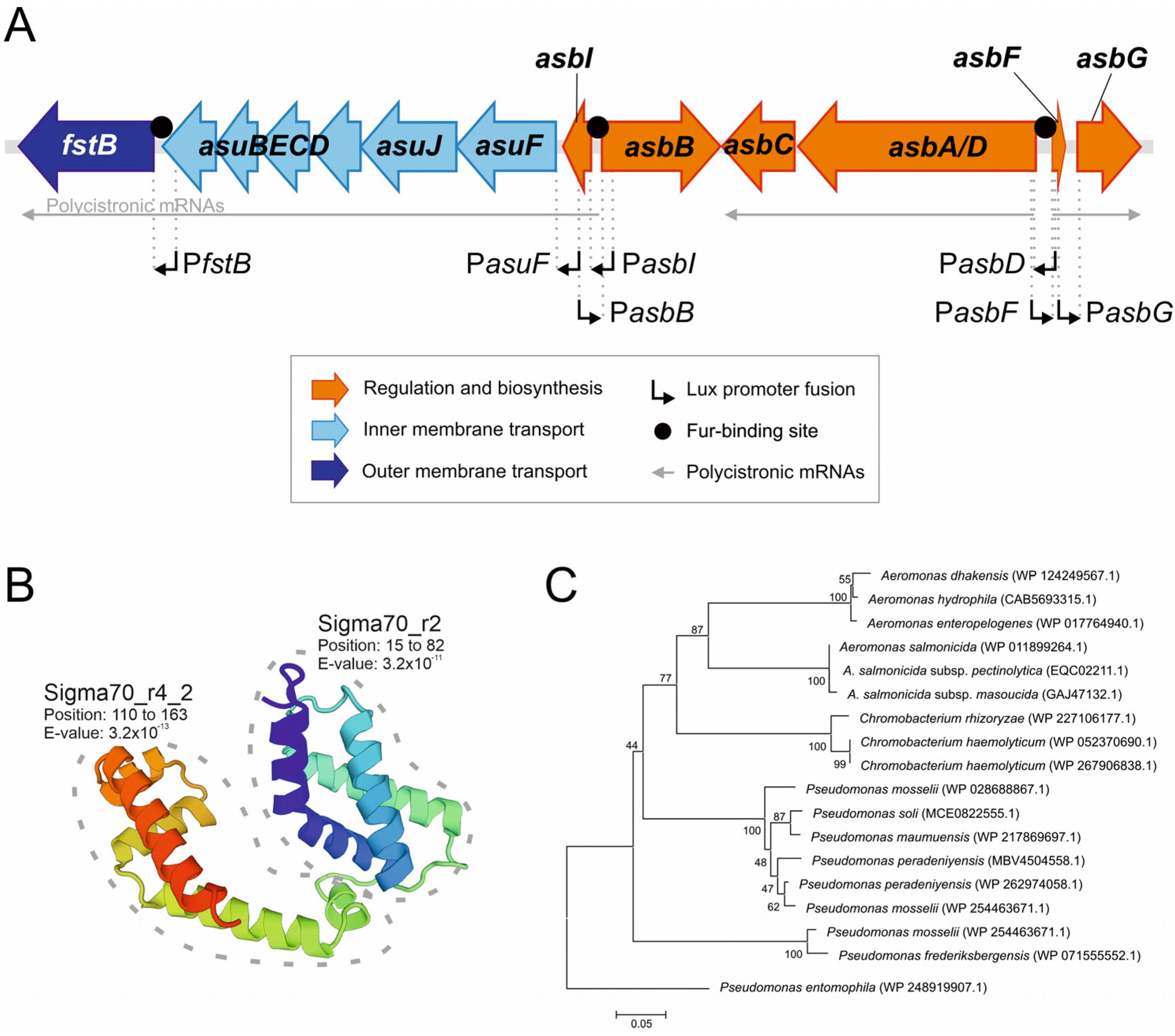
Figure 1. (A) Genetic map of the acinetobactin gene cluster in A. salmonicida. (B) Model of AsbI structure and Pfam conserved domain. The crystal structure of AlgU (6in7.1) was used as a template to build the model for AsbI. Pfam conserved domains analysis are denoted. (C) AsbI neighbor joining tree based on amino acid distances.
Ijms 24 09672 g001
Figure 2. Growth (OD600) reached after 12 h of incubation of A. salmonicida RSP74.1 wild type, RSP74.1 ΔasbD, RSP74.1 ΔasbI, and RSP74.1 ΔasbI complemented with a copy of the wild asbI gene (pDR216) in CM9 minimal medium supplemented with FeCl3 (10 μM) or with the addition of the iron chelator 2,2′-dipyridyl (90 μM). Siderophore production was measured by the CAS assay at A630. The assay was performed in triplicate to calculate the standard deviation.
Ijms 24 09672 g002
Figure 3. Transcriptional activity in the wild-type strain RSP74.1, RSP74.1 ΔasbI, and complemented strain RSP74.1 ΔasbI (pDR216 = pSEVA651-asbI) of the promoters that control the expression of acinetobactin main genes: PasbD, PasbB, PasbF, PasbG, PasbI, PasuF, and PfstB. Expression levels are showed in luminescence units. The constitutive promoter of the housekeeping gene proC (PproC) was used as reference. The luminescence activity was measured in CM9 minimal medium supplemented with FeCl3 10 µM or with 2,2′-dipyridyl 60 µM. Asterisks denote statistically significant differences (Student’s t-test), * p < 0.05.
Ijms 24 09672 g003
Figure 4. Transcriptional activity of the promoters PasbI, PasbD, PasuF, and PfstB, which control the expression of the main acinetobactin genes, in the wild-type strain RSP74.1, RSP74.1ΔfstB and RSP74.1ΔasbD mutants. Expression level is represented in luminescence units. The constitutive promoter of the gene proC (PproC) was used as reference. The activity was measured in CM9 minimal medium supplemented with 2,2′-dipyridyl 30 µM. Asterisk denote statistically significant differences (student’s t-test), * p < 0.05.
Ijms 24 09672 g004
Figure 5. Fur titration assay (FURTA) for the promoters of intergenic regions of fstB, asuF, asbI, and asbD. Promoter regions were cloned into pT7-7 plasmid and introduced into E. coli H1717. Red-pink colonies indicate a Lac+ phenotype (bind E. coli Fur) while yellow-orange colonies indicate a Lac- phenotype (the introduced sequence does not carry a Fur box). Strain H1717 carrying an empty pT7-7 was used as FURTA negative control. Promoter of Vibrio anguillarum fvtA [25] was used as FURTA positive control.
Ijms 24 09672 g005
Figure 6. Prediction of protein–protein interactions, based on STRING Database v11.5 (https://string-db.org, accessed on 15 April 2023), of AsbI with other proteins involved in acinetobactin transport. The interactions include direct (physical) and indirect (functional) associations derived from the genomic context, high-throughput experiments, co-expression, and the literature. Colored nodes indicate query protein (AsbI) and first shell of interactors (limited to 10). Line thickness indicates the strength of data support. Loci codes refer to the genome sequence data of A. salmonicida subsp. salmonicida A449 (accession number CP000644.1).
Ijms 24 09672 g006
Table 1. Strains and plasmids used in this work.
Strain/PlasmidsDescriptionReference
Aeromonas salmonicida subsp. salmonicida
RSP74.1Isolated from turbot, Portugal, Cmr[5]
RSP74.1 ΔasbDRSP74.1 defective in the acinetobactin synthesis gene asbD, Cmr[5]
RSP74.1 ΔfstBRSP74.1 defective in the acinetobactin transporter gene fstB, Cmr[11]
RSP74.1 ΔasbIRSP74.1 defective in the acinetobactin putative regulator gene asbI, CmrThis study
Escherichia coli
DH5αCloning strainLaboratory stock
H1717Strain used in FURTA assay
araD139 ΔlacU169 rpsL150 relA1 flbB5301 deoC1 ptsF25 rbsR aroB fhuF::λ placMu
[44]
Plasmids
pT7-7Cloning vector, Apr[45]
pNidKanPlasmid with sucrose selection, Kanr[46]
pDR216pSEVA651 containing wild-type asbIThis study
Table 2. Oligonucleotides (Fisher-Scientific, Barcelona, Spain) used for the amplification of the flanking regions of asbI in A. salmonicida and subsequent cloning and mutation by allelic exchange.
OligonucleotideSequence (5′ -> 3′)Size (bp)
asbI_del_1_XbaICCGTCTAGATAAACGCCTTGAAGGCATCG1018
asbI_del_2_BamHIGGCGGATCCCTCAATGGTATGGGCTACCA
asbI_del_3_BamHICCCGGATCCCCGATATAACGTCTGCCACT905
asbI_del_4_XhoIGCGCTCGAGTAAGCGTCATTTCAGGGCTG
asbHJ_compl-1_XbaIGCGTCTAGATTCCAGAGAAGGCAGATGCG5132
asbJ_compl-2_BamHIGCGGGATCCATCTGCTGCAATATGACCCC
Table 3. Oligonucleotides (Fisher-Scientific, Barcelona, Spain) used for the amplification of the promoter regions in A. salmonicida and subsequent cloning into the pSEVA236 plasmid.
OligonucleotideSequence (5′ -> 3′)Size (bp)
Promoter fusion of fstB
fstB_fusP-F-V2_KpnIGCGGGTACCGCACAAACTCAAGCCGGATC822
fstB_fusP-R-V2_XbaICCGTCTAGAAGGATGCAACAGATGGGGCT
Promoter fusion of asuF
asuF_fusP-F-V2_BamHIGCGGGATCCGAGAGGGAAGCTTGCTCCTT700
asuF_fusP-R-V2_XbaIGCGTCTAGACGCGGCTTTGGAATCAAGAA
Promoter fusion of asbI
asbI_fusP-F-V2_BamHIGCGGGATCCGGCACTCAATACCATGACGA976
asbI_fusP-R-V2_XbaIGCGTCTAGACTGTTCCTGCAAGGAGCAAG
Promoter fusion of asbD
asbD_fusPromot-1_BamHIGCGGGATCCGCAGCAAATCGCATCACTCG563
asbD_fusPromot-2_XbaIGCGTCTAGAAACCTCACCCAGTGCTGAGT
Promoter fusion of asbB
asbB_fusP-F_BamHI GCGGGATCCTGAGTGCAGGTTGCTGGGTT551
asbB_fusPromot-2_XbaIGCGTCTAGATTCATGAGTGCTCACAGGTA
Promoter fusion of asbG
asbG_fusP-F-V2_BamHIGCGGGATCCAATGATTAGCCATCAATGGC452
asbG_fusP-R-V2_XbaIGCGTCTAGACCCGGCATCTTCTGATGACA
Promoter fusion of asbF
asbF_fusP-F_BamHIGCGGGATCCTTTCAGCCACTGTCTGCATG592
asbF_fusP-R-V2_XbaICCGTCTAGAAAGTTGCTGTACGCGCTCTT
Promoter fusion of proC
proC_fusP-R-V2_XbaIGCGTCTAGAGATACTGCGGCTCATATTGC800
proC_fusP-F-V2_BamHIGCGGGATCCAGGAACGTATTGTACAGGCC
Disclaimer/Publisher’s Note: The statements, opinions and data contained in all publications are solely those of the individual author(s) and contributor(s) and not of MDPI and/or the editor(s). MDPI and/or the editor(s) disclaim responsibility for any injury to people or property resulting from any ideas, methods, instructions or products referred to in the content.