Article

Adenosine Monophosphate-Activated Protein Kinase (AMPK) Phosphorylation Is Required for 20-Hydroxyecdysone Regulates Ecdysis in Apolygus lucorum

1
Institute of Plant Protection, Jiangsu Academy of Agricultural Sciences, Nanjing 210014, China
2
College of Plant Protection, Yangzhou University, Yangzhou 225009, China
*
Authors to whom correspondence should be addressed.
These authors contributed equally to this work.
Int. J. Mol. Sci. 2023, 24(10), 8587; https://doi.org/10.3390/ijms24108587
Submission received: 28 March 2023 / Revised: 5 May 2023 / Accepted: 7 May 2023 / Published: 11 May 2023
(This article belongs to the Section Molecular Endocrinology and Metabolism)

Abstract

:
The plant mirid bug Apolygus lucorum is an omnivorous pest that can cause considerable economic damage. The steroid hormone 20-hydroxyecdysone (20E) is mainly responsible for molting and metamorphosis. The adenosine monophosphate-activated protein kinase (AMPK) is an intracellular energy sensor regulated by 20E, and its activity is regulated allosterically through phosphorylation. It is unknown whether the 20E-regulated insect’s molting and gene expression depends on the AMPK phosphorylation. Herein, we cloned the full-length cDNA of the AlAMPK gene in A. lucorum. AlAMPK mRNA was detected at all developmental stages, whereas the dominant expression was in the midgut and, to a lesser extent, in the epidermis and fat body. Treatment with 20E and AMPK activator 5-aminoimidazole-4-carboxamide-1-β-d-ribofuranoside (AlCAR) or only AlCAR resulted in activation of AlAMPK phosphorylation levels in the fat body, probed with an antibody directed against AMPK phosphorylated at Thr172, enhancing AlAMPK expression, whereas no phosphorylation occurred with compound C. Compared to compound C, 20E and/or AlCAR increased the molting rate, the fifth instar nymphal weight and shortened the development time of A. lucorum in vitro by inducing the expression of EcR-A, EcR-B, USP, and E75-A. Similarly, the knockdown of AlAMPK by RNAi reduced the molting rate of nymphs, the weight of fifth-instar nymphs and blocked the developmental time and the expression of 20E-related genes. Moreover, as observed by TEM, the thickness of the epidermis of the mirid was significantly increased in 20E and/or AlCAR treatments, molting spaces began to form between the cuticle and epidermal cells, and the molting progress of the mirid was significantly improved. These composite data indicated that AlAMPK, as a phosphorylated form in the 20E pathway, plays an important role in hormonal signaling and, in short, regulating insect molting and metamorphosis by switching its phosphorylation status.

1. Introduction

Apolygus lucorum, a global Miridae pest, occurs throughout Asia, Europe, Africa, and America, and causes significant economic losses yearly [1]. The resurgence of A. lucorum calls for more research to mitigate its damaging effects without compromising Bacillus thuringiensis cotton cultivation [2,3], and chemical control strategies involving various insecticides remain the preferred option for controlling A. lucorum, but the extensive application of insecticides has resulted in the development of A. lucorum resistant to these chemicals and is potentially harmful to the environment [2,3].
Sterol hormone 20-hydroxyecdysone (20E) is specifically biosynthesized from cholesterol and produced through the activation of AMPK and PI3K pathway, under the catalysis of a series of cytochrome P450 enzymes and is pivotal for insect molting and metamorphosis [4,5]. Ecdysone, the precursor of 20E, is synthesized and secreted by a pair of prothoracic glands in holometabolous insects and then released into the hemolymph, where it is converted to the active form 20E in peripheral tissues such as fat body, midgut, and malpighian tubules during larval stages [6,7]. The presence of ecdysone in the ovary was first identified more than 40 years ago, and now it is well-known that ovarian follicular cells produce ecdysone from scratch [8]. However, in some Lepidoptera, male gonads also release moderate amounts of ecdysone in vitro [9]. Furthermore, by binding the receptor complex of EcR-USP, 20E rapidly and highly induces the expression of primary-response genes, including nuclear receptors or transcription factors such as E75, E93, Br-C, and E74. Finally, the nuclear receptor complex of EcR-USP triggers a transcriptional cascade that induces molting and metamorphosis in insects [10,11,12]. 20E massively induces the expression of three different BmE75 isoforms in Bombyx mori, which consequentially coordinates feedback to 20E biosynthesis [10]. E93, encoded by a member of the helix-turn-helix (HTH) transcription factor family, is involved in the crosstalk of 20E signaling with juvenile hormone (JH) signaling via the JH primary response gene Kr-h1 [11]. Notably, 20E signaling predominantly mediates the occurrence of autophagy in larval tissue and organs during larval molting and larval-pupal metamorphosis [13].
Adenosine monophosphate-activated protein kinase (AMPK) is a heterotrimeric serine/threonine kinase complex comprised of one catalytic subunit α and two regulatory subunits βγ [14]. AMPK is mainly activated in response to physiological elevation of the AMP/ATP or ADP/ ATP ratio caused by energy deprivation and low nutrient levels and activates 20E in insects [15]. Furthermore, the kinase activity of AMPK is induced when the upstream kinases liver kinase B1 (LKB1) and calmodulin-dependent protein kinase-kinase β (CaMKKβ) phosphorylate AMPK on a conserved threonine residue within the activation loop of α subunit [16]. Under low intracellular ATP levels, AMP binds to the γ subunit of AMPK and further leads to a conformational change that promotes phosphorylation and inhibits dephosphorylation of Thr172 [17]. As a conservative eukaryotic energy sensor to restore intracellular ATP homeostasis, AMPK activation reprograms metabolism by switching off anabolic signaling pathways while turning on catabolic signaling pathways [18].
AMPK is highly conserved throughout eukaryotes, and its activity is regulated allosterically by AMP and through phosphorylation at Thr172 [19]. However, it is still unknown whether the 20E-regulated insect molting and gene expression depend on AMPK phosphorylation [20]. In the present study, we cloned the homolog of the A. lucorum AlAMPK gene and characterized its developmental and spatial expression profile using qRT-PCR and western blot hybridization. This study shows that 20E-induced AlAMPK phosphorylation and AlAMPK phosphorylation is essential for ecdysis and metamorphosis in A. lucorum.

2. Results

2.1. Characteristics of AlAMPK Cloned from A. lucorum

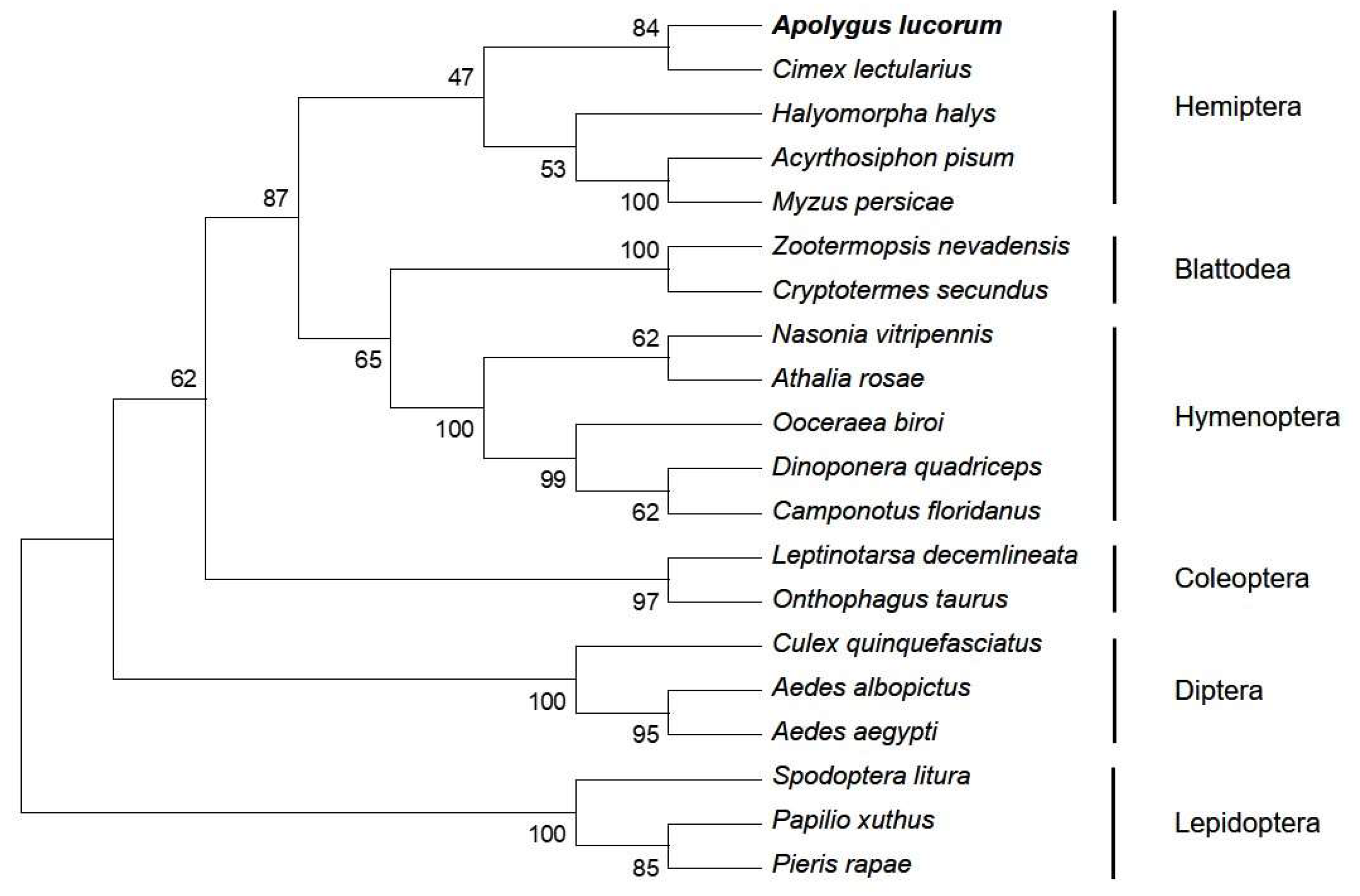
Based on the SMART cDNA library for A. lucorum and RACE, two fragments corresponding to the 5′ and 3′ ends of AlAMPK cDNA were detected. A 1980-bp nucleotide sequence representing the complete AlAMPK cDNA sequence was deposited in the GenBank database (MN514867). The full-length AlAMPK cDNA includes a 287-bp 5′-untranslated region (UTR), a 139-bp 3′-UTR, a canonical polyadenylation signal sequence (CGATAA), a poly (A) tail, and a 1554-bp ORF. The ORF encodes a polypeptide comprising 517 amino acids, with a predicted molecular weight of 58.78 kDa and a theoretical isoelectric point of 7.10. A search of the National Center for Biotechnology Information (NCBI) Conserved Domain Database (https://www.ncbi.nlm.nih.gov/cdd) (accessed on 30 March 2023) revealed three conserved domains, namely the S-TKc, UBA-AID-AMPKalpha domain, and AMPKA-C domains. The S-TKc domain (amino acids 18-270) is the catalytic domain of serine/threonine protein kinase. UBA-AID-AMPK α domain (amino acids 287-351aa), which is a ubiquitin-associated (UBA)-like autoinhibitory domain (AID) found in vertebrate 5’AMP-activated protein kinase catalytic α (AMPKα) subunits. AMPKA-C domain (amino acids 402-515), which is the C-terminal regulatory domain of 5’AMP-activated protein kinase (AMPK) α catalytic subunit, mainly involved in the formation of AMPK heterotrimers (Figure 1A). Sodium dodecyl sulfate polyacrylamide gel electrophoresis (SDS-PAGE) and fluorography demonstrated that the molecular weight of the in vitro protein was similar to the expected 60 kDa (Figure 1B). A phylogenetic analysis based on the AlAMPK sequence uncovered 20 AMPK genes in the NCBI database (Figure 2). Comparison of predicted N-terminal amino acid sequences of the phosphorylation domains of A. lucorum AMPK with their counterparts from Drosophila, mouse, and human are presented in Figure S1.

2.2. AlAMPK Was Phosphorylated by 20E

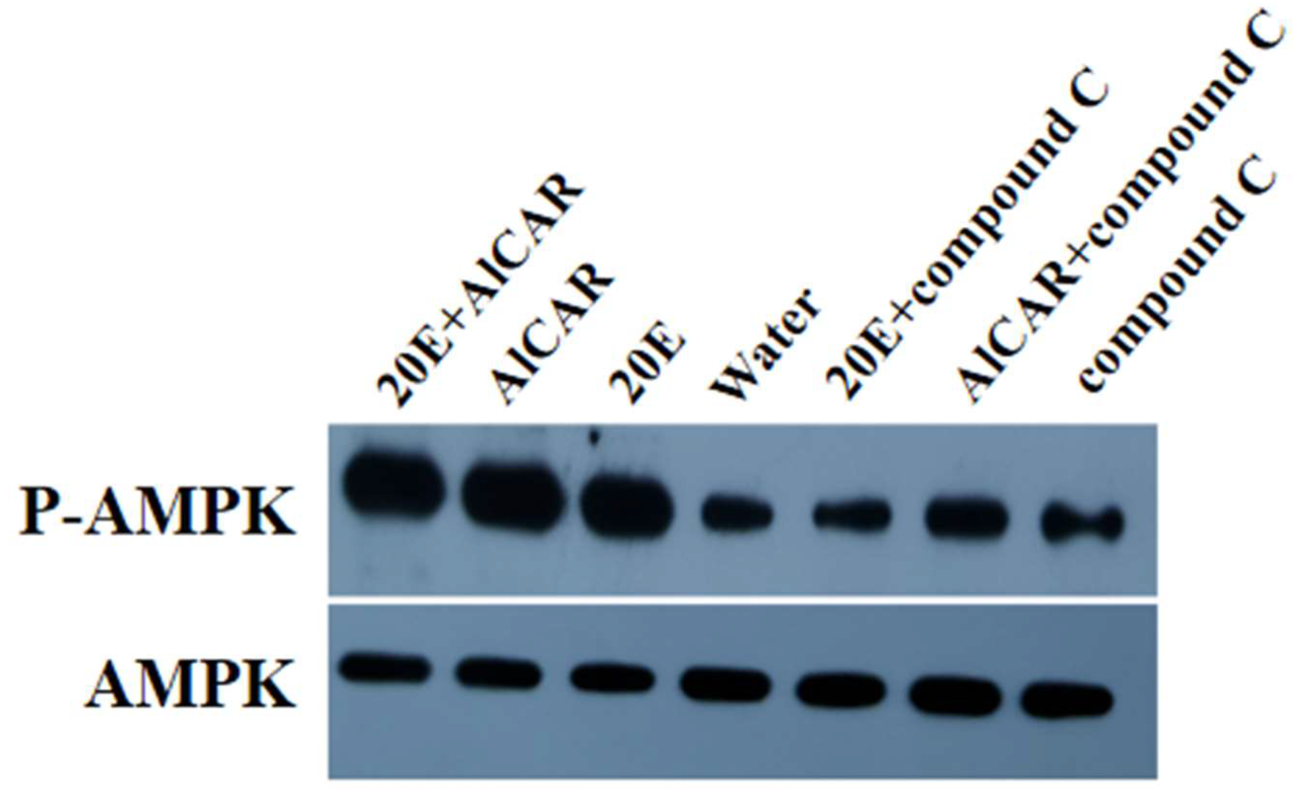
The effect of 20E on AlAMPK phosphorylation was studied in detail by examining the level of AMPK phosphorylation at Thr172. Phosphorylation that occurred at Thr172 was examined in A. lucorum AMPK through Western blot. Auto-phosphorylation of AlAMPK was significantly enhanced in the fat bodies that were cultured in vitro with 20E as compared to the control groups (Figure 3). Further evidence that the phosphorylated protein is AMPK and that the antibody specifically recognizes AMPK comes from the use of the AMPK inhibitor, compound C, and the AMPK activator, AlCAR. In vitro treatment of fat bodies from the second instar nymph with compound C for 24 h revealed a substantial reduction in antigen recognition by the anti-phospho-AMPK antibody. In contrast, treatment with 20E and AlCAR greatly increased immune activity. The total protein level was also checked using an anti-AlAMPK antibody, and results showed that it did not change with stimulation by 20E. This result indicated that the 20E could induce the phosphorylation of AlAMPK.

2.3. AlAMPK Expression Profile in the Ecdysis Stage

By quantitative real-time PCR, the expression of AlAMPK was measured in all developmental stages, including the first day of nymph to the second day of adulthood. The results showed that AlAMPK was continuously expressed throughout the whole life cycle of A. lucorum, with peaks correlating with the ecdysteroids pulse (Figure 4A). The spatial expression pattern of AlAMPK mRNA was analyzed by RT-PCR using total RNA prepared from five tissues in third-instar nymphs. The transcript levels of AlAMPK were higher in the midgut, epidermis, and fat body; in contrast, only small amounts of mRNAs for AlAMPK were detected in flying muscle and Malpighian tubules (Figure 4B).

2.4. AlAMPK Expression and Phenotypes under Spraying and RNAi Experiments

We investigated the functions of AlAMPK in A. lucorum development time and molting rate when knockdown of the mRNA expression of AlAMPK by spraying and RNAi experiments, respectively. Activation of AMPK phosphorylation by 20E and/or AlCAR in vitro enhanced the expression of AlAMPK in the nymph of A. lucorum compared to the compound C treatment (Figure 5A). Similarly, in the fat body of surviving A. lucorum nymphs after 48 h injection, qRT-PCR analysis confirmed that AlAMPK expression level was significantly reduced by treatment with dsAlAMPK compared to treatment with dsGFP and water control (Figure 5B). In the 20E spray treatment, the weight of the fifth instar nymph was 4.79 ± 0.17 mg, and the weights decreased by 10.21% and 4.23% after treatments with compound C and water, respectively. In addition, the weights decreased by 7.64% in dsAlAMPK compared to 20E injections (Figure 5C,D). In the spraying treatment, the AlCAR showed a high molting rate of up to 89.1%, followed by the 20E, 20E + AlCAR combined treatment, whereas the notable lowest role was observed by AlCAR + compound C combined treatment up to 40.3%. Similarly, in response to injection treatment, the 20E was found to play a highly influential role, reaching 89.6%, followed by dsGFP at 86.6%, whereas no influential role of compound C and dsAlAMPK was observed (Table 1).

2.5. The 20E-Induced Gene Expression under the Spray of Seven Different Compounds

Herein, we also investigated the effect of seven treatments inducing the transcription of AlAMPK in A. lucorum (Figure 6A). Among these seven different compounds, the highest expression of ECR-A was recorded under 20E + AlCAR where it reached a maximum of 3.5 folds. Induced expression of ECR-A was also observed under 20E and AlCAR compared to water. In contrast, the transcription level plummeted significantly following the 20E + Compound C, AlCAR + Compound C, and Compound C treatment. A similar expression trend was noticed for ECR-B, USP, and E75-A (Figure 6B,C).

2.6. The 20E-Induced Gene Expression under the AMPK RNAi

The dsAlAMPK significantly affected the expression level of four 20E-related genes. For instance, the expression of ECR-A induced under 20E injection, in contrast, was inhibited in dsAlAMPK treatment. Similarly, the ECR-B displayed reduced mRNA levels in dsAlAMPK compared to the 20E injection. The USP and E75-A gene plummeted substantially in dsAlAMPK, confirming the association of the AMPK gene in 20E-mediated ecdysis in A. lucorum (Figure 7).

2.7. Observation of Epidermal Structure under Spray and RNAi by TEM

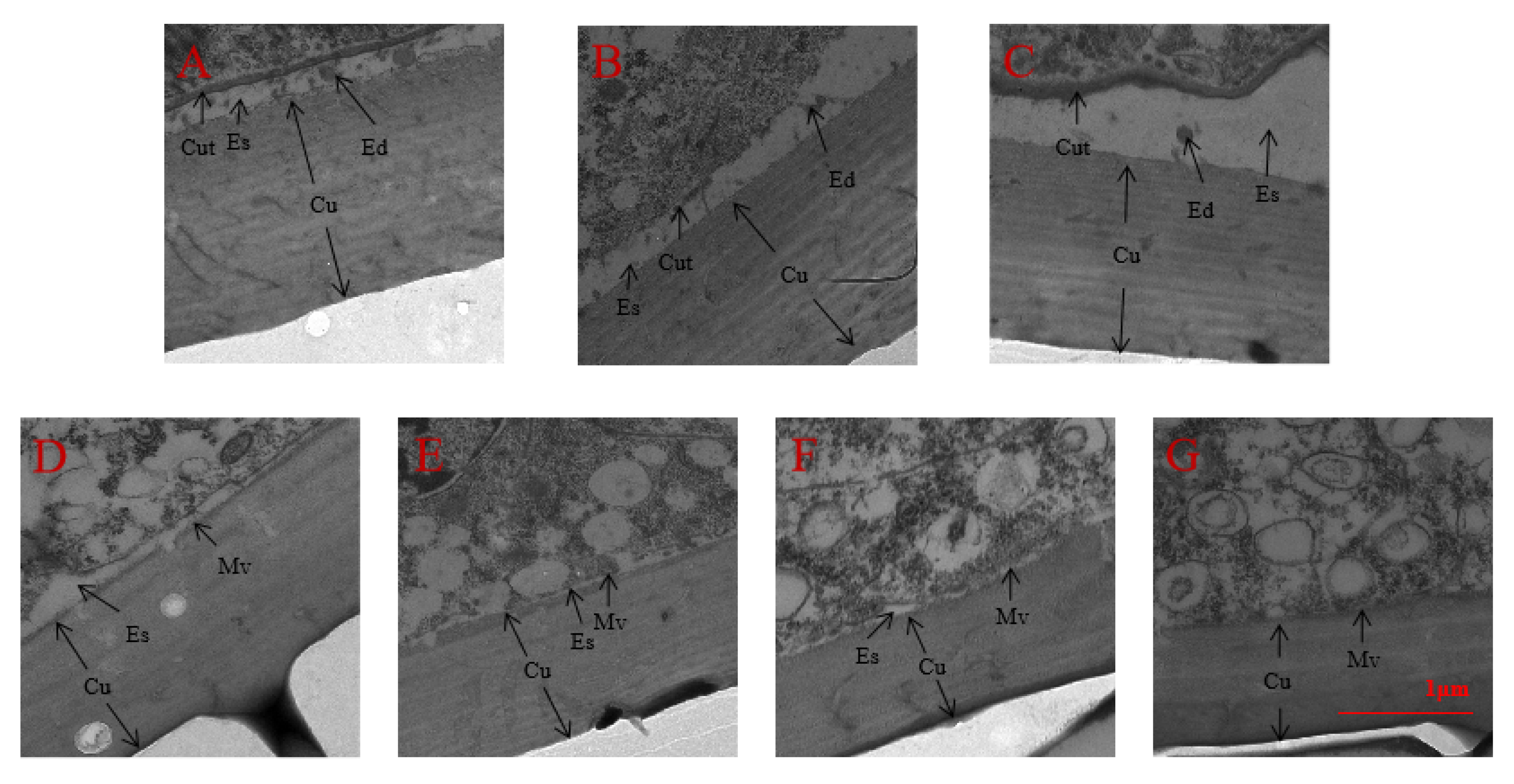
We also surveyed the variation of the epidermal structure in the 3rd instar nymphs of A. lucorum after spraying with seven chemical compounds or injection with dsRNA by TEM. The results indicated that the thickness of the nymph epidermis in 20E and/or AlCAR treatment was significantly increased, the ecdysial space started to form between the cuticle and epidermal cells, and molting progress of nymphs was improved considerably in A. lucorum, which compared to the compound C treatment (Figure 8). Similarly, the cuticle was closely connected with the epidermic cells, the ecdysial space has not yet formed after knockdown of AlAMPK by RNAi (Figure 9).

3. Discussion

The AMPK gene is a member of the gamma subunit family and is found in all domains of life. In eukaryotic cells, AMPK is a key regulatory enzyme of cellular energy homeostasis and is involved in regulating a diverse range of metabolic pathways [21]. AMPK is a serine/threonine kinase and is highly conserved throughout eukaryotes, regulated allosterically by AMP and through phosphorylation at Thr172 [22]. AMPK, in addition, also plays an important role in insect development, tissue growth, molting, and autophagy [23]. In recent decades the AMPK gene has been functionally characterized in numerous organisms such as Hyphantria cunea [24], B. mori [25], Drosophila melanogaster [26], Homo sapiens [27], and so on; however, the 20E-induced AMPK phosphorylation drives developmental programming, ecdysis, and metamorphosis in A. lucorum have not been properly understood.

3.1. AMPK Fine-Tune A. lucorum Physiology

The key role of AMPK genes in the regulation of various key parameters has been previously reported. For instance, the ecdysteroids signaling plays a major role in insect molting and metamorphosis, which is triggered by the binding of 20E to a heterodimer composed of EcR and ultra spiracle (USP) [24]. The present study revealed the AMPK role in inducing the physiological parameters of A. lucorum by dominant expression in all developmental stages with peaks correlating with the ecdysteroids pulse. Additionally, the silencing of AlAMPK by spray and RNAi delayed the nymphal growth and weight, contrary to the activation of AMPK phosphorylation by 20E and/or AlCAR, which enhanced the expression of AlAMPK in the nymph of A. lucorum. It is well known that 20E acts through insect larvae’s central nervous system (CNS) to induce wandering behavior and escape from food [28,29]. In addition, 20E subtly alters the feeding behavior of insects and, consequently, their food intake [30]. However, the induction of wandering behavior and the reduction of feeding behavior can lead to energy stress, such as sugar starvation. This, in turn, ultimately leads to an increase in the cellular AMP/ATP ratio, which activates AMPK and induces developmental parameters [31,32,33]. Our findings suggest a potential role of AlAMPK in A. lucorum development time and molting rate. The AlAMPK was expressed in all developmental stages and in all tested tissues, whereas the dominant expression was observed in the epidermis and midgut. Furthermore, when the mRNA was silenced with dsAlAMPK injection treatment, the size of the first instar nymph and second instar nymphs were reduced. Additionally, the qRT-PCR analysis confirmed that treatment with dsAlAMPK reduced AlAMPK expression in the fat body of surviving A. lucorum nymphs, compared to dsGFP and untreated treatments. Besides, 20E also increases AlAMPK mRNA expression level. A previous study showed that 20E does not directly induce fat body lipolysis in B. mori [34]. 20E reduces food consumption via an unidentified tissue (such as the brain or midgut), causing starvation and fat body lipolysis during molting and pupation in B. mori [29]; compared to the previous finding, the results of this study, we confirmed that treatment with dsAlAMPK reduced AlAMPK expression in the fat body of surviving A. lucorum nymphs.

3.2. 20E-Induced AMPK Phosphorylation Is Essential for Molting in A. lucorum

In insects, the regulation of development, molting, and metamorphosis is coordinated by various endocrine hormones and cellular signals, 20E, and the most prominent AMPK phosphorylation [35]. For instance, mitochondrial health is critical for skeletal muscle function and is improved by exercise training through mitochondrial biogenesis and removing damaged/dysfunctional mitochondria via mitophagy [36]. These changes were monitored using a novel fluorescent reporter gene, pMitoTimer, that allowed assessment of mitochondrial oxidative stress and mitophagy in vivo and were preceded by increased phosphorylation of AMPK at tyrosine 172 and of unc-51 like autophagy activating kinase 1 (Ulk1) at serine 555. Using mice expressing dominant negative and constitutively active AMPK in skeletal muscle, we demonstrate that Ulk1 activation depends on AMPK [37]. Furthermore, the study of Zhao et al. (2023) found that AMPK phosphorylation activated the BmAtg1c mRNA expression and BmAtg1c protein expression, enhancing the autophagy simultaneously peaked in the fat body during larval-pupal metamorphosis [38]. Additionally, 20E activates AMPK in the insect fat body in two ways: by up-regulating the mRNA levels of all three AMPK subunits and inducing energy stress to activate AMPK [39]. The transcription levels of all three AMPK subunits, the protein level of AMPK, and the phosphorylation level of AMPK were all elevated in the B. mori fat body and the D. melanogaster fat body during pupariation, consistent with 20E signaling. Gain-of-function and loss-of-function experiments showed that 20E activates AMPK transcriptionally [39]. In line with several other studies have been conducted but the potential role of AMPK phosphorylation in the molting of A. lucorum. Our study, for the first time, revealed the essential role of AMPK phosphorylation that induces molting also the activation of AMPK phosphorylation by 20E and/or AlCAR in vitro, enhanced the expression of AlAMPK in the nymph of A. lucorum, which compared to the compound C treatment. Additionally, also in the fat body of surviving A. lucorum nymphs after 48 h post-injection the qRT-PCR analysis confirmed that AlAMPK expression level was significantly reduced by treatment with dsAlAMPK in contrast to dsGFP and untreated. Quantitative real-time PCR measured AlAMPK expression from nymph to adult. AlAMPK expression peaks correspond to ecdysteroids pulses in the A. lucorum life cycle. RT-PCR analyzed five tissues for AlAMPK mRNA in the midgut, epidermis, and fat body transcript levels were higher than flying muscle and malpighian tubules in third-instar nymph. Although TEM revealed the variation of the epidermal structure in the surveyed third instar nymphs of A. lucorum after seven chemical compounds spraying or dsRNA injection. Furthermore, the thickness of the nymph epidermis in 20E and/or AlCAR treatments was significantly increased, the ecdysial space started between the cuticle and epidermal cells, and the molting progress of nymphs was improved considerably in A. lucorum in contrast to compound C treatment (Figure 8). Furthermore, previous studies suggested that AMPK activates when AMP levels increase in the body with decreased ATP levels and activated AMPK inhibits anabolic processes and promotes catabolism to minimize ATP utilization while promoting ATP production [39,40]. In response, our study found that the third instar nymphs’ integument ultrastructure after drip administration showed cuticle and epidermic cell effects. The apical plasma membrane area of nymph epidermic cells became smooth, the microvilli on epidermal cells’ apical plasma membrane reduced, and the ecdysial space formed between the cuticle and epidermal cells. Additionally, a dense nymphal cuticle and microvilli were observed after 20E + AlCAR and 20E treatment, also a thickened epidermis separated from epidermal cells and enlarged the ecdysial space, allowing ecdysial droplets to form a new cuticulin layer on the contrary alone 20E and AlCAR treatment resulted in narrow ecdysial.
Herein, we report that 20E is essential for ecdysis and development, and the TEM observation of epidermal structure of A. lucorum confirmed that the epidermic cells of the nymph in 20E-treated, untreated and dsGFP-treated were separated from the epidermis, the ecdysial space started to form between the cuticle and epidermal cells, and the ecdysial droplets were free in the ecdysial space. In 20E-treated, the apical plasma membrane of epidermic cells formed many bulges and a new cuticulin layer. There were sparse microvilli in the apical plasma membrane of epidermic cells in untreated and dsGFP-treated. In dsAMPK-treated, the cuticle was closely connected with the epidermic cells, the microvilli remained developed and dense. Overall, the molting progress of nymphs in the 20E-treated was significantly faster than that in the other treatment groups, and the molting progress in dsAMPK-treated was the slowest; collectively, these results suggested that the 20E is essential for ecdysis and development in A. lucorum. Taken together, our findings indicate that pathways activated in parallel by this agent then concomitantly activate AlAMPK. These results indicate that actions ascribed to AMPK after AlCAR treatment may be influenced by the concomitant modulatory actions of 20E. Collectively, these results suggested that AMPK phosphorylation is essential for molting in A. lucorum.

3.3. The Induction/Inhibition of AMPK Phosphorylation Sensitizes the Expression of 20E Downstream Genes

The steroidal hormone 20E is primarily secreted in an insect’s brain through the prothoracicotropic hormone, which further stimulates the prothoracic gland to synthesize ecdysone and plays an essential role in growth and development [39,41]. The active metabolite of ecdysone and 20E works through ecdysone receptor (EcR) and USP, E75-A and E75-B, to initiate molting and metamorphosis by regulating downstream genes [34]. Previously, the study of Roy et al. (2012) found that EcR was expressed in the PTTH-producing neurosecretory cells (PTPCs) in the larval brain of B. mori, suggesting that PTPCs function as the master cells of development under the regulation of 20E [42]. The present study revealed the hindered expression of 20E downstream genes in which ECR-B was observed with the dominant expression under 20E, AlCAR, and 20E + AlCAR combined treatment. However, the lower expression was followed by the other three genes under the same treatment. Notably, the expression plummeted in the ECR-A, ECR-B, USP, and E75-A genes under different treatments after induction/inhibition of AMPK phosphorylation. Collectively, these results revealed that manipulation with AMPK genes directly alters the functions of 20E downstream genes; however, the underlying molecular mechanisms need to be further studied.

4. Materials and Methods

4.1. Experimental Insect Rearing

A. lucorum were collected from Vicia faba grown in fields in Yancheng (33.110 N, 120.250 E) (Jiangsu China) from July to August 2020 and reared on Phaseolus vulgaris with a 10% sucrose solution in an incubator at 25  ±  1 °C with 70  ±  5% humidity under a 14: 10-h light/dark cycle.

4.2. Cloning of AlAMPK Gene

The total RNA of ten third instar nymphs was isolated using the SV Total Isolation System kit (Promega Corporation, Madison, WI, USA). First-strand cDNA was synthesized using the PrimeScript™ 1st Strand cDNA Synthesis Kit (TaKaRa Biotechnology, Dalian, Co., Ltd. Dalian, China) according to the manufacturer’s instructions. The first-strand cDNA obtained from the insect was used as the PCR template. The gene-specific primers for AlAMPK were designed based on conserved regions found in AMPK from other insect species: Cimex lectularius (GenBank accession No. XM_014407037), Halyomorpha halys (XM_024358872) and Myzus persicae (XM_022324589). The primers were AMPK-F and AMPK-R (Table S1). Following amplification, the products were separated on a 1.0% agarose gel and purified using the TIANgel Midi DNA Purification System (TianGen Biotech CO., LTD, Beijing, China). Purified DNA fragments were cloned into the pEASY-T3 cloning vector (TransGen Biotech, Beijing, China). Recombinant plasmids were isolated using the Plasmid Mini kit and sequenced. The full-length cDNA of AlAMPK was obtained by the rapid amplification of cDNA ends (RACE) using a SMARTTMRACE cDNA Amplification Kit (Clontech, Palo Alto, CA, USA). PCR products were purified, cloned, and sequenced as above. All the primers used in this study are listed in (Table S1).

4.3. In Vitro Translation

A pET28a vector containing the cDNA sequence encoding the AlAMPK gene was constructed and transformed into Escherichia coli BL21. The recombinant target protein was over-expressed and purified using nickel-nitrilotriacetic acid agarose according to the manufacturer’s protocol (ZoonBio Corporation, Nanjing, China).

4.4. Reagents, Proteins, and Antibodies

20E, permeable AMPK activator 5-aminoimidazole-4-carboxamide-1 β-d-ribofuranoside (AlCAR), and an AMPK inhibitor compound C were purchased from Sigma (Sigma-Aldrich LLC, Darmstadt, Germany). The reagents were dissolved in DMSO to obtain the desired concentrations and stored at −20 °C. Different reagents were used for 1 h before treatment was administered. The spraying assay consisted of seven experimental groups: (1) 20E (1.0 μmol/L) and AlCAR (25 μmol/L), (2) 20E only, (3) AlCAR only, (4) compound C (25 μmol/L) only, (5)20E and compound C, (6) AlCAR and compound C, and (7) water as a control.
Phospho-specific antibodies were developed and are commercially available for human AMPKα phosphorylated at Thr172 (cat. no. 2535, Cell Signaling Technology, Boston, MA, USA), as phosphorylation is essential for the activity of these enzymes. Comparison of deduced amino acid sequences of the phosphorylation domain of A. lucorum AMPK with their counterparts from Drosophila, humans, and mice showed identical phosphorylation sites, indicating high conservation among species (Figure S1). The consistency of phosphorylation sites among species suggests it is possible that commercial antibodies against mammalian kinase can be successfully used to investigate the phosphorylation of A. lucorum AMPK. Therefore, a commercial polyclonal antibody against mammalian AMPK phosphorylated at Thr172 was used in this present study. In addition, the antibodies against AlAMPK produced from the cDNA of AlAMPK ORF were preserved in our lab, used at 1:1000 dilutions, and incubated for 1 h at 37 °C.

4.5. Expression Profiles of AlAMPK mRNA Profile

qPCR was performed to profile the expression of AlAMPK across developmental stages (1st—5th instar nymph and female adult) and from selected tissues, including the epidermis, midgut, fat body, flying muscle, and Malpighian tubules. Total RNA was isolated using an SV Total Isolation System kit (Promega(Beijing) Biotech Co., Ltd., Beijing, China), and cDNA was synthesized as described above. mRNA levels were quantified by qPCR using the One Step SYBR PrimeScript RT-PCR Kit (Takara, Dalian, Liaoning, China) using the Bio-radi Cycler real-time quantitative RT-PCR detection system and the iCycleriQ real-time detection system software (version 3.0a; Bio-Rad Laboratories, Inc. Mississippi, USA) and hereafter amplification, the target gene cycle threshold (Ct) values were normalized to the reference gene by the 2−ΔΔCT method (Livak and Schmittgen, 2001) [43]. Due to the low RNA quantity from individual nymphs or adults, a mixture of 15 whole bodies of A. lucorum at each developmental stage and 25 pooled tissues of each organ were used as one sample, respectively. Each experiment was replicated three times with independent sample groups. The A. lucorum β-actin (JN616391) was used as an endogenous reference gene for data normalization. Primers for amplifying a 131 bp β-actin and 125 bp AlAMPK were selected and used for qPCR (Table S1). The mRNA expression profiles of AlAMPK across developmental stages and different tissues were assayed following the same procedure of Tan et al. (2014 a, b) [44,45].

4.6. Western Blot

Western blot was performed using proteins extracted from the fat body of surviving A. lucorum nymphs to identify phosphorylation of AlAMPK at the protein level after the above seven reagent treatments. The total proteins were extracted using a Tissue Protein Extraction Reagent Kit (Nanjing, ZoonBio Tech, Co., Ltd. Nanjing, China) according to the manufacturer’s instructions, and Bradford’s method was used to determine the protein concentration [46]. The Western blot analysis for each AlAMPK phosphorylation followed protocols described in previous studies [47].

4.7. dsRNA Synthesis and Application

Sense and antisense primers, including a T7 RNA Polymerase promoter, were designed based on the sequences of AlAMPK (Table S1). PMD-19T Vector (Takara) plasmids harboring Ace gene segments served as templates for generating double-stranded RNAs (dsRNAs) targeting AlAMPK, utilizing the T7 RiboMAX Express RNAi System (Promega) as directed by the manufacturer. The concentration of dsRNA resuspended in DEPC-treated water was adjusted to 2.0 μg/μL, and dsRNA was kept at −70 °C for further experiments. GFP dsRNA (dsGFP) used as a control was obtained as described above. Freshly molted second instar nymphs were administered 2.5 μL of AlAMPK dsRNA by injection. Three independent experiments were carried out with 300 insects per group. Control animals were administered dsGFP and untreated. After dsRNA injection, the surviving 48 h old nymphs were dissected for fat body removal. The fat bodies were frozen immediately in liquid nitrogen and stored at −80 °C. RNA samples obtained from four individuals’ fat body tissues were tested, each in triplicate.

4.8. Detection of the Effect of Spraying and RNAi

After spraying and dsRNA injection, the mRNA expression in fat bodies of AlAMPK was analyzed by RT-PCR of A. lucorum, respectively. Nymphs were individually placed in 5 cm high, 1.5 cm diameter glass vials covered with a nylon screen. Each glass vial contained a green bean and a 1 × 5 cm wet paper strip for food and water. Nymphal development and molting rate were recorded daily until adult molting or death occurred. 80 to 100 nymphs were included per treatment and control. Nymphal weights of newly molted (<4 h old) fifth-instar nymphs were recorded. In each treatment and control, 20 nymphs were weighed individually.

4.9. Transmission Electron Microscopy (TEM) after Spray and RNAi

About 24 h after molting, 110 third instar nymphs were collected into plastic cases with green beans. A topical application of 1 µL of each solution was applied on the pronotum of third-instar nymphs by microliter syringes (1 µL, Gaoge, Shanghai, China) (drip administration). Ten nymphs were treated in each group. The assay consisted of different experimental treatments described above in Section 2.4 and Section 2.7 The thoracic epidermis of nymphs treated with the chemical solution for 12 h was collected and fixed in 2.5% glutaraldehyde (pH 7.2, phosphate-buffered). The thoracic epidermis was fixed in 2.5% glutaraldehyde for 8 h, rinsed three times with 0.1 M phosphate buffer for 10 min each, and post-fixed in 1% osmium tetroxide for 2 h. Then the specimens were again rinsed three times with 0.1 M phosphate buffer for 10 min each, dehydrated separately in a series of acetone solutions (50%, 70%, 80%, and 90%) for 15 min each, and then 100% acetone (3 × 30 min). After that, the specimens were preserved in an embedding plate containing a pure embedding agent. The embedded plates were polymerized at 37 °C, 45 °C, and 60 °C for 48 h, respectively, and sectioned into 70–90 nm sections. Ultrathin sections (70 nm) were stained with uranyl acetate followed by lead citrate for 10 min and observed with a Hitachi H7650 transmission electron microscope.

4.10. Expression Profiles of Four 20E-Regulated Genes

The mRNA expression levels of AlUSP (JX675574), AlE75A (KX912697), AlEcR-A (KM401656), and AlEcR-B (KM504955) in fat body specimens from survived 48 h old nymphs A. lucorum after dsRNA injection and spraying treatments were assessed as reported by [47]. All the primers used are listed in (Table S1).

4.11. Statistical Analysis

Differences in development time and nymphs’ weight were assessed by one-way ANOVA followed by Tukey’s HSD test (p < 0.05; Statistical Analysis System version 10). Duncan’s new multiple-range test assessed gene expression differences. Finally, GraphPad Prism software (version 9.4.1, Inc., LA Jolla, CA, USA) was used, following the same procedure as Ahmad et al. (2021) [47] for graphical representation following the procedure.

5. Conclusions

The obtained results revealed that the AlAMPK regulation is crucial for ecdysis and development in A. lucorum. The full-length cDNA sequence of the AlAMPK gene was cloned and functionally characterized. The AlAMPK showed expression in all developmental stages and all tested tissues. Additionally, the knockdown of AlAMPK by RNAi delayed the nymph molting rate, reduced nymphal weight and development time, and blocked the expression of 20E downstream genes. Additionally, the 20E and AlCAR induced the thickness of the nymphal epidermis, and the molting nymphs were significantly improved. The present study clearly shows that 20E-induced AlAMPK phosphorylation is essential for ecdysis and metamorphosis in A. lucorum. Further studies are required to fully understand the regulation and pathway mechanism of phosphorylation of 20E downstream genes that regulate the ecdysis in A. lucorum.

Supplementary Materials

The following supporting information can be downloaded at: https://www.mdpi.com/article/10.3390/ijms24108587/s1.

Author Contributions

Y.T. designed the experiments, collected and analyzed the data of the experiments, and drafted the manuscript. J.Z. (Jing Zhao) helped to design the experiments. J.Z. (Jieyu Zhang) helped to analyze the data of the experiments. S.A. and D.X. helped to design the experiments and review the manuscript. G.X. helped to collect the data for the experiments. L.X. and L.G. reviewed and edited the manuscript. All authors have read and agreed to the published version of the manuscript.

Funding

This study was supported by Jiangsu Agricultural Science and Technology Innovation Fund [CX(21)3088, CX(22)2038] and Jiangsu Province Key R&D Program (Modern Agriculture) Project: Surface Project (BE2021303). The funders had no role in the study design, data collection, analysis and the decision to publish.

Institutional Review Board Statement

Not applicable.

Informed Consent Statement

Not applicable.

Data Availability Statement

Not applicable.

Conflicts of Interest

The authors reported no potential conflict of interest.

References

  1. Liu, Y.; Liu, H.; Wang, H.; Huang, T.; Liu, B.; Yang, B.; Yin, L.; Li, B.; Zhang, Y.; Zhang, S.; et al. Apolygus lucorum genome provides insights into omnivorousness and mesophyll feeding. Mol. Ecol. Resourses 2021, 21, 287–300. [Google Scholar] [CrossRef]
  2. Lu, Y.; Wu, K.; Jiang, Y.; Xia, B.; Li, P.; Feng, H.; Wyckhuys, K.A.; Guo, Y. Mirid bug outbreaks in multiple crops correlated with wide-scale adoption of Bt cotton in China. Science 2010, 328, 1151–1154. [Google Scholar] [CrossRef] [PubMed]
  3. Wu, K.; Li, W.; Feng, H.; Guo, Y. Seasonal abundance of the mirids, Lygus lucorum and Adelphocoris spp. (Hemiptera: Miridae) on Bt cotton in northern China. Crop Prot. 2002, 21, 997–1002. [Google Scholar] [CrossRef]
  4. Li, Y.B.; Li, X.R.; Yang, T.; Wang, J.X.; Zhao, X.F. The steroid hormone 20-hydroxyecdysone promotes switching from autophagy to apoptosis by increasing intracellular calcium levels. Insect Biochem. Mol. Biol. 2016, 79, 73–86. [Google Scholar] [CrossRef] [PubMed]
  5. Scieuzo, C.; Nardiello, M.; Salvia, R.; Pezzi, M.; Chicca, M.; Leis, M.; Bufo, S.A.; Vinson, S.B.; Rao, A.; Vogel, H.; et al. Ecdysteroidogenesis and development in Heliothis virescens (Lepidoptera: Noctuidae): Focus on PTTH-stimulated pathways. J. Insect Physiol. 2018, 107, 57–67. [Google Scholar] [CrossRef] [PubMed]
  6. Kefi, M.; Balabanidou, V.; Douris, V.; Lycett, G.; Feyereisen, R.; Vontas, J. Two functionally distinct CYP4G genes of Anopheles gambiae contribute to cuticular hydrocarbon biosynthesis. Insect Biochem. Mol. Biol. 2019, 110, 52–59. [Google Scholar] [CrossRef]
  7. Moulos, P.; Samiotaki, M.; Panayotou, G.; Dedos, S.G. Combinatory annotation of cell membrane receptors and signalling pathways of Bombyx mori prothoracic glands. Sci. Data 2016, 3, 160073. [Google Scholar] [CrossRef] [PubMed]
  8. Engelmann, F. 20-hydroxyecdysone, what it can do. Science 1971, 174, 1041. [Google Scholar] [CrossRef]
  9. Horike, N.; Sonobe, H. Ecdysone 20-monooxygenase in eggs of the silkworm, Bombyx mori: Enzymatic properties and developmental changes. Arch. Insect Biochem. Physiol. 1999, 41, 9–17. [Google Scholar] [CrossRef]
  10. Li, K.; Tian, L.; Guo, Z.; Guo, S.; Zhang, J.; Gu, S.H.; Palli, S.R.; Cao, Y.; Li, S. 20-Hydroxyecdysone (20E) primary response gene E75 isoforms mediate steroidogenesis autoregulation and regulate developmental timing in bombyx. J. Biol. Chem. 2016, 291, 18163–18175. [Google Scholar] [CrossRef]
  11. Liu, H.; Wang, J.; Li, S. E93 predominantly transduces 20-hydroxyecdysone signaling to induce autophagy and caspase activity in Drosophila fat body. Insect Biochem. Mol. Biol. 2014, 45, 30–39. [Google Scholar] [CrossRef] [PubMed]
  12. Liu, X.; Dai, F.; Guo, E.; Li, K.; Ma, L.; Tian, L.; Cao, Y.; Zhang, G.; Palli, S.R.; Li, S. 20-Hydroxyecdysone (20E) primary response gene E93 modulates 20E signaling to promote bombyx larval-pupal metamorphosis. J. Biol. Chem. 2015, 290, 27370–27383. [Google Scholar] [CrossRef] [PubMed]
  13. Tian, L.; Guo, E.; Wang, S.; Liu, S.; Jiang, R.J.; Cao, Y.; Ling, E.; Li, S. Developmental regulation of glycolysis by 20-hydroxyecdysone and juvenile hormone in fat body tissues of the silkworm, Bombyx mori. J. Mol. Cell Biol. 2010, 2, 255–263. [Google Scholar] [CrossRef]
  14. Steinberg, G.R.; Kemp, B.E. AMPK in Health and Disease. Physiol. Rev. 2009, 89, 1025–1078. [Google Scholar] [CrossRef] [PubMed]
  15. Jiang, Y.; Dong, Y.; Luo, Y.; Jiang, S.; Meng, F.-L.; Tan, M.; Li, J.; Zang, Y. AMPK-mediated phosphorylation on 53BP1 promotes c-NHEJ. Cell Rep. 2021, 34, 108713. [Google Scholar] [CrossRef]
  16. Hawley, S.A.; Pan, D.A.; Mustard, K.J.; Ross, L.; Bain, J.; Edelman, A.M.; Frenguelli, B.G.; Hardie, D.G. Calmodulin-dependent protein kinase kinase-beta is an alternative upstream kinase for AMP-activated protein kinase. Cell Metab. 2005, 2, 9–19. [Google Scholar] [CrossRef]
  17. Alers, S.; Löffler, A.S.; Wesselborg, S.; Stork, B. Role of AMPK-mTOR-Ulk1/2 in the regulation of autophagy: Cross talk, shortcuts, and feedbacks. Mol. Cell. Biol. 2012, 32, 2–11. [Google Scholar] [CrossRef]
  18. Lin, S.-C.; Hardie, D.G. AMPK: Sensing Glucose as well as Cellular Energy Status. Cell Metab. 2018, 27, 299–313. [Google Scholar] [CrossRef]
  19. Steinberg, G.R.; Carling, D. AMP-activated protein kinase: The current landscape for drug development. Nat. Rev. Drug Discov. 2019, 18, 527–551. [Google Scholar] [CrossRef]
  20. Nakagawa, Y.; Sonobe, H. Subchapter 98A-20-Hydroxyecdysone. In Handbook of Hormones; Takei, Y., Ando, H., Tsutsui, K., Eds.; Academic Press: San Diego, CA, USA, 2016; pp. 560-e598A–562. [Google Scholar]
  21. Mihaylova, M.M.; Shaw, R.J. The AMPK signalling pathway coordinates cell growth, autophagy and metabolism. Nat. Cell Biol. 2011, 13, 1016–1023. [Google Scholar] [CrossRef]
  22. Sanz, P. AMP-activated protein kinase: Structure and regulation. Curr. Protein Pept. Sci. 2008, 9, 478–492. [Google Scholar] [CrossRef] [PubMed]
  23. Jeon, S.-M. Regulation and function of AMPK in physiology and diseases. Exp. Mol. Med. 2016, 48, e245. [Google Scholar] [CrossRef] [PubMed]
  24. Zou, H.; Zou, H.; Li, X.; Qiu, Q.; Geng, N.; Zhang, B.; Yan, G.; Zhang, Z.; Zhang, S.; Yao, B.; et al. Metformin-induced AMPK activation suppresses larval growth and molting probably by disrupting 20E synthesis and glycometabolism in fall webworm, Hyphantria cunea Drury. Pestic. Biochem. Physiol. 2022, 183, 105083. [Google Scholar] [CrossRef] [PubMed]
  25. Wu, S.; Zhang, X.; He, Y.; Shuai, J.; Chen, X.; Ling, E. Expression of antimicrobial peptide genes in Bombyx mori gut modulated by oral bacterial infection and development. Dev. Comp. Immunol. 2010, 34, 1191–1198. [Google Scholar] [CrossRef]
  26. Sinnett, S.E.; Brenman, J.E. The Role of AMPK in Drosophila melanogaster. Exp. Suppl. 2016, 107, 389–401. [Google Scholar] [CrossRef]
  27. Kim, M.; Shen, M.; Ngoy, S.; Karamanlidis, G.; Liao, R.; Tian, R. AMPK isoform expression in the normal and failing hearts. J. Mol. Cell. Cardiol. 2012, 52, 1066–1073. [Google Scholar] [CrossRef]
  28. Dominick, O.S.; Truman, J.W. The physiology of wandering behaviour in Manduca sexta. IV. Hormonal induction of wandering behaviour from the isolated nervous system. J. Exp. Biol. 1986, 121, 133–151. [Google Scholar] [CrossRef]
  29. Wang, S.; Liu, S.; Liu, H.; Wang, J.; Zhou, S.; Jiang, R.J.; Bendena, W.G.; Li, S. 20-hydroxyecdysone reduces insect food consumption resulting in fat body lipolysis during molting and pupation. J. Mol. Cell Biol. 2010, 2, 128–138. [Google Scholar] [CrossRef]
  30. Li, Y.; Wang, X.; Hou, Y.; Zhou, X.; Chen, Q.; Guo, C.; Xia, Q.; Zhang, Y.; Zhao, P. Integrative proteomics and metabolomics analysis of insect larva brain: Novel insights into the molecular mechanism of insect wandering behavior. J. Proteome Res. 2016, 15, 193–204. [Google Scholar] [CrossRef]
  31. Shang, L.; Chen, S.; Du, F.; Li, S.; Zhao, L.; Wang, X. Nutrient starvation elicits an acute autophagic response mediated by Ulk1 dephosphorylation and its subsequent dissociation from AMPK. Proc. Natl. Acad. Sci. USA 2011, 108, 4788–4793. [Google Scholar] [CrossRef]
  32. Hardie, D.G. AMPK—Sensing Energy while Talking to Other Signaling Pathways. Cell Metab. 2014, 20, 939–952. [Google Scholar] [CrossRef]
  33. Hindupur, S.K.; González, A.; Hall, M.N. The opposing actions of target of rapamycin and AMP-activated protein kinase in cell growth control. Cold Spring Harb. Perspect. Biol. 2015, 7, a019141. [Google Scholar] [CrossRef] [PubMed]
  34. Beckstead, R.B.; Lam, G.; Thummel, C.S. The genomic response to 20-hydroxyecdysone at the onset of Drosophila metamorphosis. Genome Biol. 2005, 6, R99. [Google Scholar] [CrossRef] [PubMed]
  35. Andersen, D.S.; Colombani, J.; Léopold, P. Coordination of organ growth: Principles and outstanding questions from the world of insects. Trends Cell Biol. 2013, 23, 336–344. [Google Scholar] [CrossRef]
  36. Johnson, K.J.; Boekelheide, K. Dynamic testicular adhesion junctions are immunologically unique. I. Localization of p120 catenin in rat testis. Biol. Reprod. 2002, 66, 983–991. [Google Scholar] [CrossRef] [PubMed]
  37. Laker, R.C.; Drake, J.C.; Wilson, R.J.; Lira, V.A.; Lewellen, B.M.; Ryall, K.A.; Fisher, C.C.; Zhang, M.; Saucerman, J.J.; Goodyear, L.J.; et al. Ampk phosphorylation of Ulk1 is required for targeting of mitochondria to lysosomes in exercise-induced mitophagy. Nat. Commun. 2017, 8, 548. [Google Scholar] [CrossRef] [PubMed]
  38. Zhao, H.; Long, S.; Liu, S.; Yuan, D.; Huang, D.; Xu, J.; Ma, Q.; Wang, G.; Wang, J.; Li, S.; et al. Atg1 phosphorylation is activated by AMPK and indispensable for autophagy induction in insects. Insect Biochem. Mol. Biol. 2023, 152, 103888. [Google Scholar] [CrossRef]
  39. Yuan, D.; Zhou, S.; Liu, S.; Li, K.; Zhao, H.; Long, S.; Liu, H.; Xie, Y.; Su, Y.; Yu, F.; et al. The AMPK-PP2A axis in insect fat body is activated by 20-hydroxyecdysone to antagonize insulin/IGF signaling and restrict growth rate. Proc. Natl. Acad. Sci. USA 2020, 117, 9292–9301. [Google Scholar] [CrossRef]
  40. Horike, N.; Sakoda, H.; Kushiyama, A.; Ono, H.; Fujishiro, M.; Kamata, H.; Nishiyama, K.; Uchijima, Y.; Kurihara, Y.; Kurihara, H.; et al. AMP-activated protein kinase activation increases phosphorylation of glycogen synthase kinase 3beta and thereby reduces cAMP-responsive element transcriptional activity and phosphoenolpyruvate carboxykinase C gene expression in the liver. J. Biol. Chem. 2008, 283, 33902–33910. [Google Scholar] [CrossRef]
  41. Chiba, Y.; Oshima, K.; Arai, H.; Ishii, M.; Igarashi, Y.J.J.o.B.C. Discovery and analysis of cofactor-dependent phosphoglycerate mutase homologs as novel phosphoserine phosphatases in Hydrogenobacter thermophilus. J. Biol. Chem. 2012, 287, 11934–11941. [Google Scholar] [CrossRef]
  42. Roy, A.; Shimizu, S.; Kiya, T.; Mita, K.; Iwami, M. Identification of 20-hydroxyecdysone-inducible genes from larval brain of the silkworm, Bombyx mori, and their expression analysis. Zool. Sci. 2012, 29, 333–339. [Google Scholar] [CrossRef] [PubMed]
  43. Livak, K.J.; Schmittgen, T.D. Analysis of relative gene expression data using real-time quantitative PCR and the 2−ΔΔCT method. Methods 2001, 25, 402–408. [Google Scholar] [CrossRef] [PubMed]
  44. Tan, Y.; Xiao, L.; Sun, Y.; Zhao, J.; Bai, L. Sublethal effects of the chitin synthesis inhibitor, hexaflumuron, in the cotton mirid bug, Apolygus lucorum (Meyer-Dür). Pestic. Biochem. Physiol. 2014, 111, 43–50. [Google Scholar] [CrossRef] [PubMed]
  45. Tan, Y.; Xiao, L.; Sun, Y.; Zhao, J.; Bai, L.; Xiao, Y. Molecular characterization of soluble and membrane-bound trehalases in the cotton mirid bug, Apolygus lucorum. Arch. Insect Biochem. Physiol. 2014, 86, 107–121. [Google Scholar] [CrossRef] [PubMed]
  46. Bradford, M.M. A rapid and sensitive method for the quantitation of microgram quantities of protein utilizing the principle of protein-dye binding. Anal. Biochem. 1976, 72, 248–254. [Google Scholar] [CrossRef] [PubMed]
  47. Ahmad, S.; Jiang, L.; Zheng, S.; Chen, Y.; Zhang, J.Y.; Stanley, D.; Miao, H.; Ge, L.Q. Silencing of a putative alanine aminotransferase (ALT) gene influences free amino acid composition in hemolymph and fecundity of the predatory bug, Cyrtorhinus lividipennis Reuter. Arch. Insect Biochem. Physiol. 2021, 108, e21836. [Google Scholar] [CrossRef]
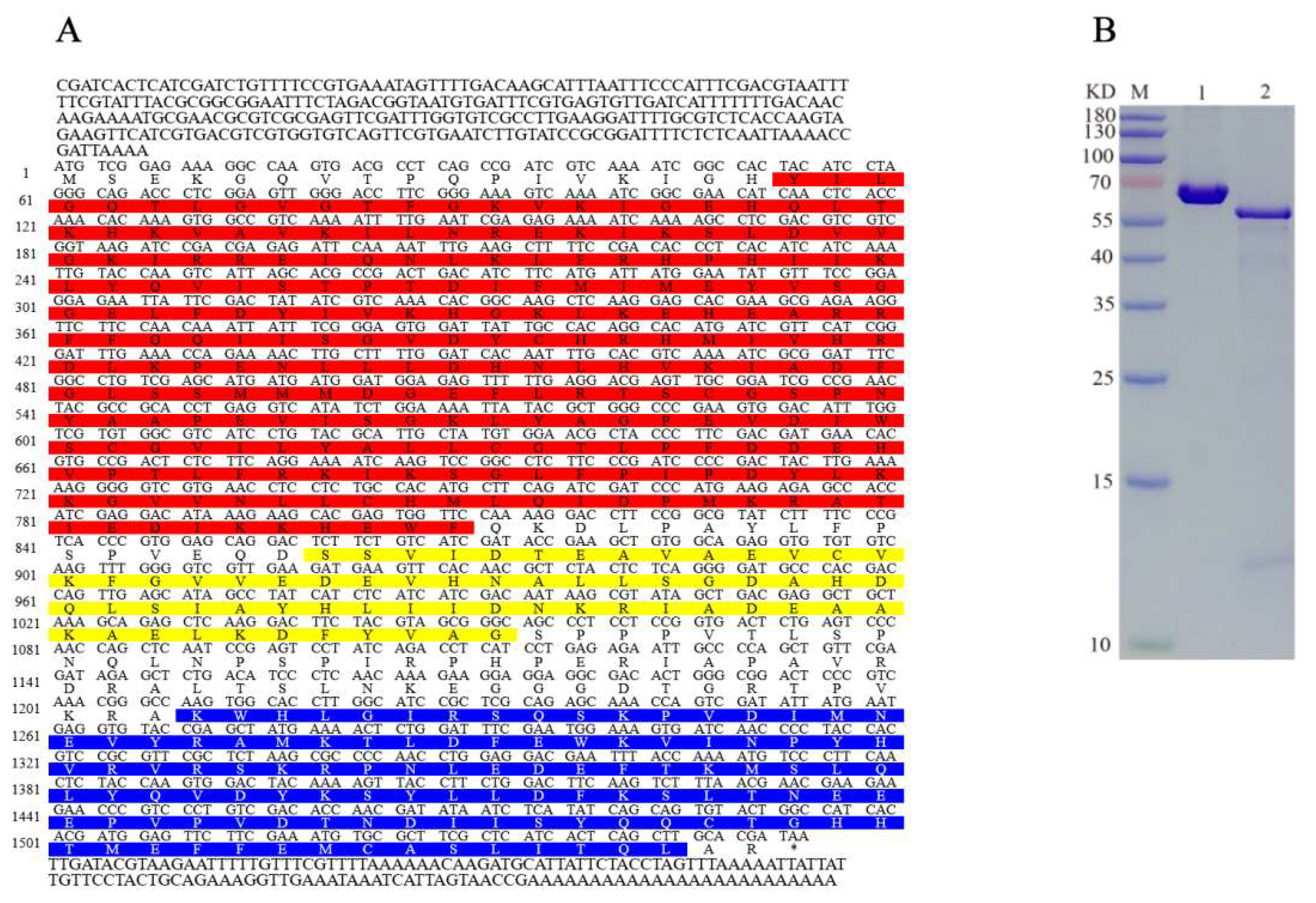
Figure 1. (A) AlAMPK cDNA and amino acid sequences. Bright red, yellow, and blue shaded amino acids indicate the S-TKc (18–270 aa), UBA-AID-AMPK alpha (287–351 aa), and AMPKA-C (402–515 aa), respectively. (B) Translation of AlAMPK in vitro. The AlAMPK translation product was resolved by 10% SDS-PAGE, followed by autoradiography. Lane 1: 0.5 mg/mL BSA, Lane 2: purified AlAMPK protein. Molecular masses of standards are shown on the left.
Ijms 24 08587 g001
Figure 2. The phylogenetic analysis of AMPK between A. lucorum with other insects’ species. The neighbor-joining (NJ) method was employed for tree generations. The Jones–Taylor–Thornton (JTT) substitution model was applied with 1000 bootstrap replicates. The gamma law assessed between-site heterogeneity. The GenBank accessions of the proteins are as follows: Apolygus lucorum AlAMPK (MN514867), Cimex lectularius ClAMPK (XP_014262523), Halyomorpha halys HhAMPK (XP_024214640), Acyrthosi phonpisum ApAMPK (XP_008183803), Myzus persicae MpAMPK2 (XP_022180281), Zootermopsis nevadensis ZnAMPK (XP_021916442), Cryptotermes secundus CsAMPK (XP_023710112), Nasonia vitripennis NvAMPK (XP_001599874), Athalia rosae ArAMPK (XP_012268666), Ooceraea biroi ObAMPK (XP_011329194), Dinoponera quadriceps DqAMPK (XP_014480794), Camponotus floridanus CfAMPK (XP_011259007), Leptinotarsa decemlineata LdAMPK (XP_023020472), Onthophagus taurus OtAMPK (XP_022908510), Culex quinquefasciatus CqAMPK (EDS36926), Aedes albopictus AaAMPK (XP_029719110), Aedes aegypti AaAMPK (XP_001652572), Spodoptera litura SlAMPK (XP_022832070), Papilio xuthus PxAMPK (KPJ05221), Pieris rapae PrAMPK (XP_022112548).
Ijms 24 08587 g002
Figure 3. 20E treatment leads to activation of AlAMPK. Fat bodies from surviving A. lucorum third nymphs were cultured in vitro for 1 h in the presence of 20E (1.0 μmol/L) and AlCAR (25 μmol/L), AlCAR only, 20E only, water as a control, 20E and compound C (25 μmol/L), AlCAR and compound C, and compound C only. Proteins were extracted from the fat bodies and subjected to Western blot analysis using antibodies against AlAMPK phosphorylated at threonine 172 and total AlAMPK.
Ijms 24 08587 g003
Figure 4. The relative developmental-stage and tissue-specific specific expression profiles of AlAMPK via qRT-PCR. (A) The relative developmental stages (day 1–day 16) expression level of AlAMPK. (B) The relative expression level of AlAMPK in the epidermis, flying muscle, Malpighian tubules, midgut, and fat body. The mRNA levels were normalized against the β-actin reference gene. Bars are mean ± SE; different letters above bars indicate highly significant differences between four treatments (p < 0.05, Tukey’s Honestly Significant Difference test).
Ijms 24 08587 g004
Figure 5. The relative mRNA expression levels of AlAMPK and fifth instar nymphal weight of the two different handling methods. (A) The relative expression level of AlAMPK after spraying 7 different compounds. (B) The relative expression level of AlAMPK after injecting dsAlAMPK, dsGFP, and 20E. (C) The fifth-instar nymphal weight after spraying 7 different compounds. (D) The fifth instar nymphal weight after injecting dsAlAMPK, dsGFP, and 20E. The content of 7 different compounds, dsRNA and 20E, is the same as above. The mRNA levels were normalized against the β-actin reference gene. Bars are mean ± SE; different letters above bars indicate highly significant differences between four treatments (p < 0.05, Tukey’s Honestly Significant Difference test).
Ijms 24 08587 g005
Figure 6. Changes in the relative expression levels of ECR-A, ECR-B, USP and E75-A (AD) under different treatments. The mRNA levels were normalized against the β-actin reference gene. The standard errors of the means for the three biologically independent replicates are represented by error bars. Bars are mean ± SE; different letters above bars indicate highly significant differences (p < 0.05, Duncan’s multiple test).
Ijms 24 08587 g006
Figure 7. Changes in the relative expression levels of 20E-regulated genes after injecting dsAlAMPK, dsGFP, and 20E (AD). The contents of dsRNA and 20E are the same as above. The mRNA levels were normalized against the β-actin reference gene. Bars are mean ± SE; different letters above bars indicate highly significant differences (p < 0.05, Duncan’s multiple test).
Ijms 24 08587 g007
Figure 8. TEM observation of epidermal structure in the 3rd instar nymphs of A. lucorum after spraying 7 different compounds. (A) 20E + AlCAR (B) AlCAR (C) 20E (D) CK, the negative control (treatment with distilled water) (E) 20E + compound C (F) AlCAR + compound C (G) compound C. Cuticle: Cu, Cuticulin: Cut, Ecdysial droplet: Ed, Ecdysial space: Es, Microvilli: Mv.
Ijms 24 08587 g008
Figure 9. TEM observation of epidermal structure in the third instar nymphs of A. lucorum after dsRNA injection. (A) dsAlAMPK, (B) dsGFP, (C) untreated (D), 20E. The contents of dsRNA and 20E are the same as above. Cuticle: Cu, Cuticulin: Cut, Ecdysial droplet: Ed, Ecdysial space: Es, Microvilli: Mv.
Ijms 24 08587 g009
Table 1. Effect of experimental treatments in A. lucorum nymphs on ecdysis and molting.
Experimental Treatment1st Nymph Instar2nd Nymph Instar3rd Nymph Instar4th Nymph Instar5th Nymph Instar
Dt
(Days)
Molting (%)Dt
(Days)
Molting (%)Dt
(Days)
Molting (%)Dt
(Days)
Molting (%)Dt
(Days)
Molting
(%)
Spraying20E + AlCAR2.66 ± 0.12 c98.52.04 ± 0.08 c97.42.09 ± 0.21 c95.22.22 ± 0.15 c87.82.53 ± 0.26 d84.8
AlCAR2.71 ± 0.14 c99.22.11 ± 0.14 c96.82.14 ± 0.16 c95.82.19 ± 0.18 c89.12.48 ± 0.14 d86.2
20E2.68 ± 0.08 c1002.01 ± 0.12 c97.52.08 ± 0.15 c94.22.14 ± 0.29 c88.72.43 ± 0.16 d85.8
Water2.82 ± 0.13 b99.52.33 ± 0.16 b95.62.46 ± 0.14 b93.22.35 ± 0.24 b85.42.81 ± 0.23 c82.6
20E + compound C2.99 ± 0.15 a85.62.76 ± 0.21 a68.52.98 ± 0.18 a55.63.07 ± 0.29 a52.23.92 ± 0.08 a42.8
AlCAR+ compound C2.96 ± 0.11 a83.22.81 ± 0.07 a73.23.01 ± 0.18 a50.82.92 ± 0.22 ab46.23.59 ± 0.32 b40.3
compound C3.07 ± 0.21 a73.62.88 ± 0.19 a62.83.42 ± 0.33 a51.2////
Injection20E--2.12 ± 0.11 a95.41.96 ± 0.15 a93.82.43 ± 0.27 a91.22.48 ± 0.26 a89.6
Untreated--2.35 ± 0.06 b96.22.15 ± 0.19 b94.22.47 ± 0.22 b92.42.68 ± 0.19 b88.4
dsGFP--2.34 ± 0.08 b94.32.19 ± 0.07 b90.22.57 ± 0.19 b91.82.62 ± 0.18 b86.6
dsAMPK--2.67 ± 0.28 c62.73.25 ± 0.22 c53.6////
Note: Dt, Development time. Different lower letters above bars indicate highly significant differences between four treatments (p < 0.05, Tukey’s Honestly Significant Difference test).
Disclaimer/Publisher’s Note: The statements, opinions and data contained in all publications are solely those of the individual author(s) and contributor(s) and not of MDPI and/or the editor(s). MDPI and/or the editor(s) disclaim responsibility for any injury to people or property resulting from any ideas, methods, instructions or products referred to in the content.