Case Report

Expanding the Etiology of Oculo–Auriculo–Vertebral Spectrum: A Novel Interstitial Microdeletion at 1p36

by
Mónica García-Castro
1,
Teresa Martinez-Merino
1,
Nuria Puente
2,3 and
José A. Riancho
2,3,*
1
Servicio de Genética, Hospital U. M. Valdecilla, 39008 Santander, Spain
2
Servicio de Medicina Interna, Hospital U. M. Valdecilla, Instituto de Investigación Marqués de Valdecilla (IDIVAL), 39008 Santander, Spain
3
Departamento de Medicina y Psiquiatría, Universidad de Cantabria, 39008 Santander, Spain
*
Author to whom correspondence should be addressed.
Int. J. Mol. Sci. 2023, 24(1), 36; https://doi.org/10.3390/ijms24010036
Submission received: 16 November 2022 / Revised: 15 December 2022 / Accepted: 18 December 2022 / Published: 20 December 2022

Abstract

:
The etiology of oculo–auriculo–vertebral spectrum (OAVS) is not well established. About half of patients show a positive family history. The etiology of familiar cases is unclear but appears genetically heterogeneous. This motivated us to report a case of OAVS with microtia, ptosis, facial microsomy, and fusion of vertebral bodies associated with a novel genetic etiology, including a deletion at 1p36.12-13. This case report expands on the genetic etiology of OAVS. Furthermore, it also expands the clinical manifestations of patients with interstitial deletions of the de 1p36.12-13 region.

1. Introduction

The abnormal embryonic development of the first and second branchial arches (around the 4th week of gestation) creates various anomalies, mainly of the head and vertebrae, that have been grouped into several syndromes, such as Treacher Collins syndrome, auriculocondylar syndrome, mandibulofacial dysostosis Guion-Almeida type, branchio-oto-renal syndrome, and Goldenhar syndrome [1,2,3,4]. Other disorders with overlapping features include the Miller, Nager, Townes–Brocks, CHARGE, and VACTER syndromes [5]. Some of these disorders have been associated with specific gene variants. For example, 80% of patients with Treacher Collins Syndrome (facial microsomy, usually symmetric, microtia, palatal cleft, eyelid abnormalities) have TCOF1 mutations. Brachio-oto-renal syndrome has been linked to EYA1 or SIX5 gene mutations. However, there is considerable variability and overlap of manifestations. Therefore, the term oculo–auriculo–vertebral spectrum (OAVS) is presently preferred. It is largely equivalent to the conditions known as Goldenhar syndrome or hemifacial microsomia syndrome.
The reported prevalence of OAVS varies between 1/3000 and 1/10,000 births [1]. The phenotype varies from mild to severe. The most common malformations are ear abnormalities (microtia, preauricular tags) with or without hearing loss, facial microsomy (usually unilateral, sometimes bilateral, but almost constantly asymmetric), orofacial clefts, ocular defects (epibulbar dermoids, coloboma, etc.), vertebral abnormalities (fused cervical vertebrae and others), and intellectual disability. Most patients have some degree of asymmetric facial microsomy. Isolated microtia, hemifacial microsomy, mild ear malformations, or hemifacial microsomy plus a family history are usually considered the minimum diagnostic criteria [1]. Patients may have other malformations, particularly of the heart or genitourinary system.
The etiology of OAVS is not well established. About one-third to one-half of cases are sporadic, and one-half show family history [1,6]. Sporadic cases have been associated with acquired disorders during pregnancy, such as maternal diabetes, smoking, placental insufficiency, or drug exposure. The etiology of familiar cases is unclear but appears genetically heterogeneous. Both recessive and dominant forms of inheritance have been described. Thus, much more information is needed to better understand the genetic landscape of these groups of conditions and the genotype–phenotype correlations that help develop deeper scientific knowledge and tailor genetic counseling. This motivated us to report a case of OAVS with a novel genetic etiology.

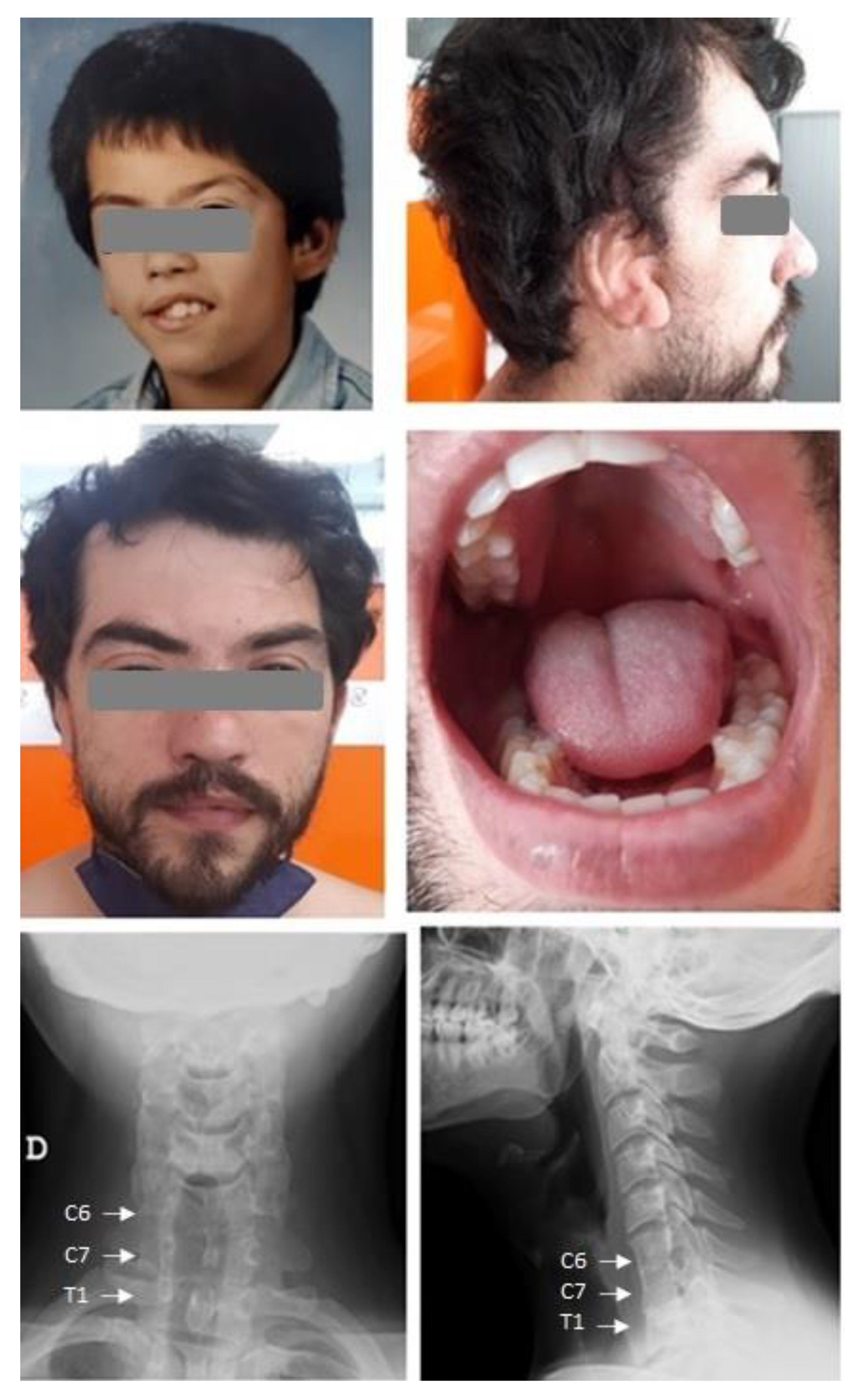
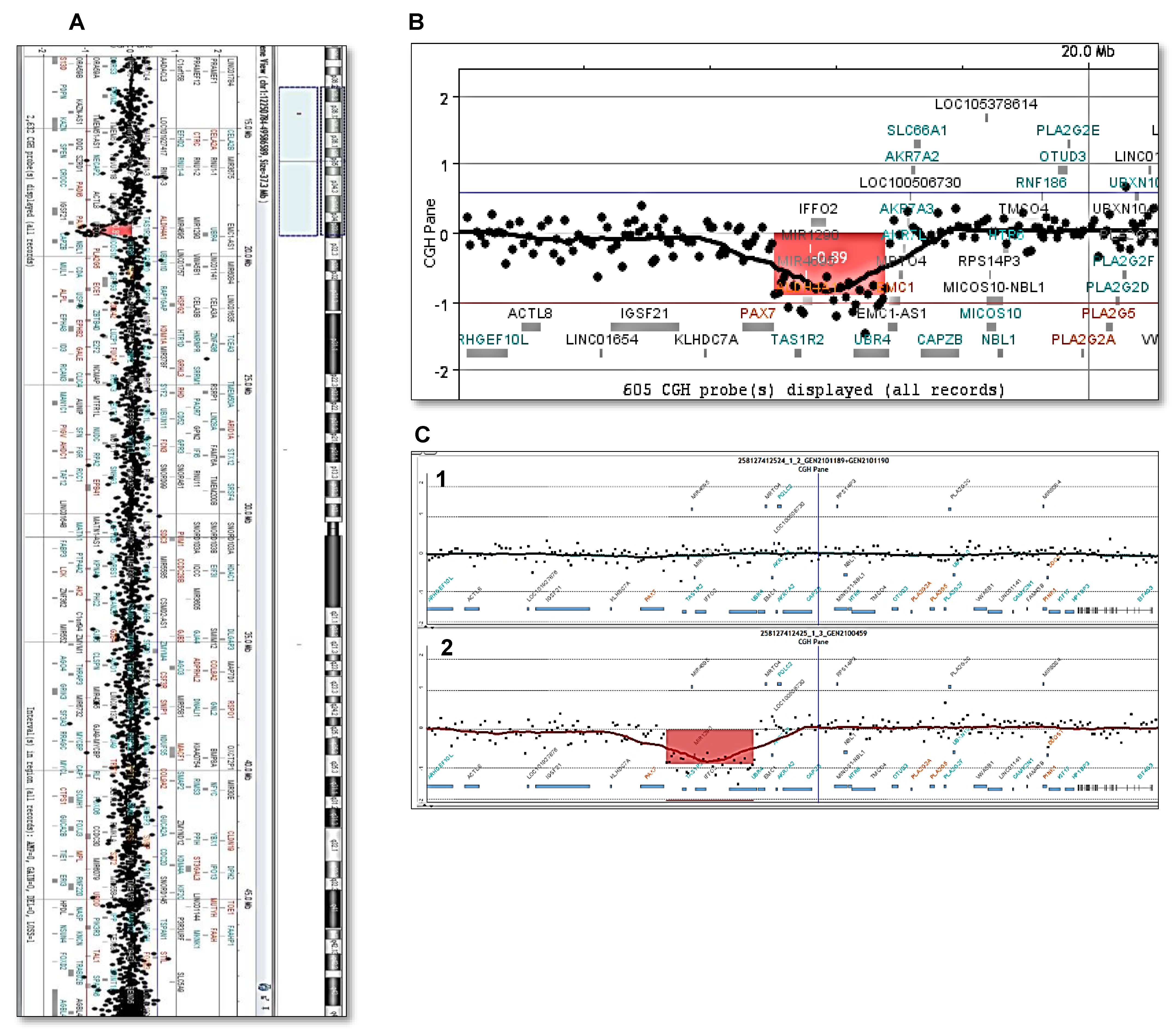
2. Case Presentation

A 33-year-old Caucasian man was sent to our Adult Rare Disease Clinic with suspicion of a malformation syndrome. He was born after an uneventful pregnancy, without known maternal diseases or toxic exposures. Physical and mental development was normal. He had a malformed rudimentary right ear and atresia of the external auditory canal that underwent reconstructive surgery. His parents recalled that the ear was rudimentary, with only the lobule present. He also had been operated on for right palpebral ptosis and used an advancement orthosis for mandibular hypoplasia. His medical history was otherwise unremarkable. He had no siblings and there was no family history of craniofacial abnormalities or other known genetic disorders.
His height, weight, and cephalic perimeter were 176 cm, 96 kg, and 57 cm, respectively. The right ear was reconstructed. There was residual right ptosis and mild left ptosis, as well as facial asymmetry, with mild left hypoplasia of the face and tongue without orofacial clefts (Figure 1). The skin was normal and there was no joint hypermobility. The rest of the physical exam was normal.
Routine blood chemistries and CBC were normal. Skeletal X-rays showed mild scoliosis and partial fusion of vertebrae C6-C7 and T1 (Figure 1). EKG showed an anterosuperior left-branch block. A cardiac ultrasound revealed no anomalies. An abdominal ultrasound was normal. A DXA scan showed moderately high bone mineral density, with Z-scores (sex and age-adjusted) of +1.5 at the lumbar spine and +1.9 at the hip.
Comparative genomic hybridization (CGH) array was performed using the Sure TAG DNA Labeling kit and Human CGH Microarray kit 4x180K (Nimgenetics, Agilent Technologies, California), following manufacturer’s instructions. Copy number variations (CNVs) were detected with the ADM-2 algorithm implemented in the Agilent Cytogenomic Software v4.0. Only CNVs with at least five consecutive probes (±40 kb) were analyzed. This analysis showed a heterozygous 437.7 kb deletion at 1p36.13 (Chr1.19085207-19522963; GRCh37, hg19) that included the OMIM genes TAS1R2 (taste receptor type 1, member 2), ALDH4A1 (aldehyde dehydrogenase, family 4, subfamily a, member 1), and UBR4 (ubiquitin protein ligase e3 component n-recognin 4), and a few other genes (IFFO2, MIR1290, and MIR4695). The study of both parents was normal (Figure 2).
Additionally, a clinical exome study of the patient showed three heterozygous variants, namely an incidental pathogenic variant of CFTR (cystic fibrosis transmembrane conductance regulator, located on 7q31) and variants of unknown significance (VOUS) in TNXB (tenascin XB, a member of the tenascin family of extracellular matrix glycoproteins, located on 6p21) and AEBP1 (AE binding protein 1, located on 7p13) genes. The CTFR variant (c.1521-1523 del CTT; p.Phe508del) had been previously associated with cystic fibrosis; thus, the patient was an asymptomatic heterozygous carrier. No cases of cystic fibrosis were reported in the family. The heterozygous TNXB missense variant (p.Gly1265Arg) was not previously reported. TNXB variants have been associated with “TNXB-related type 1 classical-like Ehlers–Danlos syndrome”, a disorder that is similar to classic Ehlers–Danlos, with generalized joint hypermobility, hyperextensible skin, and easy bruising. A recessive mode of inheritance was reported, although some heterozygous relatives showed mild manifestations [7,8]. AEBP1 encodes a member of the carboxypeptidase A protein family. The encoded protein may function as a transcriptional repressor and appears to influence the differentiation of adipocytes and smooth muscle cells, as well as the organization and remodeling of the extracellular matrix [9,10]. The AEBP1 variant (p.Glu352del) affects a poorly conserved residue and has been reported in 0.003% of Europeans. Other AEBP1 variants have also been associated with Ehlers–Danlos syndrome, such as classic-like 2, which also possesses a recessive mode of inheritance [9]. The patient did not show joint hypermobility or any other manifestation suggestive of Ehlers–Danlos syndrome. The exome study did not reveal variants in other genes known to be associated with craniofacial abnormalities.

3. Discussion

OAVS seems to be heterogeneous regarding both the genetic background and clinical manifestations. Therefore, we can speculate that a better delineation of the clinical–genetic entities will be possible in the future. However, much more information needs to be accumulated to attain that goal. Mutations of several genes have been implicated in case reports of OAVS (Table 1). They include PAX1, EYA3, MYT1, AMIGO2, ZYG11B, VWA1, SF3B2, ZIC3, YPEL1, CRKL, OTX2, and EFTUD2. Most cases were sporadic; an autosomal dominant mode of inheritance was usually suggested in familiar cases [5,11,12]. Additionally, chromosomal abnormalities have been described in a few patients with OAVS. Thus, Guida et al. [13] performed chromosomal microarray analysis in 19 patients with OAVS and identified pathogenic CNVs in two cases and VOUS in seven. Similarly, Spinelli-Silva et al. found pathogenic CNVs in 4 out of 17 cases and VOUS in another four cases. Their own literature review revealed pathogenic CNVs in 9% patients. Previously reported CNVs were located at 1p22, 5q13.2, 5q15, 10p14, 12p13, 14p31, 15q24, 22qter, and 22q11. However, the genetic variants were rarely recurrent. Deletions at the 22q11 region seem to be the most common (Table 2), although they are only present in approximately 5% of patients [14,15]. Thus, in view of these variable and overlapping genetic and clinical data, more information is needed to fully understand the etiologic landscape of OAVS and the possible genotype–phenotype relationships. Thus, here, we present a patient with OAVS and a previously unreported deletion at chromosome 1.
Terminal 1p36 is considered one of the most common terminal deletions, with an incidence of 1 in 5000 live births. The phenotype of the monosomy 1p36 syndrome is variable and includes intellectual disability, craniofacial dysmorphism, growth delay, eye problems, and hypoacusis [26].
The critical region was initially identified as a 6.3 Mb region at 1p36.33-1p36.31. A more proximal deletion syndrome was described at 1p36.23-1p36.22 by Kang et al. The five patients reported by Kang showed various combinations of microcephaly, prominent forehead, ptosis, rotated and enlarged ears, bulbous nose, digital contractures, and cardiac malformations, among others. The smallest region of overlap (SRO) was located at 1p36.22 [27]. Aagard-Nolting et al. recently reported seven patients from five families with an even more proximal deletion at 1p36.13-1p36.1. The most common manifestations were intellectual or learning disability and ptosis, which were both present in five out of seven patients. Other frequent features included epicanthus, thick eyebrows, high palate, protruding chin, misalignment of teeth, and heart malformations [28]. The SRO encompassed 1 Mb at Chr1:19077793-20081292). The genes responsible were unclear.
The present patient carried a de novo deletion that was within the SRO of Aagard-Nolting’s cases. The region included three protein-coding genes: TAS1R, ALDH4A1, and UBR4. TAS1R encodes a taste receptor; ALDH4A1 encodes a mitochondrial matrix NAD(+)-dependent dehydrogenase, which catalyzes the second step of the proline degradation pathway, converting pyrroline-5-carboxylate to glutamate; and ALDH4A1 deficiency causes hyperprolinemia, a recessive disorder presenting with epilepsy and other neurological problems. UBR4 (Ubiquitin protein ligase e3 component n-recognin 4) is highly expressed in nervous tissue and has been associated with ataxia and other neurological phenotypes. Thus, none of those genes seems to be a biologically plausible candidate to explain the patient’s phenotype. A different driver gene is likely involved. In this view, it is interesting to note that two microRNAs are also transcribed from the deleted region (miR1290 and miR4695). miR1290 is contained within intron 1 of the ALDH4A1-coding region, and it seems involved in cell proliferation through the interaction with its target genes, which may include multiple genes, such as FOXA1, IGFBP3, and others related to the Wnt and hypoxia pathways [29]. The Wnt pathway is a major regulator of the differentiation of many cell types, including mesenchymal and skeletal cells [30,31]. miR4695 may also influence the Wnt pathway by targeting TCF4, a nuclear transcription factor involved in β-catenin signaling [32]. This region is far distant from the 1p22.2-1p31.1 deletion reported by Callier in a patient with severe mental disability, narrow ears, facial asymmetry, and clinodactyly who was diagnosed as having Goldenhar syndrome [14].

4. Conclusions

This case report expands the genetic etiology of OAVS. Furthermore, it also expands the clinical manifestations of patients with interstitial deletions of de 1p36.12-13 region. More data are needed to define the genotype–phenotype links among patients with microtia and other abnormalities of the first branchial arches.

Author Contributions

Conceptualization, M.G.-C. and J.A.R.; clinical data curation, N.P. and J.A.R.; molecular data acquisition and interpretation, M.G.-C. and T.M.-M.; writing—original draft preparation, M.G.-C. and J.A.R.; writing—review and editing, all. All authors have read and agreed to the published version of the manuscript.

Funding

This research received no external funding.

Institutional Review Board Statement

The study was conducted in accordance with the Declaration of Helsinki, no procedures outside those indicated for clinical care were performed, and IRB permit was not required.

Informed Consent Statement

The patient gave informed written consent to publish this paper.

Data Availability Statement

Raw data are available from authors by reasonable request.

Conflicts of Interest

The authors declare no conflict of interest.

References

  1. Beleza-Meireles, A.; Hart, R.; Clayton-Smith, J.; Oliveira, R.; Reis, C.F.; Venâncio, M.; Ramos, F.; Sá, J.; Ramos, L.; Cunha, E.; et al. Oculo-auriculo-vertebral spectrum: Clinical and molecular analysis of 51 patients. Eur. J. Med. Genet. 2015, 58, 455–465. [Google Scholar] [CrossRef]
  2. Cohen, N.; Cohen, E.; Gaiero, A.; Zecca, S.; Fichera, G.; Baldi, F.; Giordanetto, J.F.; Mercier, J.M.; Cohen, A. Maxillofacial features and systemic malformations in expanded spectrum Hemifacial Microsomia. Am. J. Med. Genet. Part A 2017, 173, 1208–1218. [Google Scholar] [CrossRef] [PubMed]
  3. Bartel-Friedrich, S. Congenital Auricular Malformations: Description of Anomalies and Syndromes. Facial Plast. Surg. 2015, 31, 567–580. [Google Scholar] [CrossRef] [PubMed]
  4. Gendron, C.; Schwentker, A.; van Aalst, J. Genetic Advances in the Understanding of Microtia. J. Pediatr. Genet. 2016, 5, 189–197. [Google Scholar] [CrossRef] [PubMed] [Green Version]
  5. Tingaud-Sequeira, A.; Trimouille, A.; Sagardoy, T.; Lacombe, D.; Rooryck, C. Oculo-auriculo-vertebral spectrum: New genes and literature review on a complex disease. J. Med. Genet. 2022, 59, 417–427. [Google Scholar] [CrossRef]
  6. Estandia-Ortega, B.; Fernández-Hernández, L.; Alcántara-Ortigoza, M.A.; González-del Angel, A. Proposed clinical approach and imaging studies in families with oculo-auriculo-vertebral spectrum to assess variable expressivity. Am. J. Med. Genet. Part A 2022, 188, 1515–1525. [Google Scholar] [CrossRef]
  7. Brady, A.F.; Demirdas, S.; Fournel-Gigleux, S.; Ghali, N.; Giunta, C.; Kapferer-Seebacher, I.; Kosho, T.; Mendoza-Londono, R.; Pope, M.F.; Rohrbach, M.; et al. The Ehlers-Danlos syndromes, rare types. Am. J. Med. Genet. Part C 2017, 175, 70–115. [Google Scholar] [CrossRef] [Green Version]
  8. Zweers, M.C.; Bristow, J.; Steijlen, P.M.; Dean, W.B.; Hamel, B.C.; Otero, M.; Kucharekova, M.; Boezeman, J.B.; Schalkwijk, J. Haploinsufficiency of TNXB is associated with hypermobility type of Ehlers-Danlos syndrome. Am. J. Hum. Genet. 2003, 73, 214–217. [Google Scholar] [CrossRef] [Green Version]
  9. Blackburn, P.R.; Xu, Z.; Tumelty, K.E.; Zhao, R.W.; Monis, W.J.; Harris, K.G.; Gass, J.M.; Cousin, M.A.; Boczek, N.J.; Mitkov, M.V.; et al. Bi-allelic Alterations in AEBP1 Lead to Defective Collagen Assembly and Connective Tissue Structure Resulting in a Variant of Ehlers-Danlos Syndrome. Am. J. Hum. Genet. 2018, 102, 696–705. [Google Scholar] [CrossRef] [Green Version]
  10. Vishwanath, N.; Monis, W.J.; Hoffmann, G.A.; Ramachandran, B.; DiGiacomo, V.; Wong, J.Y.; Smith, M.L.; Layne, M.D. Mechanisms of aortic carboxypeptidase-like protein secretion and identification of an intracellularly retained variant associated with Ehlers-Danlos syndrome. J. Biol. Chem. 2020, 295, 9725–9735. [Google Scholar] [CrossRef]
  11. Tingaud-Sequeira, A.; Trimouille, A.; Salaria, M.; Stapleton, R.; Claverol, S.; Plaisant, C.; Bonneu, M.; Lopez, E.; Arveiler, B.; Lacombe, D.; et al. A recurrent missense variant in EYA3 gene is associated with oculo-auriculo-vertebral spectrum. Hum. Genet. 2021, 140, 933–944. [Google Scholar] [CrossRef]
  12. Carter, S.; Fellows, B.J.; Gibson, K.; Bicknell, L.S. Extending the PAX1 spectrum: A dominantly inherited variant causes oculo-auriculo-vertebral syndrome. Eur. J. Hum. Genet. 2022, 30, 1178–1181. [Google Scholar] [CrossRef]
  13. Guida, V.; Sparascio, F.P.; Bernardini, L.; Pancheri, F.; Melis, D.; Cocciadiferro, D.; Pagnoni, M.; Puzzo, M.; Goldoni, M.; Barone, C.; et al. Copy number variation analysis implicates novel pathways in patients with oculo-auriculo-vertebral-spectrum and congenital heart defects. Clin. Genet. 2021, 100, 268–279. [Google Scholar] [CrossRef]
  14. Callier, P.; Faivre, L.; Thauvin-Robinet, C.; Marle, N.; Mosca, A.L.; D’Athis, P.; Guy, J.; Masurel-Paulet, A.; Joly, L.; Guiraud, S.; et al. Array-CGH in a series of 30 patients with mental retardation, dysmorphic features, and congenital malformations detected an interstitial 1p22.2-p31.1 deletion in a patient with features overlapping the goldenhar syndrome. Am. J. Med. Genet. Part A 2008, 146, 2109–2115. [Google Scholar] [CrossRef]
  15. Spineli-Silva, S.; Sgardioli, I.C.; Dos Santos, A.P.; Bergamini, L.L.; Monlleó, I.L.; Fontes, M.I.; Félix, T.M.; Ribeiro, E.M.; Xavier, A.C.; Lustosa-Mendes, E.; et al. Genomic imbalances in craniofacial microsomia. Am. J. Med. Genet. Part C 2020, 184, 970–985. [Google Scholar] [CrossRef]
  16. Luquetti, D.V.; Heike, C.L.; Zarante, I.; Timms, A.E.; Gustafson, J.; Pachajoa, H.; Porras-Hurtado, G.L.; Ayala-Ramirez, P.; Duenas-Roque, M.M.; Jimenez, N.; et al. MYT1 role in the microtia-craniofacial microsomia spectrum. Mol. Genet. Genom. Med. 2020, 8, e1401. [Google Scholar] [CrossRef]
  17. Wang, Y.; Ping, L.; Luan, X.; Chen, Y.; Fan, X.; Li, L.; Liu, Y.; Wang, P.; Zhang, S.; Zhang, B.; et al. A Mutation in VWA1, Encoding von Willebrand Factor A Domain-Containing Protein 1, Is Associated With Hemifacial Microsomia. Front. Cell Dev. Biol. 2020, 8, 571004. [Google Scholar] [CrossRef]
  18. Rengasamy Venugopalan, S.; Farrow, E.; Sanchez–Lara, P.A.; Yen, S.; Lypka, M.; Jiang, S.; Allareddy, V. A novel nonsense substitution identified in the AMIGO2 gene in an Occulo-Auriculo-Vertebral spectrum patient. Orthod. Craniofacial. Res. 2019, 22, 163–167. [Google Scholar] [CrossRef]
  19. Zamariolli, M.; Colovati, M.; Moysés-Oliveira, M.; Nunes, N.; Caires dos Santos, L.; Alvarez Perez, A.B.; Bragagnolo, S.; Melaragno, M.I. Rare single-nucleotide variants in oculo-auriculo-vertebral spectrum (OAVS). Mol. Genet. Genom. Med. 2019, 7, e00959. [Google Scholar] [CrossRef] [Green Version]
  20. Güleray, N.; Koşukcu, C.; Oğuz, S.; Ürel Demir, G.; Taşkıran, E.Z.; Kiper, P.Ö.; Utine, G.E.; Alanay, Y.; Boduroğlu, K.; Alikaşifoğlu, M. Investigation of Genetic Causes in a Developmental Disorder: Oculoauriculovertebral Spectrum. Cleft Palate-Craniofacial. J. 2022, 59, 1114–1124. [Google Scholar] [CrossRef]
  21. Rengasamy Venugopalan, S.; Farrow, E.G.; Lypka, M. Whole-exome sequencing identified a variant in EFTUD2 gene in establishing a genetic diagnosis. Orthod. Craniofacial. Res. 2017, 20, 50–56. [Google Scholar] [CrossRef] [PubMed]
  22. Celse, T.; Tingaud-Sequeira, A.; Dieterich, K.; Siegfried, G.; Lecaignec, C.; Bouneau, L.; Fannemel, M.; Salaun, G.; Laffargue, F.; Martinez, G.; et al. OTX2 duplications: A recurrent cause of oculo-auriculo-vertebral spectrum. J. Med. Genet. 2022. [CrossRef] [PubMed]
  23. Guida, V.; Sinibaldi, L.; Pagnoni, M.; Bernardini, L.; Loddo, S.; Margiotti, K.; Digilio, M.C.; Fadda, M.T.; Dallapiccola, B.; Iannetti, G.; et al. A de novo proximal 3q29 chromosome microduplication in a patient with oculo auriculo vertebral spectrum. Am. J. Med. Genet. Part A 2015, 167A, 797–801. [Google Scholar] [CrossRef]
  24. Puvabanditsin, S.; February, M.; Francois, L.; Garrow, E.; Bruno, C.; Mehta, R. 7q21.11 Microdeletion in a Neonate With Goldenhar Syndrome: Case Report and a Literature Review. Cleft Palate-Craniofacial. J. 2016, 53, 249–252. [Google Scholar] [CrossRef]
  25. Glaeser, A.B.; Santos, A.S.; Diniz, B.L.; Deconte, D.; Rosa, R.F.M.; Zen, P.R.G. Candidate genes of oculo-auriculo-vertebral spectrum in 22q region: A systematic review. Am. J. Med. Genet. Part A 2020, 182, 2624–2631. [Google Scholar] [CrossRef]
  26. Jordan, V.K.; Zaveri, H.P.; Scott, D.A. 1p36 deletion syndrome: An update. Appl. Clin. Genet. 2015, 8, 189–200. [Google Scholar] [PubMed] [Green Version]
  27. Kang, S.H.; Scheffer, A.; Ou, Z.; Li, J.; Scaglia, F.; Belmont, J.; Lalani, S.R.; Roeder, E.; Enciso, V.; Braddock, S.; et al. Identification of proximal 1p36 deletions using array-CGH: A possible new syndrome. Clin. Genet. 2007, 72, 329–338. [Google Scholar] [CrossRef]
  28. Aagaard Nolting, L.; Brasch-Andersen, C.; Cox, H.; Kanani, F.; Parker, M.; Fry, A.E.; Loddo, S.; Novelli, A.; Dentici, M.L.; Joss, S.; et al. A new 1p36.13-1p36.12 microdeletion syndrome characterized by learning disability, behavioral abnormalities, and ptosis. Clin. Genet. 2020, 97, 927–932. [Google Scholar] [CrossRef]
  29. Kalhori, M.R.; Soleimani, M.; Arefian, E.; Alizadeh, A.M.; Mansouri, K.; Echeverria, J. The potential role of miR-1290 in cancer progression, diagnosis, prognosis, and treatment: An oncomiR or onco-suppressor microRNA? J. Cell. Biochem. 2022, 123, 506–531. [Google Scholar] [CrossRef]
  30. Banerjee, A.; Jothimani, G.; Prasad, S.V.; Marotta, F.; Pathak, S. Targeting Wnt Signaling through Small molecules in Governing Stem Cell Fate and Diseases. Endocr. Metab. Immune Disord. Drug Targets 2019, 19, 233–246. [Google Scholar] [CrossRef]
  31. Teufel, S.; Hartmann, C. Wnt-signaling in skeletal development. Curr. Top. Dev. Biol. 2019, 133, 235–279. [Google Scholar] [PubMed]
  32. Li, R.; Liu, S.; Li, Y.; Tang, Q.; Xie, Y.; Zhai, R. Long noncoding RNA AFAP1-AS1 enhances cell proliferation and invasion in osteosarcoma through regulating miR-4695-5p/TCF4-β-catenin signaling. Mol. Med. Rep. 2018, 18, 1616–1622. [Google Scholar] [CrossRef] [PubMed]
Figure 1. The patient’s pictures show ptosis, thick eyebrows, mild right face hypoplasia, bulbous nose, right hemilingual hypoplasia, and reconstructed ear in childhood (upper left) and adulthood. X-rays show C6-C7-T1 fusion.
Ijms 24 00036 g001
Figure 2. (A). CGH array showing an approximately 438 kb deletion in the 1p36.13 region (19085207_19522963) detected in this patient. (B). Cytogenomics [GRCh37] showing the TAS1R2, ALDH4A1, and UBR4 genes in the deleted region. (C). Parents’ CGH arrays did not show the deletion. The regions of interest in the parents’ DNA (C1) (mother’s and father’s DNA 1:1 mix) and the patient’s DNA (C2) are shown for comparison.
Ijms 24 00036 g002
Table 1. Genes with variants associated with OAVS.
Gene VariantsChromosomeProbandsRef.
SF3B211q13.17[5]
MYT120q13.336[5,16]
EYA31p35.32[11]
ZYG11B1p32.31[5]
VWA11p36.331[17]
ZIC213q32.31[5]
AMIGO212q13.111[18]
YPEL122q11.21-222[19]
CRKL22q11.211[19]
OTX214q22.31[19]
PAX120p11.221[12]
EFTUD217q21.312[20,21]
Table 2. Some chromosomal abnormalities associated with OAVS. Del, deletion; Dupl, duplication.
LocationProbandsRef.LocationProbandsRef.
1p22-p31 del1[15] **13q34 dupl1[15] **
2p12 dupl1[15] **14q22.3 dupl10[22]
3q29 dupl1[23]14q23 dup1[15] **
4p15.1 dupl1[20]14q31 del1[15] **
5p15 del1[15] **15q24 del1[15] **
5q13.2 del1[1]15q26.2 del1[13] **
5q31.2 dup1[13] **16p13.11 (*)1[20]
7q21.11 del1[24]16p13.3 del1[15] **
8p22 del (*)1[20]17q11 dupl1[15] **
8q13.3 del1[15] **22q11 del/dup>20[15,25] **
10p14 dupl1[1]22qter del1[1]
10q26.2 del1[15] **Xp22 del1[15] **
12p13 del1[15] **
* Additionally, present in a healthy parent. ** These copy number variations (CNVs) were regarded as pathogenic/likely pathogenic; other CNVs of unknown significance were reported in this series.
Disclaimer/Publisher’s Note: The statements, opinions and data contained in all publications are solely those of the individual author(s) and contributor(s) and not of MDPI and/or the editor(s). MDPI and/or the editor(s) disclaim responsibility for any injury to people or property resulting from any ideas, methods, instructions or products referred to in the content.

