Abstract
The aim of this paper is to provide a simple and efficient photoassisted approach to synthesize silver nanoparticles, and to elucidate the role of the key factors (synthesis parameters, such as the concentration of TSC, irradiation time, and UV intensity) that play a major role in the photochemical synthesis of silver nanoparticles using TSC, both as a reducing and stabilizing agent. Concomitantly, we aim to provide an easy way to evaluate the particle size based on Mie theory. One of the key advantages of this method is that the synthesis can be “activated” whenever or wherever silver nanoparticles are needed, by premixing the reactants and irradiating the final solution with UV radiation. UV irradiance was determined by using Keitz’s theory. This argument has been verified by premixing the reagents and deposited them in an enclosed space (away from sunlight) at 25 °C, then checking them for three days. Nothing happened, unless the sample was directly irradiated by UV light. Further, obtained materials were monitored for 390 days and characterized using scanning electron microscopy, UV-VIS, and transmission electron microscopy.
1. Introduction
Recently, metallic nanoparticles have aroused a special interest due to their physical and chemical properties, which are superior to the bulk material, as they can be modified depending on the purpose of the final material. Among such nanoparticles, AgNPs became of interest for their applicability in a variety of functions, such as antimicrobial [1,2]; water treatment [3]; contrast agents for magnetic resonance imaging, MRI imaging or computer tomography, CT [4]; catalysts [5]; biosensors [6]; SERS applications (surface enhanced Raman spectroscopy); in photovoltaic cells (three times more charge generation) [7,8], etc.
Generally, the properties of silver nanoparticles strongly depend on their morphology, e.g., AgNPs with small dimensions and spherical or quasi-spherical geometric shape release a larger amount of Ag+ due to their larger specific surface area [9]. Therefore, silver ions can react with the –SH groups of enzymes and proteins which are bound by the cell wall of the bacteria [10], and even more so, Ag+ ions released from the AgNPs can penetrate the bacterial cell wall, reaching the cytoplasm and thus degrading the chromosomal DNA [10,11]. Silver nanoprisms are suitable for SERS applications (the SERS intensity using silver nanoprisms of rhodamine 6G was 20× stronger than that of the silver nanospheres [12], photovoltaic cells (generated three times more charge) [7], DNA detectors [13], and Hg2+ sensors [14]. These applications depended not only on the adjustable intensity of the plasmonic band of the nanoprisms, but also on their small radius of curvature. On the other hand, nanoprism morphology leads to a huge enhancement of the electric field around the tips of the triangular nano-prisms [15], which makes them very useful in the aforementioned applications.
Silver nanofibers, due to their excellent optical and electrical properties [16], are very effective in applications such as OLEDs [17], relative humidity sensors, ammonia sensors [18], etc.
To date, several methods have been used to synthesize silver nanoparticles: chemical reduction [19], laser ablation [20,21,22], photochemical synthesis [23], “green” syntheses using plant extracts [19,24,25], evaporation–condensation [26], micro-emulsion [27], etc.
Of all these methods, photochemical synthesis is simple, versatile, and efficient, has a high spatial resolution [23], and does not require the addition of a chemical reducing agent. Moreover, using the photochemical synthesis, one can obtain nanoparticles in various media, such as glass [23], zeolite crystals [28], polymeric films [29], etc. The photochemical synthesis of silver nanoparticles has some advantages, including that the synthesis can be performed at room temperature [30], and that the equipment used is simple, inexpensive, and controllable. However, even with all of these advantages, the photochemical approach is not commonly used.
The use of a stabilizing agent is important in order to control the homogeneous formation of nanoparticles with the desired size and geometric shape [31,32], but also to prevent agglomeration and possible precipitation of the nanoparticles [32,33]. A multitude of reagents can be used as stabilizers, starting from tri-sodium citrate (TSC, Na3C6H5O7) [34,35], chitosan [36], poly-vinyl-pyrrolidone (PVP) [37], EDTA [38], etc.
A disadvantage of TSC is that it is also a weak reducing agent, in addition to its role as a complexing agent, thus making it difficult to obtain a monodisperse synthesis. However, the photochemical synthesis of nanoparticles using TSC as a capping agent remains popular, because it forms relatively stable complexes [39], and implicitly, one can obtain a stable solution.
Thus, the reaction mechanism involving this synthesis can be as follows (Scheme 1) [40]:
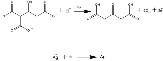
Scheme 1.
Silver reduction mechanism.
Firstly, the decomposition of trisodium citrate by the UV irradiation occurs to form acetone-1,3-dicarboxylate.
The second process is a complexing and excitation process (Scheme 2), as proposed by Shengchun Yang et al. [41]:
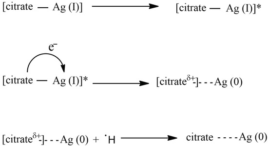
Scheme 2.
Mechanism of silver reduction by citrate.
The photochemical synthesis can be used to obtain AgNPs with different morphologies using irradiation sources with certain wavelengths [42]. For example, using a light emitting diode (LED), which irradiates at a λ of 467 nm (specific to the colour of blue), one can obtain silver nanoparticles with a majority of their morphology in the shape of a decahedron, while at a λ of 630 nm (specific to the colour of red), one can obtain silver nanoprisms [43].
There are many studies which present different approaches to photochemical synthesis of silver nanoparticles, such as using plant extracts [44], chitosan [29], TSC [34]. Most of the TSC syntheses carried out until now have offered some explanations regarding the optimization of the synthesis [45,46], but the optimization of this type of synthesis, which uses only photons to “activate” TSC reducing capabilities, without any additional reagent (as a reducing or stabilizing agent), is still an issue to be addressed. Thus, one of the main aims of this paper is to optimize the photochemical synthesis of AgNPs by adjusting different parameters, such as TSC concentration, UV intensity, UV exposure time, and the monitoring of the resulting solution.
Thus, this paper presents a photochemical method for the synthesis of AgNPs, using AgNO3 as a precursor for AgNPs and UV radiation as an “activator” for TSC, in order to begin the reduction, with TSC as the stabilizing agent. The size of the AgNPs were estimated using Mie theory. The stability of the AgNPs was studied over 390 days (13 months).
2. Results and Discussion
In this section of the article, we attempt to optimize the synthesis of silver nanoparticles by studying different synthesis parameters, such as TSC concentration, UV irradiation time, and UV intensity (irradiance calculation using Keitz’s theory). Thus, after optimizing the synthesis parameters, we continue with the electron microscopy characterization of the material (SEM/EDX, TEM), Mie theory characterization of the obtained silver nanospheres, and stability studies, which consist of periodic measurement of the optimal silver nanoparticle solution using UV-VIS spectroscopy.
2.1. Synthesis Optimization
2.1.1. TSC Concentration
We explore the influence of the TSC concentration on AgNP synthesis. Three TSC concentrations (0.032 M; 0.038 M; 0.043 M) were used. The distance (3.5 cm) from the UV lamp and the irradiation time (60 min) were kept constant. The resultant AgNPs were evaluated from their extinction spectra (Figure 1).
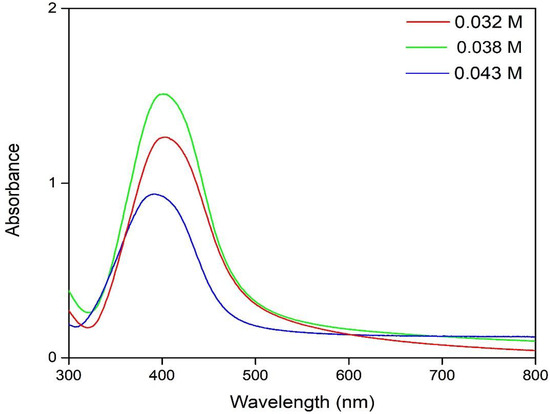
Figure 1.
The influence of the concentration of TSC on AgNP synthesis.
The localized plasmon resonance (LSPR) at ~400 nm indicates that the predominant morphology of the silver nanoparticles is spherical [47,48,49].
We conclude that the 0.038 M TSC concentration is the optimal concentration because the absorbance is the highest for that preparation, indicating that it produced the highest concentration of nanoparticles [50,51].
In the case of 0.043 M, a precipitate was observed (possibly silver), which can explain the lower absorbance in comparison to the other two samples. Thus, the subsequent research was continued with a TSC concentration of 0.038 M.
2.1.2. UV Irradiation Time
To determine the effect of the irradiation time, the TSC concentration (0.038 M), and the source–sample distance (3.5 cm) were kept constant, varying only the irradiation time. The resultant AgNPs were, again, evaluated from their extinction spectra (Figure 2).
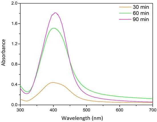
Figure 2.
The effect of UV irradiation time on AgNP synthesis.
The sample which was irradiated for 90 min exhibited the highest absorbance. However, the extinction maximum was red shifted, indicating a presence of some coarsened nanoparticles [52,53], as well as some aggregated nanoparticles [54]. The sample which was irradiated for 30 min exhibited the lowest absorbance, indicating the lowest concentration of AgNPs [50,51]. The sample which was irradiated for 60 min excited an absorbance close the that of the 90 min sample, but did not show coarsening or aggregation. Therefore, 60 min was selected as the preferred irradiation time for our studies.
2.1.3. UV Intensity
To determine the effect of the UV intensity on AgNP synthesis, the source–sample distance was varied (3.5 cm, 7 cm, 10 cm) while the TSC concentration (0.045 M) and the irradiation time (60 min) were kept constant. The resultant AgNPs were, again, evaluated from their extinction spectra (Figure 3).
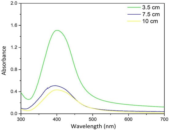
Figure 3.
The effect of the distance between the UV irradiation source and sample on AgNP synthesis.
From these extinction spectra, we concluded that the distance of 3.5 cm from the irradiation source provided the optimal UV intensity, because the concentration of AgNPs (relatively high absorbance) was much higher compared to the other two samples (7 and 10 cm, respectively).
Thus, in order to determine the necessary UV intensity to obtain an optimal synthesis, the irradiance (I) can be calculated using Keitz’s formula [34,55,56,57]:
From here, we can obtain the intensity “I”:
where:
P—UV lamp power [W]
I—radiation intensity [W m−2]
L—UV tube length [m]
D—distance [m]
α = arctan rad; sin α = ; cos α = ; sin2 α = 2 sin α cos α.
Following the calculations performed for the three distances considered for the synthesis, the results are presented in Table 1.

Table 1.
Irradiance results.
From the obtained results, one can notice a correlation between the UV-VIS spectra (Figure 2) and the obtained irradiance.
After the conversion of distance into intensity, the dosage was calculated in order to determine the optimal UV radiation dosage. For this calculus, we used the optimal irradiance (214 W/m2) and the studied irradiation time, thus resulting in the following (Table 2):

Table 2.
Radiation dosages.
From the experimental UV-VIS results, a dosage of 770,400 J/m2, which is equivalent to the distance of 3.5 cm (the optimal distance, with an irradiance of 214 W/m2), and 60 min of time represents the optimal dosage for this synthesis.
2.1.4. Summary
In conclusion, in order to obtain the optimal synthesis of AgNPs, the concentration of 0.029 mol AgNO3/L and 0.038 mol TSC/L, with an irradiation time of 60 min and a distance between the sample and UV of 3.5 cm, should be used. The sample which was synthesized with the optimal parameters was further studied.
2.2. Scanning Electronic Microscopy and Energy Dispersive X-ray Analysis
The morphology of the AgNPs was measured using SEM, and qualitative elemental analysis was performed using EDX (Figure 4).
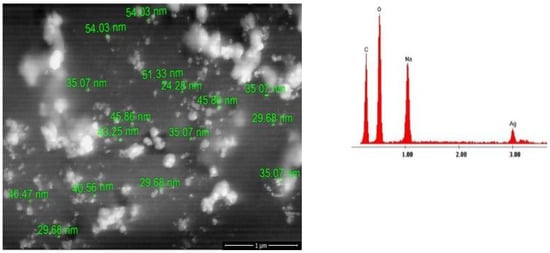
Figure 4.
SEM and EDX analysis for the AgNPs.
SEM was performed as a preliminary analysis to evaluate the morphology of the AgNPs, and to determine a preliminary particle size. From the analysis, we can conclude that the measured nanoparticles are of spherical shape, with diameters measuring ~40 nm, and that they are quite polydisperse. EDX analysis confirms the presence of Ag. Na, C, and O are specific for TSC. The SEM and EDX analyses were performed a few hours after synthesis of the AgNPs.
2.3. Transmission Electron Microscopy
The TEM analysis was performed in order to obtain a better evaluation of the nanoparticle size (the maximum, minimum, and distribution, whilst the SEM analysis was preliminary, conducted in order to obtain an idea of the nanoparticles’ shapes and sizes. In Figure 5, Figure 6 and Figure 7, the TEM micrographs are presented for the optimal sample (0.029 M AgNO3, 0.038 M TSC, irradiation time of 60 min, and a distance from the radiation source of 3.5 cm). The particle size distribution obtained based on the TEM images it is depicted in Figure 8.
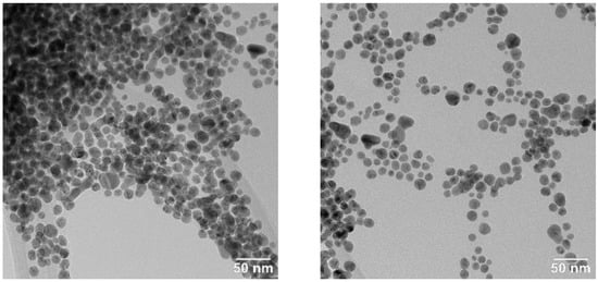
Figure 5.
TEM images from different areas of the sample, showing spherical and quasi-spherical AgNPs.
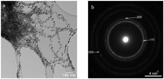
Figure 6.
An agglomeration of AgNPs (a) and the electron diffraction pattern revealing the FCC (face-centered cubic) structure of Ag (b); the most intense diffraction rings correspond to (111), (200), and (220) crystallographic planes.
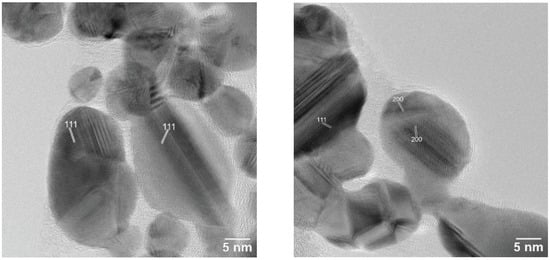
Figure 7.
HRTEM images of AgNPs; the lattice fringes correspond to (111) and (200) crystallographic planes of FCC (face-centered cubic) AgNPs.
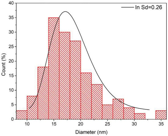
Figure 8.
Size distribution based on TEM analysis.
From the obtained results, we can conclude that the nanoparticle sizes are slightly polydisperse (which can also be seen from the UV-VIS spectra analysis), with a majority of them being around 15 nm. Even though a majority of them are around 15 nm, there are still some coarse ones present, which are ~35 nm.
2.4. Stability Study
In order to perform this study, the obtained sample was kept in the dark, at room temperature (~25 °C), and wrapped in aluminum foil in order to prevent the interference of ambient light.
The stability of the sample was studied by performing UV-VIS spectra over time. The results are shown in Figure 9.
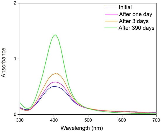
Figure 9.
Stability studies.
From the spectra, it can be observed that the absorbance increases continuously, and the full width at half maxima decreases with the increase in absorbance (the initial spectra has a FWHM of 85 nm (Gaussian). After 390 days, its full width at half maxima is 66 nm (Gaussian)), thus indicating the fact that the nanoparticles are more monodispersed [58,59]. Their diameter is also smaller compared to the initial one [35,60,61], implicitly increasing the concentration of nanoparticles as their diameter decreases. This issue can be explained by the fact that TSC has been activated by the UV irradiation [40]. Thus, this type of synthesis has the benefit of a controlled “activation” in order to obtain AgNPs, but the disadvantage is that the reduction process cannot be stopped instantaneously; the citrate continues reducing continuously until the reaction is completed. In conclusion, in order prevent this from happening, we suggest the use of an additional stabilizing (besides TSC) agent to form a more stable complex.
The solution with the obtained silver nanoparticles was monitored for 390 days, revealing changes in the UV-VIS spectra during the study period.
2.5. Absorption and Scattering by a Sphere; Mie Theory Size Assessment
In order to estimate the size of the nanoparticles, a first step involves obtaining the theoretical spectra “Cext/Csca/Cabs vs. Wavelength” for the diameter of nanoparticles, which presents the localized surface plasmon resonance at a similar wavelength to the experimental one, depicted in Figure 10 (the wavelength is closely related to the morphology of the nanoparticles) [35,62,63,64].
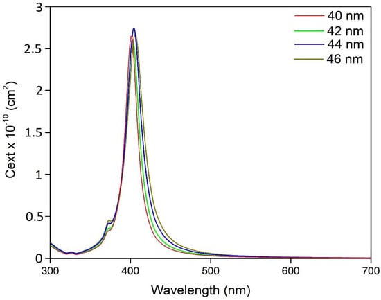
Figure 10.
Extinction cross-section for AgNPs with a diameter of 40–46 nm.
The spectra represented in this figure were obtained using the Mie Plot software [65], and they are representative of silver nanospheres with a diameter of 40–46 nm. In order to be able to compare the theoretical results with the experimental ones, it is necessary to perform the following calculations.
Thus, from the theoretical results, we will use the values obtained for Cext, to calculate the molar absorption coefficient, ϵ, for each spectrum [66,67].
Therefore: ϵ = NA C ln10 (10−3 ) where: NA = 6.0221408 × 10²³ (mole−1)
C = extinction cross section (cm2)
The obtained result can be used, together with the molar absorption coefficient, ϵ, to determine the absorbance of each spectrum using the Beer–Lambert law [68]:
where: A = Absorbance
A = ϵ c l
ϵ = molar absorption coefficient (M−1 cm−1)
c = the molar concentration of the solution (M)
l = path length (cm)
Applying this analysis, we obtained the following results, presented in Figure 11.
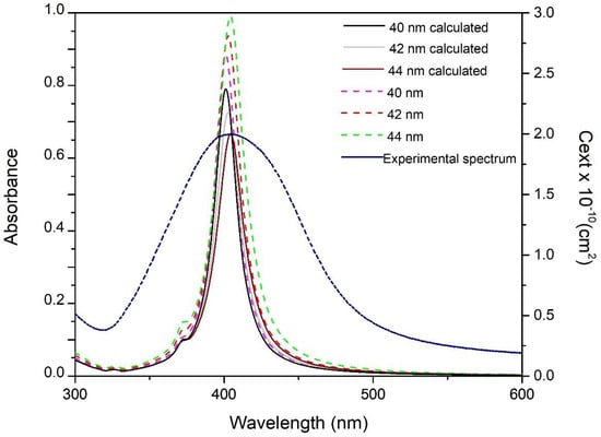
Figure 11.
Theoretical obtained spectra for AgNPs with diameters from 40–46 nm, and the experimentally obtained spectrum.
The dark-colored lines (light grey, black, and brown) are the theoretical calculated spectra, while the dotted ones represent the extinction cross-section spectrum.
To obtain a more precise comparison, the sample of AgNPs was diluted in order to achieve an absorbance of <1. Following the obtained results, it is evident that the absorbance of the theoretically calculated spectra decreases with the growth of the nanoparticles diameter, thus indicating the fact that the nanoparticle concentration decreases (lower absorbance), with the increasing size of the particles. It can also be observed that the wavelength at which the LSPR occurs (401 nm for D = 40 nm; 403 nm for D = 42 nm; 404 nm for D = 44 nm) is directly proportional to the increasing size of AgNPs, indicating a correlation between AgNP morphology and the LSPR wavelength, a fact which has been demonstrated in several papers [59,60,62,69].
Comparing the spectra between them, it results that the LSPR of the synthesized material occurs at a wavelength of 404 nm, and the theoretically obtained spectrum (the one which “overlaps” with the experimental one) has an LSPR at a wavelength of 406 nm. Therefore, we conclude that in the experimental solution, the diameter of the obtained nanoparticles is approximately ~44 ± 3 nm. It can also be seen that the synthesized material has a 99 nm FWHM with a regression coefficient of R2 = 0.99643 (Gaussian fit), a fact which indicates that the obtained nanoparticles are polydisperse [58,59,70].
Judging from the calculated spectrum and the experimental one, we can conclude that a majority of the nanoparticles present in the solution should be around ~44 nm.
Of course, it would be ideal for all AgNPs to have the same diameter (monodisperse), but this is often not possible (depending on the used reducing agent [71]). In order to obtain results as close as possible to the real values, the following steps should are necessary: (i) the rapid reduction in AgNO3 will lead to acquiring a monodisperse solution [72,73]; (ii) ultracentrifugation or ultrafiltration in order to remove the coarse colloids as well as the colloidal aggregates from the resulting solution; and (iii) the determination of the silver concentration via AAS (atomic absorption) or ICP-MS (inductively coupled plasma mass spectrometry) depending on the concentration of the silver in the solution, after the ultracentrifugation or ultrafiltration step, to determine the exact concentration of silver.
In addition, the theoretical extinction efficiency Qext, scattering efficiency Qsca, and absorption efficiency Qabs [74] for the silver nanospheres with radii from 5–100 nm (10–200 nm diameter) are presented in Figure 12.
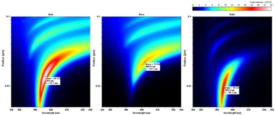
Figure 12.
Qext, Qsca, and Qabs as a function of particle radius r and wavelength λ.
The color indicates the magnitude of Qext, and, implicitly, the maximum values depending on the nanoparticle radius. Qext is the sum of Qsca and Qabs [75].
The size differences between SEM, TEM, and Mie theory could be related to the fact that the obtained nanoparticles are polydisperse (which can be seen from UV-VIS spectra, SEM, and TEM), and that the reaction did not complete during the irradiation period. The coarse nanoparticles and the aggregates could be removed from the solution by subjecting the solution to an ultracentrifugation and/or ultrafiltration process, in order to obtain a more precise Mie theory size assessment.
3. Materials and Methods
For the synthesis of silver nanoparticles, the following were used: AgNO3 (p.a. Merck) as a precursor for AgNPs, trisodium citrate dihydrate (TSC) (ACS reagent, >99% purity) as a stabilizer and as a reducer, and an UV lamp (Tungsram 18 W, having an UV output of 6 W, with λ of 254 nm) [40].
To obtain information regarding the synthesis process, the following parameters were varied: (i) TSC concentration (0.032 M; 0.038 M; 0.043 M), (ii) UV irradiation time (30 min, 60 min, and 90 min), (iii) the distance between the irradiation source and the sample (3.5 cm, 7 cm, and 10 cm), (iv) stability studies and evolution over time. All solutions were prepared using double distilled water. The synthesis of AgNPs was carried out as follows. To 30 mL of one of the TSC solutions, a volume of 1 mL of AgNO3 (0.029 M) was added dropwise with constant stirring. The resulting solution was left to be stirred for 10 min. The next step was the transfer of the solution to a Petri dish (to facilitate the penetration of the UV radiation through the entire volume of the solution). The Petri dish with the solution was subjected to UV irradiation, while keeping the solution under magnetic stirring. It is worth mentioning that the Petri dish was in the middle of the UV lamp, the UV lamp was oriented above the Petri dish, and both were enclosed in a box without any interference from outside. The schematic representation of the synthesis of AgNPs is presented in Figure 13 [55].
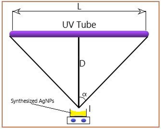
Figure 13.
Schematic representation of the synthesis.
The tube length (L) was 40 cm, and the distance traveled by the radiation (D) was 3.5 cm, 7 cm, and 10 cm, respectively, to the irradiated sample (I). The UV output of the tube was given by the manufacturer, and equaled 6 W.
The obtained sample was kept in the dark, at room temperature (~25 °C), and wrapped in aluminum foil in order to prevent the interference of ambient light. The resulting AgNP solutions were characterized as follows.
UV-vis spectra were obtained by using a Varian Cary 50 spectrophotometer. Nanoparticle morphology and composition was analyzed using scanning electron microscope (SEM) Quanta Feg 250, which was equipped with energy dispersive x-ray analysis (EDX). A more detailed analysis employed transmission electron microscopy (TEM) using the JEOL JEM-2100 with selected area electron diffraction (SAED). Long-term stability was studied via UV-VIS spectroscopy.
4. Conclusions
This study demonstrates a simple method for photoassisted synthesis of AgNPs. In this method, we used UV radiation to “activate” the reducing capabilities of TSC (unlike the classical Turkevich method, in which the solution needed to be heated in order to synthesize nanoparticles).
Additionally, the irradiance was determined, and it was observed that there exists a close correlation between the calculated irradiance and the experimental obtained extinction spectra in Section 2.3. The difference between the absorbances of the 7 cm and 10 cm spectra are not significant, and the same can be said about the calculated irradiances of the same distances. This indicates the close correlation between the irradiance and the resulting extinction maxima.
A simple and efficient theoretical determination of nanoparticle size was conducted and explained, using the theory developed by Gustav Mie, by comparing the theoretical obtained spectra and the experimental one, as well as the confirmation of the results using the SEM analysis and TEM analysis.
The stability of the obtained silver nanoparticles was studied for 390 days, and they showed good stability overall. Even though some modifications still occurred in the solution, no precipitate was observed.
One of the major advantages that this type of synthesis offers is the benefit of a controlled “activation.” Thus, the reactants may be premixed, kept in the dark, and exposed to UV irradiation when one wishes to obtain AgNPs.
Author Contributions
Conceptualization A.N., N.D. and D.M.D.-S.; Investigation, B.P., C.M., O.G., P.N., N.S.N. and M.C.; Writing, L.A.B., A.N., B.P., N.D. All authors have read and agreed to the published version of the manuscript.
Funding
This research received no external funding.
Acknowledgments
This paper was financially supported by the Project “Network of excellence in applied research and innovation for doctoral and postdoctoral programs”/InoHubDoc, project co-funded by the European Social Fund financing agreement no. POCU/993/6/13/153437.
Conflicts of Interest
Authors declare no conflict of interest.
References
- Le Ouay, B.; Stellacci, F. Antibacterial activity of silver nanoparticles: A surface science insight. Nano Today 2015, 10, 339–354. [Google Scholar] [CrossRef]
- Pascu, B.; Negrea, A.; Ciopec, M.; Duteanu, N.; Negrea, P.; Nemeş, N.S.; Seiman, C.; Marian, E.; Micle, O. A Green, Simple and Facile Way to Synthesize Silver Nanoparticles Using Soluble Starch. pH Studies and Antimicrobial Applications. Materials 2021, 14, 4765. [Google Scholar] [CrossRef] [PubMed]
- Fiorati, A.; Bellingeri, A.; Punta, C.; Corsi, I.; Venditti, I. Silver Nanoparticles for Water Pollution Monitoring and Treatments: Ecosafety Challenge and Cellulose-Based Hybrids Solution. Polymers 2020, 12, 1635. [Google Scholar] [CrossRef] [PubMed]
- Movassaghian, S.; Merkel, O.M.; Torchilin, V.P. Applications of polymer micelles for imaging and drug delivery. WIREs Nanomed. Nanobiotechnol. 2015, 7, 691–707. [Google Scholar] [CrossRef]
- Khan, F.U.; Chen, Y.; Khan, N.U.; Khan, Z.U.H.; Khan, A.U.; Ahmad, A.; Tahir, K.; Wang, L.; Khan, M.R.; Wan, P. Antioxidant and catalytic applications of silver nanoparticles using Dimocarpus longan seed extract as a reducing and stabilizing agent. J. Photochem. Photobiol. B Biol. 2016, 164, 344–351. [Google Scholar] [CrossRef]
- Varghese Alex, K.; Tamil Pavai, P.; Rugmini, R.; Shiva Prasad, M.; Kamakshi, K.; Sekhar, K.C. Green Synthesized Ag Nanoparticles for Bio-Sensing and Photocatalytic Applications. ACS Omega 2020, 5, 13123–13129. [Google Scholar] [CrossRef]
- Kulkarni, A.P.; Noone, K.M.; Munechika, K.; Guyer, S.R.; Ginger, D.S. Plasmon-Enhanced Charge Carrier Generation in Organic Photovoltaic Films Using Silver Nanoprisms. Nano Lett. 2010, 10, 1501–1505. [Google Scholar] [CrossRef]
- Tsao, C.-W.; Zheng, Y.-S.; Sun, Y.-S.; Cheng, Y.-C. Surface-enhanced Raman scattering (SERS) spectroscopy on localized silver nanoparticle-decorated porous silicon substrate. Analyst 2021, 146, 7645–7652. [Google Scholar] [CrossRef]
- Anees Ahmad, S.; Sachi Das, S.; Khatoon, A.; Tahir Ansari, M.; Afzal, M.; Saquib Hasnain, M.; Kumar Nayak, A. Bactericidal activity of silver nanoparticles: A mechanistic review. Mater. Sci. Energy Technol. 2020, 3, 756–769. [Google Scholar] [CrossRef]
- Liao, C.; Li, Y.; Tjong, S.C. Bactericidal and Cytotoxic Properties of Silver Nanoparticles. Int. J. Mol. Sci. 2019, 20, 449. [Google Scholar] [CrossRef]
- Hsueh, Y.-H.; Lin, K.-S.; Ke, W.-J.; Hsieh, C.-T.; Chiang, C.-L.; Tzou, D.-Y.; Liu, S.-T. The Antimicrobial Properties of Silver Nanoparticles in Bacillus subtilis are Mediated by Released Ag+ Ions. PLoS ONE 2015, 10, e0144306. [Google Scholar] [CrossRef]
- Ciou, S.-H.; Cao, Y.-W.; Huang, H.-C.; Su, D.-Y.; Huang, C.-L. SERS Enhancement Factors Studies of Silver Nanoprism and Spherical Nanoparticle Colloids in the Presence of Bromide Lons. J. Phys. Chem. C 2009, 113, 9520–9525. [Google Scholar] [CrossRef]
- Yang, X.; Yu, Y.; Gao, Z. A Highly Sensitive Plasmonic DNA Assay Based on Triangular Silver Nanoprism Etching. ACS Nano 2014, 8, 4902–4907. [Google Scholar] [CrossRef]
- Chen, L.; Fu, X.; Lu, W.; Chen, L. Highly Sensitive and Selective Colorimetric Sensing of Hg2+ Based on the Morphology Transition of Silver Nanoprisms. ACS Appl. Mater. Interfaces 2013, 5, 284–290. [Google Scholar] [CrossRef]
- Kelly, K.L.; Coronado, E.; Zhao, L.L.; Schatz, G.C. The Optical Properties of Metal Nanoparticles: The Influence of Size, Shape, and Dielectric Environment. J. Phys. Chem. B 2003, 107, 668–677. [Google Scholar] [CrossRef]
- Shi, Y.; He, L.; Deng, Q.; Liu, Q.; Li, L.; Wang, W.; Xin, Z.; Liu, R. Synthesis and Applications of Silver Nanowires for Transparent Conductive Films. Micromachines 2019, 10, 330. [Google Scholar] [CrossRef]
- Alshammari, A.S.; Sam, F.L.M.; Rozanski, L.J.; Mills, C.A.; Alenezi, M.R.; Beliatis, M.J.; Jayawardena, K.D.G.I.; Underwood, J.M.; Silva, S.R.P. Controlled growth and spray deposition of silver nanowires for ITO-free, flexible, and high brightness OLEDs. Phys. Status Solidi 2017, 214, 1600561. [Google Scholar] [CrossRef]
- Rashid, H.-U.; Ali, M.; Sarker, M.R.; Md Ali, S.H.; Akhtar, N.; Khan, N.A.; Asif, M.; Shah, S. Synthesis, Characterization, and Applications of Silver Nano Fibers in Humidity, Ammonia, and Temperature Sensing. Micromachines 2021, 12, 682. [Google Scholar] [CrossRef]
- Beyene, H.D.; Werkneh, A.A.; Bezabh, H.K.; Ambaye, T.G. Synthesis paradigm and applications of silver nanoparticles (AgNPs), a review. Sustain. Mater. Technol. 2017, 13, 18–23. [Google Scholar] [CrossRef]
- Kim, M.; Osone, S.; Kim, T.; Higashi, H.; Seto, T. Synthesis of Nanoparticles by Laser Ablation: A Review. Kona Powder Part. J. 2017, 34, 80–90. [Google Scholar] [CrossRef]
- Yang, D. Applications of Laser Ablation—Thin Film Deposition, Nanomaterial Synthesis and Surface Modification; BoD–Books on Demand: Norderstedt, Germany, 2016. [Google Scholar]
- Hamad, A.; Khashan, K.; Hadi, A. Laser Ablation in Different Environments and Generation of Nanoparticles; BoD–Books on Demand: Norderstedt, Germany, 2016; pp. 177–196. [Google Scholar]
- Iravani, S.; Korbekandi, H.; Mirmohammadi, S.V.; Zolfaghari, B. Synthesis of silver nanoparticles: Chemical, physical and biological methods. Res. Pharm. Sci. 2014, 9, 385–406. [Google Scholar]
- Pinto, R.J.B.; Nasirpour, M.; Carrola, J.; Oliveira, H.; Freire, C.S.R.; Duarte, I.F. Chapter 9—Antimicrobial Properties and Therapeutic Applications of Silver Nanoparticles and Nanocomposites. In Antimicrobial Nanoarchitectonics; Grumezescu, A.M., Ed.; Elsevier: Amsterdam, The Netherlands, 2017; pp. 223–259. [Google Scholar]
- Ahmed, S.; Ahmad, M.; Swami, B.L.; Ikram, S. A review on plants extract mediated synthesis of silver nanoparticles for antimicrobial applications: A green expertise. J. Adv. Res. 2016, 7, 17–28. [Google Scholar] [CrossRef]
- Backman, U. Studies on Nanoparticle Synthesis via Gas-to-Particle Conversion; VTT Publications: Espoo, Finland, 2005. [Google Scholar]
- Sosa, Y.D.; Rabelero, M.; Treviño, M.E.; Saade, H.; López, R.G. High-Yield Synthesis of Silver Nanoparticles by Precipitation in a High-Aqueous Phase Content Reverse Microemulsion. J. Nanomater. 2010, 2010, 392572. [Google Scholar] [CrossRef]
- Zaarour, M.; El Roz, M.; Dong, B.; Retoux, R.; Aad, R.; Cardin, J.; Dufour, C.; Gourbilleau, F.; Gilson, J.-P.; Mintova, S. Photochemical Preparation of Silver Nanoparticles Supported on Zeolite Crystals. Langmuir 2014, 30, 6250–6256. [Google Scholar] [CrossRef]
- Gabriel, J.S.; Gonzaga, V.A.M.; Poli, A.L.; Schmitt, C.C. Photochemical synthesis of silver nanoparticles on chitosans/montmorillonite nanocomposite films and antibacterial activity. Carbohydr. Polym. 2017, 171, 202–210. [Google Scholar] [CrossRef]
- Kazancioglu, E.O.; Aydin, M.; Arsu, N. Photochemical synthesis of nanocomposite thin films containing silver and gold nanoparticles with 2-thioxanthone thioacetic acid-dioxide and their role in photocatalytic degradation of methylene blue. Surf. Interfaces 2021, 22, 100793. [Google Scholar] [CrossRef]
- Khodashenas, B.; Ghorbani, H.R. Synthesis of silver nanoparticles with different shapes. Arab. J. Chem. 2019, 12, 1823–1838. [Google Scholar] [CrossRef]
- Restrepo, C.V.; Villa, C.C. Synthesis of silver nanoparticles, influence of capping agents, and dependence on size and shape: A review. Environ. Nanotechnol. Monit. Manag. 2021, 15, 100428. [Google Scholar] [CrossRef]
- Patel, K.; Bharatiya, B.; Mukherjee, T.; Soni, T.; Shukla, A.; Suhagia, B.N. Role of stabilizing agents in the formation of stable silver nanoparticles in aqueous solution: Characterization and stability study. J. Dispers. Sci. Technol. 2017, 38, 626–631. [Google Scholar] [CrossRef]
- Oprica, L.; Andries, M.; Sacarescu, L.; Popescu, L.; Pricop, D.; Creanga, D.; Balasoiu, M. Citrate-silver nanoparticles and their impact on some environmental beneficial fungi. Saudi J. Biol. Sci. 2020, 27, 3365–3375. [Google Scholar] [CrossRef]
- Raza, M.A.; Kanwal, Z.; Rauf, A.; Sabri, A.N.; Riaz, S.; Naseem, S. Size- and Shape-Dependent Antibacterial Studies of Silver Nanoparticles Synthesized by Wet Chemical Routes. Nanomaterials 2016, 6, 74. [Google Scholar] [CrossRef]
- Chen, Z.; Zhang, X.; Cao, H.; Huang, Y. Chitosan-capped silver nanoparticles as a highly selective colorimetric probe for visual detection of aromatic ortho-trihydroxy phenols. Analyst 2013, 138, 2343–2349. [Google Scholar] [CrossRef]
- Kumar, M.; Devi, P.; Kumar, A. Structural analysis of PVP capped silver nanoparticles synthesized at room temperature for optical, electrical and gas sensing properties. J. Mater. Sci. Mater. Electron. 2017, 28, 5014–5020. [Google Scholar] [CrossRef]
- Martinez-Andrade, J.M.; Avalos-Borja, M.; Vilchis-Nestor, A.R.; Sanchez-Vargas, L.O.; Castro-Longoria, E. Dual function of EDTA with silver nanoparticles for root canal treatment–A novel modification. PLoS ONE 2018, 13, e0190866. [Google Scholar] [CrossRef]
- Bastús, N.G.; Merkoçi, F.; Piella, J.; Puntes, V. Synthesis of Highly Monodisperse Citrate-Stabilized Silver Nanoparticles of up to 200 nm: Kinetic Control and Catalytic Properties. Chem. Mater. 2014, 26, 2836–2846. [Google Scholar] [CrossRef]
- du Toit, H.; Macdonald, T.J.; Huang, H.; Parkin, I.P.; Gavriilidis, A. Continuous flow synthesis of citrate capped gold nanoparticles using UV induced nucleation. RSC Adv. 2017, 7, 9632–9638. [Google Scholar] [CrossRef]
- Yang, S.; Wang, Y.; Wang, Q.; Zhang, R.; Ding, B. UV irradiation induced formation of Au nanoparticles at room temperature: The case of pH values. Colloids Surf. A Physicochem. Eng. Asp. 2007, 301, 174–183. [Google Scholar] [CrossRef]
- Stamplecoskie, K.G.; Scaiano, J.C. Light Emitting Diode Irradiation Can Control the Morphology and Optical Properties of Silver Nanoparticles. J. Am. Chem. Soc. 2010, 132, 1825–1827. [Google Scholar] [CrossRef]
- Saade, J.; de Araújo, C.B. Synthesis of silver nanoprisms: A photochemical approach using light emission diodes. Mater. Chem. Phys. 2014, 148, 1184–1193. [Google Scholar] [CrossRef]
- Filip, G.A.; Moldovan, B.; Baldea, I.; Olteanu, D.; Suharoschi, R.; Decea, N.; Cismaru, C.M.; Gal, E.; Cenariu, M.; Clichici, S.; et al. UV-light mediated green synthesis of silver and gold nanoparticles using Cornelian cherry fruit extract and their comparative effects in experimental inflammation. J. Photochem. Photobiol. B Biol. 2019, 191, 26–37. [Google Scholar] [CrossRef]
- Babusca, D.; Popescu, L.; Sacarescu, L.; Dorohoi, D.O.; Creanga, D.; Oprica, L.A. Two phase photochemical synthesis of silver nanoparticles and their impact on the chlorophylls. Mol. Cryst. Liq. Cryst. 2020, 698, 56–64. [Google Scholar] [CrossRef]
- Yang, L.-C.; Lai, Y.-S.; Tsai, C.-M.; Kong, Y.-T.; Lee, C.-I.; Huang, C.-L. One-Pot Synthesis of Monodispersed Silver Nanodecahedra with Optimal SERS Activities Using Seedless Photo-Assisted Citrate Reduction Method. J. Phys. Chem. C 2012, 116, 24292–24300. [Google Scholar] [CrossRef]
- De Leersnyder, I.; De Gelder, L.; Van Driessche, I.; Vermeir, P. Revealing the Importance of Aging, Environment, Size and Stabilization Mechanisms on the Stability of Metal Nanoparticles: A Case Study for Silver Nanoparticles in a Minimally Defined and Complex Undefined Bacterial Growth Medium. Nanomaterials 2019, 9, 1684. [Google Scholar] [CrossRef]
- Ogundare, S.A.; Moodley, V.; Amaku, J.F.; Ogunmoye, A.O.; Atewolara-Odule, O.C.; Olubomehin, O.O.; Awokoya, K.N.; Sanyaolu, N.O.; Ibikunle, A.A.; van Zyl, W.E. Nanocrystalline cellulose derived from melon seed shell (Citrullus colocynthis L.) for reduction and stabilization of silver nanoparticles: Synthesis and catalytic activity. Carbohydr. Polym. Technol. Appl. 2021, 2, 100134. [Google Scholar] [CrossRef]
- Verkhovskii, R.; Kozlova, A.; Atkin, V.; Kamyshinsky, R.; Shulgina, T.; Nechaeva, O. Physical properties and cytotoxicity of silver nanoparticles under different polymeric stabilizers. Heliyon 2019, 5, e01305. [Google Scholar] [CrossRef]
- Titus, D.; James Jebaseelan Samuel, E.; Roopan, S.M. Chapter 12—Nanoparticle characterization techniques. In Green Synthesis, Characterization and Applications of Nanoparticles; Shukla, A.K., Iravani, S., Eds.; Elsevier: Amsterdam, The Netherlands, 2019; pp. 303–319. [Google Scholar]
- Ibrahim, H.M.M. Green synthesis and characterization of silver nanoparticles using banana peel extract and their antimicrobial activity against representative microorganisms. J. Radiat. Res. Appl. Sci. 2015, 8, 265–275. [Google Scholar] [CrossRef]
- Ameer, F.S.; Varahagiri, S.; Benza, D.W.; Willett, D.R.; Wen, Y.; Wang, F.; Chumanov, G.; Anker, J.N. Tuning Localized Surface Plasmon Resonance Wavelengths of Silver Nanoparticles by Mechanical Deformation. J. Phys. Chem. C Nanomater. Interfaces 2016, 120, 20886–20895. [Google Scholar] [CrossRef]
- Nate, Z.; Moloto, M.J.; Mubiayi, P.K.; Sibiya, P.N. Green synthesis of chitosan capped silver nanoparticles and their antimicrobial activity. MRS Adv. 2018, 3, 2505–2517. [Google Scholar] [CrossRef]
- Balan, L.; Jin, M.; Malval, J.-P.; Chaumeil, H.; Defoin, A.; Vidal, L. Fabrication of Silver Nanoparticle-Embedded Polymer Promoted by Combined Photochemical Properties of a 2,7-Diaminofluorene Derivative Dye. Macromolecules 2008, 41, 9359–9365. [Google Scholar] [CrossRef]
- Bolton, J.R.; Santelli, M.J. Round robin test of the IUVA UV lamp efficiency testing protocol (Revised). IUVA News 2016, 18, 12–18. [Google Scholar]
- Sasges, M.; Robinson, J.; Daynouri, F. Ultraviolet Lamp Output Measurement: A Concise Derivation of the Keitz Equation. Ozone Sci. Eng. 2012, 34, 306–309. [Google Scholar] [CrossRef]
- Keitz, H.A.E. Luminance and Luminous Emittance; Macmillan Education UK: London, UK, 1971. [Google Scholar]
- Oliveira, J.P.; Prado, A.R.; Keijok, W.J.; Ribeiro, M.R.N.; Pontes, M.J.; Nogueira, B.V.; Guimarães, M.C.C. A helpful method for controlled synthesis of monodisperse gold nanoparticles through response surface modeling. Arab. J. Chem. 2020, 13, 216–226. [Google Scholar] [CrossRef]
- Mlalila, N.G.; Swai, H.S.; Hilonga, A.; Kadam, D.M. Antimicrobial dependence of silver nanoparticles on surface plasmon resonance bands against Escherichia coli. Nanotechnol. Sci. Appl. 2016, 10, 1–9. [Google Scholar] [CrossRef] [PubMed]
- Šileikaitė, A.; Puiso, J.; Prosycevas, I.; Tamulevičius, S. Investigation of Silver Nanoparticles Formation Kinetics during Reduction of Silver Nitrate with Sodium Citrate. Medziagotyra 2009, 15, 21–27. [Google Scholar]
- Sharma, R.; Dhillon, A.; Kumar, D. Mentha-Stabilized Silver Nanoparticles for High-Performance Colorimetric Detection of Al(III) in Aqueous Systems. Sci. Rep. 2018, 8, 5189. [Google Scholar] [CrossRef]
- Anandalakshmi, K.; Venugobal, J.; Ramasamy, V. Characterization of silver nanoparticles by green synthesis method using Pedalium murex leaf extract and their antibacterial activity. Appl. Nanosci. 2016, 6, 399–408. [Google Scholar] [CrossRef]
- Dell’Aglio, M.; Gaudiuso, R.; ElRashedy, R.; De Pascale, O.; Palazzo, G.; De Giacomo, A. Collinear double pulse laser ablation in water for the production of silver nanoparticles. Phys. Chem. Chem. Phys. 2013, 15, 20868–20875. [Google Scholar] [CrossRef] [PubMed]
- Siddiqi, K.S.; Husen, A.; Rao, R.A.K. A review on biosynthesis of silver nanoparticles and their biocidal properties. J. Nanobiotechnol. 2018, 16, 14. [Google Scholar] [CrossRef]
- Laven, P. 2014. Available online: http://www.philiplaven.com/mieplot.htm (accessed on 15 March 2022).
- Bumm, L.A. Size Determination of Gold Nanoparticles Using Mie Theory and Extinction Spectra. NanoLab (Phy4970). Available online: http://www.nhn.ou.edu/~bumm/NanoLab/pdf/Au_NP_spectrophotometery_activity.pdf (accessed on 10 October 2022).
- Bumm, L.A.; Johnson, M. Spectrometry—Key Conept: OU NanoLab/NSF/NUE. Available online: http://www.nhn.ou.edu/~bumm/NanoLab/ppt/Spectrophotometry.ppt (accessed on 10 October 2022).
- Worsfold, P.J.; Zagatto, E.A.G. Spectrophotometry|Overview. In Encyclopedia of Analytical Science, 3rd ed.; Worsfold, P., Poole, C., Townshend, A., Miro, M., Eds.; Academic Press: Oxford, UK, 2019; pp. 244–248. [Google Scholar]
- Saeb, A.T.M.; Alshammari, A.S.; Al-Brahim, H.; Al-Rubeaan, K.A. Production of Silver Nanoparticles with Strong and Stable Antimicrobial Activity against Highly Pathogenic and Multidrug Resistant Bacteria. Sci. World J. 2014, 2014, 704708. [Google Scholar] [CrossRef]
- Von White, G.; Kerscher, P.; Brown, R.M.; Morella, J.D.; McAllister, W.; Dean, D.; Kitchens, C.L. Green Synthesis of Robust, Biocompatible Silver Nanoparticles Using Garlic Extract. J. Nanomater. 2012, 2012, 730746. [Google Scholar] [CrossRef]
- Song, K.C.; Lee, S.M.; Park, T.S.; Lee, B.S. Preparation of colloidal silver nanoparticles by chemical reduction method. Korean J. Chem. Eng. 2009, 26, 153–155. [Google Scholar] [CrossRef]
- Riaz, M.; Mutreja, V.; Sareen, S.; Ahmad, B.; Faheem, M.; Zahid, N.; Jabbour, G.; Park, J. Exceptional antibacterial and cytotoxic potency of monodisperse greener AgNPs prepared under optimized pH and temperature. Sci. Rep. 2021, 11, 2866. [Google Scholar] [CrossRef] [PubMed]
- Gemishev, O.; Panayotova, M.; Gicheva, G.; Mintcheva, N. Green Synthesis of Stable Spherical Monodisperse Silver Nanoparticles Using a Cell-Free Extract of Trichoderma reesei. Materials 2022, 15, 481. [Google Scholar] [CrossRef] [PubMed]
- Bohren, C.F.; Huffman, D.R. Absorption and Scattering by an Arbitrary Particle; Wiley-Vch Verlag GmbH & Co KGaA: Weinheim, Germany, 1998. [Google Scholar]
- Hulst, H.C.; van de Hulst, H.C. Light Scattering by Small Particles; Courier Corporation: Chelmsford, MA, USA, 1981. [Google Scholar]
Disclaimer/Publisher’s Note: The statements, opinions and data contained in all publications are solely those of the individual author(s) and contributor(s) and not of MDPI and/or the editor(s). MDPI and/or the editor(s) disclaim responsibility for any injury to people or property resulting from any ideas, methods, instructions or products referred to in the content. |
© 2022 by the authors. Licensee MDPI, Basel, Switzerland. This article is an open access article distributed under the terms and conditions of the Creative Commons Attribution (CC BY) license (https://creativecommons.org/licenses/by/4.0/).