Article

Strigolactones Modulate Salicylic Acid-Mediated Disease Resistance in Arabidopsis thaliana

1
Graduate School of Agricultural and Life Sciences, The University of Tokyo, Tokyo 113-8657, Japan
2
Department of Bioscience and Biotechnology, Fukui Prefectural University, Fukui 910-1195, Japan
3
Graduate School of Bioresource and Bioenvironmental Sciences, Kyushu University, Fukuoka 819-0395, Japan
4
Center for Bioscience Research and Education, Utsunomiya University, Tochigi 321-8505, Japan
5
Graduate School of Life and Environmental Sciences, Osaka Prefecture University, Osaka 599-8531, Japan
*
Author to whom correspondence should be addressed.
Int. J. Mol. Sci. 2022, 23(9), 5246; https://doi.org/10.3390/ijms23095246
Submission received: 29 December 2021 / Revised: 2 May 2022 / Accepted: 6 May 2022 / Published: 8 May 2022
(This article belongs to the Special Issue Mechanism Research on Signal Transduction in Plant Immunity)

Abstract

:
Strigolactones are low-molecular-weight phytohormones that play several roles in plants, such as regulation of shoot branching and interactions with arbuscular mycorrhizal fungi and parasitic weeds. Recently, strigolactones have been shown to be involved in plant responses to abiotic and biotic stress conditions. Herein, we analyzed the effects of strigolactones on systemic acquired resistance induced through salicylic acid-mediated signaling. We observed that the systemic acquired resistance inducer enhanced disease resistance in strigolactone-signaling and biosynthesis-deficient mutants. However, the amount of endogenous salicylic acid and the expression levels of salicylic acid-responsive genes were lower in strigolactone signaling-deficient max2 mutants than in wildtype plants. In both the wildtype and strigolactone biosynthesis-deficient mutants, the strigolactone analog GR24 enhanced disease resistance, whereas treatment with a strigolactone biosynthesis inhibitor suppressed disease resistance in the wildtype. Before inoculation of wildtype plants with pathogenic bacteria, treatment with GR24 did not induce defense-related genes; however, salicylic acid-responsive defense genes were rapidly induced after pathogenic infection. These findings suggest that strigolactones have a priming effect on Arabidopsis thaliana by inducing salicylic acid-mediated disease resistance.

1. Introduction

Plants are exposed to a range of biotic and abiotic stress factors, such as pathogen attack, insect herbivory, and environmental stress. To adapt to such adverse conditions, plants have evolved unique defense mechanisms that are regulated by phytohormones such as salicylic acid (SA), jasmonic acid (JA), ethylene (ET), and abscisic acid (ABA). These phytohormones contribute to biotic stress responses through a complex network involving synergistic and antagonistic interactions, although SA- and JA/ET-mediated defense responses are important for the resistance against biotrophic and necrotrophic pathogens, respectively [1,2,3,4,5,6].
In plant immunity, pathogen-associated molecular patterns (PAMPs) are first recognized by receptors on the surface of plant cells, activating a basal resistance called PAMP-triggered immunity (PTI). Virulent pathogens have developed effector proteins that suppress this PTI mechanism of host plants; however, resistant plants have acquired a strong immune mechanism, called effector-triggered immunity (ETI), which overcomes pathogenic effectors [7]. Systemic acquired resistance (SAR), induced by SA, is a potent innate immunity system in plants that is effective against a wide range of pathogens [8]. The induction of SAR in infected plants is accompanied by SA accumulation through the increased expression of SA synthase, ISOCHORISMATE SYNTHASE 1 (ICS1), and pathogenesis-related (PR) genes [9]. Further, a reduction of the SA-receptor protein NPR1 is reportedly important for SAR induction, to which an increase in total glutathione (GSH) level contributes [10]. Additionally, probenazole and its derivative, 1, 2-benzisothiazol-3(2H)-one-1, 1-dioxide (BIT), induce SAR by activating its signaling pathway upstream of SA [11,12]. Furthermore, several studies have reported SAR as an effective strategy for disease control in plants [11,12,13,14,15,16], and priming of phytohormone-mediated disease resistance has been reported as a different type of plant immunity system. Although only weak or no expression at all of the major defense-related genes through SA- or JA-mediated signaling pathways are observed in primed plants by interaction with arbuscular mycorrhizal (AM) fungi [17,18], nonpathogenic bacteria [19,20], or treatment with certain chemicals [21], they can still respond more rapidly and strongly to pathogenic infection to protect themselves.
Strigolactones (SLs) are carotenoid-derived metabolites that act as germination stimulants on root parasitic weeds of the genus Striga [22]. Furthermore, SLs secreted by host plants induce mycelial branching in rhizosphere AM fungi and stimulate symbiosis [23]. Recently, SLs have been reported to inhibit shoot branching [24,25]. Thus, SLs have been categorized as a novel class of phytohormones.
Analysis using carotenoid-deficient mutants and carotenoid biosynthesis inhibitors suggests that SLs are synthesized from carotenoids [26]. Moreover, several mutants, including Arabidopsis more axillary growth (max) exhibiting increased shoot branching, have been identified as SL biosynthesis mutants. Arabidopsis max3 and max4 mutants have mutations in CAROTENOID CLEAVAGE DIOXYGENASE 7 (CCD7) and CCD8, respectively, and their phenotypes are restored upon treatment with synthetic SLs, indicating that they are SL-deficient mutants [25]. Additionally, AtD27, which exhibits β-carotene isomerase activity and converts all-trans-β-carotenes, is reportedly involved in SL biosynthesis. Furthermore, carlactone (CL), an SL biosynthetic intermediate, is synthesized in plants from all-trans-β-carotene using three different enzymes [27]. Mutant max1 is deficient in cytochrome P450 oxygenase CYP711A, which converts CL into carlactonic acid in the SL biosynthetic pathway [28]. Similarly, the shoot-branching mutant, SL-insensitive max2, lacks the F-box protein that plays a role in recognizing SLs [29]. In addition, the SL-insensitive mutant d14 has a mutation in the gene encoding an α/β-hydrolyzing enzyme. After binding with SL, the SL receptor D14 interacts with SCF (SKP1-CUL1-F-box) containing the F-box protein MAX2, which transmits SL-mediated signals by degrading target proteins through the ubiquitin–proteasome system [30]. The biological activities of SLs have been analyzed using SL-related mutants and bioactive chemicals. Shoot branching is inhibited by the synthetic SL-analog GR24, and it is improved by the SL-biosynthesis inhibitor TIS108, which inhibits CYP711A activity [31].
SLs are also involved in plant responses to environmental stress, as in drought [32,33], salt [34], and cold [35] resistance. As SLs are carotenoid derivatives that alleviate environmental stress as ABA does, interactions between SL and ABA have been proposed. Thus, for example, Arabidopsis SL biosynthesis-deficient (max3 and max4) and SL signaling-deficient (max2) mutants exhibit low drought tolerance and are less sensitive to ABA [36]. However, some reports indicate that Arabidopsis max2 is less drought-tolerant and more ABA-sensitive, whereas max3 and max4 are as tolerant as the wildtype (WT) [37]. Altogether, these studies suggest that MAX2-mediated SL signaling interacts with ABA signaling during stress tolerance.
Furthermore, recent studies suggest the specific role of SLs in plant responses to biotic stress: Tomato SL biosynthesis-deficient mutant ccd8 exhibits increased susceptibility to Botrytis cinerea and Alternaria alternata [38]. Similarly, rice SL signaling-deficient mutant d14 and SL biosynthesis-deficient mutant d17 are susceptible to Magnaporthe oryzae [39]. Further, Arabidopsis max2, max3, max4, and d14 mutants are highly susceptible to the biotrophic virulent bacterial pathogen Pseudomonas syringae pv. tomato DC3000 (Pst) [40,41]. The model compatible-interaction system between Arabidopsis and Pst, in which the SA-mediated defense signaling is activated upon infection, has contributed to clarifying the detailed mechanisms at work behind the plant immune system [42]. However, the mechanism underlying the effects of SLs on SA-mediated disease resistance remains unclear. Therefore, to gain a better understanding of the role of SL in disease resistance, the aim of this study is to determine the effects of SLs on SA-mediated defense responses in Arabidopsis upon Pst infection.

2. Results

2.1. Relationship between SLs and SA Signaling Pathway in Arabidopsis

To evaluate the effects of SLs on SAR induction, we examined SAR induction in the SL biosynthesis-deficient mutants max1, max3, and max4 and in the SL signaling-deficient mutant max2. SAR was induced by BIT applied using soil drench treatment to avoid its dilution during dip-inoculation with the pathogen. Furthermore, to avoid the influence of stomatal aperture on disease resistance, we covered plants with plastic wrap and maintained high humidity for 12 h before and after pathogen inoculation.
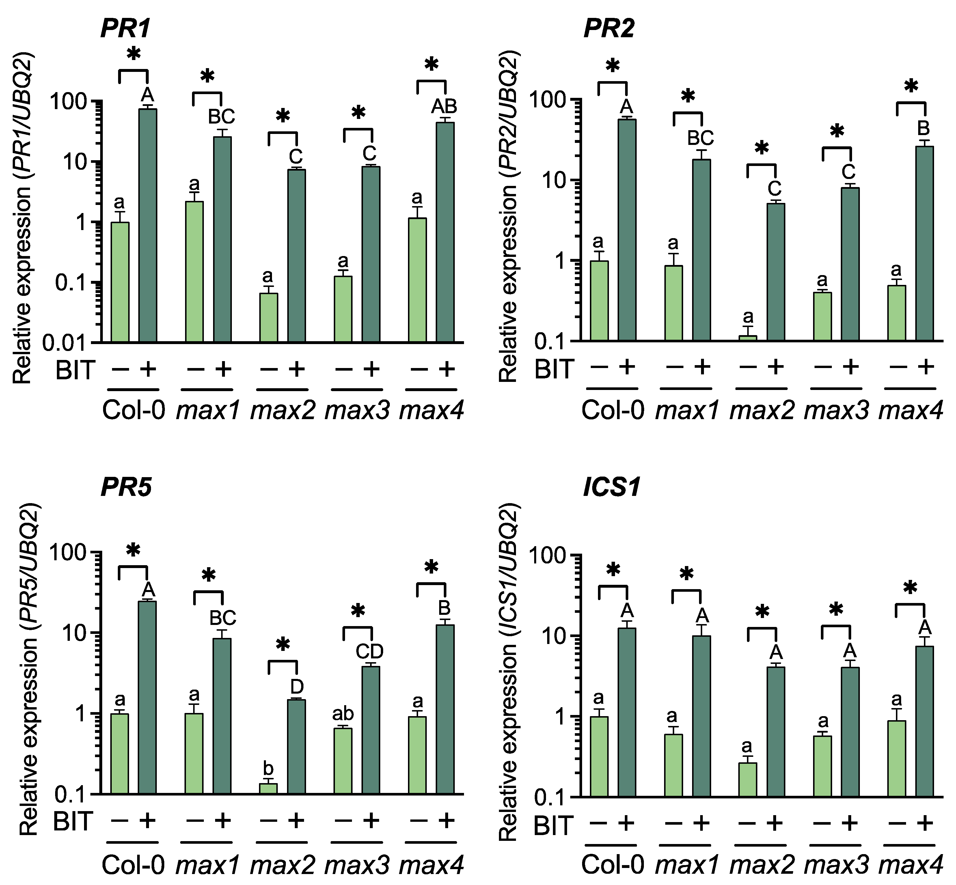
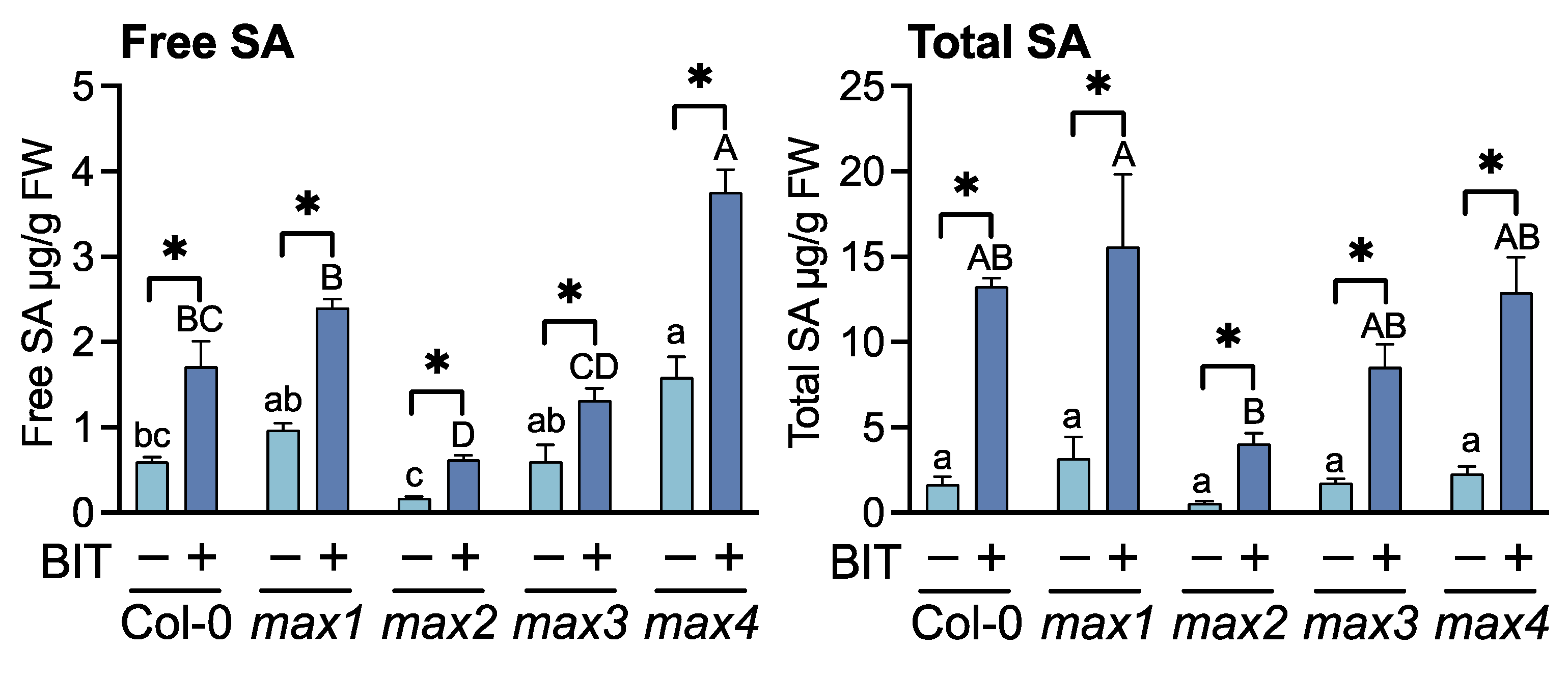
The effects of BIT treatment on SAR induction in the SL-related mutants were evaluated using a pathogenic bacterial inoculation test, defense gene expression profiling, and the estimation of endogenous SA content. Resistance to Pst was assessed by measuring bacterial growth in the leaf tissues. At 3 d after inoculation, leaves of all the SL-related mutants treated with BIT exhibited > 10-fold lower bacterial titers than those of the untreated (control) plants as well as WT (Col-0) plants (Figure 1). Reverse transcription-quantitative polymerase chain reaction (RT-qPCR) indicated that upon treatment with BIT, the expression levels of PR1 (At2g14610), PR2 (At3g57260), and PR5 (At1g75040) were upregulated in the SL-related mutants and the WT, compared to the control plants (Figure 2). Moreover, BIT treatment significantly upregulated ICS1 (At1g74710) expression and increased the levels of free and total endogenous SA (free SA + SA glucoside) in the SL-related mutants and in the WT (Figure 2 and Figure 3). These findings suggest that SAR can be induced in SL biosynthesis- and signaling-deficient mutants.
In the absence of SAR induction, bacterial growth on the leaf tissues was significantly higher in the mutant max2 than in WT plants, whereas bacterial growth in mutants max1, max3, and max4 was intermediate between those of max2 and WT (Figure 1). This finding suggested that SL-mediated signaling is partially involved in basal resistance against Pst. Although ICS1 levels in BIT-treated plants did not differ between the SL-related mutants and the WT, SA levels were altered among these plants, with max2 and max4 exhibiting the lowest and highest levels of both free and total SA, respectively (Figure 2 and Figure 3).
Consistent with SA levels, the levels of expression of PR genes after BIT treatment were significantly lower in max2 than in WT (Figure 2). Thus, BIT-mediated activation of SA biosynthesis and signaling pathways were weaker in max2 than in WT, which can be associated with high bacterial titer in the leaves of max2 (Figure 1). Additionally, the level of activation of the SA-mediated signaling pathway and disease resistance in other SL-related mutants was intermediate between those of max2 and WT (Figure 1 and Figure 2). These findings suggest that the absence of SL signaling affected the SA-mediated signaling pathway and, consequently, the degree of disease resistance against Pst. Furthermore, the difference between SL biosynthesis- and signaling-deficient mutants can be explained by the fact that the inhibition of SL-mediated signaling was stronger in the mutant max2, which is defective in the F-box protein MAX2, than in other biosynthesis-deficient mutants.

2.2. Effects of the Synthetic SL Analog GR24 on SA-Mediated Disease Resistance

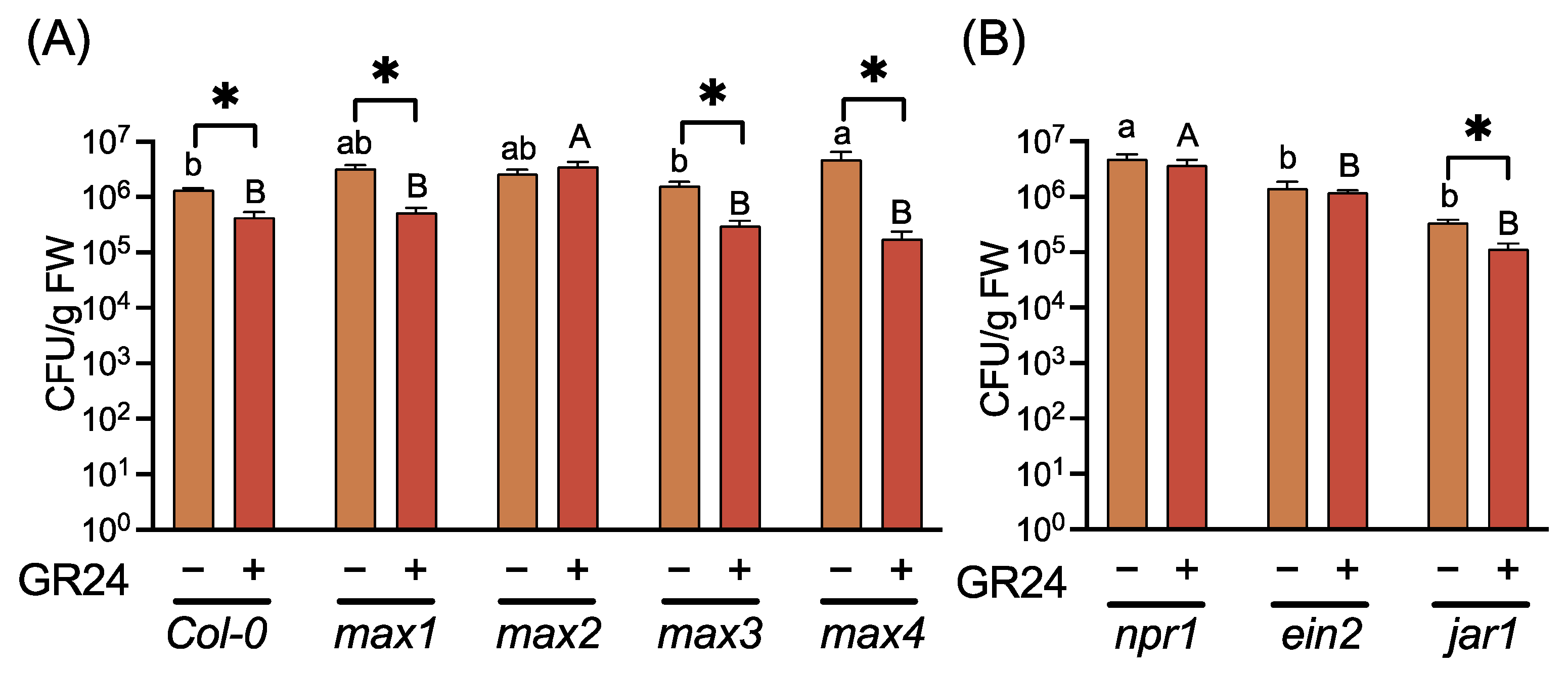
To reveal the effects of SLs on SA-mediated disease resistance, we examined the effects of GR24, a synthetic SL analog, on resistance against Pst in Arabidopsis. Treatment of WT plants with GR24 improved their resistance against Pst (Figure 4). Furthermore, GR24-treated SLs biosynthesis-deficient mutants (max1, max3, and max4) exhibited reduced bacterial growth in the leaf tissues compared to the untreated control plants. In contrast, GR24 treatment did not affect disease resistance in max2 (Figure 4). These results indicate that the activation of SL-mediated signaling by GR24 improved disease resistance in Arabidopsis.
To determine the role of disease resistance-related phytohormones in GR24-induced disease resistance, induction of resistance against Pst was analyzed in SA-, JA-, and ET-mediated signaling defective mutants npr1, jar1, and ein2, respectively. GR24 treatment induced disease resistance only in jar1, indicating that SA and ET—but not JA—were involved in GR24-induced disease resistance (Figure 4).
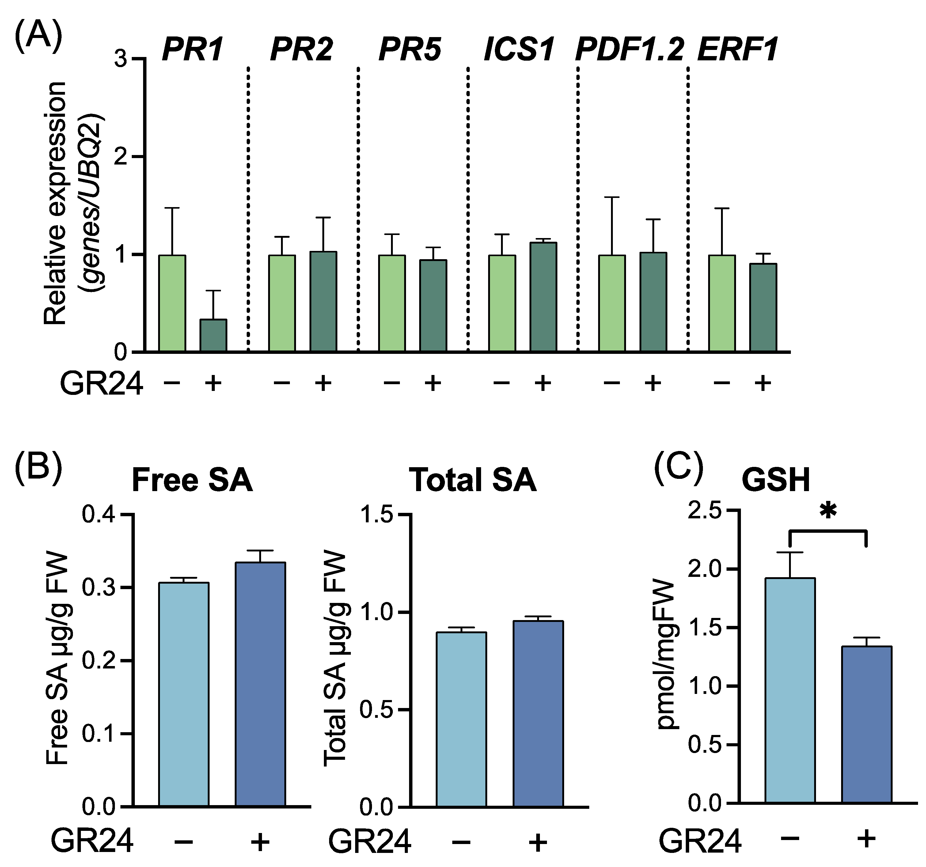
To characterize GR24-induced disease resistance, we examined physiological changes in the GR24-treated WT plants. The level of expression of SAR marker genes, namely PR1, PR2, and PR5, and ICS1 did not increase in GR24-treated plants, indicating that the mechanism of GR24-induced disease resistance was different from that of SAR (Figure 5A). Furthermore, the expression levels of PDF1.2 (At5g44420), the JA-related gene, and ERF1 (At4g17500), the ET-related gene, were not significantly different between GR24-treated and untreated control plants (Figure 5A), indicating that JA- and ET-mediated signaling pathways were not activated in GR24-treated plants. Endogenous accumulation of SAR-related metabolites was examined three days after GR24 treatment. The levels of free SA and total SA (free SA + SA glucoside) were not influenced by GR24 treatment (Figure 5B). Although an increase in GSH level is important for SA-mediated signaling in SAR induction, the level of GSH in GR24-treated plants was slightly but significantly lower than that of untreated control plants. These findings suggest that the mechanism underlying GR24-induced disease resistance was independent of SA-, JA-, or ET-mediated signaling pathways.
To determine the mechanism underlying GR24-induced resistance against Pst, defense responses were investigated by analyzing gene expression upon pathogen inoculation. Time-course analysis indicated that PR1 was significantly upregulated in GR24-treated plants, compared with control plants, at 16 and 20 h post inoculation, whereas similar levels were maintained up to 24 h (Figure 6). This finding suggests that the defense system was rapidly activated in the GR24-treated plants, which should contribute to the increased resistance against Pst. Conversely, the expression of PDF1.2 after Pst infection was not upregulated but downregulated due to activation of SA-mediated signaling (Figure 6). Therefore, GR24-induced disease resistance showed a priming effect that activated major defense pathways, such as the SA-mediated defense pathway, after infection.

2.3. Effects of the SL Biosynthesis Inhibitor TIS108 on Disease Resistance

Pathogen inoculation assays and gene expression analyses indicated that GR24 primed plants against Pst infection, suggesting that SLs positively regulated disease resistance. This was further supported by the fact that in-planta Pst growth was higher in max mutants than in WT plants (Figure 1). However, max mutants, with the absence of SLs from germination, may have some side effects on the stress response systems during their growth.
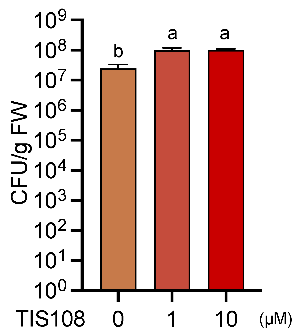
To verify the contribution of endogenous SLs to the resistance against Pst, we examined the effects of the SL-biosynthesis inhibitor TIS108 on gene expression, metabolite accumulation, and disease resistance. The expression levels of SAR-related genes PR1, PR2, PR5, and ICS1; JA-related gene PDF1.2; and ET-related gene ERF1 were not affected by TIS108 treatment (Figure 7A). Endogenous levels of free SA, total SA, and GSH were not significantly different between the TIS108-treated and untreated control plants (Figure 7B). Treatment with as low as 1 µM TIS108 significantly decreased disease resistance in WT plants compared to that of the untreated control plants, suggesting that endogenous SLs contribute to Pst resistance (Figure 8).

3. Discussion

Various aspects of plant immunity are regulated by phytohormones such as SA, JA, and ET. Previous studies have demonstrated the role of SLs in disease resistance using SLs-biosynthesis and signaling mutants of rice and Arabidopsis [39,40]. This study demonstrated that SLs positively regulated SA-mediated signaling during basal defense responses but not SAR induction. Increased disease resistance upon GR24 treatment proved to be a case of priming of the plant immune system, that is, a rapidly upregulated expression of defense genes upon infection (Figure 6). Moreover, the reduction in disease resistance upon treatment with the SL biosynthesis-inhibitor, TIS108, strongly supported that SL-mediated signaling positively affected SA-mediated disease resistance (Figure 8). These findings suggest that SLs play a positive role in plant immunity, especially in the SA-mediated disease resistance. Similar rapid upregulation of defense-related gene expression was observed in plants primed by symbioses with microbes or chemicals [18,19,21]. In turn, this study is the first demonstration of a priming effect on plants by exogenous and endogenous SLs; however, how SLs contribute to plant disease resistance in nature remains to be elucidated. Although whether SLs-biosynthesis can be activated by pathogen attack is unknown, SLs probably affect some unidentified mechanism along the pathway between PAMPs recognition and expression of defense-related genes, such as SA-biosynthesis-related genes and PR genes, which enables primed plants to counterattack pathogens more rapidly than unprimed plants (Figure 9).
As SLs comprise an important class of phytohormones with various roles in plant growth and development, there may be some side effects on the physiology of SL-related mutants due to the absence of SLs. Similarly, TIS108 treatment should also have some side effects, as it suppressed SL-mediated signaling. However, such effects did not occur in the short-time treatment with TIS108, which was supported by our finding that the expression of defense-related genes and the accumulation of SA were not affected by TIS108 treatment.
In Arabidopsis SL biosynthesis- and signaling-defective mutants, as well as in WT plants, a SAR inducer improved disease resistance by activating SA biosynthesis and SA-mediated signaling, suggesting that SLs do not affect SAR induction (Figure 1). Further, and consistently with a previous report [40], without SAR induction, these mutants were more susceptible to Pst than WT plants (Figure 1), and, compared to other SL-related mutants, the SL-signal-perception-defective mutant max2 exhibited a different phenotype. Furthermore, regardless of SAR induction, the disease resistance and SA-mediated signaling processes, such as PR gene expression and SA accumulation, were suppressed in max2 compared to WT plants (Figure 1). In contrast, SL biosynthesis-related mutants exhibited such processes at levels that were intermediate between those observed for max2 mutant and WT plants (Figure 1). These findings suggest that SLs are important, at least for SA-mediated disease resistance.
When the pathogen was inoculated via foliar spray, the levels of disease resistance were lower in max2 than in the other SLs biosynthesis-deficient mutants, which was attributed to the increased number of open stomata in the max2 mutant [40]. In this study, to avoid the influence of stomatal opening and closing, pathogen inoculation was performed by dipping the plants into the bacterial suspension after keeping 100% RH for 12 h before and after inoculation. Thus, the differences in disease resistance and SA-mediated signaling did not result from differences in stomatal opening (Figure 1). We hypothesize that the mutations in MAX2 affected SL-mediated signaling by altering SL-signal perception, thereby affecting the SA-mediated disease resistance. In this case, the differences in SA-mediated signaling processes among SL-biosynthesis-defective mutants would likely be owing to the different physiological effects of each specific mutation, a plausible idea that warrants further research.
Treatment with GR24 failed to induce disease resistance in the mutant npr1, indicating that an SA signal was involved in SL-mediated disease resistance (Figure 4), which is consistent with the altered SA-mediated disease resistance in SL-related mutants (Figure 1). However, SAR induction via the activation of SA-mediated signaling was observed in SL-related mutants, suggesting that SLs are not directly involved in SA-mediated disease resistance. Alternatively, a plausible mechanism involves the regulation of SA-mediated signaling by SL during basal resistance, including PTI. However, further investigation is necessary to confirm the operation of this mechanism.
Treatment with GR24 effectively induced disease resistance in jar1 but not in ein2, indicating that ET but not JA was involved in SL-mediated disease resistance (Figure 4). Further, some reports indicate that ET is involved in SL-mediated signaling; thus, for example, SL-induced seed germination in Striga hermonthica (Delile) Benth. requires ET biosynthesis and signaling [43]. Additionally, SL promotes ET accumulation in etiolated Arabidopsis seedlings by upregulating the expression of 1-aminocyclopropane-1-carboxylic acid oxidase (ACO) [44]. Similarly, SL promotes ET biosynthesis in Arabidopsis roots [45]. In this study, ET-responsive ERF1 was not induced in GR24-treated plant leaves (Figure 5), suggesting that ET was not involved in SL-mediated priming.
In contrast, several other reports have underlined the importance of ET in SA-mediated disease resistance; however, such findings remain controversial. In Arabidopsis, co-treatment with an ET biosynthetic precursor (1-aminocyclopropane-1-carboxylic acid) and SA upregulated PR1 relative to SA treatment alone [46]. Conversely, ein3 eil1, a double mutant of ET-responsive transcription factors, constantly accumulated SA, indicating that ET negatively affects SA-mediated signaling pathways [47]. Nonetheless, GR24-induced disease resistance required ET-mediated signaling through EIN2 (Figure 5), suggesting that ET improved SA-mediated disease resistance after pathogen infection of primed plants.
Nutrient stress, such as low phosphate or nitrogen, induces SL biosynthesis, which alters SL-mediated responses in AM fungi symbioses, which effectively increase the soil volume explored by plant roots in search for phosphorus. However, the trade-off between growth and disease resistance is well known. In contrast to plants growing under soil nutrient-rich conditions (i.e., excess phosphate or nitrogen for better growth), which are more susceptible to diseases, plants growing under soil nutrient-poor conditions exhibit higher levels of expression of defense-related genes after infection [48]. Overall, SL synthesized under soil nutrient-poor conditions may not only promote symbiosis with AM fungi but effectively prime plants for infection as well. This effect might be possible because priming does not affect growth, while it effectively protects plants from pathogen attack by accelerating the induction of SA-, JA-, or other phytohormone-mediated disease resistance mechanisms.

4. Materials and Methods

4.1. Plant Material and Chemical Treatment

Arabidopsis thaliana Col-0 and seven mutants with the same ecotype, including max1-1 [24], max2-1, max3-1, max4-1, npr1-2, jar1-1, and ein2-1, were used for the experiments [9]. Plants were grown in sterilized potting soil (Nihon hiryo, Fujioka, Japan) in plastic pots (5 cm × 5 cm × 5 cm) inside a growth chamber under a 16:8 h light: dark photoperiod at 22 °C and 60% RH.
BIT [11] solutions (1 mg/mL for soil drenching and 1 mM for spraying) were prepared by dissolving the BIT powder in sterilized distilled water. GR24 (rac-GR24) and TIS108 [31] solutions (1 mM) were prepared from 50 mM GR24 in acetone and 50 mM TIS108 in DMSO, respectively, by diluting with sterilized distilled water. Three-week-old plants were treated with chemicals as indicated in the Figures.

4.2. Pathogenicity Assays

Control and chemical-treated plants were inoculated with Pst as previously described [49].

4.3. Estimation of SA Levels

Three-week-old plants were treated with chemicals for three days. Leaf samples (ca. 100 mg) were harvested and used for extraction and measurement of SA as previously described [12]. Briefly, leaf tissues were homogenized in 1 mL of 90% methanol and then extracted using 1 mL of 100% methanol. These extracts were pooled and dried at 40 °C. The dried residues were extracted in 4 mL of water at 80 °C for 15 min, and equal aliquots (1 mL) were used for free and total SA estimation. One aliquot was extracted using 2.5 mL ethyl acetate–cyclohexane (1:1) following the addition of 50 µL of concentrated HCl, and the upper layer was dried and dissolved in 1 mL of 20% methanol in 20 mM sodium acetate buffer (pH 5). This solution was then subjected to high-performance liquid chromatography (HPLC) analysis to determine free SA levels. Then, 1 mL of β-glucosidase (Merck, Darmstadt, Germany) solution (3 units/mL) was added to the remaining aliquots and incubated for 6 h. These aliquots were prepared for HPLC analysis using the above procedure to determine total SA levels. SA analysis was performed using an Agilent 1220 Infinity LC (Agilent, Snata Clara, CA, USA) equipped with a ZORBAX Eclipse Plus C18 (4.6 × 150 mm; Agilent) and run with 20% methanol in 20 mM sodium acetate buffer (pH 5) at a flow rate of 1 mL/min. SA was detected and fluorometrically quantified at 295 nm excitation and 370 nm emission wavelengths.

4.4. GSH Measurement

Three-week-old plants were treated with chemicals for three days. Leaf samples (ca. 100 mg) were harvested and used for extraction and measurement of GSH as previously described [50].

4.5. RT-qPCR

Gene expression profiles were determined using leaf samples. Total RNA isolation, cDNA synthesis, and RT-qPCR were performed as previously described [49]. Gene-specific primers used in this study are listed in Supplementary Table S1. Transcript levels were normalized using the expression of UBQ2 in the same samples [51].

Supplementary Materials

The following supporting information can be downloaded at: https://www.mdpi.com/article/10.3390/ijms23095246/s1.

Author Contributions

Conceptualization, M.K. and H.N. (Hideo Nakashita); methodology, H.N. (Hidemitsu Nakamura), A.M.-N. and H.N. (Hideo Nakashita); validation, M.K., M.F., K.Y., T.N., H.N. (Hidemitsu Nakamura), K.A., A.M.-N., T.A. and H.N. (Hideo Nakashita); investigation, M.K., M.F. and K.S.; resources, T.N., H.N. (Hidemitsu Nakamura) and T.A.; writing—original draft preparation, M.K.; writing—review and editing, H.N. (Hidemitsu Nakamura), T.A. and H.N. (Hideo Nakashita); visualization, M.K.; project administration, T.A. and H.N. (Hideo Nakashita); funding acquisition, K.Y., T.N., K.A., T.A. and H.N. (Hideo Nakashita). All authors have read and agreed to the published version of the manuscript.

Funding

This work received partial support for K.Y., T.N., K.A., T.A. and H.N. (Hideo Nakashita) from the Ministry of Agriculture, Forestry, and Fisheries of Japan under the Science and Technology Research Promotion Program for Agriculture, Forestry, Fisheries, and Food Industry (27004A); also, from Grant-in-Aid for JSPS Fellows 19J14665 for M.F., and from JSPS KAKENHI Grant Numbers 18K05656 for H.N. (Hideo Nakashita).

Institutional Review Board Statement

Not applicable.

Informed Consent Statement

Not applicable.

Data Availability Statement

Data are available on request.

Acknowledgments

We thank H. Takeuchi and A. Sugimoto (Fukui Pref. Univ.) for their kind support in plant culture.

Conflicts of Interest

The authors declare no conflict of interest.

References

  1. Yang, J.; Duan, G.; Li, C.; Liu, L.; Han, G.; Zhang, Y.; Wang, C. The Crosstalks between Jasmonic Acid and Other Plant Hormone Signaling Highlight the Involvement of Jasmonic Acid as a Core Component in Plant Response to Biotic and Abiotic Stresses. Front. Plant Sci. 2019, 10, 1349. [Google Scholar] [CrossRef] [PubMed]
  2. Yasuda, M.; Ishikawa, A.; Jikumaru, Y.; Seki, M.; Umezawa, T.; Asami, T.; Maruyama-Nakashita, A.; Kudo, T.; Shinozaki, K.; Yoshida, S.; et al. Antagonistic Interaction between Systemic Acquired Resistance and the Abscisic Acid–mediated Abiotic Stress Response in Arabidopsis. Plant Cell 2008, 20, 1678–1692. [Google Scholar] [CrossRef] [PubMed]
  3. Kusajima, M.; Yasuda, M.; Kawashima, A.; Nojiri, H.; Yamane, H.; Nakajima, M.; Akutsu, K.; Nakashita, H. Suppressive Effect of Abscisic Acid on Systemic Acquired Resistance in Tobacco Plants. J. Gen. Plant Pathol. 2010, 76, 161–167. [Google Scholar] [CrossRef]
  4. Kusajima, M.; Okumura, Y.; Fujita, M.; Nakashita, H. Abscisic Acid Modulates Salicylic Acid Biosynthesis for Systemic Acquired Resistance in Tomato. Biosci. Biotechnol. Biochem. 2017, 81, 1850–1853. [Google Scholar] [CrossRef] [PubMed]
  5. Shigenaga, A.M.; Berens, M.L.; Tsuda, K.; Argueso, C.T. Towards Engineering of Hormonal Crosstalk in Plant Immunity. Curr. Opin. Plant Biol. 2017, 38, 164–172. [Google Scholar] [CrossRef]
  6. Lorenzo, O.; Piqueras, R.; Sánchez-Serrano, J.J.; Solano, R. ETHYLENE RESPONSE FACTOR1 Integrates Signals from Ethylene and Jasmonate Pathways in Plant Defense. Plant Cell 2003, 15, 165–178. [Google Scholar] [CrossRef]
  7. Schwessinger, B.; Zipfel, C. News from the Frontline: Recent Insights into PAMP-Triggered Immunity in Plants. Curr. Opin. Plant Biol. 2008, 11, 389–395. [Google Scholar] [CrossRef]
  8. Tripathi, D.; Raikhy, G.; Kumar, D. Chemical Elicitors of Systemic Acquired resistance—Salicylic Acid and its Functional Analogs. Curr. Plant Biol. 2019, 17, 48–59. [Google Scholar] [CrossRef]
  9. Wildermuth, M.C.; Dewdney, J.; Wu, G.; Ausubel, F.M. Isochorismate Synthase is Required to Synthesize Salicylic Acid for Plant Defence. Nature 2001, 414, 562–565. [Google Scholar] [CrossRef]
  10. Mou, Z.; Fan, W.; Dong, X. Inducers of Plant Systemic Acquired Resistance Regulate NPR1 Function through Redox Changes. Cell 2003, 113, 935–944. [Google Scholar] [CrossRef]
  11. Nakashita, H.; Yoshioka, K.; Yasuda, M.; Nitta, T.; Arai, Y.; Yoshida, S.; Yamaguchi, I. Probenazole Induces Systemic Acquired Resistance in Tobacco through Salicylic Acid Accumulation. Physiol. Mol. Plant Pathol. 2002, 61, 197–203. [Google Scholar] [CrossRef]
  12. Yoshioka, K.; Nakashita, H.; Klessig, D.F.; Yamaguchi, I. Probenazole Induces Systemic Acquired Resistance in Arabidopsis with a Novel Type of Action. Plant J. 2001, 25, 149–157. [Google Scholar] [CrossRef]
  13. Yasuda, M.; Nakashita, H.; Yoshida, S. Tiadinil, a Novel Class of Activator of Systemic Acquired Resistance, Induces Defense Gene Expression and Disease Resistance in Tobacco. J. Pest. Sci. 2004, 29, 46–49. [Google Scholar] [CrossRef]
  14. Umemura, K.; Satou, J.; Iwata, M.; Uozumi, N.; Koga, J.; Kawano, T.; Koshiba, T.; Anzai, H.; Mitomi, M. Contribution of Salicylic Acid Glucosyltransferase, OsSGT1, to Chemically Induced Disease Resistance in Rice Plants. Plant J. 2009, 57, 463–472. [Google Scholar] [CrossRef]
  15. Lawton, K.A.; Friedrich, L.; Hunt, M.; Weymann, K.; Delaney, T.; Kessmann, H.; Staub, T.; Ryals, J. Benzothiadiazole Induces Disease Resistance in Arabidopsis by Activation of the Systemic Acquired Resistance Signal Transduction Pathway. Plant J. Cell Mol. Biol. 1996, 10, 71–82. [Google Scholar] [CrossRef] [PubMed]
  16. Nakashita, H. Studies on Regulation of Plant Physiology by Pesticides. J. Pest. Sci. 2021, 46, 393–398. [Google Scholar] [CrossRef] [PubMed]
  17. Jung, S.C.; Martinez-Medina, A.; Lopez-Raez, J.A.; Pozo, M.J. Mycorrhiza-Induced Resistance and Priming of Plant Defenses. J. Chem. Ecol. 2012, 38, 651–664. [Google Scholar] [CrossRef]
  18. Fujita, M.; Kusajima, M.; Fukagawa, M.; Okumura, Y.; Nakajima, M.; Akiyama, K.; Asami, T.; Yoneyama, K.; Kato, H.; Nakashita, H. Response of Tomatoes Primed by Mycorrhizal Colonization to Virulent and Avirulent Bacterial Pathogens. Sci. Rep. 2022, 12, 4686. [Google Scholar] [CrossRef]
  19. Pieterse, C.M.; Van Wees, S.C.; Hoffland, E.; Van Pelt, J.A.; Van Loon, L.C. Systemic Resistance in Arabidopsis Induced by Biocontrol Bacteria is Independent of Salicylic Acid Accumulation and Pathogenesis-Related Gene Expression. Plant Cell 1996, 8, 1225–1237. [Google Scholar]
  20. Fujita, M.; Kusajima, M.; Okumura, Y.; Nakajima, M.; Minamisawa, K.; Nakashita, H. Effects of Colonization of a Bacterial Endophyte, Azospirillum sp. B510, on Disease Resistance in Tomato. Biosci. Biotechnol. Biochem. 2017, 81, 1657–1662. [Google Scholar] [CrossRef]
  21. Buswell, W.; Schwarzenbacher, R.E.; Luna, E.; Sellwood, M.; Chen, B.; Flors, V.; Pétriacq, P.; Ton, J. Chemical Priming of Immunity without Costs to Plant Growth. New Phytol. 2018, 218, 1205–1216. [Google Scholar] [CrossRef] [PubMed]
  22. Cook, C.E.; Whichard, L.P.; Turner, B.; Wall, M.E.; Egley, G.H. Germination of Witchweed (Striga lutea Lour.): Isolation and Properties of a Potent Stimulant. Science 1966, 154, 1189–1190. [Google Scholar] [CrossRef] [PubMed]
  23. Akiyama, K.; Matsuzaki, K.; Hayashi, H. Plant Sesquiterpenes Induce Hyphal Branching in Arbuscular Mycorrhizal Fungi. Nature 2005, 435, 824–827. [Google Scholar] [CrossRef] [PubMed]
  24. Gomez-Roldan, V.; Fermas, S.; Brewer, P.B.; Puech-Pagès, V.; Dun, E.A.; Pillot, J.; Letisse, F.; Matusova, R.; Danoun, S.; Portais, J. Strigolactone Inhibition of Shoot Branching. Nature 2008, 455, 189–194. [Google Scholar] [CrossRef]
  25. Umehara, M.; Hanada, A.; Yoshida, S.; Akiyama, K.; Arite, T.; Takeda-Kamiya, N.; Magome, H.; Kamiya, Y.; Shirasu, K.; Yoneyama, K. Inhibition of Shoot Branching by New Terpenoid Plant Hormones. Nature 2008, 455, 195–200. [Google Scholar] [CrossRef]
  26. Matusova, R.; Rani, K.; Verstappen, F.W.; Franssen, M.C.; Beale, M.H.; Bouwmeester, H.J. The Strigolactone Germination Stimulants of the Plant-Parasitic Striga and Orobanche spp. are Derived from the Carotenoid Pathway. Plant Physiol. 2005, 139, 920–934. [Google Scholar] [CrossRef]
  27. Seto, Y.; Sado, A.; Asami, K.; Hanada, A.; Umehara, M.; Akiyama, K.; Yamaguchi, S. Carlactone is an Endogenous Biosynthetic Precursor for Strigolactones. Proc. Natl. Acad. Sci. USA 2014, 111, 1640–1645. [Google Scholar] [CrossRef]
  28. Booker, J.; Sieberer, T.; Wright, W.; Williamson, L.; Willett, B.; Stirnberg, P.; Turnbull, C.; Srinivasan, M.; Goddard, P.; Leyser, O. MAX1 Encodes a Cytochrome P450 Family Member that Acts Downstream of MAX3/4 to Produce a Carotenoid-Derived Branch-Inhibiting Hormone. Dev. Cell. 2005, 8, 443–449. [Google Scholar] [CrossRef]
  29. Stirnberg, P.; van De Sande, K.; Leyser, H.O. MAX1 and MAX2 Control Shoot Lateral Branching in Arabidopsis. Development 2002, 129, 1131–1141. [Google Scholar] [CrossRef]
  30. De Cuyper, C.; Struk, S.; Braem, L.; Gevaert, K.; De Jaeger, G.; Goormachtig, S. Strigolactones, Karrikins and Beyond. Plant Cell Environ. 2017, 40, 1691–1703. [Google Scholar] [CrossRef]
  31. Ito, S.; Umehara, M.; Hanada, A.; Yamaguchi, S.; Asami, T. Effects of Strigolactone-Biosynthesis Inhibitor TIS108 on Arabidopsis. Plant Signal. Behav. 2013, 8, e24193. [Google Scholar] [CrossRef] [PubMed]
  32. Mostofa, M.G.; Li, W.; Nguyen, K.H.; Fujita, M.; Tran, L.P. Strigolactones in Plant Adaptation to Abiotic Stresses: An Emerging Avenue of Plant Research. Plant Cell Environ. 2018, 41, 2227–2243. [Google Scholar] [CrossRef] [PubMed]
  33. Shirani Bidabadi, S.; Sharifi, P. Strigolactone and Methyl Jasmonate-Induced Antioxidant Defense and the Composition Alterations of Different Active Compounds in Dracocephalum Kotschyi Boiss Under Drought Stress. J. Plant Growth Regul. 2021, 40, 878–889. [Google Scholar] [CrossRef]
  34. Sharifi, P.; Bidabadi, S.S. Strigolactone could Enhances Gas-Exchange through Augmented Antioxidant Defense System in Salvia nemorosa, L. Plants Subjected to Saline Conditions Stress. Ind. Crops Prod. 2020, 151, 112460. [Google Scholar] [CrossRef]
  35. Cooper, J.W.; Hu, Y.; Beyyoudh, L.; Yildiz Dasgan, H.; Kunert, K.; Beveridge, C.A.; Foyer, C.H. Strigolactones Positively Regulate Chilling Tolerance in Pea and in Arabidopsis. Plant Cell Environ. 2018, 41, 1298–1310. [Google Scholar] [CrossRef]
  36. Van Ha, C.; Leyva-González, M.A.; Osakabe, Y.; Tran, U.T.; Nishiyama, R.; Watanabe, Y.; Tanaka, M.; Seki, M.; Yamaguchi, S.; Van Dong, N. Positive Regulatory Role of Strigolactone in Plant Responses to Drought and Salt Stress. Proc. Natl. Acad. Sci. USA 2014, 111, 851–856. [Google Scholar]
  37. Bu, Q.; Lv, T.; Shen, H.; Luong, P.; Wang, J.; Wang, Z.; Huang, Z.; Xiao, L.; Engineer, C.; Kim, T.H. Regulation of Drought Tolerance by the F-Box Protein MAX2 in Arabidopsis. Plant Physiol. 2014, 164, 424–439. [Google Scholar] [CrossRef]
  38. Torres-Vera, R.; García, J.M.; Pozo, M.J.; López-Ráez, J.A. Do Strigolactones Contribute to Plant Defence? Mol. Plant Pathol. 2014, 15, 211–216. [Google Scholar] [CrossRef]
  39. Nasir, F.; Tian, L.; Shi, S.; Chang, C.; Ma, L.; Gao, Y.; Tian, C. Strigolactones Positively Regulate Defense against Magnaporthe oryzae in Rice (Oryza sativa). Plant Physiol. Biochem. 2019, 142, 106–116. [Google Scholar] [CrossRef]
  40. Kalliola, M.; Jakobson, L.; Davidsson, P.; Pennanen, V.; Waszczak, C.; Yarmolinsky, D.; Zamora, O.; Palva, E.T.; Kariola, T.; Kollist, H. Differential Role of MAX2 and Strigolactones in Pathogen, Ozone, and Stomatal Responses. Plant Dir. 2020, 4, e00206. [Google Scholar] [CrossRef]
  41. Piisilä, M.; Keceli, M.A.; Brader, G.; Jakobson, L.; Jõesaar, I.; Sipari, N.; Kollist, H.; Palva, E.T.; Kariola, T. The F-Box Protein MAX2 Contributes to Resistance to Bacterial Phytopathogens in Arabidopsis thaliana. BMC Plant Biol. 2015, 15, 53. [Google Scholar] [CrossRef] [PubMed]
  42. Katagiri, F.; Thilmony, R.; He, S.Y. The Arabidopsis thaliana-Pseudomonas syringae Interaction. Am. Soc. Plant Biol. 2002, 1, e0039. [Google Scholar] [CrossRef] [PubMed]
  43. Sugimoto, Y.; Ali, A.M.; Yabuta, S.; Kinoshita, H.; Inanaga, S.; Itai, A. Germination Strategy of Striga hermonthica Involves Regulation of Ethylene Biosynthesis. Physiol. Plant. 2003, 119, 137–145. [Google Scholar] [CrossRef]
  44. Lee, H.Y.; Yoon, G.M. Strigolactone Elevates Ethylene Biosynthesis in Etiolated Arabidopsis Seedlings. Plant Signal. Behav. 2020, 15, 1805232. [Google Scholar] [CrossRef]
  45. Kapulnik, Y.; Resnick, N.; Mayzlish-Gati, E.; Kaplan, Y.; Wininger, S.; Hershenhorn, J.; Koltai, H. Strigolactones Interact with Ethylene and Auxin in Regulating Root-Hair Elongation in Arabidopsis. J. Exp. Bot. 2011, 62, 2915–2924. [Google Scholar] [CrossRef]
  46. De Vos, M.; Van Zaanen, W.; Koornneef, A.; Korzelius, J.P.; Dicke, M.; Van Loon, L.C.; Pieterse, C.M. Herbivore-Induced Resistance Against Microbial Pathogens in Arabidopsis. Plant Physiol. 2006, 142, 352–363. [Google Scholar] [CrossRef]
  47. Chen, H.; Xue, L.; Chintamanani, S.; Germain, H.; Lin, H.; Cui, H.; Cai, R.; Zuo, J.; Tang, X.; Li, X. ETHYLENE INSENSITIVE3 and ETHYLENE INSENSITIVE3-LIKE1 Repress SALICYLIC ACID INDUCTION DEFICIENT2 Expression to Negatively Regulate Plant Innate Immunity in Arabidopsis. Plant Cell 2009, 21, 2527–2540. [Google Scholar] [CrossRef]
  48. Campos-Soriano, L.; Bundó, M.; Bach-Pages, M.; Chiang, S.; Chiou, T.; San Segundo, B. Phosphate Excess Increases Susceptibility to Pathogen Infection in Rice. Mol. Plant Pathol. 2020, 21, 555–570. [Google Scholar] [CrossRef]
  49. Kusajima, M.; Fujita, M.; Yamakawa, H.; Ushiwatari, T.; Mori, T.; Tsukamoto, K.; Hayashi, H.; Maruyama-Nakashita, A.; Che, F.; Nakashita, H. Characterization of Plant Immunity-Activating Mechanism by a Pyrazole Derivative. Biosci. Biotechnol. Biochem. 2020, 84, 1427–1435. [Google Scholar] [CrossRef]
  50. Morikawa-Ichinose, T.; Kim, S.; Allahham, A.; Kawaguchi, R.; Maruyama-Nakashita, A. Glucosinolate Distribution in the Aerial Parts of Sel1-10, a Disruption Mutant of the Sulfate Transporter SULTR1; 2, in Mature Arabidopsis thaliana Plants. Plants 2019, 8, 95. [Google Scholar] [CrossRef]
  51. Maruyama-Nakashita, A.; Nakamura, Y.; Yamaya, T.; Takahashi, H. A Novel Regulatory Pathway of Sulfate Uptake in Arabidopsis Roots: Implication of CRE1/WOL/AHK4-mediated Cytokinin-dependent Regulation. Plant J. 2004, 38, 779–789. [Google Scholar] [CrossRef] [PubMed]
Figure 1. Effects of BIT treatment on growth of Pseudomonas syringae pv. tomato DC3000 in Arabidopsis leaf tissues. Three-week-old plants were treated with water (control) (–) or BIT (1 mg/pot) (+) using the soil drenching method 3 d before inoculation with Pst (2 × 10⁵ CFU/mL). Leaves were homogenized at 3 d post-inoculation, and the number of CFU was estimated using their growth on nutrient broth agar plates. Values are means ± SE from six replicates, each with three leaves. Significant differences between the control and BIT-treated leaves are indicated by * (unpaired t-test, p < 0.05). Different letters indicate significant differences between accessions of the same treatment (Tukey’s multiple comparison test, p < 0.05).
Ijms 23 05246 g001
Figure 2. Expression of defense-related genes in BIT-treated plants. Leaves of three-week-old plants were sprayed with water (control) (–) or 1 mM BIT (+) and collected after 3 d. Transcript levels were normalized using the expression of UBQ2 (At2g36170) in the same samples. Relative mRNA levels among treatments are presented. Values are means ± SE (n = 3). Significant differences between the control and BIT-treated plants are indicated by * (unpaired t-test, p < 0.05). Different letters indicate significant differences between accessions of the same treatment (Tukey’s multiple comparison test, p < 0.05).
Ijms 23 05246 g002
Figure 3. Accumulation of free and total SA in BIT-treated plants. Plants were treated with water (control) (–) or 1 mM BIT (+). Leaves were harvested 3 d after treatment, and free and total SA (free SA + SA glucoside) levels were quantified using high-performance liquid chromatography. Significant differences between control and BIT-treated plants are indicated by * (unpaired t-test, p < 0.05). Different letters indicate significant differences between accessions of the same treatment (Tukey’s multiple comparison test, p < 0.05).
Ijms 23 05246 g003
Figure 4. Effects of GR24 treatment on growth of Pseudomonas syringae pv. tomato DC3000 in Arabidopsis leaf tissues. Three-week-old plants of WT and SL-related mutants (A) and SA-, ET-, and JA-related mutants (B) were treated with 0.02% acetone (control) (–) or 10 µM GR24 (+) by drenching the soil 3 d before inoculation with Pst (2 × 10⁵ CFU/mL). Leaves were homogenized 3 d post-inoculation, and the number of CFUs was estimated from their growth on nutrient broth agar plates. Values are means ± SE of six replicates, each with three leaves. Significant differences between control and GR24-treated plants are indicated by * (unpaired t-test, p < 0.05). Different letters indicate significant differences between accessions of the same treatment (Tukey’s multiple comparison test, p < 0.05).
Ijms 23 05246 g004
Figure 5. Analysis of defense-related gene expression and metabolite accumulation in Arabidopsis thaliana treated with GR24. Three-week-old plants were treated with 0.02% acetone (control) (–) or 10 µM GR24 (+) using the soil drenching method, and leaves were collected 3 d after treatment. (A) Expression levels of PR1, PR2, PR5, ICS1, PDF1.2, and ERF1 in GR24-treated plants. Transcript levels were normalized using the expression of UBQ2 in the same samples. Relative mRNA levels of each gene among the treatments are presented. Values are means ± SE (n = 4). (B,C) Accumulation of free and total SA and GSH in GR24-treated plants. The levels of free and total SA (free SA + SA glucoside) and GSH were quantified using high-performance liquid chromatography. Significant differences between control and GR24-treated plants are indicated by * (unpaired t-test, p < 0.05).
Ijms 23 05246 g005
Figure 6. Expression of PR1 and PDF1.2 after pathogen infection in GR24-treated plants. Three-week-old plants were treated with 0.02% acetone (control) or 10 µM GR24 by drenching the soil 3 d before inoculation with Pst (2 × 10⁷ CFU/mL). Leaves were collected 16, 20, and 24 h after Pst inoculation. Closed circle, water-treated control plants; closed square, GR24-treated plants. Significant differences between control and GR24 treatments are indicated by * (unpaired t-test, p < 0.05).
Ijms 23 05246 g006
Figure 7. Analysis of defense-related gene expression and metabolite accumulation in Arabidopsis thaliana treated with TIS108. Three-week-old plants were treated with 0,02% dimethyl sulfoxide (DMSO) (–) or 10 µM TIS108 (+) using the soil drenching method, and leaves were collected 3 d after treatment. (A) Expression levels of PR1, PR2, PR5, ICS1, PDF1.2, and ERF1 in TIS108-treated plants. Transcript levels were normalized using the expression of UBQ2 in the same samples. Relative mRNA levels of each gene among the treatments are presented. Values are means ± SE (n = 4). (B,C) Accumulation of free and total SA and GSH in TIS108-treated plants. The levels of free and total SA (free SA + SA glucoside) and GSH were quantified using high-performance liquid chromatography.
Ijms 23 05246 g007
Figure 8. Effects of strigolactone biosynthesis inhibitor TIS108 on Pst growth in Arabidopsis leaf tissues. Three-week-old plants were treated with 0.02% dimethyl sulfoxide (DMSO) (control) or 1 or 10 µM TIS108 by drenching the soil 3 d before inoculation with Pst (2 × 10⁵ CFU/mL). Leaves were homogenized 3 d post-inoculation, and the number of CFUs was estimated using their growth on nutrient broth agar plates. Values are means ± SE of six replicates, each with three leaves. Different letters indicate significant differences (Tukey’s multiple comparison test, p < 0.05).
Ijms 23 05246 g008
Figure 9. Proposed model for modulation mechanism of SA-mediated defense signaling by strigolactones. SCFMAX2 is SKP1-CUL1-F-box complex containing MAX2 as F-box protein.
Ijms 23 05246 g009
Publisher’s Note: MDPI stays neutral with regard to jurisdictional claims in published maps and institutional affiliations.