
Bone Healing Materials in the Treatment of Recalcitrant Nonunions and Bone Defects


Abstract
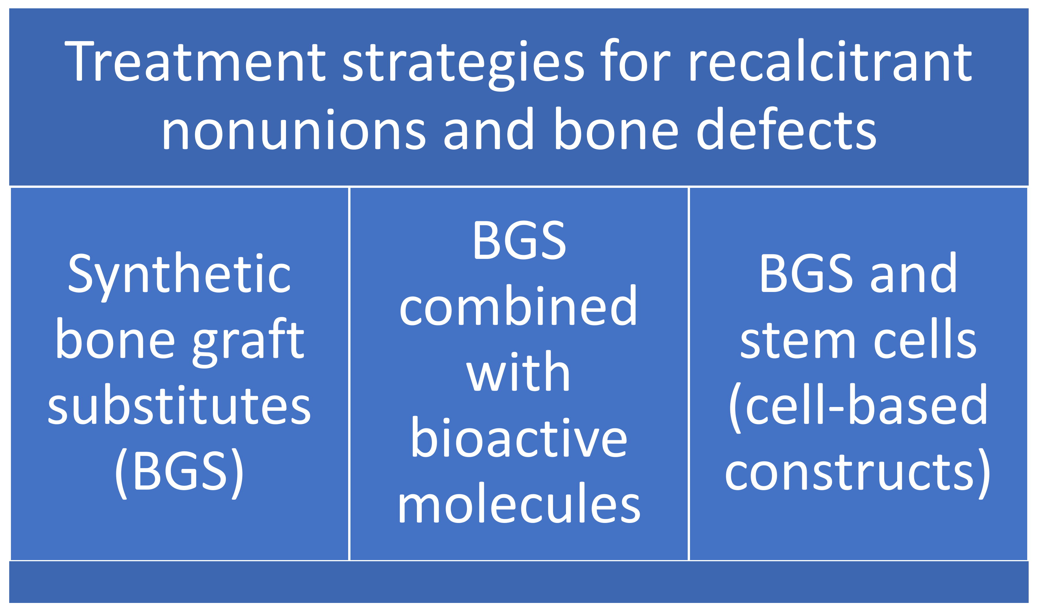
:1. Introduction
2. Synthetic Bone Graft Substitutes (BGS)
2.1. Biomaterials
2.2. Bone Tissue Engineering Scaffolds
3. BGS Combined with Bioactive Molecules
3.1. Bone Morphogenetic Proteins (BMPs)
3.2. Alternative Carriers for Growth Factor Delivery
3.3. Small Molecules as Regulators of Bone Mass
3.3.1. Parathyroid Hormone (PTH)
3.3.2. KUR-111, KUR-112, and KUR-113
4. Bone Graft Substitutes and Stem Cells (Cell-Based Constructs)
4.1. Bone Marrow Stromal Cells (BMSCs)
4.2. Adipose-Derived Mesenchymal Cells (ASCs)
4.3. Periosteum-Derived Stem Cells (PDSCs)
4.4. Three-Dimensional (3D) Bioprinting of Hydrogels and Cells or Bioactive Molecules
5. Conclusions
Funding
Institutional Review Board Statement
Informed Consent Statement
Conflicts of Interest
References
- Ho-Shui-Ling, A.; Bolander, J.; Rustom, L.E.; Johnson, A.W.; Luyten, F.P.; Picart, C. Bone regeneration strategies: Engineered scaffolds, bioactive molecules and stem cells current stage and future perspectives. Biomaterials 2018, 180, 143–162. [Google Scholar] [CrossRef]
- Caplan, A.I. Mesenchymal stem cells. J. Orthop. Res. 1991, 9, 641–650. [Google Scholar] [CrossRef]
- Rodriguez-Merchan, E.C.; Forriol, F. Nonunion: General principles and experimental data. Clin. Orthop. Relat. Res. 2004, 419, 4–12. [Google Scholar] [CrossRef]
- Rodriguez-Merchan, E.C.; Gomez-Castresana, F. Internal fixation of nonunions. Clin. Orthop. Relat. Res. 2004, 419, 13–20. [Google Scholar] [CrossRef] [PubMed]
- Hernigou, P.; Pariat, J.; Queinnec, S.; Homma, Y.; Flouzat Lachaniette, C.H.; Chevallier, N.; Rouard, H. Supercharging irradiated allografts with mesenchymal stem cells improves acetabular bone grafting in revision arthroplasty. Int. Orthop. 2014, 38, 1913–1921. [Google Scholar] [CrossRef]
- Gómez-Barrena, E.; Padilla-Eguiluz, N.G.; Rosset, P. Frontiers in non-union research. EFORT Open Rev. 2020, 5, 574–583. [Google Scholar] [CrossRef]
- Rodríguez-Merchán, E.C. A review of recent developments in the molecular mechanisms of bone healing. Int. J. Mol. Sci. 2021, 22, 767. [Google Scholar] [CrossRef]
- Sohn, H.-S.; Oh, J.-K. Review of bone graft and bone substitutes with an emphasis on fracture surgeries. Biomater. Res. 2019, 23, 9. [Google Scholar] [CrossRef] [PubMed] [Green Version]
- Qi, J.; Yu, T.; Hu, B.; Wu, H.; Ouyang, H. Current biomaterial-based bone tissue engineering and translational medicine. Int. J. Mol. Sci. 2021, 22, 10233. [Google Scholar] [CrossRef] [PubMed]
- Ireland, H.; Gay, M.H.P.; Baldomero, H.; De Angelis, B.; Baharvand, H.; Lowdell, M.W.; Passweg, J.; Martin, I.; International Society for Cellular Therapy (ISCT); Tissue Engineering and Regenerative Medicine International Society—Europe (TERMIS-EU); et al. The survey on cellular and tissue-engineered therapies in Europe and neighboring Eurasian countries in 2014 and 2015. Cytotherapy 2018, 20, 1–20. [Google Scholar] [CrossRef]
- Sawadkar, P.; Mohanakrishnan, J.; Rajasekar, P.; Rahmani, B.; Kohli, N.; Bozec, L.; Garcia-Gareta, E. A synergistic relationship between Polycaprolactone and natural polymers enhances the physical properties and biological activity of scaffolds. ACS Appl. Mater. Interfaces 2020, 12, 13587–13597. [Google Scholar] [CrossRef] [PubMed]
- Li, L.; Li, J.; Guo, J.; Zhang, H.; Zhang, X.; Yin, C.; Wang, L.; Zhu, Y.; Yao, Q. 3D molecularly functionalized cell-free biomimetic scaffolds for osteochondral regeneration. Adv. Funct. Mater. 2018, 29, 1807356. [Google Scholar] [CrossRef]
- Dong, Q.; Zhang, M.; Zhou, X.; Shao, Y.; Li, J.; Wang, L.; Chu, C.; Xue, F.; Yao, Q.; Bai, J. 3D-printed Mg-incorporated PCL-based scaffolds: A promising approach for bone healing. Mater. Sci. Eng. C Mater. Biol. Appl. 2021, 129, 112372. [Google Scholar] [CrossRef]
- Wiese, A.; Pape, H.C. Bone defects caused by high-energy injuries, bone loss, infected nonunions, and nonunions. Orthop. Clin. N. Am. 2010, 41, 1–4. [Google Scholar] [CrossRef]
- Fardjahromi, M.A.; Nazari, H.; Tafti, S.M.A.; Razmjou, A.; Mukhopadhyay, S.; Warkiani, M.E. Metal-organic framework-based nanomaterials for bone tissue engineering and wound healing. Mater. Today Chem. 2022, 23, 100670. [Google Scholar] [CrossRef]
- Lei, H.; Yi, T.; Fan, H.; Pei, X.; Wu, L.; Xing, F.; Li, M.; Liu, L.; Zhou, C.; Fan, Y.; et al. Customized additive manufacturing of porous Ti6Al4V scaffold with microtopological structures to regulate cell behavior in bone tissue engineering. Mater. Sci. Eng. C 2021, 120, 111789. [Google Scholar] [CrossRef]
- Carluccio, D.; Xu, C.; Venezuela, J.; Cao, Y.X.; Kent, D.; Bermingham, M.; Demir, A.G.; Previtali, B.; Ye, Q.S.; Dargusch, M. Additively manufactured iron-manganese for biodegradable porous load-bearing bone scaffold applications. Acta Biomater. 2020, 103, 346–360. [Google Scholar] [CrossRef]
- Bian, D.; Qin, L.; Lin, W.; Shen, D.; Qi, H.; Shi, X.; Zhang, G.; Liu, H.; Yang, H.; Wang, J.; et al. Magnetic resonance (MR) safety and compatibility of a novel iron bioresorbable scaffold. Bioact. Mater. 2020, 5, 260–274. [Google Scholar] [CrossRef]
- Dong, J.; Li, Y.; Lin, P.; Leeflang, M.A.; Asperen, S.; Yu, K.; Tümer, N.; Norder, B.; Zadpoor, A.A.; Zhou, J. Solvent-cast 3D printing of magnesium scaffolds. Acta Biomater. 2020, 114, 497–514. [Google Scholar] [CrossRef]
- Zhang, L.; Jia, G.Z.; Tang, M.; Chen, C.X.; Niu, J.L.; Huang, H.; Kang, B.; Pei, J.; Zeng, H.; Yuan, G.Y. Simultaneous enhancement of anti-corrosion, biocompatibility, and antimicrobial activities by hierarchically-structured brushite/Ag3PO4-coated Mgbased scaffolds. Mater. Sci. Eng. C 2020, 111, 110779. [Google Scholar] [CrossRef]
- Cockerill, I.; Su, Y.; Sinha, S.; Qin, Y.; Zheng, Y.; Young, M.L.; Zhu, D. Porous zinc scaffolds for bone tissue engineering applications: A novel additive manufacturing and casting approach. Mater. Sci. Eng. C 2020, 110, 110738. [Google Scholar] [CrossRef] [PubMed]
- Pearson, J.J.; Gerken, N.; Bae, C.; Lee, K.B.; Satsangi, A.; McBride, S.; Appleford, M.R.; Dean, D.D.; Hollinger, J.O.; Ong, J.L.; et al. In vivo hydroxyapatite scaffold performance in infected bone defects. J. Biomed. Mater. Res. B 2020, 108, 1157–1166. [Google Scholar] [CrossRef] [PubMed]
- Calabresea, G.; Petralia, S.; Franco, D.; Nocito, G.; Fabbi, F.; Forte, L.; Guglielmino, S.; Squarzoni, S.; Traina, F.; Conoci, S. A new Ag-nanostructured hydroxyapatite porous scaffold: Antibacterial effect and cytotoxicity study. Mater. Sci. Eng. C 2021, 118, 111394. [Google Scholar] [CrossRef]
- Rustom, L.E.; Poellmann, M.J.; Johnson, A.J.W. Mineralization in micropores of calcium phosphate scaffolds. Acta Biomater. 2019, 83, 435–455. [Google Scholar] [CrossRef]
- Zhang, S.W.; Wu, X.W.; Chen, J.D.; Lin, K.L. The development of collagen based composite scaffolds for bone regeneration. Bioact. Mater. 2018, 3, 129–138. [Google Scholar] [CrossRef] [PubMed]
- Baptista, R.; Guedes, M. Morphological and mechanical characterization of 3D printed PLA scaffolds with controlled porosity for trabecular bone tissue replacement. Mater. Sci. Eng. C 2021, 118, 111528. [Google Scholar] [CrossRef]
- Williams, J.M.; Adewunmi, A.; Schek, R.M.; Flanagan, C.L.; Krebsbach, P.H.; Feinberg, S.E.; Hollister, S.J.; Das, S. Bone tissue engineering using polycaprolactone scaffolds fabricated via selective laser sintering. Biomaterials 2005, 26, 4817–4827. [Google Scholar] [CrossRef]
- Guo, X.; Xue, M.; Chen, F.; Guo, Q.; Zhou, X.; Lin, H.; Chen, Y. Local delivery and controlled release of miR-34a loaded in hydroxyapatite/mesoporous organosilica nanoparticles composite-coated implant wire to accelerate bone fracture healing. Biomaterials 2022, 280, 121300. [Google Scholar] [CrossRef]
- Hasan, R.; Schaner, K.; Mulinti, P.; Brooks, A. A bioglass-based antibiotic (vancomycin) releasing bone void filling putty to treat osteomyelitis and aid bone healing. Int. J. Mol. Sci. 2021, 22, 7736. [Google Scholar] [CrossRef]
- Roseti, L.; Parisi, V.; Petretta, M.; Cavallo, C.; Desando, G.; Bartolotti, I.; Grigolo, B. Scaffolds for bone tissue engineering: State of the art and new perspectives. Mater. Sci. Eng. C 2017, 78, 1246–1262. [Google Scholar] [CrossRef]
- Su, X.; Wang, T.; Guo, S. Applications of 3D printed bone tissue engineering scaffolds in the stem cell field. Regen. Ther. 2021, 16, 63–72. [Google Scholar] [CrossRef] [PubMed]
- Wang, C.; Huang, W.; Zhou, Y.; He, L.B.; He, Z.; Chen, Z.L.; He, X.; Tian, S.; Liao, J.M.; Lu, B.H.; et al. 3D printing of bone tissue engineering scaffolds. Bioact. Mater. 2020, 5, 82–91. [Google Scholar] [CrossRef] [PubMed]
- Wubneh, A.; Tsekoura, E.K.; Ayranci, C.; Uludag, H. Current state of fabrication technologies and materials for bone tissue engineering. Acta Biomater. 2018, 80, 1–30. [Google Scholar] [CrossRef] [PubMed]
- Pang, L.; Paxton, N.C.; Ren, J.Y.; Liu, F.; Zhan, H.F.; Woodruff, M.A.; Bo, A.X.; Gu, Y.T. Development of mechanically enhanced polycaprolactone composites by a functionalized titanate nanofiller for melt electrowriting in 3D printing. ACS Appl. Mater. Interfaces 2020, 12, 47993–48006. [Google Scholar] [CrossRef] [PubMed]
- Campana, V.; Milano, G.; Pagano, E.; Barba, M.; Cicione, C.; Salonna, G.; Lattanzi, W.; Logroscino, G. Substitutes in orthopaedic surgery: From basic science to clinical practice. J. Mater. Sci. Mater. Med. 2014, 25, 2445–2461. [Google Scholar] [CrossRef]
- Katagiri, T.; Yamaguchi, A.; Komaki, M.; Abe, E.; Takahashi, N.; Ikeda, T.; Rosen, V.; Wozney, J.M.; Fujisawa-Sehara, A.; Suda, T. Bone morphogenetic protein-2 converts the differentiation pathway of C2C12 myoblasts into the osteoblast lineage. J. Cell Biol. 1994, 127, 1755–1766. [Google Scholar] [CrossRef] [Green Version]
- Suliman, S.; Xing, Z.; Wu, X.; Xue, Y.; Pedersen, T.O.; Sun, Y.; Døskeland, A.P.; Nickel, J.; Waag, T.; Lygre, H.; et al. Release and bioactivity of bone morphogenetic protein-2 are affected by scaffold binding techniques in vitro and in vivo. J. Control Release 2015, 197, 148–157. [Google Scholar] [CrossRef]
- Oryan, A.; Alidadi, S.; Moshiri, A.; Bigham-Sadegh, A. Bone morphogenetic proteins: A powerful osteoinductive compound with non-negligible side effects and limitations. Biofactors 2014, 40, 459–481. [Google Scholar] [CrossRef]
- Martina, M.; Hutmacher, D.W. Biodegradable polymers applied in tissue engineering research: A review. Polym. Int. 2007, 56, 145–157. [Google Scholar] [CrossRef]
- Lungu, A.; Titorencu, I.; Albu, M.G.; Florea, N.M.; Vasile, E.; Iovu, H.; Jinga, V.; Simonescu, M. The effect of BMP-4 loaded in 3D collagen-hyaluronic acid scaffolds on biocompatibility assessed with MG 63 osteoblast-like cells. Dig. J. Nanomater. Biostruct. 2011, 6, 1897–1908. [Google Scholar]
- Schmidmaier, G.; Schwabe, P.; Wildemann, B.; Haas, N.P. Use of bone morphogenetic proteins for treatment of non-unions and future perspectives. Injury 2007, 38, S35–S41. [Google Scholar] [CrossRef]
- Arrighi, I.; Mark, S.; Alvisi, M.; von Rechenberg, B.; Hubbell, J.A.; Schense, J.C. Bone healing induced by local delivery of an engineered parathyroid hormone prodrug. Biomaterials 2009, 30, 1763–1771. [Google Scholar] [CrossRef] [PubMed]
- Fuerst, A.; Derungs, S.; von Rechenberg, B.; Auer, J.A.; Schense, J.; Watson, J. Use of a parathyroid hormone peptide (PTH(1-34))-enriched fibrin hydrogel for the treatment of a subchondral cystic lesion in the proximal interphalangeal joint of a warmblood filly. J. Vet. Med. A Physiol. Pathol. Clin. Med. 2007, 54, 107–112. [Google Scholar] [CrossRef]
- Kowalczewski, C.J.; Saul, J.M. Biomaterials for the delivery of growth Factors and other therapeutic agents in tissue engineering approaches to bone regeneration. Front. Pharmacol. 2018, 9, 513. [Google Scholar] [CrossRef] [Green Version]
- Lissenberg-Thunnissen, S.N.; David, J.J.; Sier, C.F.M.; Schipper, I.B. Use and efficacy of bone morphogenetic proteins in fracture healing. Int. Orthop. 2011, 35, 1271–1280. [Google Scholar] [CrossRef] [PubMed] [Green Version]
- Urist, M.R. Bone: Formation by autoinduction. Science 1965, 150, 893–899. [Google Scholar] [CrossRef] [PubMed]
- Brown, K.V.; Li, B.; Guda, T.; Perrien, D.S.; Guelcher, S.A.; Wenke, J.C. Improving bone formation in a rat femur segmental defect by controlling bone morphogenetic protein-2 release. Tissue Eng. Part A 2011, 17, 1735–1746. [Google Scholar] [CrossRef]
- Sharma, A.; Meyer, F.; Hyvonen, M.; Best, S.M.; Cameron, R.E.; Rushton, N. Osteoinduction by combining bone morphogenetic protein (BMP)-2 with a bioactive novel nanocomposite. Bone Jt. Res. 2012, 1, 145–151. [Google Scholar] [CrossRef]
- Agarwal, R.; García, A.J. Biomaterial strategies for engineering implants for enhanced osseointegration and bone repair. Adv. Drug Deliv. Rev. 2015, 94, 53–62. [Google Scholar] [CrossRef] [Green Version]
- Iqbal, N.; Khan, A.S.; Asif, A.; Yar, M.; Haycock, J.W.; Rehman, I.U. Recent concepts in biodegradable polymers for tissue engineering paradigms: A critical review. Int. Mater. Rev. 2018, 64, 91–126. [Google Scholar] [CrossRef] [Green Version]
- Lombardi, G.; Di Somma, C.; Rubino, M.; Faggiano, A.; Vuolo, L.; Guerra, E.; Contaldi, P.; Savastano, S.; Colao, A. The roles of parathyroid hormone in bone remodeling: Prospects for novel therapeutics. J. Endocrinol. Investig. 2011, 34, 18–22. [Google Scholar]
- Devine, M.J.; Mierisch, C.M.; Jang, E.; Anderson, P.C.; Balian, G. Transplanted bone marrow cells localize to fracture callus in a mouse model. J. Orthop. Res. 2002, 20, 1232–1239. [Google Scholar] [CrossRef]
- Cowan, C.M.; Shi, Y.Y.; Aalami, O.O.; Chou, Y.F.; Mari, C.; Thomas, R.; Quarto, N.; Contag, C.H.; Wu, B.; Longaker, M.T. Adipose-derived adult stromal cells heal critical-size mouse calvarial defects. Nat. Biotechnol. 2004, 22, 560–567. [Google Scholar] [CrossRef] [PubMed]
- McIntosh, K.R.; Lopez, M.J.; Borneman, J.N.; Spencer, N.D.; Anderson, P.A.; Gimble, J.M. Immunogenicity of allogeneic adipose-derived stem cells in a rat spinal fusion model. Tissue Eng. 2009, 15, 2677–2686. [Google Scholar] [CrossRef] [PubMed]
- Lopez, M.J.; McIntosh, K.R.; Spencer, N.D.; Borneman, J.N.; Horswell, R.; Anderson, P.; Yu, G.; Gaschen, L.; Gimble, J.M. Acceleration of spinal fusion using syngeneic and allogeneic adult adipose derived stem cells in a rat model. J. Orthop. Res. 2009, 27, 366–373. [Google Scholar] [CrossRef] [Green Version]
- Lambrechts, T.; Papantoniou, I.; Rice, B.; Schrooten, J.; Luyten, F.P.; Aerts, J.M. Large-scale progenitor cell expansion for multiple donors in a monitored hollow fibre bioreactor. Cytotherapy 2016, 18, 1219–1233. [Google Scholar] [CrossRef]
- Ji, C.; Qiu, M.; Ruan, H.; Li, C.; Cheng, L.; Wang, J.; Li, C.; Qi, J.; Cui, W.; Deng, L. Transcriptome analysis revealed the symbiosis niche of 3D scaffolds to accelerate bone defect healing. Adv. Sci. 2022, 9, e2105194. [Google Scholar] [CrossRef]
- Elsafadi, M.; Manikandan, M.; Atteya, M.; Hashmi, J.A.; Iqbal, Z.; Aldahmash, A.; Alfayez, M.; Kassem, M.; Mahmood, A. Characterization of cellular and molecular heterogeneity of bone marrow stromal cells. Stem Cell Int. 2016, 2016, 9378081. [Google Scholar] [CrossRef] [Green Version]
- Bianco, P.; Riminucci, M.; Gronthos, S.; Robey, P.G. Bone marrow stromal stem cells: Nature, biology, and potential applications. Stem Cell 2001, 19, 180–192. [Google Scholar] [CrossRef] [Green Version]
- Hernigou, P.; Mathieu, G.; Poignard, A.; Manicom, O.; Beaujean, F.; Rouard, H. Percutaneous autologous bone-marrow grafting for nonunions. Surgical technique. J. Bone Jt. Surg. Am. 2006, 88, 322–327. [Google Scholar] [CrossRef] [Green Version]
- Bunnell, B.A.; Flaat, M.; Gagliardi, C.; Patel, B.; Ripoll, R. Adipose-derived stem cells: Isolation, expansion and differentiation. Methods 2008, 45, 115–120. [Google Scholar] [CrossRef] [PubMed] [Green Version]
- Tabatabaei Qomi, R.; Sheykhhasan, M. Adipose-derived stromal cell in regenerative medicine: A review. World J. Stem Cells 2017, 9, 107–117. [Google Scholar] [CrossRef] [PubMed]
- Aust, L.; Devlin, B.; Foster, S.J.; Halvorsen, Y.D.; Hicok, K.; du Laney, T.; Sen, A.; Willingmyre, G.D.; Gimble, J.M. Yield of human adipose-derived adult stem cells from liposuction aspirates. Cytotherapy 2004, 6, 7–14. [Google Scholar] [CrossRef]
- Zuk, P.A.; Zhu, M.; Mizuno, H.; Huang, J.; Futrell, J.W.; Katz, A.J.; Benhaim, P.; Lorenz, H.P.; Hedrick, M.H. Multilineage cells from human adipose tissue: Implications for cell-based therapies. Tissue Eng. 2001, 7, 211–228. [Google Scholar] [CrossRef] [Green Version]
- Cui, L.; Yin, S.; Liu, W.; Li, N.; Zhang, W.; Cao, Y. Expanded adipose-derived stem cells suppress mixed lymphocyte reaction by secretion of prostaglandin E2. Tissue Eng. 2007, 13, 1185–1195. [Google Scholar] [CrossRef] [PubMed]
- Puissant, B.; Barreau, C.; Bourin, P.; Clavel, C.; Corre, J.; Bousquet, C.; Taureau, C.; Cousin, B.; Abbal, M.; Laharrague, P.; et al. Immunomodulatory effect of human adipose tissue-derived adult stem cells: Comparison with bone marrow mesenchymal stem cells. Br. J. Haematol. 2005, 129, 118–129. [Google Scholar] [CrossRef]
- Bailey, A.M.; Kapur, S.; Katz, A.J. Characterization of adipose-derived stem cells: An update. Curr. Stem Cell Res. Ther. 2010, 5, 95–102. [Google Scholar] [CrossRef]
- Oedayrajsingh-Varma, M.J.; van Ham, S.M.; Knippenberg, M.; Helder, M.N.; Klein-Nulend, J.; Schouten, T.E.; Ritt, M.J.; van Milligen, F.J. Adipose tissuederived mesenchymal stem cell yield and growth characteristics are affected by the tissue-harvesting procedure. Cytotherapy 2006, 8, 166–177. [Google Scholar] [CrossRef]
- Roberts, S.J.; van Gastel, N.; Carmeliet, G.; Luyten, F.P. Uncovering the periosteum for skeletal regeneration: The stem cell that lies beneath. Bone 2015, 70, 10–18. [Google Scholar] [CrossRef]
- Moore, S.R.; Milz, S.; Knothe Tate, M.L. Periosteal thickness and cellularity in mid-diaphyseal cross-sections from human femora and tibiae of aged donors. J. Anat. 2014, 224, 142–149. [Google Scholar] [CrossRef]
- Allen, M.R.; Hock, J.M.; Burr, D.B. Periosteum: Biology, regulation, and response to osteoporosis therapies. Bone 2004, 35, 1003–1012. [Google Scholar] [CrossRef] [PubMed] [Green Version]
- Clarke, B. Normal bone anatomy and physiology. Clin. J. Am. Soc. Nephrol. 2008, 3 (Suppl. 3), S131–S139. [Google Scholar] [CrossRef] [PubMed] [Green Version]
- Colnot, C. Skeletal cell fate decisions within periosteum and bone marrow during bone regeneration. J. Bone Miner. Res. 2009, 24, 274–282. [Google Scholar] [CrossRef] [PubMed]
- Chang, H.; Knothe Tate, M.L. Concise review: The periosteum: Tapping into a reservoir of clinically useful progenitor cells. Stem Cell Transl. Med. 2012, 1, 480–491. [Google Scholar] [CrossRef]
- De Bari, C.; Dell’Accio, F.; Vanlauwe, J.; Eyckmans, J.; Khan, I.M.; Archer, C.W.; Jones, E.A.; McGonagle, D.; Mitsiadis, T.A.; Pitzalis, C.; et al. Mesenchymal multipotency of adult human periosteal cells demonstrated by single-cell lineage analysis. Arthritis Rheum. 2006, 54, 1209–1221. [Google Scholar] [CrossRef] [Green Version]

| Strategy | Advantages and Disadvantages |
|---|---|
| BIOMATERIALS Titanium; iron; magnesium; zinc; bioceramics (hydroxyapatite); tricalcium phosphate; biopolymers (collagen, polylactic acid, polycaprolactone (PCL)); and biocomposites (HA/MONs@miR-34a composite coating, Bioglass (BG)-based ABVF-BG (antibiotic-releasing bone void filling) putty). | Compared to other types of biomaterials, polycaprolactone (PCL) exhibits high design flexibility at a low melting temperature and slow biodegradation for long-term service [11]. However, PCL scaffolds have limitations on biofunctional sites, resulting in ineffective cellular responses [12]. |
| THREE-DIMENSIONAL (3D) PRINTED SCAFFOLDS Three-dimensional printing to customize the design of patient-specific BGS, and 3D printed cell-free Mg (magnesium)/PCL (polycaprolactone) scaffolds. | Mg/PCL scaffolds exhibited good biocompatibility, enhanced osteogenic and angiogenic activity, and a good ability to form new bone [13]. In vitro experiments indicated that the 3% Mg/PCL scaffolds had adequate mechanical properties, good biocompatibility, and good osteogenic and angiogenic activities. In addition, in vivo studies demonstrated that Mg/PCL scaffolds promoted tissue ingrowth and new bone formation. A study found that 3D printed cell-free Mg/PCL scaffolds are promising for bone healing [14]. |
| NANOSCALE METAL–ORGANIC FRAMEWORK (NANO-MOF) SCAFFOLD | Recent advances in the fabrication of nanoscale metal–organic framework (nano-MOF) scaffolds have made it possible to enhance the properties of scaffolds in bone tissue engineering [15]. |
| Strategy | Advantages and Disadvantages |
|---|---|
| BONE MORPHOGENETIC PROTEINS (BMPs) | BMPs are involved in all stages of fracture healing by inducing the differentiation of MSCs into chondrogenic and osteogenic lineages, stimulating angiogenesis, and increasing alkaline phosphatase activity [35]. BMPs can produce heterotopic ossification in muscle tissue, as surrounding mesenchymal lineage progenitor cells in adjacent muscle differentiate into osteoblasts and cause mineral deposition under BMP stimulation [36]. Elevated levels of BMP-2 can also activate osteoclasts, leading to bone resorption [37]. In spine surgery, side effects such as inflammation, bone cysts, and neurological impairment have been reported [38]. |
| ALTERNATIVE CARRIERS FOR GROWTH FACTOR DELIVERY NATURAL POLYMERS: collagen; fibrin; chitosan; hyaluronic acid; gelatin; alginate. SYNTHETIC POLYMERS: aliphatic polymers (polylactic acid (PLA), polyglycolic acid (PGA), polylactic-coglycolide (PLGA), poly(e-caprolactone) (PCL), poly-p-dioxanone); copolymers (consisting of glycolide/trimethylene carbonate). SYNTHETIC BONE GRAFTS: can be synthesized through solvent casting and particulate leaching (SCPL), freeze drying, thermally induced phase separation (TIPS), gas foaming, electrospinning, hydrogel formation, and additive manufacturing. | Natural polymers such as collagen, fibrin, chitosan, hyaluronic acid, gelatin, and alginate have clear advantages due to their inherent biocompatibility and bioactivity, but they lack the mechanical properties necessary for load-bearing applications. In addition, they have fixed degradation rates, are difficult to harvest and sterilize, and exhibit batch-to-batch variability. In some cases, they can transmit pathogens and induce an immunogenic response [39]. The use of a natural component such as collagen for this purpose appeared to be very promising due to its biodegradability, biocompatibility, and ability to support mineralization and cell ingrowth in an osteoconductive manner [40]. However, there are several serious side effects in clinical applications associated with this carrier system. Placement of the absorbable collagen sponges (ACS) during surgery is often difficult, and, in some cases, secondary displacement of the collagen sponge occurs [41]. |
| SMALL MOLECULES AS REGULATORS OF BONE MASS Parathyroid hormone (PTH), KUR-111, KUR-112, and KUR-113. | PTH was initially tested in femur and humerus defects of female sheep, where it was shown to be both osteoconductive and osteoinductive [42]. KUR-113 was developed for fractures at risk of incomplete healing. It was initially tested in tibial shaft fractures in a Phase II clinical trial and later for spinal fusion in patients with degenerative disc disease [43]. |
| Strategy | Advantages and Disadvantages |
|---|---|
| Bone marrow stromal cells (BMSCs) | Injection of BMSCs into a stabilized fracture appears to contribute to direct ossification [52]. |
| Adipose-derived mesenchymal cells (ASCs) | In preclinical studies, ASCs were seeded onto scaffolds for critical-size mouse calvarial defects, demonstrating significant intramembranous bone formation and areas of complete bone regeneration, with a contribution of 84–99% from the implanted cells [53]. ASCs have also shown promising results for spinal fusion, with lower infiltration of inflammatory cells along with superior fusion compared to scaffolds without ASCs [54,55]. |
| Periosteum-derived stem cells (PDSCs) | Under serum-containing conditions, human PDSCs (hPDSCs) have shown an in vitro expansion potential of up to 30 population doublings, with cells showing a fibroblast-like morphology and a population doubling time of approximately 55 h [56]. |
| Three-dimensional bioprinting of hydrogels and cells or bioactive molecules | The tricalcium phosphate (TCP) scaffold was able to specifically enhance the expression of genes of the osteoclast differentiation pathway and the extracellular space to promote osteoclast differentiation and favor the process of bone remodeling [57]. |
Publisher’s Note: MDPI stays neutral with regard to jurisdictional claims in published maps and institutional affiliations. |
© 2022 by the author. Licensee MDPI, Basel, Switzerland. This article is an open access article distributed under the terms and conditions of the Creative Commons Attribution (CC BY) license (https://creativecommons.org/licenses/by/4.0/).
Share and Cite
Rodríguez-Merchán, E.C. Bone Healing Materials in the Treatment of Recalcitrant Nonunions and Bone Defects. Int. J. Mol. Sci. 2022, 23, 3352. https://doi.org/10.3390/ijms23063352
Rodríguez-Merchán EC. Bone Healing Materials in the Treatment of Recalcitrant Nonunions and Bone Defects. International Journal of Molecular Sciences. 2022; 23(6):3352. https://doi.org/10.3390/ijms23063352
Chicago/Turabian StyleRodríguez-Merchán, Emérito Carlos. 2022. "Bone Healing Materials in the Treatment of Recalcitrant Nonunions and Bone Defects" International Journal of Molecular Sciences 23, no. 6: 3352. https://doi.org/10.3390/ijms23063352
APA StyleRodríguez-Merchán, E. C. (2022). Bone Healing Materials in the Treatment of Recalcitrant Nonunions and Bone Defects. International Journal of Molecular Sciences, 23(6), 3352. https://doi.org/10.3390/ijms23063352