Article

GhENODL6 Isoforms from the Phytocyanin Gene Family Regulated Verticillium Wilt Resistance in Cotton

State Key Laboratory of North China Crop Improvement and Regulation, Key Laboratory for Crop Germplasm Resources of Hebei, Hebei Agricultural University, Baoding 071001, China
*
Authors to whom correspondence should be addressed.
These authors contributed equally to this work.
Int. J. Mol. Sci. 2022, 23(6), 2913; https://doi.org/10.3390/ijms23062913
Submission received: 5 February 2022 / Revised: 4 March 2022 / Accepted: 5 March 2022 / Published: 8 March 2022
(This article belongs to the Topic Plant Functional Genomics and Crop Genetic Improvement)

Abstract

:
Verticillium wilt (VW), a fungal disease caused by Verticillium dahliae, currently devastates cotton fiber yield and quality seriously, yet few resistance germplasm resources have been discovered in Gossypium hirsutum. The cotton variety Nongda601 with suitable VW resistance and high yield was developed in our lab, which supplied elite resources for discovering resistant genes. Early nodulin-like protein (ENODL) is mainly related to nodule formation, and its role in regulating defense response has been seldom studied. Here, 41 conserved ENODLs in G. hirsutum were identified and characterized, which could divide into four subgroups. We found that GhENODL6 was upregulated under V. dahliae stress and hormonal signal and displayed higher transcript levels in resistant cottons than the susceptible. The GhENODL6 was proved to positively regulate VW resistance via overexpression and gene silencing experiments. Overexpression of GhENODL6 significantly enhanced the expressions of salicylic acid (SA) hormone-related transcription factors and pathogenicity-related (PR) protein genes, as well as hydrogen peroxide (H2O2) and SA contents, resulting in improved VW resistance in transgenic Arabidopsis. Correspondingly, in the GhENODL6 silenced cotton, the expression levels of both phenylalanine ammonia lyase (PAL) and 4-coumarate-CoA ligase (4CL) genes significantly decreased, leading to the reduced SA content mediating by the phenylalanine ammonia lyase pathway. Taken together, GhENODL6 played a crucial role in VW resistance by inducing SA signaling pathway and regulating the production of reactive oxygen species (ROS). These findings broaden our understanding of the biological roles of GhENODL and the molecular mechanisms underlying cotton disease resistance.

1. Introduction

Cotton is one of the most important textile crops worldwide and is regarded as the backbone of the economy of many developing countries [1]. Verticillium wilt (VW), caused by V. dahliae, a soil-borne hemi-biotrophic fungus, can lead to plant vascular discoloration and serious economic loss, and there is no effective fungicide available to control it. The yield damage of cotton caused by VW is approximately 10–35% each year [2]. For example, about 300 million hectares of cotton are subject to VW in China, and the economic loss is more than 12 billion Chinese Yuan per year [3,4]. Using genetic resistance is regarded as the most effective method in controlling VW in G. hirsutum, which accounts for over 90% of the yield in production; however, screening efforts globally examining thousands of cotton germplasm show few accessions displaying resistance [3,5]. Recently, we developed a resistant cultivar, Nongda601 (ND601), that could maintain a high yield in the field disease nursery [6], which supplied us with the possibility of further mining VW resistance genes. The high-quality genome of modern cultivated tetraploid cotton G. hirsutum released by our lab also promotes excavating functional genes and genome-wide analysis of gene families [5].
Plants have evolved sophisticated resistance mechanisms to defend against a diversity of biotic stresses [7]. In cotton, the defense mechanisms primarily depend on the defense structures, including thick cuticle, synthesis of phenolic compounds, reinforcement of cell wall structure, accumulation of reactive oxygen species (ROS), release of phytoalexins, and the hypersensitive response [8,9,10]. In addition, several signaling pathways participate in mediating the communications between the plant and pathogen interaction. Salicylic acid (SA) is one of the key defense-related hormones and may be induced by different pathogens and mediate gene expression in response to stresses [2,11]. ROS could regulate multiple signaling in a highly coordinated manner [12], including antioxidants, kinases, defense genes, the influx of Ca2+ ions, synthesis of plant hormones (such as SA, JA, and ET) [13], and directly involved in signal transduction and defense response, resulting in inhibiting fungal propagation [14]. It was reported that the Cys-rich repeat protein 1 (CRR1) protected chitinase 28 (Chi28) from cleavage by V. dahliae secretory Ser protease 1 (VdSSEP1) to facilitate defense against the fungal pathogen V. dahliae [15]. GhLAC15 enhanced VW resistance via an increase in defense-induced lignification, arabinose, and xylose accumulation in the cotton cell wall [16]. GbCYP86A1-1 positively regulated VW resistance by cell wall modification and activation of immune pathways [17]. Pectin lyase could improve cotton resistance by inducing cell apoptosis of V. dahliae [18]. To date, transgenic cotton with broad-spectrum disease resistance has not been released, although many attempts have been made, and the lack of powerful resistance genes has been a possible factor hindering the progress of disease-resistant transgenic cotton [19]. Thus, it is still necessary to deepen our understanding of the key factors and regulation mechanism of cotton resistance to VW and to mine more novel resistance genes for the future efforts of cotton improvement.
The accumulation of ROS is considered to be the earliest event caused by the plant-pathogen interaction, which could control and inhibit the growth of pathogens. ROS is also considered to be the crosslinking molecules in plant cell walls that block the entry of pathogens and act as local and systemic secondary messengers to trigger additional immune responses [20]. Hydrogen peroxide (H2O2) can promote the expression of the glutathione S-transferase (GST1) gene in adjacent cells, preliminarily demonstrating the signal transduction function of H2O2 in cells [21]. H2O2 also induces the increased activity of benzoate 2-hydroxylase that is required for the synthesis of SA necessary for the development of systemic acquired resistance (SAR) [22]. These findings suggested that H2O2 regulated and participated in SA signaling during signal transduction [23].
Several early nodulin-like proteins (ENODL) belonging to phytocyanin (PC) have been identified via VW-induced transcriptome analyses [24], which aroused our interest in the identification and analysis of this novel gene family. PCs can be divided into four main subclasses according to the identity of copper ligand residues: uclacyanins (UCs), stellacyanins (SCs), plantacyanins (PLCs), and early nodulin-like proteins (ENODLs) [25]. Miwa et al. analyzed PC genes in morning glory and found that they were involved in plant organ differentiation [26]. Cao et al. revealed diverse expression patterns of the PC genes in response to salt and drought stresses in maize and discovered that all nine detected maize PC genes were down-regulated under salt treatment, of which five PCs were down-regulated under drought treatment [27]. Xu et al. identified 30 putative PC genes in the Phalaenopsis equestris through comprehensive bioinformatics analysis, and expression analysis revealed that nine PC genes were highly expressed in flowers, stems, and roots [28]. Ma et al. identified the PC family in rice and appraised the expression profiles of OsPC genes at different development stages and under abiotic stresses [29]. However, in cotton, there is no report of PC family in response to V. dahliae, and the function of important PC family members are also obscure. Therefore, identifying the functional potential of such gene family and the key member with important functions is essential. Here, we identified 113 PC genes and 41 ENODL genes in cotton and determined the interactions between the ENODL subgroup and V. dahliae. Subsequently, we verified the function of a candidate gene, GhENODL6, in resistance to VW and revealed its molecular mechanisms of the disease resistance associated with the SA and ROS pathway.

2. Results

2.1. Identification, Sequence Features, and Phylogeny of the Cotton GhPCs Gene Family

To determine the PC genes in G. hirsutum, we performed a genome-wide prediction of GhPC genes by BLAST analysis of 38 AtPC against the G. hirsutum genome using the tBLASTn algorithm, SMART, and the Pfam (PF02298) [30]. A total of 113 GhPC genes from G. hirsutum were identified and numbered according to genome location. The open reading frames (ORFs) of GhPC genes ranged from 321 bp (GhPC1) to 2496 bp (GhPC41), the predicted molecular weight varied from 11.763 kDa (GhPC1) to 88.307 kDa (GhPC41), and the theoretical pI varied from 4.128 (GhPC34) to 10.573 (GhPC12) (Table S1). To investigate the evolutionary relationships of the GhPCs proteins, sequences of the 113 GhPCs and Arabidopsis AtPCs proteins [31] were used to construct a neighbor-joining (NJ) phylogenetic tree. According to evolutionary relationships, 113 GhPCs were classified into five subgroups (subgroups a to c within class I and subgroups d and e within class II) (Figure 1), of which 69 GhPCs genes within class I had copper-binding sites with four conserved amino acid residues (His, Cys, His and Met/Gln), and the 69 proteins could be further divided into three subgroups, including 25 UCs (subgroup a), 32 SCs (subgroup b), and 12 PLCs (subgroup c), respectively. The remaining 44 GhPCs in class II included 41 ENODLs (subgroup d), and 3 unknown PCNL-containing proteins (subgroup e) were gathered due to the lack of key copper-binding amino acid residues. Moreover, 40 GhENODLs had putative N-glycosylation sites. Except for the PCNL domain, 31 ENODLs have presumed AG glycomodules in the PAST-rich region (rich in Pro, Ala, Ser, and Thr) (Table S2).

2.2. GhPC Genes Have Distinct Expression Patterns upon V. dahliae Stress

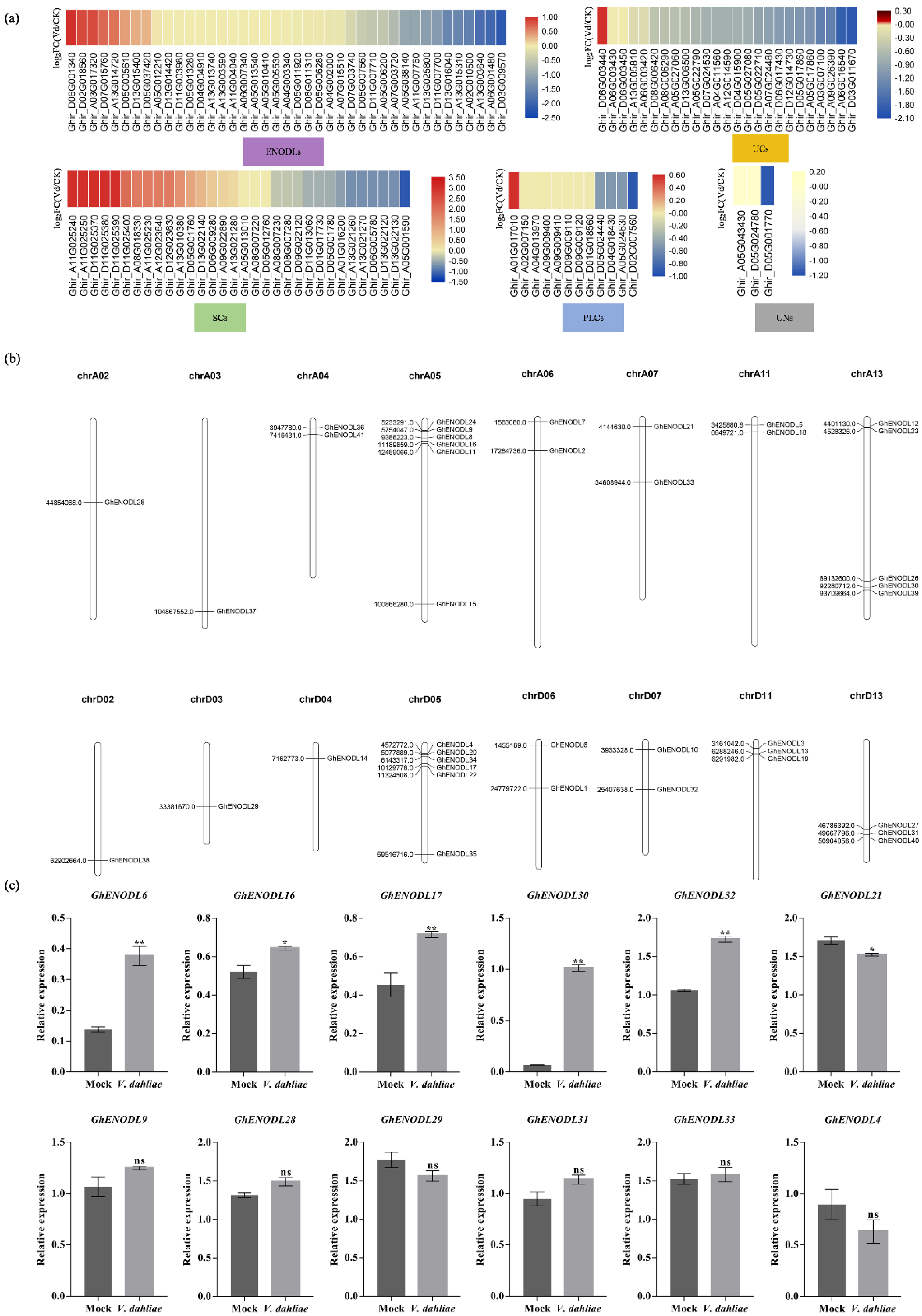
Chromosome location analysis revealed that 41 GhENODLs were unevenly distributed on 16 chromosomes (Figure 2b), of which 35 GhENODLs contained signal peptides, and 22 were found to be GPI-anchored proteins, suggesting that these proteins might localize in the plasma membrane. In order to investigate the response of GhPCs to V. dahliae infection, we analyzed the transcriptional change of GhPCs based on our previous RNA-seq data [32]. Interestingly, we found that GhPC genes in different subgroups displayed diversity expression patterns, and only most GhENODLs in subgroup d showed obvious expression change under V. dahliae stress (Figure 2a). In detail, as many as 25 out of 41 GhENODL was upregulated at 2 h post inoculation (hpi), indicating that these genes potentially early regulated VW resistance (Figure 2a). Among the differentially expressed GhENODLs, GhENODL6 displayed maximal fold-changes post inoculation based on both RNA-seq data and q-PCR results (Figure 2c); thus, GhENODL6 was further used for functional study.

2.3. GhENODL6 Is a Plasma Membrane Protein

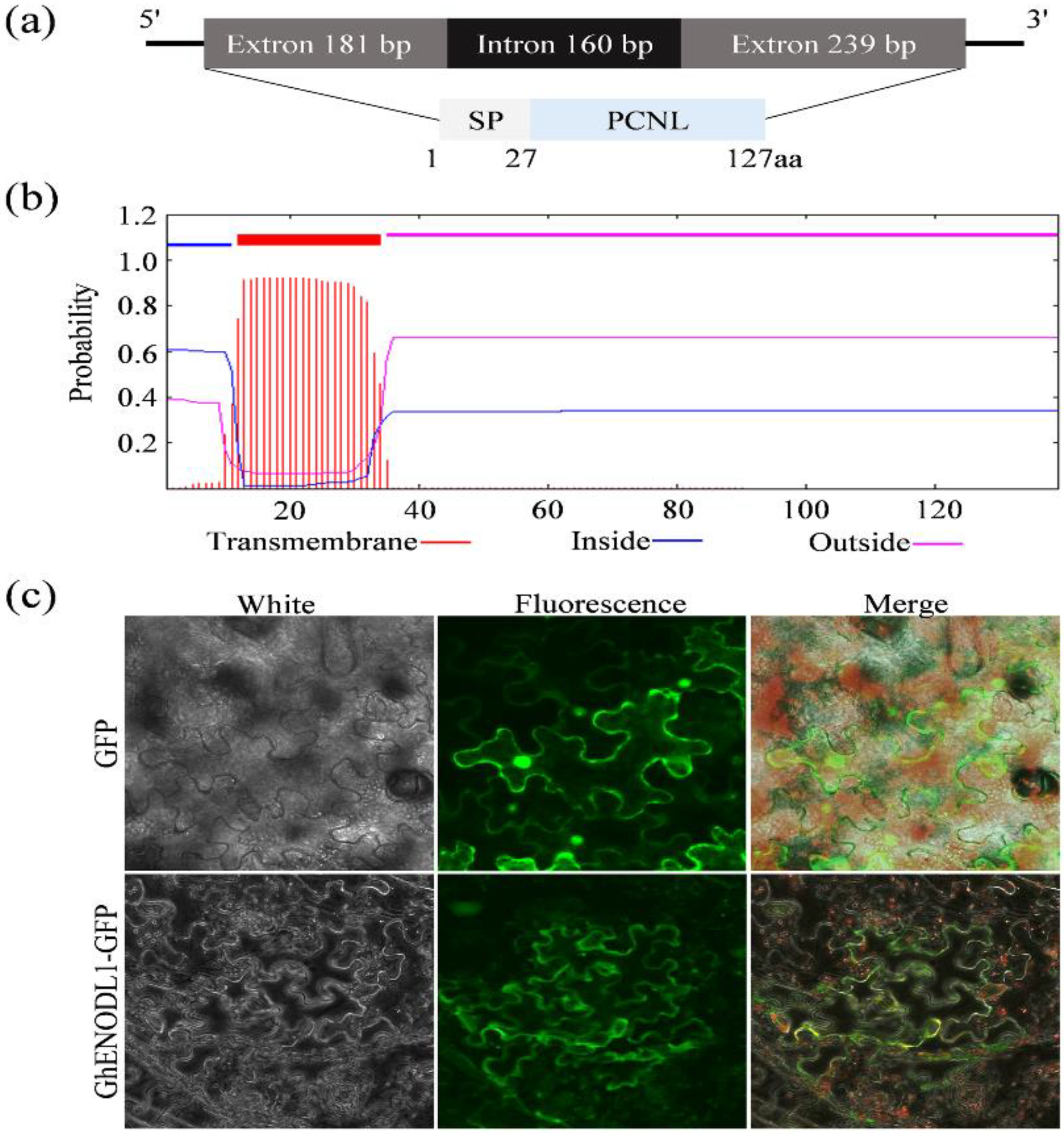
The GhENODL6 (GhM_D06G0164; Ghir_D06G001340) has an open reading frame (ORF) of 420 bp encoding a 139 amino acid, the molecular weight of GhENODL6 predicted by the online tools ProtParam (http://web.expasy.org/protpara 12 March 2021) is 15.71 kDa, and the theoretical PI is 9.97. GhENODL6 contained 27-residue signal peptide (SP), an 86-residue PCNL domain and a transmembrane helix (12-34aa) (Figure 3a,b). To confirm its subcellular localization, the fusion protein of GhENODL6 with GFP under the constitutive CaMV 35 s promoter was successfully expressed in the epidermal cells of tobacco and onions, respectively, and we vividly observed the fluorescence signal both in cytoplasmic and membrane (Figure 3c and Figure S1), indicating that GhENODL6 was a secretory protein. Though it belonged to the PC family and was similar to the ENODL, GhENODL6 lacked the copper-binding residues (Figure S2), suggesting that GhENODL6 was an atypical copper ions member.

2.4. GhENODL6 Expression Is Upregulated in Response to V. dahliae and Hormonal Signal in Cotton

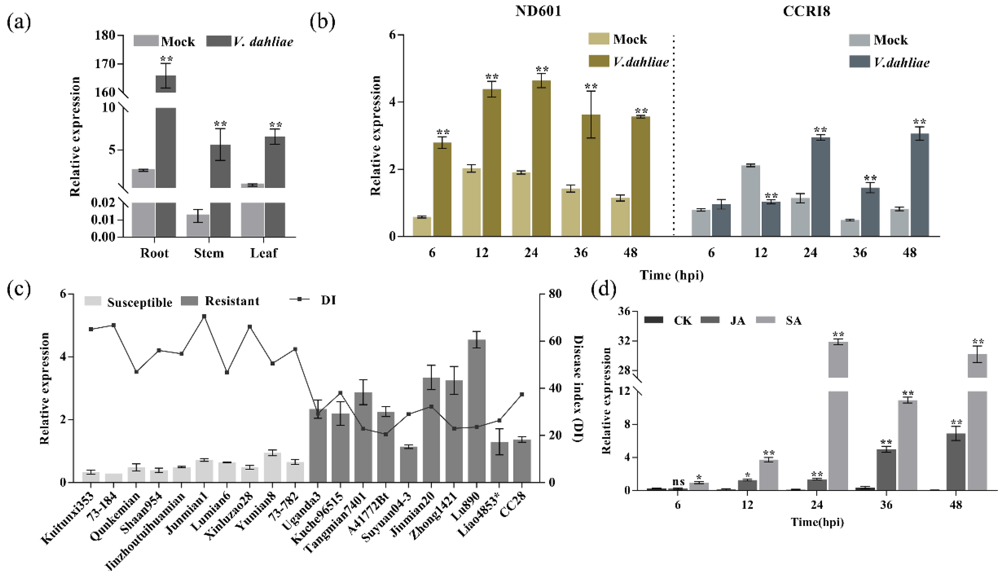
In order to investigate the GhENODL6 expression in different tissues and VW-resistant cottons, we detected GhENODL6 transcript levels and found that GhENODL6 predominantly expressed in the root (Figure 4a), and GhENODL6 could obviously induce upon V. dahliae, with higher expression level in resistant ND601 than in susceptible CCRI8 (Figure 4b). This trend was also evidenced in multiple cottons with different resistance [33], indicating that GhENODL6 should positively regulate VW resistance (Figure 4c). Based on the hormonal signal treatment experiment, we found that GhENODL6 dramatically upregulated expression upon foliar spray with JA and SA, especially under treatment by SA (Figure 4d), suggesting that GhENODL6 is involved in SA and/or JA signaling pathway.

2.5. GhENODL6 Positively Regulated Verticillium Wilt Resistance in Plants

To confirm the function of GhENODL6 in response to V. dahliae, we firstly overexpressed it in Arabidopsis and obtained T3 pure transgenic lines. Of the nine independent T3 transgenic lines, three stable overexpressing lines selected via RT-PCR and Western blot analysis were inoculated with V. dahliae (Figure 5a,b). Twenty days after inoculation by strong pathogenic strain Lx2-1 [34], typical symptoms of VW displayed evident in the infected wild-type (WT) plants but were much less pronounced in GhENODL6 transgenic lines (Figure 5c). The statistical analysis showed that the average DI of transgenic lines was 32.0, significantly lower than that of WT, with a 45.8% decrease (Figure 5d). Additionally, the fungal biomass in the transgenic plants was markedly lower than in the WT (Figure 5e). Based on the above evidence, we concluded that overexpression of GhENODL6 in Arabidopsis could improve the VW resistance of plants.
To further confirm the GhENODL6 function in cotton, we successfully knocked down endogenous GhENODL6 via virus-induced gene silencing (VIGS) that was successfully established in this research (Figure 6a). At 10 days post agroinfiltration, the transcript of GhENODL6 was significantly suppressed, decreasing 75% of expression level in silenced plants (Figure 6b). The silenced and mock cotton displayed a significant difference in disease symptoms at 20 days post inoculation (dpi) under Lx2-1 stress, as indicated in DI and disease-grade statistical results (Figure 6c–e). Moreover, the fungal biomass in silenced plants was significantly higher than that in mock plants (Figure 6f,g). The VIGS experiment suggested that suppressing GhENODL6 could make cotton disease reaction type from tolerant to susceptible. Combining the results of transgenic Arabidopsis and silenced cotton, we proved that GhENODL6 positively modulated plant VW resistance, resulting in disease resistance from susceptible to tolerant grade.

2.6. GhENODL6 Regulates Defense Genes Involving in SA Signaling

To identify the molecular basis of GhENODL6 in response to the pathogen, RNA sequencing (RNA-seq) was carried out to analyze the transcriptome profile of transgenic plant leaves in the absence of V. dahliae. Comparative transcriptome analysis between transgenic plants and WT, a total of 4235 differential expression genes (q < 0.05) were identified, including 2170 upregulated and 2065 down-regulated genes. Gene ontology (GO) analysis showed that a subset of genes involved in defense response to fungus, response to immune response, secondary metabolite biosynthetic process, response to oxidative stress, response to antibiotic, glucosinolate biosynthetic process, response to SA, response to ROS, and defense response by callose deposition were enriched (Figure 7a and Table S3). We further compared the differentially expressed genes (DEG) between transgenic plants and WT at 24 hpi when GhENODL peaked expressing under V. dahliae stress. Interestingly, the number of DEGs showed remarkable differences. In detail, 4235 differential genes were found in the GhENODL6-4 transgenic line, and these differential genes were mainly related to the response to a variety of abiotic factors, as well as carbohydrate catabolic process, defense response, incompatible interaction, response to SA and ROS (Table S4). In contrast, 783 differential genes were found in WT, in addition to the response to abiotic factors, genes mainly involved in response to extracellular stimulus defense response to fungus (Table S5). Compared with the WT, more resistance genes were activated in GhENODL transgenic plants, and KEGG revealed phenylpropanoid biosynthesis was enriched (Figure 7b). Given the fact that the phenylalanine ammonia lyase pathway is the main synthesis pathway of SA in cotton [35] and the results of qPCR in Figure 4e and the RNA-seq data, we deduced that GhENODL6 is probably involved in SA signaling. Thus, the SA pathway-regulated genes, EDS1, PR1, PR2, PR5, and PR10, were detected via qPCR (Figure 8a–e). Compared to the WT, the transcript levels of the above genes were significantly upregulated in transgenic lines, indicating that overexpression GhENODL6 could influence SA-mediated defense response. In contrast, the expression of those genes was observably hindered after suppressing GhENODL6 in silenced cotton (Figure 8g–k). The SA content also exhibited consistent trends, with increasing in the overexpression Arabidopsis and decreasing in the silenced cotton, respectively (Figure 8f,l). The expression levels of PAL, 4CL involved in the phenylalanine ammonia lyase pathway were also decreased in the silenced plant, and the activity of PAL was significantly lower than the control group (Figure 8m–r). At the same time, the silenced plants and WT were treated by 1 mM SA via exogenous spraying, and we found that the silenced plants displayed improving disease resistance, with significantly lower DI than the WT (Figure 8s,t), suggesting that exogenous SA compensated the resistance of GhENODL6-silenced plants. Taken together, the above results indicated that GhENODL6 defense response against V. dahliae was via activating the SA pathway.

2.7. GhENODL6 Contributed to the Production of ROS

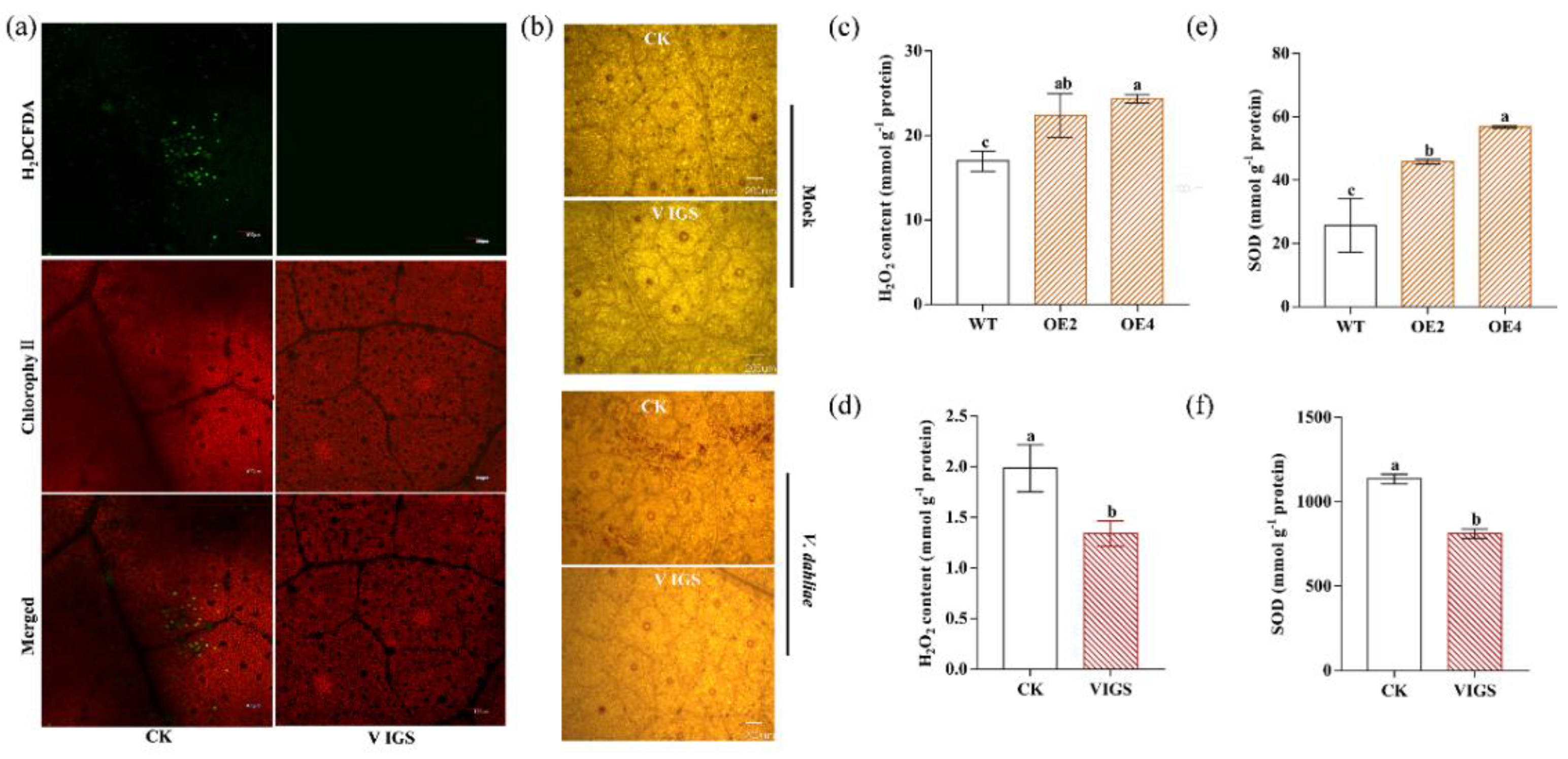
It was well known that there was an important relationship between the production of ROS and the defense response of plants against pathogens. In order to determine whether GhENODL6 is involved in the production of ROS, Arabidopsis and VIGS cotton plants were inoculated with V. dahliae and stained with 2,7′-dichlorodihydrofluorescein diacetate (H2DCFDA) and 3-3′-diaminobenzidine (DAB), and the results demonstrated that the ROS levels were remarkably higher in mock cotton leaves than in silenced cotton leaves (Figure 9a,b), indicating that GhENODL6 was involved in the production of ROS in the immune response of plants to V. dahliae. We also determined H2O2 and superoxide dismutase (SOD) both in transgenic Arabidopsis and silenced cotton under V. dahliae stress. We found that both H2O2 content and SOD activity in the transgenic lines were significantly higher than in WT, while the silenced cotton displayed decreased trends compared to the mock cotton plants (Figure 9c–f). These results suggested that GhENODL6 was related to ROS production during plant immune responses to V. dahliae.

3. Discussion

PCs are ancient blue copper-binding proteins in plants. They act as electron transporters and bind to a single type I copper atom. PCs play important roles in plant development and stress resistance and can be divided into four subfamilies: UCs, SCs, PLCs, and ENODLs, and all PCs have a PCNL domain [27]. The structure of ENODL is similar to that of the other three subfamilies, while ENODL lacks amino acid residues that bind to copper. The vast majority of ENODL are chimeric arabinogalactosins (AGPs) [28,36] and relate nodule development [37], pollen germination [38] and osmotic tolerance [39]. Until now, there is no report of ENODLs involved in disease resistance. This research was the first to link cotton ENODL with VW resistance, and this will broaden our knowledge toward ENODLs’ biologically specific function in crops.
In this study, the ENODLs sub-family was identified responding more to V. dahliae than the other three subgroups (SCs, UCs, and PLCs). There was rare information about the functions of ENODLs in plants. Here, we found that the GhENODL6 was significantly induced by V. dahliae from 2 hpi, suggesting its potential function on early regulating VW resistance. We also found that GhENODL6 expression peaked at 24 hpi, which guided us to sample the materials at 24 hpi by V. dahliae for comparing the DEGs between transgenic and WT plants, which should reflect the relatively big expression of the defense genes between transgenic and WT plants. GhENODL6 was proved to positively regulate VW resistance and displayed transcriptome differences between resistant and susceptible cottons. Functional studies firstly demonstrated that GhENODL6 could improve plant VW resistance via the SA signal pathway and ROS production, which could make the plant disease reaction susceptible to tolerance. These results indicated that GhENODL6 was a functionally novel gene in defense against VW resistance.
SA is a key defense-related hormone in plants, which plays an important role in SAR and broad-spectrum durable disease resistance [40,41]. Among the reported plant defense hormones, the roles of SA, JA, and ethylene (Eth) have been established, and these hormones play important roles in biological stress signaling pathways, including ROS and MAPK kinases [8]. In cotton, most SA biosynthesis relies on the phenylalanine ammonia lyase pathway [35]. In this study, according to qPCR results and RNA-seq data, we inferred that GhENODL6 could activate the phenylpropanoid pathway and promote SA synthesis. Both hypersensitive reaction (HR) and SAR were associated with increased expression of a wide range of defense genes, including those encoding various types of pathogen-related (PR) proteins. The induction of the PR gene is closely related to the occurrence of disease resistance, so the PR gene is a common molecular marker of defense response [34]. Meanwhile, as an important marker gene in response to SA, PR genes play an essential role in plant immune response [42]. In this study, our findings demonstrated that GhENODL6 responded quickly to SA signaling and maintained a high expression level, suggesting that GhENODL6 was related to the SA signaling pathway. The activation of SA signals in stressed plants can stimulate the expression of downstream disease resistance genes and provide protection [43]. There are many genes related to the SA signaling pathway, including NPR1, PR1, PR2, WRKYs, and GSTF6 [44]. To verify this, we tested the expression level of SA-related marker genes in overexpressing GhENODL6 transgenic Arabidopsis and silenced cotton. The expression of SA signaling pathway-related genes (EDS1, PR1, and PR2) and the increase in SA content confirmed our hypothesis. In order to further clarify the connection between GhENODL6 and SA signal, we treated silenced cotton and mock plants with exogenous SA and observed that exogenous SA significantly increased the resistance of silenced plants to V. dahliae. These findings demonstrated that GhENODL6 participated in the SA signaling pathway and functioned in VW resistance.
Plants infected by pathogenic bacteria and fungi would produce ROS, of which H2O2 played multiple roles in the interactions between the host and pathogens and was considered to be an antibacterial agent in the defense response of plants [45,46]. It also further participated in different signaling pathways related to defense mechanisms, such as triggering allergic reactions, accumulation of phytoantitoxins, and activation of many other defense response genes [47]. In order to overcome oxidative damage, plants have an antioxidant defense system composed of a variety of enzymes, such as SOD, which can remove, neutralize and scavenge active oxygen by converting superoxide anion radical (O2.-) into H2O2 and O2 [48]. In this study, the H2O2 content and endogenous SOD activity of GhENODL6 transgenic Arabidopsis and GhENODL6 silenced cotton after V. dahliae infection were determined. The H2O2 content and SOD activity of transgenic Arabidopsis were substantially higher than WT; as expected, silencing GhENODL6 reduced H2O2 accumulation and endogenous SOD enzyme activity. Thus, we deduced that the role of GhENODL6 in plant fungal resistance might also be due to the accumulation of H2O2 regulated by SOD activity.
This study is the first to discuss the relationship between ENODL and disease resistance. The function of GhENODL6 was demonstrated through gene transcription, gene overexpression, and gene silencing experiments, highlighting the GhENODL6 novel role in VW resistance. The molecular network of GhENODL6 is still unclear at present. Based on bioinformatics prediction and yeast two-hybrid assay, we preliminary obtained several candidates interactors, including 14-3-3 protein 6, ethylene-responsive transcription factor RAP2-7, and CBL-interacting serine/threonine-protein kinase 3, which inspired us to study the mechanism underlying GhENODL6 responding to disease resistance in the future.

4. Methods

4.1. Plant Growth and V. dahliae Culture

Seeds of G. hirsutum cv. CCRI8 and cv. ND601 (G100729) were delinted with H2SO4 (98%) and rinsed in water, then soaked in distilled water for 1 day and then germinated on wet gauzes for another day at 25 °C. Germinant seeds were transferred to pots containing vermiculite in a controlled environment (25 °C, 16 h/8 h (day/night), and 75% relative humidity). Arabidopsis were grown in growth chamber with a climate (23 °C, 16 h/8 h (day/night), and 60–70% relative humidity) [49]. The defoliating V. dahliae isolate (Lx2-1) was grown on potato dextrose agar medium for 7 days and then cultured in Czapek’s broth at 150 rpm, 25 °C for 1 week. When inoculating spores, adjust the concentration to 1 × 10⁷ conidia/mL with deionized water. For specific methods, refer to Yang et al. that pipetting conidia to the roots using injection method [50].

4.2. Identification of GhPCs and Bioinformatics Analysis

We first used the plastocyanin-like domain (PCNL) from Pfam (PF02298) (http://pfam.sanger.ac.uk/ 12 March 2021) to screen the protein databases in Arabidopsis and cotton. The sequences were then confirmed by the databases NCBI CD-Search. Subsequently. The N-terminal signal peptide (SP) of all proteins was examined using SignalP 3.0 [51]. Big-PI Plant Predictor was used to predict the glycosylphosphatidylinositol (GPI) modification site [52]. Moreover, we also used NetNGlyc 1.0 Server to predict the N-glycosylation sites [53]. Putative arabinogalactan (AG) glycomodules were predicted mainly following the previously described criteria [54].

4.3. Multiple Sequence Alignment of PCNLs and Phylogenetic Analysis

The Clustal W [55] program was used to perform multiple sequence alignments of all PCs protein sequences in Arabidopsis and cotton. The phylogenetic tree was constructed using the neighbor-joining (NJ) method of MEGA 7.0 software, and the bootstrap replications were set to 1000. The gene structures, chromosomal distribution, conserved motifs were analyzed referred to Liu et al. [56].

4.4. Expression Analysis of GhPC Genes

The transcriptome database of ND601 infected by V. dahliae was obtained from our laboratory [32]. A heatmap of PC gene expression clusters was constructed using TBtools software, and the expression values of PC genes were presented as fragments per kilobase of exon model per million mapped reads (FPKM). Finally, log2 (FPKM) values after averaging two replicates were displayed.

4.5. Gene Cloning and Bioinformatics Analysis

The root tissue of cotton seedlings was ground with liquid nitrogen, and total RNA and DNA were extracted using an EASYspi plant RNA rapid extraction kit (AidLab, Beijing, China) and EasyPure® Plant Genomic DNA Kit (TransGen, Beijing, China), respectively. cDNA was obtained by reverse transcription using Prime Script II 1st Strand cDNA Synthesis Kit (Takara, Dalian, Japan). We designed a pair of primers for PCR amplification based on the homologous sequence of upland cotton TM-1 in Cotton FGD (https://cottonfgd.org 12 March 2021). The primers are shown in Table S6. The sequence of GhENODL6 was analyzed by Blast in the NCBI database (http://www.ncbi.nlm.nih.gov/ 12 March 2021), and the functional domain of the protein encoded by the gene was analyzed at the same time. The protein signal peptide was predicted by online software SignalP 4.0 (http://www.cbs.dtu.dk/services/SignalP 12 March 2021). The protein transmembrane domain was analyzed by TMpred (http://www.ch.embnet.org/software/TMPRED_form.html 12 March 2021).

4.6. Real-Time PCR and Expression Analysis

Cotton seedlings were treated under multiple conditions. Cotton seedlings were cultivated in nutritious soil and were treated with hormone spraying at the 4 leaf stage. Ethephon and SA were diluted to 5 and 10 mM, respectively, and sprayed evenly on the leaves of cotton seedlings and moisturized with a transparent plastic cover. The leaves of cotton seedlings were taken at 6, 12, 24, 36, and 48 h, respectively, and stored at −80 °C. Subsequently, cotton seedlings were grown in nutrient medium and inoculated with the root dip method. The specific operation is as mentioned before [57]. After inoculation, the cotton seedlings were cultured under the conditions of 25 °C, 14 h light/10 h darkness, and 70% relative humidity. The root tissues of cotton seedlings were taken at 6, 12, 24, 36, and 48 h after inoculation and stored at −80 °C. The above tissues were sampled at each time point and repeated three times. Distilled water treatment was used as a control. Total RNA was extracted from roots, stems, and leaves for tissue expression analysis.
The real-time PCR reaction system (20 μL) consists of 1 μL cDNA template (<100 ng), 10 μL SYBR® Premix Ex TaqTM (TaKaRa), forward and backward primers 0.8 μL (10 μmol/L), and 7.4 μL sterilized double distilled water). The reaction was carried out in CFX96 real-time PCR detection system (Bio-Rad), and the program was set up as follows: 95 °C, 15 s, 95 °C, 10 s, 58 °C, 10 s, 72 °C 15 s, 40 cycles. Poly ubiquitin 14 (UBQ14) was used as the internal reference. Each reaction was repeated three times. The primers used are shown in Supplementary Table S1. The comparative CT (2−ΔΔCT) method was used for relative quantitative analysis. GraphPad Prism® 6 software was used for statistical analysis and drawing [58].

4.7. Subcellular Location

In order to detect the subcellular localization of GhENODL6, we used tobacco for transient expression. The target gene was cloned into pMDC43-GFP vector by gateway method [59] and verified by sequencing. It was transferred into Agrobacterium tumefaciens strain GV3101 by the freeze-thaw method [60]. The transient transformation of tobacco leaf epidermis cells was described by predecessors [61]. The leaves were observed with a confocal laser scanning microscope (Fluo View FV1000; Olympus, Tokyo, Japan) about 2–3 days later.

4.8. Generation and Analysis of Transgenic Arabidopsis

The coding sequence of GhENODL6 was amplified using PCR from the cDNA of upland cotton ND601 and cloned into a pGWB414 vector stored in our laboratory. The recombinant plasmid was transformed into Arabidopsis thaliana plants by the floral dip method, and transgenic plants were selected on MS medium containing 50 mg/mL kanamycin [62,63]. Gene-specific primers were used to isolate homozygous plants and confirm transcription status and normal protein expression. T3 transgenic pure lines were obtained for subsequent experiments.
The resistance of Arabidopsis thaliana transformed by GhENODL6 to V. dahliae was determined by the root dip method [64]. The 20-day-old Arabidopsis thaliana was uprooted, soaked in a suspension containing 1 × 10⁷ conidia per milliliter for 5 min, and then replanted in the soil. The disease index (DI) is graded according to a previous report [65]. The resistance mechanism of GhENODL6 in transgenic Arabidopsis thaliana was analyzed by the determination of physiological indicators after inoculation. In addition, qRT-PCR was used to compare the expression of the internal defense-related genes of GhENODL6 transgenic Arabidopsis with WT, with the AtEF-1α gene as internal standard [40,66].

4.9. VIGS Experiment in Cotton

Design primers GhENODL6-V-F and GhENODL6-V-R (Table S6), containing restriction sites EcoRI and KpnI for GhENODL6 ORF sequence by PCR amplification and then ligated into VIGS vector pTRV2 [67]. The recombinant plasmid was transformed into Agrobacterium GV3101 competent cells by the electric shock method [68]. In the cotton seedling stage, about 7–10 days (cotyledons fully unfolded, true leaves did not grow), with chloroplasts alterados1 gene (CLA1) as a marker gene, injected with Agrobacterium tumefaciens culture. The cotton VIGS was carried out according to the method previously described [69]. About 15 days after the VIGS procedure, detecting the silencing efficiency and then the plants were inoculated with V. dahliae. The experiments were carried out using at least 30 plants per treatment and repeated three times. The DI was calculated as described previously [64].

4.10. ROS Dyeing

ROS staining adopts a reactive oxygen detection kit (aladdin). Dilute DCFH-DA 1:1000 with PBS buffer to make the final concentration 10 uM. DCFH-DA completely immersed the cotton leaves and reacted at 28 °C for 20 min [70]. Before observation, the leaves were washed with double distilled water at least three times to remove the reagents on the surface and then observed the staining signal with a laser confocal microscope [71]. In order to observe the accumulation of H2O2, using the plant hydrogen peroxide DAB staining kit (servicebio), fresh cotton leaves were taken, incubated in 1 mg/mL, pH 3.8 DAB solution for 8 h, and then decolorized in 95% ethanol [72].

4.11. Determination of H2O2 and SOD Content

V. dahliae was inoculated into 4-week-old Arabidopsis thaliana plants by the method of Zhang et al. [24]. Then the leaves of the transgenic plant were taken at 6, 12, 24, 36, and 48 h, respectively. The activity of H2O2 and SOD was determined according to the kit’s instructions (Jiancheng Biotech Inc., Nanjing, China). The protein content was determined using the BCA protein detection kit (Sangon Biotech, Shanghai, China).

4.12. Determination of SA Content

In order to determine the concentration of endogenous SA in Arabidopsis thaliana and cotton, 100 mg leaf samples were extracted with methanol. The contents of SA were determined by ESI-HPLC-MS/MS and the standard curve method [34].

4.13. Transcriptome Sequencing

GhENODL6 transgenic Arabidopsis plants and WT were grown in vermiculite for four weeks; the whole plants were collected treatment with Lx2-1 or water. Total RNA was isolated, and the libraries for sequencing were constructed. Sequencing was performed on the Illumina HiSeq2000 platform. GO analysis of the differentially expressed genes in the biological process was conducted using the AgriGO software [73]. GO terms with a corrected p-value less than 0.05 were considered significantly enriched by differentially expressed genes. GO is divided into three independent ontologies, which are biological process (BP), molecular function (MF), and cellular component (CC). The gene ontology database we used was Intepro, which was made up of CATH-Gene3D, CDD, MobiDB, HAMAP, PANTHER, PIRSF, ProDom, PROSITE, SFLD, SMART, SUPERFAMILY, TIGRFAMS, and Pfam. The Intepro was a common database for all the plants. The background was constituted by the whole annotated gene sequence of Arabidopsis, and the output of enrichment needed padj <0.05. All samples contained three biological repeats. Other relevant analyses were referred to previous results [17].

4.14. Primers for Gene Cloning, Vector Construction and Expression Analysis

All primers used in this study are listed in Table S6. The primers used in this study were designed via Primer Premier 5 software and primer blast analysis.

4.15. Statistical Analysis

All experiments were performed at least three times for each determination. Statistical analysis was performed using Graph Pad Prism® 6 software (Graph Pad, San Diego, CA, USA). Data were evaluated by analysis of variance (ANOVA method) followed by Dunn’s multiple comparisons test. The p-value ≤ 0.05 is considered to be statistically significant.

Supplementary Materials

The following supporting information can be downloaded at: https://www.mdpi.com/article/10.3390/ijms23062913/s1.

Author Contributions

Z.M. designed the research; M.Z. wrote the manuscript; Z.M. and Y.Z. revised the manuscript; Y.Z., X.W., Z.S. and D.Z. analyzed the data. M.Z., J.Y., Z.W., B.C. and X.Z. performed the experiments. J.W., H.K., L.W. and G.Z. prepared experiment materials. All authors have read and agreed to the published version of the manuscript.

Funding

This work was supported by the Key Research and Development Program of Hebei province (21326314D) to Zhiying Ma and The National Youth Talent Support Program to Yan Zhang.

Institutional Review Board Statement

Not applicable.

Informed Consent Statement

Not applicable.

Data Availability Statement

The data presented in this study are available within the article and Supplementary Materials.

Conflicts of Interest

The authors declare no conflict of interest.

References

  1. Mo, H.; Wang, X.; Zhang, Y.; Zhang, G.; Zhang, J.; Ma, Z. Cotton polyamine oxidase is required for spermine and camalexin signalling in the defence response to Verticillium dahliae. Plant J. 2015, 83, 962–975. [Google Scholar] [CrossRef] [PubMed] [Green Version]
  2. Shaban, M.; Miao, Y.; Ullah, A.; Khan, A.Q.; Menghwar, H.; Khan, A.H.; Ahmed, M.M.; Tabassum, M.A.; Zhu, L. Physiological and molecular mechanism of defense in cotton against Verticillium dahliae. Plant Physiol. Biochem. 2018, 125, 193–204. [Google Scholar] [CrossRef] [PubMed]
  3. Zhang, J.; Sanogo, S.; Flynn, R.; Baral, J.B.; Bajaj, S.; Hughs, S.E.; Percy, R.G. Germplasm evaluation and transfer of Verticillium wilt resistance from Pima (Gossypium barbadense) to Upland cotton (G. hirsutum). Euphytica 2011, 187, 147–160. [Google Scholar] [CrossRef]
  4. Tong, S.; Yuan, M.; Liu, Y.; Li, X.; Jin, D.; Cheng, X.; Lin, D.; Ling, H.; Yang, D.; Wang, Y. Ergosterol-targeting fusion antifungal peptide significantly increases the Verticillium wilt resistance of cotton. Plant Biotechnol. J. 2020, 19, 926–936. [Google Scholar] [CrossRef]
  5. Ma, Z.; Zhang, Y.; Wu, L.; Zhang, G.; Sun, Z.; Li, Z.; Jiang, Y.; Ke, H.; Chen, B.; Liu, Z. High-quality genome assembly and resequencing of modern cotton cultivars provide resources for crop improvement. Nat. Genet. 2021, 53, 1385–1391. [Google Scholar] [CrossRef]
  6. Wang, G.; Zhang, G.; Wu, L.; Wang, X.; Zhang, Y.; Wu, J.; Ma, Z. New cotton variety Nong da 601 with disease resistance: Insect resisitance and high yield. Seed 2015, 34, 3. [Google Scholar]
  7. Andersen, E.J.; Ali, S.; Byamukama, E.; Yen, Y.; Nepal, M.P. Disease resistance mechanisms in plants. Genes 2018, 9, 339. [Google Scholar] [CrossRef] [Green Version]
  8. Bari, R.; Jones, J.D. Role of plant hormones in plant defence responses. Plant Mol. Biol. 2009, 69, 473–488. [Google Scholar] [CrossRef]
  9. Vlot, A.C.; Klessig, D.F.; Park, S.W. Systemic acquired resistance: The elusive signal(s). Curr. Opin. Plant Biol. 2008, 11, 436–442. [Google Scholar] [CrossRef] [Green Version]
  10. Cheng, H.Q.; Han, L.B.; Yang, C.L.; Wu, X.M.; Zhong, N.Q.; Wu, J.H.; Wang, F.X.; Wang, H.Y.; Xia, G.X. The cotton MYB108 forms a positive feedback regulation loop with CML11 and participates in the defense response against Verticillium dahliae infection. J. Exp. Bot. 2016, 67, 1935–1950. [Google Scholar] [CrossRef] [Green Version]
  11. Kouzai, Y.; Kimura, M.; Watanabe, M.; Kusunoki, K.; Osaka, D.; Suzuki, T.; Matsui, H.; Yamamoto, M.; Ichinose, Y.; Toyoda, K. Salicylic acid-dependent immunity contributes to resistance against Rhizoctonia solani, a necrotrophic fungal agent of sheath blight, in rice and Brachypodium distachyon. New Phytol. 2018, 217, 771–783. [Google Scholar] [CrossRef] [PubMed] [Green Version]
  12. Vinay, K.; Tushar, K.; Mansi, S.; Shabir, H.W. ROS-induced signaling and gene expression in crops under salinity stress. In Reactive Oxygen Species and Antioxidant Systems in Plants. Role and Regulation under Abiotic Stress; Springer: Singapore, 2017; pp. 159–184. [Google Scholar]
  13. Camejo, D.; Guzman-Cedeno, A.; Moreno, A. Reactive oxygen species, essential molecules, during plant-pathogen interactions. Plant Physiol. Biochem. 2016, 103, 10–23. [Google Scholar] [CrossRef] [PubMed]
  14. Fukuda, H. Programmed cell death of tracheary elements as a paradigm in plants. Plant Mol. Biol. Rep. 2000, 44, 245–253. [Google Scholar] [CrossRef] [PubMed]
  15. Han, L.B.; Li, Y.B.; Wang, F.X.; Wang, W.Y.; Liu, J.; Wu, J.H.; Zhong, N.Q.; Wu, S.J.; Jiao, G.L.; Wang, H.Y. The cotton apoplastic protein CRR1 stabilizes chitinase 28 to facilitate defense against the fungal pathogen Verticillium dahliae. Plant Cell 2019, 31, 520–536. [Google Scholar] [CrossRef]
  16. Zhang, Y.; Wu, L.; Wang, X.; Chen, B.; Zhao, J.; Cui, J.; Li, Z.; Yang, J.; Wu, L.; Wu, J. The cotton laccase gene GhLAC15 enhances Verticillium wilt resistance via an increase in defence-induced lignification and lignin components in the cell walls of plants. Mol. Plant Pathol. 2019, 20, 309–322. [Google Scholar] [CrossRef] [Green Version]
  17. Wang, G.; Xu, J.; Li, L.; Guo, Z.; Si, Q.; Zhu, G.; Wang, X.; Guo, W. GbCYP86A1-1 from Gossypium barbadense positively regulates defence against Verticillium dahliae by cell wall modification and activation of immune pathways. Plant Biotechnol. J. 2020, 18, 222–238. [Google Scholar] [CrossRef] [Green Version]
  18. Zhang, J.; Yu, X.; Zhang, C.; Zhang, Q.; Sun, Y.; Zhu, H.; Tang, C. Pectin lyase enhances cotton resistance to Verticillium wilt by inducing cell apoptosis of Verticillium dahliae. J. Hazard. Mater. 2021, 404, 124029. [Google Scholar] [CrossRef]
  19. Jun, Z.; Zhang, Z.; Gao, Y.; Zhou, L.; Fang, L.; Chen, X.; Ning, Z.; Chen, T.; Guo, W.; Zhang, T. Overexpression of GbRLK, a putative receptor-like kinase gene, improved cotton tolerance to Verticillium wilt. Sci. Rep. 2015, 5, 15048. [Google Scholar] [CrossRef] [Green Version]
  20. Gilroy, S.; Suzuki, N.; Miller, G. A tidal wave of signals: Calcium and ROS at the forefront of rapid systemic signaling. Trends Plant Sci. 2014, 19, 623–630. [Google Scholar] [CrossRef]
  21. Levine, A.; Tenhaken, R.; Dixon, R.; Lamb, C. H2O2 from the oxidative burst orchestrates the plant hypersensitive diseases resistance response. Cell 1994, 79, 583. [Google Scholar] [CrossRef]
  22. Yalpani, N.; Silverman, P.; Wilson, T. Salicylic acid is a systemic signal and an inducer of pathogenesis-related proteins in virus-infected tobacco. Plant Cell 1991, 3, 809–818. [Google Scholar] [PubMed] [Green Version]
  23. Leon, J. Hydrogen peroxide stimulates salicylic acid biosynthesis in tobacco. Plant Physiol. 1995, 108, 1673–1678. [Google Scholar] [CrossRef] [PubMed] [Green Version]
  24. Zhang, Y.; Wang, X.F.; Ding, Z.G.; Ma, Q.; Zhang, G.R.; Zhang, S.L.; Li, Z.K.; Wu, L.Q.; Zhang, G.Y.; Ma, Z.Y. Transcriptome profiling of Gossypium barbadense inoculated with Verticillium dahliae provides a resource for cotton improvement. BMC Genom. 2013, 14, 637. [Google Scholar] [CrossRef] [PubMed] [Green Version]
  25. Li, J.; Gao, G.; Zhang, T.; Wu, X. The putative phytocyanin genes in Chinese cabbage (Brassica rapa L.) genome-wide identification, classification and expression analysis. Mol. Genet. Genom. 2013, 288, 1–20. [Google Scholar] [CrossRef]
  26. Yoshizaki, M.; Furumoto, T.; Hata, S.; Shinozaki, M.; Izui, K. Characterization of a novel gene encoding a phytocyanin-related protein in morning glory (Pharbitis nil). Biochem. Biophys. Res. Commun. 2000, 268, 466–470. [Google Scholar] [CrossRef]
  27. Cao, J.; Li, X.; Lv, Y.; Ding, L. Comparative analysis of the phytocyanin gene family in 10 plant species. a focus on Zea mays. Front. Plant Sci. 2015, 6, 515. [Google Scholar] [CrossRef] [Green Version]
  28. Xu, L.; Wang, X.J.; Wang, T.; Li, L.B. Genome-wide identification.; classification.; and expression analysis of the phytocyanin gene family in Phalaenopsis equestris. Biol. Plant. 2017, 61, 445–452. [Google Scholar] [CrossRef]
  29. Ma, H.; Zhao, H.; Liu, Z.; Zhao, J. The phytocyanin gene family in rice (Oryza sativa L.): Genome-wide identification.; classification and transcriptional analysis. PLoS ONE 2011, 6, e25184. [Google Scholar] [CrossRef]
  30. Xu, Q.; Dunbrack, R.L., Jr. Assignment of protein sequences to existing domain and family classification systems: Pfam and the PDB. Bioinformatics 2012, 28, 2763–2772. [Google Scholar] [CrossRef]
  31. Mashiguchi, K.; Asami, T.; Suzuki, Y. Genome-wide identification.; structure and expression studies, and mutant collection of 22 early nodulin-like protein genes in Arabidopsis. Biosci. Biotech. Biochem. 2009, 3, 2452–2459. [Google Scholar] [CrossRef] [Green Version]
  32. Wang, G.; Wang, X.; Zhang, Y.; Yang, J.; Li, Z.; Wu, L.; Wu, J.; Wu, N.; Liu, L.; Liu, Z.; et al. Dynamic characteristics and functional analysis provide new insights into long non-coding RNA responsive to Verticillium dahliae infection in Gossypium hirsutum. BMC Plant Biol. 2021, 21, 68. [Google Scholar] [CrossRef] [PubMed]
  33. Ma, Z.; He, S.; Wang, X.; Sun, J.; Zhang, Y.; Zhang, G.; Wu, L.; Li, Z.; Liu, Z.; Sun, G. Resequencing a core collection of upland cotton identifies genomic variation and loci influencing fiber quality and yield. Nat. Genet. 2018, 50, 803–813. [Google Scholar] [CrossRef] [PubMed]
  34. Chen, B.; Zhang, Y.; Yang, J.; Zhang, M.; Ma, Z. The G-protein α subunit GhGPA positively regulates Gossypium hirsutum resistance to Verticillium dahliae via induction of SA and JA signaling pathways and ROS accumulation. Crop J. 2020, 9, 11. [Google Scholar] [CrossRef]
  35. Mo, S.; Zhang, Y.; Wang, X.; Yang, J.; Sun, Z.; Zhang, D.; Chen, B.; Wang, G.; Ke, H.; Liu, Z. Cotton GhSSI2 isoforms from the stearoyl acyl carrier protein fatty acid desaturase family regulate Verticillium wilt resistance. Mol. Plant Pathol. 2021, 22, 1041–1056. [Google Scholar] [CrossRef]
  36. Khan, J.A.; Wang, Q.; Sjolund, R.D.; Schulz, A.; Thompson, G.A. An early nodulin-like protein accumulates in the sieve element plasma membrane of Arabidopsis. Plant Physiol. 2007, 143, 1576–1589. [Google Scholar] [CrossRef] [Green Version]
  37. Serova, T.A.; Tsyganova, A.V.; Tsyganov, V.E. Early nodule senescence is activated in symbiotic mutants of pea (Pisum sativum L.) forming ineffective nodules blocked at different nodule developmental stages. Protoplasma 2018, 255, 1443–1459. [Google Scholar] [CrossRef]
  38. Hou, Y.; Guo, X.; Cyprys, P.; Zhang, Y.; Bleckmann, A.; Cai, L.; Huang, Q.; Luo, Y.; Gu, H.; Dresselhaus, T. Maternal ENODLs are required for pollen tube reception in Arabidopsis. Curr. Biol. 2016, 26, 2343–2350. [Google Scholar] [CrossRef] [Green Version]
  39. Wu, H.; Shen, Y.; Hu, Y.; Tan, S.; Lin, Z. A phytocyanin-related early nodulin-like gene, BcBCP1, cloned from Boea crassifolia enhances osmotic tolerance in transgenic tobacco. Plant Physiol. 2011, 168, 935–943. [Google Scholar] [CrossRef]
  40. Fu, Z.Q.; Yan, S.; Saleh, A.; Wang, W.; Ruble, J.; Oka, N.; Mohan, R.; Spoel, S.H.; Tada, Y.; Zheng, N. NPR3 and NPR4 are receptors for the immune signal salicylic acid in plants. Nature 2012, 486, 228–232. [Google Scholar] [CrossRef] [Green Version]
  41. Cui, H.; Gobbato, E.; Kracher, B.; Qiu, J.; Bautor, J.; Parker, J.E. A core function of EDS1 with PAD4 is to protect the salicylic acid defense sector in Arabidopsis immunity. New Phytol. 2016, 213, 1802–1817. [Google Scholar] [CrossRef] [Green Version]
  42. Weigel, R.R.; Pfitzner, U.M.; Gatz, C. Interaction of NIMIN1 with NPR1 modulates PR gene expression in Arabidopsis. Plant Cell 2005, 17, 1279–1291. [Google Scholar] [CrossRef] [Green Version]
  43. Rietz, S.; Stamm, A.; Malonek, S. Different roles of enhanced disease susceptibility1 (EDS1) bound to and dissociated from phytoalexin deficient4 (PAD4) in Arabidopsis immunity. New Phytol. 2011, 191, 107–119. [Google Scholar] [CrossRef]
  44. Ding, Y.; Sun, T.; Ao, K.; Peng, Y.; Zhang, Y.; Li, X.; Zhang, Y. Opposite roles of salicylic acid receptors NPR1 and NPR3/NPR4 in transcriptional regulation of plant immunity. Cell 2018, 173, 1454–1467. [Google Scholar] [CrossRef] [PubMed]
  45. Sachdev, S.; Ansari, S.A.; Ansari, M.I.; Fujita, M.; Hasanuzzaman, M. Abiotic stress and reactive oxygen species. generation, signaling, and defense mechanisms. Antioxidants 2021, 10, 277. [Google Scholar] [CrossRef] [PubMed]
  46. Ali, M.; Cheng, Z.; Ahmad, H.; Hayat, S. Reactive oxygen species (ROS) as defenses against a broad range of plant fungal infections and case study on ROS employed by crops against Verticillium dahliae wilts. J. Plant Interact. 2018, 13, 353–363. [Google Scholar] [CrossRef] [Green Version]
  47. Wirthmueller, L.; Zhang, Y.; Jones, J.D.; Parker, J.E. Nuclear accumulation of the Arabidopsis immune receptor RPS4 is necessary for triggering EDS1-dependent defense. Curr. Biol. 2007, 17, 2023–2029. [Google Scholar] [CrossRef] [PubMed] [Green Version]
  48. Liu, H.; Li, Y.; Hu, Y.; Yang, Y.; Zhang, W.; He, M.; Li, X.; Zhang, C.; Kong, F.; Liu, X. EDS1-interacting J protein 1 is an essential negative regulator of plant innate immunity in Arabidopsis. Plant Cell 2021, 33, 153–171. [Google Scholar] [CrossRef]
  49. Yang, J.; Ke, H.F.; Zhang, Y.; Ji, L.L.; Huang, L.Z.; Zhang, C.Y.; Wang, X.F.; Ma, Z.Y. Genome-wide identification of cyclophilin genes in Gossypium hirsutum and functional characterization of a CYP with antifungal activity against Verticillium dahliae. BMC Plant Biol. 2019, 19, 272. [Google Scholar] [CrossRef]
  50. Yang, J.; Ji, L.; Wang, X.; Zhang, Y.; Wu, L.; Yang, Y.; Ma, Z. Overexpression of 3-deoxy-7-phosphoheptulonate synthase gene from Gossypium hirsutum enhances Arabidopsis resistance to Verticillium wilt. Plant Cell Rep. 2015, 34, 1429–1441. [Google Scholar] [CrossRef]
  51. Bendtsen, J.D.; Nielsen, H.; Hei, G.; Brunak, S.S. Improved prediction of signal peptides: SignalP 3.0. J. Mol. Cell Biol. 2004, 340, 783–795. [Google Scholar]
  52. Eisenhaber, B.; Wildpaner, M.; Schultz, C.J.; Borner, G.; Eisenhaber, D.F. Glycosylphosphatidylinositol lipid anchoring of plant proteins: Sensitive prediction from sequence-and genome-wide studies for Arabidopsis and rice. Plant Physiol. 2003, 133, 1691–1701. [Google Scholar] [CrossRef] [PubMed] [Green Version]
  53. Gupta, R.; Brunak, S. Prediction of glycosylation across the human proteome and the correlation to protein function. Pac. Symp. Biocomput. 2002, 7, 310–322. [Google Scholar]
  54. Schultz, C.J.; Rumsewicz, M.P.; Johnson, K.L.; Jones, B.J.; Gaspar, Y.M.; Bacic, A. Using genomic resources to guide research directions: The arabinogalactan protein gene family as a test case. Plant Physiol. 2002, 129, 1448–1463. [Google Scholar] [CrossRef] [PubMed] [Green Version]
  55. Larkin, M. Clustal W and Clustal X v. 2.0. Bioinformatics 2007, 23, 2947–2948. [Google Scholar] [CrossRef] [Green Version]
  56. Liu, Z.; Wang, X.; Sun, Z.; Zhang, Y.; Meng, C.; Chen, B.; Wang, G.; Ke, H.; Wu, J.; Yan, Y. Evolution, expression and functional analysis of cultivated allotetraploid cotton DIR genes. BMC Plant Biol. 2021, 21, 89. [Google Scholar] [CrossRef] [PubMed]
  57. Yang, J.; Ma, Q.; Zhang, Y.; Wang, X.; Zhang, G.; Ma, Z. Molecular cloning and functional analysis of GbRvd, a gene in Gossypium barbadense that plays an important role in conferring resistance to Verticillium wilt. Gene 2016, 575, 687–694. [Google Scholar] [CrossRef]
  58. Yang, Y.; Ling, X.; Chen, T.; Cai, L.; Liu, T.; Wang, J.; Fan, X.; Ren, Y.; Yuan, H.; Zhu, W. A cotton Gbvdr5 gene encoding a leucine-rich-repeat receptor-like protein confers resistance to Verticillium dahliae in transgenic Arabidopsis and upland cotton. Plant Mol. Biol. Rep. 2014, 33, 987–1001. [Google Scholar] [CrossRef]
  59. Smedley, M.A.; Harwood, W.A. Gateway(R)-compatible plant transformation vectors. Methods Mol. Biol. 2015, 1, 3–16. [Google Scholar]
  60. Sree, V.S.; Manoharan, M.; Ranjit, K.T.; Savtthri, H.S.; Lakshmi, S.G. Agrobacterium-mediated transformation of tomato (Lycopersicon esculentum var. Pusa Ruby) with coat-protein gene of Physalis Mottle Tymovirus. J. Plant Physiol. 2000, 156, 106–110. [Google Scholar] [CrossRef]
  61. Sparkes, I.A.; Runions, J.; Kearns, A.; Hawes, C. Rapid, transient expression of fluorescent fusion proteins in tobacco plants and generation of stably transformed plants. Nat. Protoc. 2006, 1, 2019–2025. [Google Scholar] [CrossRef]
  62. Bent, S.J.; Ca, A.F. Floral dip a simplified method for Agrobacterium-mediated transformation of Arabidopsis thaliana. Plant J. 1998, 16, 735–743. [Google Scholar]
  63. Zhang, X.; Henriques, R.; Lin, S.S.; Niu, Q.W.; Chua, N.H. Agrobacterium-mediated transformation of Arabidopsis thaliana using the floral dip method. Nat. Protoc. 2006, 1, 641–646. [Google Scholar] [CrossRef] [PubMed]
  64. Li, C.; He, X.; Luo, X.; Xu, L.; Liu, L.; Min, L.; Jin, L.; Zhu, L.; Zhang, X. Cotton WRKY1 mediates the plant defense-to-development transition during infection of cotton by Verticillium dahliae by activating JASMONATE ZIM-DOMAIN1 expression. Plant Physiol. 2014, 166, 2179–2194. [Google Scholar] [CrossRef] [PubMed] [Green Version]
  65. Zhang, B.; Yang, Y.; Chen, T.; Yu, W.; Liu, T.; Li, H.; Fan, X.; Ren, Y.; Shen, D.; Liu, L. Island cotton Gbve1 gene encoding a receptor-like protein confers resistance to both defoliating and non-defoliating isolates of Verticillium dahliae. PLoS ONE 2012, 7, e51091. [Google Scholar] [CrossRef] [PubMed] [Green Version]
  66. Ghorbel, M.; Zribi, I.; Missaoui, K.; Drira-Fakhfekh, M.; Azzouzi, B.; Brini, F. Differential regulation of the durum wheat Pathogenesis-related protein (PR1) by Calmodulin TdCaM1.3 protein. Mol. Biol. Rep. 2021, 48, 347–362. [Google Scholar] [CrossRef]
  67. Reyes, M.I.; Flores-Vergara, M.A.; Guerra-Peraza, O.; Rajabu, C.; Desai, J.; Hiromoto-Ruiz, Y.H.; Ndunguru, J.; Hanley-Bowdoin, L.; Kjemtrup, S.; Ascencio-Ibanez, J.T. A VIGS screen identifies immunity in the Arabidopsis Pla-1 accession to viruses in two different genera of the Geminiviridae. Plant J. 2017, 92, 796–807. [Google Scholar] [CrossRef] [Green Version]
  68. Ren, F.; Zhang, Z.; Fan, X. Construction of a plant expression vector of the Grapevine virus B coat protein gene and transformation into Nicotiana tabacum. Plant Protect. 2018, 44, 11–16. [Google Scholar]
  69. Gao, X.; Wheeler, T.; Li, Z.; Kenerley, C.M.; He, P.; Shan, L. Silencing GhNDR1 and GhMKK2 compromises cotton resistance to Verticillium wilt. Plant J. 2011, 66, 293–305. [Google Scholar] [CrossRef] [Green Version]
  70. Athira, S.S.; Biby, E.T.; Mohanan, P.V. Dextran stabilized fullerene soot induced toxicity on alveolar epithelial cells (A549 cells). Environ. Res. 2020, 188, 109716. [Google Scholar] [CrossRef]
  71. Chai, Q.; Shang, X.; Wu, S.; Zhu, G.; Cheng, C.; Cai, C.; Wang, X.; Guo, W. 5-Aminolevulinic acid dehydratase gene dosage affects programmed cell death and immunity. Plant Physiol. 2017, 175, 511–528. [Google Scholar] [CrossRef] [Green Version]
  72. Li, X.; Liu, N.; Sun, Y.; Wang, P.; Hou, Y. The cotton GhWIN2 gene activates the cuticle biosynthesis pathway and influences the salicylic and jasmonic acid biosynthesis pathways. BMC Plant Biol. 2019, 19, 379. [Google Scholar] [CrossRef] [PubMed]
  73. Du, Z.; Zhou, X.; Ling, Y.; Zhang, Z.; Su, Z. agriGO: A GO analysis toolkit for the agricultural community. Nucleic Acids Res. 2010, 38, W64–W70. [Google Scholar] [CrossRef] [PubMed] [Green Version]
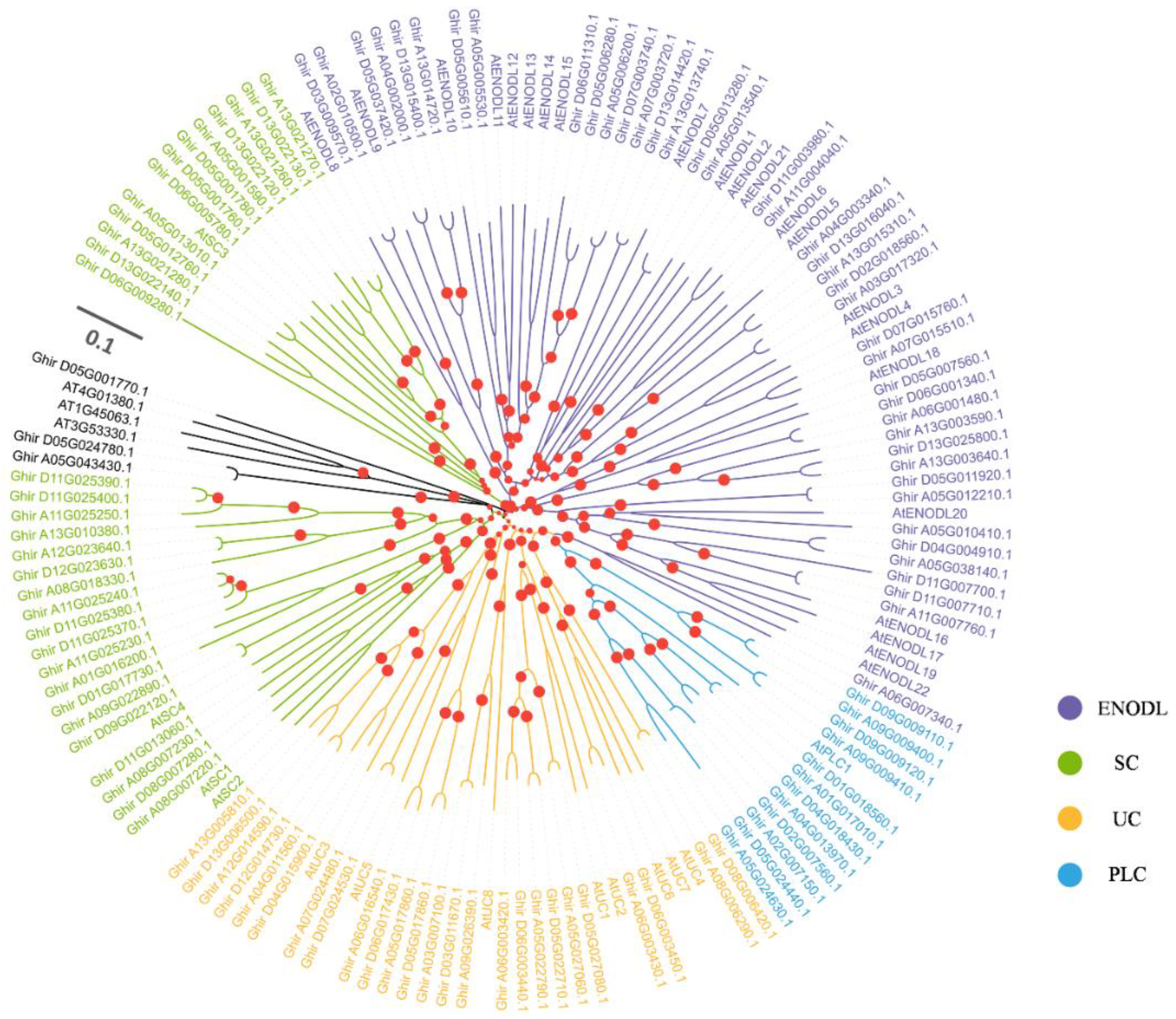
Figure 1. Phylogenetic analysis of PCs from two different plants. The phylogenetic tree was generated from the alignment result of the full-length amino acid sequences by the neighbor-joining (NJ) method. All PCs members, together with homologs of A. thaliana, were classified into four distinct clades shown in different colors.
Ijms 23 02913 g001
Figure 2. Expression analysis and chromosomal localization of ENODLs. (a) Expression patterns of ENODL genes in response to V. dahliae (using VDlog2 (FPKM)/CKlog2 (FPKM) to draw the graph) red: positive regulation, green: negative regulation. (b) Chromosomal distribution of GhENODLs. (c) Verification of the expression of GhENODL genes under the stress of V. dahlia (*, p < 0.05 **, p < 0.01). ns, not significant.
Ijms 23 02913 g002
Figure 3. Characterization of GhENODL6. (a) Schematic structure of GhENODL6. (b) Prediction of the transmembrane helix in GhENODL6 protein. (c) Subcellular localization of GFP alone or GhENODL6-GFP fusion in tobacco leaves transiently transformed by Agrobacterium infiltration. The green fluorescence was monitored using a confocal laser scanning microscope.
Ijms 23 02913 g003
Figure 4. Expression analysis of GhENODL6. (a) Expression analysis of GhENODL6 in different organs of cotton plant under Verticillium wilt stress. (b) Expression analysis of GhENODL6 in ND601 and CCRI8 roots under VW stress. (c) Expression of GhENODL6 in ten resistant and ten susceptible upland cotton cultivars upon infection with V. dahliae. (d) Expression analysis of GhENODL6 with the treatment of water, JA, and SA, respectively. All experiments were repeated at least three times. Values are means with standard deviation (SD) (n = 3 biological replicates). Error bars represent the SD of three biological replicates. Asterisks indicate statistically significant differences according to Student’s t-test (two-tailed) (*, p < 0.05 **, p < 0.01). All experiments were repeated at least three times. Vd, V. dahliae; CK, control; ns, not significant.
Ijms 23 02913 g004
Figure 5. Overexpression of GhENODL6 in Arabidopsis improved resistance to V. dahliae. (a) Relative expression levels of GhENODL6 in different Arabidopsis strains. (b) Western blot result of GhENODL6 in transgenic lines and WT. The membranes were stained with Coomassie brilliant blue (CBB) as loading control. (c) Disease manifestations and diseased plants of wild-type and two homozygous transgenic plants (OE2, OE4) at 25 dpi. (d) Disease index of different strains at 25 dpi. (e) Relative expression levels of plant fungus biomass at 10 dpi. Error bars represent the SD of three biological replicates. Asterisks indicate statistically significant differences by Student’s t-test (two-tailed) (**, p < 0.01). All experiments were repeated at least three times.
Ijms 23 02913 g005
Figure 6. Silence detection of GhENODL6. (a) The CLA1 gene was used as a positive control with an albino phenotype on leave after VIGS in cotton. (b) Relative expression levels of GhENODL6 in TRV.00 and TRV.GhENODL6 plants. (ce) The VW and statistics of the seedling number with different disease-grade symptoms of silenced and TRV.00 plants at 25 dpi and disease index of seedlings at 25 dpi. (f) V. dahliae isolation from the infected stems. (g) Plant fungal biomass and relative expression levels. Error bars indicate the SE of three biological replicates. Asterisks indicate statistically significant differences as determined by Dunnett’s multiple comparisons test (**, p < 0.01).
Ijms 23 02913 g006
Figure 7. RNA-seq reveals that GhENODL6 overexpression affects immunity-related pathways of Arabidopsis. (a) GO enrichment analysis of the differentially expressed genes in plants of GhENODL6-4 transgenic line and the WT control. Padj. Corrected p-value after multiple hypothesis testing. GeneRatio: ratio of the number of differential genes annotated to the GO number to the total number of differential genes. (b) KEGG enrichment analysis of the difference in plants of GhENODL6-4 transgenic line and the WT control after challenged with V. dahliae for 24 h. KEGG terms were separately shown by enrichment analysis. p-value of 0.05 adjusted by multiple hypothesis testing.
Ijms 23 02913 g007
Figure 8. Detection of SA pathway genes. (ae) qRT-PCR analysis of the expression of SA pathway genes in wild-type, transgenic Arabidopsis line. (gk) qRT-PCR analysis of the expression of SA pathways in controls and silenced cotton plants. (f,l) Measurement of SA content in transgenic Arabidopsis and GhENODL6-silenced cotton. The leaves of Arabidopsis and cotton inoculated by V. dahliae for 2 dpi were used for SA. (mq) qRT-PCR analysis of the expression of PAL pathways in controls and silenced cotton plants. (r) PAL active identification. (s) Disease symptoms induced with V. dahliae on the GhENODL6-silenced plant and assessment of disease Index (DI). Photos were taken at 25 dpi. (t) V. dahliae isolation from the infected stems and plant fungal biomass and relative expression levels. Values are means with SD (n = 3 biological replicates). Error bars represent the SD of three biological replicates (p < 0.05). All experiments were repeated at least three times. Lowercase letters represent statistically significant differences (p < 0.05) according to Tukey’s HSD test. All experiments were repeated at least three times (**, p < 0.01).
Ijms 23 02913 g008
Figure 9. Determination of H2O2 content and activities at 2 dpi. (a) Detection of ROS levels in cotton leaves by H2DCFDA staining. Then 10 mM H2DCFDA ROS—specific fluorescent indicator was used for the staining. Bars = 100 um. (b) H2O2 visualization in cotton leaves by staining with DAB, showing more H2O2 was produced in the TRV.00 than the silent plants. Bars = 200 um. (cf) Measurements of the contents of H2O2 and SOD in cotton and Arabidopsis leaves at 2 dpi. Values are means with SD (n = 3 biological replicates). Error bars represent the SD of three biological replicates. Lowercase letters represent statistically significant differences (p < 0.05) according to Tukey’s HSD test.
Ijms 23 02913 g009
Publisher’s Note: MDPI stays neutral with regard to jurisdictional claims in published maps and institutional affiliations.