


Integrated Metabolomics and Lipidomics Reveal High Accumulation of Glycerophospholipids in Human Astrocytes under the Lipotoxic Effect of Palmitic Acid and Tibolone Protection

 , ,
, , 
Abstract
1. Introduction
2. Results
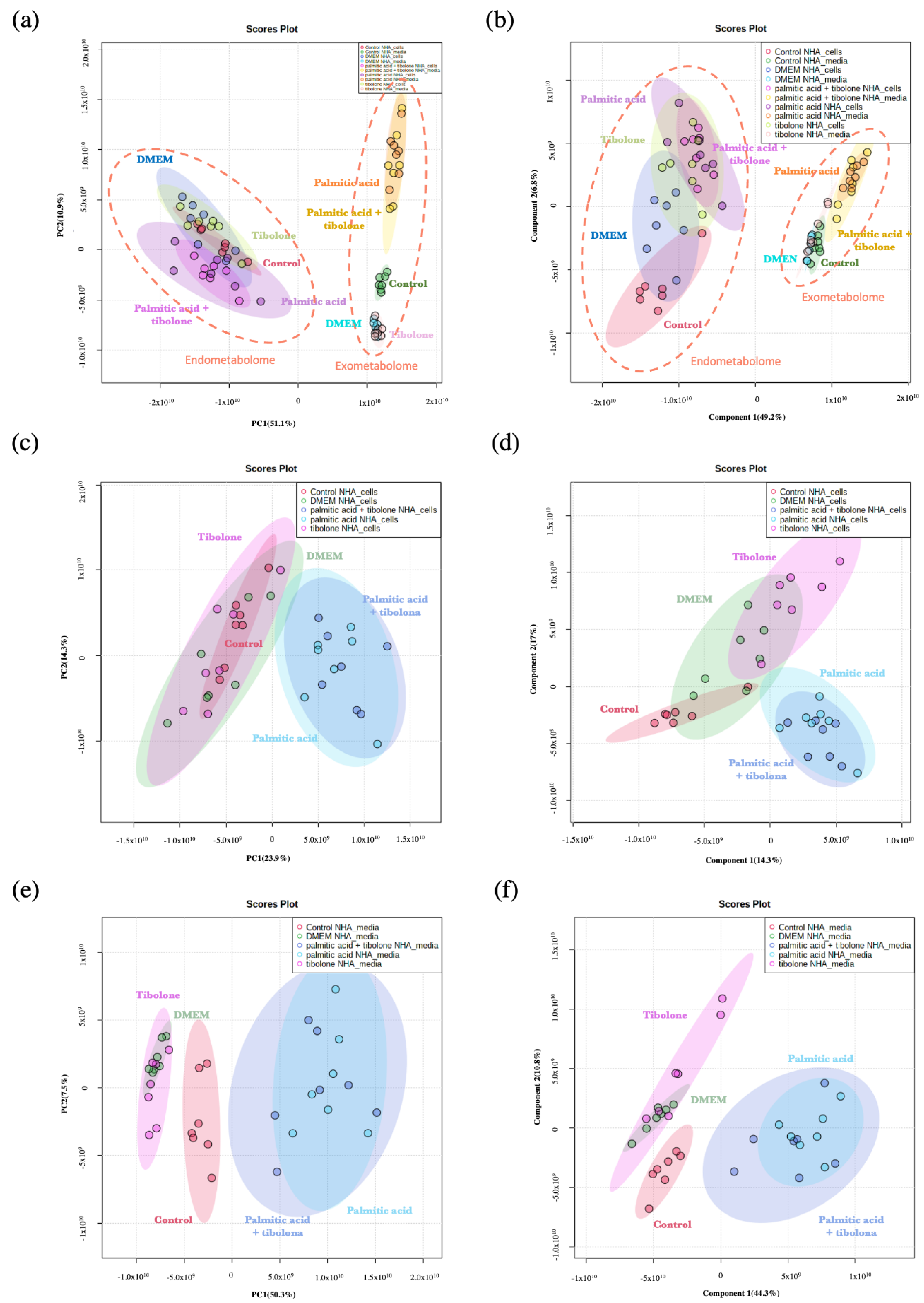
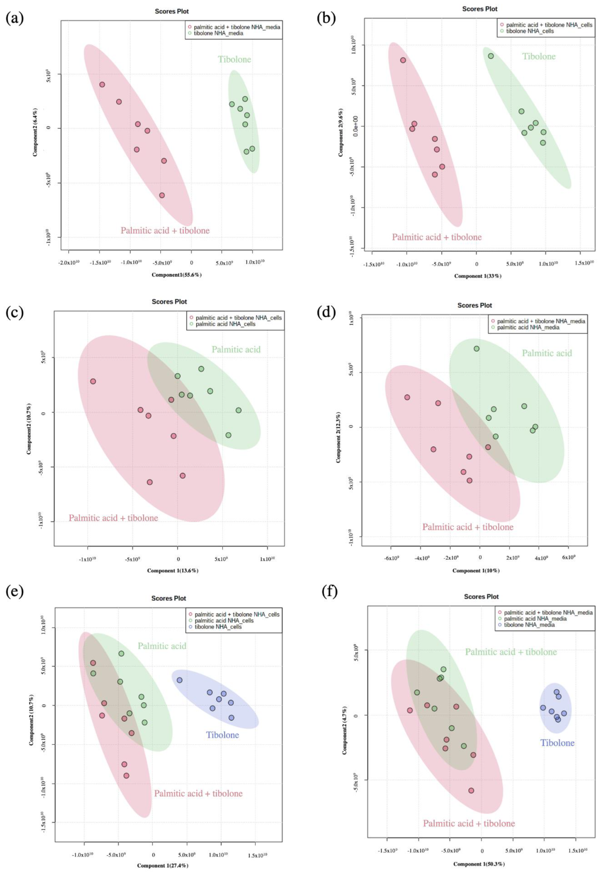
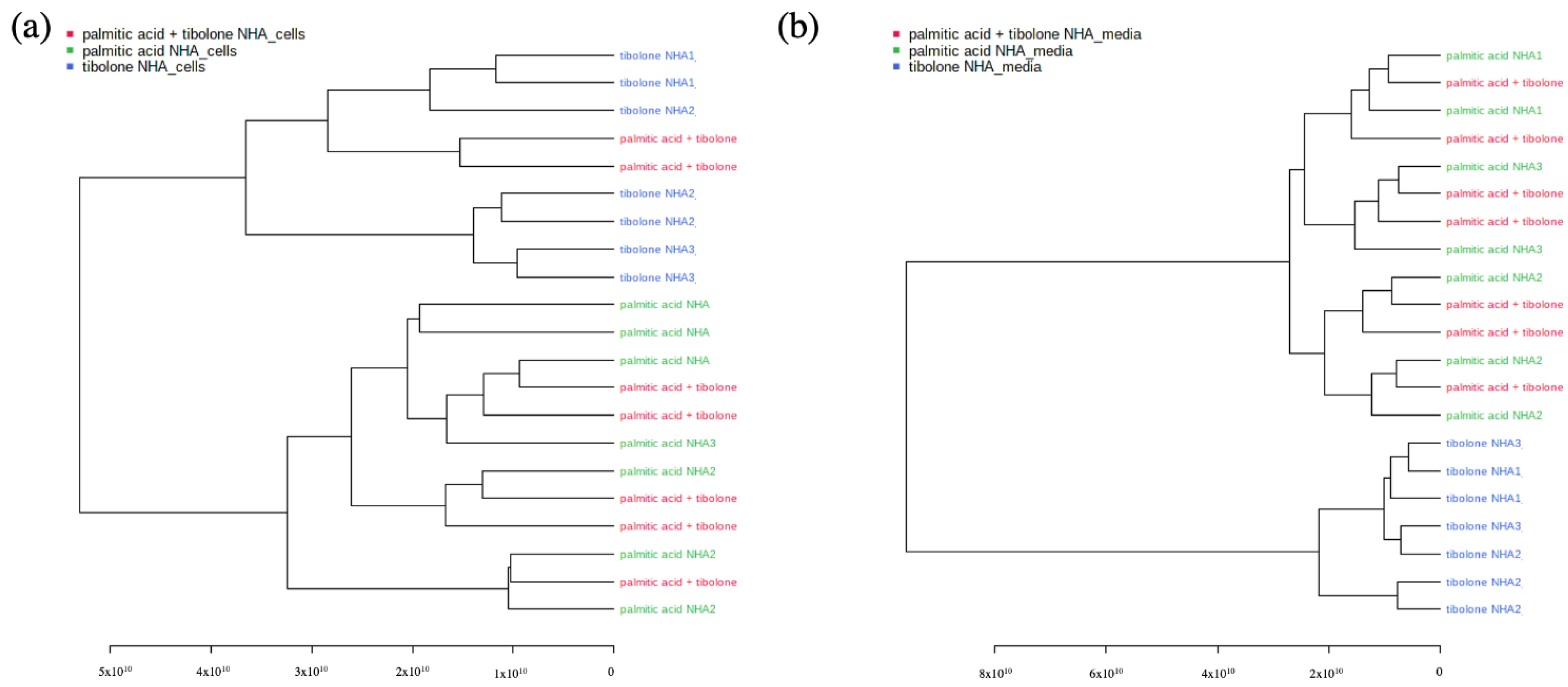
2.1. Applied Treatments Show Different Organization Patterns in PCA and PLS-DA Analysis
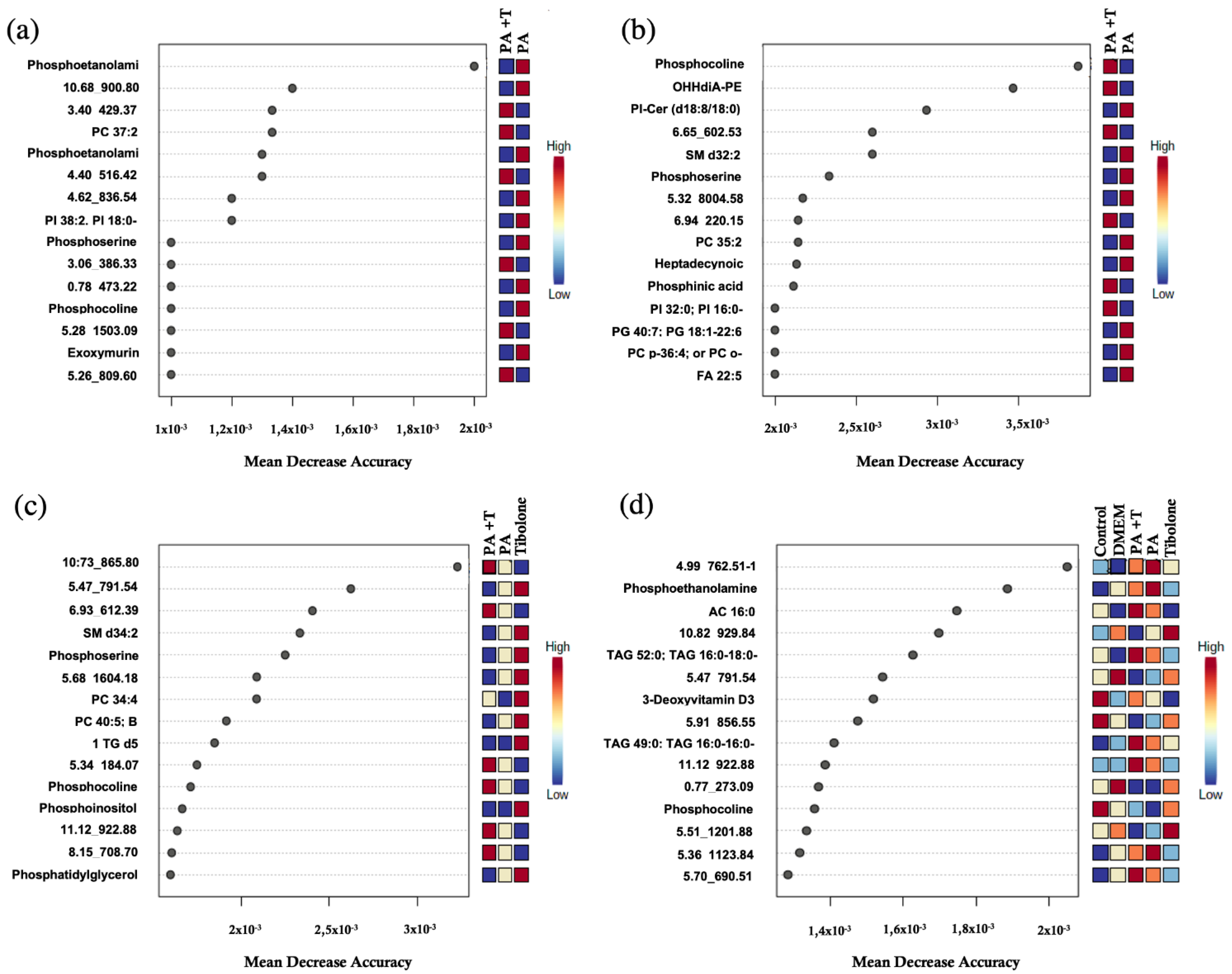
2.2. Implementation of Machine Learning Techniques Enhances the Identification of Metabolites Related to Biological Processes
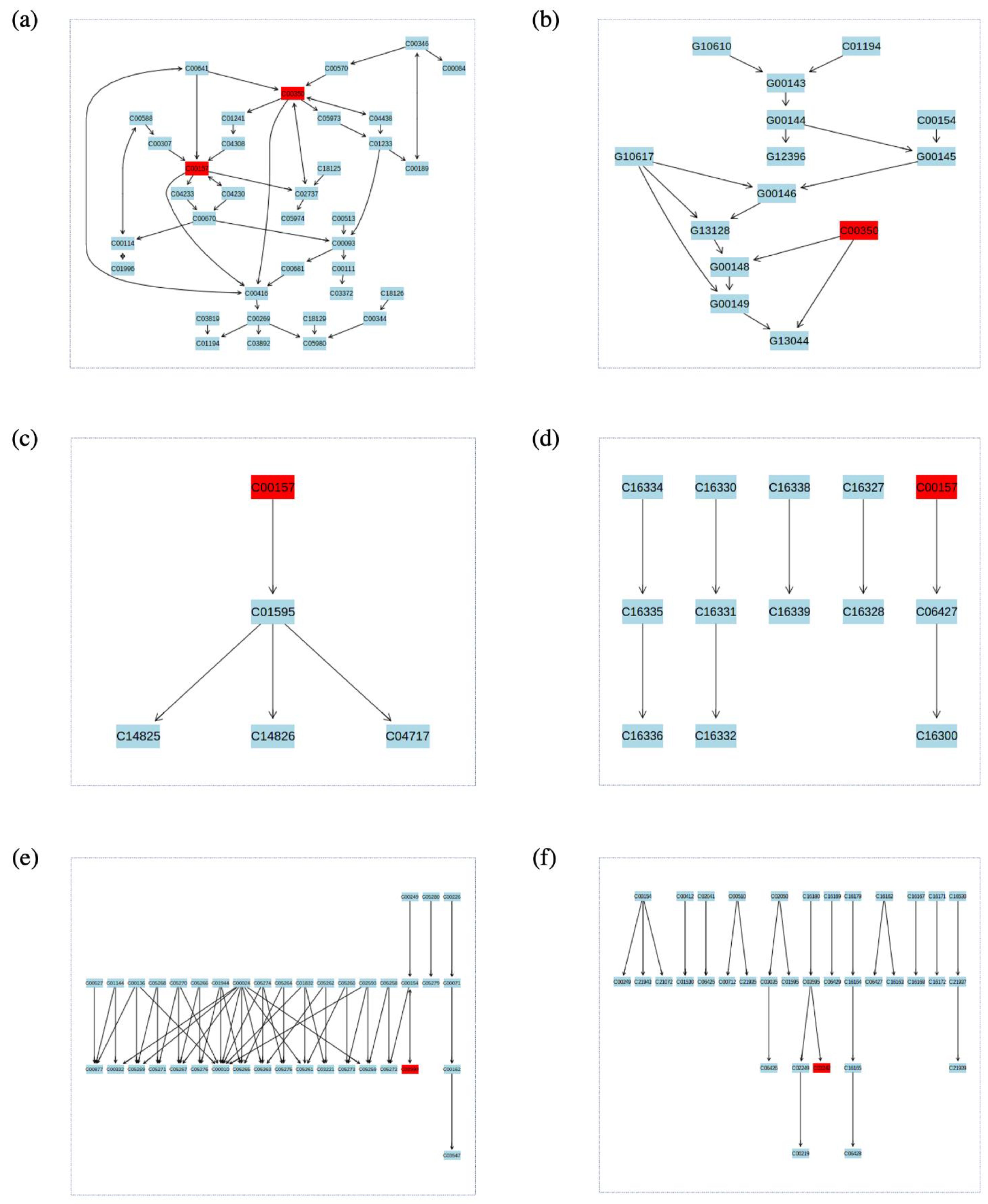
2.3. Identified Metabolic Pathways
3. Discussion
4. Materials and Methods
4.1. Cell Cultures
4.2. Palmitic Acid Treatment
4.3. Tibolone Pre-Treatment
4.4. Metabolite Extraction
4.5. Derivatization of Samples/Standards
4.6. Lipidomics Instrumental Analysis
4.7. Data Processing
4.8. Data Analysis
4.9. Machine Learning Approach
4.10. Enrichment Analysis
5. Conclusions
Supplementary Materials
Author Contributions
Funding
Institutional Review Board Statement
Informed Consent Statement
Acknowledgments
Conflicts of Interest
References
- Unger, R.; Orci, L. Lipotoxic diseases of nonadipose tissues in obesity. Int. J. Obes. 2000, 24, S28–S32. [Google Scholar] [CrossRef] [Scilit] [PubMed]
- Savary, S.; Trompier, D.; Andreoletti, P.; le Borgne, F.; Demarquoy, J.; Lizard, G. Fatty acids-induced lipotoxicity and inflammation. Curr. Drug Metab. 2012, 13, 1358–1370. [Google Scholar] [CrossRef] [Scilit] [PubMed]
- Ford, J. Saturated fatty acid metabolism is key link between cell division, cancer, and senescence in cellular and whole organism aging. Age 2010, 32, 231–237. [Google Scholar] [CrossRef] [Scilit] [PubMed]
- Liu, L.; Martin, R.; Chan, C. Palmitate-activated astrocytes via serine palmitoyltransferase increase BACE1 in primary neurons by sphingomyelinases. Neurobiol. Aging 2013, 34, 540–550. [Google Scholar] [CrossRef] [Scilit]
- Almaguel, F.G.; Liu, J.W.; Pacheco, F.J.; Casiano, C.A.; de Leon, M. Activation and reversal of lipotoxicity in PC12 and rat cortical cells following exposure to palmitic acid. J. Neurosci. Res. 2009, 87, 1207–1218. [Google Scholar] [CrossRef] [Scilit]
- Ortiz-Rodriguez, A.; Acaz-Fonseca, E.; Boya, P.; Arevalo, M.A.; Garcia-Segura, L.M. Lipotoxic effects of palmitic acid on astrocytes are associated with autophagy impairment. Mol. Neurobiol. 2019, 56, 1665–1680. [Google Scholar] [CrossRef] [Scilit]
- Hidalgo-Lanussa, O.; Ávila-Rodriguez, M.; Baez-Jurado, E.; Zamudio, J.; Echeverria, V.; Garcia-Segura, L.M.; Barreto, G.E. Tibolone reduces oxidative damage and inflammation in microglia stimulated with palmitic acid through mechanisms involving estrogen receptor beta. Mol. Neurobiol. 2018, 55, 5462–5477. [Google Scholar] [CrossRef] [Scilit]
- Alvarez, J.I.; Katayama, T.; Prat, A. Glial influence on the blood brain barrier. Glia 2013, 61, 1939–1958. [Google Scholar] [CrossRef] [Scilit]
- Cabezas, R.; Ávila, M.; Gonzalez, J.; El-Bachá, R.S.; Báez, E.; García-Segura, L.M.; Coronel, J.C.J.; Capani, F.; Cardona-Gomez, G.P.; Barreto, G.E. Astrocytic modulation of blood brain barrier: Perspectives on Parkinson’s disease. Front. Cell. Neurosci. 2014, 8, 211. [Google Scholar] [CrossRef] [Scilit]
- De Keyser, J.; Mostert, J.P.; Koch, M.W. Dysfunctional astrocytes as key players in the pathogenesis of central nervous system disorders. J. Neurol. Sci. 2008, 267, 3–16. [Google Scholar] [CrossRef] [Scilit]
- Min, K.J.; Yang, M.S.; Kim, S.U.; Jou, I.; Joe, E.H. Astrocytes induce hemeoxygenase-1 expression in microglia: A feasible mechanism for preventing excessive brain inflammation. J. Neurosci. Off. J. Soc. Neurosci. 2006, 26, 1880–1887. [Google Scholar] [CrossRef] [Scilit] [PubMed]
- Maciejczyk, M.; Żebrowska, E.; Zalewska, A.; Chabowski, A. Redox balance, antioxidant defense, and oxidative damage in the hypothalamus and cerebral cortex of rats with high fat diet-induced insulin resistance. Oxidative Med. Cell. Longev. 2018, 2018, 6940515. [Google Scholar] [CrossRef] [Scilit] [PubMed]
- Lebon, V.; Petersen, K.F.; Cline, G.W.; Shen, J.; Mason, G.F.; Dufour, S.; Behar, K.L.; Shulman, G.I.; Rothman, D.L. Astroglial contribution to brain energy metabolism in humans revealed by 13C nuclear magnetic resonance spectroscopy: Elucidation of the dominant pathway for neurotransmitter glutamate repletion and measurement of astrocytic oxidative metabolism. J. Neurosci. 2002, 22, 1523. [Google Scholar] [CrossRef] [Scilit]
- Allaman, I.; Bélanger, M.; Magistretti, P.J. Astrocyte-neuron metabolic relationships: For better and for worse. Trends Neurosci. 2011, 34, 76–87. [Google Scholar] [CrossRef] [Scilit] [PubMed]
- Lee, J.A.; Hall, B.; Allsop, J.; Alqarni, R.; Allen, S.P. Lipid metabolism in astrocytic structure and function. Semin. Cell Dev. Biol. 2021, 112, 123–136. [Google Scholar] [CrossRef] [Scilit] [PubMed]
- Bhaduri, A.; Neumann, E.K.; Kriegstein, A.R.; Sweedler, J.V. Identification of lipid heterogeneity and diversity in developing human brain. JACS Au. 2021, 1, 2261–2270. [Google Scholar] [CrossRef] [Scilit] [PubMed]
- Turovsky, E.A.; Varlamova, E.G.; Gudkov, S.V.; Plotnikov, E.Y. The protective mechanism of deuterated linoleic acid involves the activation of the Ca2+ signaling system of astrocytes in ischemia in vitro. Int. J. Mol. 2021, 22, 13216. [Google Scholar] [CrossRef] [Scilit] [PubMed]
- Gupta, S.; Knight, A.G.; Gupta, S.; Keller, J.N.; Bruce-Keller, A.J. Saturated long chain fatty acids activate inflammatory signaling in astrocytes. J. Neurochem. 2012, 120, 1060. [Google Scholar] [CrossRef] [Scilit]
- Sofroniew, M.V.; Vinters, H.V. Astrocytes: Biology and pathology. Acta Neuropathol. 2010, 119, 7. [Google Scholar] [CrossRef] [Scilit] [PubMed]
- Sofroniew, M.V. Astrogliosis. Cold Spring Harb. Perspect. Biol. 2015, 7, a020402. [Google Scholar] [CrossRef] [Scilit]
- Medeiros, A.R.S.D.; Lamas, A.Z.; Caliman, I.F.; Dalpiaz, P.L.; Firmes, L.B.; Abreu, G.R.D.; Moysés, M.R.; Lemos, E.M.; Reis, A.M.D.; Bissoli, N. Tibolone has anti-inflammatory effects in estrogen-deficient female rats on the natriuretic peptide system and TNF-alpha. Regul. Pept. 2012, 179, 55–60. [Google Scholar] [CrossRef] [Scilit] [PubMed]
- Rodriguez, M.Á.; Garcia-Segura, L.M.; Cabezas, R.; Torrente, D.; Capani, F.; Gonzalez, J.; Barreto, G.E. Tibolone protects T98G cells from glucose deprivation. J. Steroid Biochem. Mol. Biol. 2014, 144, 294–303. [Google Scholar] [CrossRef] [Scilit] [PubMed]
- Pinto-Almazán, R.; Segura-Uribe, J.; Farfán-García, E.D.; Guerra-Araiza, C. Effects of tibolone on the central nervous system: Clinical and experimental approaches. BioMed Res. Int. 2017, 2017, 8630764. [Google Scholar] [CrossRef] [Scilit]
- Martin-Jimenez, C.; Gonzalez, J.; Vesga, D.; Aristizabal, A.; Barrero, G. Tibolone ameliorates the lipotoxic effect of palmitic acid in normal human astrocytes. Neurotox. Res. 2020, 38, 585–595. [Google Scholar] [CrossRef] [Scilit] [PubMed]
- Kind, T.; Liu, K.H.; Lee, D.Y.; DeFelice, B.; Meissen, J.K.; Fiehn, O. LipidBlast in silico tandem mass spectrometry database for lipid identification. Nat. Methods 2013, 10, 755–758. [Google Scholar] [CrossRef] [Scilit]
- Tsugawa, H.; Cajka, T.; Kind, T.; Ma, Y.; Higgins, B.; Ikeda, K.; Kanazawa, M.; VanderGheynst, J.; Fiehn, O.; Arita, M. MS-DIAL: Data-independent MS/MS deconvolution for comprehensive metabolome analysis. Nat. Methods 2015, 12, 523–526. [Google Scholar] [CrossRef] [Scilit]
- Ruiz-Perez, D.; Guan, H.; Madhivanan, P.; Mathee, K.; Narasimhan, G. So you think you can PLS-DA? BMC Bioinform. 2020, 21, 1–10. [Google Scholar] [CrossRef] [Scilit]
- Fahy, E.; Subramaniam, S.; Murphy, R.; Nishijima, M.; Raetz, C.; Shimizu, T.; Spene, F.; van Meer, G.; Wakelam, M.; Dennis, E. Update of the LIPID MAPS comprehensive classification system for lipids. J. Lipid Res. 2009, 50, S9–S14. [Google Scholar] [CrossRef] [Scilit]
- Kanehisa, M.; Goto, S. KEGG: Kyoto Encyclopedia of Genes and Genomes; Oxford University Press: Oxford, UK, 2000; Volume 28, pp. 27–30. [Google Scholar]
- Hidalgo-Lanussa, O.; Baez-Jurado, E.; Echeverria, V.; Ashraf, G.; Sahebkar, A.; Garcia-Segura, L.; Melcangi, R.; Barreto, G. Lipotoxicity, neuroinflammation, glia cells and oestrogenic compounds. J. Neuroendocrinol. 2019, 32, 12776. [Google Scholar]
- Klein, J. Membrane breakdown in acute and chronic neurodegeneration: Focus on choline-containing phospholipids. J. Neural. Transm. 2000, 107, 1027–1063. [Google Scholar] [CrossRef] [Scilit]
- Walter, A.; Korth, U.; Hilgert, M.; Hartmann, J.; Weichel, O.; Hilgert, M.; Fassbender, K.; Schmitt, A.; Klein, J. Glycerophosphocholine is elavated in cerebrospinal fluid of Alzheimer patients. Neurobiol. Aging 2004, 25, 1299–1303. [Google Scholar] [CrossRef] [Scilit] [PubMed]
- Alaamery, M.; Albesher, N.; Aljawini, N.; Alsuwailm, M.; Massadeh, S.; Wheeler, M.; Chao, C.; Quintana, F. Role of shingolipid metabolism in neurodegeneration. J. Neurochem. 2021, 158, 25–35. [Google Scholar] [CrossRef] [Scilit]
- Yadav, R.S.; Tiwari, N.K. Lipid integration in neurodegeneration: An overview of Alzheimer’s disease. Mol. Neurobiol. 2014, 50, 168–176. [Google Scholar] [CrossRef] [Scilit] [PubMed]
- Wong, E.; Ng, C.; Goh, K.; Vadivelu, J.; Ho, B.; Loke, M. Metabolomic analysis of low and high biofilm-forming Helicobacter pylori strains. Sci. Rep. 2018, 8, 1409. [Google Scholar] [CrossRef] [Scilit] [PubMed]
- Landel, V.; Stephan, D.; Cui, X.; Eyles, D.; Feron, F. Differential expression of vitamin D-associated enzymes and receptors in brain cell subtypes. J. Steroid Biochem. 2018, 177, 129–134. [Google Scholar] [CrossRef] [Scilit]
- Durk, M.; Han, K.; Chow, E.; Ahrens, R.; Henderson, J.; Fraser, P.; Pang, K. 1α,25-Dihydroxyvitamin D3 reduces cerebral amyloid-β accumulation and improves cognition in mouse models of Alzheimer’s disease. J. Neurosci. 2014, 34, 7091–7101. [Google Scholar] [CrossRef] [Scilit]
- Annweiler, C.; Dursun, E.; Féron, F.; Gezen-Ak, D.; Kalueff, A.; Littlejohns, T.; Llewellyn, D.; Millet, P.; Scott, T.; Tucker, K.; et al. Vitamin D and cognition in older adults: Updated international recommendations. J. Intern. Med. 2015, 277, 45–57. [Google Scholar] [CrossRef] [Scilit]
- Di Somma, C.; Scarano, E.; Barrea, L.; Zhukouskaya, V.; Savastano, S.; Mele, C.; Scacchi, M.; Aimaretti, G.; Colao, A.; Marzullo, P. Vitamin D and neurological diseases: An endocrine view. Int. J. Mol. Sci. 2017, 18, 2482. [Google Scholar] [CrossRef] [Scilit]
- Farooqui, A.; Horrocks, L.; Farooqui, T. Glycerophospholipids in brain: Their metabolism, incorporation into membranes, functions, and involvement in neurological disorders. Chem. Phys. Lipids 2000, 106, 1–29. [Google Scholar] [CrossRef] [Scilit]
- Kao, Y.; Ho, P.; Tu, Y.; Jou, I.; Tsai, K. Lipids and Alzheimer’s Disease. Int. J. Mol. Sic. 2020, 21, 1505. [Google Scholar] [CrossRef] [Scilit]
- Snowden, S.; Ebshiana, A.; Hye, A.; An, Y.; Pletnikova, O.; O’Brien, R.; Troncoso, J.; Legido-Quigley, C.; Thambisetty, M. Association between fatty acid metabolism in the brain and Alzheimer disease neuropathology and cognitive performance: A nontargeted metabolomic study. PLoS Med. 2017, 14, e1002266. [Google Scholar] [CrossRef] [Scilit] [PubMed]
- Aizawa, F.; Nishinaka, T.; Yamashita, T.; Nakamoto, K.; Koyama, Y.; Kasuya, F.; Tokuyama, S. Astrocytes release polyunsaturated fatty acids by lipopolysaccharide stimuli. Biol. Pharm. Bull. 2016, 39, 1100–1106. [Google Scholar] [CrossRef] [Scilit] [PubMed]
- Ioannou, M.S.; Jackson, J.; Sheu, S.H.; Chang, C.L.; Weigel, A.V.; Liu, H.; Pasolli, H.A.; Xu, C.S.; Pang, S.; Matthies, D.; et al. Neuronastrocyte metabolic soupling protects against activity-induced fatty acid toxicity. Cell 2019, 177, 152–1535. [Google Scholar] [CrossRef] [Scilit] [PubMed]
- Guttenplan, K.A.; Weigel, M.K.; Prakash, P.; Wijewardhane, P.R.; Hasel, P.; Rufen-Blanchette, U.; Münch, A.E.; Blum, J.A.; Fine, J.; Neal, M.C.; et al. Neurotoxic reactive astrocytes induce cell death via saturated lipids. Nature 2021, 599, 102–107. [Google Scholar] [CrossRef] [Scilit] [PubMed]
- Astarita, G.; Stocchero, M.; Paglia, G. Unbiased lipidomics and metabolomics of human brain samples. Methods Mol. Biol. 2018, 1750, 255–269. [Google Scholar] [PubMed]
- Zhuo, C.; Hou, W.; Tian, H.; Wang, L.; Li, R. Lipidomics of the brain, retina, and biofluids: From the biological landscape to potential clinical application in schizophrenia. Transl. Psychiatry 2020, 10, 391. [Google Scholar] [CrossRef] [Scilit]
- Castellanos, D.B.; Martín-Jiménez, C.A.; Rojas-Rodríguez, F.; Barreto, G.E.; González, J. Brain lipidomics as a rising field in neurodegenerative contexts: Perspective with machine learning approaches. Front. Neuroendocrinol. 2021, 61, 100899. [Google Scholar] [CrossRef] [Scilit]
- Rieske, P.; Augelli, B.J.; Stawski, R.; Gaughan, J.; Azizi, S.A.; Krynska, B. A population of human brain cells expressing phenotypic markers of more than one lineage can be induced in vitro to differentiate into mesenchymal cells. Exp. Cell Res. 2009, 315, 462–473. [Google Scholar] [CrossRef] [Scilit]
- Azizi, S.A.; Krynska, B. Derivation of Neuronal Cells from Fetal Normal Human Astrocytes (NHA). 2013. Available online: https://pubmed.ncbi.nlm.nih.gov/23975823/ (accessed on 1 February 2022).
- Kind, T.; Fiehn, O. Metabolomic database annotations via query of elemental compositions: Mass accuracy is insufficient even at less than 1 ppm. BMC Bioinform. 2006, 7, 1–10. [Google Scholar] [CrossRef] [Scilit]
- Matyash, V.; Liebisch, G.; Kurzchalia, T.V.; Shevchenko, A.; Schwudke, D. Lipid extraction by methyl-tert-butyl ether for high-throughput lipidomics. J. Lipid Res. 2008, 49, 1137–1146. [Google Scholar] [CrossRef] [Scilit]
- Checa, A.; Bedia, C.; Jaumot, J. Lipidomic data analysis: Tutorial, practical guidelines and applications. Anal. Chim. 2015, 885, 1–16. [Google Scholar] [CrossRef] [Scilit] [PubMed]
- Di Guida, R.; Engel, J.; Allwood, J.W.; Weber, R.J.; Jones, M.R.; Sommer, U.; Viant, M.R.; Dunn, W.B. Non-targeted UHPLC-MS metabolomic data processing methods: A comparative investigation of normalisation, missing value imputation, transformation and scaling. Metab. Off. J. Metab. Soc. 2016, 12, 1–14. [Google Scholar] [CrossRef] [Scilit] [PubMed]
- Sarbu, C.; Pop, H. Principal component analysis versus fuzzy principal component analysis. Talanta 2005, 65, 1215–1220. [Google Scholar] [CrossRef] [Scilit] [PubMed]
- Xia, J.; Psychogios, N.; Young, N.; Wishart, D. MetaboAnalyst: A web server for metabolomic data analysis and interpretation. Nucleic Acids Res. 2009, 37, 652–660. [Google Scholar] [CrossRef] [Scilit] [PubMed]
- Xia, J.; Wishart, D. Using MetaboAnalyst 3.0 for comprehensive metabolomics data analysis. Curr. Protocls Bioinform. 2016, 55, 10–14. [Google Scholar] [CrossRef] [Scilit] [PubMed]
- Banerjee, P.; Ghosh, S.; Dutta, M.; Subramani, E.; Khalpada, J.; Choudhury, S.; Chakravarty, B.; Chaudhury, K. Identification of key contributory factors responsible for vascular dysfunction in idiopathic recurrent spontaneous miscarriage. PLoS ONE 2013, 8, e80940. [Google Scholar] [CrossRef] [Scilit]
- Menze, B.H.; Kelm, B.M.; Masuch, R.; Himmelreich, U.; Bachert, P.; Petrich, W.; Hamprecht, F.A. A comparison of random forest and its Gini importance with standard chemometric methods for the feature selection and classification of spectral data. BMC Bioinform. 2009, 10, 1–16. [Google Scholar] [CrossRef] [Scilit]
- Kim, S.; Chen, J.; Cheng, T.; Gindulyte, A.; He, J.; He, S.; Li, Q.; Shoemaker, B.A.; Thiessen, P.A.; Yu, B.; et al. PubChem in 2021: New data content and improved web interfaces. Nucleic Acids Res. 2021, 49, 1388–1395. [Google Scholar] [CrossRef] [Scilit]
- Caspi, R.; Altman, T.; Billington, R.; Dreher, K.; Foerster, H.; Fulcher, C.A.; Holland, T.A.; Keseler, I.M.; Kothari, A.; Kubo, A.; et al. The MetaCyc database of metabolic pathways and enzymes and the BioCyc collection of pathways. Genome Databases 2014, 42, D459–D471. [Google Scholar]
- Wishart, D.S.; Tzur, D.; Knox, C.; Eisner, R.; Guo, A.C.; Young, N.; Cheng, D.; Jewell, K.; Arndt, D.; Sawhney, S.; et al. HMDB: The human metabolome database. Nucleic Acids Res. 2007, 35, D521–D526. [Google Scholar] [CrossRef] [Scilit]
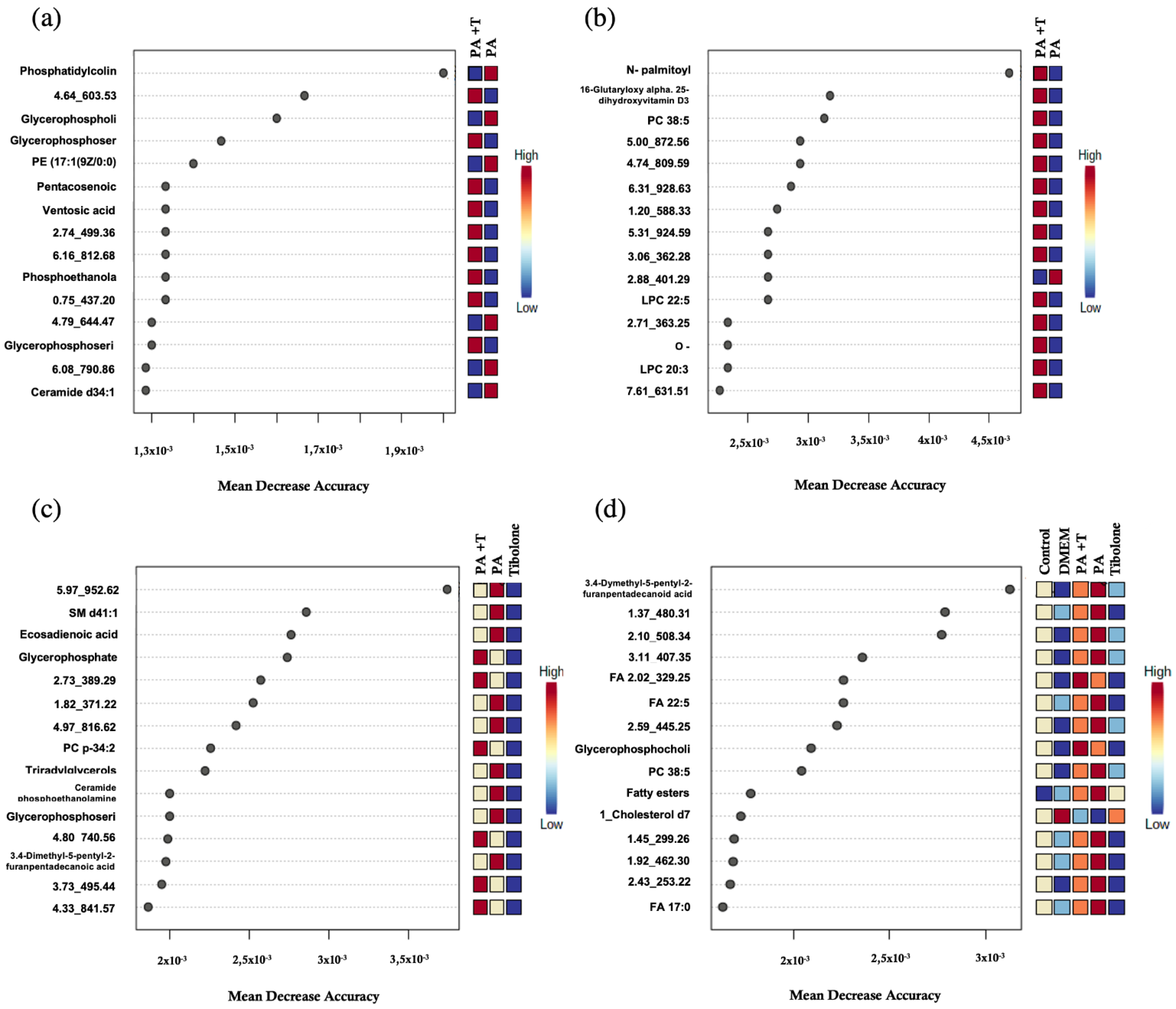

| Treatments | m/z | Adducts | Formula | Δm/z (ppm) | Compound Type |
|---|---|---|---|---|---|
| PA + T & PA | 702.5087 | [M-H]- | Phosphoethanolamine | ||
| 800.6167 | [M+H]+ | C45H86NO8P | 0 | PC 37:2; | |
| 780,5547 | [M-H]- | C44H80NO8P | 0 | Phosphoethanolamine | |
| 889.5812 | [M-H]- | C47H87O13P | 0 | PI 38:2; PI 18:0-20:2; | |
| 770.5337 | [M-H]- | C42H78NO9P | 1 | Phosphoserine | |
| 828.5766 | [M-H]- | C45H84NO10P | 1 | Phosphocholine | |
| 531.4769 | [M+H]+ | C35H62O3 | 1 | Epoxymurin A/ 30-(-2-(O-2-hydroxy-ethane)-3-hydroxy-propane)-hopane | |
| 524.3719 | [M+H]+ | C26H54NO7P | 2 | Phosphocholine | |
| PA + T & T | 636.3497 | [M-H]- | OHHdiA-PE | ||
| 810.5923 | [M+H]+ | C42H84NO11P | 8 | PI-Cer(d18:0/18:0)/PI-Cer(d20:0/16:0) | |
| 673.526 | [M+H]+ | C37H73N2O6P | 3 | SM d32:2; | |
| 798.5584 | [M+H]+ | C44H80NO9P | 7 | Phosphoserine | |
| 830.5904 | [M+HAc-H]- | PC 35:2; | |||
| 267.2332 | [M-H]- | C17H32O2 | 1 | Heptadecynoic acid | |
| 480.309 | [M-H]- | C23H48NO7P | 1 | Phosphinic acid | |
| 809.5197 | [M-H]- | C41H79O13P | 1 | PI 32:0; PI 16:0-16:0; | |
| 819.5187 | [M-H]- | C46H77O10P | 1 | PG 40:7; PG 18:1-22:6; | |
| 824.5814 | [M+ HAc-H]- | PC p-36:4; or PC o-36:5; | |||
| 329.2489 | [M-H]- | C22H34O2 | 1 | FA 22:5; | |
| 759.5662 | [M+HAc-H]- | SM d34:2; | |||
| PA + T & PA & T | 816.5759 | [M+HAc-H]- | Phosphoserine | ||
| 754.5376 | [M+H]+ | C42H76NO8P | 1 | PC 34:4; | |
| 836.618 | [M+H]+ | C48H86NO8P | 2 | PC 40:5; B | |
| 753.5475 | [M+H]+ | C43H77O8P | 6 | Phosphocholine | |
| 797.5145 | [M-H]- | C47H75O8P | 2 | Phosphoinositol | |
| 771.5181 | [M-H]- | C42H77O10P | 0 | Phosphatidylglycerol | |
| 890.7687/874.7944/869.8343 | [M+K]+_[M+Na]+_[M+NH4]+ | C54H97D5O6 | 8 | 1_TG d5 17:0/17:1/17:0; iSTD | |
| 756.553 | [M+H]+ | C42H78NO8P | 1 | Phosphoethanolamine | |
| Complete | 400.343 | [M+H]+ | C23H45NO4 | 2 | AC 16:0; |
| 885.7905/880.8353 | [M+Na]+_[M+NH4]+ | C55H106O6 | 3 | TAG 52:0; TAG 16:0-18:0-18:0; | |
| 369.3513 | [M+H]+ | C27H44 | 1 | 3-Deoxyvitamin D3 | |
| 838.7822 | [M+NH4]+ | TAG 49:0; TAG 16:0-16:0-17:0; | |||
| 766.5727 | [M+H]+ | C44H80NO7P | 2 | Phosphocholine |
| Treatments | m/z | Adducts | Formula | Δm/z (ppm) | Compound |
|---|---|---|---|---|---|
| PA + T & PA | 836.6165 | [M+H]+ | Phosphatidylcholine | ||
| 835.5348 | [M-H]- | Glycerophospholipids | |||
| 802.5609 | [M-H]- | Glycerophosphoserines | |||
| 466.2932 | [M-H]- | C22H46NO7P | 2 | PE(17:1(9Z)/0:0)/PC(14:1(9Z)/0:0) | |
| 381.3737 | [M-H]- | C25H50O2 | 0 | Pentacosenoic acid/Mycolipenic acid (C25) | |
| 405.3214 | [M-H]- | C22H44O6 | 1 | Ventosic acid | |
| 730.57 | [M-H]- | Phosphoethanolamin/Glycerophosphocholines | |||
| 842.5916 | [M-H]- | Glycerophosphoserines | |||
| 572.4818/596.528 | [M+Cl]- _[M+HAc-H]- | C34H67NO3 | 1 | Ceramide d34:1; | |
| PA + T & T | 354.3014 | [M-H]- | C21H41NO3 | 0 | N-palmitoyl proline/N-oleoyl alanine |
| 547.3674 | [M+H]+ | C32H50O7 | 8 | 16-Glutaryloxy-1alpha,25-dihydroxyvitamin D3/16-Glutaryloxy-1alpha,25-dihydroxy-20-epivitamin D3 | |
| 866.5909 | [M+HAc-H]- | C48H84NO10P | 0 | PC 38:5; A | |
| 628.3619 | [M+HAc-H]- | C30H52NO7P | LPC 22:5; | ||
| 398.3272 | [M+H]+ | C23H43NO4 | 2 | O-palmitoleoylcarnitine/trans-Hexadec-2-enoyl carnitine | |
| 604.3614 | [M+HAc-H]- | LPC 20:3; | |||
| PA + T & PA & T | 859.6912 | [M+HAc-H]- | SM d41:1; | ||
| 307.2637 | [M-H]- | C20H36O2 | 2 | FA 20:2; (eicosadienoic acid) | |
| 787.6146 | [M+H]+ | Glycerophosphates | |||
| 800.5814 | [M+HAc-H]- | PC p-34:2; or PC o-34:3; | |||
| 811.6775 | [M-H]- | Triradylglycerols | |||
| 715.5757 | [M-H]- | Ceramide phosphoethanolamines | |||
| 814.5593 | [M-H]- | Glycerophosphoserines | |||
| 407.3524 | [M-H]- | C26H48O3 | 2 | 3,4-Dimethyl-5-pentyl-2-furanpentadecanoic acid | |
| Complete | 407.3532 | [M-H]- | C26H48O3 | 2 | 3,4-Dimethyl-5-pentyl-2-furanpentadecanoic acid |
| 407.3524 | [M-H]- | C26H48O3 | 2 | 3,4-Dimethyl-5-pentyl-2-furanpentadecanoic acid | |
| 329.2489 | [M-H]- | C22H34O2 | 1 | FA 22:5; | |
| 538.351 | [M-H]- | Glycerophosphocholines/Glycerophosphoserines | |||
| 808.5882 | [M+H]+ | C46H82NO8P | 4 | PC 38:5; B | |
| 537.4896 | [M-H]- | Fatty esters | |||
| 376.3969 | [M+H-H2O]+ | C27H39D7O | 5 | 1_Cholesterol d7 iSTD | |
| 269.2489 | [M-H]- | C17H34O2 | 1 | FA 17:0; (margaric acid) |
Publisher’s Note: MDPI stays neutral with regard to jurisdictional claims in published maps and institutional affiliations. |
© 2022 by the authors. Licensee MDPI, Basel, Switzerland. This article is an open access article distributed under the terms and conditions of the Creative Commons Attribution (CC BY) license (https://creativecommons.org/licenses/by/4.0/).
Share and Cite
Cabezas, R.; Martin-Jiménez, C.; Zuluaga, M.; Pinzón, A.; Barreto, G.E.; González, J. Integrated Metabolomics and Lipidomics Reveal High Accumulation of Glycerophospholipids in Human Astrocytes under the Lipotoxic Effect of Palmitic Acid and Tibolone Protection. Int. J. Mol. Sci. 2022, 23, 2474. https://doi.org/10.3390/ijms23052474
Cabezas R, Martin-Jiménez C, Zuluaga M, Pinzón A, Barreto GE, González J. Integrated Metabolomics and Lipidomics Reveal High Accumulation of Glycerophospholipids in Human Astrocytes under the Lipotoxic Effect of Palmitic Acid and Tibolone Protection. International Journal of Molecular Sciences. 2022; 23(5):2474. https://doi.org/10.3390/ijms23052474
Chicago/Turabian StyleCabezas, Ricardo, Cynthia Martin-Jiménez, Martha Zuluaga, Andrés Pinzón, George E. Barreto, and Janneth González. 2022. "Integrated Metabolomics and Lipidomics Reveal High Accumulation of Glycerophospholipids in Human Astrocytes under the Lipotoxic Effect of Palmitic Acid and Tibolone Protection" International Journal of Molecular Sciences 23, no. 5: 2474. https://doi.org/10.3390/ijms23052474
APA StyleCabezas, R., Martin-Jiménez, C., Zuluaga, M., Pinzón, A., Barreto, G. E., & González, J. (2022). Integrated Metabolomics and Lipidomics Reveal High Accumulation of Glycerophospholipids in Human Astrocytes under the Lipotoxic Effect of Palmitic Acid and Tibolone Protection. International Journal of Molecular Sciences, 23(5), 2474. https://doi.org/10.3390/ijms23052474

