Article

Global N6-Methyladenosine Profiling Revealed the Tissue-Specific Epitranscriptomic Regulation of Rice Responses to Salt Stress

1
Institute of Crop Sciences, National Key Facility for Crop Gene Resources and Genetic Improvement, Chinese Academy of Agricultural Sciences, Beijing 100081, China
2
Biotechnology Research Institute, Chinese Academy of Agricultural Sciences, Beijing 100081, China
3
College of Agronomy, Anhui Agricultural University, Hefei 230036, China
4
National Nanfan Research Institute (Sanya), Chinese Academy of Agricultural Sciences, Sanya 572024, China
*
Authors to whom correspondence should be addressed.
Int. J. Mol. Sci. 2022, 23(4), 2091; https://doi.org/10.3390/ijms23042091
Submission received: 10 December 2021 / Revised: 29 January 2022 / Accepted: 10 February 2022 / Published: 14 February 2022
(This article belongs to the Collection Recent Advances in Plant Molecular Science in China 2021)

Abstract

:
N6-methyladenosine (m6A) methylation represents a new layer of the epitranscriptomic regulation of plant development and growth. However, the effects of m6A on rice responses to environmental stimuli remain unclear. In this study, we performed a methylated-RNA immunoprecipitation sequencing analysis and compared the changes in m6A methylation and gene expression in rice under salt stress conditions. Salt stress significantly increased the m6A methylation in the shoots (p value < 0.05). Additionally, 2537 and 2304 differential m6A sites within 2134 and 1997 genes were identified in the shoots and roots, respectively, under salt stress and control conditions. These differential m6A sites were largely regulated in a tissue-specific manner. A unique set of genes encoding transcription factors, antioxidants, and auxin-responsive proteins had increased or decreased m6A methylation levels only in the shoots or roots under salt stress, implying m6A may mediate salt tolerance by regulating transcription, ROS homeostasis, and auxin signaling in a tissue-specific manner. Integrating analyses of m6A modifications and gene expression changes revealed that m6A changes regulate the expression of genes controlling plant growth, stress responses, and ion transport under saline conditions. These findings may help clarify the regulatory effects of m6A modifications on rice salt tolerance.

1. Introduction

RNA molecules, which have dual roles as information carriers and gene expression regulators, play crucial roles in cellular mechanisms affecting genetic messages, biochemical reactions, and the cell cytoskeleton in all living organisms [1]. RNA modifications are post-transcriptional regulatory processes that provide a biochemical basis for functional diversity. Of the more than 100 types of RNA modifications, the most important is N6-methyladenosine (m6A) in eukaryotic rRNA, tRNA, snRNA, and mRNA [2]. The m6A methylation, which is the most prevalent modification in mRNA, affects plant development and growth by regulating the processing, transport, stability, and translation of mRNA [3].
The m6A modification, which is a reversible biological process mediated by writers/methylases and erasers/demethylases, is recognized by readers/RNA-binding proteins [4]. Studies revealed that m6A methylation is a highly conserved mRNA modification in plants and animals. More specifically, m6A is generated by the consensus sequence RRACH (R is G or A; H is U, A, or C), and this modification is highly enriched near the stop codon or 3′ untranslated region (UTR) in plants and animals [5,6] as well as around the start codon in plants [7,8].
Recent gene functional studies regarding m6A writers, readers, and erasers highlighted an important role for m6A mRNA modifications in plant gene regulation during responses to abiotic stresses. Gene expression studies demonstrated that a few of these writers, erasers, and readers in Arabidopsis thaliana and rice are differentially modulated by diverse abiotic stresses [7,9,10]. The A. thaliana reader ECT2 affects the heat stress response by modifying mRNA stability and relocating to stress granules [11,12]. The knockdown of the RNA demethylase ALKBH6 in A. thaliana increases the sensitivity of plants to salt, drought, and heat stresses [13]. Overexpressing PtrMTA can enhance the drought tolerance of Populus species by altering trichome and root development [14]. The molecular mechanisms underlying the responses of these m6A-related genes to abiotic stress need to be more thoroughly investigated.
Global epitranscriptome studies revealed that m6A mRNA methylation in plants is highly sensitive to abiotic stresses, including drought, salt, and heat. Moreover, m6A methylation can increase the stability of salt-tolerance-related transcripts, thereby regulating salt tolerance in A. thaliana [15]. Further genome-wide m6A profiling using mutants of m6A writer components highlighted the importance of m6A methylation for A. thaliana salt tolerance. More specifically, m6A sites and 3′ UTR length are closely associated with transcript stability during adaptations to stress [16]. In various maize genotypes, the diversity in the extent of the m6A modifications of a unique set of transcripts influences the regulation of drought responses [17]. Additionally, the number and extent of m6A methylations of the mRNA of salt-resistance genes affect the salt tolerance of A. thaliana and sweet sorghum [18,19].
Rice is glycophytic in nature and highly sensitive to salt stress. High salinity stimulates many resistance-related responses, including morphological, physiological, and molecular changes in cells as well as epigenetic events [20,21,22]. The development of the methylated-RNA immunoprecipitation high-throughput sequencing (MeRIP-seq) platform enabled the transcriptome-wide m6A methylation profiling of two rice tissues, which revealed the tissue specificity of m6A methylation and the correlation between m6A peak enrichment and gene expression [7]. To date, there has been no report describing the m6A methylation related to rice responses to salt stress. In the present study, the MeRIP-seq platform was used to investigate the m6A methylation changes in response to salt stress in the shoots and roots of FL478, which is a good source of salt tolerance at the seedling stage in rice. Our results elucidated the tissue-specific and stress-modulated aspects of m6A mRNA methylation, thereby expanding our understanding of the post-transcriptional regulation of salt stress tolerance in rice.

2. Results

2.1. Physiological Indices of FL478 under Salt Stress

The physiological traits of FL478 under control and salt stress conditions were compared. Relative to the corresponding levels under normal growth conditions, proline and malondialdehyde contents and superoxide dismutase activity increased in response to salt stress, whereas relative electrolyte leakage and Na+/K+ contents decreased. In contrast, there was no significant change in the chlorophyll content following the exposure to salt stress (Figure 1). These results indicated that FL478 grew relatively well (i.e., without any obvious salt-induced damages) during the 48 h treatment with NaCl, but there were significant changes to specific physiological indices.

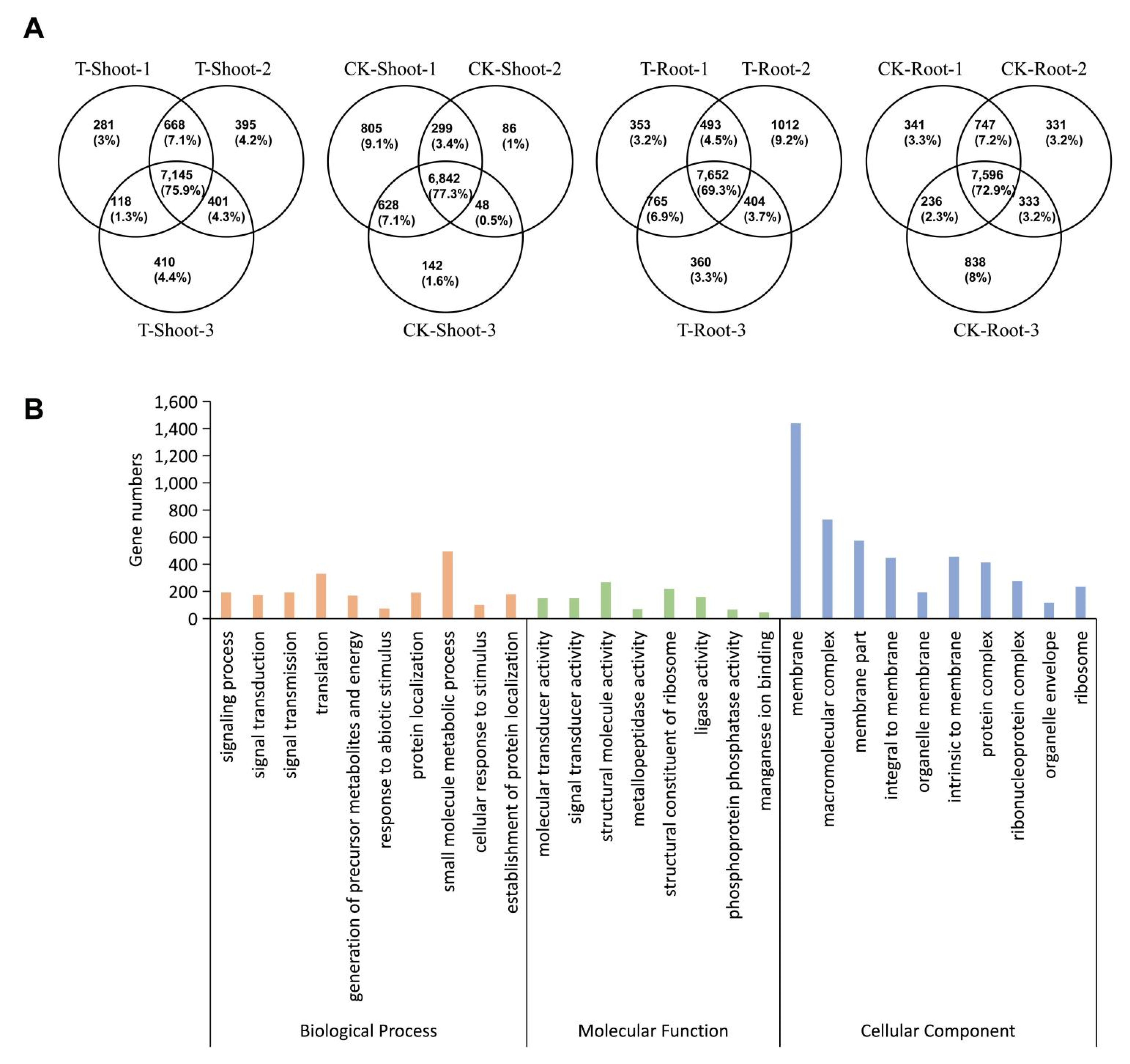
2.2. Identification and Analysis of m6A Methylation Sites

To profile the transcriptome-wide m6A mRNA methylation in FL478, 12 m6A-immunoprecipitation (IP) and corresponding input (non-IP control) libraries were constructed and sequenced for the FL478 shoots and roots under salt stress and control conditions. After removing the adapter sequences and low-quality bases, 36.6–60.0 million clean reads were obtained per sample (Table S1). The clean reads were aligned to the rice reference genome using HISAT2 software and the MACS program was used to identify methylation sites. For each sample, the number of RNA and gene sequences with m6A peaks (Figure 2A) revealed that salt stress resulted in an increase in m6A methylation in the FL478 shoots and roots, which is basically consistent with the results obtained using the EpiQuik m6A RNA Methylation Quantification Kit (Figure 2B). A further analysis of the transcripts with m6A methylation peaks indicated that most transcripts (76.32–79.45%) had one m6A peak, 18.51–21.35% had two m6A peaks, 1.86–2.22% had three peaks, and 0.15–0.30% had four peaks; transcripts with more than four m6A peaks were rare (Figure 2C). Genes encoding a vesicle transporter (Os03g57760), deoxyfructose 5-phosphate reducing isomerase (Os01g01710), heat shock protein DnaJ (Os03g14040), and E3 ubiquitin ligase (Os06g19680) with one, two, two, and three methylation peaks, respectively, were selected to verify the MeRIP data by m6A immunoprecipitation (IP)-qRT-PCR (Figure 2D,E). As expected, these genes were substantially enriched after the mRNA immunoprecipitation with the m6A-specific antibody (relative to the input control) (Figure 2E). Accordingly, our m6A-seq data were accurate and robust.
Only m6A peaks consistently detected in all three biological replicates for each sample were designated as high-confidence m6A peaks. A total of 7596 and 6842 genes with high-confidence m6A peaks were identified in the roots and shoots, respectively, under normal growth conditions, whereas 7652 and 7145 genes with high-confidence m6A peaks were identified in the roots and shoots, respectively, under salt stress conditions (Figure 3A). The GO enrichment analysis of these m6A-containing genes revealed a potential role for m6A modifications in multiple signaling pathways and cellular processes (Figure 3B). Collectively, these findings demonstrated that m6A methylation is widespread in rice mRNA, and m6A-containing transcripts are related to diverse biological processes.

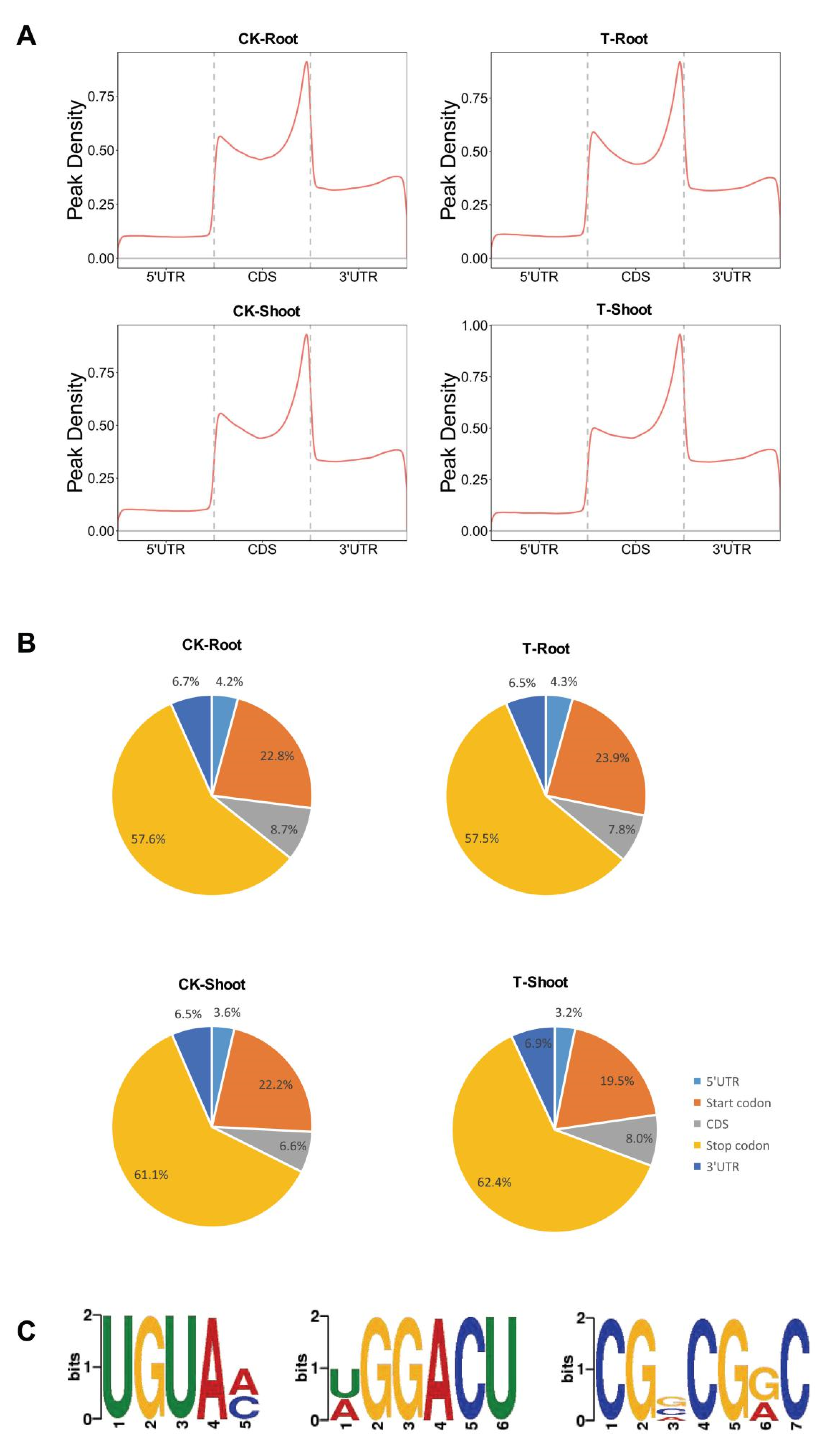
2.3. m6A Distribution among Sequence Motifs in the Transcriptome of FL478

To further clarify the distribution of m6A peaks in the rice root and shoot transcriptomes, we mapped the metagene region methylation sites using the metaplot R package and determined the extent of the methylation in various transcript regions [5′ and 3′ UTRs and coding sequences (CDSs)] (Figure 4A). The m6A modification was highly enriched around the start and termination codons, which is basically in accordance with the m6A distribution revealed in other plant studies [7,8,16]. To confirm the distribution of m6A methylation within transcripts, we divided transcripts into the following five non-overlapping fragments: transcription initiation site (TSS), 5′ UTR, CDS, termination codon, and 3′ UTR. Each m6A RNA methylation peak was assigned to one of the five transcriptional fragments. The m6A peaks were significantly enriched in the stop codon fragment (100-nucleotide fragment centered on the stop codon), with 57.5–62.4% of the peaks in different samples included in this fragment (Figure 4B). Additionally, m6A methylation peaks were highly prevalent in the start codon region (100-nucleotide fragment centered on the start codon), which is inconsistent with the previously reported enrichment of m6A RNA methylation in tomato fruit and A. thaliana [16,23]. Overall, there were no major differences in the distribution of m6A peaks among the samples. Moreover, m6A distribution appears to differ significantly among diverse species.
To identify the sequence motifs enriched within the m6A peaks, we used the DREME software to visualize the sequence characteristics of m6A methylation regions. The following three conserved sequences were detected in these regions: UGUAM (M = A/C), which is a plant-specific conserved motif [16]; CGVCGRC (V = A/C/G, R = A/G); and DGGACU (D = A/G/U), which is similar to a conserved motif DRACH (D = A/G/U, H = A/C/U) in animals [24]. These findings reflected the conservation of sequence motifs associated with m6A methylation among plants and animals (Figure 4C).

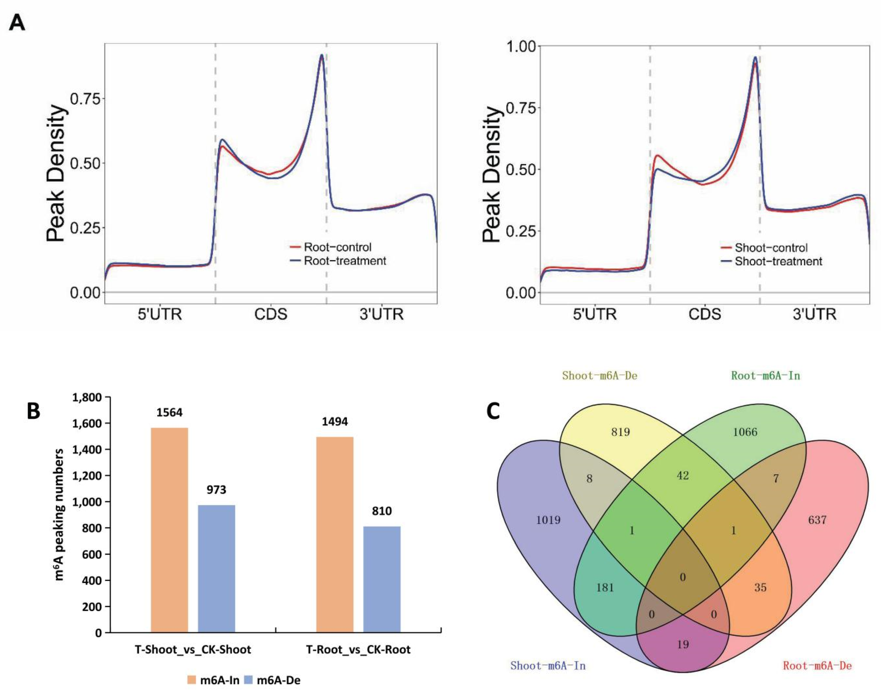
2.4. Tissue-Specific Changes to m6A Methylation in Response to Salt Stress

The extent of the m6A methylation of whole transcripts was plotted using the metaplot R package. We then analyzed the salt-stress-induced m6A methylation changes in different regions (5′ and 3′ UTRs and CDSs) in the roots and leaves. In general, transcripts in the shoots had decreased and increased m6A levels around the start and stop codons, respectively. In contrast, the transcripts in the roots had increased m6A levels around the start codon (Figure 5A).
We further screened the sites with significant changes in m6A methylation following the exposure to salt stress. High salinity resulted in significant increases/decreases in m6A methylation at 1564/973 sites and 1494/810 sites in the shoots and roots, respectively (Figure 5B). Correspondingly, there were 1228/906 and 1296/699 genes with increased/decreased m6A methylation in the salt-stressed FL478 shoots and roots, respectively (Tables S2–S5). A Venn diagram analysis revealed the degree of m6A methylation depended on the tissue for a substantial proportion of these genes under salt stress conditions (Figure 5C). Only 182 and 36 genes had the same m6A methylation changes in both examined tissues. Notably, some genes had the opposite m6A methylation changes in the shoots and roots under saline conditions. Specifically, 42 genes had increased m6A levels in the roots, but decreased m6A levels in the shoots, whereas 19 genes had increased m6A levels in the shoots but decreased m6A levels in the roots (Figure 5C, Tables S6 and S7). These results indicated that m6A modifications in rice are largely tissue-specifically regulated in response to salt stress.
A GO enrichment analysis was performed to functionally characterize the tissue-specific genes with differential m6A changes induced by salt stress. The genes with increased m6A methylation exclusively in the shoots were assigned the following GO terms: regulation of transcription, chloroplast, developmental process, plasma membrane, response to stimulus, and oxidoreductase activity. The genes with decreased m6A methylation only in the shoots were annotated with the following GO terms: signal transducer activity, response to chemical stimulus, receptor activity, and response to hormone stimulus. The genes with increased m6A methylation exclusively in the roots were assigned the following GO terms: oxidoreductase activity, carbohydrate metabolic process, electron carrier activity, cell wall organization or biogenesis, and response to chemical stimulus. The genes with decreased m6A methylation only in the roots were annotated with the following GO terms: signal transducer activity, receptor activity, and ATP-dependent helicase activity (Table S8). The results of the GO enrichment analysis suggested that genes with tissue-specific m6A changes are functionally related to a wide range of biological processes.

2.5. A Unique Set of Genes with Tissue-Specific m6A Changes Contributed to Salt Tolerance

As mentioned above, many genes in specific functional categories had tissue-specific m6A levels under salt stress (Figure 5C and Table S8). The 45 genes encoding transcriptional regulators with increased m6A methylation levels only in salt-stressed shoots included transcription factor (TF) genes (13 ERFs, 7 WRKYs, 4 TIFYs, and 4 ZIPs) (Table S9). Additionally, 9 and 23 genes encoding proteins with oxidoreductase activity had increased m6A methylation levels exclusively in the shoots and roots, respectively, under salt stress. These nine genes in the shoots included OsACO3, Os2ODD7, Os2ODD8, OsLOX7, and OsLOX8. The 23 genes in the roots included OsNOX5, OsNOX9, and 20 peroxidase-encoding genes (Table S9). All of these genes with increased m6A methylation levels may be involved in the m6A-mediated post-transcriptional regulation of salt tolerance.
Interestingly, among the genes encoding proteins responsive to phytohormones, 8 and 13 had salt-induced increased m6A levels exclusively in the shoots and roots, respectively, and 9 genes had salt-induced decreased m6A levels in the roots only. Most of these genes are involved in auxin signaling pathways, including OsIAA, OsSAUR, OsARF, and OsAUX genes (Table S10).

2.6. Main Effects of m6A Methylation in Rice Responses to Salt Stress

On the basis of a comparative analysis of the RNA-seq data, we identified the DEGs in the shoots and roots under control and salt stress conditions. In response to the exposure to high salinity, 481 and 330 genes had up-regulated and down-regulated expression levels, respectively, in the shoots, whereas 779 and 951 genes had up-regulated and down-regulated expression levels, respectively, in the roots. To further clarify the mechanism underlying the m6A methylation in response to salt stress, we analyzed the expression of genes homologous to A. thaliana writer and eraser genes. An examination of the corresponding transcriptome data indicated that compared with the control expression levels, three of the eight writer genes were differentially expressed under salt stress conditions. The expression levels of the MT homolog OsMTA2 (LOC_Os02g45110) and the VIRILIZER homolog OsVIRILIZER (LOC_Os03g35340) were significantly down-regulated in the roots and shoots, respectively. In contrast, the expression of the MT homolog OsMTA4 (LOC_Os10g31030) was highly up-regulated in the shoots. Moreover, three of the seven eraser genes were involved in the response to salt stress. The expression levels of the ALKBH8 homolog OsALKBH8 (LOC_Os11g43610) and the ALKBH10A homolog OsALKBH10A (LOC_Os05g33310) decreased significantly in the shoots, whereas the expression of the ALKBH9A homolog OsALKBH9A (LOC_Os06g04660) clearly increased in the shoots (Figure S1). Thus, some m6A ‘writers’ and ‘erasers’ may participate in rice responses to salt stress by regulating the changes in m6A levels.
We also analyzed the expression profiles of genes encoding m6A-binding proteins in the shoots and roots under control and salt stress conditions. Compared with the control expression levels, six YTHDF genes were differentially expressed in the shoots or roots in response to salt stress. The expression of two YTHDF genes (ECT11 and ECT6) and one YTHDF gene (ECT3) decreased in the roots and shoots, respectively, whereas the expression of three YTHDF genes (ECT2, ECT8, and ECT9) increased in the shoots under saline conditions (Figure S1). Hence, methylation recognition proteins (readers) are regulated by salt stress and may mediate the responses of genes with differential salt-induced m6A levels by binding to m6A sites.

2.7. m6A Modifications Involved in the Regulation of Gene Expression in FL478 under Salt Stress Conditions

Recent studies on gene functions demonstrated that m6A mRNA modifications are important for the regulation of gene expression in plants responding to abiotic stresses [25,26]. To clarify the relationship between m6A modifications in mRNA and gene expression, we analyzed the DEGs with differential m6A methylation changes in the shoots or roots under salt stress. Of the 57 DEGs with increased m6A methylation in the shoots, the expression levels of 50 and 7 genes were up-regulated and down-regulated, respectively, under salt stress. Among the 29 DEGs with decreased m6A methylation in the shoots, the expression levels of 11 and 18 were up-regulated and down-regulated, respectively, under salt stress. The 56 DEGs with increased m6A methylation in the roots comprised 40 and 16 genes with up-regulated and down-regulated expression levels, respectively, under salt stress. Of the 51 DEGs with decreased m6A methylation in the roots, 13 and 38 had up-regulated and down-regulated expression levels, respectively, under salt stress (Figure 6A and Table S11). Genes encoding a pectinesterase inhibitor domain protein (Os02g053700), cellulose synthase (Os09g0478300), and the auxin response protein OsIAA6 (Os01g0741900) had increased m6A methylation and up-regulated expression levels in the shoots under salt stress (relative to the corresponding control levels) (Figure 6B). Although the exposure to salt stress resulted in increased m6A methylation in RePRP1.1 (Os05g0226900) and OsGA20ox1 (Os03g0856700) and decreased m6A methylation in RL14 (Os10g0558900) in the roots, the expression levels of all these genes were up-regulated in the roots under salt stress (relative to the corresponding control levels) (Figure 6B). Therefore, m6A methylation was positively correlated with gene expression changes in a unique set of DEGs in FL478 under saline conditions.
A GO enrichment analysis revealed that these DEGs with differential m6A changes encode proteins that affect tissue development, ion transport, and stress responses. The DEGs in the shoots included OsIAA6, OsNCED4, OsLEA24, OsCSLE6, OsMPH1, OsLAT4, OsPUP3, and OsSLD (Table S11), which reportedly modulate growth and development as well as abiotic stress tolerance in rice [27,28,29,30,31,32]. The differentially m6A methylated DEGs in salt-stressed roots, such as OsGA20ox1, OsACA6, OsmiR396d, OsPAIR3, OsFAD3, OsWAK3, OsWAK25, OsMYBR1, OsGORK, and OsHMA3, may affect salt stress responses at the transcriptional and post-transcriptional levels.

3. Discussion

High salinity is a serious environmental stress that limits global crop growth and yield. Rice is highly sensitive to salt stress, especially at the seedling and reproductive stages. Several studies clarified the physiological, cellular, and molecular mechanisms mediating rice responses to salt stress [20,21,22]. However, the molecular basis of the m6A-mediated salt stress response in rice has not been elucidated. In the present study, we performed a high-throughput m6A MeRIP-seq analysis and compared the changes in m6A methylation and gene expression in the shoots and roots of a salt-tolerant rice genotype under normal growth and saline conditions. The results revealed the importance of m6A modifications on salt stress responses and provided novel insights into the molecular mechanisms regulating rice salt tolerance at the post-transcriptional level.
Earlier research confirmed that m6A methylation varies among tissues and is distributed unevenly on rice chromosomes [7]. In this study, we observed that m6A methylation peaks are enriched in the start codon region (Figure 4) in addition to being widely prevalent in the stop codon region [16,23], reflecting the diversity in the distribution patterns among species. However, our results suggest that m6A motifs are highly conserved in plants and animals [5,6]. We identified three m6A motifs, UGUAM (M = A/C), DGGACU (D = A/G/U), and CGVCGRC (V = A/C/G, R = A/G), of which UGUAM (M = A/C) and DGGACU (D = A/G/U) are conserved in plant and animals, respectively [16,24]. Whether CGVCGRC (V = A/C/G, R = A/G) is a specific motif in rice remains to be determined.
Post-transcriptional regulatory processes, including m6A modifications, influence plant responses to abiotic stresses. Previous studies demonstrated that salinity stress can increase the degree of m6A methylation in A. thaliana [15,16]. In the present study, the m6A methylation level increased substantially in the shoots and roots of the salt-tolerant FL478 rice plants under salt stress. Moreover, the salt-induced increase in m6A methylation in different genic regions appears to vary among tissues. We detected increased m6A levels around the stop and start codons in the shoots and roots, respectively, implying m6A methylation modulates the post-transcriptional regulation of rice responses to salt stress in a tissue-specific manner. An earlier investigation involving A. thaliana revealed the tissue-specific changes to the RNA structurome under salt stress conditions [18]. Tissue-specific methylation changes might affect transcript stability or translation efficiency, but their precise effects on salt stress responses must be further elucidated.
Transcriptome-wide m6A-seq analyses detected distinct m6A modifications in different plant organs or tissues [7,33]. Consistent with these findings, we observed that the m6A methylation of many genes was specifically differentially regulated in the salt-stressed shoots or roots. Notably, a set of TF genes, including those encoding ERF, WRKY, TIFY, and bZIP family members, had increased m6A methylation levels exclusively in the shoots (Table S9). Transcription factors, such as ERF and WRKY TFs, are often involved in the transcriptional regulation associated with plant responses to abiotic and biotic stresses [34,35,36]. Additionally, TIFY and bZIP TFs mediate tissue growth and stress responses [37,38]. The m6A methylation levels of the genes encoding these TFs increased only in the shoots, suggesting m6A methylation positively affects the transcriptional regulation of salt tolerance. Moreover, different subsets of genes encoding proteins with antioxidant functions had increased m6A methylation levels in the shoots and roots, including 20 genes encoding peroxidases in the roots (Table S9). These genes, especially the peroxidase genes, may be responsible for maintaining reactive oxygen species (ROS) homeostasis in plants under stress conditions [39]. Thus, m6A modifications may contribute to the ROS regulation related to rice responses to salt stress. Furthermore, phytohormones (e.g., auxin) are crucial for the coordination of biological processes mediating plant development and stress responses [40]. Genes encoding auxin-responsive proteins (OsIAAs, OsSAURs, and OsAUXs) and auxin-responsive factors (OsARFs) in rice reportedly affect shoot and root growth as well as stress responses in a tissue-specific or developmental stage-specific manner [41]. The m6A levels of these gene family members increased or decreased significantly in the shoots or roots (Table S10), implying m6A modifications actively contribute to the tissue-specific regulation of salinity stress responses. These results suggest that m6A methylation helps regulate the transcription, redox homeostasis, and phytohormone contents associated with rice plant responses to high salinity.
The core components of the m6A system are the genes encoding methyltransferases (writers, MT), demethylases (erasers), and m6A-binding proteins (readers) [4,42,43,44,45,46,47]. Several of these genes were identified in A. thaliana and rice on the basis of an analysis of animal homologous genes [10,48]. In the current study, the expression levels of some of the writer and eraser genes were differentially regulated in the shoots and roots under salt stress. For example, OsMTA4 expression was up-regulated in the shoots, whereas the OsMTA2 and OsVIRILIZER expression levels were down-regulated in the roots and shoots, respectively, under salt stress. In response to the salt stress treatment, the expression levels of the eraser-encoding genes OsALKBH8 and OsALKBH10 were down-regulated in the shoots, but the expression of another eraser gene was up-regulated in the shoots. Accordingly, the expression level of a single writer or eraser gene was not directly related to the degree of m6A methylation in the shoots and roots determined using the EpiQuik m6A kit (Figure 2B) and by the m6A-IP-seq analysis. Accumulating evidence obtained from mammalian studies has indicated that the contrasting effects of m6A modifications on the regulation of gene expression are due to different ‘reader’ proteins [49,50]. In A. thaliana, YTHD05, YTHD06, and YTHD07 expression levels are up-regulated by heat, cold, hypoxia, or flooding stress, whereas YTHD10 expression is inhibited by cold, drought, salt, or osmotic stress [10]. In this study, compared with the control levels, 6 of 12 YTHDF genes were differentially expressed in the shoots or roots under salt stress. The m6A readers are more responsive to abiotic stress than the writers and erasers, implying that the recognition of m6A methylation marks is more important than the introduction or removal of m6A methylation marks during plant adaptations to stress [10]. How different m6A marks are recognized by specific reader proteins and how this interaction contributes to the regulation of target gene expression in response to different environmental stimuli must be determined.
Several studies revealed that m6A modifications are important for salt stress responses because of the resulting increases in mRNA abundance and the stability of stress-responsive genes in A. thaliana [15]. In the present study, we identified a unique set of DEGs with increased or decreased m6A methylation levels in the shoots or roots under salt stress, suggesting that m6A is not involved in large-scale transcriptome-level changes in response to environmental stimuli. Among the DEGs in the shoots, OsIAA6, OsNCED4, OsPSY3, OsGA20ox1, and OsLEA24 are related to phytohormone signaling pathways and stress responses [27,28,51], whereas OsCSLE6, OsSLD, OsPUP3, and OsMPH1 influence tissue growth and development [29,30,31,32]. Several of the DEGs in the roots, including OsGORK, OsHMA3, OsHAK4, and OsPIP2-4, are related to ion transport [52,53,54,55]. The m6A methylation levels of these DEGs were similarly increased or decreased in rice plants exposed to salt stress, implying that m6A modifications are involved in the coordinated regulation of the gene expression related to plant growth, stress responses, and ion transport under saline conditions. Although we detected a positive correlation between gene expression and m6A methylation changes (e.g., the m6A methylation level of most of the up-regulated genes increased in FL478 under salt stress), how m6A methylation contributes to the changes in gene expression during plant adaptations to salt stress will need to be further clarified.

4. Materials and Methods

4.1. Plant Materials and Growth Conditions

A salinity-tolerant genotype (FL478) was used in this study. The surfaces of FL478 seeds were disinfected by soaking in 0.3% sodium hypochlorite. The seeds were then immersed in water for approximately 3 days at 28 °C until they germinated, after which they were maintained in water (pH 5.5) for 5 days. The seedlings were subsequently cultured in Yoshida nutrient solution under the following conditions: 16 h light (28 °C)/8 h dark (26 °C) cycle, with a light intensity of 600 μmol × m−2 × s−1 [56,57]. Seedlings at the four-leaf stage were treated with 120 mM NaCl for 48 h and then the shoots and roots were collected for analyses of physiological indices and m6A mRNA methylation.

4.2. Analysis of Physiological Indices

Leaves were collected from FL478 seedlings exhibiting similar growth potential under the same environmental conditions. The malondialdehyde and proline contents were measured as previously described by Song et al. [58] and Bates et al. [59]. Superoxide dismutase activity was estimated as described by Ouyang et al., with minor modifications [60]. The chlorophyll content was calculated according to the Arnon formula [61], with equal amounts of anhydrous ethanol and acetone used as the extraction solution. Relative electrolyte leakage was measured as follows. Leaves (5 cm × 5 cm) were thoroughly rinsed with deionized water to remove dirt particles from the surface and then fully immersed in deionized water at room temperature for 12 h. The conductivity was measured and recorded as R1. The leaves were maintained in boiling water for 30 min and then cooled to room temperature. The conductivity was measured and recorded as R2. Relative conductivity was calculated as R1/R2 × 100%. Potassium and sodium ion contents were analyzed using a standard curve prepared before the experiment. The dry weight of each sample was determined before the sample was added to a 50 mL tube containing 100 mM acetic acid solution. The tube was placed in a water bath shaker set at 90 °C for 2 h to extract potassium and sodium ions. The supernatant was analyzed three times using an atomic absorption spectrometer.

4.3. RNA Sequencing (RNA-Seq) and Data Analysis

A high-throughput RNA-seq analysis was performed by Cloud-Seq Biotech Inc. (Shanghai, China). Briefly, rRNA was removed from the extracted total RNA using the NEBNext rRNA Depletion Kit (New England Biolabs, Inc., Ipswich, MA, USA). The RNA-seq libraries were constructed using the NEBNext® Ultra™ II Directional RNA Library Prep Kit (New England Biolabs, Inc., Ipswich, MA, USA). The qualitative and quantitative analyses of the libraries were performed using the 2100 Bioanalyzer (Agilent Technologies, Inc., Santa Clara, CA, USA). The libraries were sequenced on an Illumina NovaSeq 6000 instrument (Illumina, Inc., Santiago, CA, USA). The quality of the generated 150 bp paired-end reads was evaluated on the basis of a Q score of 30 (Q30). After trimming the 3′ adapters and eliminating the low-quality reads using the Cutadapt software (v1.9.3) [62] with the cutoff -m 20, -q 15, the retained high-quality clean reads were aligned to the rice reference genome (IRGSP-1.0) using HISAT2 (v2.0.4) [63]. The HTSeq software (v0.9.1) with default settings was used to obtain the raw count [64] and edgeR was used for normalizing data by TMM method. The differentially expressed genes (DEGs) were identified according to the p-value and expression-level fold-change (p-value < 0.05 and fold change > 2) [65]. The DEGs were functionally characterized by conducting a Gene Ontology (GO) and pathway enrichment analysis.

4.4. MeRIP-Seq Analysis

The m6A MeRIP-seq analysis was performed by Cloud-Seq Biotech Inc. (Shanghai, China). Briefly, m6A RNA immunoprecipitation was completed using the GenSeq™ m6A RNA IP Kit (GenSeq Inc., Shanghai, China). The input sample without immunoprecipitation and the m6A IP samples were used for generating the RNA-seq library with the NEBNext® Ultra II Directional RNA Library Prep Kit (New England Biolabs, Inc., Ipswich, MA, USA). The library quality was evaluated using the 2100 Bioanalyzer (Agilent Technologies, Inc., Santa Clara, CA, USA) and then the library was sequenced on the Illumina NovaSeq 6000 instrument (Illumina, Inc., Santiago, CA, USA) to produce 150 bp paired-end reads. Read quality was controlled on the basis of Q30. After removing 3′ adapters and low-quality reads using the Cutadapt software (v1.9.3) [62] with the cutoff -m 20, -q 15, the clean reads were aligned to the reference genome using the HISAT2 software (v2.0.4) [63] with default settings. Methylated RNA sites (peaks) were identified using the MACS program with a cutoff of p < 0.00001. Differentially methylated sites were identified with diffReps with a cutoff of p < 0.00001 and fold change > 2. The RNA sites detected by both programs that overlapped mRNA sequences were identified and filtered using home-made scripts. The differentially methylated protein-coding genes underwent a GO and pathway enrichment analysis. Genes that were simultaneously differentially expressed and m6A methylated (or demethylated) in response to salt stress were included in a correlation analysis of MeRIP and RNA-seq data.

4.5. Analysis of MeRIP-Seq Data by Quantitative Real-Time (qRT)-PCR

The MeRIP data generated using the GenSeq® m6A MeRIP kit (Cloud-Seq Biotech Inc., China) were analyzed by conducting a qRT-PCR analysis using SYBR® Premix Ex Taq™ II (TaKaRa, Kyoto, Japan) and the Applied Biosystems® 7500 thermocycler (Thermo Fisher Scientific, Waltham, MA, USA). The relative expression level of each gene was calculated according to the 2−ΔΔCt method [66]. Standard deviations were calculated on the basis of three technical replicates (n = 6). The OsUBQ5 gene was used as an internal control to normalize target gene expression levels.

5. Conclusions

Our genome-wide m6A methylation profiling demonstrated that salt stress enhances m6A methylation in rice in a distinct tissue-specific manner. The m6A methylation sites were largely differentially regulated in the shoots and roots in response to salt stress. The GO enrichment analysis of the genes with m6A methylation changes induced by salt stress revealed that m6A modifications likely mediate salt tolerance by regulating transcription, ROS homeostasis, and auxin signaling in a tissue-specific manner. A comparative analysis of m6A methylation and gene expression changes following an exposure to salt stress indicated m6A modifications synergistically regulate the gene expression associated with plant growth, stress responses, and ion transport under salt stress conditions.

Supplementary Materials

The following supporting information can be downloaded at: https://www.mdpi.com/article/10.3390/ijms23042091/s1.

Author Contributions

Conceptualization, B.F. and W.W.; methodology, Y.W.; software, J.W. and F.D.; validation, Y.W., Y.L. and F.D.; formal analysis, X.Z.; investigation, Y.W.; resources, J.X.; data curation, B.F.; writing—original draft preparation, Y.W. and B.F.; writing—review and editing, Z.L. and W.W.; supervision, B.F.; funding acquisition, B.F. and W.W. All authors have read and agreed to the published version of the manuscript.

Funding

This work was supported by the National Natural Science Foundation of China (No. 31971928), the National Key Research and Development Program of China (2020YFE0202300), the CAAS Innovative Team Award to BYF and WSW, and the Hainan Provincial Joint Project of Sanya Yazhou Bay Science and Technology City (320LH044).

Institutional Review Board Statement

Not applicable.

Informed Consent Statement

Not applicable.

Data Availability Statement

The original data presented in the study are included in the article and Supplementary Materials. Further inquiries can be directed to the corresponding authors.

Conflicts of Interest

The authors declare that the research was conducted in the absence of any commercial or financial relationships that could be construed as a potential conflict of interest.

References

  1. Sharp, P.A. The Centrality of RNA. Cell 2009, 136, 577–580. [Google Scholar] [CrossRef] [PubMed] [Green Version]
  2. Roundtree, I.A.; Evans, M.E.; Pan, T.; He, C. Dynamic RNA Modifications in Gene Expression Regulation. Cell 2017, 169, 1187–1200. [Google Scholar] [CrossRef] [PubMed] [Green Version]
  3. Arribas-Hernandez, L.; Brodersen, P. Occurrence and functions of m6A and other covalent modifications in plant mRNA. Plant Physiol. 2020, 182, 79–96. [Google Scholar] [CrossRef] [PubMed] [Green Version]
  4. Yue, H.; Nie, X.J.; Yan, Z.G.; Weining, S. N6-methyladenosine regulatory machinery in plants: Composition, function and evolution. Plant Biotechnol. J. 2019, 17, 1194–1208. [Google Scholar] [CrossRef]
  5. Shen, L.S.; Liang, Z.; Gu, X.F.; Chen, Y.; Teo, Z.W.; Hou, X.; Cai, W.M.; Dedon, P.C.; Liu, L.; Yu, H. N6-methyladenosine RNA modification regulates shoot stem cell fate in Arabidopsis. Dev. Cell 2016, 38, 186–200. [Google Scholar] [CrossRef] [Green Version]
  6. Fu, Y.; Dominissini, D.; Rechavi, G.; He, C. Gene expression regulation mediated through reversible m6A RNA methylation. Nat. Rev. Genet. 2014, 15, 293–306. [Google Scholar] [CrossRef]
  7. Li, Y.L.; Wang, X.L.; Li, C.P.; Hu, S.; Yu, J.; Song, S. Transcriptome-wide N6-methyladenosine profiling of rice callus and leaf reveals the presence of tissue-specific competitors involved in selective mRNA modification. RNA Biol. 2014, 11, 1180–1188. [Google Scholar] [CrossRef] [Green Version]
  8. Luo, G.Z.; MacQueen, A.; Zheng, G.Q.; Duan, H.; Dore, L.C.; Lu, Z.; Liu, J.; Chen, K.; Jia, G.; Bergelson, J.; et al. Unique features of the m6A methylome in Arabidopsis thaliana. Nat. Commun. 2014, 5, 5630. [Google Scholar] [CrossRef] [Green Version]
  9. Duan, H.C.; Wei, L.H.; Zhang, C.; Wang, Y.; Chen, L.; Lu, Z.; Chen, P.R.; He, C.; Jia, G. ALKBH10B is an RNA N6-methyladenosine demethylase affecting Arabidopsis floral transition. Plant Cell 2017, 29, 2995–3011. [Google Scholar] [CrossRef] [Green Version]
  10. Hu, J.Z.; Manduzio, S.; Kang, H.S. Epitranscriptomic RNA methylation in plant development and abiotic stress responses. Front. Plant Sci. 2019, 10, 500. [Google Scholar] [CrossRef] [Green Version]
  11. Scutenaire, J.; Deragon, J.M.; Jean, V.; Benhamed, M.; Raynaud, C.; Favory, J.J.; Merret, R.; Bousquet-Antonelli, C. The YTH domain protein ECT2 is an m6A reader required for normal trichome branching in Arabidopsis. Plant Cell 2018, 30, 986–1005. [Google Scholar] [CrossRef] [PubMed] [Green Version]
  12. Wei, L.H.; Song, P.Z.; Wang, Y.; Lu, Z.; Tang, Q.; Yu, Q.; Xiao, Y.; Zhang, X.; Duan, H.C.; Jia, G. The m6A reader ECT2 controls trichome morphology by affecting mRNA stability in Arabidopsis. Plant Cell 2018, 30, 968–985. [Google Scholar] [CrossRef] [PubMed] [Green Version]
  13. Huong, T.T.; Ngoc, L.N.T.; Kang, H. Functional characterization of a putative RNA demethylase ALKBH6 in Arabidopsis growth and abiotic stress responses. Int. J. Mol. Sci. 2020, 21, 6707. [Google Scholar] [CrossRef] [PubMed]
  14. Lu, L.; Zhang, Y.; He, Q.Z.; Qi, Z.; Zhang, G.; Xu, W.; Yi, T.; Wu, G.; Li, R. MTA, an RNA m6A methyltransferase, enhances drought tolerance by regulating the development of trichomes and roots in poplar. Int. J. Mol. Sci. 2020, 21, 2462. [Google Scholar] [CrossRef] [Green Version]
  15. Anderson, S.J.; Kramer, M.C.; Gosai, S.J.; Yu, X.; Vandivier, L.E.; Nelson, A.; Anderson, Z.D.; Beilstein, M.A.; Fray, R.G.; Lyons, E.; et al. N6-methyladenosine inhibits local ribonucleolytic cleavage to stabilize mRNAs in Arabidopsis. Cell Rep. 2018, 25, 1146–1157. [Google Scholar] [CrossRef] [Green Version]
  16. Hu, J.; Cai, J.; Park, S.J.; Lee, K.; Li, Y.; Chen, Y.; Yun, J.Y.; Xu, T.; Kang, H. N6-methyladenosine mRNA methylation is important for salt stress tolerance in Arabidopsis. Plant J. 2021, 106, 1759–1775. [Google Scholar] [CrossRef]
  17. Miao, Z.Y.; Zhang, T.; Qi, Y.H.; Song, J.; Han, Z.; Ma, C. Evolution of the RNA N6-methyladenosine methylome mediated by genomic duplication. Plant Physiol. 2020, 182, 345–360. [Google Scholar] [CrossRef] [Green Version]
  18. Tack, D.C.; Su, Z.; Yu, Y.; Bevilacqua, P.C.; Assmann, S.M. Tissue-specific changes in the RNA structurome mediate salinity response in Arabidopsis. RNA 2020, 26, 492–511. [Google Scholar] [CrossRef] [Green Version]
  19. Zheng, H.X.; Sun, X.; Li, J.L.; Song, Y.; Song, J.; Wang, F.; Liu, L.; Zhang, X.; Sui, N. Analysis of N6-methyladenosine reveals a new important mechanism regulating the salt tolerance of sweet sorghum. Plant Sci. 2021, 304, 110801. [Google Scholar] [CrossRef] [PubMed]
  20. Ganie, S.A.; Molla, K.A.; Henry, R.J.; Bhat, K.V.; Mondal, T.K. Advances in understanding salt tolerance in rice. Theor. Appl. Genet. 2019, 132, 851–870. [Google Scholar] [CrossRef]
  21. Zheng, D.Y.; Wang, L.; Chen, L.F.; Pan, X.; Lin, K.; Fang, Y.; Wang, X.E.; Zhang, W. Salt-responsive genes are differentially regulated at the chromatin levels between seedlings and roots in Rice. Plant Cell Physiol. 2019, 60, 1790–1803. [Google Scholar] [CrossRef] [PubMed]
  22. Wang, W.S.; Zhao, X.Q.; Pan, Y.J.; Zhu, L.; Fu, B.; Li, Z. DNA methylation changes detected by methylation-sensitive amplified polymorphism in two contrasting rice genotypes under salt stress. J. Genet. Genom. 2011, 38, 419–424. [Google Scholar] [CrossRef] [PubMed]
  23. Zhou, L.L.; Tian, S.P.; Qin, G.Z. RNA methylomes reveal the m6A-mediated regulation of DNA demethylase gene SlDML2 in tomato fruit ripening. Genome Biol. 2019, 20, 156. [Google Scholar] [CrossRef] [PubMed] [Green Version]
  24. Liang, Z.; Riaz, A.; Chachar, S.; Ding, Y.; Du, H.; Gu, X. Epigenetic modifications of mRNA and DNA in plants. Mol. Plant 2020, 13, 14–30. [Google Scholar] [CrossRef] [PubMed]
  25. Saletore, Y.; Meyer, K.; Korlach, J.; Vilfan, I.D.; Jaffrey, S.; Mason, C.E. The birth of the Epitranscriptome: Deciphering the function of RNA modifications. Genome Biol. 2012, 13, 175. [Google Scholar] [CrossRef] [PubMed] [Green Version]
  26. Meyer, K.D.; Jaffrey, S.R. The dynamic epitranscriptome: N6-methyladenosine and gene expression control. Nat. Rev. Mol. Cell Biol. 2014, 15, 313–326. [Google Scholar] [CrossRef] [PubMed] [Green Version]
  27. Jung, H.; Lee, D.K.; Choi, Y.D.; Kim, J.K. OsIAA6, a member of the rice Aux/IAA gene family, is involved in drought tolerance and tiller outgrowth. Plant Sci. 2015, 236, 304–312. [Google Scholar] [CrossRef]
  28. Hwang, S.G.; Lee, C.Y.; Tseng, C.S. Heterologous expression of rice 9-cis-epoxycarotenoid dioxygenase 4 (OsNCED4) in Arabidopsis confers sugar oversensitivity and drought tolerance. Bot. Stud. 2018, 59, 2. [Google Scholar] [CrossRef]
  29. Wang, L.; Guo, K.; Li, Y.; Tu, Y.; Hu, H.; Wang, B.; Cui, X.; Peng, L. Expression profiling and integrative analysis of the CESA/CSL superfamily in rice. BMC Plant Biol. 2010, 10, 282. [Google Scholar] [CrossRef] [Green Version]
  30. Zhang, Y.; Yu, C.; Lin, J.; Liu, J.; Liu, B.; Wang, J.; Huang, A.; Li, H.; Zhao, T. OsMPH1 regulates plant height and improves grain yield in rice. PLoS ONE 2017, 12, e0180825. [Google Scholar] [CrossRef] [Green Version]
  31. Qi, Z.; Xiong, L. Characterization of a purine permease family gene OsPUP7 involved in growth and development control in rice. J. Integr. Plant Biol. 2013, 55, 1119–1135. [Google Scholar] [CrossRef] [PubMed]
  32. Sato, M.; Nagano, M.; Jin, S.; Miyagi, A.; Yamaguchi, M.; Kawai-Yamada, M.; Ishikawa, T. Plant-unique cis/trans isomerism of long-chain base unsaturation is selectively required for aluminum tolerance resulting from glucosylceramide-dependent plasma membrane fluidity. Plants 2019, 9, 19. [Google Scholar] [CrossRef] [PubMed] [Green Version]
  33. Wan, Y.; Tang, K.; Zhang, D.; Xie, S.; Zhu, X.; Wang, Z.; Lang, Z. Transcriptome-wide high-throughput deep m(6)A-seq reveals unique differential m(6)A methylation patterns between three organs in Arabidopsis thaliana. Genome Biol. 2015, 16, 272. [Google Scholar] [CrossRef] [Green Version]
  34. Sharoni, A.M.; Nuruzzaman, M.; Satoh, K.; Shimizu, T.; Kondoh, H.; Sasaya, T.; Choi, I.R.; Omura, T.; Kikuchi, S. Gene structures, classification and expression models of the AP2/EREBP transcription factor family in rice. Plant Cell Physiol. 2011, 52, 344–360. [Google Scholar] [CrossRef] [PubMed]
  35. Abiri, R.; Shaharuddin, N.A.; Maziah, M.; Yusof, Z.N.B.; Atabaki, N.; Sahebi, M.; Valdiani, A.; Kalhori, N.; Azizi, P.; Hanafi, M.M. Role of ethylene and the APETALA 2/ethylene response factor superfamily in rice under various abiotic and biotic stress conditions. Environ. Exp. Bot. 2017, 134, 33–44. [Google Scholar] [CrossRef] [Green Version]
  36. Viana, V.E.; Busanello, C.; da Maia, L.C.; Pegoraro, C.; Costa de Oliveira, A. Activation of rice WRKY transcription factors: An army of stress fighting soldiers? Curr. Opin. Plant Biol. 2018, 45, 268–275. [Google Scholar] [CrossRef] [PubMed]
  37. Ye, H.; Du, H.; Tang, N.; Li, X.; Xiong, L. Identification and expression profiling analysis of TIFY family genes involved in stress and phytohormone responses in rice. Plant Mol. Biol. 2009, 71, 291–305. [Google Scholar] [CrossRef]
  38. Zhang, Y.P.; Zhou, J.H.; Wang, L. Mini review roles of the bZIP gene family in rice. Genet. Mol. Res. 2014, 13, 3025–3036. [Google Scholar] [CrossRef]
  39. Gill, S.S.; Tuteja, N. Reactive oxygen species and antioxidant machinery in abiotic stress tolerance in crop plants. Plant Physiol. Biochem. 2020, 48, 909–930. [Google Scholar] [CrossRef]
  40. Naser, V.; Shani, E. Auxin response under osmotic stress. Plant Mol. Biol. 2016, 91, 661–672. [Google Scholar] [CrossRef]
  41. Jain, M.; Khurana, J.P. Transcript profiling reveals diverse roles of auxin-responsive genes during reproductive development and abiotic stress in rice. FEBS J. 2009, 276, 3148–3162. [Google Scholar] [CrossRef] [PubMed]
  42. Sledz, P.; Jinek, M. Structural insights into the molecular mechanism of the m6A writer complex. eLife 2016, 5, e18438. [Google Scholar] [CrossRef] [PubMed]
  43. Wang, X.; Feng, J.; Xue, Y.; Guan, Z.; Zhang, D.; Liu, Z.; Gong, Z.; Wang, Q.; Huang, J.; Tang, C. Structural basis of N6-adenosine methylation by the METTL3-METTL14 complex. Nature 2016, 534, 260. [Google Scholar] [CrossRef] [PubMed]
  44. Niessen, M.; Schneiter, R.; Nothiger, R. Molecular identification of virilizer, a gene required for the expression of the sex-determining gene sex-lethal in Drosophila melanogaster. Genetics 2001, 157, 679–688. [Google Scholar] [CrossRef] [PubMed]
  45. Yue, Y.A.; Liu, J.; Cui, X.L.; Cao, J.; Luo, G.; Zhang, Z.; Cheng, T.; Gao, M.; Shu, X.; Ma, H.; et al. VIRMA mediates preferential m6A mRNA methylation in 3’ UTR and near stop codon and associates with alternative polyadenylation. Cell Discov. 2018, 4, 10. [Google Scholar] [CrossRef] [PubMed] [Green Version]
  46. Fedeles, B.I.; Singh, V.; Delaney, J.C.; Li, D.; Essigmann, J.M. The AlkB family of Fe(II)/alpha-ketoglutarate-dependent dioxygenases: Repairing nucleic acid alkylation damage and beyond. J. Biol. Chem. 2015, 290, 20734–20742. [Google Scholar] [CrossRef] [Green Version]
  47. Alemu, E.A.; He, C.; Klungland, A. ALKBHs-facilitated RNA modifications and de-modifications. DNA Repair 2016, 44, 87–91. [Google Scholar] [CrossRef] [Green Version]
  48. Shi, H.; Wei, J.; He, C. Where, when, and how: Context-dependent functions of RNA methylation writers, readers, and erasers. Mol. Cell 2019, 74, 640–650. [Google Scholar] [CrossRef]
  49. Wang, X.; Lu, Z.; Gomez, A.; Hon, G.C.; Yue, Y.; Han, D.; Fu, Y.; Parisien, M.; Dai, Q.; Jia, G.; et al. N6-methyladenosine-dependent regulation of messenger RNA stability. Nature 2014, 505, 117–120. [Google Scholar] [CrossRef]
  50. Welsch, R.; Wüst, F.; Bär, C.; Al-Babili, S.; Beyer, P. A third phytoene synthase is devoted to abiotic stress-induced abscisic acid formation in rice and defines functional diversification of phytoene synthase genes. Plant Physiol. 2008, 147, 367–380. [Google Scholar] [CrossRef] [Green Version]
  51. Wang, X.S.; Zhu, H.B.; Jin, G.L.; Liu, H.L.; Wu, W.R.; Zhu, J. Genome-scale identification and analysis of LEA genes in rice (Oryza sativa L.). Plant Sci. 2007, 172, 414–420. [Google Scholar] [CrossRef]
  52. Cai, H.; Huang, S.; Che, J.; Yamaji, N.; Ma, J.F. The tonoplast-localized transporter OsHMA3 plays an important role in maintaining Zn homeostasis in rice. J. Exp. Bot. 2019, 70, 2717–2725. [Google Scholar] [CrossRef] [PubMed] [Green Version]
  53. Liu, J.; Shabala, S.; Shabala, L.; Zhou, M.; Meinke, H.; Venkataraman, G.; Chen, Z.; Zeng, F.; Zhao, Q. Tissue-specific regulation of Na+ and K+ transporters explains genotypic differences in salinity stress tolerance in rice. Front. Plant Sci. 2019, 10, 1361. [Google Scholar] [CrossRef] [PubMed] [Green Version]
  54. Wang, Z.; Hong, Y.; Zhu, G.; Li, Y.; Niu, Q.; Yao, J.; Hua, K.; Bai, J.; Zhu, Y.; Shi, H.; et al. Loss of salt tolerance during tomato domestication conferred by variation in a Na+/K+ transporter. EMBO J. 2020, 39, e103256. [Google Scholar] [CrossRef] [PubMed]
  55. Kumar, K.; Mosa, K.A.; Chhikara, S.; Musante, C.; White, J.C.; Dhankher, O.P. Two rice plasma membrane intrinsic proteins, OsPIP2;4 and OsPIP2;7, are involved in transport and providing tolerance to boron toxicity. Planta 2014, 239, 187–198. [Google Scholar] [CrossRef]
  56. Yoshida, S.; Forno, D.A.; Cock, J.H.; Gomez, K.A. Routine procedure for growing rice plants in culture solution. In Laboratory Manual for Physiological Studies of Rice; The International Rice Research Institute: Los Baños, Philippines, 1976; pp. 61–66. [Google Scholar]
  57. Wang, Y.; Du, F.; Wang, J.; Li, Y.; Zhang, Y.; Zhao, X.; Zheng, T.; Li, Z.; Xu, J.; Wang, W.; et al. Molecular dissection of the gene OsGA2ox8 conferring osmotic stress tolerance in rice. Int. J. Mol. Sci. 2021, 22, 9107. [Google Scholar] [CrossRef]
  58. Song, S.Y.; Chen, Y.; Chen, J.; Dai, X.Y.; Zhang, W.H. Physiological mechanisms underlying OsNAC5-dependent tolerance of rice plants to abiotic stress. Planta 2011, 234, 331–345. [Google Scholar] [CrossRef]
  59. Bates, L.S.; Waldren, R.P.; Teare, I.D. Rapid determination of free proline for water-stress studies. Plant Soil 1973, 39, 205–207. [Google Scholar] [CrossRef]
  60. Ouyang, S.Q.; Liu, Y.F.; Liu, P.; Lei, G.; He, S.J.; Ma, B.; Zhang, W.K.; Zhang, J.S.; Chen, S.Y. Receptor-like kinase OsSIK1 improves drought and salt stress tolerance in rice (Oryza sativa) plants. Plant J. 2010, 62, 316–329. [Google Scholar] [CrossRef]
  61. Arnon, D.I. Copper enzymes in isolated chloroplasts. Polyphenoloxidase in Beta vulgaris. Plant Physiol. 1949, 24, 1–15. [Google Scholar] [CrossRef] [Green Version]
  62. Martin, M. Cutadapt removes adapter sequences from high-throughput sequencing reads. EMBnet. J. 2011, 17, 2226–6089. [Google Scholar] [CrossRef]
  63. Kim, D.; Langmead, B.; Salzberg, S.L. HISAT: A fast spliced aligner with low memory requirements. Nat. Methods 2015, 12, 357–360. [Google Scholar] [CrossRef] [PubMed] [Green Version]
  64. Anders, S.; Pyl, P.T.; Huber, W. HTSeq-a Python framework to work with high-throughput sequencing data. Bioinformatics 2015, 31, 166–169. [Google Scholar] [CrossRef] [PubMed]
  65. Robinson, M.D.; McCarthy, D.J.; Smyth, G.K. edgeR: A Bioconductor package for differential expression analysis of digital gene expression data. Bioinformatics 2010, 26, 139–140. [Google Scholar] [CrossRef] [Green Version]
  66. Livak, K.J.; Schmittgen, T.D. Analysis of relative gene expression data using real-time quantitative PCR and the 2(-Delta Delta C(T)) method. Methods 2001, 25, 402–408. [Google Scholar] [CrossRef]
Figure 1. Physiological indices of FL478 plants under salt stress (T) and control (CK) conditions. Error bars indicate the standard deviation for three replicates. Asterisks indicate a significant difference between the control and salt stress conditions (* p < 0.05, ** p < 0.01; Student’s t-test).
Ijms 23 02091 g001
Figure 2. Identification and analysis of m6A methylation sites in rice shoots and roots under salt stress (T) and control (CK) conditions. (A) Number of m6A peaks and m6A-modified transcripts in rice shoots and roots under control and salt stress conditions; (B) m6A levels in the rice shoots and roots under control and salt stress conditions (* p < 0.05; Student’s t-test); (C) Proportions of the m6A-modified transcripts with one to more than four m6A peaks; (D) Examples of m6A-modified transcripts containing one m6A peak, two m6A peaks, and three m6A peaks. Rectangles (gray dotted lines) indicate the positions of the m6A peaks (peaks 1–8). Black lines indicate the positions of the amplified fragments in the following m6A-immunoprecipitation (IP)-qRT-PCR analysis; (E) Validation of the m6A peaks presented in panel D by m6A-IP-qRT-PCR.
Ijms 23 02091 g002
Figure 3. Transcriptome-wide m6A methylation profiles in rice shoots and roots under salt stress (T) and control (CK) conditions. (A) Venn diagrams presenting the overlap of m6A peaks identified in three independent m6A-seq experiments; (B) Gene Ontology (GO) analysis of the m6A-containing transcripts identified during the m6A-seq analysis. The functional characterization involved the annotation of GO terms from the biological process, molecular function, and cellular component categories.
Ijms 23 02091 g003
Figure 4. Characteristics of m6A localization and sequence motifs in rice shoots and roots under salt stress (T) and control (CK) conditions. (A) Metagenomic profiles of peak distributions along the transcripts comprising three rescaled non-overlapping segments [5′ untranslated region (UTR), coding sequence (CDS), and 3′ UTR]; (B) Proportions of m6A peaks in five non-overlapping transcript segments: transcription initiation site (TSS), 5′ UTR, CDS, termination codon, and 3′ UTR; (C) Sequence motifs identified within m6A peaks using the DREME software.
Ijms 23 02091 g004
Figure 5. Changes in m6A peaks in response to salt stress. (A) Salt stress-induced m6A methylation changes in different gene regions (5′ UTR, CDS, and 3′ UTR) in the roots and shoots; (B) Number of genes with m6A methylations or demethylations in response to salt stress; (C) Venn diagram analysis of the genes with increased/decreased m6A levels (In-m6A/De-m6A) in the shoots and roots under salt stress conditions.
Ijms 23 02091 g005
Figure 6. Comparative analysis of the differentially expressed genes and the genes with decreased and increased m6A methylation levels in the shoots and roots of FL478 plants under salt stress conditions. (A) Venn diagram analysis of the up-regulated (Up-gene)/down-regulated genes (Dn-gene) and the genes with increased/decreased m6A levels (In-m6A/De-m6A) in the shoots and roots under salt stress conditions; (B) Peak m6A levels and expression patterns of six genes in the shoots and roots under salt stress (T) and control (CK) conditions.
Ijms 23 02091 g006
Publisher’s Note: MDPI stays neutral with regard to jurisdictional claims in published maps and institutional affiliations.

