Article

Genome-Wide Analysis of LBD Transcription Factor Genes in Dendrobium catenatum

1
School of Life Sciences, Key Laboratory of Yunnan for Biomass Energy and Biotechnology of Environment, Yunnan Normal University, Kunming 650500, China
2
The Germplasm Bank of Wild Species, Kunming Institute of Botany, Chinese Academy of Sciences, Kunming 650201, China
3
University of Chinese Academy of Sciences, Beijing 100049, China
*
Authors to whom correspondence should be addressed.
Int. J. Mol. Sci. 2022, 23(4), 2089; https://doi.org/10.3390/ijms23042089
Submission received: 12 January 2022 / Revised: 3 February 2022 / Accepted: 11 February 2022 / Published: 14 February 2022
(This article belongs to the Special Issue Plant Genome Dynamics)

Abstract

:
The LATERAL ORGAN BOUNDARIES DOMAIN (LBD) gene family comprises plant-specific transcription factors that control cell proliferation and differentiation during growth and development in many plant species. However, to date, no studies of the LBD gene family in Dendrobium catenatum have been reported. In this study, a genome-wide analysis of LBD genes was performed in D. catenatum and 24 LBD genes were identified. The genes were classified into two classes (I and II) based on phylogenetic relationships and motif structure. Subcellular localization analysis for DcaLBD6 and DcaLBD18 from class I and DcaLBD37 and DcaLBD41 from class II revealed that the proteins were localized in the nucleus. Transient expression analysis of DcaLBD6, DcaLBD18, DcaLBD37, and DcaLBD41 indicated that class I and class II members have opposite roles in regulating VASCULAR-RELATED NAC-DOMAIN 7 (VND7) expression. DcaLBD genes showed diverse expression patterns in response to different phytohormone treatments. Heat maps revealed diverse patterns of DcaLBD gene expression in different organs. These results lay the foundation for further detailed studies of the LBD gene family in D. catenatum.

1. Introduction

The LATERAL ORGAN BOUNDARIES DOMAIN (LBD) transcription factors play important roles in the growth and development of many plant species. LBD genes are involved in the initiation, growth, metabolic regulation, and secondary growth of roots, stems, leaves, and corollas, establishment of the boundary between lateral organ and terminal meristem primordia, and have an important influence on the formation and development of aboveground and belowground organs in higher plants. In addition, LBD genes are involved in anthocyanin and nitrogen metabolism [1,2,3,4].
The LBD gene family is plant specific and is characterized by a highly conserved LATERAL ORGAN BOUNDARIES (LOB) domain. The length of the LOB domain is about 100 amino acids. The LOB domain comprises a C-block, which contains conserved cysteine residues in the CX2CX6CX3C motif required for DNA-binding activity. A Gly–Ala–Ser (GAS) block and a complete leucine zipper-like coiled-coil motif (LX6LX3LX6L) are responsible for protein dimerization [5,6]. On the basis of protein sequence analyses, LBD proteins can be classified into two groups (class I and class II). All LBD proteins contain the C-block, but LBD proteins of class I also include a leucine zipper structure, whereas class II proteins lack a complete leucine zipper structure and cannot form a spiral coil [7,8,9]. Previous studies have demonstrated that LBD proteins play important roles in plant growth, development, signal transduction, and stress response development [10,11,12,13,14,15,16].
Dendrobium catenatum has a broad distribution and a long history as a traditional medicinal plant in China, and has attracted considerable attention from local and international researchers on account of its high medicinal value [17]. In recent years, pharmacological research on the species has mainly focused on its antioxidant, anti-tumor, immunoregulatory, antifatigue, and diabetes-alleviating properties [18,19].
In previous studies, LBD transcription factors have been identified in many plant species. For example, Malus domestica has 58 LBD genes [20], Fragaria vesca has 35 LBD genes [21], Vitis vinifera has 40 LBD genes [22], Zea mays has 44 LBD genes [23], Morus notabilis has 31 LBD genes [24], Brassica rapa var. rapa has 59 LBD genes [25], and Hordeum vulgare has 24 LBD genes [26]. In the present study, we identified LBD genes in D. catenatum, then analyzed their structure, physicochemical properties, phylogenetic relationships, conserved motif profiles, subcellular localization, and expression patterns. Overall, this study provides valuable information for future structural and functional studies of LBD genes in D. catenatum.

2. Results

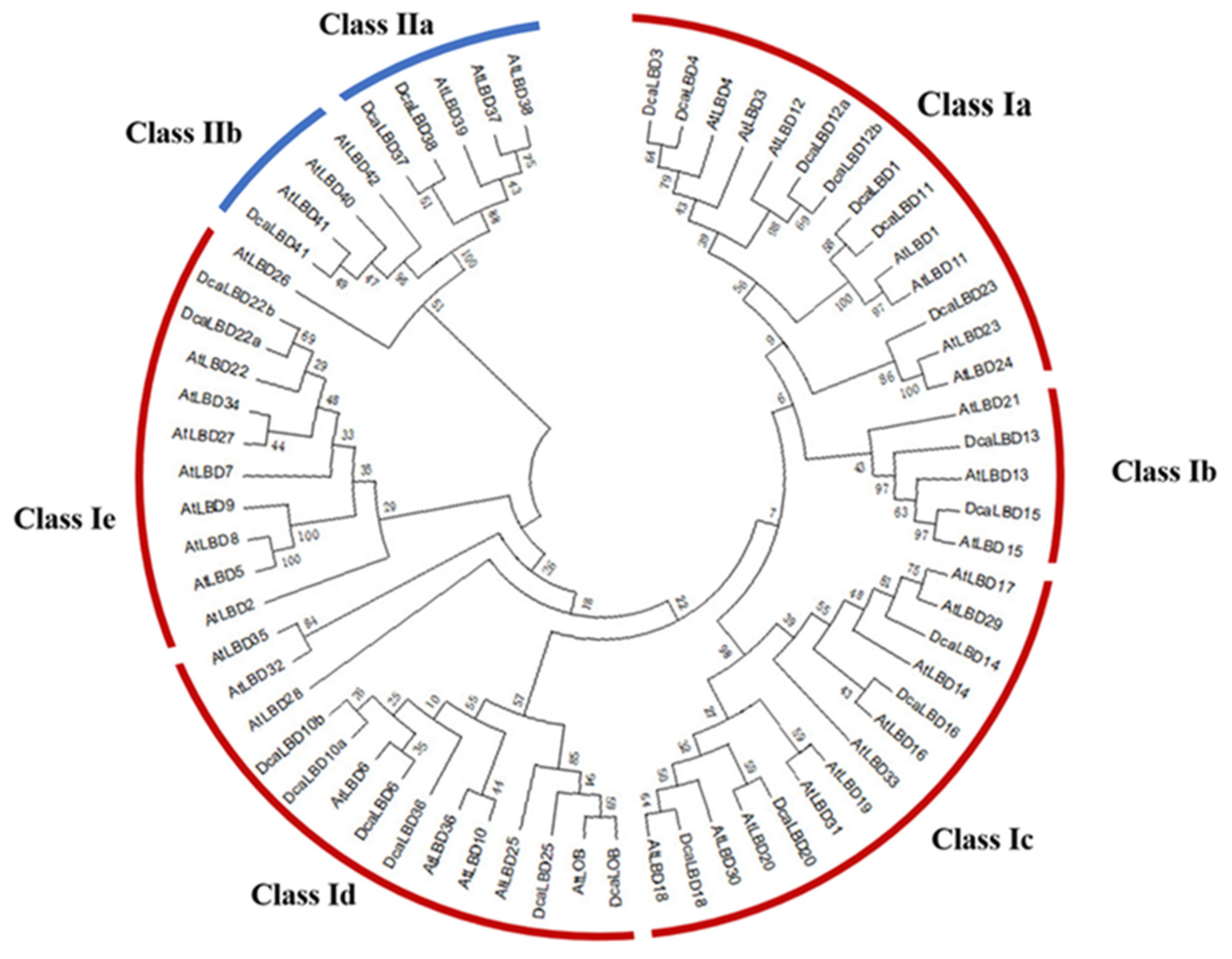
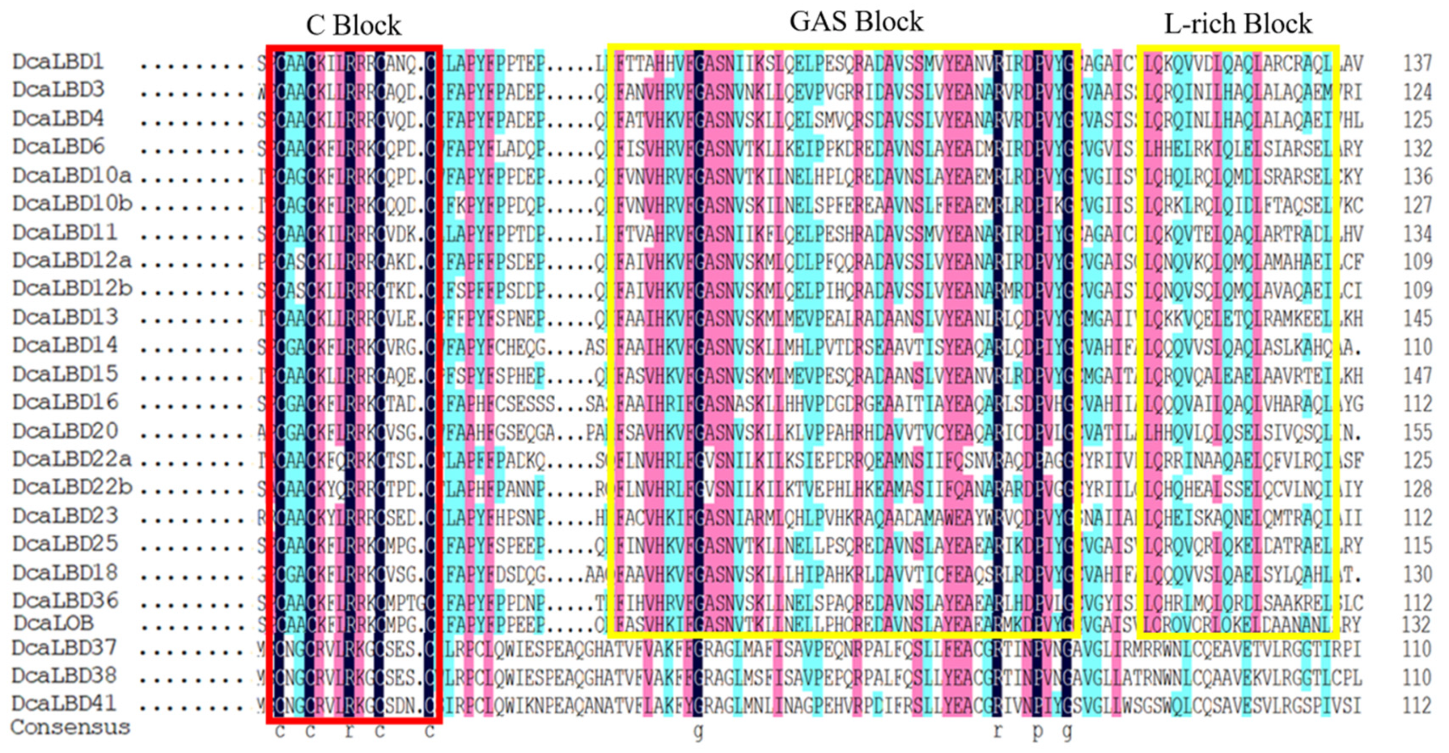
2.1. Identification of LBD Family Genes in D. catenatum

To identify the LBD proteins in the genome of D. catenatum, a local BLAST search of the hidden Markov models (HMMs) of the SMART and Pfam databases was conducted. In total, 24 LBD genes from the complete D. catenatum genome were isolated. All of the deduced LBD proteins possessed a conserved LOB domain. We reconstructed a phylogenetic tree from an alignment of LBD protein sequences from Arabidopsis thaliana and D. catenatum. The DcaLBD genes were annotated based on the similarity of the protein sequences to those of A. thaliana (Figure 1).
On the basis of the phylogenetic analysis, the LBD proteins of D. catenatum were resolved into two monophyletic groups (classes I and II), of which 21 LBD proteins belonged to class I and three LBD proteins belonged to class II. Sequence analysis revealed that AtLBD1, 3, 4, 6, 10, 11, 12, 13, 14, 15, 16, 18, 20, 22, 23, 25, 36, 37, 38, 41, and LOB have orthologs in D. catenatum of which DcaLBD10, 12, and 22 comprised multiple orthologs.
The length of the DcaLBD proteins ranged from 148 to 307 aa, the molecular weight ranged from 17.06 to 34.33 KDa, the theoretical isoelectric point value ranged from 4.61 to 9.33, and the calculated grand average of hydrophobicity value ranged from −0.529 to 0.114. Most of the proteins were hydrophobic with the exception of DcaLBD4 and DcaLBD16 (Table 1).

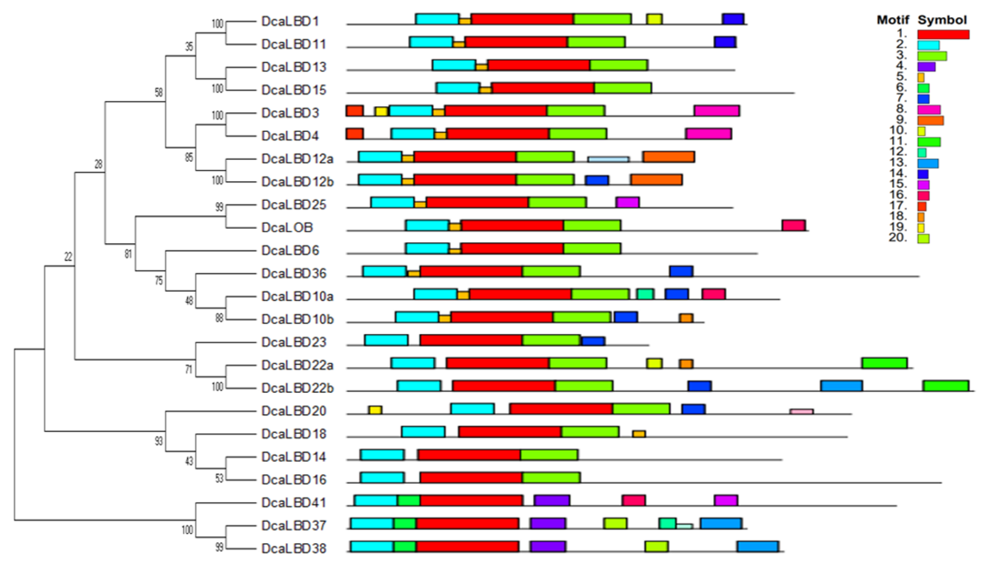
2.2. Motif Analysis and Gene Structure

To further explore the diversity of LBD genes in D. catenatum, the conserved motifs of the DcaLBD proteins were analyzed. A neighbor-joining (NJ) tree was constructed based on a multiple alignment of the DcaLBD protein sequences. The MEME online tool was used to predict the conserved motif composition of the DcaLBD proteins. The number of motifs ranged from three to seven. A LOB domain, motif 2, was detected in all DcaLBDs. In addition, motif 3 was detected in all class I members, indicating that the class I proteins have a specific structure (Figure 2).
On this basis, we further analyzed the DcaLBD protein sequences using the DNAMAN tool. The multiple sequence alignment indicated that a sequence comprising more than 100 amino acids was conserved in all DcaLBDs (Figure 3). For the class I proteins, a string consisting of a C-block, GAS-block, and L-rich block was detected. The C-block in DcaLBDs can be summarized as CX2CX6CX3C. The GAS-block began with a FX2V/AH motif and ended with a DPV/IYG motif. All class II DcaLBDs contained the conserved C-block similar to class I proteins and lacked the GAS-block and Leu-zipper-like domain.

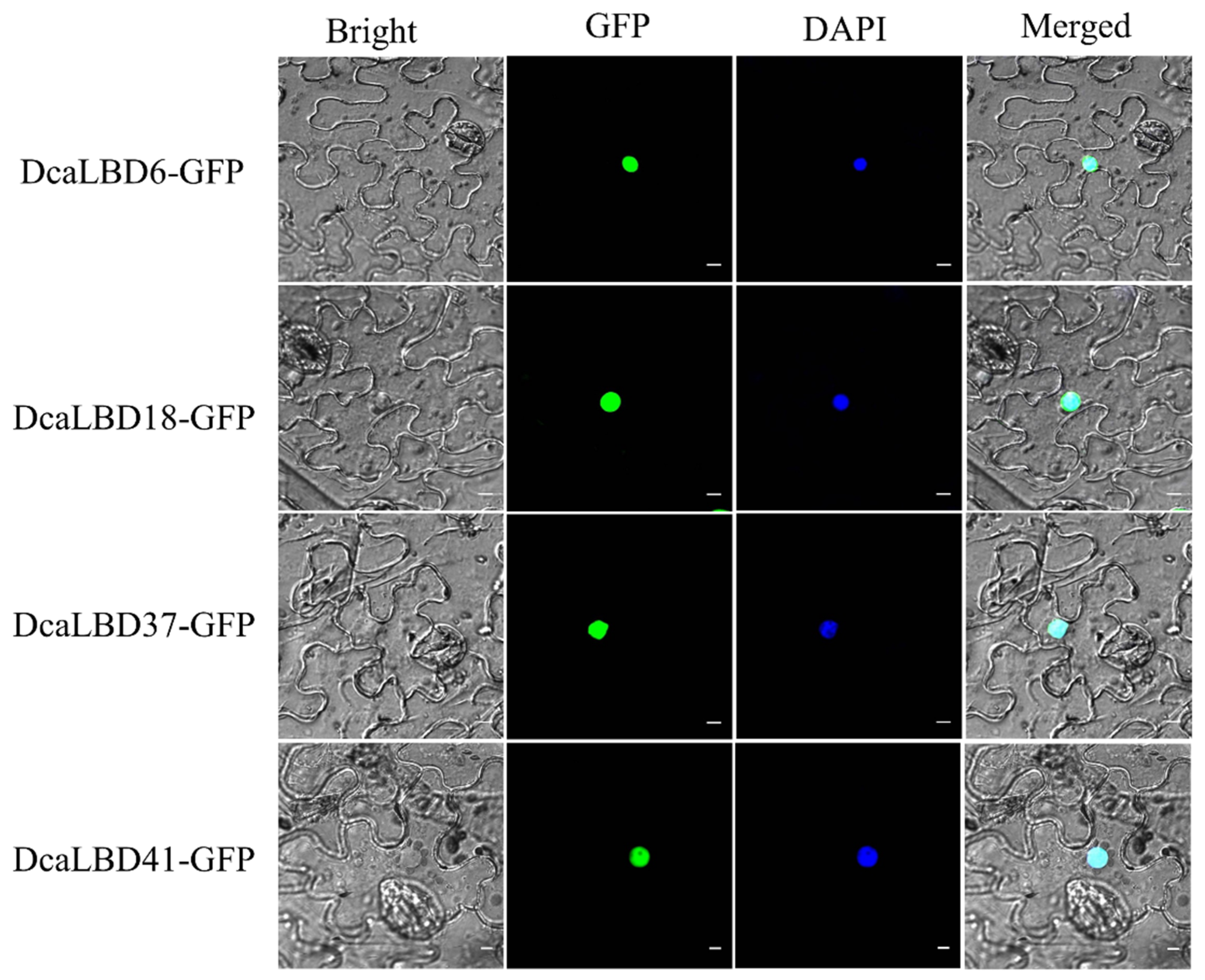
2.3. Subcellular Localization

We predicted that the LBD gene family were transcription factors localized in the nucleus to play roles in regulating plant growth and development. To support this hypothesis, we conducted a subcellular localization analysis for DcaLBD6 and DcaLBD18 from class I and DcaLBD37 and DcaLBD41 from class II. The respective DcaLBD gene was fused with the green fluorescent protein (GFP) gene and co-injected into Nicotiana benthamiana leaves. The transfected plants were cultured in a greenhouse for 3 days. The fluorescence in the injected leaves was observed under a microscope (Olympus FV1000, Tokyo, Japan) after 4′,6-diamidino-2-phenylindole (DAPI) injection. Fluorescent signal from the DcaLBD–GFP protein was detected in the nucleus, consistent with the proteins functioning as transcription factors (Figure 4).

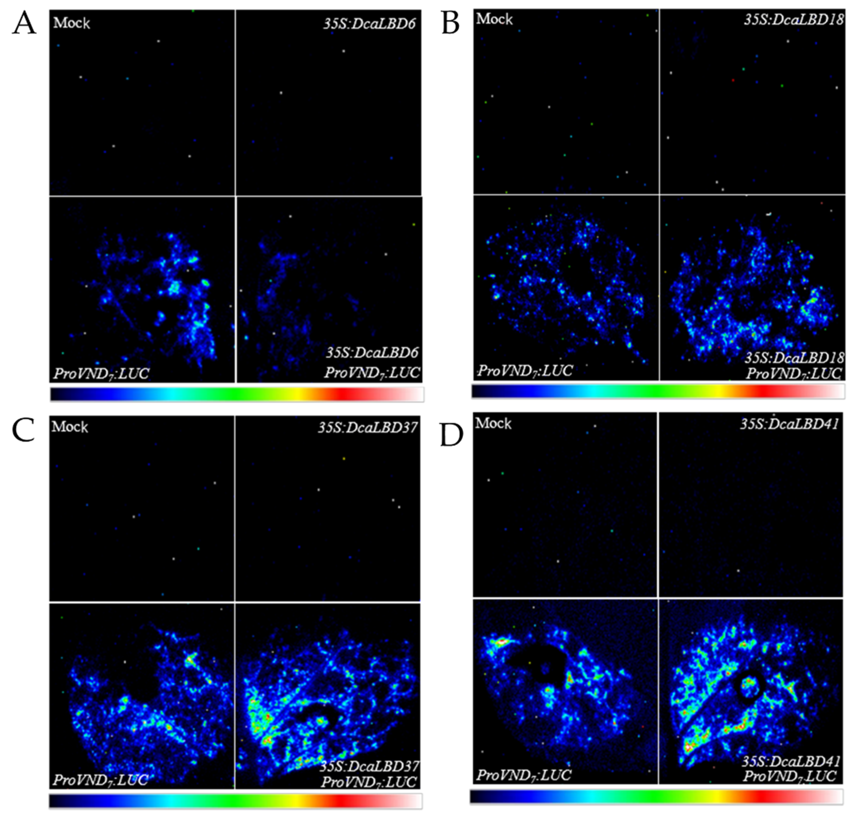
2.4. Class I and II LBDs Regulate VND7 Expression

Previous studies have shown that members of the LBD family have a positive feedback-regulatory effect on the master regulator VASCULAR-RELATED NAC-DOMAIN 7 (VND7) [27]. Therefore, we speculated that the DcaLBD family members may also regulate the expression of VND7. To test this hypothesis, we co-injected the ProAtVND7:LUC promoter with class I (35S:DcaLBD6 and 35S:DcaLBD18) or class II (35S:DcaLBD37 and 35S:DcaLBD41) gene constructs (Figure 5). When the ProAtVND7:LUC reporter plasmid was co-injected with the 35S:DcaLBD37 or 35S:DcaLBD41 effector plasmid, intense luciferase (LUC) fluorescence signal was detected. These results indicated that DcaLBD37 and DcaLBD41 directly activated expression of AtVND7. In contrast, when the ProAtVND7:LUC reporter plasmid was co-injected with the 35S:DcaLBD6 effector plasmid, faint LUC fluorescence signal was detected, but when injected without the effector plasmid the LUC fluorescence signal was weaker. Thus, DcaLBD6 restrained expression of AtVND7. Co-injection of the ProAtVND7:LUC reporter plasmid with the 35S:DcaLBD18 effector plasmid resulted in LUC fluorescence signal similar to that of the ProAtVND7:LUC. These results are consistent with previous studies of LBD family members in suggesting that DcaLBD family members exhibit different functions or even antagonistic roles [28].

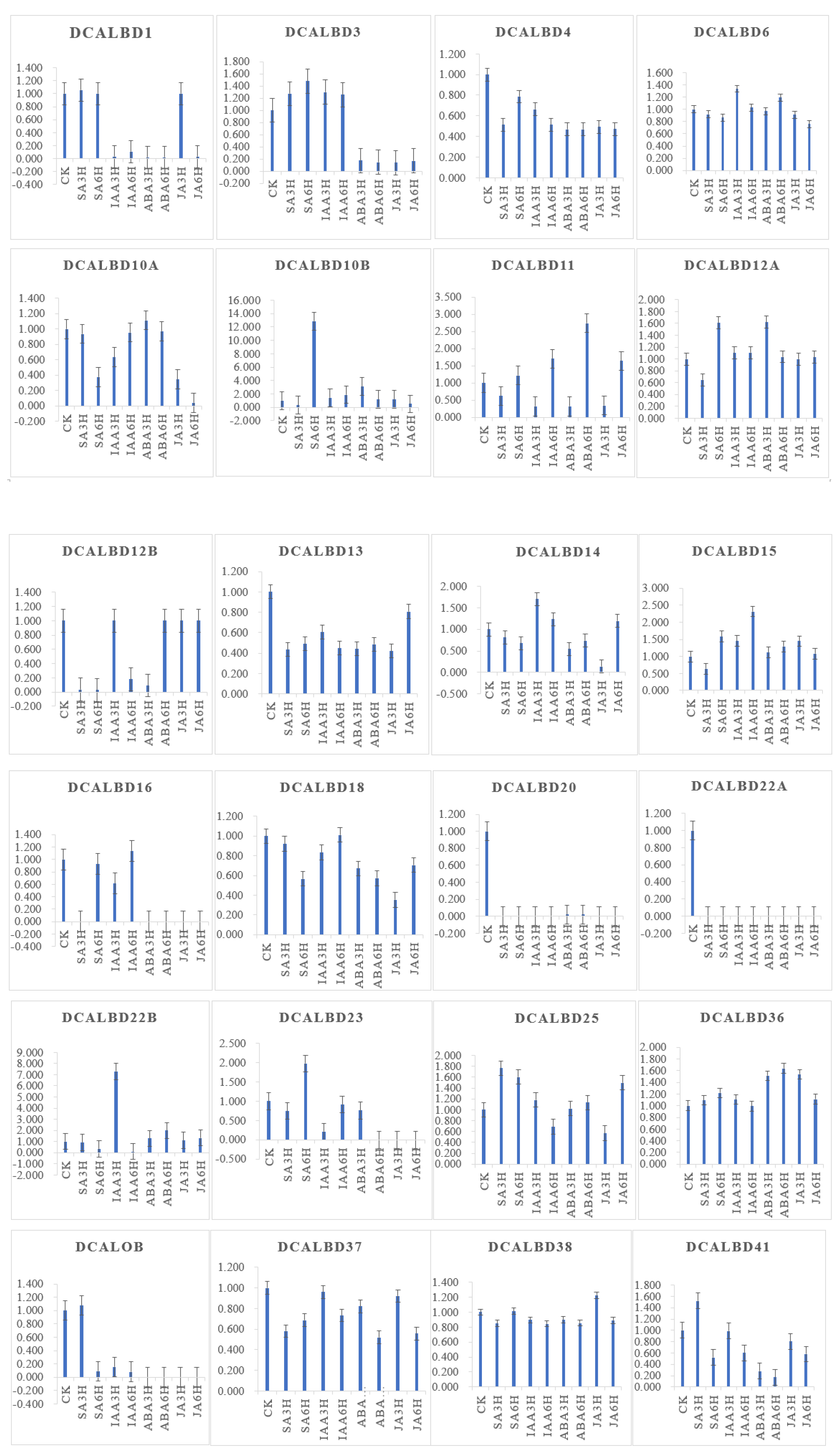
2.5. Expression Profiles of LBD Genes in D. catenatum

To investigate the function of LBD genes in D. catenatum, we used transcriptomic data to determine changes in the expression of the DcaLBD family genes under different phytohormone treatments.
DcaLBD13 showed the highest relative expression level without treatment (Figure 6). The expression level of DcaLBD13 decreased to varying degrees under the various phytohormone treatments. After treatment with indole-3-acetic acid (IAA) for 3 h, DcaLBD6 showed the highest expression level. After treatment with IAA for 6 h, the expression level of DcaLBD18 was the highest. Among class II genes, DcaLBD38 showed the highest expression level without treatment. After treatment with jasmonic acid (JA) for 3 h, the expression level of DcaLBD38 was increased. The expression levels of DcaLBD37 were increased after phytohormone treatment. DcaLBD41 showed the highest expression level after treatment with salicylic acid (SA) for 3 h.
Heat maps were generated to indicate the relative expression of the DcaLBD family members in different organs (Figure 7). Among class I genes, DcaLBD22b showed the highest expression level in the pollinium. DcaLBD6 was more highly expressed in the pollinium than in other organs. The expression of DcaLBD18 was higher in the white portion of the root than in other organs. Among class II genes, DcaLBD38 showed the highest expression level in the gynostemium. The expression level of DcaLBD37 and DcaLBD41 in the pollinium and green root tip were higher than that in other organs.

3. Discussion

As plant-specific transcription factors, the LBD family has been studied extensively in plants. Plants have evolved from low to high, and the number of LBD family genes shows a trend of expanding from scratch. The number of LBDs in different species of plants is different, and the genome size of lower plants is relatively small and the structure is relatively simple, so the number of LBD gene family members is relatively small. Previous studies have shown that the LBD gene family members are absent in Chlamydomonas reinhardtii and Volvox carteri, while 26 LBD gene family members are present in physcomitrella patens in bryophytes, 15 LBD gene family members are present in selaginella moellendorrffii in ferns, and more than 20 LBD gene family members are present in most angiosperms [29]. For example, A. thaliana has 43 LBD genes and D. catenatum has 24 LBD genes. In previous studies, there are 4, 1, 1 LBD gene family members were found from Nitella mirabilis, Coleochaete Orbicularis and Spirogyra pratensis. The fact that there are several LBD gene family members in Charales suggests that LBD proteins were already exist before algae evolved towards terrestrial plants. Charales is the closet ancestor of higher embryo plants [30]. After two rounds of genetic replication, these primitive gene lineages formed the ancestral genes of partial gymnosperms and angiosperms. In the process of terrestrial plant evolution, the overall replication and discrete replication of the whole genome greatly increased the number of LBD genes in angiosperms, and the frequent changes in protein replacement and expression patterns promoted the diversification of LBD genes, so the LBD transcription factor family differentiation produced a variety of biologically functional proteins [31]. These results show that LBD gene expands the number of genes and diversifies its functions through a complex gene replication process in the process of family evolution. The LBD gene family members in A. thaliana and D. catenatum as angiosperms are more numerous and functionally more complete.
In Arabidopsis, AtLBDs are classified into two classes, of which members of class I have a leucine zipper structure, whereas class II members lack a complete leucine zipper structure [7]. Thus, the functions of class I and II LBD genes differ. Class I members are associated with organ development and stress resistance [10,11,12,13]; for example, LBD15 is associated with drought resistance and regulates the expression of VND7 [28]. In contrast, the functions of class II members are involved in metabolism [2]. Therefore, the LBD gene family members exhibit functional diversity. In the present study, DcaLBDs were classifiable into classes I and II based on the characteristic motifs. We also observed that DcaLBD genes regulated the expression of VND7, which is a NAC-domain transcription factor that regulates xylem formation [32]. Previous studies on Arabidopsis have shown that AtLBD6 has an important regulatory effect on the symmetrical development of leaves and the normal development of vascular system [6]. Overexpression of TcLBD6 can lead to a significant reduction in development of lateral roots. AtLBD16 and AtLBD18 jointly regulate the development of Arabidopsis lateral roots under the upstream regulation of AtARF [33], whereas AtLBD6 does not show this function. These results suggest that LBD6-related genes are typically pleiotropic genes, and that physiological effects in plants may differ. Eucalyptus grandis LBD genes differ in degree of influence on secondary growth; overexpression of EgLBD37 results in a significant increase in secondary xylem development, whereas overexpression of EgLBD29 leads to a significant increase in phloem fiber development [34]. These results suggest that DcaLBD37 and DcaLBD41 might be involved in xylem formation via regulation of the expression of VND7. These findings suggest a potential mechanism for the function of DcaLBD genes. However, the roles of DcaLBD genes and their mechanism of action need to be further studied in the future.
Plant hormones affect the expression of genes through diverse mechanisms [35,36]. The expression patterns of DcaLBD genes differed among plant organs, indicating that different DcaLBD genes were involved in different organ development processes. It further suggests that the functions of LBD gene family members are diverse. The positive feedback-regulatory effect on LBD gene and NAC (NAM/ATAF/CUC) protein regulates growth of A. thaliana by controlling the differentiation of xylem cells. LBD16, LBD18, and LBD29 are also involved in regulating the formation of lateral roots in Oryza sativa and Zea mays [7,23]. In this study, the heatmap of expression analysis of DcaLBD genes in different organs (Figure 7) also showed that the expression of DcaLBD16 and DcaLBD18 in the root was higher than that of other organs. These results suggest that the LBD gene family plays an important role in secondary growth processes that affect the quality of plant development. Under the influence of IAA [36], the expression of DcaLBD15, DcaLBD16 and DcaLBD18 increased, which was in line with the expected results. AtLBD10 and AtLBD22 were involved in pollen formation, and the expression of DcaLBD10 and DcaLBD22 in pollinium was significantly higher than in other parts of the Figure 7, and the results were also in line with expectations [14]. The function of MdLBD11 in Malus domestica is similarly to AtLBD11 [20], where the expression of LBD11 may affect plant phenotype includes abnormal traits such as leaf curling upwards, delayed flowering, flowering downwards, and siliques. In this study, the expression data of DcaLBD11 in labellum is also significantly higher than that of other organs, and it can be inferred that the function of DcaLBD11 may be similar to that of A. thaliana and Malus domestica.
In summary, 24 LBD genes were identified in D. catenatum and classified into classes I and II. Expression analysis suggested that DcaLBD transcription factors perform various functions. The present results provide an important foundation for further study of the functions of DcaLBD genes. At present, the research on the LBD transcription factors mainly focuses on the discovery of new members and the study of biological functions, but the study of its origin, evolution and the analysis of the characteristics of each subclass needs to be deepened [31].

4. Materials and Methods

4.1. Plant Material and Phytohormone Treatments

Plants of Dendrobium catenatum were grown in a sand:soil mixture (1:3, w/w). The plants were cultivated in a greenhouse maintained at 23 °C under relative humidity of 65–75% and a 12 h (daytime, 08:00–20:00) photoperiod (50 μmolm2 s1). Treatments for D. catenatum followed the methods of Zhang [37]. Non-treated plants were used as the control. The collected samples were immediately frozen in liquid nitrogen and stored at −80 °C until further use.

4.2. Identification of LBD Family Genes in D. catenatum

To identify the members of the LBD gene family in D. catenatum, three different approaches were performed. First, proteins data for the whole genome sequence of D. catenatum were downloaded from OrchidBase (http://orchidbase.itps.ncku.edu.tw/est/Dendrobium). All known Arabidopsis LBD gene and protein sequences were downloaded from TAIR 12.0 (www.arabidopsis.org) [38]. Local BLASTP and TBLASTN (Basic Local Alignment Search Tool, http://blast.ncbi.nlm.nih.gov) tools [39], available from the National Center for Biotechnology Information (NCBI) website, were used to search the genome sequence of D. catenatum with the known AtLBD sequences as the query. These searches identified the initial candidate genes containing putative LOB domains in D. catenatum. Second, the HMM profile (accession number PF03195) from the Pfam database (http://pfam.sanger.ac.uk) [40] was applied to confirm the presence of the conserved LOB domain in each candidate LOB motif. Finally, DNAMAN 7.0 software was used for multiple sequence alignment and amino acid sequence analysis of D. catenatum LBD proteins and the LOB domain motif (CX2CX6CX3C). The sequences of the identified LBD genes were confirmed using the conserved domain search tool. The retrieved sequences of candidate D. catenatum LBD genes that lacked a LOB domain were discarded.

4.3. Gene Structure and Conserved Motif Analysis

The conserved motifs of DcaLBD proteins were analyzed using the Multiple Em for Motif Elucidation (MEME) website (http://meme-suite.org/index.html) [41]. The search parameters were set as follows: the optimal motif order width was set from 6 to 50, the maximum number of motifs identified was 20 motifs, and all other parameters were set to the default value. In addition, the sequence was used is appeared at least once.

4.4. Phylogenetic Analysis and Physicochemical Properties

The LOB domain nucleotide sequences of DcaLBD and AtLBD genes were used in the phylogenetic analysis. We used MEGA 7.0 software [42] to generate a multiple sequence alignment and perform a neighbor-joining (NJ) analysis with 1000 bootstrap replicates. Using Expasy Protparam tool (ExPASy–ProtParam tool) [43] to analyze the identified DcaLBDs, including their CDS lengths, the protein sizes, protein molecular weight (MW), isoelectric points (pI), and grand average of hydrophilicity (GRAVITY) of DcaLBD genes.

4.5. Subcellular Localization

The CDS of DcaLBD6, 18, 37, and 41 was amplified using Phanta® Max Super-Fidelity DNA Polymerase (Vazyme Biotech Co., Ltd., Nanjing, China) with relevant primers (Table S1). The ClonExpress® II One Step Cloning Kit (Vazyme Biotech Co., Ltd.) was used to insert the CDS into the pRI101-GFP vector. The 35S:GFP-DcaLBD constructs were transferred into Agrobacterium tumefaciens strain EHA105 using electroporation and then injected into Nicotiana benthamiana leaves. The transfected plants were cultivated for 3 days in a greenhouse. The injected leaf blade after 4′,6-diamidino-2-phenylindole (DAPI) injection was excised and observed with a laser confocal microscope [25,33].

4.6. Transient Expression in Nicotiana benthamiana Leaves

Transactivation activity detection assays were performed using 35S:GFP-DcaLBD6, 18, 37, and 41 as the effector and ProVND7:LUC as the reporter. The 35S:GFP-DcaLBD6, 18, 37, and 41 constructs were co-injected with ProAtVND7:LUC into N. benthamiana leaves with a syringe. The transfected plants were cultured in a greenhouse for 3 days. Before observation, 1 mM luciferin solution was sprayed evenly on the surface of the injected N. benthamiana leaves. The leaf blades were then incubated at a low temperature in the dark for 3 min. Luciferase luminescence was captured using a Tanon-5200 Chemiluminescent Imaging System with a low-light cooled CCD camera [44].

4.7. Expression Analysis

Transcriptomic data from phytohormone-treated D. catenatum were downloaded from the Biodiversity Data Center (iflora.cn). The data is collated and a histogram is made by Excel, with ck as 1. The expression level of the DcaLBD genes in different organs (flower bud, sepal, labellum, pollinium, gynostemium, stem, leaf, root, green root tip, and white portion of the root) of D. catenatum were retrieved from OrchidBase (http://orchidbase.itps.ncku.edu.tw/est/). A heat map for tissue-specific expression of the DcaLBD genes was constructed using Tbtools [45]. All data were adjusted using normalize genes. Hierarchical clustering was performed using the default parameters [46].

5. Conclusions

In this study, 24 LBD transcription factors were identified in the genome of D. catenatum and a comprehensive analysis of this gene family was performed. The genes were classified into classes I and II and showed functional diversity; even members within the same class showed opposite functions. The expression patterns of DcaLBD genes showed diverse responses to phytohormone treatments and differential expression patterns in different organs. This study provides a basis for selection of candidate genes to elucidate the functional roles of DcaLBD in the growth and development of D. catenatum.

Supplementary Materials

The following are available online at https://www.mdpi.com/article/10.3390/ijms23042089/s1.

Author Contributions

L.M. and X.S. conceived and designed the experiments. R.J., C.L. and X.Q. performed the experiments and collected the data. R.J. wrote the manuscript. L.M., Y.W. and X.S. revised the paper. All authors have read and agreed to the published version of the manuscript.

Funding

This research was supported by the grants from the Beijing DR PLANT Biotechnology Co., Ltd. (2019H078), the Project of Science & Technology of Guizhou Province (NY[2017]2524).

Institutional Review Board Statement

Not applicable.

Informed Consent Statement

Not applicable.

Data Availability Statement

Not applicable.

Conflicts of Interest

The authors declare no conflict of interest.

References

  1. Fan, M.; Xu, C.; Xu, K.; Hu, Y. LATERAL ORGAN BOUNDARIES DOMAIN transcription factors direct callus formation in Arabidopsis regeneration. Cell Res. 2012, 22, 1169–1180. [Google Scholar] [CrossRef] [Green Version]
  2. Rubin, G.; Tohge, T.; Matsuda, F.; Saito, K.; Scheible, W.R. Members of the LBD family of transcription factors repress anthocyanin synthesis and affect additional nitrogen responses in Arabidopsis. Plant Cell 2009, 21, 3567–3584. [Google Scholar] [CrossRef] [Green Version]
  3. Shuai, B.; Reynaga-Peña, C.G.; Springer, P.S. The Lateral Organ Boundaries Gene Defines a Novel, Plant-Specific Gene Family. Plant Physiol. 2002, 129, 747–761. [Google Scholar] [CrossRef] [Green Version]
  4. Majer, C.; Hochholdinger, F. Defining the boundaries: Structure and function of LOB domain proteins. Trends Plant Sci. 2011, 16, 47–52. [Google Scholar] [CrossRef]
  5. Xu, C.; Luo, F.; Hochholdinger, F. LOB Domain Proteins: Beyond Lateral Organ Boundaries. Trends Plant Sci. 2015, 21, 159–167. [Google Scholar] [CrossRef]
  6. Iwakawa, H.; Ueno, Y.; Semiarti, E.; Onouchi, H.; Kojima, S.; Tsukaya, H.; Hasebe, M.; Soma, T.; Ikezaki, M.; Machida, C.; et al. The ASYMMETRIC LEAVES2 Gene of Arabidopsis thaliana, Required for Formation of a Symmetric Flat Leaf Lamina, Encodes a Member of a Novel Family of Proteins Characterized by Cysteine Repeats and a Leucine Zipper. Plant Cell Physiol. 2002, 43, 467–478. [Google Scholar] [CrossRef] [Green Version]
  7. Yang, Y.; Yu, X.; Wu, P. Comparison and evolution analysis of two rice subspecies LATERAL ORGAN BOUNDARIES domain gene family and their evolutionary characterization from Arabidopsis. Mol. Phylogenet. Evol. 2006, 39, 248–262. [Google Scholar] [CrossRef]
  8. Yordanov, Y.S.; Regan, S.; Busov, V. Members of the LATERAL ORGAN BOUNDARIES DOMAIN Transcription Factor Family Are Involved in the Regulation of Secondary Growth in Populus. Plant Cell 2010, 22, 3662–3677. [Google Scholar] [CrossRef] [Green Version]
  9. Lu, N.; Zhang, P.; Zhang, Q.; Qiao, R.; He, Q.; Li, H.B.; Wang, Y.; Guo, J.; Zhang, D.; Duan, Z.; et al. Electric-field control of tri-state phase transformation with a selective dual-ion switch. Nature 2017, 546, 124–128. [Google Scholar] [CrossRef] [Green Version]
  10. Cabrera, J.; Diaz-Manzano, F.E.; Sanchez, M.; Rosso, M.N.; Melillo, T.; Goh, T.; Fukaki, H.; Cabello, S.; Hofmann, J.; Fenoll, C.; et al. A role for LATERAL ORGAN BOUNDARIES-DOMAIN 16 during the interaction Arabidopsis-Meloidogyne spp. provides a molecular link between lateral root and root-knot nematode feeding site development. New Phytol. 2014, 203, 632–645. [Google Scholar] [CrossRef]
  11. Chen, J.; Moreau, C.; Liu, Y.; Kawaguchi, M.; Hofer, J.; Ellis, N.; Chen, R. Conserved genetic determinant of motor organ identity in Medicago truncatula and related legumes. Proc. Natl. Acad. Sci. USA 2012, 109, 11723–11728. [Google Scholar] [CrossRef] [PubMed] [Green Version]
  12. Feng, Z.; Sun, X.; Wang, G.; Liu, H.; Zhu, J. LBD29 regulates the cell cycle progression in response to auxin during lateral root formation in Arabidopsis thaliana. Ann. Bot. 2012, 110, 1–10. [Google Scholar] [CrossRef] [PubMed] [Green Version]
  13. Kim, J.; Lee, H.W. Direct activation of EXPANSIN14 by LBD18 in the gene regulatory network of lateral root formation in Arabidopsis. Plant Signal. Behav. 2013, 8, e22979. [Google Scholar] [CrossRef] [PubMed] [Green Version]
  14. Kim, M.-J.; Kim, M.; Lee, M.R.; Park, S.K.; Kim, J. LATERAL ORGAN BOUNDARIES DOMAIN(LBD)10interacts withSIDECAR POLLEN/LBD27to control pollen development in Arabidopsis. Plant J. 2015, 81, 794–809. [Google Scholar] [CrossRef] [PubMed]
  15. Lee, H.W.; Kang, N.Y.; Pandey, S.K.; Cho, C.; Lee, S.H.; Kim, J. Dimerization in LBD16 and LBD18 Transcription Factors Is Critical for Lateral Root Formation. Plant Physiol. 2017, 174, 301–311. [Google Scholar] [CrossRef] [Green Version]
  16. Lee, H.W.; Kim, M.-J.; Park, M.Y.; Han, K.-H.; Kim, J. The Conserved Proline Residue in the LOB Domain of LBD18 Is Critical for DNA-Binding and Biological Function. Mol. Plant 2013, 6, 1722–1725. [Google Scholar] [CrossRef] [PubMed] [Green Version]
  17. Hou, B.; Luo, J.; Zhang, Y.; Niu, Z.; Xue, Q.; Ding, X. Iteration expansion and regional evolution: Phylogeography of Dendrobium officinale and four related taxa in southern China. Sci. Rep. 2017, 7, 43525. [Google Scholar] [CrossRef] [Green Version]
  18. Yan, L.; Wang, X.; Liu, H.; Tian, Y.; Lian, J.; Yang, R.; Hao, S.; Wang, X.; Yang, S.; Li, Q.; et al. The Genome of Dendrobium officinale Illuminates the Biology of the Important Traditional Chinese Orchid Herb. Mol. Plant 2014, 8, 922–934. [Google Scholar] [CrossRef] [Green Version]
  19. Tang, H.; Zhao, T.; Sheng, Y.; Zheng, T.; Fu, L.; Zhang, Y. Dendrobium officinale Kimura et Migo: A Review on Its Ethnopharmacology, Phytochemistry, Pharmacology, and Industrialization. Evid. Based Complement. Altern. Med. 2017, 2017, 7436259. [Google Scholar] [CrossRef] [Green Version]
  20. Wang, X.; Zhang, S.; Su, L.; Liu, X.; Hao, Y. A Genome-Wide Analysis of the LBD (LATERAL ORGAN BOUNDARIES Domain) Gene Family in Malus domestica with a Functional Characterization of MdLBD11. PLoS ONE 2013, 8, e57044. [Google Scholar] [CrossRef] [Green Version]
  21. Chen, X.; Wang, J.; Zhao, M.; Yuan, H. Identification and expression analysis of LATERAL ORGAN BOUNDARIES DOMAIN (LBD) transcription factor genes in Fragaria vesca. Can. J. Plant Sci. 2017, 98, 288–299. [Google Scholar] [CrossRef] [Green Version]
  22. Cao, H.; Liu, C.-Y.; Liu, C.-X.; Zhao, Y.-L.; Xu, R.-R. Genomewide analysis of the lateral organ boundaries domain gene family in Vitis vinifera. J. Genet. 2016, 95, 515–526. [Google Scholar] [CrossRef] [PubMed]
  23. Majer, C.; Xu, C.; Berendzen, K.W.; Hochholdinger, F. Molecular interactions of ROOTLESS CONCERNING CROWN AND SEMINAL ROOTS, a LOB domain protein regulating shoot-borne root initiation in maize (Zea mays L.). Philos. Trans. R. Soc. Lond. B Biol. Sci. 2012, 367, 1542–1551. [Google Scholar] [CrossRef] [PubMed] [Green Version]
  24. Luo, Y.; Ma, B.; Zeng, Q.; Xiang, Z.; He, N. Identification and characterization of Lateral Organ Boundaries Domain genes in mulberry, Morus notabilis. Meta. Gene. 2016, 8, 44–50. [Google Scholar] [CrossRef] [PubMed]
  25. Du, J.; Hu, S.; Yu, Q.; Wang, C.; Yang, Y.; Sun, H.; Yang, Y.; Sun, X. Genome-Wide Identification and Characterization of BrrTCP Transcription Factors in Brassica rapa ssp. rapa. Front. Plant Sci. 2017, 8, 1588. [Google Scholar] [CrossRef] [Green Version]
  26. Guo, B.J.; Wang, J.; Lin, S.; Tian, Z.; Zhou, K.; Luan, H.Y.; Lyu, C.; Zhang, X.-Z.; Xu, R.-G. A genome-wide analysis of the ASYMMETRIC LEAVES2/LATERAL ORGAN BOUNDARIES (AS2/LOB) gene family in barley (Hordeum vulgare L.). J. Zhejiang Univ Sci. B 2016, 17, 763–774. [Google Scholar] [CrossRef] [Green Version]
  27. Ohashi-Ito, K.; Iwamoto, K.; Fukuda, H. LOB DOMAIN-CONTAINING PROTEIN 15 Positively Regulates Expression of VND7, a Master Regulator of Tracheary Elements. Plant Cell Physiol. 2018, 59, 989–996. [Google Scholar] [CrossRef] [Green Version]
  28. Sun, X.; Feng, Z.; Meng, L.; Zhu, J.; Geitmann, A. Arabidopsis ASL11/LBD15 is involved in shoot apical meristem development and regulates WUS expression. Planta 2013, 237, 1367–1378. [Google Scholar] [CrossRef]
  29. Chanderbali, A.S.; He, F.; Soltis, P.S.; Soltis, D.E. Out of the Water: Origin and Diversification of the LBD Gene Family. Mol. Biol Evol. 2015, 32, 1996–2000. [Google Scholar] [CrossRef] [Green Version]
  30. Bowman, J.L.; Floyd, S.K.; Sakakibara, K. Green genes-comparative genomics of the green branch of life. Cell 2007, 129, 229–234. [Google Scholar] [CrossRef] [Green Version]
  31. Kong, Y.; Xu, P.; Jing, X.; Chen, L.; Li, L.; Li, X. Decipher the ancestry of the plant-specific LBD gene family. BMC Genom. 2017, 18 (Suppl. 1), 951. [Google Scholar] [CrossRef] [PubMed] [Green Version]
  32. Endo, H.; Yamaguchi, M.; Tamura, T.; Nakano, Y.; Nishikubo, N.; Yoneda, A.; Kato, K.; Kubo, M.; Kajita, S.; Katayama, Y.; et al. Multiple classes of transcription factors regulate the expression of VASCULAR-RELATED NAC-DOMAIN7, a master switch of xylem vessel differentiation. Plant Cell Physiol. 2015, 56, 242–254. [Google Scholar] [CrossRef] [PubMed] [Green Version]
  33. Okushima, Y.; Fukaki, H.; Onoda, M.; Theologis, A.; Tasaka, M. ARF7 and ARF19 regulate lateral root formation via direct activation of LBD/ASL genes in Arabidopsis. Plant Cell 2007, 19, 118–130. [Google Scholar] [CrossRef] [PubMed] [Green Version]
  34. Lu, Q.; Shao, F.; Macmillan, C.; Wilson, I.W.; Van Der Merwe, K.; Hussey, S.G.; Myburg, A.A.; Dong, X.; Qiu, D. Genomewide analysis of the lateral organ boundaries domain gene family in Eucalyptus grandis reveals members that differentially impact secondary growth. Plant Biotechnol. J. 2017, 16, 124–136. [Google Scholar] [CrossRef] [PubMed] [Green Version]
  35. Thatcher, L.F.; Powell, J.J.; Aitken, E.A.; Kazan, K.; Manners, J.M. The Lateral Organ Boundaries Domain Transcription Factor LBD20 Functions in Fusarium Wilt Susceptibility and Jasmonate Signaling in Arabidopsis. Plant Physiol. 2012, 160, 407–418. [Google Scholar] [CrossRef] [Green Version]
  36. Mangeon, A.; Bell, E.M.; Lin, W.-C.; Jablonska, B.; Springer, P.S. Misregulation of the LOB domain gene DDA1 suggests possible functions in auxin signalling and photomorphogenesis. J. Exp. Bot. 2010, 62, 221–233. [Google Scholar] [CrossRef] [Green Version]
  37. Zhang, L.; Li, C.; Yang, D.; Wang, Y.; Yang, Y.; Sun, X. Genome-Wide Analysis of the TCP Transcription Factor Genes in Dendrobium catenatum Lindl. Int. J. Mol. Sci. 2021, 22, 10269. [Google Scholar] [CrossRef]
  38. Lamesch, P.; Berardini, T.Z.; Li, D.; Wilks, C.; Sasidharan, R.; Muller, R.; Dreher, K.; Alexander, D.-L.; Garcia-Hernandez, M.; Karthikeyan, A.S.; et al. The Arabidopsis Information Resource (TAIR): Improved gene annotation and new tools. Nucleic Acids Res. 2012, 40, D1202–D1210. [Google Scholar] [CrossRef]
  39. Altschul, S.F.; Gish, W.; Miller, W.; Myers, E.W.; Lipman, D.J. Basic local alignment search tool. J. Mol. Biol. 1990, 215, 403–410. [Google Scholar] [CrossRef]
  40. Finn, R.D.; Bateman, A.; Clements, J.; Coggill, P.; Eberhardt, R.Y.; Eddy, S.R.; Heger, A.; Hetherington, K.; Holm, L.; Mistry, J.; et al. Pfam: The protein families database. Nucleic Acids Res. 2014, 42, D222–D230. [Google Scholar] [CrossRef] [Green Version]
  41. Bailey, T.L.; Johnson, J.; Grant, C.E. Noble WS. The MEME Suite. Nucleic Acids Res. 2015, 43, W39–W49. [Google Scholar] [CrossRef] [PubMed] [Green Version]
  42. Kumar, S.; Stecher, G.; Tamura, K. MEGA7: Molecular Evolutionary Genetics Analysis Version 7.0 for Bigger Datasets. Mol. Biol. Evol. 2016, 33, 1870–1874. [Google Scholar] [CrossRef] [PubMed] [Green Version]
  43. Artimo, P.; Jonnalagedda, M.; Arnold, K.; Baratin, D.; Csardi, G.; de Castro, E.; Duvaud, S.; Flegel, V.; Fortier, A.; Gasteiger, E.; et al. ExPASy: SIB bioinformatics resource portal. Nucleic Acids Res. 2012, 40, W597–W603. [Google Scholar] [CrossRef] [PubMed]
  44. Chen, H.; Zou, Y.; Shang, Y.; Lin, H.; Wang, Y.; Cai, R.; Tang, X.; Zhou, J.-M. Firefly luciferase complementation imaging assay for protein-protein interactions in plants. Plant Physiol. 2008, 146, 368–376. [Google Scholar] [CrossRef] [PubMed] [Green Version]
  45. Chen, C.; Chen, H.; Zhang, Y.; Thomas, H.R.; Frank, M.H.; He, Y.; Xia, R. TBtools, a Toolkit for Biologists integrating various HTS-data handling tools with a user-friendly interface. Mol. Plant 2020, 13, 1194–1202. [Google Scholar] [CrossRef]
  46. Anders, S.; Huber, W. Differential expression analysis for sequence count data. Genome Biol. 2010, 11, R106. [Google Scholar] [CrossRef] [Green Version]
Figure 1. Phylogenetic tree of LBD proteins from D. catenatum and Arabidopsis was generated using the neighbor-joining (NJ) method implemented in MEGA 7.0 software. Bootstrap analysis was conducted with 1000 iterations.
Ijms 23 02089 g001
Figure 2. Genomic structure and motif composition of D. catenatum LBDs, phylogenetic tree of D. catenatum LBD proteins. The conserved motifs in D. catenatum LBD proteins were identified using MEME, each motif is shown in a specific color.
Ijms 23 02089 g002
Figure 3. LBD-domain alignment and corresponding logo of DcaLBDs. The conserved C–block in all DcaLBDs protein is indicated by a red box. The GAS block and L-rich block in class II of DcaLBDs protein are indicated by yellow boxes.
Ijms 23 02089 g003
Figure 4. Subcellular localization of 35S:DcaLBD-GFP in Nicotiana benthamiana leaves. DcaLBD6-GFP, DcaLBD18-GFP, DcaLBD37-GFP and DcaLBD41-GFP were localized in the nucleus. Bar = 10 μm.
Ijms 23 02089 g004
Figure 5. Transient expression analysis of DcaLBD6, DcaLBD18, DcaLBD37 and DcaLBD41 activities. (A) VND7 was repressed by DcaLBD6. (B) VND7 was activated slightly by DcaLBD18. (C) VND7 was activated by DcaLBD37. (D) VND7 was activated by DcaLBD41.
Ijms 23 02089 g005
Figure 6. Expression analysis of DcaLBD genes after treatment with ABA, IAA, JA and SA. ABA: abscisic acid, IAA: indole-3-acetic acid, JA: jasmonic acid, SA: salicylic acid. ck: control, 3 h: treatment with phytohormone after 3 h, 6 h: treatment with phytohormone after 6 h.
Ijms 23 02089 g006
Figure 7. Expression analysis of DcaLBD genes in different organs.
Ijms 23 02089 g007
Table 1. Identification and characteristics of LBD genes in D. catenatum.
Gene NameAccession NumberCDS Length (bp)Protein Size (aa)MW (kD)PIGRAVY
DcaLBD1XP_020695176.159119621.65.77−0.172
DcaLBD3XP_020704279.157919220.848.98−0.049
DcaLBD4XP_020673109.156718820.276.930.114
DcaLBD6XP_020676861.160620121.378.56−0.115
DcaLBD10aXP_020702209.163921222.857.64−0.323
DcaLBD10bXP_020700451.152817519.539.28−0.225
DcaLBD11XP_020701018.157619120.796.40−0.020
DcaLBD12aXP_020682070.151317018.846.07−0.152
DcaLBD12bXP_020701020.149516418.366.94−0.291
DcaLBD13XP_020682962.157319021.068.27−0.247
DcaLBD14XP_020677496.164221323.326.11−0.139
DcaLBD15XP_020682881.266021923.828.85−0.235
DcaLBD16XP_020701180.187629131.139.330.047
DcaLBD18XP_020699320.173824525.768.26−0.212
DcaLBD20XP_020693551.174424726.936.35−0.300
DcaLBD22aXP_020686925.183427731.154.61−0.465
DcaLBD22bXP_020682680.192430734.335.15−0.443
DcaLBD23XP_028557256.144714817.068.70−0.418
DcaLBD25XP_028549413.168122624.665.96−0.345
DcaLBD36XP_020698423.184328031.146.81−0.529
DcaLOBXP_020678334.157018921.328.26−0.108
DcaLBD37XP_028551170.159119621.766.17−0.297
DcaLBD38XP_020688010.164521423.378.74−0.257
DcaLBD41XP_020703175.181026929.177.56−0.442
Publisher’s Note: MDPI stays neutral with regard to jurisdictional claims in published maps and institutional affiliations.

