Review

Carotenoids from Marine Sources as a New Approach in Neuroplasticity Enhancement

1
Department of General Biochemistry, Faculty of Biology and Environmental Protection, University of Lodz, Pomorska 141/143, 90-236 Lodz, Poland
2
Biohazard Prevention Centre, Faculty of Biology and Environmental Protection, University of Lodz, Pomorska 141/143, 90-236 Lodz, Poland
*
Author to whom correspondence should be addressed.
Int. J. Mol. Sci. 2022, 23(4), 1990; https://doi.org/10.3390/ijms23041990
Submission received: 31 December 2021 / Revised: 2 February 2022 / Accepted: 8 February 2022 / Published: 11 February 2022
(This article belongs to the Special Issue Role of Natural Compounds in Neurological Diseases)

Abstract

:
An increasing number of people experience disorders related to the central nervous system (CNS). Thus, new forms of therapy, which may be helpful in repairing processes’ enhancement and restoring declined brain functions, are constantly being sought. One of the most relevant physiological processes occurring in the brain for its entire life is neuroplasticity. It has tremendous significance concerning CNS disorders since neurological recovery mainly depends on restoring its structural and functional organization. The main factors contributing to nerve tissue damage are oxidative stress and inflammation. Hence, marine carotenoids, abundantly occurring in the aquatic environment, being potent antioxidant compounds, may play a pivotal role in nerve cell protection. Furthermore, recent results revealed another valuable characteristic of these compounds in CNS therapy. By inhibiting oxidative stress and neuroinflammation, carotenoids promote synaptogenesis and neurogenesis, consequently presenting neuroprotective activity. Therefore, this paper focuses on the carotenoids obtained from marine sources and their impact on neuroplasticity enhancement.

1. Introduction

In the past few decades, the quality of human life has significantly improved due to advances in medicine, lifestyle and nutrition changes. At the same time, it contributed to the augmented lifespan and, therefore, the growing number of the elderly, in whom, with age, ailments from miscellaneous central nervous system (CNS) diseases occur. In this regard, scientists have started to put more emphasis on being better acquainted with the processes leading to nervous tissue pathology, which would allow them to develop effective drugs to improve neurological recovery. The studies on carotenoids with potent antioxidant and anti-inflammatory power, which could be possible applications in neuro-intervention, have been conducted for quite some time. Furthermore, recent results have indicated they also own neuroprotective activity and stimulate synaptogenesis and neurogenesis, by, for instance, inhibiting oxidative stress and neuroinflammation, which makes them important compounds in neuroplasticity enhancement and CNS therapy [1].
The nervous system’s capacity to undergo maturation, modify its structure and function, adapting to both physiological and pathological variations in the environment, is known as neuroplasticity. Without this ability, any brain would be unable to develop from infancy through to adulthood or recover from injury [2]. It is a complex physiological process, characterized by a limited scope, happening in the brain for its whole life. Therefore, it comprises neurogenesis, synaptogenesis, and neurochemical variations of the CNS. Brain plasticity is realized mainly by modulating genetic, molecular and cellular mechanisms that influence synaptic connections and neural circuitry formation [3]. High clinical hopes in regulating neuroplasticity processes are raised by both pharmacotherapies and biological therapies, which neurorestorative activity is realized through a synergistic effect occurring between neurogenesis and synaptogenesis.
Neurogenesis, regulated by a wide range of factors, including neurotrophins, neurotransmitters and growth factors, is one of the components of brain plasticity. Neurons, in that process, are generated from neural stem cells (NSCs) and integrated into existing neuronal circuits [4]. NSCs are required for the adequate functioning of neurogenesis by retaining their self-renew moldability and generating neuronal precursors throughout life. In the adult brain, neurogenesis is mainly localized in the subventricular zone (SVZ) and the subgranular zone (SGZ), which are responsible for memory, learning, and olfactory sensation [5]. However, with age, the ability of the newly formed nerve cells to survive as well as the rate of neurogenesis decreases. Adulthood neurogenesis perturbation contributes to various human disorders, such as cognitive impairment or neurodegenerative diseases [6]. In turn, synaptogenesis, which creates new neural connections, occurs throughout life. In adults, synaptogenesis remains a local event, founded on creating new connections and improving existing synaptic pathways [7].
Growth factors, including brain-derived neurotrophic factor (BDNF), nerve growth factor (NGF), and fibroblast growth factor (FGF), play prominent roles in modulating brain plasticity by activating signaling pathways. Examples include: phosphoinositide-3-kinase–protein kinase B/protein kinase B (PI3K/Akt), mitogen-activated protein kinase/extracellular signal-regulated kinase 1/2 (MAPK/Erk), and phospholipase C/inositol trisphosphate/Ca2+/calmodulin-dependent protein kinase II (PLC/IP3/CAMKII), involved in neuron proliferation and survival as well as neuroprotection [8,9,10,11]. Mature forms of neurotrophin bind to a member of the tyrosine kinase receptor family, the tropomyosin receptor kinase (Trk) and to a representative of the tumor necrosis factor receptor superfamily, p75 receptor. They regulate survival, proper development, normal neuronal function, and synaptic strength and plasticity through them [12,13]. Trk receptors (TrkA, TrkB and TrkC) are composed of ligand-binding domains, the transmembrane domain and the cytoplasmic domain. Those domains contain several sites of tyrosine phosphorylation that recruit intermediates in intracellular signaling cascades [14]. The direct proteins binding to Trk receptors leads to tyrosine kinases activation and, in consequence, activates several proteins, including Ras, Ras-related protein 1 (Rap-1), as well as pathways regulated by MAPK, PI3K, and PLC-γ [15]. Unlike the Trk receptors, which autophosphorylate after ligand engagement, the p75 receptor does not contain a catalytic domain to autoactivate. Therefore, it functions mainly via interactions with other effector proteins, mainly by signaling, promoted by the Trk receptors and modulating their functions [16]. Ras proteins activate the PI3K/Akt pathway causing the activation of the expression of genes involved in brain plasticity or MAPK/Erk pathway. That leads to the transcription of protein factors engaged in neurogenesis and synaptogenesis, including cAMP response element-binding protein (CREB), Myc, and ribosomal S6 kinase (RSK) [17].
PI3K activation, stimulated by Ras, is a critical signaling pathway responsible for neurons survival [18]. PI3K generates phosphatidyl inositides accountable for activating protein kinase Akt, also termed protein kinase B (PKB). It has an influence on many proteins involved in regulating cell survival. For instance, PKB inhibits apoptosis by Bcl2-associated agonist of cell death (BAD) phosphorylation [19]. Akt also influences the nuclear factor kappa B (NFκB) pathway. After stimulation with specific or unspecific signals (oxidative stress, inflammatory cytokines), nuclear factor kappa B inhibitor (IκB) phosphorylated by the IKK complex leads to the ubiquitination and proteasomal degradation of the IκB protein and the NFκB stimulation. That mechanism activates the transcription of various target genes, many of which are inflammatory and immunoregulatory, which modulates the neurons’ survival [20].
Akt is associated with both inhibition and promotion of apoptosis, by phosphorylating the transcription factor forkhead 1 (FKHRL1), which regulates apoptosis-promoting proteins expression, and by the negative regulation of glycogen synthase 3β kinase (GSK-3β), respectively. Furthermore, PI3K signaling may also be initiated in the Ras-independent manner when PI3 kinase binds to the growth factor receptor-bound protein 2 (Grb-2)-associated-binding protein 1 (Gab-1) activated by phosphorylated Grb-2 [15,21].
MAPK/Erk pathway, induced by Ras, is activated by the Src homology and containing protein (Shc)/Grb-2/son of sevenless (SOS). Besides, it can also be stimulated by Trk, which phosphorylates fibroblast growth factor receptor substrate 2 (FRS-2), causing its binding to the adapter molecule crk, which associates with the guanyl-nucleotide exchange factor (C3G), that in turn stimulates Rap1. Protein Rap1 activates the Erk kinase signaling pathway. The Erk kinase, through the RSK and MAP pathway, phosphorylates CREB and other transcription factors, regulating genes expression responsible, among others, for the neurons’ survival [15]. In addition, the Trk receptor phosphorylation leads to the PLC-γ1 activation, causing diacylglycerol (DAG) and IP3 formation [22]. IP3 induces Ca2+ reservoirs release and thus increases its cytoplasmic level, indirectly prompting the action of many enzymes, including CAMK and calcium-modulated protein (calmodulin)-dependent phosphatase [23]. In contrast, DAG stimulates DAG-dependent protein kinase C isoforms (PKCδ) activity, which induces the MAPK/Erk pathway [24].
Since multiple brain processes are affected by natural substances like carotenoids, including neurogenesis, synaptic plasticity, and neuronal connectivity, the therapy, based on these compounds, seems to be a promising treatment strategy for CNS diseases. Furthermore, several carotenoids, generously present in marine organisms and easily digestible, exhibit positive effects on brain function enhancement. Therefore, this work aims to review the latest research on the use of carotenoids from marine sources in neuroplasticity enhancement.

2. Neurorestorative Actions of Marine Carotenoids

Basically, carotenoids consist of a polyisoprenoid structure, a long-conjugated chain of double bond and an end group at both ends of the chain [25]. They can be categorized into carotenes, containing a hydrocarbon chain and xanthophylls, oxygen derivatives of carotenes, forming hydroxyl, epoxide, and keto groups [26]. The functions of carotenoids from aquatic habitats are largely specified by their molecular properties such as size, geometry, functional groups, and other traits [25] Kliknij lub naciśnij tutaj, aby wprowadzić tekst. Most of them are lipophilic and can cross the blood-brain barrier (BBB), which is fundamental during neuroplasticity enhancement, treatment of brain injuries or the prevention of brain disorders with these molecules [27,28]. The typical structure of carotenoids and different end groups are shown in Figure 1.
In humans, carotenoids play different significant functions in the brain and have several medicinal properties, including neuroplasticity enhancement [29,30,31,32]. They wield essential roles in immunity, take part in the antioxidant defense system, improve brain function, and are linked to a reduced risk of acquiring chronic diseases. The pharmacological properties, such as antioxidant, anti-inflammatory, and anti-apoptotic potentials of marine carotenoids, endorse their protecting effectiveness against oxidative stress, neuroinflammation and mitochondrial dysfunction, which are known to be implicated in brain injuries or neurodegenerative diseases pathophysiology [33]. Marine carotenoids are suggested to impact gene expression and cell function through multiple mechanisms, especially by: interacting with several transcription factors, including BDNF, NGF, NFκB; modulating signaling pathways, such as the NFκB, MAPK, and the nuclear factor erythroid 2-related factor 2 (Nrf2), associated with inflammatory and oxidative stress responses; and scavenging of reactive oxygen species (ROS) [34]. Hence, these compounds can act either directly on biological molecules and systems or indirectly through the expression of different genes engaged in, among others, antioxidant responses.
Due to their conjugated double-bond structure, carotenoids from the aquatic environment are strong scavengers of singlet oxygen and peroxyl radicals. They act as chemical quenchers of singlet oxygen. Three major types of reactions of free radical scavenging by carotenoids are: electron transfer between the free radical and carotenoid, whereby a carotenoid radical cation or carotenoid radical anion is formed; radical adduct formation; hydrogen atom relocation leading to a neutral carotenoid radical [35].
In addition to their scavenging function toward ROS, carotenoids may also operate through more indirect tracks, including Nrf2, NFκB, or MAPK signaling pathways [36,37].
The Keap1-Nrf2 pathway plays a vital role in the cellular defense against ROS. Moreover, Nrf2 signaling is an important molecular mechanism for neuroprotection and it modulates the activation of immune cells, including microglia. Under normal conditions, Keap1 promotes ubiquitination and degradation of Nrf2, thus maintaining it in an inactive form in the cytosol. During redox imbalance, the Keap1-Nrf2 association is disrupted, Nrf2 ubiquitination is inhibited, leading to its accumulation in the cell and translocation to the nucleus. There, it binds to the antioxidant response element (ARE), leading to antioxidant and cytoprotective enzymes expression. Marine carotenoids interact with Keap1 by changing its conformation, resulting in enhancement of antioxidant activity [38,39,40].
NFκB is accountable for the transcription of various genes that regulate inflammatory responses. Under resting conditions, NFκB is bound to IκB, which resides in the cytoplasm. However, during chronic neuroinflammation, carotenoids or their derivatives may block NFκB activation by interaction with cysteine residues of the IKK and/or NFκB subunits. That NFκB activation blocking causes the target genes transcription repression and thus diminishes inflammation and increases neurons survival [41].
In addition to the NFκB pathway, the anti-inflammatory effects of marine carotenoids are also found through regulating other pathways, including Akt and MAPK pathways, which control synaptic plasticity in the adult brain. Carotenoids from aquatic habitats may increase the phosphorylation of Akt, and phosphorylated Akt regulates Nrf2 and NFκB, influencing gene expression [42,43]. Thereby it alleviates oxidative stress or inflammation-associated damage in brain cells [44]. In the MAPK/Erk pathway, phosphorylated Erk translocates into the nucleus where it activates transcription factors such as Elk-1 and Msk. That activation of the transcription factors regulates synaptic plasticity and, consequently, it may contribute to the neuroplasticity enhancement [45,46].
Besides, marine carotenoids may impact the level of several neurotrophic factors, including NGF or BDNF. BDNF, the major neurotrophin in the brain, being able to activate intracellular signaling via binding to its receptors, is critical in the proper functioning of the nervous system because it regulates neuronal survival and differentiation, learning and memory. Additionally, BDNF plays a role in proteins’ transcription and translation, which are engaged in synapse development. BDNF is also an underlying agent in the pathology of neural diseases like Alzheimer’s disease, schizophrenia and depression [47,48]. In turn, NGF plays a crucial role in developmental and adult neurobiology for its significant regulatory activity at nerve cells survival, growth, and differentiation in the CNS [49]. Carotenoid supplementation might increase systemic levels of BDNF reduced during neuroinflammation, and the plausible mechanism for this effect is marine carotenoids anti-inflammatory capability [50,51,52]. They also promote the secretion of NGF and BDNF from NSCs [42]. BDNF can block neuronal apoptosis by inducing phosphorylation of Akt during excitotoxicity [53]. In contrast, NGF regulates apoptosis by activating PI3K/Akt and MAPK/Erk pathways [54]. NGF may interact with TrkA and activate Erk signaling to phosphorylate some proteins, as Bcl-2-like protein 11, and inactivate their pro-apoptotic function [55].
Carotenoids derived mainly from marine sources, such as astaxanthin (AST), fucoxanthin (FUC), but also the rare siphonaxanthin or myxol, have lately shown antioxidant and inflammatory effects, which help enhance cognitive function and neuroprotection. Since carotenoids are hydrophobic antioxidants, their main action mechanism is found within biological membranes and depends on their structural features and membrane composition. The results showed that AST, having two polar hydroxyl groups, is anchored across the membranes with polar functional groups oriented outside. Hence it exhibits more effective protection against oxidation by peroxyl radicals than β-carotene or lutein, which are oriented parallel to the membrane surface [56]. Recent reports claim that AST delays or ameliorates cognitive impairment associated with normal ageing or alleviates various neurodegenerative diseases’ pathophysiology [57,58]. AST proved its neuroprotective potential by preventing brain damage in progeny exposed to prenatal epilepsy seizures by inducing the expression of CREB and BDNF in the hippocampus of newborn rats [59]. Additionally, another typical marine carotenoid—FUC reduced Aβ-induced damage in a cultured cell model through apoptotic factors downregulation, inflammatory cytokine-mediating action inhibition, and simultaneous ROS reduction [60].
Besides, other carotenoids also positively impact CNS recovery. High levels of lutein and zeaxanthin within the brain can improve cognitive function in the elderly by their neuroprotection ability to neuronal mortality reduction [61]. Additionally, β-carotene exhibited its potential in the treatment of acute spinal cord injury, the inhibition of the NFκB pathway reduced the progression of secondary injury events [62]. Moreover, lycopene proved to improve neurological function recovery by suppressing neuronal death and neuroinflammation in spinal cord ischemia/reperfusion injury rat models [63]. The schematic depiction of marine carotenoids actions on signaling pathways related to neuroplasticity is presented in Figure 2.
Increased ROS causes oxidative stress inside cells. Marine carotenoids can lower ROS level, thereby mitigating cellular damage and inhibiting inflammatory responses as well as participating in the maintenance of neuronal plasticity. In the MAPK/Erk pathway, phosphorylated Erk translocates into the nucleus where it activates transcription factors responsible for synaptic plasticity regulation. Certain carotenoids from aquatic ecosystems increase the phosphorylation of Akt, and phosphorylated Akt regulates Nrf2 and NFκB, influencing gene expression. Thereby it alleviates oxidative stress or inflammation associated damage in brain cells. Under natural conditions, Nrf2 is kept inactive by its repressor protein Keap1 in the cytosol. During redox imbalance, the Keap1-Nrf2 linkage is disrupted, and marine carotenoids seem to change Keap1 conformation, resulting in Nrf2 liberation and translocation to the nucleus. There, it binds to the ARE, causing antioxidant and cytoprotective enzymes expression. Under resting conditions, NFκB is bound to IκB (inhibitors of NFκB), which resides in the cytoplasm. However, during exposure to specific or unspecific signals (oxidative stress, inflammatory cytokines), the IKK complex phosphorylates IκB protein which leads to its ubiquitination and proteasomal degradation and the NFκB pathway activation. NFκB translocates into the nucleus, where it could bind to DNA sequences, activating the transcription of various target genes, many of which are inflammatory and immunoregulatory. During chronic neuroinflammation, blocking NFκB activation, for instance, by some marine carotenoids, will lead to the transcription of the target genes repression and thus reduce inflammation and increase neurons survival. In addition, these carotenoids may impact the level of several neurotrophic factors which can regulate survival signaling pathways.
Importantly, it has been shown that marine carotenoids may modulate autophagy, which is associated with the neutralization of damaged organelles, including misfolded proteins in the CNS [64]. Some carotenoids, depending on the conditions and the type of research material, showed a different regulating potential of autophagy. Lutein in rat Müller cells inhibited autophagy through the mTOR pathway [65], while in retinal pigment epithelial cells it activated autophagy through Beclin-1 overexpression [66]. Similarly, crocin, depending on the amount of oxygen, influenced autophagy in two ways: hypoxia activated it, while reperfusion extinguished it [67]. The autophagy-inhibiting potential is also shown by astaxanthin [68] and lycopene [69], while FUC demonstrates the autophagy-promoting activity [70]. This inconclusive evidence of carotenoids modulating autophagy implies further research into the phenomenon. This seems to be particularly important in relation to supporting the management of neurodegenerative diseases.

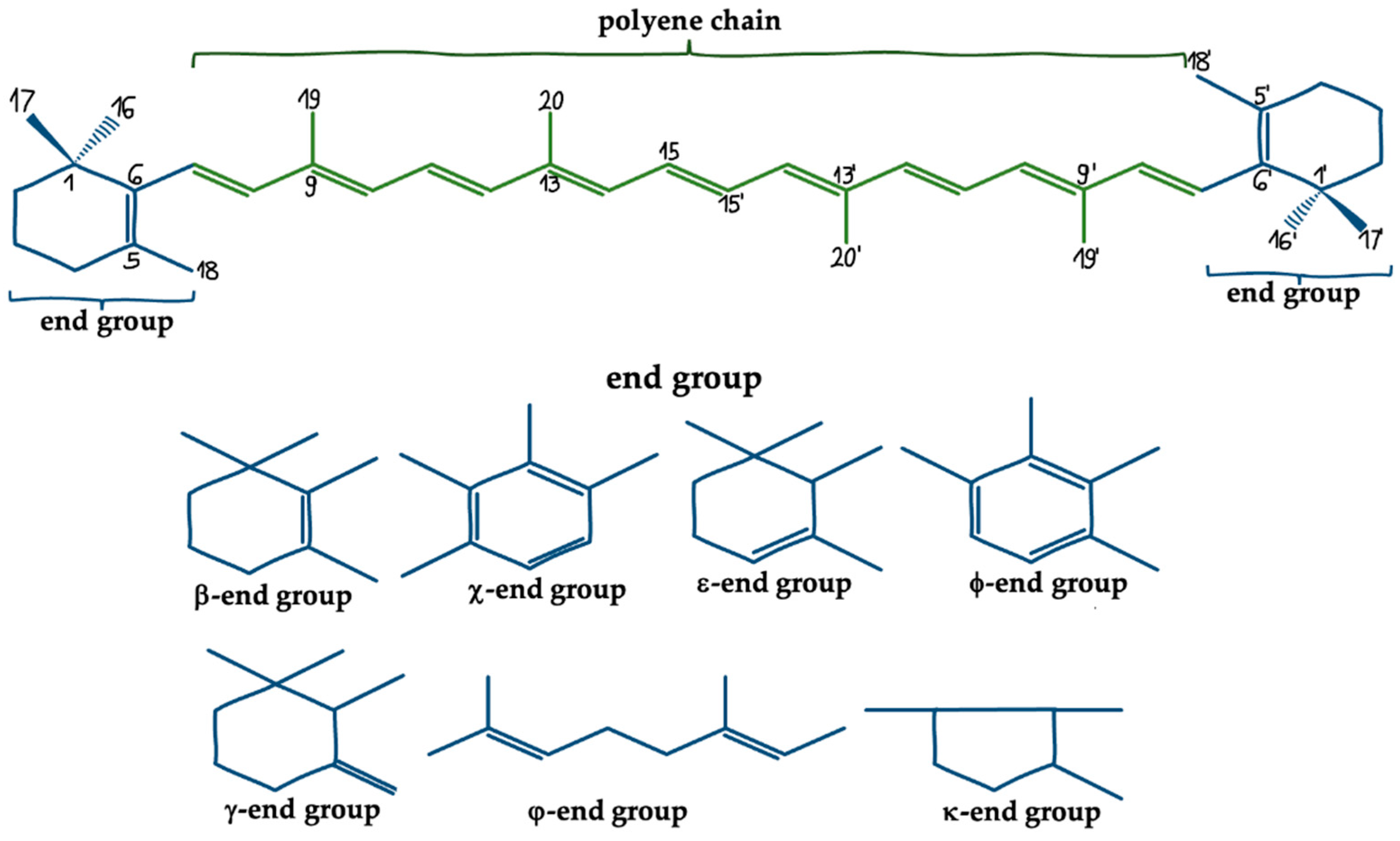
3. Marine Carotenoids

Marine and oceanic fauna and flora represent an enormous wealth of potential therapeutic agents, and amongst them, over 250 nautical carotenoids, which, generally, serve as natural, lipid-soluble pigments responsible for nature’s varied and vivid colors [71]. Humans cannot synthesize carotenoids de novo, therefore they must be obtained through the diet and converted into functional metabolites. Moreover, considering their low bioavailability in humans, different strategies, for instance, encapsulation in liposomes, micelles, or nanogels, increasing their absorption efficiency in the digestive tract have been developed [72,73]. Bioactive metabolites of marine algae, fungi, diatoms, and other marine organisms have been identified as pharmaceuticals with a wide variety of uses [71]. The significant role of marine carotenoids in neuroplasticity is underlined by the fact that there are currently several clinical studies conducted on the effects of carotenoids on cognitive impairment, neuroprotection, oxidative stress, and neurodegenerative diseases [74,75,76].

3.1. Fucoxanthin

One of the most promising carotenoids to be used in CNS diseases is FUC, the source of which is brown algae, mainly Sargassum siliquastrum, Undaria pinnatifida, Hijikia fusiformis, Alaria crassifolia, Laminaria japonica, and Cladosiphon okamuranus [77]. FUC is a naturally occurring compound with a brown color, showing the activity of provitamin A. This xanthophyll contains in its structure an epoxy group and conjugated carbonyl groups in the polyene chain. This structure translates into the antioxidant properties of FUC [78,79]. The biological activity of FUC is associated with a strong anti-inflammatory [80,81], antioxidant [82], anticancer and cell cycle suppressing [83,84], antidiabetic [85], hepatoprotective [86] and cardioprotective effects [87].
The neuroprotective effect of FUC has been confirmed in several in vitro and preclinical studies. It is proposed that Nrf2 signaling is the most important molecular mechanism for neuroprotection in FUC [88,89,90]. Hu et al. [88] evaluated the neurorestorative properties of FUC in a rat stroke model. The animals were administered this carotenoid at a dose of 30, 60, and 90 mg/kg 1 h prior to ischemia induction, and then rat cortical neurons were harvested and treated with 5, 10, and 20 µM FUC. It was observed that FUC dose-dependently reduced neurological deficits and infarct volume. Moreover, FUC blocked apoptosis by reducing the elevated cleaved caspase (C-CASP) 3 and Bcl-2/Bax ratio and also decreased oxidative stress by increasing SOD activity. The neuroprotective properties of FUC were confirmed in an in vitro study where a dose-dependency reduction of ROS accumulation and apoptosis was demonstrated through activating the Nrf2/HO-1 pathway initiated by Nrf2 nuclear translocation and increased levels of HO-1 [88]. Wu et al. [89] also suggested an impact of FUC on the activation of Nrf2 signaling, albeit by inhibiting the interaction of the Keap1 repressor protein with Nrf2. In 6-hydroxydopamine (6-OHDA) induced PC12 cells, FUC reduced ROS accumulation, cell apoptosis and membrane potential interference, as well as dose-dependently enhanced the activity of antioxidant enzymes: glutamate-cysteine ligase modifier subunit, nicotinamide heme oxygenase-1, and glutamate-cysteine ligase catalytic subunit. The FUC was then administered to the zebrafish treated with 6-OHDA. It was observed that FUC improved the granular region of the brain injury and enhanced the total swimming distance of the larvae [89]. Activation of the Nrf2/ARE pathway was also confirmed in a study by Zhang et al. [70], who found that FUC suppressed secondary brain injury, cerebral edema, neurological deficits, and apoptosis in a mouse traumatic brain injury (TBI) model. Moreover, in primary neurons, FUC promoted neuronal survival and inhibited oxidative stress by activating Nrf/ARE signaling, while in Nrf/ knockout mice, the neuroprotective effect of FUC was abolished, and activation of autophagy was observed [70]. Furthermore, in vitro studies demonstrated that FUC attenuated LPS-induced neuroinflammation by decreasing secretion of inflammatory mediators, including tumor necrosis factor α (TNF-α), NO, interleukin (IL) 1β, IL-6, and prostaglandin E2 (PGE2) [90]. Suppression of MAPK/AP-1 and Akt/NF-κB pathways, reduction of expression of inducible nitric oxide synthase (iNOS) and cyclooxygenase 2 (COX2) are also involved in the anti-inflammatory activity of FUC [90]. Moreover, FUC was also observed to suppress neuroinflammation by affecting NLRP3 inflammasome by inhibiting C-CASP 1. Expression and oligomerization of that apoptosis-associated speck-like protein containing a C-terminal caspase recruitment domain (ASC) are the major components of the inflammasome. Interestingly, it was proposed that FUC may also modulate the initiation step of the inflammasome signaling pathways as FUC was noted to reduce pro-IL-1β and phosphorylated IκBα expression [81]. Lin et al. [91] showed that FUC attenuates cognitive impairment induced by scopolamine in a mouse Alzheimer’s Disease (AD) model. Treatment of mice with scopolamine increased acetylcholinesterase (AChE) activity, as well as decreased BDNF expression and choline acetyltransferase activity, which was reversed by FUC. Moreover, it was shown that FUC directly inhibited AChE (IC50 81.2 µM) in a non-competitive manner, and based on molecular docking. It was found that FUC interacted with the peripheral anionic site of AChE [91]. A growing body of data from animal studies demonstrates the enormous potential of FUC in preventing disease or managing human health. Nevertheless, despite significant progress in characterizing its potential health-promoting effects, much research is still required to establish the appropriate protocol for human administration.

3.2. Astaxanthin

AST (3,3’-dihydroxy-β, β-carotene-4,4’-dione) is a natural xanthophylls [92,93]. Its predominant source in the diet are fish and seafood, mainly crawfish, crabs, shrimps, salmon, and pink trout [93,94,95]. The natural sources of AST are the algae Haematoccocus pluvalis [96,97], Chlorella zofingensis, as well as the yeast Xanthophyllomyces dendrorhous [77,95,96]. Eight isoprene units containing 40 carbon atoms form the compound’s chemical structure. The polyene carbon chain is terminated at both ends with β-ionone rings, each of which has one ketone group and one hydroxyl group in its structure. These groups are responsible for AST’s greater stability and polarity in relation to other known carotenoids [93]. It was proven that carotenoids containing a greater number of oxygen molecules with the same number of double bonds are characterized by higher photostability and more tremendous antioxidant potential [98]. The chemical structure of AST is responsible for its physicochemical and health-promoting properties. The hydrophobic carbon chain containing 9 conjugated double bonds and 2 unconjugated β-ionon rings is accountable for the quenching of ROS. The presence of oxygen in the end rings influences the hydrophilic properties of astaxanthin, contributing to the neutralization of free radicals and other oxidative substances in the aquatic environment. Such a hydrophilic-hydrophobic-hydrophilic structure of AST is analogous to the cell membrane, which allows it to be distributed across its entire width, enabling the removal of free radicals and ROS both on the surface of the membrane and inside it [92,93]. AST is characterized by a greater resistance to light and high temperature compared to other carotenoids and a free, rapid crossing of the BBB in animals [97].
The promising pro-health actions of AST include antioxidant, anti-inflammatory, antitumor, hepato-, cardio, and neuroprotective effects [77,78,79,93,94,95,96,97,98]. The potent antioxidant effect of AST manifests in high oxidative potential, production of chelate complexes with metals, and in the presence of metal ions generation of neutral radicals and aggregation into ester forms [99]. Wu et al. [100] assessed the effect of AST (intragastric administration; at the dose of 0.02% of daily diet 3 times a week) on D-galactose-induced brain aging in rats. It was observed that AST suppressed oxidative stress by enhancing the activity of antioxidant enzymes, including superoxide dismutase (SOD) and glutathione peroxidase, enhancing total antioxidant capacity and thiol levels. In addition, a reduction in the levels of antioxidant damage markers such as malondialdehyde (MDA), 8-hydroxy-2-deoxyguanosine, and protein carbonyl groups was observed in the brains of rats. Furthermore, AST augmented an anti-apoptotic index—Bcl2/Bax ratio as well as suppressed neuroinflammation as expressed by decreased COX2 expression. Moreover, it was found that the neuroregenerative properties of AST were associated with a reduction in histopathological changes in the hippocampus and an increase in BDNF expression in both the hippocampus and the brain of aging rats [100]. In a recent study, Aslankoc et al. [101] found that AST (oral administration, 100 mg/kg for 7 days) was protective against methotrexate damage in the hippocampus, cerebral cortex, cerebellar cortex, and blood in rats. In the control group, there was increased oxidative stress in the hippocampus, cerebral cortex, and blood, manifested by an increase in the total oxidative state (TOS) and a decrease in total antioxidant status (TAS), in contrast to the study group, in which AST alleviated oxidative stress. Moreover, AST suppressed histopathological changes in the hippocampus, cerebral cortex, and cerebellar cortex, including congestion, edema, and degenerative changes noted in the controls. In addition, AST was demonstrated to have anti-apoptotic and anti-inflammatory properties related to increased expression of myelin basic protein (MBP) and decreased CASP 3 levels, growth related oncogene (GRO), granulocyte colony-stimulating factor (GCSF), and iNOS [101]. Zhao et al. [102] suggested that AST could be a potential therapeutic agent for the treatment of neuropathic pain. C57BL/6 mice with spinal nerve ligation were administered intraperitoneally with AST at a dose of 5 mg/kg or 10 mg/kg from the 5th postoperative day for 23 days. It was shown that AST partially relieved neuropathic pain, and the analgesic effect was demonstrated on day 7. Subsequently, an in vitro study (spinal dorsal horns taken 11 days after spinal nerve ligation) found that AST reduced microglia activation and the expression of proinflammatory cytokines leading to inhibition of neuroinflammation. The anti-inflammatory effect of AST was manifested by the inhibition of p38 and Erk1/2 phosphorylation, as well as NFκB p65 nuclear translocation [102].
The promising use of AST in enhancing cognition is still being intensively researched. AST (administered orally at a dose of 25 mg/kg 5 times a week for 25 days) was shown to ameliorate memory impairment induced by doxorubicin in rats. Moreover, AST restored doxorubicin-induced histological changes in brain tissue, including degeneration and nuclear pyknosis in fascia dentata, hilus, and subiculum of the hippocampus, as well as focal hemorrhage in the area that separates the hippocampus from the striatum. Moreover, it was observed that the neuroprotective effect of AST is associated with a decrease in AChE activation, inhibition of oxidative stress, and overactive apoptotic processes [103]. In turn, Zhu et al. [104] assessed the effects of AST on cognition, oxidative stress, and neuroinflammation in mice with vascular dementia. AST (at a dose of 50 mg/kg, 100 mg/kg 200 mg/kg for 30 days) was shown to attenuate cognitive deficits in a dose-dependent manner as well as reduce oxidative stress, as observed by increasing SOD activity and reducing MDA. Moreover, a decrease in IL-1β expression and an enhancement of IL-4 expression were noted in the study group [104]. A recent study by Loganathan et al. [105] demonstrated that the astaxanthin-s-allylcysteine (AST-SAC) diester has a neuroprotective effect in alleviating cognitive deficits in diabetic rats by preventing spatial memory loss, as well as reducing brain tissue damage by inhibiting AChE activity, mitochondrial dysfunction, and oxidative stress. Moreover, in an in vitro study (SH-SY5Y neuronal cells treated with high glucose concentration), AST (at a dose of 5 μM, 10 μM and 15 μM) in a dose-dependent manner promoted neuronal viability by reducing the expression of pro-apoptotic proteins, leading to inhibition of apoptosis, increasing the level of endogenous antioxidant compounds reducing ROS generation, and by preventing mitochondrial dysfunction. The mechanism of preventing mitochondrial dysfunction goes on through modulating the membrane potential and the activity of oxidative phosphorylation complexes [105]. A randomized clinical trial, which enrolled 96 people (age 45–65) with mild memory impairment, investigated the effect of the AST-rich Haematococcus pluvialis extract. It was shown that in the group receiving AST at a dose of 6 mg/day and 12 mg/day for 12 weeks, improvement in performed tasks was significantly faster. That improvement was manifested by increased psychomotor speed, which is a marker of physical and mental coordination. Importantly, this study did not report any side effects from AST [106].
In addition to the research concerning the neuroprotective properties of AST alone in the treatment of CNS diseases, combination therapies are currently under consideration. Ata Yaseen Abdulqader et al. [107] assessed the effect of using AST in combination with valproic acid (VPA) in rats with pentylenetetrazole-induced epilepsy. VPA was shown to counteract the histopathological damage and behavioral disturbances induced by pentylenetetrazole. In contrast, AST alone (oral administration at a dose of 100 mg/kg) showed antiepileptic properties and increased anti-inflammatory activity compared to VPA alone. On the other hand, the use of AST/VPA combination therapy intensified the anti-epileptic effect, which was noted on the basis of the reduction of oxidative stress, the level of glutathione and TNF-α. Importantly, AST/VPA augmented the improvement in animal behavioral changes compared to VPA alone [107].

3.3. Siphonaxanthin

Siphonaxanthin (3,3′,19-trihydroxy-7,8-dihydro-8-oxo-a-carotene), a keto-carotenoid present in edible green algae including Codium fragile, Caulerpa lentillifera, and Umbraulva japonica, constitutes approximately 0.03–0.1% of their dry weight [108]. Siphonaxanthin contains an additional hydroxyl group that could contribute to its strong apoptosis-inducing effect [109]. The biological functions of this ketocarotenoid are associated with antioxidant activity [110], anti-inflammatory effect [111], suppression of cell viability, induction of apoptosis, more potent anti-angiogenic activity than FUC [112,113,114], and antiobesity [115,116]. However, additional in vivo studies, are needed to validate siphonaxanthin’s bioavailability and biological action.
Dambeck and Sandmann [110] showed that siphonaxanthin exerts an efficient outcome against the radical formation and lipid peroxidation [110]. Studies performed on the transfected human monocytic cell line, which was treated with siphonaxanthin at 1.0 µM concentration for 24 h, showed the anti-inflammatory effect of this carotenoid, manifested by the significant inhibition of the LPS- and TNF-α-induced NFκB activation. Moreover, pretreatment with siphonaxanthin at 1.0 µM concentration significantly suppressed the IL-1β-induced NFκB activation [117]. Ganesan et al. [113] revealed that siphonaxanthin might inhibit FGF-2 signaling. Human endothelial cells treated with siphonaxanthin at 0.1 and 0.5 µM concentration for 6 h exhibited the inhibition of FGF-2-induced intracellular proliferation and survival signals by down-regulating the FGF-2-induced phosphorylation of Akt and the Erk1/2 [113]. FGF signaling often leads to the concurrent activation of both the Raf/MAPK and the PI3K/Akt pathways, which, as was mentioned, are crucial for neurons survival or synaptic plasticity [118]. In addition, this carotenoid, at a concentration of 20 μM, reduced human leukemia cell viability (p < 0.05) within 6 h of treatment, inducing the apoptosis by decreasing expression of Bcl-2 and increasing activation of CASP 3 [112].

3.4. Mytiloxanthin

Mytiloxanthin (3,3′,8′-trihydroxy-7,8-didehydro-β,κ-caroten-6′-one), a metabolite of FUC, is another carotenoid with high antioxidant properties. Mytiloxanthin is widely distributed in marine mussels, oysters, and tunicates [77,119]. As it was mentioned before, carotenoids singlet oxygen-quenching activity depends on the number of conjugated double bonds, polyene chain structures, and functional groups, therefore Mytiloxanthin, which has more conjugated double bond than FUC, is suggested to have stronger quenching activity for singlet oxygen.
Maoka et al. [120] investigated the anti-oxidative activities of mytiloxanthin, they revealed that it exhibits a high quenching activity of singlet oxygen (61.6%) similar to that of AST (61.0%). Moreover, the researchers showed this compound has more powerful inhibitory activity on lipid peroxidation (20% formation of lipid hydroperoxide) than AST (24%), FUC (32%), and β-carotene (38%), at a final concentration of 167 μM [120].

3.5. Saproxanthin and Myxol

The two monocyclic carotenoids, seldom found in nature, (3R)-Saproxanthin and (3R,2′S)-Myxol, are produced by Saprospira grandis, marine bacterial strain 04OKA-13-27 and Anabaena variabilis ATCC 29413, marine bacterial strain P99-3, YM6-073, respectively [121]. These tetraterpenes are reported to possess neuroprotection against L-glutamate toxicity, lipid peroxidation prevention, and have powerful antioxidant potential, for instance, by their orientation in the head-group region of the phospholipids that form the bilayer, which leads to the reinforcement and stabilization of biological membranes. Therefore, these carotenoids may induce a reduction of membrane permeability to oxygen and may enhance protection against radical-induced peroxidation [122,123].
As was revealed by Shindo et al. [121] the antioxidant activity of saproxanthin and myxol is stronger than that of β-carotene or zeaxanthin. The team used the rat brain homogenate model to evaluate the inhibitory activities against lipid peroxidation by saproxanthin, myxol, and zeaxanhin. Saproxanthin showed the most potent effect with IC50 value 2.1, whereas myxol and zeaxanhin were 6.2 and 13.5 μM, respectively. Furthermore, the researchers tested saproxanthin and myxol inhibitory activities against L-glutamate toxicity in embryonic rat retinal neuron hybrid cells, and their concentration necessary to reduce glutamate-induced cell death by 50% (EC50 values) was 3.1 and 8.1 μM, respectively. At the same time the protective effect of AST and β-carotene was >500 and >100 μM, respectively [121]. The results show that these rare carotenoids might be expected to be useful for ameliorating tissue damage resulting from free radicals’ generation and subsequent cell membrane peroxidative deterioration, as well as possessing potent neuroprotective effect against L-glutamate toxicity and they may be helpful in cerebral ischemic disease treatment.
The biological effect of marine carotenoids described above are summarized in the Table 1 and Table 2.

4. Conclusions

There is considerable scientific and social interest in the use of natural compounds in the prevention and treatment of many diseases, including neurological disorders. In recent years, the demand for biologically active nutraceuticals implies the search and development of natural sources of these molecules. This led to an interest in marine compounds as unused and new natural sources. Marine sources are a great wealth of bioactive substances that have a beneficial effect on the human body, including carotenoids, which has been indicated in many scientific reports. There is strong scientific evidence that the marine carotenoids fucoxanthin and astaxanthin support the CNS. In turn, noteworthy as raising high hopes are mytiloxanthin, saproxanthin, and myxol. The enhancement of neuroplasticity by marine carotenoids is mainly related to anti-inflammatory and antioxidant effects. Nevertheless, activation of pathways related to neurogenesis and synaptogenesis mean that these compounds appear to have great therapeutic potential. However, it is necessary to conduct further preclinical and clinical studies that will be able to accurately determine the mechanism of their action, as well as dosing in particular CNS diseases.

Funding

This research received no external funding.

Institutional Review Board Statement

Not applicable.

Informed Consent Statement

Not applicable.

Data Availability Statement

No new data were created or analyzed in this study. Data sharing is not applicable to this article.

Conflicts of Interest

The authors declare no conflict of interest.

References

  1. Davinelli, S.; Ali, S.; Solfrizzi, V.; Scapagnini, G.; Corbi, G. Carotenoids and Cognitive Outcomes: A Meta-Analysis of Randomized Intervention Trials. Antioxidants 2021, 10, 223. [Google Scholar] [CrossRef]
  2. Mateos-Aparicio, P.; Rodríguez-Moreno, A. The Impact of Studying Brain Plasticity. Front. Cell. Neurosci. 2019, 13, 66. [Google Scholar] [CrossRef] [PubMed] [Green Version]
  3. Johnston, M.v.; Ishida, A.; Ishida, W.N.; Matsushita, H.B.; Nishimura, A.; Tsuji, M. Plasticity and Injury in the Developing Brain. Brain Dev. 2009, 31, 1. [Google Scholar] [CrossRef] [PubMed] [Green Version]
  4. Katsimpardi, L.; Lledo, P.M. Regulation of Neurogenesis in the Adult and Aging Brain. Curr. Opin. Neurobiol. 2018, 53, 131–138. [Google Scholar] [CrossRef] [PubMed]
  5. Niklison-Chirou, M.V.; Agostini, M.; Amelio, I.; Melino, G. Regulation of Adult Neurogenesis in Mammalian Brain. Int. J. Mol. Sci. 2020, 21, 4869. [Google Scholar] [CrossRef]
  6. Isaev, N.K.; Stelmashook, E.v.; Genrikhs, E.E. Neurogenesis and Brain Aging. Rev. Neurosci. 2019, 30, 573–580. [Google Scholar] [CrossRef] [PubMed] [Green Version]
  7. Bourgeois, J.-P. Synaptogenèses et Épigenèses Cérébrales. Méd./Sci. 2005, 21, 428–433. [Google Scholar] [CrossRef] [PubMed]
  8. Kowiański, P.; Lietzau, G.; Czuba, E.; Waśkow, M.; Steliga, A.; Moryś, J. BDNF: A Key Factor with Multipotent Impact on Brain Signaling and Synaptic Plasticity. Cell. Mol. Neurobiol. 2018, 38, 579. [Google Scholar] [CrossRef]
  9. Dyer, A.H.; Vahdatpour, C.; Sanfeliu, A.; Tropea, D. The Role of Insulin-Like Growth Factor 1 (IGF-1) in Brain Development, Maturation and Neuroplasticity. Neuroscience 2016, 325, 89–99. [Google Scholar] [CrossRef]
  10. Zechel, S.; Werner, S.; Unsicker, K.; Halbach, O. von B. und Expression and Functions of Fibroblast Growth Factor 2 (FGF-2) in Hippocampal Formation. Neuroscientist 2010, 16, 357–373. [Google Scholar] [CrossRef]
  11. Keefe, K.M.; Sheikh, I.S.; Smith, G.M. Targeting Neurotrophins to Specific Populations of Neurons: NGF, BDNF, and NT-3 and Their Relevance for Treatment of Spinal Cord Injury. Int. J. Mol. Sci. 2017, 18, 548. [Google Scholar] [CrossRef] [PubMed]
  12. Carter, A.R.; Chen, C.; Schwartz, P.M.; Segal, R.A. Brain-Derived Neurotrophic Factor Modulates Cerebellar Plasticity and Synaptic Ultrastructure. J. Neurosci. 2002, 22, 1316. [Google Scholar] [CrossRef] [PubMed]
  13. Uren, R.T.; Turnley, A.M. Regulation of Neurotrophin Receptor (Trk) Signaling: Suppressor of Cytokine Signaling 2 (SOCS2) Is a New Player. Front. Mol. Neurosci. 2014, 7, 39. [Google Scholar] [CrossRef] [PubMed] [Green Version]
  14. Ivanisevic, L.; Saragovi, H.U. Neurotrophins. Handb. Biol. Act. Pept. 2013, 1639–1646. [Google Scholar] [CrossRef]
  15. Huang, E.J.; Reichardt, L.F. Trk Receptors: Roles in Neuronal Signal Transduction. Annu. Rev. Biochem. 2003, 72, 609–642. [Google Scholar] [CrossRef] [Green Version]
  16. Meeker, R.B.; Williams, K.S. The P75 Neurotrophin Receptor: At the Crossroad of Neural Repair and Death. Neural Regen. Res. 2015, 10, 721. [Google Scholar] [CrossRef]
  17. Simanshu, D.K.; Nissley, D.v.; McCormick, F. RAS Proteins and Their Regulators in Human Disease. Cell 2017, 170, 17. [Google Scholar] [CrossRef] [Green Version]
  18. Sánchez-Alegría, K.; Flores-León, M.; Avila-Muñoz, E.; Rodríguez-Corona, N.; Arias, C. PI3K Signaling in Neurons: A Central Node for the Control of Multiple Functions. Int. J. Mol. Sci. 2018, 19, 3725. [Google Scholar] [CrossRef] [Green Version]
  19. Hemmings, B.A.; Restuccia, D.F. PI3K-PKB/Akt Pathway. Cold Spring Harb. Perspect. Biol. 2012, 4, a011189. [Google Scholar] [CrossRef] [Green Version]
  20. Liu, T.; Zhang, L.; Joo, D.; Sun, S.C. NF-ΚB Signaling in Inflammation. Signal Transduct. Target. Ther. 2017, 2, 17023. [Google Scholar] [CrossRef] [Green Version]
  21. Cantley, L.C. The Phosphoinositide 3-Kinase Pathway. Science 2002, 296, 1655–1657. [Google Scholar] [CrossRef] [PubMed]
  22. Vetter, M.L.; Martin-Zanca, D.; Parada, L.F.; Bishop, J.M.; Kaplan, D.R. Nerve Growth Factor Rapidly Stimulates Tyrosine Phosphorylation of Phospholipase C-Gamma 1 by a Kinase Activity Associated with the Product of the Trk Protooncogene. Proc. Natl. Acad. Sci. USA 1991, 88, 5650. [Google Scholar] [CrossRef] [PubMed] [Green Version]
  23. Reichardt, L.F. Neurotrophin-Regulated Signalling Pathways. Philos. Trans. R. Soc. B Biol. Sci. 2006, 361, 1545. [Google Scholar] [CrossRef] [PubMed] [Green Version]
  24. Corbit, K.C.; Foster, D.A.; Rosner, M.R. Protein Kinase Cδ Mediates Neurogenic but Not Mitogenic Activation of Mitogen-Activated Protein Kinase in Neuronal Cells. Mol. Cell. Biol. 1999, 19, 4209. [Google Scholar] [CrossRef] [PubMed] [Green Version]
  25. Britton, G. Structure and Properties of Carotenoids in Relation to Function. FASEB J. 1995, 9, 1551–1558. [Google Scholar] [CrossRef]
  26. Meléndez-Martínez, A.J.; Mapelli-Brahm, P.; Hornero-Méndez, D.; Vicario, I.M. CHAPTER 1 Structures, Nomenclature and General Chemistry of Carotenoids and Their Esters. Food Chem. Funct. Anal. 2019, 1–50. [Google Scholar] [CrossRef]
  27. Katayama, S.; Ogawa, H.; Nakamura, S. Apricot Carotenoids Possess Potent Anti-Amyloidogenic Activity in Vitro. J. Agric. Food Chem. 2011, 59, 12691–12696. [Google Scholar] [CrossRef]
  28. Grimmig, B.; Kim, S.H.; Nash, K.; Bickford, P.C.; Douglas Shytle, R. Neuroprotective Mechanisms of Astaxanthin: A Potential Therapeutic Role in Preserving Cognitive Function in Age and Neurodegeneration. GeroScience 2017, 39, 19. [Google Scholar] [CrossRef]
  29. Chisté, R.C.; Freitas, M.; Mercadante, A.Z.; Fernandes, E. Carotenoids Inhibit Lipid Peroxidation and Hemoglobin Oxidation, but Not the Depletion of Glutathione Induced by ROS in Human Erythrocytes. Life Sci. 2014, 99, 52–60. [Google Scholar] [CrossRef]
  30. Shaish, A.; Harari, A.; Kamari, Y.; Soudant, E.; Harats, D.; Ben-Amotz, A. A Carotenoid Algal Preparation Containing Phytoene and Phytofluene Inhibited LDL Oxidation In Vitro. Plant Foods Hum. Nutr. 2008, 63, 83–86. [Google Scholar] [CrossRef]
  31. Pérez-Gálvez, A.; Mínguez-Mosquera, M.I. Esterification of Xanthophylls and Its Effect on Chemical Behavior and Bioavailability of Carotenoids in the Human. Nutr. Res. 2005, 25, 631–640. [Google Scholar] [CrossRef]
  32. Papandreou, M.A.; Kanakis, C.D.; Polissiou, M.G.; Efthimiopoulos, S.; Cordopatis, P.; Margarity, M.; Lamari, F.N. Inhibitory Activity on Amyloid-β Aggregation and Antioxidant Properties of Crocus Sativus Stigmas Extract and Its Crocin Constituents. J. Agric. Food Chem. 2006, 54, 8762–8768. [Google Scholar] [CrossRef] [PubMed]
  33. Stahl, W.; Sies, H. Bioactivity and Protective Effects of Natural Carotenoids. Biochim. Biophys. Acta (BBA)-Mol. Basis Dis. 2005, 1740, 101–107. [Google Scholar] [CrossRef] [PubMed] [Green Version]
  34. Kaulmann, A.; Bohn, T. Carotenoids, Inflammation, and Oxidative Stress—Implications of Cellular Signaling Pathways and Relation to Chronic Disease Prevention. Nutr. Res. 2014, 34, 907–929. [Google Scholar] [CrossRef]
  35. Woodall, A.A.; Lee, S.W.M.; Weesie, R.J.; Jackson, M.J.; Britton, G. Oxidation of Carotenoids by Free Radicals: Relationship between Structure and Reactivity. Biochim. Biophys. Acta (BBA)-Gen. Subj. 1997, 1336, 33–42. [Google Scholar] [CrossRef]
  36. Ben-Dor, A.; Steiner, M.; Gheber, L.; Danilenko, M.; Dubi, N.; Linnewiel, K.; Zick, A.; Sharoni, Y.; Levy, J. Carotenoids Activate the Antioxidant Response Element Transcription System. Mol. Cancer Ther. 2005, 4, 177–186. [Google Scholar]
  37. Palozza, P.; Serini, S.; Torsello, A.; di Nicuolo, F.; Piccioni, E.; Ubaldi, V.; Pioli, C.; Wolf, F.I.; Calviello, G. β-Carotene Regulates NF-ΚB DNA-Binding Activity by a Redox Mechanism in Human Leukemia and Colon Adenocarcinoma Cells. J. Nutr. 2003, 133, 381–388. [Google Scholar] [CrossRef] [Green Version]
  38. Kim, J.; Cha, Y.N.; Surh, Y.J. A Protective Role of Nuclear Factor-Erythroid 2-Related Factor-2 (Nrf2) in Inflammatory Disorders. Mutat. Res./Fundam. Mol. Mech. Mutagenes. 2010, 690, 12–23. [Google Scholar] [CrossRef]
  39. Kansanen, E.; Kuosmanen, S.M.; Leinonen, H.; Levonenn, A.L. The Keap1-Nrf2 Pathway: Mechanisms of Activation and Dysregulation in Cancer. Redox Biol. 2013, 1, 45–49. [Google Scholar] [CrossRef] [Green Version]
  40. Jung, K.A.; Kwak, M.K. The Nrf2 System as a Potential Target for the Development of Indirect Antioxidants. Molecules 2010, 15, 7266–7291. [Google Scholar] [CrossRef] [Green Version]
  41. Piovan, A.; Filippini, R.; Corbioli, G.; Costa, V.D.; Giunco, E.M.V.; Burbello, G.; Pagetta, A.; Giusti, P.; Zusso, M. Carotenoid Extract Derived from Euglena Gracilis Overcomes Lipopolysaccharide-Induced Neuroinflammation in Microglia: Role of NF-ΚB and Nrf2 Signaling Pathways. Mol. Neurobiol. 2021, 58, 3515. [Google Scholar] [CrossRef] [PubMed]
  42. Huang, C.; Gan, D.; Fan, C.; Wen, C.; Li, A.; Li, Q.; Zhao, J.; Wang, Z.; Zhu, L.; Lu, D. The Secretion from Neural Stem Cells Pretreated with Lycopene Protects against Tert-Butyl Hydroperoxide-Induced Neuron Oxidative Damage. Oxidative Med. Cell. Longev. 2018, 2018, 5490218. [Google Scholar] [CrossRef] [PubMed] [Green Version]
  43. Park, H.A.; Hayden, M.M.; Bannerman, S.; Jansen, J.; Crowe-White, K.M. Anti-Apoptotic Effects of Carotenoids in Neurodegeneration. Molecules 2020, 25, 3453. [Google Scholar] [CrossRef] [PubMed]
  44. Zhao, B.; Ren, B.; Guo, R.; Zhang, W.; Ma, S.; Yao, Y.; Yuan, T.; Liu, Z.; Liu, X. Supplementation of Lycopene Attenuates Oxidative Stress Induced Neuroinflammation and Cognitive Impairment via Nrf2/NF-ΚB Transcriptional Pathway. Food Chem. Toxicol. 2017, 109, 505–516. [Google Scholar] [CrossRef]
  45. Thomas, G.M.; Huganir, R.L. MAPK Cascade Signalling and Synaptic Plasticity. Nat. Rev. Neurosci. 2004, 5, 173–183. [Google Scholar] [CrossRef]
  46. Besnard, A.; Galan-Rodriguez, B.; Vanhoutte, P.; Caboche, J. Elk-1 a Transcription Factor with Multiple Facets in the Brain. Front. Neurosci. 2011, 5, 35. [Google Scholar] [CrossRef] [Green Version]
  47. Cattaneo, A.; Cattane, N.; Begni, V.; Pariante, C.M.; Riva, M.A. The Human BDNF Gene: Peripheral Gene Expression and Protein Levels as Biomarkers for Psychiatric Disorders. Transl. Psychiatry 2016, 6, e958. [Google Scholar] [CrossRef]
  48. Dincheva, I.; Lynch, N.B.; Lee, F.S. The Role of BDNF in the Development of Fear Learning. Depress. Anxiety 2016, 33, 907. [Google Scholar] [CrossRef] [Green Version]
  49. Rocco, M.L.; Soligo, M.; Manni, L.; Aloe, L. Nerve Growth Factor: Early Studies and Recent Clinical Trials. Curr. Neuropharmacol. 2018, 16, 1455. [Google Scholar] [CrossRef]
  50. Stringham, N.T.; Holmes, P.V.; Stringham, J.M. Lutein Supplementation Increases Serum Brain-Derived Neurotrophic Factor (BDNF) in Humans. FASEB J. 2016, 30, 689.3. [Google Scholar] [CrossRef]
  51. Prakash, A.; Kumar, A. Implicating the Role of Lycopene in Restoration of Mitochondrial Enzymes and BDNF Levels in β-Amyloid Induced Alzheimer’s Disease. Eur. J. Pharmacol. 2014, 741, 104–111. [Google Scholar] [CrossRef] [PubMed]
  52. Sahin, K.; Orhan, C.; Tuzcu, M.; Sahin, N.; Juturu, V. Regular Exercise Training with Lutein/Zeaxanthin Isomers Regulates Brain Transcription Factors and Neurotrophic and Synaptic Proteins in Rats (P06-020-19). Curr. Dev. Nutr. 2019, 3. [Google Scholar] [CrossRef]
  53. Dong, Y.L.; Pu, K.J.; Duan, W.J.; Chen, H.C.; Chen, L.X.; Wang, Y.M. Involvement of Akt/CREB Signaling Pathways in the Protective Effect of EPA against Interleukin-1β-Induced Cytotoxicity and BDNF down-Regulation in Cultured Rat Hippocampal Neurons. BMC Neurosci. 2018, 19, 52. [Google Scholar] [CrossRef] [PubMed]
  54. Putcha, G.v.; Moulder, K.L.; Golden, J.P.; Bouillet, P.; Adams, J.A.; Strasser, A.; Johnson, E.M. Induction of BIM, a Proapoptotic BH3-Only BCL-2 Family Member, Is Critical for Neuronal Apoptosis. Neuron 2001, 29, 615–628. [Google Scholar] [CrossRef] [Green Version]
  55. Biswas, S.C.; Greene, L.A. Nerve Growth Factor (NGF) Down-Regulates the Bcl-2 Homology 3 (BH3) Domain-Only Protein Bim and Suppresses Its Proapoptotic Activity by Phosphorylation. J. Biol. Chem. 2002, 277, 49511–49516. [Google Scholar] [CrossRef] [Green Version]
  56. Jomova, K.; Valko, M. Health Protective Effects of Carotenoids and Their Interactions with Other Biological Antioxidants. Eur. J. Med. Chem. 2013, 70, 102–110. [Google Scholar] [CrossRef]
  57. Wen, X.; Huang, A.; Hu, J.; Zhong, Z.; Liu, Y.; Li, Z.; Pan, X.; Liu, Z. Neuroprotective Effect of Astaxanthin against Glutamate-Induced Cytotoxicity in HT22 Cells: Involvement of the Akt/GSK-3β Pathway. Neuroscience 2015, 303, 558–568. [Google Scholar] [CrossRef]
  58. Xu, L.; Zhu, J.; Yin, W.; Ding, X. Astaxanthin Improves Cognitive Deficits from Oxidative Stress, Nitric Oxide Synthase and Inflammation through Upregulation of PI3K/Akt in Diabetes Rat. Int. J. Clin. Exp. Pathol. 2015, 8, 6083. [Google Scholar]
  59. Lu, Y.; Wang, X.; Feng, J.; Xie, T.; Si, P.; Wang, W. Neuroprotective Effect of Astaxanthin on Newborn Rats Exposed to Prenatal Maternal Seizures. Brain Res. Bull. 2019, 148, 63–69. [Google Scholar] [CrossRef]
  60. Xiang, S.; Liu, F.; Lin, J.; Chen, H.; Huang, C.; Chen, L.; Zhou, Y.; Ye, L.; Zhang, K.; Jin, J.; et al. Fucoxanthin Inhibits β-Amyloid Assembly and Attenuates β-Amyloid Oligomer-Induced Cognitive Impairments. J. Agric. Food Chem. 2017, 65, 4092–4102. [Google Scholar] [CrossRef]
  61. Johnson, E.J.; Vishwanathan, R.; Schalch, W.; Poon, L.; Wittwer, J.; Johnson, M.A.; Hausman, D.; Davey, A.; Green, R.; Gearing, M.; et al. Brain Levels of Lutein (L) and Zeaxanthin (Z) Are Related to Cognitive Function in Centenarians. FASEB J. 2011, 25, 975.21. [Google Scholar] [CrossRef]
  62. Zhou, L.; Ouyang, L.; Lin, S.; Chen, S.; Liu, Y.J.; Zhou, W.; Wang, X. Protective Role of β-Carotene against Oxidative Stress and Neuroinflammation in a Rat Model of Spinal Cord Injury. Int. Immunopharmacol. 2018, 61, 92–99. [Google Scholar] [CrossRef] [PubMed]
  63. Hua, Y.; Xu, N.; Ma, T.; Liu, Y.; Xu, H.; Lu, Y. Anti-Inflammatory Effect of Lycopene on Experimental Spinal Cord Ischemia Injury via Cyclooxygenase-2 Suppression. Neuroimmunomodulation 2019, 26, 84–92. [Google Scholar] [CrossRef] [PubMed]
  64. Rahman, M.A.; Rhim, H. Therapeutic Implication of Autophagy in Neurodegenerative Diseases. BMB Rep. 2017, 50, 345. [Google Scholar] [CrossRef] [Green Version]
  65. Fung, F.K.C.; Law, B.Y.K.; Lo, A.C.Y. Lutein Attenuates Both Apoptosis and Autophagy upon Cobalt (II) Chloride-Induced Hypoxia in Rat Műller Cells. PLoS ONE 2016, 11, e0167828. [Google Scholar] [CrossRef]
  66. Chang, C.J.; Lin, J.F.; Hsiao, C.Y.; Chang, H.H.; Li, H.J.; Chang, H.H.; Lee, G.A.; Hung, C.F. Lutein Induces Autophagy via Beclin-1 Upregulation in IEC-6 Rat Intestinal Epithelial Cells. Am. J. Chin. Med. 2017, 45, 1273–1291. [Google Scholar] [CrossRef]
  67. Zeng, C.; Li, H.; Fan, Z.; Zhong, L.; Guo, Z.; Guo, Y.; Xi, Y. Crocin-Elicited Autophagy Rescues Myocardial Ischemia/Reperfusion Injury via Paradoxical Mechanisms. Am. J. Chin. Med. 2016, 44, 515–530. [Google Scholar] [CrossRef]
  68. Shen, M.; Chen, K.; Lu, J.; Cheng, P.; Xu, L.; Dai, W.; Wang, F.; He, L.; Zhang, Y.; Chengfen, W.; et al. Protective Effect of Astaxanthin on Liver Fibrosis through Modulation of TGF- β 1 Expression and Autophagy. Mediat. Inflamm. 2014, 2014, 954502. [Google Scholar] [CrossRef] [Green Version]
  69. Buyuklu, M.; Kandemir, F.M.; Ozkaraca, M.; Set, T.; Bakirci, E.M.; Topal, E.; Ileriturk, M.; Turkmen, K. Benefical Effects of Lycopene against Contrast Medium-Induced Oxidative Stress, Inflammation, Autophagy, and Apoptosis in Rat Kidney. Hum. Exp. Toxicol. 2015, 34, 487–496. [Google Scholar] [CrossRef]
  70. Zhang, L.; Wang, H.; Fan, Y.; Gao, Y.; Li, X.; Hu, Z.; Ding, K.; Wang, Y.; Wang, X. Fucoxanthin Provides Neuroprotection in Models of Traumatic Brain Injury via the Nrf2-ARE and Nrf2-Autophagy Pathways. Sci. Rep. 2017, 7, 1–15. [Google Scholar] [CrossRef] [Green Version]
  71. Maoka, T. Carotenoids as Natural Functional Pigments. J. Nat. Med. 2020, 74, 1. [Google Scholar] [CrossRef] [PubMed] [Green Version]
  72. Li, H.; Xu, Y.; Sun, X.; Wang, S.; Wang, J.; Zhu, J.; Wang, D.; Zhao, L. Stability, Bioactivity, and Bioaccessibility of Fucoxanthin in Zein-Caseinate Composite Nanoparticles Fabricated at Neutral PH by Antisolvent Precipitation. Food Hydrocoll. 2018, 84, 379–388. [Google Scholar] [CrossRef]
  73. Huang, Z.; Xu, L.; Zhu, X.; Hu, J.; Peng, H.; Zeng, Z.; Xiong, H. Stability and Bioaccessibility of Fucoxanthin in Nanoemulsions Prepared from Pinolenic Acid-Contained Structured Lipid. Int. J. Food Eng. 2017, 13. [Google Scholar] [CrossRef] [Green Version]
  74. Effect of an Antioxidants Mix on Cognitive Performance and Well Being: The Bacopa, Licopene, Astaxantina, Vitamin B12-Full Text View-ClinicalTrials.Gov. Available online: https://clinicaltrials.gov/ct2/show/NCT03825042?term=astaxanthin&draw=3&rank=23 (accessed on 8 November 2021).
  75. Effect of Astaxanthin on the Patients with Alzheimer Disease-Full Text View-ClinicalTrials.Gov. Available online: https://clinicaltrials.gov/ct2/show/NCT05015374?term=astaxanthin&draw=2&rank=1 (accessed on 8 November 2021).
  76. A Randomised, Double-Blind, Placebo-Controlled, Parallel Study of the Effect of BrainPhyt on Cognitive Function in Healthy Older Subjects-Full Text View-ClinicalTrials.Gov. Available online: https://clinicaltrials.gov/ct2/show/NCT04832412?term=fucoxanthin&draw=2&rank=5 (accessed on 8 November 2021).
  77. Galasso, C.; Corinaldesi, C.; Sansone, C. Carotenoids from Marine Organisms: Biological Functions and Industrial Applications. Antioxidants 2017, 6, 96. [Google Scholar] [CrossRef] [Green Version]
  78. Rodrigues, E.; Mariutti, L.R.B.; Mercadante, A.Z. Scavenging Capacity of Marine Carotenoids against Reactive Oxygen and Nitrogen Species in a Membrane-Mimicking System. Mar. Drugs 2012, 10, 1784. [Google Scholar] [CrossRef]
  79. Riccioni, G.; D’Orazio, N.; Franceschelli, S.; Speranza, L. Marine Carotenoids and Cardiovascular Risk Markers. Mar. Drugs 2011, 9, 1166. [Google Scholar] [CrossRef] [Green Version]
  80. D’Orazio, N.; Gammone, M.A.; Gemello, E.; de Girolamo, M.; Cusenza, S.; Riccioni, G. Marine Bioactives: Pharmacological Properties and Potential Applications against Inflammatory Diseases. Mar. Drugs 2012, 10, 812. [Google Scholar] [CrossRef] [Green Version]
  81. Lee, A.H.; Shin, H.Y.; Park, J.H.; Koo, S.Y.; Kim, S.M.; Yang, S.H. Fucoxanthin from Microalgae Phaeodactylum Tricornutum Inhibits Pro-Inflammatory Cytokines by Regulating Both NF-ΚB and NLRP3 Inflammasome Activation. Sci. Rep. 2021, 11, 1–12. [Google Scholar] [CrossRef]
  82. Ha, A.W.; Na, S.J.; Kim, W.K. Antioxidant Effects of Fucoxanthin Rich Powder in Rats Fed with High Fat Diet. Nutr. Res. Pract. 2013, 7, 475. [Google Scholar] [CrossRef] [Green Version]
  83. Rengarajan, T.; Rajendran, P.; Nandakumar, N.; Balasubramanian, M.P.; Nishigaki, I. Cancer Preventive Efficacy of Marine Carotenoid Fucoxanthin: Cell Cycle Arrest and Apoptosis. Nutrients 2013, 5, 4978. [Google Scholar] [CrossRef] [Green Version]
  84. Das, S.K.; Hashimoto, T.; Kanazawa, K. Growth Inhibition of Human Hepatic Carcinoma HepG2 Cells by Fucoxanthin Is Associated with Down-Regulation of Cyclin D. Biochim. Biophys. Acta (BBA)-Gen. Subj. 2008, 1780, 743–749. [Google Scholar] [CrossRef] [PubMed] [Green Version]
  85. Maeda, H.; Hosokawa, M.; Sashima, T.; Miyashita, K. Dietary Combination of Fucoxanthin and Fish Oil Attenuates the Weight Gain of White Adipose Tissue and Decreases Blood Glucose in Obese/Diabetic KK-A y Mice. J. Agric. Food Chem. 2007, 55, 7701–7706. [Google Scholar] [CrossRef] [PubMed]
  86. Mayer, C.; Côme, M.; Blanckaert, V.; Zittelli, G.C.; Faraloni, C.; Nazih, H.; Ouguerram, K.; Mimouni, V.; Chénais, B. Effect of Carotenoids from Phaeodactylum Tricornutum on Palmitate-Treated HepG2 Cells. Molecules 2020, 25, 2845. [Google Scholar] [CrossRef] [PubMed]
  87. Chang, P.M.; Li, K.L.; Lin, Y.C. Fucoidan–Fucoxanthin Ameliorated Cardiac Function via IRS1/GRB2/SOS1, GSK3β/CREB Pathways and Metabolic Pathways in Senescent Mice. Mar. Drugs 2019, 17, 69. [Google Scholar] [CrossRef] [PubMed] [Green Version]
  88. Hu, L.; Chen, W.; Tian, F.; Yuan, C.; Wang, H.; Yue, H. Neuroprotective Role of Fucoxanthin against Cerebral Ischemic/Reperfusion Injury through Activation of Nrf2/HO-1 Signaling. Biomed. Pharmacother. 2018, 106, 1484–1489. [Google Scholar] [CrossRef] [PubMed]
  89. Wu, W.; Han, H.; Liu, J.; Tang, M.; Wu, X.; Cao, X.; Zhao, T.; Lu, Y.; Niu, T.; Chen, J.; et al. Fucoxanthin Prevents 6-OHDA-Induced Neurotoxicity by Targeting Keap1. Oxidative Med. Cell. Longev. 2021, 2021, 6688708. [Google Scholar] [CrossRef]
  90. Zhao, D.; Kwon, S.H.; Chun, Y.S.; Gu, M.Y.; Yang, H.O. Anti-Neuroinflammatory Effects of Fucoxanthin via Inhibition of Akt/NF-ΚB and MAPKs/AP-1 Pathways and Activation of PKA/CREB Pathway in Lipopolysaccharide-Activated BV-2 Microglial Cells. Neurochem. Res. 2016, 42, 667–677. [Google Scholar] [CrossRef]
  91. Lin, J.; Huang, L.; Yu, J.; Xiang, S.; Wang, J.; Zhang, J.; Yan, X.; Cui, W.; He, S.; Wang, Q. Fucoxanthin, a Marine Carotenoid, Reverses Scopolamine-Induced Cognitive Impairments in Mice and Inhibits Acetylcholinesterase in Vitro. Mar. Drugs 2016, 14. [Google Scholar] [CrossRef] [Green Version]
  92. Higuera-Ciapara, I.; Félix-Valenzuela, L.; Goycoolea, F.M. Astaxanthin: A Review of Its Chemistry and Applications. Crit. Rev. Food Sci. Nutr. 2007, 46, 185–196. [Google Scholar] [CrossRef]
  93. Brotosudarmo, T.H.P.; Limantara, L.; Setiyono, E. Heriyanto Structures of Astaxanthin and Their Consequences for Therapeutic Application. Int. J. Food Sci. 2020, 2020, 2156582. [Google Scholar] [CrossRef]
  94. Ambati, R.R.; Moi, P.S.; Ravi, S.; Aswathanarayana, R.G. Astaxanthin: Sources, Extraction, Stability, Biological Activities and Its Commercial Applications—A Review. Mar. Drugs 2014, 12, 128. [Google Scholar] [CrossRef] [PubMed]
  95. Kumar, S.; Kumar, R.; Kumari, A.; Panwar, A. Astaxanthin: A Super Antioxidant from Microalgae and Its Therapeutic Potential. J. Basic Microbiol. 2021. [Google Scholar] [CrossRef] [PubMed]
  96. Raza, S.H.A.; Naqvi, S.R.Z.; Abdelnour, S.A.; Schreurs, N.; Mohammedsaleh, Z.M.; Khan, I.; Shater, A.F.; Abd El-Hack, M.E.; Khafaga, A.F.; Quan, G.; et al. Beneficial Effects and Health Benefits of Astaxanthin Molecules on Animal Production: A Review. Res. Vet. Sci. 2021, 138, 69–78. [Google Scholar] [CrossRef]
  97. Aoi, W.; Naito, Y.; Sakuma, K.; Kuchide, M.; Tokuda, H.; Maoka, T.; Toyokuni, S.; Oka, S.; Yasuhara, M.; Yoshikawa, T. Astaxanthin Limits Exercise-Induced Skeletal and Cardiac Muscle Damage in Mice. Antioxid. Redox Signal. 2004, 5, 139–144. [Google Scholar] [CrossRef]
  98. Camera, E.; Mastrofrancesco, A.; Fabbri, C.; Daubrawa, F.; Picardo, M.; Sies, H.; Stahl, W. Astaxanthin, Canthaxanthin and β-Carotene Differently Affect UVA-Induced Oxidative Damage and Expression of Oxidative Stress-Responsive Enzymes. Exp. Dermatol. 2009, 18, 222–231. [Google Scholar] [CrossRef]
  99. Focsan, A.L.; Polyakov, N.E.; Kispert, L.D. Photo Protection of Haematococcus Pluvialis Algae by Astaxanthin: Unique Properties of Astaxanthin Deduced by EPR, Optical and Electrochemical Studies. Antioxidants 2017, 6, 80. [Google Scholar] [CrossRef] [Green Version]
  100. Wu, W.; Wang, X.; Xiang, Q.; Meng, X.; Peng, Y.; Du, N.; Liu, Z.; Sun, Q.; Wang, C.; Liu, X. Astaxanthin Alleviates Brain Aging in Rats by Attenuating Oxidative Stress and Increasing BDNF Levels. Food Funct. 2013, 5, 158–166. [Google Scholar] [CrossRef]
  101. Aslankoc, R.; Ozmen, O.; Yalcın, A. Astaxanthin Ameliorates Damage to the Cerebral Cortex, Hippocampus and Cerebellar Cortex Caused by Methotrexate. Biotech. Histochem. 2021, 1–12. [Google Scholar] [CrossRef]
  102. Zhao, L.; Tao, X.; Song, T. Astaxanthin Alleviates Neuropathic Pain by Inhibiting the MAPKs and NF-ΚB Pathways. Eur. J. Pharmacol. 2021, 912, 174575. [Google Scholar] [CrossRef]
  103. Emad El-Agamy, S.; Kamal Abdel-Aziz, A.; Wahdan, S.; Esmat, A.; Azab, S.S. Astaxanthin Ameliorates Doxorubicin-Induced Cognitive Impairment (Chemobrain) in Experimental Rat Model: Impact on Oxidative, Inflammatory, and Apoptotic Machineries. Mol. Neurobiol. 2018, 55, 5727–5740. [Google Scholar] [CrossRef]
  104. Zhu, N.; Liang, X.; Zhang, M.; Yin, X.; Yang, H.; Zhi, Y.; Ying, G.; Zou, J.; Chen, L.; Yao, X.; et al. Astaxanthin Protects Cognitive Function of Vascular Dementia. Behav. Brain Funct. 2020, 16, 1–10. [Google Scholar] [CrossRef] [PubMed]
  105. Loganathan, C.; Sakayanathan, P.; Thayumanavan, P. Astaxanthin-s-Allyl Cysteine Diester against High Glucose-Induced Neuronal Toxicity in Vitro and Diabetes-Associated Cognitive Decline in Vivo: Effect on P53, Oxidative Stress and Mitochondrial Function. NeuroToxicology 2021, 86, 114–124. [Google Scholar] [CrossRef] [PubMed]
  106. Katagiri, M.; Satoh, A.; Tsuji, S.; Shirasawa, T. Effects of Astaxanthin-Rich Haematococcus Pluvialis Extract on Cognitive Function: A Randomised, Double-Blind, Placebo-Controlled Study. J. Clin. Biochem. Nutr. 2012, 51, 102. [Google Scholar] [CrossRef] [PubMed] [Green Version]
  107. Ata Yaseen Abdulqader, Y.; Abdel Kawy, H.S.; Mohammed Alkreathy, H.; Abdullah Rajeh, N. The Potential Antiepileptic Activity of Astaxanthin in Epileptic Rats Treated with Valproic Acid. Saudi Pharm. J. SPJ 2021, 29, 418. [Google Scholar] [CrossRef] [PubMed]
  108. Ricketts, T.R. The Structures of Siphonein and Siphonaxanthin from Codium Fragile. Phytochemistry 1971, 10, 155–160. [Google Scholar] [CrossRef]
  109. Sugawara, T.; Ganesan, P.; Li, Z.; Manabe, Y.; Hirata, T. Siphonaxanthin, a Green Algal Carotenoid, as a Novel Functional Compound. Mar. Drugs 2014, 12, 3660. [Google Scholar] [CrossRef] [Green Version]
  110. Dambeck, M.; Sandmann, G. Antioxidative Activities of Algal Keto Carotenoids Acting as Antioxidative Protectants in the Chloroplast. Photochem. Photobiol. 2014, 90, 814–819. [Google Scholar] [CrossRef]
  111. Manabe, Y.; Takii, Y.; Sugawara, T. Siphonaxanthin, a Carotenoid from Green Algae, Suppresses Advanced Glycation End Product-Induced Inflammatory Responses. J. Nat. Med. 2019, 74, 127–134. [Google Scholar] [CrossRef]
  112. Ganesan, P.; Noda, K.; Manabe, Y.; Ohkubo, T.; Tanaka, Y.; Maoka, T.; Sugawara, T.; Hirata, T. Siphonaxanthin, a Marine Carotenoid from Green Algae, Effectively Induces Apoptosis in Human Leukemia (HL-60) Cells. Biochim. Biophys. Acta (BBA)-Gen. Subj. 2011, 1810, 497–503. [Google Scholar] [CrossRef]
  113. Ganesan, P.; Matsubara, K.; Sugawara, T.; Hirata, T. Marine Algal Carotenoids Inhibit Angiogenesis by Down-Regulating FGF-2-Mediated Intracellular Signals in Vascular Endothelial Cells. Mol. Cell. Biochem. 2013, 380, 1–9. [Google Scholar] [CrossRef]
  114. Ganesan, P.; Matsubara, K.; Ohkubo, T.; Tanaka, Y.; Noda, K.; Sugawara, T.; Hirata, T. Anti-Angiogenic Effect of Siphonaxanthin from Green Alga, Codium Fragile. Phytomedicine 2010, 17, 1140–1144. [Google Scholar] [CrossRef] [PubMed] [Green Version]
  115. Zheng, J.; Manabe, Y.; Sugawara, T. Siphonaxanthin, a Carotenoid from Green Algae Codium Cylindricum, Protects Ob/Ob Mice Fed on a High-Fat Diet against Lipotoxicity by Ameliorating Somatic Stresses and Restoring Anti-Oxidative Capacity. Nutr. Res. 2020, 77, 29–42. [Google Scholar] [CrossRef] [PubMed]
  116. Zheng, J.; Li, Z.; Manabe, Y.; Kim, M.; Goto, T.; Kawada, T.; Sugawara, T. Siphonaxanthin, a Carotenoid From Green Algae, Inhibits Lipogenesis in Hepatocytes via the Suppression of Liver X Receptor α Activity. Lipids 2018, 53, 41–52. [Google Scholar] [CrossRef]
  117. Manabe, Y.; Tomonaga, N.; Maoka, T.; Sugawara, T. Multivariate Analysis Reveals That Unsubstituted β-Ring and C8-Keto Structures Are Important Factors for Anti-Inflammatory Activity of Carotenoids. Nutrients 2021, 13, 3699. [Google Scholar] [CrossRef]
  118. Eswarakumar, V.P.; Lax, I.; Schlessinger, J. Cellular Signaling by Fibroblast Growth Factor Receptors. Cytokine Growth Factor Rev. 2005, 16, 139–149. [Google Scholar] [CrossRef]
  119. Khare, A.; Moss, G.P.; Weedon, B.C.L. Mytiloxanthin and Isomytiloxanthin, Two Novel Acetylenic Carotenoids. Tetrahedron Lett. 1973, 14, 3921–3924. [Google Scholar] [CrossRef]
  120. Maoka, T.; Nishino, A.; Yasui, H.; Yamano, Y.; Wada, A. Anti-Oxidative Activity of Mytiloxanthin, a Metabolite of Fucoxanthin in Shellfish and Tunicates. Mar. Drugs 2016, 14, 93. [Google Scholar] [CrossRef] [Green Version]
  121. Shindo, K.; Kikuta, K.; Suzuki, A.; Katsuta, A.; Kasai, H.; Yasumoto-Hirose, M.; Matsuo, Y.; Misawa, N.; Takaichi, S. Rare Carotenoids, (3R)-Saproxanthin and (3R,2′S)-Myxol, Isolated from Novel Marine Bacteria (Flavobacteriaceae) and Their Antioxidative Activities. Appl. Microbiol. Biotechnol. 2007, 74, 1350–1357. [Google Scholar] [CrossRef]
  122. Subczynski, W.K.; Markowska, E.; Sielewiesiuk, J. Effect of Polar Carotenoids on the Oxygen Diffusion-Concentration Product in Lipid Bilayers. An EPR Spin Label Study. Biochim. Biophys. Acta (BBA)-Biomembr. 1991, 1068, 68–72. [Google Scholar] [CrossRef]
  123. Woodall, A.A.; Britton, G.; Jackson, M.J. Carotenoids and Protection of Phospholipids in Solution or in Liposomes against Oxidation by Peroxyl Radicals: Relationship between Carotenoid Structure and Protective Ability. Biochim. Biophys. Acta (BBA)-Gen. Subj. 1997, 1336, 575–586. [Google Scholar] [CrossRef]
Figure 1. The chemical structure of carotenoids and their end groups.
Ijms 23 01990 g001
Figure 2. Marine carotenoids actions on signaling pathways enhancing neuroplasticity. Abbreviations: Akt—protein kinase B; ARE—antioxidant response element; crk—CT10 regulator of kinase; C3G—guanyl-nucleotide exchange factor; DAG—diacylglycerol; Erk—extracellular signal-regulated kinase; FRS-2—fibroblast growth factor receptor substrate 2; Grb-2—growth factor receptor-bound protein 2; IκB—inhibitor of NFκB; IKK—IκB kinase; IP3—inositol trisphosphate; Keap1—kelch-like-ECH-associated protein 1; MAPK—mitogen-activated protein kinase; MEK—mitogen-activated protein kinase kinase; NFκB—nuclear factor kappa B; Nrf2—nuclear factor erythroid 2-related factor 2; PI3K—phosphoinositide-3-kinase–protein kinase B; PKC—protein kinase C; PLC—phospholipase C; Raf—rapidly accelerated fibrosarcoma; Rap1—Ras-related protein 1; ROS—Reactive oxygen species; Shc—Src homology and containing protein; SOS—son of sevenless; Trk—tropomyosin receptor kinase.
Ijms 23 01990 g002
Table 1. In vitro studies of biological roles of marine carotenoids.
CarotenoidEffectModelBioactive ConcentrationTargetRef.
Fucoxanthinneuroprotectionrat cortical neurons5, 10 and 20 μMNrf2 signaling[88]
neuroprotectionPC12 cells0.5, 1, 2 and 5 μMNrf2 signaling[89]
anti-neuroinflammationBV-2 microglial cells5, 10, and 20 μMMAPKs and NF-κB signaling[90]
anti-neuroinflammationbone marrow-derived macrophages, bone marrow-derived dendritic cells, astrocytes40 μMNF-κB and NLRP3 inflammasome signaling[81]
Astaxanthinanti-neuroinflammationBV2 cells, PC12 cells, primary astrocytes5 or 10 μMMAPKs and NF-κB signaling[102]
neuronal viabilityhuman neuronal cell line
SH-SY5Y
5, 10 and 15 μMpro-apoptotic proteins[105]
Siphonaxanthinanti-neuroinflammationhuman monocytic cells1 μM for 24 hNF-κB signaling[117]
neuron survival synaptic plasticityhuman endothelial cells0.1 and 0.5 μM for 6 hFGF-2 signaling[113]
anti-proliferativehuman leukemia cells20 μMBcl-2, CASP 3[112]
Saproxanthin and Myxolneuroprotectionembryonic rat retinal neuron hybrid cells3.1 and 8.1 μM, respectivelyL-glutamate toxicity[121]
Table 2. In vivo and clinical studies of biological roles of marine carotenoids.
CarotenoidEffectModelBioactive ConcentrationTargetRef.
Fucoxanthinneuroprotectionrat stroke30, 60 and 90 mg/kgNrf2 signaling[88]
neuroprotectionzebrafish6.25, 12.5, 25 and 50 μg/mLNrf2 signaling[89]
neuroprotectiontraumatic brain injury mice50, 100 and 200 mg/kgNrf2/ARE signaling[70]
cognitive impairments
attenuation
Alzheimer’s Disease mice50, 100 and 200 mg/kgAChE, BDNF[91]
Astaxanthinantioxidation anti-neuroinflammation, neuroregenerationrats’ brain0.02% of daily diet,
3 times a week
antioxidant enzymes COX2, BDNF[100]
anti-apoptotic, anti-inflammation, oxidative stress alleviationrats100 mg/kg for 7 daysMBP, CASP 3, iNOS[101]
neuropathic pain alleviationC57BL/6 mice5 or 10 mg/kg for 23 daysMAPKs and NF-κB signaling[102]
neuroprotectionrats25 mg/kg 5 times a week for 25 daysAChE[103]
oxidative stress alleviationvascular dementia mice50, 100 and 200 mg/kg for 30 daysSOD, MDA, IL-4, IL-1β[104]
psychomotor speed improvementpeople with mild memory impairment6 and 12 mg/day for 12 weeks[106]
Antiepileptic anti-inflammationepileptic rats100 mg/kg[107]
Publisher’s Note: MDPI stays neutral with regard to jurisdictional claims in published maps and institutional affiliations.

