
Experimental Chemotherapy-Induced Mucositis: A Scoping Review Guiding the Design of Suitable Preclinical Models


 and
and 
Abstract
1. Introduction
2. Results
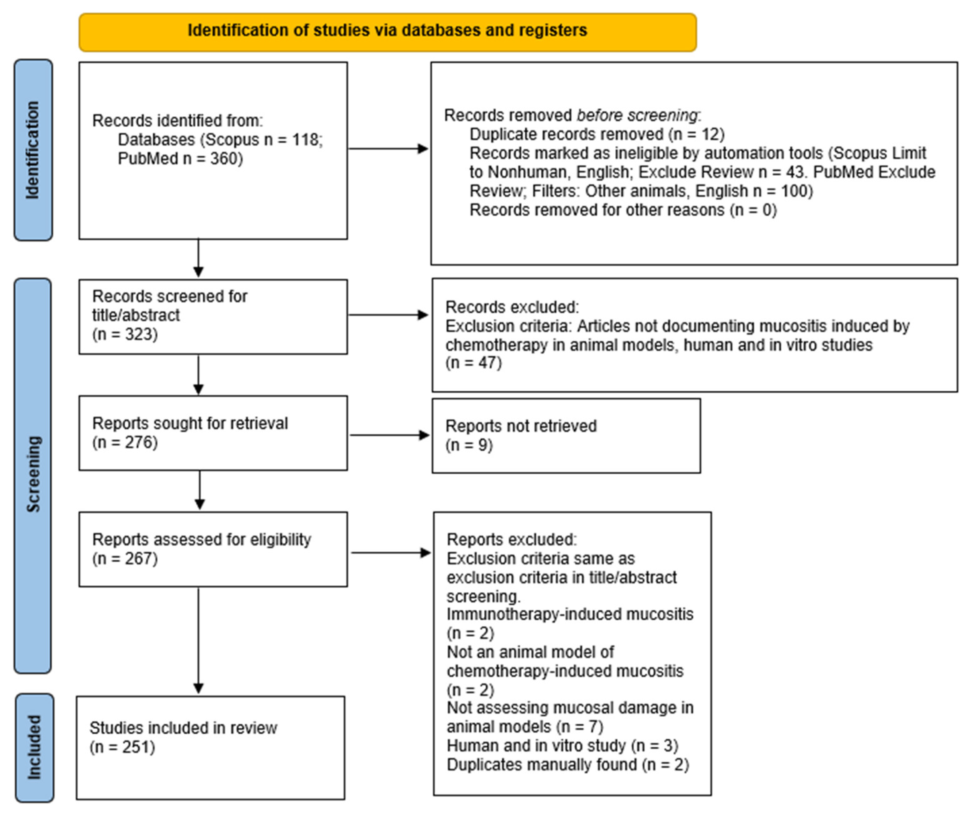
2.1. Search Results
2.2. Chemotherapy-Induced Intestinal Mucositis
2.2.1. Assessment of Intestinal Mucositis
2.2.2. Animal Models of Intestinal Mucositis Related to Specific Chemotherapeutic Drugs
5-Fluorouracil (5-FU)
Irinotecan
Platinum-Based Chemotherapy Drugs
Methotrexate
Other Chemotherapeutic Agents
2.3. Chemotherapy-Induced Oral Mucositis
2.3.1. Assessment of Oral Mucositis
2.3.2. Animal Models of Oral Mucositis Related to Specific Chemotherapeutic Drugs
5-Fluorouracil
Irinotecan
Topotecan
Methotrexate
Other Chemotherapeutic Agents
2.3.3. Induction of Oral Mucositis in Animal Models
3. Discussion
3.1. Translatability of Models
3.2. Pathophysiology
3.3. Optimal Dosage
3.4. Limitations
4. Methods
4.1. Protocol and Search Strategy
4.2. Eligibility Criteria
4.3. Data Selection and Collection
5. Conclusions
Supplementary Materials
Author Contributions
Funding
Data Availability Statement
Acknowledgments
Conflicts of Interest
References
- Rubenstein, E.B.; Peterson, D.E.; Schubert, M.; Keefe, D.; McGuire, D.; Epstein, J.; Elting, L.S.; Fox, P.C.; Cooksley, C.; Sonis, S.T.; et al. Clinical practice guidelines for the prevention and treatment of cancer therapy-induced oral and gastrointestinal mucositis. Cancer 2004, 100, 2026–2046. [Google Scholar] [CrossRef] [PubMed]
- Blijlevens, N.; Donnelly, J.; De Pauw, B. Mucosal barrier injury: Biology, pathology, clinical counterparts and consequences of intensive treatment for haematological malignancy: An overview. Bone Marrow Transplant. 2000, 25, 1269–1278. [Google Scholar] [CrossRef] [PubMed]
- Knox, J.J.; Puodziunas, A.L.; Feld, R. Chemotherapy-induced oral mucositis. Drugs Aging 2000, 17, 257–267. [Google Scholar] [CrossRef] [PubMed]
- Chen, C.; Zhang, Q.; Yu, W.; Chang, B.; Le, A. Oral Mucositis: An Update on Innate Immunity and New Interventional Targets. J. Dent. Res. 2020, 99, 1122–1130. [Google Scholar] [CrossRef] [PubMed]
- Page, M.J.; McKenzie, J.E.; Bossuyt, P.M.; Boutron, I.; Hoffmann, T.C.; Mulrow, C.D.; Shamseer, L.; Tetzlaff, J.M.; Akl, E.A.; Brennan, S.E.; et al. The PRISMA 2020 statement: An updated guideline for reporting systematic reviews. Int. J. Surg. 2021, 88, 105906. [Google Scholar] [CrossRef]
- Gautam, R.; Singh, M.; Gautam, S.; Rawat, J.K.; A Saraf, S.; Kaithwas, G. Rutin attenuates intestinal toxicity induced by Methotrexate linked with anti-oxidative and anti-inflammatory effects. BMC Complement. Altern. Med. 2016, 16, 99. [Google Scholar] [CrossRef]
- Chang, C.-W.; Lee, H.-C.; Li, L.-H.; Chiau, J.-S.C.; Wang, T.-E.; Chuang, W.-H.; Chen, M.-J.; Wang, H.-Y.; Shih, S.-C.; Liu, C.-Y.; et al. Fecal Microbiota Transplantation Prevents Intestinal Injury, Upregulation of Toll-Like Receptors, and 5-Fluorouracil/Oxaliplatin-Induced Toxicity in Colorectal Cancer. Int. J. Mol. Sci. 2020, 21, 386. [Google Scholar] [CrossRef]
- Chang, C.-W.; Liu, C.-Y.; Lee, H.-C.; Huang, Y.-H.; Li, L.-H.; Chiau, J.-S.C.; Wang, T.-E.; Chu, C.-H.; Shih, S.-C.; Tsai, T.-H.; et al. Lactobacillus casei Variety rhamnosus Probiotic Preventively Attenuates 5-Fluorouracil/Oxaliplatin-Induced Intestinal Injury in a Syngeneic Colorectal Cancer Model. Front. Microbiol. 2018, 9, 983. [Google Scholar] [CrossRef]
- Luk, G.D.; Vaughan, W.P.; Burke, P.J.; Baylin, S.B. Diamine oxidase as a plasma marker of rat intestinal mucosal injury and re-generation after administration of 1-β-D-arabinofuranosylcytosine. Cancer Res. 1981, 41, 2334–2337. [Google Scholar]
- Moriyama, K.; Kouchi, Y.; Morinaga, H.; Irimura, K.; Hayashi, T.; Ohuchida, A.; Goto, T.; Yoshizawa, Y. Diamine oxidase, a plasma biomarker in rats to GI tract toxicity of oral fluorouracil anti-cancer drugs. Toxicology 2006, 217, 233–239. [Google Scholar] [CrossRef]
- Zhang, S.; Liu, Y.; Xiang, D.; Yang, J.; Liu, D.; Ren, X.; Zhang, C. Assessment of dose-response relationship of 5-fluorouracil to murine intestinal injury. Biomed. Pharmacother. 2018, 106, 910–916. [Google Scholar] [CrossRef] [PubMed]
- Cardani, D.; Sardi, C.; La Ferla, B.; D’Orazio, G.; Sommariva, M.; Marcucci, F.; Olivero, D.; Tagliabue, E.; Koepsell, H.; Nicotra, F.; et al. Sodium glucose cotransporter 1 ligand BLF501 as a novel tool for management of gas-trointestinal mucositis. Mol. Cancer 2014, 13, 23. [Google Scholar] [CrossRef] [PubMed]
- Zhan, Y.; Xu, C.; Liu, Z.; Yang, Y.; Tan, S.; Jiang, J.; Liu, H.; Chen, J.; Wu, B. β-Arrestin1 inhibits chemotherapy-induced intestinal stem cell apoptosis and mucositis. Cell Death Dis. 2016, 7, e2229. [Google Scholar] [CrossRef] [PubMed]
- Kato, S.; Hamouda, N.; Kano, Y.; Oikawa, Y.; Tanaka, Y.; Matsumoto, K.; Amagase, K.; Shimakawa, M. Probiotic Bifidobacterium bifidum G9-1 attenuates 5-fluorouracil-induced intestinal mucositis in mice via suppression of dysbiosis-related secondary inflammatory responses. Clin. Exp. Pharmacol. Physiol. 2017, 44, 1017–1025. [Google Scholar] [CrossRef]
- Soares, P.M.G.; Lima-Junior, R.C.P.; Mota, J.M.S.C.; Justino, P.F.C.; Brito, G.A.C.; Ribeiro, R.A.; Cunha, F.Q.; Souza, M.H.L.P. Role of plate-let-activating factor in the pathogenesis of 5-fluorouracil-induced intestinal mucositis in mice. Cancer Chemother Pharmacol. 2011, 68, 713–720. [Google Scholar] [CrossRef]
- Wu, Z.-Q.; Han, X.-D.; Wang, Y.; Yuan, K.-L.; Jin, Z.-M.; Di, J.-Z.; Yan, J.; Pan, Y.; Zhang, P.; Huang, X.-Y.; et al. Interleukin-1 receptor antagonist reduced apoptosis and attenuated intestinal mucositis in a 5-fluorouracil chemotherapy model in mice. Cancer Chemother. Pharmacol. 2010, 68, 87–96. [Google Scholar] [CrossRef]
- Sano, T.; Utsumi, D.; Amagase, K.; Matsumoto, K.; Tominaga, M.; Higuchi, K.; Takeuchi, T.; Kato, S. Lafutidine, a histamine H2 receptor antagonist with mucosal protective properties, attenuates 5-fluorouracil-induced intestinal mucositis in mice through activation of extrinsic primary afferent neurons. J. Physiol. Pharmacol. 2017, 68, 79–90. [Google Scholar]
- Yasuda, M.; Kato, S.; Yamanaka, N.; Iimori, M.; Utsumi, D.; Kitahara, Y.; Iwata, K.; Matsuno, K.; Amagase, K.; Yabe-Nishimura, C.; et al. Potential role of the NADPH oxidase NOX1 in the pathogenesis of 5-fluorouracil-induced intestinal mucositis in mice. Am. J. Physiol. Liver Physiol. 2012, 302, G1133–G1142. [Google Scholar] [CrossRef]
- Justino, P.F.C.; Melo, L.F.M.; Nogueira, A.F.; Morais, C.M.; Mendes, W.O.; Franco, A.X.; Souza, E.P.; Ribeiro, R.A.; Souza, M.H.L.P.; Soares, P.M.G. Regulatory role of Lactobacillus acidophilus on inflammation and gastric dysmotility in intestinal mucositis induced by 5-fluorouracil in mice. Cancer Chemother. Pharmacol. 2015, 75, 559–567. [Google Scholar] [CrossRef]
- Yasuda, M.; Kato, S.; Yamanaka, N.; Iimori, M.; Matsumoto, K.; Utsumi, D.; Kitahara, Y.; Amagase, K.; Horie, S.; Takeuchi, K. 5-HT3 receptor antagonists ameliorate 5-fluorouracil-induced intestinal mucositis by suppression of apoptosis in murine intestinal crypt cells. J. Cereb. Blood Flow Metab. 2012, 168, 1388–1400. [Google Scholar] [CrossRef]
- Gou, H.; Gu, L.; Shang, B.; Xiong, Y.; Wang, C. Protective effect of Bu-Zhong-Yi-Qi decoction, the water extract of Chinese traditional herbal medicine, on 5-fluorouracil-induced intestinal mucositis in mice. Hum. Exp. Toxicol. 2016, 35, 1243–1251. [Google Scholar] [CrossRef] [PubMed]
- Wu, Z.; Han, X.; Qin, S.; Zheng, Q.; Wang, Z.; Xiang, D.; Zhang, J.; Lu, H.; Wu, M.; Zhu, S.; et al. Interleukin 1 receptor antagonist reduces lethality and intestinal toxicity of 5-Fluorouracil in a mouse mucositis model. Biomed. Pharmacother. 2011, 65, 339–344. [Google Scholar] [CrossRef] [PubMed]
- Zhan, Y.S.; Tan, S.W.; Mao, W.; Jiang, J.; Liu, H.L.; Wu, B. Chemotherapy mediates intestinal injury via p53/p53 upregulated mod-ulator of apoptosis (PUMA) signaling pathway. J. Dig. Dis. 2014, 15, 425–434. [Google Scholar] [CrossRef] [PubMed]
- Jain, U.; Midgen, C.A.; Woodruff, T.M.; Schwaeble, W.J.; Stover, C.M.; Stadnyk, A.W. Properdin deficiency protects from 5-fluorouracil-induced small intestinal mucositis in a complement activation-independent, interleukin-10-dependent mechanism. Clin. Exp. Immunol. 2017, 188, 36–44. [Google Scholar] [CrossRef]
- Huang, T.-Y.; Chu, H.-C.; Lin, Y.-L.; Ho, W.-H.; Hou, H.-S.; Chao, Y.-C.; Liao, C.-L. Minocycline attenuates 5-fluorouracil-induced small intestinal mucositis in mouse model. Biochem. Biophys. Res. Commun. 2009, 389, 634–639. [Google Scholar] [CrossRef]
- Coutinho, J.O.P.A.; Quintanilha, M.F.; Campos, M.R.A.; Ferreira, E.; de Menezes, G.C.A.; Rosa, L.H.; Rosa, C.A.; Vital, K.D.; Fernandes, S.O.A.; Cardoso, V.N.; et al. Antarctic Strain of Rhodotorula mucilaginosa UFMGCB 18,377 At-tenuates Mucositis Induced by 5-Fluorouracil in Mice. Probiotics Antimicrob. Proteins 2022, 14, 486–500. [Google Scholar] [CrossRef]
- Gao, J.; Gao, J.; Qian, L.; Wang, X.; Wu, M.; Zhang, Y.; Ye, H.; Zhu, S.; Yu, Y.; Han, W. Activation of p38-MAPK by CXCL4/CXCR3 axis contributes to p53-dependent intestinal apoptosis initiated by 5-fluorouracil. Cancer Biol. Ther. 2014, 15, 982–991. [Google Scholar] [CrossRef]
- Azevedo, O.; Oliveira, R.A.C.; Oliveira, B.C.; Zaja-Milatovic, S.; Araújo, C.V.; Wong, D.V.T.; Costa, T.B.; Lucena, H.B.M.; Lima-Júnior, R.C.P.; A Ribeiro, R.; et al. Apolipoprotein E COG 133 mimetic peptide improves 5-fluorouracil-induced intestinal mucositis. BMC Gastroenterol. 2012, 12, 35. [Google Scholar] [CrossRef]
- Zeeshan, M.; Atiq, A.; Ain, Q.U.; Ali, J.; Khan, S.; Ali, H. Evaluating the mucoprotective effects of glycyrrhizic acid-loaded polymeric nanoparticles in a murine model of 5-fluorouracil-induced intestinal mucositis via suppression of inflammatory mediators and oxidative stress. Inflammopharmacology 2021, 29, 1539–1553. [Google Scholar] [CrossRef]
- Hamouda, N.; Sano, T.; Oikawa, Y.; Ozaki, T.; Shimakawa, M.; Matsumoto, K.; Amagase, K.; Higuchi, K.; Kato, S. Apoptosis, Dysbiosis and Expression of Inflammatory Cytokines are Sequential Events in the Development of 5-Fluorouracil-Induced Intestinal Mucositis in Mice. Basic Clin. Pharmacol. Toxicol. 2017, 121, 159–168. [Google Scholar] [CrossRef]
- Matsumoto, K.; Nakajima, T.; Sakai, H.; Kato, S.; Sagara, A.; Arakawa, K.; Tashima, K.; Narita, M.; Horie, S. Increased Expression of 5-HT3 and NK1 Receptors in 5-Fluorouracil-Induced Mucositis in Mouse Jejunum. Am. J. Dig. Dis. 2013, 58, 3440–3451. [Google Scholar] [CrossRef] [PubMed]
- Yoneda, J.; Nishikawa, S.; Kurihara, S. Oral administration of cystine and theanine attenuates 5-fluorouracil-induced intestinal mucositis and diarrhea by suppressing both glutathione level decrease and ROS production in the small intestine of mu-cositis mouse model. BMC Cancer 2021, 21, 1343. [Google Scholar] [CrossRef] [PubMed]
- Maioli, T.U.; Silva, B.D.M.; Dias, M.N.; Paiva, N.C.; Cardoso, V.N.; Fernandes, S.O.; Carneiro, C.M.; Martins, F.D.S.; Generoso, S.D.V. Pretreatment with Saccharomyces boulardii does not prevent the experimental mucositis in Swiss mice. J. Negat. Results Biomed. 2014, 13, 6. [Google Scholar] [CrossRef] [PubMed]
- Koizumi, R.; Azuma, K.; Izawa, H.; Morimoto, M.; Ochi, K.; Tsuka, T.; Imagawa, T.; Osaki, T.; Ito, N.; Okamoto, Y.; et al. Oral Administration of Surface-Deacetylated Chitin Nanofibers and Chitosan Inhibit 5-Fluorouracil-Induced Intestinal Mucositis in Mice. Int. J. Mol. Sci. 2017, 18, 279. [Google Scholar] [CrossRef] [PubMed]
- Magalhães, T.A.F.M.; de Souza, M.O.; Gomes, S.V.; e Silva, R.M.; Martins, F.D.S.; de Freitas, R.N.; Amaral, J.F.D. Açaí (Euterpe oleracea Martius) Promotes Jejunal Tissue Regeneration by Enhancing Antioxidant Response in 5-Fluorouracil-Induced Mucositis. Nutr. Cancer 2021, 73, 523–533. [Google Scholar] [CrossRef] [PubMed]
- Sumiyoshi, M.; Suzuki, T.; Kimura, Y. Protective effects of water-soluble low-molecular-weight beta-(1,3-1,6)d-glucan purified from Aureobasidium pullulans GM-NH-1A1 against UFT toxicity in mice. J. Pharm. Pharmacol. 2009, 61, 795–800. [Google Scholar]
- Huang, F.S.; Kemp, C.J.; Williams, J.L.; Erwin, C.R.; Warner, B.W. Role of epidermal growth factor and its receptor in chemothera-py-induced intestinal injury. Am. J. Physiol. Gastrointest. Liver Physiol. 2002, 282, G432–G442. [Google Scholar] [CrossRef]
- Kato, S.; Hayashi, S.; Kitahara, Y.; Nagasawa, K.; Aono, H.; Shibata, J.; Utsumi, D.; Amagase, K.; Kadowaki, M. Saireito (TJ-114), a Japanese traditional herbal medicine, reduces 5-fluorouracil-induced intestinal mucositis in mice by inhibiting cyto-kine-mediated apoptosis in intestinal crypt cells. PLoS ONE 2015, 10, e0116213. [Google Scholar] [CrossRef]
- Tucker, J.M.; Davis, C.; E Kitchens, M.; A Bunni, M.; Priest, D.G.; Spencer, H.; Berger, F.G. Response to 5-fluorouracil chemotherapy is modified by dietary folic acid deficiency in ApcMin/+ mice. Cancer Lett. 2002, 187, 153–162. [Google Scholar] [CrossRef]
- Chen, K.-J.; Huang, Y.-L.; Kuo, L.-M.; Chen, Y.-T.; Hung, C.-F.; Hsieh, P.-W. Protective role of casuarinin from Melastoma malabath-ricum against a mouse model of 5-fluorouracil-induced intestinal mucositis: Impact on inflammation and gut microbiota dysbiosis. Phytomed. Int. J. Phytother. Phytopharm. 2022, 101, 154092. [Google Scholar]
- Boushey, R.P.; Yusta, B.; Drucker, D.J. Glucagon-like peptide (GLP)-2 reduces chemotherapy-associated mortality and enhances cell survival in cells expressing a transfected GLP-2 receptor. Cancer Res. 2001, 61, 687–693. [Google Scholar] [PubMed]
- Yue, X.; Wen, S.; Long-Kun, D.; Man, Y.; Chang, S.; Min, Z.; Shuang-Yu, L.; Xin, Q.; Jie, M.; Liang, W. Three important short-chain fatty acids (SCFAs) attenuate the inflammatory response induced by 5-FU and maintain the integrity of intestinal mucosal tight junction. BMC Immunol. 2022, 23, 19. [Google Scholar] [CrossRef] [PubMed]
- Kim, K.-A.; Kakitani, M.; Zhao, J.; Oshima, T.; Tang, T.; Binnerts, M.; Liu, Y.; Boyle, B.; Park, E.; Emtage, P.; et al. Mitogenic Influence of Human R-Spondin1 on the Intestinal Epithelium. Science 2005, 309, 1256–1259. [Google Scholar] [CrossRef] [PubMed]
- Bertolini, M.; Ranjan, A.; Thompson, A.; Diaz, P.I.; Sobue, T.; Maas, K.; Dongari-Bagtzoglou, A. Candida albicans induces mucosal bacterial dysbiosis that promotes invasive infection. PLoS Pathog. 2019, 15, e1007717. [Google Scholar] [CrossRef] [PubMed]
- Oliveira, M.M.B.; de Araújo, A.A.; Ribeiro, S.B.; Mota, P.C.M.d.S.; Marques, V.B.; Rebouças, C.d.S.M.; Figueiredo, J.G.; Barra, P.B.; Brito, G.A.D.C.; Leitão, R.F.D.C.; et al. Losartan improves intestinal mucositis induced by 5-fluorouracil in mice. Sci. Rep. 2021, 11, 23241. [Google Scholar] [CrossRef]
- Wang, L.; Wang, R.; Wei, G.-Y.; Wang, S.-M.; Du, G.-H. Dihydrotanshinone attenuates chemotherapy-induced intestinal mucositis and alters fecal microbiota in mice. Biomed. Pharmacother. 2020, 128, 110262. [Google Scholar] [CrossRef]
- Wang, L.; Song, B.; Hu, Y.; Chen, J.; Zhang, S.; Chen, D.; Wang, J. Puerarin Ameliorates 5-Fluorouracil–Induced Intestinal Mucositis in Mice by Inhibiting JAKs. J. Pharmacol. Exp. Ther. 2021, 379, 147–155. [Google Scholar] [CrossRef]
- Cai, B.; Pan, J.; Chen, H.; Chen, X.; Ye, Z.; Yuan, H.; Sun, H.; Wan, P. Oyster polysaccharides ameliorate intestinal mucositis and improve metabolism in 5-fluorouracil-treated S180 tumour-bearing mice. Carbohydr. Polym. 2021, 256, 117545. [Google Scholar] [CrossRef]
- Yeung, C.; Chiau, J.C.; Cheng, M.; Chan, W.; Chang, S.; Chang, Y.; Jiang, C.; Lee, H. Modulations of probiotics on gut microbiota in a 5-fluorouracil-induced mouse model of mucositis. J. Gastroenterol. Hepatol. 2020, 35, 806–814. [Google Scholar] [CrossRef]
- Wenqin, D.; Yaodong, Z.; Wanji, S.; FengLi, Z.; Li, S.; Haili, J.; Ping, L.; Mei, Z. Armillariella Oral Solution Ameliorates Small Intestinal Damage in a Mouse Model of Chemotherapy-Induced Mucositis. Nutr. Cancer 2019, 71, 1142–1152. [Google Scholar] [CrossRef]
- Huang, L.; Chiau, J.-S.C.; Cheng, M.-L.; Chan, W.-T.; Jiang, C.-B.; Chang, S.-W.; Yeung, C.-Y.; Lee, H.-C. SCID/NOD mice model for 5-FU induced intestinal mucositis: Safety and effects of probiotics as therapy. Pediatr. Neonatol. 2019, 60, 252–260. [Google Scholar] [CrossRef] [PubMed]
- Yan, X.-X.; Li, H.-L.; Zhang, Y.-T.; Wu, S.-Y.; Lu, H.-L.; Yu, X.-L.; Meng, F.-G.; Sun, J.-H.; Gong, L.-K. A new recombinant MS-superoxide dismutase alleviates 5-fluorouracil-induced intestinal mucositis in mice. Acta Pharmacol. Sin. 2020, 41, 348–357. [Google Scholar] [CrossRef] [PubMed]
- Liu, Z.; Xie, W.; Li, M.; Teng, N.; Liang, X.; Zhang, Z.; Yang, Z.; Wang, X. Oral Administration of Polaprezinc Attenuates Fluoroura-cil-induced Intestinal Mucositis in a Mouse Model. Basic Clin. Pharmacol. Toxicol. 2017, 121, 480–486. [Google Scholar] [CrossRef] [PubMed]
- Chartier, L.C.; Howarth, G.S.; Mashtoub, S. Chemotherapy-induced mucositis development in a murine model of coli-tis-associated colorectal cancer. Scand. J. Gastroenterol. 2020, 55, 47–54. [Google Scholar] [CrossRef] [PubMed]
- Ferreira, T.M.; Leonel, A.J.; Melo, M.A.; Santos, R.R.G.; Cara, D.C.; Cardoso, V.N.; Correia, M.I.T.D.; Alvarez-Leite, J.I. Oral Supplementation of Butyrate Reduces Mucositis and Intestinal Permeability Associated with 5-Fluorouracil Administration. Lipids 2012, 47, 669–678. [Google Scholar] [CrossRef] [PubMed]
- Bastos, R.; Pedroso, S.; Vieira, A.; Moreira, L.; França, C.; Cartelle, C.; Arantes, R.; Generoso, S.; Cardoso, V.; Neves, M.; et al. Saccharomyces cerevisiae UFMG A-905 treatment reduces intestinal damage in a murine model of irinotecan-induced mucositis. Benef. Microbes 2016, 7, 549–557. [Google Scholar] [CrossRef]
- Shen, S.-R.; Chen, W.-J.; Chu, H.-F.; Wu, S.-H.; Wang, Y.-R.; Shen, T.-L. Amelioration of 5-fluorouracil-induced intestinal mucositis by Streptococcus thermophilus ST4 in a mouse model. PLoS ONE 2021, 16, e0253540. [Google Scholar] [CrossRef]
- Wang, X.; Zhu, S.; Qian, L.; Gao, J.; Wu, M.; Zhang, Y.; Chan, G.L.; Yu, Y.; Han, W. IL-1Ra selectively protects intestinal crypt epithelial cells, but not tumor cells, from chemotoxicity via p53-mediated upregulation of p21WAF1 and p27KIP1. Pharmacol. Res. 2014, 82, 21–33. [Google Scholar] [CrossRef]
- Zhao, G.; Williams, J.; Washington, M.K.; Yang, Y.; Long, J.; Townsend, S.D.; Yan, F. 2′-Fucosyllactose Ameliorates Chemothera-py-Induced Intestinal Mucositis by Protecting Intestinal Epithelial Cells Against Apoptosis. Cell. Mol. Gastroenterol. Hepatol. 2022, 13, 441–457. [Google Scholar] [CrossRef]
- Carvalho, R.D.; Breyner, N.; Menezes-Garcia, Z.; Rodrigues, N.M.; Lemos, L.; Maioli, T.U.; Souza, D.D.G.; Carmona, D.; de Faria, A.M.C.; Langella, P.; et al. Secretion of biologically active pancreatitis-associated protein I (PAP) by genetically modified dairy Lactococcus lactis NZ9000 in the prevention of intestinal mucositis. Microb. Cell Factories 2017, 16, 27. [Google Scholar] [CrossRef]
- Ali, J.; Khan, A.U.; Shah, F.A.; Ali, H.; Islam, S.U.; Kim, Y.S.; Khan, S. Mucoprotective effects of Saikosaponin-A in 5-fluorouracil-induced intestinal mucositis in mice model. Life Sci. 2019, 239, 116888. [Google Scholar] [CrossRef] [PubMed]
- Levit, R.; De Giori, G.S.; Leblanc, A.D.M.D.; Leblanc, J.G. Folate-producing lactic acid bacteria reduce inflammation in mice with induced intestinal mucositis. J. Appl. Microbiol. 2018, 125, 1494–1501. [Google Scholar] [CrossRef] [PubMed]
- Levit, R.; de Giori, G.S.; LeBlanc, A.D.M.D.; LeBlanc, J.G. Protective effect of the riboflavin-overproducing strain Lactobacillus plantarum CRL2130 on intestinal mucositis in mice. Nutrition 2018, 54, 165–172. [Google Scholar] [CrossRef] [PubMed]
- Wang, C.; Yang, S.; Gao, L.; Wang, L.; Cao, L. Carboxymethyl pachyman (CMP) reduces intestinal mucositis and regulates the intestinal microflora in 5-fluorouracil-treated CT26 tumour-bearing mice. Food Funct. 2018, 9, 2695–2704. [Google Scholar] [CrossRef] [PubMed]
- Wang, X.; Gao, J.; Qian, L.; Zhu, S.; Wu, M.; Zhang, Y.; Guan, W.; Ye, H.; Yu, Y.; Han, W. Exogenous IL-1Ra attenuates intestinal mucositis induced by oxaliplatin and 5-fluorouracil through suppression of p53-dependent apoptosis. Anti-Cancer Drugs 2015, 26, 35–45. [Google Scholar] [CrossRef] [PubMed]
- Fideles, L.d.S.; de Miranda, J.A.L.; Martins, C.d.S.; Barbosa, M.L.L.; Pimenta, H.B.; Pimentel, P.V.d.S.; Teixeira, C.S.; Scafuri, M.A.S.; Façanha, S.d.O.; Barreto, J.E.F.; et al. Role of Rutin in 5-Fluorouracil-Induced Intestinal Mucositis: Prevention of Histological Damage and Reduction of Inflammation and Oxidative Stress. Molecules 2020, 25, 2786. [Google Scholar] [CrossRef]
- Leocádio, P.C.L.; Antunes, M.M.; Teixeira, L.G.; Leonel, A.J.; Alvarez-Leite, J.I.; Machado, D.C.C.; Generoso, S.V.; Cardoso, V.N.; Correia, M.I.T.D. L-Arginine Pretreatment Reduces Intestinal Mucositis as Induced by 5-FU in Mice. Nutr. Cancer 2015, 67, 486–493. [Google Scholar] [CrossRef]
- Levit, R.; de Giori, G.S.; LeBlanc, A.D.M.D.; LeBlanc, J.G. Evaluation of vitamin-producing and immunomodulatory lactic acid bacteria as a potential co-adjuvant for cancer therapy in a mouse model. J. Appl. Microbiol. 2021, 130, 2063–2074. [Google Scholar] [CrossRef]
- Chen, Y.; Zheng, H.; Zhang, J.; Wang, L.; Jin, Z.; Gao, W. Protective effect and potential mechanisms of Wei-Chang-An pill on high-dose 5-fluorouracil-induced intestinal mucositis in mice. J. Ethnopharmacol. 2016, 190, 200–211. [Google Scholar] [CrossRef]
- Li, C.-H.; Ko, J.-L.; Ou, C.-C.; Lin, W.-L.; Yen, C.-C.; Hsu, C.-T.; Hsiao, Y.-P. The Protective Role of GMI, an Immunomodulatory Protein from Ganoderma microsporum, on 5-Fluorouracil-Induced Oral and Intestinal Mucositis. Integr. Cancer Ther. 2019, 18, 1534735419833795. [Google Scholar] [CrossRef]
- Zhang, L.; Jin, Y.; Peng, J.; Chen, W.; Lisha, L.; Lin, J. Qingjie Fuzheng Granule attenuates 5-fluorouracil-induced intestinal mucosal damage. Biomed. Pharmacother. 2019, 118, 109223. [Google Scholar] [CrossRef] [PubMed]
- Chen, K.-J.; Chen, Y.-L.; Ueng, S.-H.; Hwang, T.-L.; Kuo, L.-M.; Hsieh, P.-W. Neutrophil elastase inhibitor (MPH-966) improves intes-tinal mucosal damage and gut microbiota in a mouse model of 5-fluorouracil-induced intestinal mucositis. Biomed. Pharmacother. 2021, 134, 111152. [Google Scholar] [CrossRef] [PubMed]
- Lee, J.M.; Yoo, I.K.; Lee, J.M.; Kim, S.H.; Choi, H.S.; Kim, E.S.; Keum, B.; Seo, Y.S.; Jeen, Y.T.; Chun, H.J.; et al. Dipeptidyl-peptidase-4 (DPP-4) inhibitor ameliorates 5-flurouracil induced intestinal mucositis. BMC Cancer 2019, 19, 1016. [Google Scholar] [CrossRef] [PubMed]
- Hytting-Andreasen, R.; Balk-Møller, E.; Hartmann, B.; Pedersen, J.; Windeløv, J.A.; Holst, J.J.; Kissow, H. Endogenous glucagon-like peptide- 1 and 2 are essential for regeneration after acute intestinal injury in mice. PLoS ONE 2018, 13, e0198046. [Google Scholar] [CrossRef] [PubMed]
- Chen, G.; Zeng, H.; Li, X.; Liu, J.; Li, Z.; Xu, R.; Ma, Y.; Liu, C.; Xue, B. Activation of G protein coupled estrogen receptor prevents chemotherapy-induced intestinal mucositis by inhibiting the DNA damage in crypt cell in an extracellular signal-regulated kinase 1- and 2- dependent manner. Cell Death Dis. 2021, 12, 1034. [Google Scholar] [CrossRef] [PubMed]
- Kim, H.J.; Kim, J.H.; Moon, W.; Park, J.; Park, S.J.; Song, G.A.; Han, S.H.; Lee, J.H. Rebamipide Attenuates 5-Fluorouracil-Induced Small Intestinal Mucositis in a Mouse Model. Biol. Pharm. Bull. 2015, 38, 179–183. [Google Scholar] [CrossRef]
- Xiang, D.-C.; Yang, J.-Y.; Xu, Y.-J.; Zhang, S.; Li, M.; Zhu, C.; Zhang, C.-L.; Liu, D. Protective effect of Andrographolide on 5-Fu induced intestinal mucositis by regulating p38 MAPK signaling pathway. Life Sci. 2020, 252, 117612. [Google Scholar] [CrossRef]
- Han, X.; Wu, Z.; Di, J.; Pan, Y.; Zhang, H.; Du, Y.; Cheng, Z.; Jin, Z.; Wang, Z.; Zheng, Q.; et al. CXCL9 attenuated chemotherapy-induced intestinal mucositis by inhibiting proliferation and reducing apoptosis. Biomed. Pharmacother. 2011, 65, 547–554. [Google Scholar] [CrossRef]
- Yeung, C.-Y.; Chan, W.-T.; Jiang, C.-B.; Cheng, M.-L.; Liu, C.-Y.; Chang, S.-W.; Chiang Chiau, J.-S.; Lee, H.-C. Amelioration of chemo-therapy-induced intestinal mucositis by orally administered probiotics in a mouse model. PLoS ONE 2015, 10, e0138746. [Google Scholar]
- Lu, H.; Liu, H.; Wang, J.; Shen, J.; Weng, S.; Han, L.; Sun, T.; Qian, L.; Wu, M.; Zhu, S.; et al. The chemokine CXCL9 exacerbates chemotherapy-induced acute intestinal damage through inhibition of mucosal restitution. J. Cancer Res. Clin. Oncol. 2015, 141, 983–992. [Google Scholar] [CrossRef]
- Torres, D.M.; Tooley, K.L.; Butler, R.N.; Smith, C.L.; Geier, M.S.; Howarth, G.S. Lyprinol™ only partially improves indicators of small intestinal integrity in a rat model of 5-fluorouracil-induced mucositis. Cancer Biol. Ther. 2008, 7, 295–302. [Google Scholar] [CrossRef] [PubMed]
- Mashtoub, S.; Tran, C.D.; Howarth, G.S. Emu oil expedites small intestinal repair following 5-fluorouracil-induced mucositis in rats. Exp. Biol. Med. 2013, 238, 1305–1317. [Google Scholar] [CrossRef] [PubMed]
- Lindsay, R.J.; Geier, M.S.; Yazbeck, R.; Butler, R.N.; Howarth, G.S. Orally administered emu oil decreases acute inflammation and alters selected small intestinal parameters in a rat model of mucositis. Br. J. Nutr. 2010, 104, 513–519. [Google Scholar] [CrossRef]
- Kissow, H.; Viby, N.-E.; Hartmann, B.; Holst, J.J.; Timm, M.; Thim, L.; Poulsen, S.S. Exogenous glucagon-like peptide-2 (GLP-2) pre-vents chemotherapy-induced mucositis in rat small intestine. Cancer Chemother. Pharmacol. 2012, 70, 39–48. [Google Scholar] [CrossRef] [PubMed]
- Prisciandaro, L.D.; Geier, M.S.; Butler, R.N.; Cummins, A.G.; Howarth, G.S. Probiotic factors partially improve parameters of 5-fluorouracil-induced intestinal mucositis in rats. Cancer Biol. Ther. 2011, 11, 671–677. [Google Scholar] [CrossRef] [PubMed]
- Machida, M.; Shiga, S.; Machida, T.; Ohno, M.; Iizuka, K.; Hirafuji, M. Potentiation of Glucagon-Like Peptide-2 Dynamics by Methotrexate Administration in Rat Small Intestine. Biol. Pharm. Bull. 2019, 42, 1733–1740. [Google Scholar] [CrossRef] [PubMed]
- Al-Asmari, A.K.; Khan, A.Q.; Al-Asmari, S.A.; Al-Rawi, A.; Al-Omani, S. Alleviation of 5-fluorouracil-induced intestinal mucositis in rats by vitamin E via targeting oxidative stress and inflammatory markers. J. Complement. Integr. Med. 2016, 13, 377–385. [Google Scholar] [CrossRef] [PubMed]
- Logan, R.M.; Stringer, A.M.; Bowen, J.M.; Gibson, R.J.; Sonis, S.T.; Keefe, D.M.K. Is the pathobiology of chemotherapy-induced ali-mentary tract mucositis influenced by the type of mucotoxic drug administered? Cancer Chemother. Pharmacol. 2009, 63, 239–251. [Google Scholar] [CrossRef]
- George, R.P.; Barker, T.; Lymn, K.A.; Bigatton, D.A.; Howarth, G.S.; Whittaker, A.L. A Judgement Bias Test to Assess Affective State and Potential Therapeutics in a Rat Model of Chemotherapy-Induced Mucositis. Sci. Rep. 2018, 8, 8193. [Google Scholar] [CrossRef]
- Whittaker, A.L.; Lymn, K.A.; Wallace, G.L.; Howarth, G.S. Differential Effectiveness of Clinically-Relevant Analgesics in a Rat Model of Chemotherapy-Induced Mucositis. PLoS ONE 2016, 11, e0158851. [Google Scholar] [CrossRef]
- Saegusa, Y.; Ichikawa, T.; Iwai, T.; Goso, Y.; Okayasu, I.; Ikezawa, T.; Shikama, N.; Saigenji, K.; Ishihara, K. Changes in the mucus barrier of the rat during 5-fluorouracil-induced gastrointestinal mucositis. Scand. J. Gastroenterol. 2008, 43, 59–65. [Google Scholar] [CrossRef]
- Kissow, H.; Hartmann, B.; Holst, J.J.; Poulsen, S.S. Glucagon-like peptide-1 as a treatment for chemotherapy-induced mucositis. Gut 2013, 62, 1724–1733. [Google Scholar] [CrossRef] [PubMed]
- Tsuji, E.; Hiki, N.; Nomura, S.; Fukushima, R.; Kojima, J.-I.; Ogawa, T.; Mafune, K.-I.; Mimura, Y.; Kaminishi, M. Simultaneous onset of acute inflammatory response, sepsis-like symptoms and intestinal mucosal injury after cancer chemotherapy. Int. J. Cancer 2003, 107, 303–308. [Google Scholar] [CrossRef] [PubMed]
- Cheah, K.Y.; Howarth, G.S.; Bastian, S.E.P. Grape Seed Extract Dose-Responsively Decreases Disease Severity in a Rat Model of Mucositis; Concomitantly Enhancing Chemotherapeutic Effectiveness in Colon Cancer Cells. PLoS ONE 2014, 9, e85184. [Google Scholar] [CrossRef] [PubMed]
- Yazbeck, R.; Howarth, G.S.; Borges, L.; Geier, M.S.; Smith, C.L.; Davidson, G.P.; Butler, R.N. Non-invasive detection of a palifer-min-mediated adaptive response following chemotherapy-induced damage to the distal small intestine of rats. Cancer Biol. Ther. 2011, 12, 399–406. [Google Scholar] [CrossRef] [PubMed][Green Version]
- Bajic, J.E.; Howarth, G.S.; Mashtoub, S.; Whittaker, A.L.; Bobrovskaya, L.; Hutchinson, M.R. Neuroimmunological complications arising from chemotherapy-induced gut toxicity and opioid exposure in female dark agouti rats. J. Neurosci. Res. 2022, 100, 237–250. [Google Scholar] [CrossRef]
- Takechi, H.; Mawatari, K.; Harada, N.; Nakaya, Y.; Asakura, M.; Aihara, M.; Takizawa, H.; Goto, M.; Nishino, T.; Minato, T.; et al. Glutamine protects the small intestinal mucosa in anticancer drug-induced rat enteritis model. J. Med. Investig. 2014, 61, 59–64. [Google Scholar] [CrossRef]
- Renck, D.; Santos, A.A.J.; Machado, P.; Petersen, G.O.; Lopes, T.G.; Santos, D.S.; Campos, M.M.; Basso, L.A. Human uridine phosphor-ylase-1 inhibitors: A new approach to ameliorate 5-fluorouracil-induced intestinal mucositis. Investig. New Drugs 2014, 32, 1301–1307. [Google Scholar] [CrossRef]
- Yamaguchi, K.; Ono, K.; Hitomi, S.; Ito, M.; Nodai, T.; Goto, T.; Harano, N.; Watanabe, S.; Inoue, H.; Miyano, K.; et al. Distinct TRPV1-and TRPA1-based mechanisms underlying enhancement of oral ul-cerative mucositis-induced pain by 5-fluorouracil. Pain 2016, 157, 1004–1020. [Google Scholar] [CrossRef]
- Zheng, H.; Gao, J.; Man, S.; Zhang, J.; Jin, Z.; Gao, W. The protective effects of Aquilariae Lignum Resinatum extract on 5-Fuorouracil-induced intestinal mucositis in mice. Phytomedicine 2019, 54, 308–317. [Google Scholar] [CrossRef]
- Medeiros, A.D.C.; Azevedo, Í.M.; Lima, M.L.; Araújo Filho, I.; Moreira, M.D. Effects of simvastatin on 5-fluorouracil-induced gastrointestinal mucositis in rats. Rev. Col. Bras. Cir. 2018, 45, e1968. [Google Scholar] [CrossRef] [PubMed]
- Li, B.-R.; Shao, S.-Y.; Yuan, L.; Jia, R.; Sun, J.; Ji, Q.; Sui, H.; Zhou, L.-H.; Zhang, Y.; Liu, H.; et al. Effects of mild moxibustion on intestinal microbiome and NLRP3 inflammasome in rats with 5-fluorouracil-induced intestinal mucositis. J. Integr. Med. 2021, 19, 144–157. [Google Scholar] [CrossRef] [PubMed]
- Abalo, R.; Uranga, J.; Pérez-García, I.; De Andrés, R.; Girón, R.; Vera, G.; López-Pérez, A.; Martín-Fontelles, M. May cannabinoids prevent the development of chemotherapy-induced diarrhea and intestinal mucositis? Experimental study in the rat. Neurogastroenterol. Motil. 2017, 29, e12952. [Google Scholar] [CrossRef] [PubMed]
- Chen, H.; Zhang, F.; Li, R.; Liu, Y.; Wang, X.; Zhang, X.; Xu, C.; Li, Y.; Guo, Y.; Yao, Q. Berberine regulates fecal metabolites to ameliorate 5-fluorouracil induced intestinal mucositis through modulating gut microbiota. Biomed. Pharmacother. 2020, 124, 109829. [Google Scholar] [CrossRef]
- Mashtoub, S.; Lampton, L.S.; Eden, G.L.; Cheah, K.Y.; Lymn, K.A.; Bajic, J.E.; Howarth, G.S. Emu Oil Combined with LyprinolTM Re-duces Small Intestinal Damage in a Rat Model of Chemotherapy-Induced Mucositis. Nutr. Cancer 2016, 68, 1171–1180. [Google Scholar] [CrossRef]
- Al-Asmari, A.K.; Al-Zahrani, A.M.; Khan, A.Q.; Al-Shahrani, H.M.; Ali Al Amri, M. Taurine ameliorates 5-flourouracil-induced intestinal mucositis, hepatorenal and reproductive organ damage in Wistar rats: A biochemical and histological study. Hum. Exp. Toxicol. 2016, 35, 10–20. [Google Scholar] [CrossRef]
- Whitford, E.J.; Cummins, A.G.; Butler, R.N.; Prisciandaro, L.D.; Fauser, J.K.; Yazbeck, R.; Lawrence, A.; Cheah, K.Y.; Wright, T.H.; Lymn, K.A.; et al. Effects of Streptococcus thermophilus TH-4 on intestinal mucositis induced by the chemotherapeutic agent, 5-Fluorouracil (5-FU). Cancer Biol. Ther. 2009, 8, 505–511. [Google Scholar] [CrossRef][Green Version]
- M Logan, R.; MStringer, A.; MBowen, J.; JGibson, R.; TSonis, S.; MKKeefe, D. Serum levels of NF-κB and pro-inflammatory cytokines following administration of mucotoxic drugs. Cancer Biol. Ther. 2008, 7, 1139–1145. [Google Scholar] [CrossRef]
- Gerhard, D.; Sousa, F.J.d.S.S.d.; Andraus, R.A.C.; Pardo, P.E.; Nai, G.A.; Neto, H.B.; Messora, M.R.; Maia, L.P. Probiotic therapy reduces inflammation and improves intestinal morphology in rats with induced oral mucositis. Braz. Oral Res. 2017, 31, e71. [Google Scholar] [CrossRef]
- Manzano, M.; Bueno, P.; Rueda, R.; Ramirez-Tortosa, C.; Prieto, P.; Lopez-Pedrosa, J. Intestinal Toxicity Induced by 5-Fluorouracil in Pigs: A New Preclinical Model. Chemotherapy 2007, 53, 344–355. [Google Scholar] [CrossRef]
- Wong, D.V.T.; Holanda, R.B.F.; Cajado, A.G.; Bandeira, A.M.; Pereira, J.F.B.; Amorim, J.O.; Torres, C.S.; Ferreira, L.M.M.; Lopes, M.H.S.; Oliveira, R.T.G.; et al. TLR4 deficiency upregulates TLR9 expression and enhances irinotecan-related intestinal mucositis and late-onset diarrhoea. Br. J. Pharmacol. 2021, 178, 4193–4209. [Google Scholar] [CrossRef] [PubMed]
- Lian, Q.; Xu, J.; Yan, S.; Huang, M.; Ding, H.; Sun, X.; Bi, A.; Ding, J.; Sun, B.; Geng, M. Chemotherapy-induced intestinal inflammatory responses are mediated by exosome secretion of double-strand DNA via AIM2 inflammasome activation. Cell Res. 2017, 27, 784–800. [Google Scholar] [CrossRef] [PubMed]
- Chen, S.; Yueh, M.-F.; Bigo, C.; Barbier, O.; Wang, K.; Karin, M.; Nguyen, N.; Tukey, R.H. Intestinal glucuronidation protects against chemotherapy-induced toxicity by irinotecan (CPT-11). Proc. Natl. Acad. Sci. USA 2013, 110, 19143–19148. [Google Scholar] [CrossRef] [PubMed]
- Wessner, B.; Strasser, E.-M.; Koitz, N.; Schmuckenschlager, C.; Unger-Manhart, N.; Roth, E. Green Tea Polyphenol Administration Partly Ameliorates Chemotherapy-Induced Side Effects in the Small Intestine of Mice. J. Nutr. 2007, 137, 634–640. [Google Scholar] [CrossRef]
- de Alencar, N.M.N.; Bitencourt, F.D.S.; de Figueiredo, I.S.T.; Luz, P.B.; Lima-Júnior, R.C.P.; Aragão, K.S.; Magalhães, P.J.C.; Brito, G.A.D.C.; Ribeiro, R.A.; de Freitas, A.P.F.; et al. Side-Effects of Irinotecan (CPT-11), the Clinically Used Drug for Colon Cancer Therapy, Are Eliminated in Experimental Animals Treated with Latex Proteins from Calotropis procera (Apocynaceae). Phytother. Res. PTR 2017, 31, 312–320. [Google Scholar] [CrossRef]
- Arifa, R.D.N.; Brito, C.B.; de Paula, T.P.; Lima, R.L.; Menezes-Garcia, Z.; Cassini-Vieira, P.; Boas, F.A.V.; Queiroz-Junior, C.M.; da Silva, J.M.; da Silva, T.A.; et al. Eosinophil plays a crucial role in intestinal mucositis induced by antineoplastic chemotherapy. Immunology 2022, 165, 355–368. [Google Scholar] [CrossRef]
- Pedroso, S.H.S.P.; Vieira, A.T.; Bastos, R.W.; Oliveira, J.S.; Cartelle, C.T.; Arantes, R.M.E.; Soares, P.M.G.; Generoso, S.V.; Cardoso, V.N.; Teixeira, M.M.; et al. Evaluation of mucositis induced by irinotecan after microbial colonization in germ-free mice. Microbiology 2015, 161, 1950–1960. [Google Scholar] [CrossRef]
- Arifa, R.D.; Madeira, M.F.; de Paula, T.P.; Lima, R.L.; Tavares, L.D.; Menezes-Garcia, Z.; Fagundes, C.T.; Rachid, M.A.; Ryffel, B.; Zamboni, D.S.; et al. Inflammasome Activation Is Reactive Oxygen Species Dependent and Mediates Irinotecan-Induced Mucositis through IL-1β and IL-18 in Mice. Am. J. Pathol. 2014, 184, 2023–2034. [Google Scholar] [CrossRef]
- Lima-Júnior, R.C.P.; Freitas, H.C.; Wong, D.V.T.; Wanderley, C.W.S.; Nunes, L.G.; Leite, L.L.; Miranda, S.P.; Souza, M.H.L.P.; Brito, G.; Magalhaes, P.; et al. Targeted inhibition of IL-18 attenuates irinotecan-induced intestinal mu-cositis in mice. Br. J. Pharmacol. 2014, 171, 2335–2350. [Google Scholar] [CrossRef]
- Lima-Júnior, R.C.P.; Figueiredo, A.A.; Freitas, H.C.; Melo, M.L.P.; Wong, D.V.T.; Leite, C.A.V.G.; Medeiros, R.P.; Marques-Neto, R.D.; Vale, M.L.; Brito, G.A.C.; et al. Involvement of nitric oxide on the pathogenesis of irinotecan-induced intestinal mucositis: Role of cytokines on inducible nitric oxide synthase activation. Cancer Chemother. Pharmacol. 2012, 69, 931–942. [Google Scholar] [CrossRef]
- Secombe, K.R.; Crame, E.E.; Tam, J.S.Y.; Wardill, H.R.; Gibson, R.J.; Coller, J.K.; Bowen, J.M. Intestinal toll-like receptor 4 knockout alters the functional capacity of the gut microbiome following irinotecan treatment. Cancer Chemother. Pharmacol. 2022, 89, 275–281. [Google Scholar] [CrossRef] [PubMed]
- Boeing, T.; de Souza, P.; Speca, S.; Somensi, L.B.; Mariano, L.N.B.; Cury, B.J.; dos Anjos, M.F.; Quintão, N.L.M.; Dubuqoy, L.; Desreumax, P.; et al. Luteolin prevents irinotecan-induced intestinal mucositis in mice through antioxidant and anti-inflammatory properties. J. Cereb. Blood Flow Metab. 2020, 177, 2393–2408. [Google Scholar] [CrossRef] [PubMed]
- Boeing, T.; Gois, M.B.; de Souza, P.; Somensi, L.B.; Sant Ana, D.D.M.G.; da Silva, L.M. Irinotecan-induced intestinal mucositis in mice: A histopathological study. Cancer Chemother. Pharmacol. 2021, 87, 327–336. [Google Scholar] [CrossRef] [PubMed]
- Gallotti, B.; Galvao, I.; Leles, G.; Quintanilha, M.F.; Souza, R.O.; Miranda, V.C.; Rocha, V.M.; Trindade, L.M.; Jesus, L.C.L.; Mendes, V.; et al. Effects of dietary fibre intake in chemotherapy-induced mucositis in murine model. Br. J. Nutr. 2021, 126, 853–864. [Google Scholar] [CrossRef] [PubMed]
- Arifa, R.D.N.; de Paula, T.P.; Madeira, M.F.M.; Lima, R.L.; Garcia, Z.M.; Ÿvila, T.V.; Pinho, V.; Barcelos, L.S.; Pinheiro, M.V.B.; Ladeira, L.O.; et al. The reduction of oxidative stress by nanocomposite Fullerol decreases mu-cositis severity and reverts leukopenia induced by Irinotecan. Pharmacol. Res. 2016, 107, 102–110. [Google Scholar] [CrossRef] [PubMed]
- Guabiraba, R.; Besnard, A.; Menezes, G.B.; Secher, T.; Jabir, M.; Amaral, S.S.; Braun, H.; Lima-Junior, R.C.P.; A Ribeiro, R.; Cunha, F.Q.; et al. IL-33 targeting attenuates intestinal mucositis and enhances effective tumor chemotherapy in mice. Mucosal Immunol. 2014, 7, 1079–1093. [Google Scholar] [CrossRef] [PubMed]
- Fernandes, C.; Wanderley, C.W.S.; Silva, C.M.S.; Muniz, H.A.; Teixeira, M.A.; Souza, N.R.P.; Cândido, A.G.F.; Falcão, R.B.; Souza, M.H.L.P.; Almeida, P.R.C.; et al. Role of regulatory T cells in irinotecan-induced intestinal mucositis. Eur. J. Pharm. Sci. 2018, 115, 158–166. [Google Scholar] [CrossRef] [PubMed]
- Ouyang, M.; Luo, Z.; Zhang, W.; Zhu, D.; Lu, Y.; Wu, J.; Yao, X. Protective effect of curcumin against irinotecan induced intestinal mucosal injury via attenuation of NF κB activation, oxidative stress and endoplasmic reticulum stress. Int. J. Oncol. 2019, 54, 1376–1386. [Google Scholar] [CrossRef] [PubMed]
- Cechinel-Zanchett, C.C.; Boeing, T.; Somensi, L.B.; Steimbach, V.M.B.; Campos, A.; Krueger, C.D.M.A.; Schultz, C.; Sant’Ana, D.D.M.G.; Cechinel-Filho, V.; da Silva, L.M.; et al. Flavonoid-rich fraction of Bauhinia forficata Link leaves prevents the intestinal toxic effects of irinotecan chemotherapy in IEC-6 cells and in mice. Phytother. Res. 2019, 33, 90–106. [Google Scholar] [CrossRef] [PubMed]
- Wardill, H.R.; Gibson, R.J.; Van Sebille, Y.Z.; Secombe, K.R.; Coller, J.K.; White, I.A.; Manavis, J.; Hutchinson, M.R.; Staikopoulos, V.; Logan, R.M.; et al. Irinotecan-Induced Gastrointestinal Dysfunction and Pain Are Mediated by Common TLR4-Dependent Mechanisms. Mol. Cancer Ther. 2016, 15, 1376–1386. [Google Scholar] [CrossRef]
- Quintanilha, M.F.; Miranda, V.C.; Souza, R.O.; Gallotti, B.; Cruz, C.; Santos, E.A.; Alvarez-Leite, J.I.; Jesus, L.C.; Azevedo, V.; Trindade, L.M.; et al. Bifidobacterium longum subsp. longum 5(1A) attenuates intestinal injury against irinotecan-induced mucositis in mice. Life Sci. 2022, 289, 120243. [Google Scholar] [CrossRef] [PubMed]
- Howarth, G.S.; Tooley, K.L.; Davidson, G.P.; Butler, R.N. A non-invasive method for detection of intestinal mucositis induced by different classes of chemotherapy drugs in the rat. Cancer Biol. Ther. 2006, 5, 1189–1195. [Google Scholar] [CrossRef] [PubMed][Green Version]
- Bowen, J.M.; Gibson, R.J.; Stringer, A.M.; Chan, T.W.; Prabowo, A.S.; Cummins, A.G.; Keefe, D.M. Role of p53 in irinotecan-induced intestinal cell death and mucosal damage. Anti-Cancer Drugs 2007, 18, 197–210. [Google Scholar] [CrossRef] [PubMed]
- Al-Dasooqi, N.; Bowen, J.M.; Gibson, R.J.; Logan, R.M.; Stringer, A.M.; Keefe, D.M. Selection of housekeeping genes for gene ex-pression studies in a rat model of irinotecan-induced mucositis. Chemotherapy 2011, 57, 43–53. [Google Scholar] [CrossRef] [PubMed]
- Bateman, E.; Weaver, E.; Klein, G.; Wignall, A.; Wozniak, B.; Plews, E.; Mayo, B.; White, I.; Keefe, D. Serum-derived bovine immuno-globulin/protein isolate in the alleviation of chemotherapy-induced mucositis. Support. Care Cancer Off. 2016, 24, 377–385. [Google Scholar]
- Al-Dasooqi, N.; Bowen, J.M.; Gibson, R.J.; Logan, R.M.; Stringer, A.M.; Keefe, D.M. Irinotecan-induced alterations in intestinal cell kinetics and extracellular matrix component expression in the dark agouti rat. Int. J. Exp. Pathol. 2011, 92, 357–365. [Google Scholar] [CrossRef]
- Al-Dasooqi, N.; Gibson, R.; Bowen, J.M.; Logan, R.M.; Stringer, A.; Keefe, D.M. Matrix metalloproteinases are possible mediators for the development of alimentary tract mucositis in the dark agouti rat. Exp. Biol. Med. 2010, 235, 1244–1256. [Google Scholar] [CrossRef]
- Gibson, R.J.; Bowen, J.M.; Keefe, D.M. Palifermin reduces diarrhea and increases survival following irinotecan treatment in tumor-bearing DA rats. Int. J. Cancer 2005, 116, 464–470. [Google Scholar] [CrossRef]
- Tentori, L.; Leonetti, C.; Scarsella, M.; Muzi, A.; Mazzon, E.; Vergati, M.; Forini, O.; Lapidus, R.; Xu, W.; Dorio, A.S.; et al. Inhibition of poly(ADP-ribose) polymerase prevents irinotecan-induced intestinal damage and enhances irinotecan/temozolomide efficacy against colon carcinoma. FASEB J. 2006, 20, 1709–1711. [Google Scholar] [CrossRef]
- Thorpe, D.; Butler, R.; Sultani, M.; Vanhoecke, B.; Stringer, A. Irinotecan-induced mucositis is associated with goblet cell dysreg-ulation and neural cell damage in a tumour bearing DA rat model. Pathol. Oncol. Res. 2020, 26, 955–965. [Google Scholar] [CrossRef]
- Thorpe, D.; Sultani, M.; Stringer, A. Irinotecan induces enterocyte cell death and changes to muc2 and muc4 composition during mucositis in a tumour-bearing DA rat model. Cancer Chemother. Pharmacol. 2019, 83, 893–904. [Google Scholar] [CrossRef] [PubMed]
- Fakiha, K.; Coller, J.K.; Logan, R.M.; Gibson, R.J.; Bowen, J.M. Amitriptyline prevents CPT-11-induced early-onset diarrhea and colonic apoptosis without reducing overall gastrointestinal damage in a rat model of mucositis. Support. Care Cancer 2019, 27, 2313–2320. [Google Scholar] [CrossRef] [PubMed]
- Yu, C.; Zhou, B.; Xia, X.; Chen, S.; Deng, Y.; Wang, Y.; Wu, L.; Tian, Y.; Zhao, B.; Xu, H.; et al. Prevotella copri is associated with carboplatin-induced gut toxicity. Cell Death Dis. 2019, 10, 714. [Google Scholar] [CrossRef] [PubMed]
- Deng, L.; Zeng, H.; Hu, X.; Xiao, M.; He, D.; Zhang, Y.; Jin, Y.; Hu, Y.; Zhu, Y.; Gong, L.; et al. Se@Albumin nanoparticles ameliorate intestinal mucositis caused by cisplatin via gut microbi-ota-targeted regulation. Nanoscale 2021, 13, 11250–11261. [Google Scholar] [CrossRef] [PubMed]
- Deng, L.; Zhou, X.; Lan, Z.; Tang, K.; Zhu, X.; Mo, X.; Zhao, Z.; Zhao, Z.; Wu, A.M. Simotang Alleviates the Gastrointestinal Side Effects of Chemotherapy by Altering Gut Microbiota. J. Microbiol. Biotechnol. 2022, 32, 405–418. [Google Scholar] [CrossRef]
- Araújo, R.S.; Silveira, A.L.M.; Souza, L.D.S.E.; Freire, R.H.; de Souza, C.M.; Reis, D.C.; Costa, B.R.C.; Sugimoto, M.A.; Silveira, J.N.; Martins, F.D.S.; et al. Intestinal toxicity evaluation of long-circulating and pH-sensitive lipo-somes loaded with cisplatin. Eur. J. Pharm. Sci. 2017, 106, 142–151. [Google Scholar]
- Liao, P.-L.; Huang, S.-H.; Hung, C.-H.; Huang, W.-K.; Tsai, C.-H.; Kang, J.-J.; Wang, H.-P.; Cheng, Y.-W. Efficacy of Azatyrosine-Phenylbutyric Hydroxamides, a Histone Deacetylase Inhibitor, on Chemotherapy-Induced Gastrointestinal Mucositis. Int. J. Mol. Sci. 2019, 20, 249. [Google Scholar] [CrossRef]
- Jin, S.; Guan, T.; Wang, S.; Hu, M.; Liu, X.; Huang, S.; Liu, Y. Dioscin Alleviates Cisplatin-Induced Mucositis in Rats by Modulating Gut Microbiota, Enhancing Intestinal Barrier Function and Attenuating TLR4/NF-κB Signaling Cascade. Int. J. Mol. Sci. 2022, 23, 4431. [Google Scholar] [CrossRef]
- Bilg, A.O.; Topal, I.; Akbul, U.E.; Cimen, O.; Kolkiran, A.; Akturan, S.; Cim, F.K.; Cankaya, M.; Eden, A.O.; Suleyman, Z. Effect of Rutin on Cisplatin-induced Small Intestine (Jejunum) Damage in Rats. Int. J. Pharmacol. 2018, 14, 1136–1144. [Google Scholar] [CrossRef]
- Wu, Y.; Wu, J.; Lin, Z.; Wang, Q.; Li, Y.; Wang, A.; Shan, X.; Liu, J. Administration of a Probiotic Mixture Ameliorates Cispla-tin-Induced Mucositis and Pica by Regulating 5-HT in Rats. J. Immunol. Res. 2021, 2021, 9321196. [Google Scholar] [CrossRef]
- Tazuke, Y.; Maeda, K.; Wasa, M.; Satoko, N.; Fukuzawa, M. Protective mechanism of glutamine on the expression of proliferating cell nuclear antigen after cisplatin-induced intestinal mucosal injury. Pediatr. Surg. Int. 2011, 27, 151–158. [Google Scholar] [CrossRef] [PubMed]
- Nose, S.; Wasa, M.; Tazuke, Y.; Owari, M.; Fukuzawa, M. Cisplatin upregulates glutamine transport in human intestinal epithelial cells: The protective mechanism of glutamine on intestinal mucosa after chemotherapy. JPEN J. Parenter. Enter. Nutr. 2010, 34, 530–537. [Google Scholar] [CrossRef] [PubMed]
- Zenitani, M.; Sasaki, T.; Oue, T. Kampo medicines Rikkunshito and Hangeshashinto prevent cisplatin-induced intestinal mu-cosal injury in rats. J. Pediatr. Surg. 2021, 56, 1211–1218. [Google Scholar] [CrossRef] [PubMed]
- Donald, E.L.; Stojanovska, L.; Apostolopoulos, V.; Nurgali, K. Resveratrol alleviates oxidative damage in enteric neurons and associated gastrointestinal dysfunction caused by chemotherapeutic agent oxaliplatin. Maturitas 2017, 105, 100–106. [Google Scholar] [CrossRef] [PubMed]
- Xia, T.; Zhang, J.; Han, L.; Jin, Z.; Wang, J.; Li, X.; Man, S.; Liu, C.; Gao, W. Protective effect of magnolol on oxaliplatin-induced intes-tinal injury in mice. Phytother. Res. PTR 2019, 33, 1161–1172. [Google Scholar] [CrossRef] [PubMed]
- Gao, Y.; Sun, Q.; Yang, X.; Lu, W.; Zhao, Y.; Ge, W.; Yang, Y.; Xu, X.; Zhang, J. Orally administered salecan ameliorates methotrexate-induced intestinal mucositis in mice. Cancer Chemother. Pharmacol. 2019, 84, 105–116. [Google Scholar] [CrossRef]
- Tran, C.D.; Sundar, S.; Howarth, G.S. Dietary zinc supplementation and methotrexate-induced small intestinal mucositis in metallothionein-knockout and wild-type mice. Cancer Biol. Ther. 2009, 8, 1662–1667. [Google Scholar] [CrossRef][Green Version]
- Frank, M.; Hennenberg, E.M.; Eyking, A.; Rünzi, M.; Gerken, G.; Scott, P.; Parkhill, J.; Walker, A.W.; Cario, E. TLR Signaling Modulates Side Effects of Anticancer Therapy in the Small Intestine. J. Immunol. 2015, 194, 1983–1995. [Google Scholar] [CrossRef]
- Musa, N.S.E.; Howarth, G.S.; Tran, C.D. Zinc Supplementation Alone Is Effective for Partial Amelioration of Methotrexate-induced Intestinal Damage. Altern. Ther. Health Med. 2015, 21 (Suppl. S2), 22–31. [Google Scholar]
- De Koning, B.A.E.; Sluis, M.v.d.; Lindenbergh-Kortleve, D.J.; Velcich, A.; Pieters, R.; Büller, H.A.; Einerhand, A.W.C.; Renes, I.B. Methotrexate-induced mucositis in mucin 2-deficient mice. J. Cell Physiol. 2007, 210, 144–152. [Google Scholar] [CrossRef]
- Yilmaz, E.; Azizoglu, Z.B.; Aslan, K.; Erdem, S.; Haliloglu, Y.; Suna, P.A.; Yay, A.H.; Deniz, K.; Tasdemir, A.; Per, S.; et al. Therapeutic effects of vitamin D and IL-22 on methotrexate-induced mucositis in mice. Anti-Cancer Drugs 2022, 33, 11–18. [Google Scholar] [CrossRef] [PubMed]
- Shi, C.-J.; Wen, X.-S.; Gao, H.-F.; Liu, Z.-H.; Xu, X.-K.; Li, L.-F.; Shen, T.; Xian, C.J. Steamed root of Rehmannia glutinosa Libosch (Plantaginaceae) alleviates methotrexate-induced intestinal mucositis in rats. J. Ethnopharmacol. 2016, 183, 143–150. [Google Scholar] [CrossRef] [PubMed]
- Yamamoto, A.; Itoh, T.; Nasu, R.; Kajiwara, E.; Nishida, R. Sodium alginate inhibits methotrexate-induced gastrointestinal mu-cositis in rats. Biol. Pharm. Bull. 2013, 36, 1528–1534. [Google Scholar] [CrossRef] [PubMed]
- Shiga, S.; Machida, T.; Yanada, T.; Machida, M.; Hirafuji, M.; Iizuka, K. The role of nitric oxide in small intestine differs between a single and a consecutive administration of methotrexate to rats. J. Pharmacol. Sci. 2020, 143, 30–38. [Google Scholar] [CrossRef] [PubMed]
- Kesik, V.; Uysal, B.; Kurt, B.; Kismet, E.; Koseoglu, V. Ozone ameliorates methotrexate-induced intestinal injury in rats. Cancer Biol. Ther. 2009, 8, 1623–1628. [Google Scholar] [CrossRef]
- Clarke, J.M.; Pelton, N.C.; Bajka, B.H.; Howarth, G.S.; Read, L.C.; Butler, R.N. Use of the 13C-sucrose breath test to assess chemo-therapy-induced small intestinal mucositis in the rat. Cancer Biol. Ther. 2006, 5, 34–38. [Google Scholar] [CrossRef][Green Version]
- Gibson, R.J.; Keefe, D.M.; Clarke, J.M.; Regester, G.O.; Thompson, F.M.; Goland, G.J.; Edwards, B.G.; Cummins, A.G. The effect of keratinocyte growth factor on tumour growth and small intestinal mucositis after chemotherapy in the rat with breast cancer. Cancer Chemother. Pharmacol. 2002, 50, 53–58. [Google Scholar] [CrossRef]
- Harsha, W.T.F.; Kalandarova, E.; McNutt, P.; Irwin, R.; Noel, J. Nutritional Supplementation with Transforming Growth Factor-β, Glutamine, and Short Chain Fatty Acids Minimizes Methotrexate-Induced Injury. J. Pediatr. Gastroenterol. Nutr. 2006, 42, 53–58. [Google Scholar] [CrossRef]
- Alamir, I.; Boukhettala, N.; Aziz, M.; Breuillé, D.; Déchelotte, P.; Coëffier, M. Beneficial effects of cathepsin inhibition to prevent chemotherapy-induced intestinal mucositis. Clin. Exp. Immunol. 2010, 162, 298–305. [Google Scholar] [CrossRef]
- Kolli, V.K.; Abraham, P.; Isaac, B.; Kasthuri, N. Preclinical Efficacy of Melatonin to Reduce Methotrexate-Induced Oxidative Stress and Small Intestinal Damage in Rats. Dig. Dis. Sci. 2013, 58, 959–969. [Google Scholar] [CrossRef]
- Chen, B.; Dragomir, M.P.; Fabris, L.; Bayraktar, R.; Knutsen, E.; Liu, X.; Tang, C.; Li, Y.; Shimura, T.; Ivkovic, T.C.; et al. The Long Noncoding RNA CCAT2 Induces Chromosomal Instability Through BOP1-AURKB Signaling. Gastroenterology 2020, 159, 2146–2162.e33. [Google Scholar] [CrossRef] [PubMed]
- Sukhotnik, I.; Shteinberg, D.; Ben Lulu, S.; Bashenko, Y.; Mogilner, J.G.; Ure, B.M.; Shaoul, R.; Shamian, B.; Coran, A.G. Transforming growth factor-alpha stimulates enterocyte proliferation and accelerates intestinal recovery following methotrexate-induced intestinal mucositis in a rat and a cell culture model. Pediatr. Surg. Int. 2008, 24, 1303–1311. [Google Scholar] [CrossRef] [PubMed]
- Van’t Land, B.; Van Beek, N.; Van Den Berg, J.J.; M’Rabet, L. Lactoferrin reduces methotrexate-induced small intestinal damage, possibly through inhibition of GLP-2-mediated epithelial cell proliferation. Dig. Dis. Sci. 2004, 49, 425–433. [Google Scholar] [CrossRef] [PubMed]
- Fijlstra, M.; Rings, E.H.H.M.; Verkade, H.J.; van Dijk, T.H.; Kamps, W.A.; Tissing, W.J.E. Lactose maldigestion during methotrexate-induced gastrointestinal mucositis in a rat model. Am. J. Physiol. Liver Physiol. 2011, 300, G283–G291. [Google Scholar] [CrossRef][Green Version]
- Ferreira, A.R.D.S.; Wardill, H.R.; Havinga, R.; Tissing, W.J.E.; Harmsen, H.J.M. Prophylactic Treatment with Vitamins C and B2 for Methotrexate-Induced Gastrointestinal Mucositis. Biomolecules 2020, 11, 34. [Google Scholar] [CrossRef]
- Ozcicek, F.; Kara, A.V.; Akbas, E.M.; Kurt, N.; Yazici, G.N.; Cankaya, M.; Mammadov, R.; Ozcicek, A.; Suleyman, H. Effects of anakinra on the small intestine mucositis induced by methotrexate in rats. Exp. Anim. 2020, 69, 144–152. [Google Scholar] [CrossRef]
- Tooley, K.L.; Howarth, G.S.; Lymn, K.A.; Butler, R.N. Optimization of the non-invasive 13C-sucrose breath test in a rat model of methotrexate-induced mucositis. Cancer Chemother. Pharmacol. 2010, 65, 913–921. [Google Scholar] [CrossRef]
- Fijlstra, M.; Tissing, W.J.E.; Stellaard, F.; Verkade, H.J.; Rings, E.H.H.M. Reduced absorption of long-chain fatty acids during metho-trexate-induced gastrointestinal mucositis in the rat. Clin. Nutr. Edinb. Scotl. 2013, 32, 452–459. [Google Scholar] [CrossRef]
- Koppelmann, T.; Pollak, Y.; Ben-Shahar, Y.; Gorelik, G.; Sukhotnik, I. The Mechanisms of the Anti-Inflammatory and An-ti-Apoptotic Effects of Omega-3 Polyunsaturated Fatty Acids during Methotrexate-Induced Intestinal Damage in Cell Line and in a Rat Model. Nutrients 2021, 13, 888. [Google Scholar] [CrossRef]
- Tooley, K.L.; Howarth, G.S.; Lymn, K.A.; Lawrence, A.; Butler, R.N. Oral ingestion of Streptococcus thermophilus does not affect mucositis severity or tumor progression in the tumor-bearing rat. Cancer Biol. Ther. 2011, 12, 131–138. [Google Scholar] [CrossRef]
- Arslan, A.; Ozcicek, A.; Suleyman, B.; Coban, T.A.; Cimen, F.K.; Nalkiran, H.S.; Kuzucu, M.; Altuner, D.; Cetin, N.; Suleyman, H. Effects of nimesulide on the small intestine mucositis induced by methotrexate in rats. Exp. Anim. 2016, 65, 329–336. [Google Scholar] [CrossRef] [PubMed]
- Carneiro-Filho, B.A.; Lima, I.P.F.; Araujo, D.H.; Cavalcante, M.C.; Carvalho, G.H.P.; Brito, G.; Lima, V.; Monteiro, S.M.N.; Santos, F.N.; Ribeiro, R.A.; et al. Intestinal Barrier Function and Secretion in Methotrexate-Induced Rat Intestinal Mucositis. Dig. Dis. Sci. 2004, 49, 65–72. [Google Scholar] [CrossRef] [PubMed]
- Sukhotnik, I.; Geyer, T.; Pollak, Y.; Mogilner, J.G.; Coran, A.G.; Berkowitz, D. The Role of Wnt/β-Catenin Signaling in Enterocyte Turnover during Methotrexate-Induced Intestinal Mucositis in a Rat. PLoS ONE 2014, 9, e110675. [Google Scholar] [CrossRef]
- Pinto, C.; Horta, L.; Soares, A.; Carvalho, B.; Ferreira, E.; Lages, E.; Ferreira, L.; Faraco, A.; Santiago, H.; Goulart, G. Nanoencapsulated Doxorubicin Prevents Mucositis Development in Mice. Pharmaceutics 2021, 13, 1021. [Google Scholar] [CrossRef] [PubMed]
- Kimura, Y.; Sawai, N.; Okuda, H. Antitumour activity and adverse reactions of combined treatment with chitosan and doxo-rubicin in tumour-bearing mice. J. Pharm. Pharmacol. 2001, 53, 1373–1378. [Google Scholar] [CrossRef]
- Sheahan, B.J.; Theriot, C.M.; Cortes, J.E.; Dekaney, C.M. Prolonged oral antimicrobial administration prevents doxorubicin-induced loss of active intestinal stem cells. Gut Microbes 2022, 14, 2018898. [Google Scholar] [CrossRef]
- Nexoe, A.B.; Pedersen, A.A.; von Huth, S.; Sorensen, G.L.; Holmskov, U.; Jiang, P.-P.; Detlefsen, S.; Husby, S.; Rathe, M. No effect of deleted in malignant brain tumors 1 deficiency on chemotherapy induced murine intestinal mucositis. Sci. Rep. 2021, 11, 14687. [Google Scholar] [CrossRef] [PubMed]
- Anderson, C.J.; Medina, C.B.; Barron, B.J.; Karvelyte, L.; Aaes, T.L.; Lambertz, I.; Perry, J.S.A.; Mehrotra, P.; Gonçalves, A.; Lemeire, K.; et al. Microbes exploit death-induced nutrient release by gut epithelial cells. Nature 2021, 596, 262–267. [Google Scholar] [CrossRef]
- Rigby, R.J.; Carr, J.; Orgel, K.; King, S.L.; Lund, P.K.; Dekaney, C.M. Intestinal bacteria are necessary for doxorubicin-induced intes-tinal damage but not for doxorubicin-induced apoptosis. Gut Microbes 2016, 7, 414–423. [Google Scholar] [CrossRef]
- Andersen, M.C.E.; Johansen, M.W.; Nissen, T.; Nexoe, A.B.; Madsen, G.I.; Sorensen, G.L.; Holmskov, U.; Schlosser, A.; Moeller, J.B.; Husby, S.; et al. FIBCD1 ameliorates weight loss in chemotherapy-induced murine mucositis. Support. Care Cancer 2021, 29, 2415–2421. [Google Scholar] [CrossRef]
- Beukema, M.; Jermendi, É.; Koster, T.; Kitaguchi, K.; de Haan, B.J.; van den Berg, M.A.; Faas, M.M.; Schols, H.A.; de Vos, P. Attenuation of Doxorubicin-Induced Small Intestinal Mucositis by Pectins is Dependent on Pectin’s Methyl-Ester Number and Distribution. Mol. Nutr. Food Res. 2021, 65, e2100222. [Google Scholar] [CrossRef] [PubMed]
- Morelli, D.; Ménard, S.; Colnaghi, M.I.; Balsari, A. Oral administration of anti-doxorubicin monoclonal antibody prevents chem-otherapy-induced gastrointestinal toxicity in mice. Cancer Res. 1996, 56, 2082–2085. [Google Scholar] [PubMed]
- Wang, H.; Brook, C.L.; Whittaker, A.L.; Lawrence, A.; Yazbeck, R.; Howarth, G.S. Effects of Streptococcus thermophilus TH-4 in a rat model of doxorubicin-induced mucositis. Scand. J. Gastroenterol. 2013, 48, 959–968. [Google Scholar] [CrossRef] [PubMed]
- Shen, R.L.; Pontoppidan, P.E.L.; Rathe, M.; Jiang, P.; Hansen, C.F.; Buddington, R.K.; Heegaard, P.M.H.; Müller, K.; Sangild, P.T. Milk diets influence doxorubicin-induced intestinal toxicity in piglets. Am. J. Physiol. Liver Physiol. 2016, 311, G324–G333. [Google Scholar] [CrossRef] [PubMed]
- Rtibi, K.; Selmi, S.; Grami, D.; Amri, M.; Sebai, H.; Marzouki, L. Contribution of oxidative stress in acute intestinal mucositis induced by 5 fluorouracil (5-FU) and its pro-drug capecitabine in rats. Toxicol. Mech. Methods 2018, 28, 262–267. [Google Scholar] [CrossRef] [PubMed]
- Méndez Utz, V.E.; Pérez Visñuk, D.; Perdigón, G.; de Moreno de LeBlanc, A. Milk fermented by Lactobacillus casei CRL431 administered as an immune adjuvant in models of breast cancer and metastasis under chemotherapy. Appl. Microbiol. Bio-Technol. 2021, 105, 327–340. [Google Scholar] [CrossRef]
- Van Sebille, Y.Z.; Gibson, R.J.; Wardill, H.R.; Carney, T.J.; Bowen, J.M. Use of zebrafish to model chemotherapy and targeted therapy gastrointestinal toxicity. Exp. Biol. Med. 2019, 244, 1178–1185. [Google Scholar] [CrossRef]
- Tang, S.; Ma, X.; Lu, J.; Zhang, Y.; Liu, M.; Wang, X. Preclinical toxicology and toxicokinetic evaluation of ailanthone, a natural product against castration-resistant prostate cancer, in mice. Fitoterapia 2019, 136, 104161. [Google Scholar] [CrossRef]
- Castellino, S.; Elion, G.B.; Griffith, O.W.; Dewhirst, M.; Kurtzberg, J.; Cattley, R.C.; Scott, P.; Bigner, D.D.; Friedman, H.S. Development of a model of melphalan-induced gastrointestinal toxicity in mice. Cancer Chemother. Pharmacol. 1993, 31, 376–380. [Google Scholar] [CrossRef]
- EL Pontoppidan, P.; Shen, R.L.; Cilieborg, M.S.; Jiang, P.; Kissow, H.; Petersen, B.L.; Thymann, T.; Heilmann, C.; Müller, K.; Sangild, P.T. Bovine Colostrum Modulates Myeloablative Chemotherapy–Induced Gut Toxicity in Piglets. J. Nutr. 2015, 145, 1472–1480. [Google Scholar] [CrossRef]
- Zhang, P.; Liu, J.; Xiong, B.; Zhang, C.; Kang, B.; Gao, Y.; Li, Z.; Ge, W.; Cheng, S.; Hao, Y.; et al. Microbiota from alginate oligosaccharide-dosed mice successfully mitigated small intestinal mucositis. Microbiome 2020, 8, 112. [Google Scholar] [CrossRef] [PubMed]
- Zuo, T.; Li, X.; Chang, Y.; Duan, G.; Yu, L.; Zheng, R.; Xue, C.; Tang, Q. Dietary fucoidan of Acaudina molpadioides and its enzy-matically degraded fragments could prevent intestinal mucositis induced by chemotherapy in mice. Food Funct. 2015, 6, 415–422. [Google Scholar] [CrossRef]
- Xiang, D.; Guo, Y.; Zhang, J.; Gao, J.; Lu, H.; Zhu, S.; Wu, M.; Yu, Y.; Han, W. Interleukin-1 receptor antagonist attenuates cyclo-phosphamide-induced mucositis in a murine model. Cancer Chemother. Pharmacol. 2011, 67, 1445–1453. [Google Scholar] [CrossRef] [PubMed]
- Zuo, T.; Cao, L.; Li, X.; Zhang, Q.; Xue, C.; Tang, Q. The Squid Ink Polysaccharides Protect Tight Junctions and Adherens Junctions from Chemotherapeutic Injury in the Small Intestinal Epithelium of Mice. Nutr. Cancer 2015, 67, 364–371. [Google Scholar] [CrossRef] [PubMed]
- Liu, T.; Wu, Y.; Wang, L.; Pang, X.; Zhao, L.; Yuan, H.; Zhang, C. A More Robust Gut Microbiota in Calorie-Restricted Mice Is Associated with Attenuated Intestinal Injury Caused by the Chemotherapy Drug Cyclophosphamide. mBio 2019, 10, e02903-18. [Google Scholar] [CrossRef]
- Moriya, T.; Fukatsu, K.; Noguchi, M.; Okamoto, K.; Murakoshi, S.; Saitoh, D.; Miyazaki, M.; Hase, K.; Yamamoto, J. Intravenous ad-ministration of high-dose Paclitaxel reduces gut-associated lymphoid tissue cell number and respiratory immunoglobulin A concentrations in mice. Surg. Infect. 2014, 15, 50–57. [Google Scholar] [CrossRef]
- Ramos, M.G.; Bambirra, E.A.; Cara, D.C.; Vieira, E.C.; Alvarez-Leite, J.I. Oral administration of short-chain fatty acids reduces the intestinal mucositis caused by treatment with Ara-C in mice fed commercial or elemental diets. Nutr. Cancer 1997, 28, 212–217. [Google Scholar] [CrossRef]
- Ypsilantis, P.; Tentes, I.; Assimakopoulos, S.F.; Kortsaris, A.; Scopa, C.D.; Simopoulos, C. Mesna ameliorates intestinal mucosa damage after ifosfamide administration in the rabbit at a dose-Related manner. J. Surg. Res. 2004, 121, 84–91. [Google Scholar] [CrossRef]
- Sasu, A.; Herman, H.; Mariasiu, T.; Rosu, M.; Balta, C.; Anghel, N.; Miutescu, E.; Cotoraci, C.; Hermenean, A. Protective effects of silymarin on epirubicin-induced mucosal barrier injury of the gastrointestinal tract. Drug Chem. Toxicol. 2015, 38, 442–451. [Google Scholar] [CrossRef]
- Cao, Y.-N.; Wang, Y.; Zhang, L.; Hou, Y.; Shan, J.; Li, M.; Chen, C.; Zhou, Y.; Shan, E.; Wang, J. Protective effect of endoplasmic reticulum stress inhibition on 5-fluorouracil-induced oral mucositis. Eur. J. Pharmacol. 2022, 919, 174810. [Google Scholar] [CrossRef]
- Sottili, M.; Mangoni, M.; Gerini, C.; Salvatore, G.; Castiglione, F.; Desideri, I.; Bonomo, P.; Meattini, I.; Greto, D.; Loi, M.; et al. Peroxisome proliferator activated receptor-gamma stimulation for prevention of 5-fluorouracil-induced oral mucositis in mice. Head Neck 2018, 40, 577–583. [Google Scholar] [CrossRef] [PubMed]
- Liu, Y.; Qi, X.; Wang, Y.; Li, M.; Yuan, Q.; Zhao, Z. Inflammation-targeted cannabidiol-loaded nanomicelles for enhanced oral mucositis treatment. Drug Deliv. 2022, 29, 1272–1281. [Google Scholar] [CrossRef] [PubMed]
- Takeuchi, I.; Kamiki, Y.; Makino, K. Tacids reduces the intestinal muherapeutic efficacy of rebamipide-loaded PLGA nanoparticles coated with chitosan in a mouse model for oral mucositis induced by cancer chemotherapy. Colloids Surf. B Biointerfaces 2018, 167, 468–473. [Google Scholar] [CrossRef] [PubMed]
- Chang, C.-T.; Hsiang, C.-Y.; Ho, T.-Y.; Wu, C.-Z.; Hong, H.-H.; Huang, Y.-F. Comprehensive Assessment of Host Responses to 5-Fluorouracil-Induced Oral Mucositis through Transcriptomic Analysis. PLoS ONE 2015, 10, e0135102. [Google Scholar] [CrossRef] [PubMed]
- Sun, H.; Zhou, Y.; Ma, R.; Zhang, J.; Shan, J.; Chen, Y.; Li, X.; Shan, E. Metformin protects 5-Fu-induced chemotherapy oral mucositis by reducing endoplasmic reticulum stress in mice. Eur. J. Pharm. Sci. 2022, 173, 106182. [Google Scholar] [CrossRef] [PubMed]
- Gupta, N.; Quah, S.; Yeo, J.; Ferreira, J.; Tan, K.; Hong, C. Role of oral flora in chemotherapy-induced oral mucositis In Vivo. Arch. Oral Biol. 2019, 101, 51–56. [Google Scholar] [CrossRef]
- Cuba, L.D.F.; Salum, F.G.; Guimarães, F.S.; Cherubini, K.; Borghetti, R.L.; de Figueiredo, M.A.Z. Cannabidiol on 5-FU-induced oral mucositis in mice. Oral Dis. 2020, 26, 1483–1493. [Google Scholar] [CrossRef]
- Gupta, N.; Ferreira, J.; Hong, C.H.L.; Tan, K.S. Lactobacillus reuteri DSM 17938 and ATCC PTA 5289 ameliorates chemothera-py-induced oral mucositis. Sci. Rep. 2020, 10, 16189. [Google Scholar] [CrossRef]
- Katagiri, H.; Fukui, K.; Nakamura, K.; Tanaka, A. Systemic hematogenous dissemination of mouse oral candidiasis is induced by oral mucositis. Odontology 2018, 106, 389–397. [Google Scholar] [CrossRef]
- Shimamura, Y.; Takeuchi, I.; Terada, H.; Makino, K. Therapeutic Effect of GGsTop, Selective Gamma-glutamyl Transpeptidase Inhibitor, on a Mouse Model of 5-Fluorouracil-induced Oral Mucositis. Anticancer Res. 2019, 39, 201–206. [Google Scholar] [CrossRef]
- Tancharoen, S.; Shakya, P.; Narkpinit, S.; Dararat, P.; Kikuchi, K. Anthocyanins Extracted from Oryza sativa L. Prevent Fluorouracil-Induced Nuclear Factor-κB Activation in Oral Mucositis: In Vitro and In Vivo Studies. Int. J. Mol. Sci. 2018, 19, 2981. [Google Scholar] [CrossRef] [PubMed]
- Thieme, S.; Ribeiro, J.T.; dos Santos, B.G.; Zieger, R.D.A.; Severo, M.L.B.; Martins, M.A.T.; Matté, C.; Martins, M.D. Comparison of photobiomodulation using either an intraoral or an extraoral laser on oral mucositis induced by chemotherapy in rats. Support. Care Cancer 2020, 28, 867–876. [Google Scholar] [CrossRef] [PubMed]
- Lima, G.D.M.G.; Severo, M.C.; Santana-Melo, G.D.F.; Carvalho, M.A.; Vilela-Goulart, M.D.G.; Salgado, M.A.C.; Gomes, M.F. Amniotic membrane as a biological dressing for 5-fluoruracil-induced oral mucositis in rats. Int. J. Oral. Maxillofac. Surg. 2015, 44, 845–851. [Google Scholar] [CrossRef] [PubMed]
- Miyano, K.; Eto, M.; Hitomi, S.; Matsumoto, T.; Hasegawa, S.; Hirano, A.; Nagabuchi, K.; Asai, N.; Uzu, M.; Nonaka, M.; et al. The Japanese herbal medicine Hangeshashinto enhances oral keratinocyte migration to facilitate healing of chemotherapy-induced oral ulcerative mucositis. Sci. Rep. 2020, 10, 625. [Google Scholar] [CrossRef]
- Tanideh, N.; Davarmanesh, M.; Andisheh-Tadbir, A.; Ranjbar, Z.; Mehriar, P.; Koohi-Hosseinabadi, O. Healing acceleration of oral mucositis induced by 5-fluorouracil with Pistacia atlantica (bene) essential oil in hamsters. J. Oral Pathol. Med. 2017, 46, 725–730. [Google Scholar] [CrossRef]
- Leitão, R.F.C.; Ribeiro, R.A.; Lira, A.M.S.; Silva, L.R.; Bellaguarda, E.A.L.; Macedo, F.D.B.; Sousa, R.B.; Brito, G.A.C. Glutamine and ala-nyl-glutamine accelerate the recovery from 5-fluorouracil-induced experimental oral mucositis in hamster. Cancer Chemother. Pharmacol. 2008, 61, 215–222. [Google Scholar] [CrossRef]
- Fonseca, K.M.; RodriguesCosta, D.M.; da Silva, V.F.; de Carvalho, J.L.; Oliveira, A.P.; Sousa, F.B.D.M.; Lopes, A.L.F.; Martins, C.D.S.; Chaves, L.D.S.; Nicolau, L.A.D.; et al. Anti-inflammatory effect of l-cysteine (a semi-essential amino acid) on 5-FU-induced oral mucositis in hamsters. Amino Acids 2021, 53, 1415–1430. [Google Scholar] [CrossRef]
- Lima, V.; Brito, G.; Cunha, F.D.Q.; Rebouças, C.; Falcão, B.; Augusto, R.; Souza, M.; Leitão, B.; Ribeiro, R. Effects of the tumour necrosis factor-α inhibitors pentoxifylline and thalidomide in short-term experimental oral mucositis in hamsters. Eur. J. Oral Sci. 2005, 113, 210–217. [Google Scholar] [CrossRef]
- Medeiros, C.A.C.X.; Leitão, R.F.C.; Macedo, R.N.; Barboza, D.R.M.M.; Gomes, A.S.; Nogueira, N.A.P.; Alencar, N.M.N.; Ribeiro, R.A.; Brito, G.A.C. Effect of atorvastatin on 5-fluorouracil-induced experimental oral mucositis. Cancer Chemother. Pharmacol. 2011, 67, 1085–1100. [Google Scholar] [CrossRef]
- Clarke, J.; Butler, R.; Howarth, G.; Read, L.; Regester, G. Exposure of oral mucosa to bioactive milk factors reduces severity of chemotherapy-induced mucositis in the hamster. Oral Oncol. 2002, 38, 478–485. [Google Scholar] [CrossRef]
- Yoshino, F.; Yoshida, A.; Nakajima, A.; Wada-Takahashi, S.; Takahashi, S.-S.; Lee, M.C.-I. Alteration of the Redox State with Reactive Oxygen Species for 5-Fluorouracil-Induced Oral Mucositis in Hamsters. PLoS ONE 2013, 8, e82834. [Google Scholar] [CrossRef] [PubMed]
- Sonis, S.; Muska, A.; O’Brien, J.; VanVugt, A.; Langer-Safer, P.; Keith, J. Alteration in the frequency, severity and duration of chemotherapy-induced mucositis in hamsters by interleukin-11. Oral Oncol. 1995, 31, 261–266. [Google Scholar] [CrossRef] [PubMed]
- Freitas, A.P.F.; Bitencourt, F.S.; Brito, G.A.C.; de Alencar, N.M.N.; Ribeiro, R.A.; Lima-Júnior, R.C.P.; Ramos, M.V.; Vale, M.L. Protein fraction of Calotropis procera latex protects against 5-fluorouracil-induced oral mucositis associated with downregulation of pivotal pro-inflammatory mediators. Naunyn-Schmiedeberg’s Arch. Pharmacol. 2012, 385, 981–990. [Google Scholar] [CrossRef] [PubMed]
- Horii, K.; Kanayama, T.; Miyamoto, H.; Kohgo, T.; Tsuchimochi, T.; Shigetomi, T.; Yokoi, M. Platelet-rich fibrin has a healing effect on chemotherapy-induced mucositis in hamsters. Oral Surg. Oral Med. Oral Pathol. Oral Radiol. 2014, 117, 445–453. [Google Scholar] [CrossRef]
- Park, J.W.; Oh, J.; Ko, S.J.; Chang, M.S.; Kim, J. Effects of Onchung-eum, an Herbal Prescription, on 5-Fluorouracil–Induced Oral Mucositis. Integr. Cancer Ther. 2018, 17, 1285–1296. [Google Scholar] [CrossRef]
- Wilder-Smith, P.; Hammer-Wilson, M.J.; Zhang, J.; Wang, Q.; Osann, K.; Chen, Z.; Wigdor, H.; Schwartz, J.; Epstein, J. In Vivo Imaging of Oral Mucositis in an Animal Model Using Optical Coherence Tomography and Optical Doppler Tomography. Clin. Cancer Res. 2007, 13, 2449–2454. [Google Scholar] [CrossRef]
- Schmidt, T.R.; Curra, M.; Wagner, V.P.; Martins, M.A.T.; de Oliveira, A.C.; Batista, A.C.; Valadares, M.C.; Marreto, R.N.; Martins, M.D. Mucoadhesive formulation containing Curcuma longa L. reduces oral mucositis induced by 5-fluorouracil in hamsters. Phytother. Res. PTR 2019, 33, 881–890. [Google Scholar] [CrossRef]
- Da Cruz, É.D.P.; Campos, L.; da Silva Pereira, F.; Magliano, G.C.; Benites, B.M.; Arana-Chavez, V.E.; Ballester, R.Y.; Simões, A. Clinical, biochemical and histological study of the effect of antimicrobial photodynamic therapy on oral mucositis induced by 5-fluorouracil in hamsters. Photodiagn. Photodyn. Ther. 2015, 12, 298–309. [Google Scholar] [CrossRef]
- Koohi-Hosseinabadi, O.; Ranjbar, Z.; Sepehrimanesh, M.; AndisheTadbir, A.; Poorbaghi, S.L.; Bahranifard, H.; Tanideh, N.; Koohi-Hosseinabadi, M.; Iraji, A. Biochemical, hematological, and pathological related healing effects of Elaeagnus angustifolia hydroalcoholic extract in 5-fluorouracil-induced oral mucositis in male golden hamster. Environ. Sci. Pollut. Res. 2017, 24, 24447–24453. [Google Scholar] [CrossRef] [PubMed]
- Morvan, F.O.; Baroukh, B.; Ledoux, D.; Caruelle, J.-P.; Barritault, D.; Godeau, G.; Saffar, J.-L. An Engineered Biopolymer Prevents Mucositis Induced by 5-Fluorouracil in Hamsters. Am. J. Pathol. 2004, 164, 739–746. [Google Scholar] [CrossRef]
- Davarmanesh, M.; Miri, R.; Haghnegahdar, S.; Tadbir, A.A.; Tanideh, N.; Saghiri, M.A.; Garcia-Godoy, F.; Asatourian, A. Protective effect of bilberry extract as a pretreatment on induced oral mucositis in hamsters. Oral Surg. Oral Med. Oral Pathol. Oral Radiol. 2013, 116, 702–708. [Google Scholar] [CrossRef] [PubMed]
- Sacono, N.T.; Costa, C.A.; Bagnato, V.S.; Abreu-E-Lima, F.C. Light-emitting diode therapy in chemotherapy-induced mucositis. Lasers Surg. Med. 2008, 40, 625–633. [Google Scholar] [CrossRef] [PubMed]
- Clarke, J.; Edwards, B.; Srpek, L.; Regester, G. Evaluation of bovine lactoferrin as a topical therapy for chemotherapy-induced mucositis in the golden Syrian hamster. Oral Oncol. 1999, 35, 197–202. [Google Scholar] [CrossRef] [PubMed]
- Sonis, S.T.; Costa, J.W.; Evitts, S.M.; Lindquist, L.E.; Nicolson, M. Effect of epidermal growth factor on ulcerative mucositis in hamsters that receive cancer chemotherapy. Oral Surg. Oral Med. Oral Pathol. 1992, 74, 749–755. [Google Scholar] [CrossRef] [PubMed]
- Cotomacio, C.C.; Calarga, C.C.; Yshikawa, B.K.; Arana-Chavez, V.E.; Simões, A. Wound healing process with different photobio-modulation therapy protocols to treat 5-FU-induced oral mucositis in hamsters. Arch. Oral Biol. 2021, 131, 105250. [Google Scholar] [CrossRef] [PubMed]
- Mitsuhashi, H.; Suemaru, K.; Li, B.; Cui, R.; Araki, H. Evaluation of topical external medicine for 5-fluorouracil-induced oral mucositis in hamsters. Eur. J. Pharmacol. 2006, 551, 152–155. [Google Scholar] [CrossRef]
- Sonis, S.T.; Lindquist, L.; Van Vugt, A.; A Stewart, A.; Stam, K.; Qu, G.Y.; Iwata, K.K.; Haley, J.D. Prevention of chemotherapy-induced ulcerative mucositis by transforming growth factor beta 3. Cancer Res. 1994, 54, 1135–1138. [Google Scholar]
- Sonis, S.T.; Van Vugt, A.G.; Brien, J.P.; Muska, A.D.; Bruskin, A.M.; Rose, A.; Haley, J.D. Transforming growth factor-beta 3 mediated modulation of cell cycling and attenuation of 5-fluorouracil induced oral mucositis. Oral Oncol. 1997, 33, 47–54. [Google Scholar] [CrossRef]
- Cotomacio, C.C.; Campos, L.; De Souza, D.N.; Arana-Chavez, V.E.; Simões, A. Dosimetric study of photobiomodulation therapy in 5-FU-induced oral mucositis in hamsters. J. Biomed. Opt. 2017, 22, 18003. [Google Scholar] [CrossRef]
- Sonis, S.T.; Tracey, C.; Shklar, G.; Jenson, J.; Florine, D. An animal model for mucositis induced by cancer chemotherapy. Oral Surg. Oral Med. Oral Pathol. 1990, 69, 437–443. [Google Scholar] [CrossRef]
- Skeff, M.A.; Brito, G.A.C.; de Oliveira, M.G.; Braga, C.M.; Cavalcante, M.M.; Baldim, V.; Holanda-Afonso, R.C.; Silva-Boghossian, C.M.; Colombo, A.P.; Ribeiro, R.A.; et al. S-Nitrosoglutathione Accelerates Recovery from 5-Fluorouracil-Induced Oral Mucositis. PLoS ONE 2014, 9, e113378. [Google Scholar] [CrossRef]
- Freire, M.D.R.S.; Freitas, R.; Colombo, F.; Valença, A.; Marques, A.M.C.; Sarmento, V.A. LED and laser photobiomodulation in the prevention and treatment of oral mucositis: Experimental study in hamsters. Clin. Oral. Investig. 2014, 18, 1005–1013. [Google Scholar] [CrossRef]
- Leitão, R.F.C.; Ribeiro, R.A.; Bellaguarda, E.A.L.; Macedo, F.D.B.; Silva, L.R.; Oriá, R.; Vale, M.; Cunha, F.Q.; Brito, G.A.C. Role of nitric oxide on pathogenesis of 5-fluorouracil induced experimental oral mucositis in hamster. Cancer Chemother. Pharmacol. 2007, 59, 603–612. [Google Scholar] [CrossRef]
- Al-Azri, A.R.; Gibson, R.J.; Bowen, J.M.; Stringer, A.M.; Keefe, D.M.; Logan, R.M. Involvement of matrix metalloproteinases (MMP-3 and MMP-9) in the pathogenesis of irinotecan-induced oral mucositis. J. Oral Pathol. Med. 2015, 44, 459–467. [Google Scholar] [CrossRef]
- Bayar Muluk, N.; Kaymaz, F.F.; Cakar, A.N. Effects of topotecan treatment on nasal, buccal, and lingual mucosa in the rabbit: Light and transmission electron microscopic evaluation. Eur. Arch. Oto-Rhino-Laryngol. 2007, 264, 197–203. [Google Scholar] [CrossRef]
- A LaFerriere, C.; Pang, D.S. Review of Intraperitoneal Injection of Sodium Pentobarbital as a Method of Euthanasia in Laboratory Rodents. J. Am. Assoc. Lab. Anim. Sci. 2020, 59, 254–263. [Google Scholar] [CrossRef]
- Bertolini, M.; Sobue, T.; Thompson, A.; Dongari-Bagtzoglou, A. Chemotherapy Induces Oral Mucositis in Mice Without Additional Noxious Stimuli. Transl. Oncol. 2017, 10, 612–620. [Google Scholar] [CrossRef]
- Bowen, J.M.; Gibson, R.J.; Keefe, D.M. Animal Models of Mucositis: Implications for Therapy. J. Support. Oncol. 2011, 9, 161–168. [Google Scholar] [CrossRef]
- Sonis, S.T.; Elting, L.S.; Keefe, D.; Peterson, D.E.; Schubert, M.; Hauer-Jensen, M.; Bekele, B.N.; Raber-Durlacher, J.; Donnelly, J.P.; Ru-benstein, E.B. Perspectives on cancer therapy-induced mucosal injury: Pathogenesis, measurement, epidemiology, and con-sequences for patients. Cancer Interdiscip. Int. J. Am. Cancer Soc. 2004, 100, 1995–2025. [Google Scholar]
- Lee, C.S.; Ryan, E.J.; Doherty, G.A. Gastro-intestinal toxicity of chemotherapeutics in colorectal cancer: The role of inflammation. World J. Gastroenterol. 2014, 20, 3751. [Google Scholar] [CrossRef]
- Nguyen, H.; Sangha, S.; Pan, M.; Shin, D.H.; Park, H.; Mohammed, A.I.; Cirillo, N. Oxidative Stress and Chemoradiation-Induced Oral Mucositis: A Scoping Review of In Vitro, In Vivo and Clinical Studies. Int. J. Mol. Sci. 2022, 23, 4863. [Google Scholar] [CrossRef]


| Animal Models | Number of Articles | Dosage of Drug | References |
|---|---|---|---|
| Mice | 72 | 100 ng/kg i.p. a; 25–450 mg/kg i.p.; 50–200 mg/kg i.v. injection/infusion; 50 mg/kg orally. | [7,8,11,12,13,14,15,16,17,18,19,20,21,22,23,24,25,26,27,28,29,30,31,32,33,34,35,36,37,38,39,40,41,42,43,44,45,46,47,48,49,50,51,52,53,54,55,56,57,58,59,60,61,62,63,64,65,66,67,68,69,70,71,72,73,74,75,76,77,78,79,80] |
| Rats | 30 | 40–400 mg/kg i.p.; 20–50 mg/kg orally; | [81,82,83,84,85,86,87,88,89,90,91,92,93,94,95,96,97,98,99,100,101,102,103,104,105,106,107,108,109] |
| Pigs | 1 | 12 mg/kg orally | [110] |
| Total | 103 |
| Animal Models | Number of Articles | Dosage of Drug | References |
|---|---|---|---|
| Mice | 22 | 10–270 mg/kg i.p. | [46,111,112,113,114,115,116,117,118,119,120,121,122,123,124,125,126,127,128,129,130,131] |
| Rats | 12 | 20–200 mg/kg i.p. | [108,132,133,134,135,136,137,138,139,140,141,142] |
| Total | 34 |
| Animal Models | Number of Articles | Dosage of Drug | References | |
|---|---|---|---|---|
| Carboplatin | Mice | 1 | 100 mg/kg i.p. | [143] |
| Cisplatin | Mice | 5 | 2–11 mg/kg i.p. | [75,144,145,146,147] |
| Rats | 6 | 5–7 mg/kg i.p. | [148,149,150,151,152,153] | |
| Oxaliplatin | Mice | 4 | 1–5 mg/kg i.p. | [7,8,154,155] |
| Total | 16 |
| Animal Models | Number of Articles | Dosage of Drug | References |
|---|---|---|---|
| Mice | 6 | 20–500 mg/kg i.p.; 12.5 mg/kg s.c. | [156,157,158,159,160,161] |
| Rats | 27 | 2.5–90 mg/kg i.p.; 1.5–3.5 mg/kg s.c.; 1.5 mg/kg i.m.; 20–150 mg/kg i.v.; 5 mg/kg orally | [6,86,88,108,157,162,163,164,165,166,167,168,169,170,171,172,173,174,175,176,177,178,179,180,181,182,183] |
| Total | 33 |
| Animal Models | Number of Articles | Dosage of Drug | References |
|---|---|---|---|
| Mice | 13 | 10–100 mg/kg i.p.; 50 mg/kg intravenously (i.v.) | [44,70,210,211,212,213,214,215,216,217,218,219,220] |
| Rats | 8 | 40–150 mg/kg i.p. | [88,99,108,109,221,222,223,224] |
| Hamsters | 29 | 40–100 mg/kg i.p. | [225,226,227,228,229,230,231,232,233,234,235,236,237,238,239,240,241,242,243,244,245,246,247,248,249,250,251,252,253] |
| Total | 50 |
| Additional Stimuli | Number of Articles | References |
|---|---|---|
| Mechanical irritation | 33 | [70,109,217,221,222,225,226,227,228,229,230,232,233,234,235,236,237,238,239,240,241,242,243,244,245,246,247,248,249,250,251,252,253] |
| Chemical injury | 8 | [99,212,213,219,220,223,224,231] |
| No additional stimuli | 12 | [44,88,108,200,210,211,214,215,216,218,254,255] |
| Total | 53 |
Publisher’s Note: MDPI stays neutral with regard to jurisdictional claims in published maps and institutional affiliations. |
© 2022 by the authors. Licensee MDPI, Basel, Switzerland. This article is an open access article distributed under the terms and conditions of the Creative Commons Attribution (CC BY) license (https://creativecommons.org/licenses/by/4.0/).
Share and Cite
Huang, J.; Hwang, A.Y.M.; Jia, Y.; Kim, B.; Iskandar, M.; Mohammed, A.I.; Cirillo, N. Experimental Chemotherapy-Induced Mucositis: A Scoping Review Guiding the Design of Suitable Preclinical Models. Int. J. Mol. Sci. 2022, 23, 15434. https://doi.org/10.3390/ijms232315434
Huang J, Hwang AYM, Jia Y, Kim B, Iskandar M, Mohammed AI, Cirillo N. Experimental Chemotherapy-Induced Mucositis: A Scoping Review Guiding the Design of Suitable Preclinical Models. International Journal of Molecular Sciences. 2022; 23(23):15434. https://doi.org/10.3390/ijms232315434
Chicago/Turabian StyleHuang, Junhua, Alan Yaw Min Hwang, Yuting Jia, Brian Kim, Melania Iskandar, Ali Ibrahim Mohammed, and Nicola Cirillo. 2022. "Experimental Chemotherapy-Induced Mucositis: A Scoping Review Guiding the Design of Suitable Preclinical Models" International Journal of Molecular Sciences 23, no. 23: 15434. https://doi.org/10.3390/ijms232315434
APA StyleHuang, J., Hwang, A. Y. M., Jia, Y., Kim, B., Iskandar, M., Mohammed, A. I., & Cirillo, N. (2022). Experimental Chemotherapy-Induced Mucositis: A Scoping Review Guiding the Design of Suitable Preclinical Models. International Journal of Molecular Sciences, 23(23), 15434. https://doi.org/10.3390/ijms232315434