Brief Report

CD200 as a Potential New Player in Inflammation during Rotator Cuff Tendon Injury/Repair: An In Vitro Model

by
Raffaella Giancola
1,
Francesco Oliva
2,3,
Marialucia Gallorini
4,
Noemi Michetti
1,
Clarissa Gissi
5,
Fadl Moussa
6,7,
Cristina Antonetti Lamorgese Passeri
8,
Alessia Colosimo
6 and
Anna Concetta Berardi
8,*
1
Department of Haematology, Transfusion Medicine and Biotechnologies, Cytofluorimetry and Cell Sorting Service, Ospedale Spirito Santo, 65122 Pescara, Italy
2
Department of Musculoskeletal Disorders, Faculty of Medicine and Surgery, University of Salerno, 84084 Baronissi, Italy
3
Clinica Ortopedica, Ospedale San Giovanni di Dio e Ruggi D’Aragona, 84131 Salerno, Italy
4
Department of Pharmacy, University G. d’Annunzio, 66100 Chieti, Italy
5
Department of Medicine, University of Udine, 33100 Udine, Italy
6
Faculty of Bioscience and Technology for Food, Agriculture and Environment, University of Teramo, 64100 Teramo, Italy
7
Doctoral School of Science and Technology, Lebanese University, Beirut 1107, Lebanon
8
Department of Haematology, Transfusion Medicine and Biotechnologies, Laboratory of Stem Cells, Ospedale Spirito Santo, 65122 Pescara, Italy
*
Author to whom correspondence should be addressed.
Int. J. Mol. Sci. 2022, 23(23), 15165; https://doi.org/10.3390/ijms232315165
Submission received: 7 October 2022 / Revised: 28 November 2022 / Accepted: 29 November 2022 / Published: 2 December 2022
(This article belongs to the Section Molecular Biology)

Abstract

:
Rotator cuff tendon (RCT) disease results from multifactorial mechanisms, in which inflammation plays a key role. Pro-inflammatory cytokines and tendon stem cell/progenitor cells (TSPCs) have been shown to participate in the inflammatory response. However, the underlying molecular mechanism is still not clear. In this study, flow cytometry analyses of different subpopulations of RCT-derived TSPCs demonstrate that after three days of administration, TNFα alone or in combination with IFNγ significantly decreases the percentage of CD146+CD49d+ and CD146+CD49f+ but not CD146+CD109+ TSPCs populations. In parallel, the same pro-inflammatory cytokines upregulate the expression of CD200 in the CD146+ TSPCs population. Additionally, the TNFα/IFNγ combination modulates the protein expression of STAT1, STAT3, and MMP9, but not fibromodulin. At the gene level, IRF1, CAAT (CAAT/EBPbeta), and DOK2 but not NF-κb, TGRF2 (TGFBR2), and RAS-GAP are modulated. In conclusion, although our study has several important limitations, the results highlight a new potential role of CD200 in regulating inflammation during tendon injuries. In addition, the genes analyzed here might be new potential players in the inflammatory response of TSPCs.

1. Introduction

Tendon healing after an acute injury is an ineffective process, rarely restoring complete mechanical functionality of the damaged tissue. Studies suggest that the early inflammatory response during the first stage of tendon healing plays a crucial role in the onset and progression of tendinopathy [1,2,3]. Indeed, the enhanced expression of pro-inflammatory cytokines and the consequent persistent inflammatory response has been linked to tendinopathy [2]. However, the role of sustained cytokine signaling under inflammatory conditions in the development, progression, and resolution of tendon injuries remains controversial [4,5].
Resident tendon stem/progenitor-cells (TSPCs) represent 1% to 4% of the total tendon cell population and express a cluster of differentiation (CD)146, CD90, and CD44 [6,7], as well as tenocyte-specific markers, such as scleraxis (Scx) [8].
Although it has been shown that cytokines secreted at the injured site during inflammation affect resident TSPCs, which regulate tendon repair through the c-Jun N-terminal kinase (JNK)/signal transducer and activator of transcription 3 (STAT3) signaling pathways, most of the mechanisms are still unclear [9,10]. The glycoprotein CD200 type-1, belonging to the immunoglobulin supergene family, is one of several cell transmembrane proteins playing an active role during inflammation. Recently, CD200 was found to inhibit immune responses by engaging the CD200 inhibitory receptor (CD200R1), whose expression is restricted to myeloid-derived antigen-presenting cells (APCs) and some T-cell populations [11,12,13,14]. CD200 expression can be induced by pro-inflammatory cytokines, such as TNF-α and IFN-γ, in an NF-kB-, STAT1-, and IRF-1-dependent manner [14,15]. Interestingly, CD200 is expressed in several tissues/cells, including CD146+ stem cells originating from the Achilles and patellar tendons [16]. However, there are no data for CD200 expression and cytokine response in rotator cuff-derived tendon cells (RCTCs).
Moreover, given the ability of stem/progenitor cells from other sources to modulate inflammation [7,17], the role of TSPCs in the process of tendon inflammation deserves further investigation.
The aim of the present study was to analyze in vitro response of RCTCs to TNFα and IFNγ pro-inflammatory cytokines. We herein report the effect of pro-inflammatory cytokines on TSPCs surface-markers expression, as well as on the mRNA and protein levels of selected target genes that are involved in inflammation.

2. Results

2.1. Identification of Cell Surface Markers Characterizing the TSPC Population

Since we found that RCTCs modify their antigen expression during serial passaging (P), especially after P5, we only used cells at P2 for reproducible outcomes. To identify a TSPC subpopulation in isolated RCTCs, we found that most cells expressed high levels of CD146, CD90, CD44, and known TSPC markers [7,8]. Moreover, we found low expression of α4 and α6 integrins (CD49d, CD49f) and a medium expression of glycophosphatidylinositol-anchored protein CD109, which is known to bind and regulate transforming growth factor-beta (TGF-beta) signaling. Interestingly, we found low expression of CD200 and no expression of CD45, which was consistent with the connective tissue origin of tendon-derived cells (Figure 1A). Our data confirm the presence of a subpopulation of TSPCs (named CD146+TSPCs) [6,7] in isolated RCTCs expressing low basal surface CD200.

2.2. CD146+TSPCs Response to TNFα and IFNγ

To analyze the effects of TNFα and IFNγ stimulation on CD146+TSPCs in the RCTCs, we treated RCTCs with TNFα or IFNγ or both for 3 days. We observed RCTCs morphology changing from a spindle shape to a rather roundish one, only with the combination of TNFα and IFNγ, and not with either cytokine alone (Figure 1B). Neither single nor combination cytokine treatment altered CD146 marker expression in TSPCs (Figure 1C,D).

2.3. TNFα and IFNγ Increased the Expression of CD146+CD200+ TSPCs

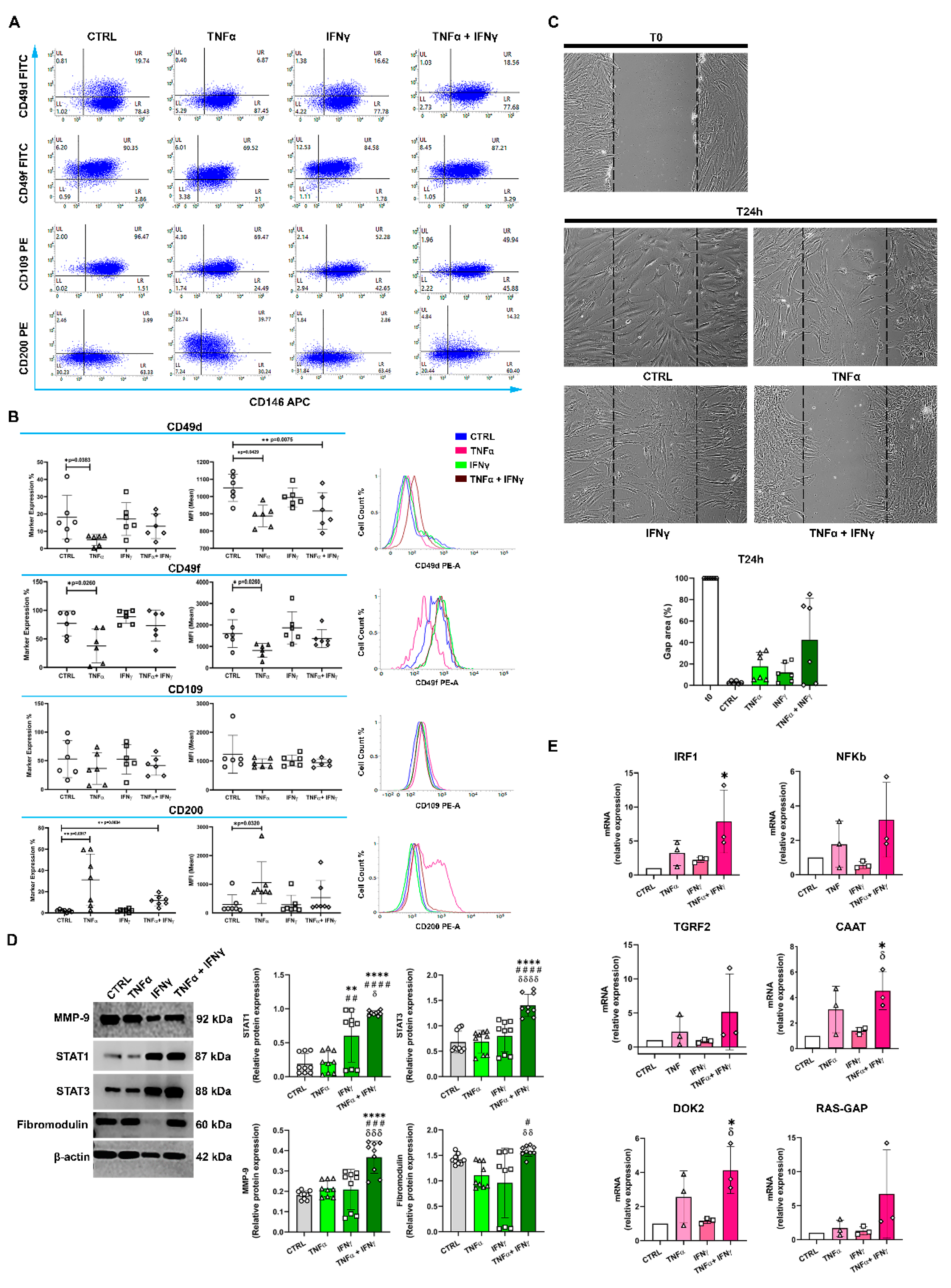
The percentage of CD146+CD49d+ cells decreased significantly after TNFα stimulation (p ≤ 0.04), but did not change after IFNγ administration (Figure 2B), and decreased only slightly with combination treatment. Analogously, the MFI values for CD146+CD49d+ cells decreased significantly with TNFα alone (p ≤ 0.05) or TNFα in combination with IFNγ (p ≤ 0.01), but not with IFNγ alone (Figure 2B). The percentage of CD146+CD49f+ cells decreased significantly with TNFα (p ≤ 0.03), increased moderately with IFNγ alone, and remained largely unchanged with the TNFα/IFNγ combination. A similar trend was observed in the corresponding MFI values. TNFα or TNFα+IFNγ did not significantly affect the proportion of CD146+ CD109+ TSPCs (Figure 2A,B). Finally, a significant increase in the percentage of the CD146+CD200+ cell population was observed upon TNFα stimulation (p ≤ 0.0017) and combination TNFα/IFNγ treatment (p ≤ 0.01) but not with IFNγ alone (Figure 2A). MFI values for this cell subpopulation showed analogous results, with a significant increase only after TNFα stimulation (p ≤ 0.032) (Figure 2B).

2.4. The In Vitro Gap Repair Assay

To measure the repair capacity of RCTCs, we used an in vitro gap repair assay. As shown in Figure 2C, 24 h treatment of RCTCs with cytokine alone or in combination showed a non-significant decrease in cell migration compared to the unstimulated cells. Notably, RCTCs treated with the TNFα/IFNγ combination showed slower gap closure.

2.5. IFNγ+TNFα Increased the Expression of STAT1, STAT3, and MMP9 Proteins

We then measured downstream signaling in response to cytokine stimulation. While TNFα did not significantly increase STAT1 expression in RCTCs, IFNγ significantly increased it (p ≤ 0.001), while the TNFα/IFNγ combination increased STAT1 levels even more (p ≤ 0.0001). Both STAT3 and MMP9 protein levels were significantly increased only with the TNFα/IFNγ combination (p ≤ 0.0001) (Figure 2D), while neither cytokine nor the combination of them significantly affected fibromodulin levels (Figure 2D).

2.6. IFNγ+TNFα Increased the Expression of IRF1, CAAT and DOK2 mRNA

IRF1 mRNA expression was not significantly modulated in RCTCs with TNFα and IFNγ alone, but it increased significantly with combination treatment (p ≤ 0.05). On the other hand, NF-κB and TGFR2 mRNA levels were not significantly modulated by IFNγ+TNFα. The levels of CAAT and DOK2 mRNA increased slightly with TNFα, but not with IFNγ, but increased significantly with combination treatment (p ≤ 0.05) (Figure 2E). RAS-GAP mRNA expression was not significantly modulated by cytokines in RCTCs.

3. Discussion

The role of inflammation in tendon injury/repair remains poorly understood. This study shows that RCTCs contain a population expressing CD146, CD90, and CD44 TSPC surface-markers. Additionally, they co-express CD49d (integrinα-4); CD49f (integrinα-6), a known and specific stem-cell population marker [18,19]; CD109; and, interestingly, the CD200 ligand. In vitro stimulation on RCTCs using pro-inflammatory cytokines TNFα and IFNγ only revealed significant modulation of CD146+CD49d+ and CD146+CD49f+ expression using TNFα alone. This result likely indicates CD146+TSPC activation, which may influence recruitment and survival and, for CD49f, self-renewal regulation in TSPCs, as previously reported [19,20]. However, migration results showed no significant difference for 24 h pro-inflammatory cytokine stimulation in in vitro culture, compared to the control. Noticeably, migration capacity for the whole RCTC population decreased using TNFα and IFNγ in combination. These findings, together with morphological observations, suggest that the above cytokines may induce biochemical and molecular cellular changes, thus requiring further studies. In previous research, CD109 inhibition suppressed inflammation, by reducing pro-inflammatory factor production, cell migration, invasion, chemo-attractive potential, and osteoclast differentiation [21]. Here, CD109 expression was not modified by pro-inflammatory cytokines. A novel finding was that TNFα alone or TNFα+IFNγ significantly increased CD200 marker expression in the CD146+TSPCs. Similar results have been found in mesenchymal stem/stromal cells [22]. Previous studies highlighted a fundamental regulatory role in controlling inflammation for the CD200 ligand interacting with CD200R [11,12,13,14,15]. Our results suggest an active role for TSPCs in regulating inflammatory processes during tendon injury/repair, through the interaction of CD200, expressed on CD146+TSPCs, with CD200R, located on immune-competent cells. STAT1 and STAT3, members of the cytoplasmic family of transcription-factor (STAT) signal-transducers and activators, have been associated with inflammatory pathologies, including tendinopathy [23].
Our study clearly demonstrates that co-administration of TNFα and IFNγ induces a significant increase in STAT1 and STAT3 protein levels. These results agree with previous research showing crosstalk between TNFα and IFNγ signaling pathways and suggest the molecular control of STAT1 availability to tumor necrosis factor receptor 1 (TNFR1) [24]. STAT1 and STAT3 are known to play antagonistic roles and disruption of their balanced interaction redirects cells from survival to apoptotic death, or from inflammatory to anti-inflammatory response [25]. Most importantly, STAT3 has been shown to play a key role in healing tendons [9]. TNFα and IFNγ have been reported to affect metalloproteinase (MMP) synthesis, and their ability to upregulate MMP9 expression leads to matrix destruction and remodeling [6,26]. Accordingly, our data show a significant increase in MMP9 protein levels after TNFα and IFNγ co-stimulation. Proteoglycan fibromodulin, a critical component of the ECM involved in collagen assembly and tendon repair [27], was not modulated by TNFα and IFNγ in our study.
TNFα and IFNγ have been shown to induce the expression of IRF1 (ubiquitously expressed in human cells), associated with STAT pathway activation [28,29,30,31]. Additionally, increased IRF1 expression is also found in tendinopathy [32]. Accordingly, our results indicate that TNFα and IFNγ together induce a significant increase in IRF1 mRNA in RCTCs. NF-κB, which has already been shown to play a role in inflammation, is activated by pro-inflammatory cytokines, including TNFα and IFNγ. NF-κB expression is also dependent on IRF1 activation, and increased NF-κB levels are detected in early RC tendinopathy [33,34,35,36]. In our study, NF-κB was not significantly modulated by TNFα and IFNγ cytokines. Similarly, no significant modulation of TGFR2 (TGFBR2) was observed in our in vitro model, although knockout of the TGFBR2 gene in tenocytes has been shown to attenuate development of tendinopathy [37].
The activity and expression levels of CAAT/Enhancer-binding protein beta (C/EBPbeta), involved in the maintenance of normal function and response to injury, are regulated by several inflammatory agents, including TNFα and IFNγ [38]. Here, for the first time, we demonstrate that co-administration of TNFα and IFNγ significantly modulates CAAT mRNA expression in RCTCs. Furthermore, DOK2, which may have a role in various physiological functions, including both innate and adaptive immunities, could also act as a negative regulator of cell proliferation when stimulated by cytokines [39]. Accordingly, we have shown the significant modulation of DOK2 mRNA after TNFα and IFNγ stimulation in RCTCs. These results suggest a possible role for DOK2 in tendinopathy. Finally, we investigated RAS-GAP mRNA expression after pro-inflammatory cytokine administration in RCTCs, since it is involved in many aspects of cell biology. In our in vitro study, RAS-GAP mRNA was not significantly modulated by TNFα and IFNγ in RCTCs.
Our study has the following limitations: (1) tendon repair, in vitro or in vivo, using KO or over expression approaches, should be analyzed in order to infer any “potential” mechanistic role of one or the other markers (CD200 or others); (2) to determine the potential involvement of CD200 in reduced cell migration in cells treated with two cytokines, it would be better to use lentivirus and see whether this would affect the phenotype; (3) rather than performing qRT-PCR on a few selected markers, it would be potentially more interesting to perform RNAseq analysis, which could lead to the identification, potentially, of previously unknown targets; (4) further research is needed to explain why if both STAT1 and STAT3 are upregulated and how they can have antagonistic activities. Despite these considerations, this study enhances the understanding of RCTC populations in inflammatory conditions, including stem/progenitor subpopulations, and suggests an important role for CD200 among the various markers. Further studies are necessary to evaluate the role of genes whose mRNA expression is increased by TNFα and IFNγ, such as IRF1, C/EBPbeta, and DOK2, and to deeply understand how CD200 activation may regulate inflammation. Identifying underlying molecular mechanisms may provide the basis for the development of innovative therapies for RC tendinopathy.

4. Materials and Methods

4.1. Rotator Cuff Tendon-Derived Cells Cultures

RCTCs that were isolated from the same 10 patients described in our previous work [40] and cryopreserved in liquid nitrogen were used. The isolation protocol was described previously [40,41,42]. The cell phenotype was confirmed by assessing the expression of a tenocyte-specific gene (scleraxis) and genes for collagens α1(I), α2(I), and α1(III) by real-time PCR, as previously described (not shown) [43].
For the present study, cells at passage 0 were thawed out and sub-cultured in alpha-MEM with 10% heat-inactivated FBS and 1% penicillin/streptomycin (Gibco, MA, USA) at 37 °C and 5% CO2. Cells at passage 2 (P2) were used to avoid phenotypic drift [44], were seeded at 1.5 × 10⁵ cells/flask in a 25 cm2 culture flask, and were allowed to adhere overnight. Afterward, cells were exposed to complete alpha-MEM (untreated control) or stimulated by cytokines at a final concentration of 10 ng/mL as previously described [45]. In detail, TNFα alone, IFNγ alone, or IFNγ and TNFα in combination (PeproTech, London, UK) were added to the medium.

4.2. Flow Cytometry

RCTCs were stained with a panel of fluorochrome-conjugated, monoclonal antibodies: CD45-FITC, CD90-FITC, CD49d-PE, CD49f-PE, CD109-PE, (BD Pharmingen, San Diego, CA, USA) CD44-FITC, CD146-APC, and CD200-PE (Miltenyi Biotech, Bergisch Gladbach, Germany). Cells were acquired with a BD FACSLyric II flow cytometer (BD Biosciences, CA, USA) equipped with a 488 nm, 640 nm, and 405 nm laser. Events were analyzed using Suite1.5 and FlowJo 10.6.2 software (BD Biosciences). Results are shown as cell positivity percentages or as mean fluorescence intensities (MFIs).

4.3. In Vitro Gap Repair Assay

Cells were grown to confluence in 24-well plates, and the scratch was made using a sterile P10 pipette tip, creating a cell-free area, as described before [46]. Cultures were treated in reduced FBS conditions (1% FBS) and images were acquired immediately after wounding (T0) and after 24 h through bright field microscopy (NIKON, Melville, NY, USA). Images were analyzed by ImageJ (Version 1.49 v, RRID:SCR_003070; NIH, Bethesda, MD, USA), and cell-free areas were marked; outcomes are represented as a percentage of the initial wound area.

4.4. Immunoblotting

At 24 h, cells were harvested and lysed as previously reported [42]. Twenty micrograms of whole-protein fraction were loaded on a 12% sodium dodecyl sulfate-polyacrylamide gel followed by Western blot. Nitrocellulose membranes were blocked and probed overnight at 4 °C with mouse monoclonal anti-STAT1 and anti-STAT3, rabbit polyclonal anti-fibromodulin (1:1000; Abcam, UK), mouse monoclonal anti-MMP-9 (1:200; Santa Cruz Biotechnology, Santa Cruz, CA, USA), and anti-β-actin antibodies (1:5000; Merck, Darmstadt, Germany). Immunoreactive bands were identified as already reported [42].

4.5. Gene Expression Analysis

Total RNA was extracted using Total RNA Purification Kit (NORGEN Biotek) as previously described [47,48] and reported in Supplementary Materials. Quantitative real-time PCR (qPCR) was carried out using primer sequences for IRF1, NFKb, TGRF2, CAAT, DOK2, RAS-GAP, and ACTB genes. Primer sequences are listed in Supplementary Materials Table S1, as well as methods for RT-qPCR analysis. Relative gene expression was calculated by comparative Ct (ΔΔCt) method and converted to relative expression ratio (2−ΔΔCt) (Figure S1).

4.6. Statistics

Statistical analysis was performed using GraphPad Prism 6.0 software (GraphPad Software, San Diego, CA, USA). For immunophenotype data, results are expressed as median with interquartile range. Statistical differences were determined either using Mann–Whitney nonparametric t-tests between two groups with only one variable, or with Kruskal–Wallis non-parametric ANOVA with Dunn’s post-test for multiple variables. For Western blot and qRT-PCR analyses, individual values from independent densitometric measurements were summarized as means ± standard deviations (S.D.), and statistics were performed using one-way analysis of variance (ANOVA) followed by Tukey’s multiple comparison test. Values of p ≤ 0.05 were considered statistically significant.

Supplementary Materials

The supporting information can be downloaded at: https://www.mdpi.com/article/10.3390/ijms232315165/s1. References [49,50,51,52,53] are cited in the supplementary materials.

Author Contributions

Conceptualization, A.C.B.; methodology, R.G., M.G., N.M., F.M., C.A.L.P. and A.C.; software, M.G. and C.G.; validation, R.G., M.G., N.M., F.M., A.C. and A.C.B.; formal analysis, M.G., N.M. and F.M.; resources, R.G., F.O. and A.C.B.; data curation, M.G., A.C. and A.C.B.; writing—original draft preparation, M.G., A.C. and A.C.B; writing—review and editing, C.G., M.G., A.C. and A.C.B.; supervision, A.C.B.; funding acquisition, F.O. and A.C.B. All authors have read and agreed to the published version of the manuscript.

Funding

This research received no external funding.

Institutional Review Board Statement

The study used human cells represented by thawed material that was cryopreserved during our previous studies [14,15,16]. In those papers (see Section 4), it was stated that all the procedures were approved by the Ethical Committee of Rome Tor Vergata University, that all patients gave written informed consent, and that all samples were anonymized before being processed. The study was conducted in accordance with the Declaration of Helsinki and approved by the Ethical Committee of Rome Tor Vergata University.

Informed Consent Statement

Informed consent was obtained from all subjects involved in the study.

Acknowledgments

This work was supported by Programma Operativo Nazionale (PON) Ricerca e Innovazione 2014–2020, Fondo Sociale Europeo, Azione I. 2 “Attrazione e Mobilità Internazionale dei Ricercatori” (Marialucia Gallorini). This work was supported by the University of Salerno (Francesco Oliva).

Conflicts of Interest

The authors declare no conflict of interest.

References

  1. Chisari, E.; Rehak, L.; Khan, W.S.; Maffulli, N. Tendon healing is adversely affected by low-grade inflammation. J. Orthop. Surg. Res. 2021, 16, 700. [Google Scholar] [CrossRef] [PubMed]
  2. Chisari, E.; Rehak, L.; Khan, W.S.; Maffulli, N. Tendon healing in presence of chronic low-level inflammation: A systematic review. Br. Med. Bull. 2019, 132, 97–116. [Google Scholar] [CrossRef]
  3. Cipollaro, L.; Sahemey, R.; Oliva, F.; Maffulli, N. Immunohistochemical features of rotator cuff tendinopathy. Br. Med. Bull. 2019, 130, 105–123. [Google Scholar] [CrossRef] [PubMed]
  4. Riley, G.P. Gene expression and matrix turnover in overused and damaged tendons. Scand. J. Med. Sci. Sports 2005, 15, 241–251. [Google Scholar] [CrossRef] [PubMed] [Green Version]
  5. Dakin, S.G.; Martinez, F.O.; Yapp, C.; Wells, G.; Oppermann, U.; Dean, B.J.; Smith, R.D.; Wheway, K.; Watkins, B.; Roche, L.; et al. Inflammation activation and resolution in human tendon disease. Sci. Transl. Med. 2015, 7, 311ra173. [Google Scholar] [CrossRef] [Green Version]
  6. Kendal, A.R.; Layton, T.; Al-Mossawi, H.; Appleton, L.; Dakin, S.; Brown, R.; Loizou, C.; Rogers, M.; Sharp, R.; Carr, A. Multi-omic single cell analysis resolves novel stromal cell populations in healthy and diseased human tendon. Sci. Rep. 2020, 10, 13939. [Google Scholar] [CrossRef]
  7. Russo, V.; El Khatib, M.; Prencipe, G.; Citeroni, M.R.; Faydaver, M.; Mauro, A.; Berardinelli, P.; Cerveró-Varona, A.; Haidar-Montes, A.A.; Turriani, M.; et al. Tendon Immune Regeneration: Insights on the Synergetic Role of Stem and Immune Cells during Tendon Regeneration. Cells 2022, 11, 434. [Google Scholar] [CrossRef]
  8. Bi, Y.; Ehirchiou, D.; Kilts, T.M.; Inkson, C.A.; Embree, M.C.; Sonoyama, W.; Li, L.; Leet, A.I.; Seo, B.M.; Zhang, L.; et al. Identification of tendon stem/progenitor cells and the role of the extracellular matrix in their niche. Nat. Med. 2007, 13, 1219. [Google Scholar] [CrossRef]
  9. Tarafder, S.; Chen, E.; Jun, Y.; Kao, K.; Hee Sim, K.; Back, J.; Lee, F.Y.; Lee, C.H. Tendon stem/progenitor cells regulate inflammation in tendon healing via JNK and STAT3 signaling. FASEB J. 2017, 31, 3991–3998. [Google Scholar] [CrossRef] [Green Version]
  10. Vinhas, A.; Rodrigues, M.T.; Gomes, M.E. Exploring Stem Cells and Inflammation in Tendon Repair and Regeneration. Adv. Exp. Med. Biol. 2018, 1089, 37–46. [Google Scholar] [CrossRef]
  11. Hoek, R.M.; Ruuls, S.R.; Murphy, C.A.; Wright, G.J.; Goddard, R.; Zurawski, S.M.; Blom, B.; Homola, M.E.; Streit, W.J.; Brown, M.H.; et al. Downregulation of the macrophage lineage through interaction with OX2 (CD200). Science 2000, 290, 1768–1771. [Google Scholar] [CrossRef] [PubMed]
  12. Wright, G.J.; Puklavec, M.J.; Willis, A.C.; Hoek, R.M.; Sedgwick, J.D.; Brown, M.H.; Barclay, A.N. Lymphoid/neuronal cell surface OX2 glycoprotein recognizes a novel receptor on macrophages implicated in the control of their function. Immunity 2000, 13, 233–242. [Google Scholar] [CrossRef] [PubMed] [Green Version]
  13. Gorczynski, R.; Chen, Z.; Kai, Y.; Lee, L.; Wong, S.; Marsden, P.A. CD200 is a ligand for all members of the CD200R family of immunoregulatory molecules. J. Immunol. 2004, 172, 7744–7749. [Google Scholar] [CrossRef] [PubMed] [Green Version]
  14. Kotwica-Mojzych, K.; Jodłowska-Jędrych, B.; Mojzych, M. CD200:CD200R Interactions and Their Importance in Immunoregulation. Int. J. Mol. Sci. 2021, 22, 1602. [Google Scholar] [CrossRef]
  15. Chen, Z.; Marsden, P.A.; Gorczynski, R.M. Role of distal enhancer in the transcriptional responsiveness of the human CD200 gene to interferon-y and tumor necrosis factor-α. Mol. Immunol. 2009, 46, 1951–1963. [Google Scholar] [CrossRef]
  16. Grol, M.W.; Haelterman, N.A.; Lim, J.; Munivez, E.M.; Archer, M.; Hudson, D.M.; Tufa, S.F.; Keene, D.R.; Lei, K.; Park, D.; et al. Tendon and motor phenotypes in the Crtap−/− mouse model of recessive osteogenesis imperfecta. eLife 2021, 26, e63488. [Google Scholar] [CrossRef]
  17. Alshoubaki, Y.K.; Nayer, B.; Das, S.; Martino, M.M. Modulation of the Activity of Stem and Progenitor Cells by Immune Cells. Stem Cells Transl. Med. 2022, 11, 248–258. [Google Scholar] [CrossRef]
  18. Baiula, M.; Spampinato, S.; Gentilucci, L.; Tolomelli, A. Novel Ligands Targeting α4β1 Integrin: Therapeutic Applications and Perspectives. Front. Chem. 2019, 7, 489. [Google Scholar] [CrossRef]
  19. Krebsbach, P.H.; Villa-Diaz, L.G. The Role of Integrin α6 (CD49f) in Stem Cells: More than a Conserved Biomarker. Stem Cells Dev. 2017, 26, 1090–1099. [Google Scholar] [CrossRef]
  20. Kumar, S.; Ponnazhagan, S. Bone homing of mesenchymal stem cells by ectopic alpha 4 integrin expression. FASEB J. 2007, 21, 3917–3927. [Google Scholar] [CrossRef]
  21. Mii, S.; Hoshino, A.; Enomoto, A.; Murakumo, Y.; Ito, M.; Yamaguchi, A.; Takahashi, M. CD109 deficiency induces osteopenia with an osteoporosis-like phenotype in vivo. Genes Cells 2018, 23, 590–598. [Google Scholar] [CrossRef] [PubMed]
  22. Pontikoglou, C.; Langonné, A.; Ba, M.A.; Varin, A.; Rosset, P.; Charbord, P.; Sensébé, L.; Deschaseaux, F. CD200 expression in human cultured bone marrow mesenchymal stem cells is induced by pro-osteogenic and pro-inflammatory cues. J. Cell. Mol. Med. 2016, 20, 655–665. [Google Scholar] [CrossRef] [PubMed]
  23. Butturini, E.; Carcereri de Prati, A.; Mariotto, S. Redox Regulation of STAT1 and STAT3 Signaling. Int. J. Mol. Sci. 2020, 21, 7034. [Google Scholar] [CrossRef] [PubMed]
  24. Wesemann, D.R.; Benveniste, E.N. STAT-1 alpha and IFN-gamma as modulators of TNF-alpha signaling in macrophages: Regulation and functional implications of the TNF receptor 1-STAT-1 alpha complex. J. Immunol. 2003, 171, 5313–5319. [Google Scholar] [CrossRef] [Green Version]
  25. Hu, Y.S.; Han, X.; Liu, X.H. STAT3: A Potential Drug Target for Tumor and Inflammation. Curr. Top. Med. Chem. 2019, 19, 1305–1317. [Google Scholar] [CrossRef]
  26. Del Buono, A.; Oliva, F.; Osti, L.; Maffulli, N. Metalloproteases and tendinopathy. Muscles Ligaments Tendons J. 2013, 3, 51–57. [Google Scholar] [CrossRef]
  27. Delalande, A.; Gosselin, M.P.; Suwalski, A.; Guilmain, W.; Leduc, C.; Berchel, M.; Jaffrès, P.A.; Baril, P.; Midoux, P.; Pichon, C. Enhanced Achilles tendon healing by fibromodulin gene transfer. Nanomedicine 2015, 11, 1735–1744. [Google Scholar] [CrossRef]
  28. Vila-del Sol, V.; Punzón, C.; Fresno, M. IFN-gamma-induced TNF-alpha expression is regulated by interferon regulatory factors 1 and 8 in mouse macrophages. J. Immunol. 2008, 181, 4461–4470. [Google Scholar] [CrossRef] [Green Version]
  29. Feng, H.; Zhang, Y.B.; Gui, J.F.; Lemon, S.M.; Yamane, D. Interferon regulatory factor 1 (IRF1) and anti-pathogen innate immune responses. PLoS Pathog. 2021, 17, 1009220. [Google Scholar] [CrossRef]
  30. Michalska, A.; Blaszczyk, K.; Wesoly, J.; Bluyssen, H.A.R. A Positive Feedback Amplifier Circuit That Regulates Interferon (IFN)-Stimulated Gene Expression and Controls Type I and Type II IFN Responses. Front. Immunol. 2018, 9, 1135. [Google Scholar] [CrossRef]
  31. Bonelli, M.; Dalwigk, K.; Platzer, A.; Olmos Calvo, I.; Hayer, S.; Niederreiter, B.; Holinka, J.; Sevelda, F.; Pap, T.; Steiner, G.; et al. IRF1 is critical for the TNF-driven interferon response in rheumatoid fibroblast-like synoviocytes: JAKinibs suppress the interferon response in RA-FLSs. Exp. Mol. Med. 2019, 51, 1–11. [Google Scholar] [CrossRef] [PubMed] [Green Version]
  32. Dakin, S.G.; Newton, J.; Martinez, F.O.; Hedley, R.; Gwilym, S.; Jones, N.; Reid, H.A.B.; Wood, S.; Wells, G.; Appleton, L.; et al. Chronic inflammation is a feature of Achilles tendinopathy and rupture. Br. J. Sports Med. 2018, 52, 359–367. [Google Scholar] [CrossRef] [PubMed]
  33. Courtois, G.; Gilmore, T.D. Mutations in the NF-KappaB signaling pathways: Implications for the human disease. Oncogene 2006, 25, 6831–6843. [Google Scholar] [CrossRef] [Green Version]
  34. Rius, J.; Guma, M.; Schachtrup, C.; Akassoglou, K.; Zinkernagel, A.S.; Nizet, V.; Johnson, R.S.; Haddad, G.G.; Karin, M. NF-kappaB links innate immunity to the hypoxic response through transcriptional regulation of HIF-1alpha. Nature 2008, 453, 807–811. [Google Scholar] [CrossRef] [PubMed] [Green Version]
  35. Abraham, A.C.; Shah, S.A.; Golman, M.; Song, L.; Li, X.; Kurtaliaj, I.; Akbar, M.; Millar, N.L.; Abu-Amer, Y.; Galatz, L.M.; et al. Targeting the NF-κB signaling pathway in chronic tendon disease. Sci. Transl. Med. 2019, 11, eaav4319. [Google Scholar] [CrossRef]
  36. Xu, K.; Lin, C.; Ma, D.; Chen, M.; Zhou, X.; He, Y.; Moqbel, S.A.A.; Ma, C.; Wu, L. Spironolactone Ameliorates Senescence and Calcification by Modulating Autophagy in Rat Tendon-Derived Stem Cells via the NF-κB/MAPK Pathway. Oxidative Med. Cell. Longev. 2021, 2021, 5519587. [Google Scholar] [CrossRef]
  37. Wang, X.; Liu, S.; Yu, T.; An, S.; Deng, R.; Tan, X.; Crane, J.; Zhang, W.; Pan, D.; Wan, M.; et al. Inhibition of Integrin αvβ6 Activation of TGF-β Attenuates Tendinopathy. Adv. Sci. 2022, 9, e2104469. [Google Scholar] [CrossRef]
  38. Ko, C.Y.; Chang, W.C.; Wang, J.M. Biological roles of CCAAT/Enhancer-binding protein delta during inflammation. J. Biomed. Sci. 2015, 22, 6. [Google Scholar] [CrossRef] [Green Version]
  39. Suzu, S.; Tanaka-Douzono, M.; Nomaguchi, K.; Yamada, M.; Hayasawa, H.; Kimura, F.; Motoyoshi, K. p56(dok-2) as a cytokine-inducible inhibitor of cell proliferation and signal transduction. EMBO J. 2000, 19, 5114–5122. [Google Scholar] [CrossRef] [Green Version]
  40. Oliva, F.; Berardi, A.C.; Misiti, S.; Verga Falzacappa, C.; Iacone, A.; Maffulli, N. Thyroid hormones enhance growth and counteract apoptosis in human tenocytes isolated from rotator cuff tendons. Cell Death Dis. 2013, 4, e705. [Google Scholar] [CrossRef]
  41. Osti, L.; Berardocco, M.; di Giacomo, V.; Di Bernardo, G.; Oliva, F.; Berardi, A.C. Hyaluronic acid increases tendon derived cell viability and collagen type I expression in vitro: Comparative study of four different Hyaluronic acid preparations by molecular weight. BMC Musculoskelet. Disord. 2015, 16, 284. [Google Scholar] [CrossRef] [PubMed] [Green Version]
  42. Gallorini, M.; Berardi, A.C.; Gissi, C.; Cataldi, A.; Osti, L. Nrf2-mediated cytoprotective effect of four different hyaluronic acids by molecular weight in human tenocytes. J. Drug Target. 2020, 28, 212–224. [Google Scholar] [CrossRef] [PubMed]
  43. Berardi, A.C.; Oliva, F.; Berardocco, M.; la Rovere, M.; Accorsi, P.; Maffulli, N. Thyroid hormones increase collagen I and cartilage oligomeric matrix protein (COMP) expression in vitro human tenocytes. Muscles Ligaments Tendons J. 2014, 4, 285–291. [Google Scholar] [CrossRef] [PubMed]
  44. Yao, L.; Bestwick, C.S.; Bestwick, L.A.; Maffulli, N.; Aspden, R.M. Phenotypic drift in human tenocyte culture. Tissue Eng. 2006, 12, 1843–1849. [Google Scholar] [CrossRef]
  45. Stolk, M.; Klatte-Schulz, F.; Schmock, A.; Minkwitz, S.; Wildemann, B.; Seifert, M. New insights into tenocyte-immune cell interplay in an in vitro model of inflammation. Sci. Rep. 2017, 7, 9801. [Google Scholar] [CrossRef]
  46. Gissi, C.; Radeghieri, A.; Antonetti Lamorgese Passeri, C.; Gallorini, M.; Calciano, L.; Oliva, F.; Veronesi, F.; Zendrini, A.; Cataldi, A.; Bergese, P.; et al. Extracellular vesicles from rat-bone-marrow mesenchymal stromal/stem cells improve tendon repair in rat Achilles tendon injury model in dose-dependent manner: A pilot study. PLoS ONE 2020, 15, e0229914. [Google Scholar] [CrossRef]
  47. Colosimo, A.; Di Rocco, G.; Curini, V.; Russo, V.; Capacchietti, G.; Berardinelli, P.; Mattioli, M.; Barboni, B. Characterization of the methylation status of five imprinted genes in sheep gametes. Anim. Genet. 2009, 40, 900–908. [Google Scholar]
  48. Cimini, C.; Moussa, F.; Taraschi, A.; Ramal-Sanchez, M.; Colosimo, A.; Giulia Capacchietti, G.; Mokh, S.; Valbonetti, L.; Tagaram, I.; Bernabò, N.; et al. Pre-treatment of swine oviductal epithelial cells with progesterone increases the sperm fertilizing ability in an IVF model. Animals 2022, 12, 1191. [Google Scholar] [CrossRef]
  49. Pfaffl, M.W. A new mathematical model for relative quantification in real-time RT-PCR. Nucleic Acids Res. 2001, 29, e45. [Google Scholar] [CrossRef]
  50. Ragni, E.; Perucca Orfei, C.; Bowles, A.C.; de Girolamo, L.; Correa, D. Reliable Reference Genes for Gene Expression Assessment in Tendon-Derived Cells under Inflammatory and Pro-Fibrotic/Healing Stimuli. Cells 2019, 8, 1188. [Google Scholar] [CrossRef] [Green Version]
  51. Klatte-Schulz, F.; Pauly, S.; Scheibel, M.; Greiner, S.; Gerhardt, C.; Schmidmaier, G.; Wildemann, B. Influence of age on the cell biological characteristics and the stimulation potential of male human tenocyte-like cells. Eur. Cells Mater. 2012, 24, 74–89. [Google Scholar] [CrossRef]
  52. Hellebrekers, D.M.; Castermans, K.; Viré, E.; Dings, R.P.; Hoebers, N.T.; Mayo, K.H.; Oude Egbrink, M.G.; Molema, G.; Fuks, F.; van Engeland, M.; et al. Epigenetic regulation of tumor endothelial cell anergy: Silencing of intercellular adhesion molecule-1 by histone modifications. Cancer Res. 2006, 66, 10770–10777. [Google Scholar] [CrossRef] [Green Version]
  53. Cohen, S.; Mosig, R.; Moshier, E.; Pereira, E.; Rahaman, J.; Prasad-Hayes, M.; Halpert, R.; Billaud, J.N.; Dottino, P.; Martignetti, J.A. Interferon regulatory factor 1 is an independent predictor of platinum resistance and survival in high-grade serous ovarian carcinoma. Gynecol. Oncol. 2014, 134, 591–598. [Google Scholar] [CrossRef]
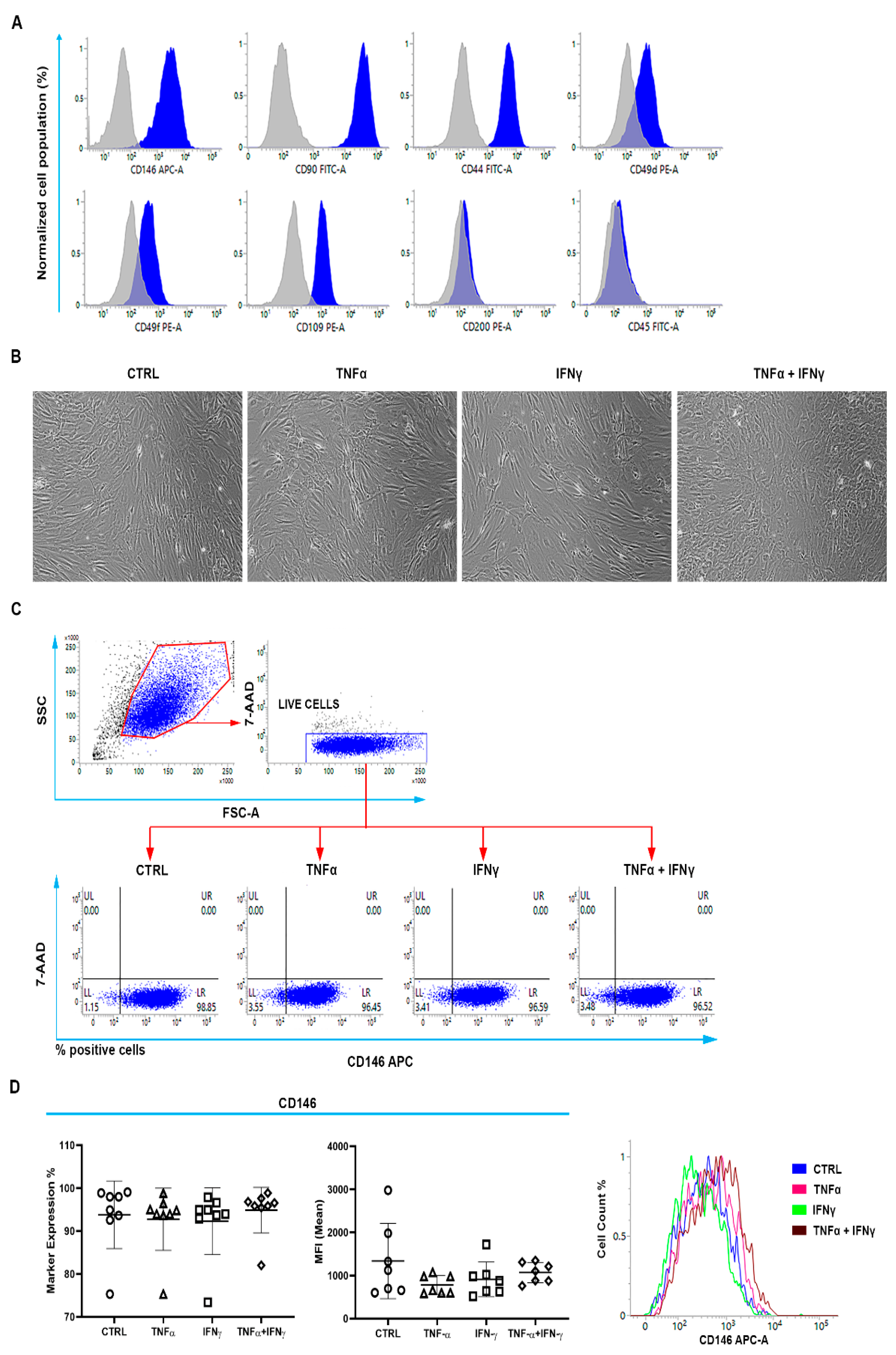
Figure 1. Immunophenotypic profile of human rotator-cuff-tendon-derived cells (RCTCs) by flow cytometry. (A) Cells were stained for a panel of the cluster of designation (CD): CD146, CD90, CD44, CD49d, CD49f, CD109, CD200, and CD45. Peaks of fluorescence emission were obtained by flow cytometry, and their right-shifted peak (blue) represents the positivity for the marker analyzed with respect to the isotype negative control (grey peak). CD146 (74% ± 16.1), CD90 (99% ± 0.3), CD44 (100% ± 0.4), CD49d (58% ± 26.6), CD49f (74% ± 8.8), and CD109 (73% ± 19.8). (B) Microscopic analysis of cell morphology after 3 days of in vitro stimulation of proinflammatory cytokines. Representative images of cells exposed to treatments were acquired by phase-contrast microscopy. 100× magnification. (C) Gating strategy. The SSC (side scatter)/FSC (forward scatter) dot plot allows the gating of the cell population by means of its morphological parameters. Cells were afterward stained with the 7-AAD (7-aminoactinomycin) to exclude dead cells from further analyses (CD146). No altered expression of CD146 was promoted after stimulation with single or combined pro-inflammatory cytokines. (D) Graphs represent the percentage of CD146 expression and the MFI (mean fluorescence intensity) of cells exposed to treatments. Fluorescence emission peaks related to CD146 were obtained by flow cytometry. CTRL = untreated cells.
Ijms 23 15165 g001
Figure 2. Analysis of inflammation markers in the CD146+TSPCs population. (A) RCTCs, stained positive for CD146, were afterward co-stained for the cluster of designation (CD)49d, CD49f, CD109, and CD200. Representative dot plots show the distribution of the cell population in response to treatments. (B) Graphs represent the percentage of marker expression and the MFI (mean fluorescence intensity) related to CD49d, CD49f, CD109, and CD200. Relative emission peaks were obtained by flow cytometry. The right shift of peaks represents a higher positivity for markers. (C) Migration of RCT-derived cells in response to the various treatments immediately after the stimulus (T0) and after 24 h. Representative images obtained by phase-contrast microscopy. The bar graph represents the percentage of cells covering the gap (empty) area. 100× magnification. (D) Protein expression of MMP (metalloproteinase)-9, STAT (Signal transducer and activator of transcription)1, STAT3, and fibromodulin detected by Western blotting after 24 h. β-actin is used as a loading control. Bar graphs display densitometric values normalized on the ones of the loading control (relative expression). (E) Graphs represent relative gene expressions of IRF (Interferon Regulatory Factor)-1, NFkb (Nuclear factor kappa subunit b), TGRF2 (TGFBR2-Transforming growth receptor factor 2), CAAT (CAAT/Enhancer-binding protein beta), DOK2 (docking protein 2), and RAS-GAP (Ras GTPase activating protein). * = p < 0.05; ** = p < 0.01; and **** = p < 0.0001 between CTRL and treated cells. # = p < 0.05; ## = p < 0.01; ### = p < 0.001 and #### = p < 0.0001 between cells treated with TNFα and cells in the presence of other treatments. δ = p < 0.05; δδ = p < 0.01; δδδ = p < 0.001 and δδδδ = p < 0.0001 between cells treated with IFNγ and cells in the presence of other treatments.
Ijms 23 15165 g002
Publisher’s Note: MDPI stays neutral with regard to jurisdictional claims in published maps and institutional affiliations.