Article

Na+/K+- and Mg2+-ATPases and Their Interaction with AMPA, NMDA and D2 Dopamine Receptors in an Animal Model of Febrile Seizures

by
María Crespo
1,
David Agustín León-Navarro
1,* and
Mairena Martín
2
1
Department of Inorganic, Organic Chemistry and Biochemistry, Faculty of Chemical and Technological Sciences, Regional Centre of Biomedical Research (CRIB), Universidad de Castilla-La Mancha, 13071 Ciudad Real, Spain
2
Department of Inorganic, Organic Chemistry and Biochemistry, Faculty of Chemical and Technological Sciences, School of Medicine of Ciudad Real, Regional Centre of Biomedical Research (CRIB), Universidad de Castilla-La Mancha, 13071 Ciudad Real, Spain
*
Author to whom correspondence should be addressed.
Int. J. Mol. Sci. 2022, 23(23), 14638; https://doi.org/10.3390/ijms232314638
Submission received: 19 September 2022 / Revised: 5 November 2022 / Accepted: 15 November 2022 / Published: 24 November 2022
(This article belongs to the Special Issue Advances in Neurodevelopmental Disorders (NDDs) Research)

Abstract

:
Febrile seizures (FS) are one of the most common seizure disorders in childhood which are classified into short and prolonged, depending on their duration. Short FS are usually considered as benign. However, epidemiological studies have shown an association between prolonged FS and temporal lobe epilepsy. The development of animal models of FS has been very useful to investigate the mechanisms and the consequences of FS. One of the most used, the “hair dryer model”, has revealed that prolonged FS may lead to temporal lobe epilepsy by altering neuronal function. Several pieces of evidence suggest that Na+/ K+-ATPase and Mg2+-ATPase may play a role in this epileptogenic process. In this work, we found that hyperthermia-induced seizures (HIS) significantly increased the activity of Na+/ K+-ATPase and Mg2+-ATPase five and twenty days after hyperthermic insult, respectively. These effects were diminished in response to AMPA, D2 dopamine A1 and A2A receptors activation, respectively. Furthermore, HIS also significantly increased the protein level of the AMPA subunit GluR1. Altogether, the increased Na+/ K+-ATPase and Mg2+-ATPase agree well with the presence of protective mechanisms. However, the reduction in ATPase activities in the presence of NMDA and AMPA suggest an increased propensity for epileptic events in adults.

1. Introduction

Febrile seizures (FS) are one of the most common seizure disorders in childhood between 6 months and 5 years, affecting about 2–4% of children. FS are brought on by fever but without evidence of infection of the central nervous system [1,2]. FS can be classified into two main types according to their duration: short FS, which last less than 15 min, and prolonged FS, which last longer than 15 min. Generally, short FS are considered benign. However, the outcome of prolonged FS is more controversial. Thus, whereas retrospective studies have shown an association between prolonged FS and temporal lobe epilepsy, prospective studies have failed to find such a relationship [3,4].
The development of animal models of FS has been very useful to investigate their mechanisms and consequences. One of the most used is the “hair dryer model”, in which hyperthermia-induced seizures (HIS) are evoked in neonatal rats by an adjustable stream of heated air [5]. This animal model has revealed that prolonged fever seizures lead to temporal lobe epilepsy in 30% of rats [6]. Moreover, although the mechanisms by which prolonged FS could contribute to epilepsy remains unknown, no cell death and neuronal circuit reorganization seems to be involved, suggesting changes in neuronal function as the main mechanism in the epileptogenic process during development [4,7,8]. In that sense, multiple works carried out in recent years have revealed that HIS evoke changes in metabotropic receptors, such as adenosine A1 and A2A receptors [9,10,11,12,13], metabotropic glutamate receptor [14] or GABAB receptor [15]. Ionotropic receptors, including NMDA and GABAA receptors, are also modulated by FS [16,17,18,19].
Several pieces of evidence suggest that the Na+/ K+-ATPase may play a key role in the epileptogenic process. Thus, fundamental cellular processes commonly associated to seizure episodes, such as neuronal excitability or neurotransmitter release, are regulated by Na+/ K+-ATPase activity. Moreover, multiple works using epileptic animal models or human brain have found mutations in the Na+/ K+-ATPase α subunit gene [20,21]. Moreover, changes in Na+/ K+-ATPase activity have also been found in rodent brain after pentylentetrazol-induced seizures, and in epileptic human brain [22,23]. On the other hand, a variety of studies have shown that Na+/ K+-ATPase activity can be modulated by neurotransmitter receptors; this process seems to involve a direct interaction in the case of glutamate ionotropic receptor NMDA and AMPA, forming macrocomplexes for NMDA [24] or binding directly to the Na+/ K+-ATPase subunit alpha in the case of AMPA [25]. On the other hand, metabotropic receptors seem to involve mechanisms mediated by protein kinases activities through secondary messenger signaling cascades [26,27].
The role of Mg2+-ATPase in the epileptogenic process is less known. This pump contributes to maintaining a high intracellular concentration of magnesium. Since these ions are crucial for several cellular processes such as enzymatic reactions, cellular signaling and ion channel functions [28] it seems plausible that Mg2+-ATPase could be altered in response to HIS.
In the present work, we analyzed the activity of both Na+/ K+ and Mg2+-ATPases in cortical plasma membranes from rat brain at five and twenty days after HIS and at 2-months-old. Furthermore, we studied the status of AMPA, NMDA and dopamine receptor and the effect of their activation on Na+/ K+-ATPase and Mg2+-ATPase activities.

2. Results

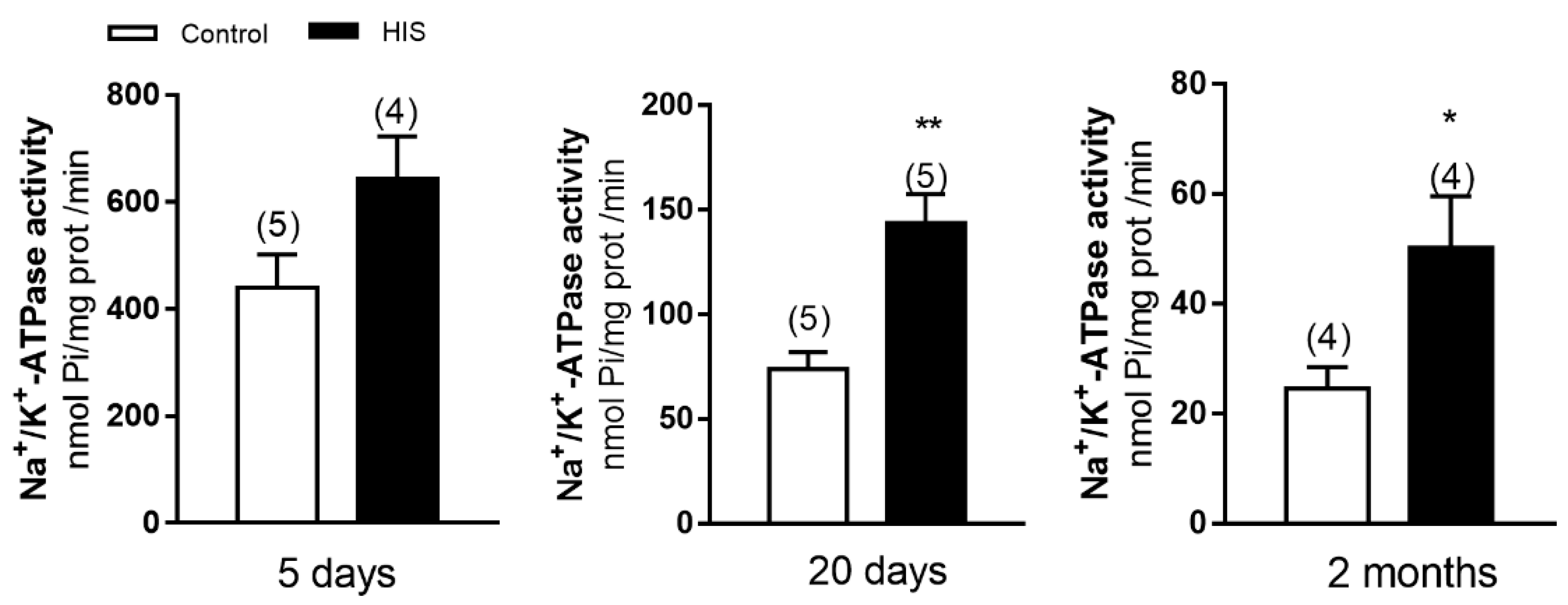
2.1. Na+/K+-ATPase Activity in Cortical Plasma Membranes after HIS

To know whether HIS can alter Na+/K+-ATPase activity, we measured the enzymatic activity at 5 and 20 days after hyperthermic insult and in 2-month-old animals. As shown in Figure 1, no significant variation was detected in Na+/K+-ATPase activity 5 days after HIS (439.8 ± 62.79 nmol of Pi /mg prot /min vs. 644.5 ± 78.74 nmol of Pi /mg prot /min, p > 0.05). However, a significant increase was observed 20 days after hyperthermic seizures (73.7 ± 8.22 nmol of Pi /mg prot /min vs. 143.5 ± 14.08 nmol of Pi /mg prot /min, p < 0.05) and in 2-month-old animals after hyperthermic seizures (24 ± 4 nmol of Pi /mg prot /min vs. 50 ± 9 nmol of Pi /mg prot /min, p < 0.05) (Figure 1).

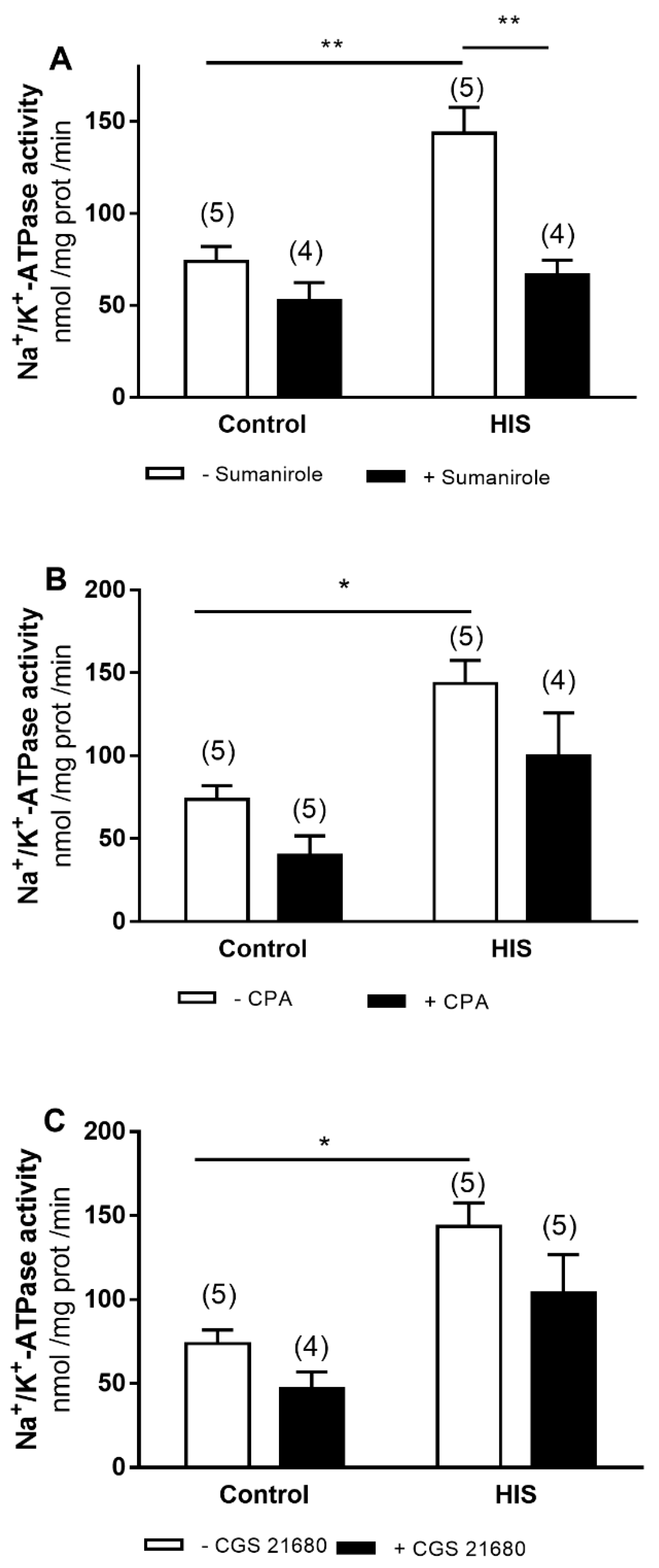
2.2. Modulation of Na+/K+-ATPase Activity by G-Protein-Coupled Receptor after HIS

Next, we analyzed whether the activation of different G-protein-coupled receptors could modify the increased Na+/K+-ATPase activity observed 20 days after HIS. Thus, we assayed three agonists: sumanirole, CPA and GGS 21680, which act through dopamine D2 and adenosine A1 and A2A receptors, respectively. A two-way ANOVA was carried out to analyze the effect of HIS and agonists on Na+/K+-ATPase activity. This analysis revealed that there was a significant effect of sumanirole [F(1,14) = 20.73, p = 0.0005], HIS [(F(1,14) = 15.11, p = 0.0016] and sumanirole x HIS interaction [F(1,14) = 6.74, p = 0.0211] on Na+/K+-ATPase activity (Figure 2A). The Bonferroni’s post test showed that HIS induced a significant increase in the activity of Na+/K+-ATPase in control rats, and that this effect was significantly reduced by sumanirole (Figure 2A).
With respect to CPA, a selective A1 receptor agonist, the two-way ANOVA analysis revealed that HIS and agonist significantly altered the activity of Na+/K+-ATPase [HIS: F(1,15) = 18.37, p = 0.0006; CPA: F(1.15) = 6.517, p = 0.0221] without observed HIS x agonist interaction (Figure 2B). Similar results were obtained when CGS 21680, selective A2A receptor agonist, was assayed. [HIS: F(1,15) = 16.8, p = 0.0009; CGS 21680: F(1.15) = 4.58, p = 0.0492] (Figure 2C). Bonferroni’s post-test only showed an increase in Na+/K+-ATPase activity in the control group after HIS for all agonists assayed.

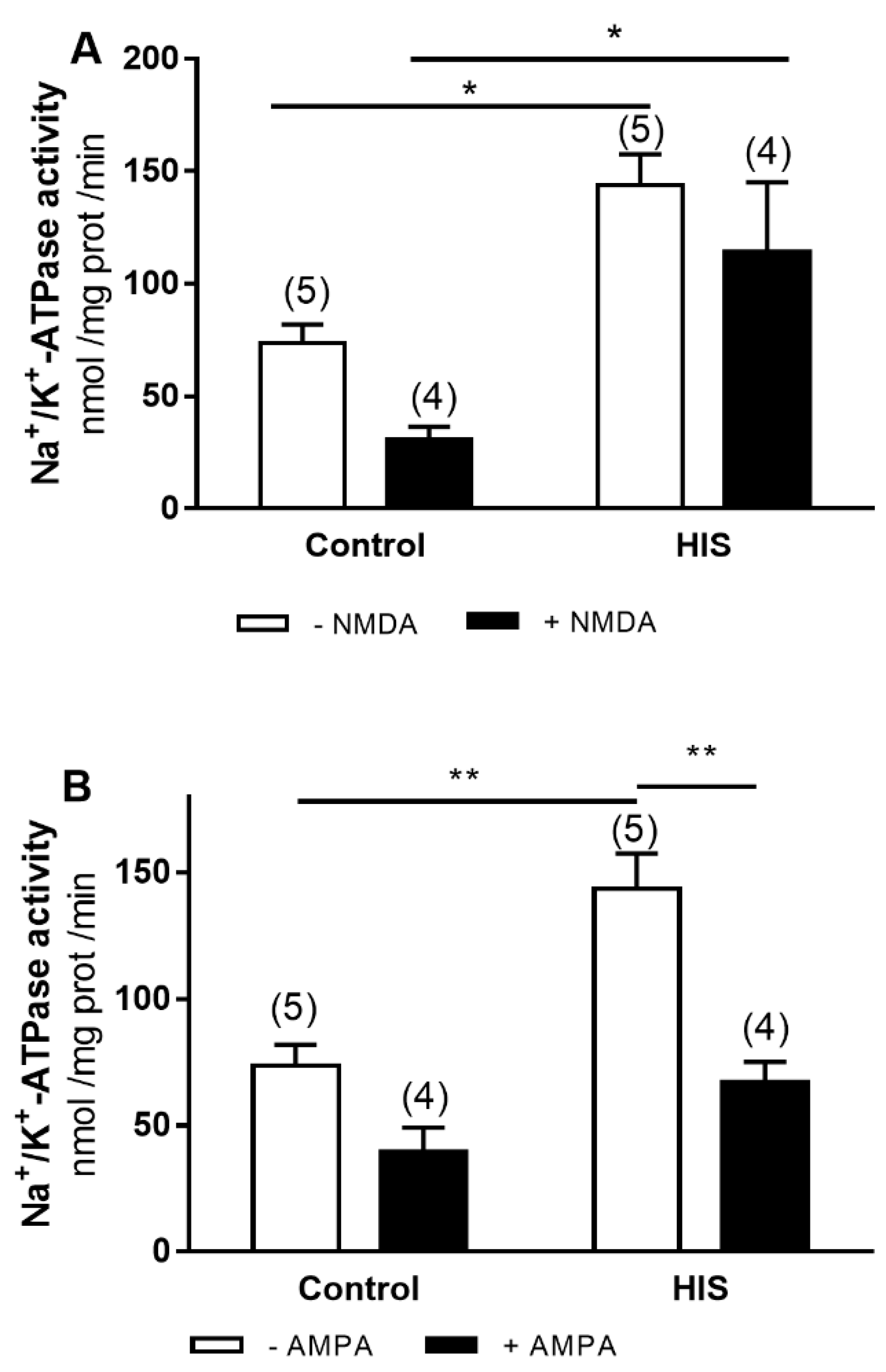
2.3. Modulation of Na+/K+-ATPase Activity by Ionotropic Glutamate Receptor after HIS

To evaluate the effect of ionotropic glutamate receptor, we used AMPA and NMDA, selective agonists of the different types of the corresponding ionotropic receptors. The two-way ANOVA analyses revealed that both HIS and agonists [NMDA: F(1,14) = 4.729, p = 0.0473 and AMPA: F(1,14) = 26.56, p = 0.0001] significantly altered Na+/K+-ATPase activity. No significant interaction of HIS x agonist was observed for NMDA and AMPA receptor (Figure 3A,B). Bonferroni’s post hoc test showed that the significant increase in Na+/K+-ATPase activity induced by HIS was significantly reduced in the presence of AMPA (p < 0.01) (Figure 3B).

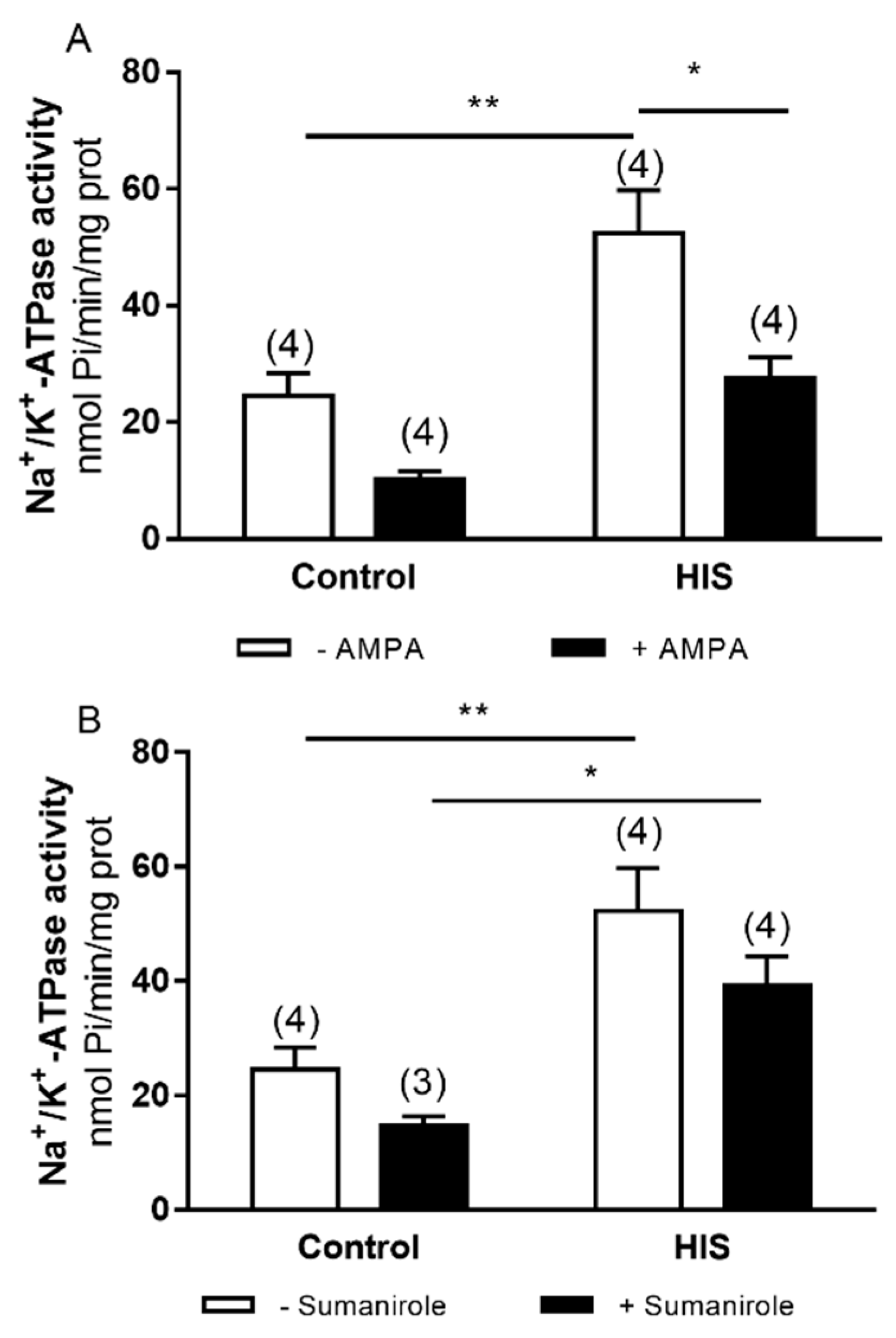
2.4. Modulation of Na+/K+-ATPase Activity after HIS by AMPA and Sumanirole in 2-Month-Old Animals

The effect of ionotropic glutamate receptor AMPA and G-protein-coupled receptor dopamine D2 receptor was shown in the Na+/K+-ATPase activity. The two-way ANOVA analyses revealed a significant difference in HIS and agonist AMPA [AMPA: F (1,12) = 17.97, p = 0.0011] (Figure 4A) in Na+/K+-ATPase activity. However, no significative interaction of HIS x agonist was found.
Bonferroni’s post hoc test showed a significant increase in Na+/K+-ATPase activity after HIS in the control group in Figure 4A and B (p < 0.01), with an increase in Na+/K+-ATPase activity in the sumanirole group in Figure 4B (p < 0.05). In Figure 4B, a decrease was observed in Na+/K+-ATPase activity after HIS in the presence of agonist (p < 0.05).

2.5. Mg2+-ATPase Activity in Cortical Plasma Membrane after HIS

The analysis of Mg2+-ATPase activity revealed that it was significantly increased 5 days after HIS (175.4 ± 12.94 nmol of Pi /mg prot /min vs. 270.9 ± 7.98 nmol of Pi /mg prot /min, p < 0.01), whereas no significant variation was found 20 days following hyperthermic seizures (136.1 ± 13.91 nmol of Pi /mg prot /min vs. 131.7 ± 20.46 nmol of Pi /mg prot /min, p > 0.05) and in 2-month-old animals (134.0 ± 10.88 nmol of Pi /mg prot /min vs. 126.1 ± 8.40 nmol of Pi /mg prot /min, p > 0.05) (Figure 5).

2.6. Modulation of Mg2+-ATPase Activity by G-Protein-Coupled Receptor after HIS

We also analyzed whether the increase in Mg2+-ATPase activity induced by HIS could be modulated by sumanirole, CPA or CGS 21680. The two-way ANOVA analyses showed that HIS caused a significant change in Mg2+-ATPase activity [Figure 6A: F(1.16) = 18.23, p = 0.0006; Figure 6B: F(1.16) = 15.28, p = 0.0013; Figure 6C: F(1.16) = 14.7, p = 0.0115]; moreover, agonist x HIS interaction was detected for CPA and CGS 21680 agonist [CPA: F(1.16) = 6.452, p = 0.0218; CPA x HIS: F(1.16) = 5.604, p = 0.0309; CGS 21680: F(1.16) = 8.037, p = 0.0119; CGS 21680 x HIS: F(1.16) = 5.475, p = 0.0326].
Bonferroni’s post hoc test showed an increase in Mg2+-ATPase activity for sumanirole after HIS in the control group (p < 0.05), while in the presence of CPA and CGS 21680 we observed an increase in Mg2+-ATPase activity after HIS in the control group (p < 0.01), together with a decrease in this activity after HIS in the presence of both agonists (p < 0.05).

2.7. Modulation of Mg2+-ATPase by Ionotropic Receptors after HIS

Finally, we analyzed the effect of ionotropic glutamate receptors activation on the increase in Mg2+-ATPase activity evoked by HIS. The two-way ANOVA revealed a significant effect of HIS [Figure 7A F(1,16) = 8.866, p = 0.0089; and Figure 7B F(1,16) = 20.58, p = 0.0003] on Mg2+-ATPase activity, similarly for NMDA, agonist and agonist x HIS [NMDA: F(1.16) = 7.788, p = 0.0131; NMDA x HIS: F(1.16) = 6.937, p = 0.0181] (Figure 7A).
Bonferroni’s post hoc test showed an increase in Mg2+-ATPase activity after HIS in the control group (p < 0.01). Moreover, Mg2+-ATPase activity was increased in the presence of AMPA (AMPA: p < 0.05). However, a significative decrease was observed after HIS with NMDA (p < 0.01).

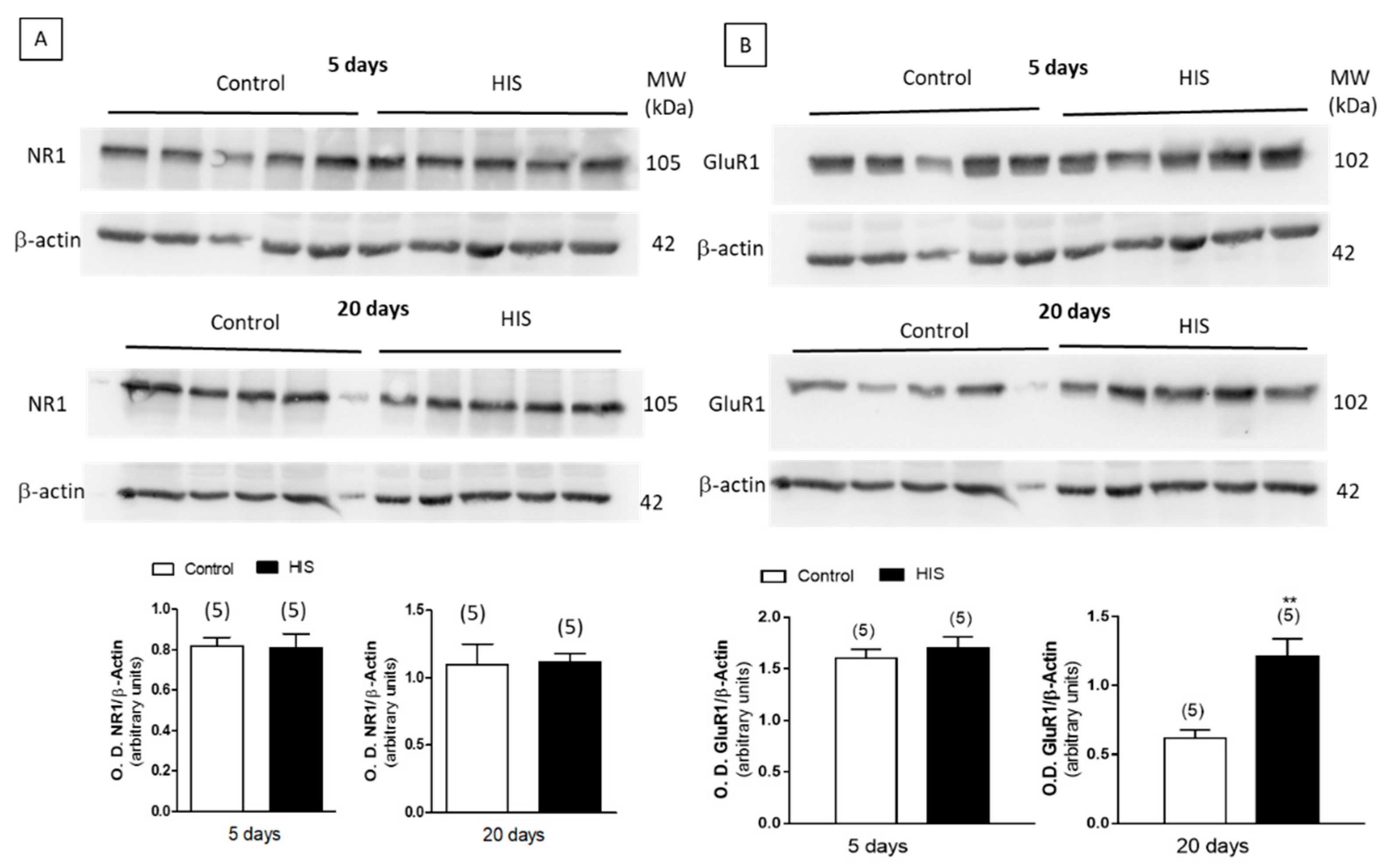
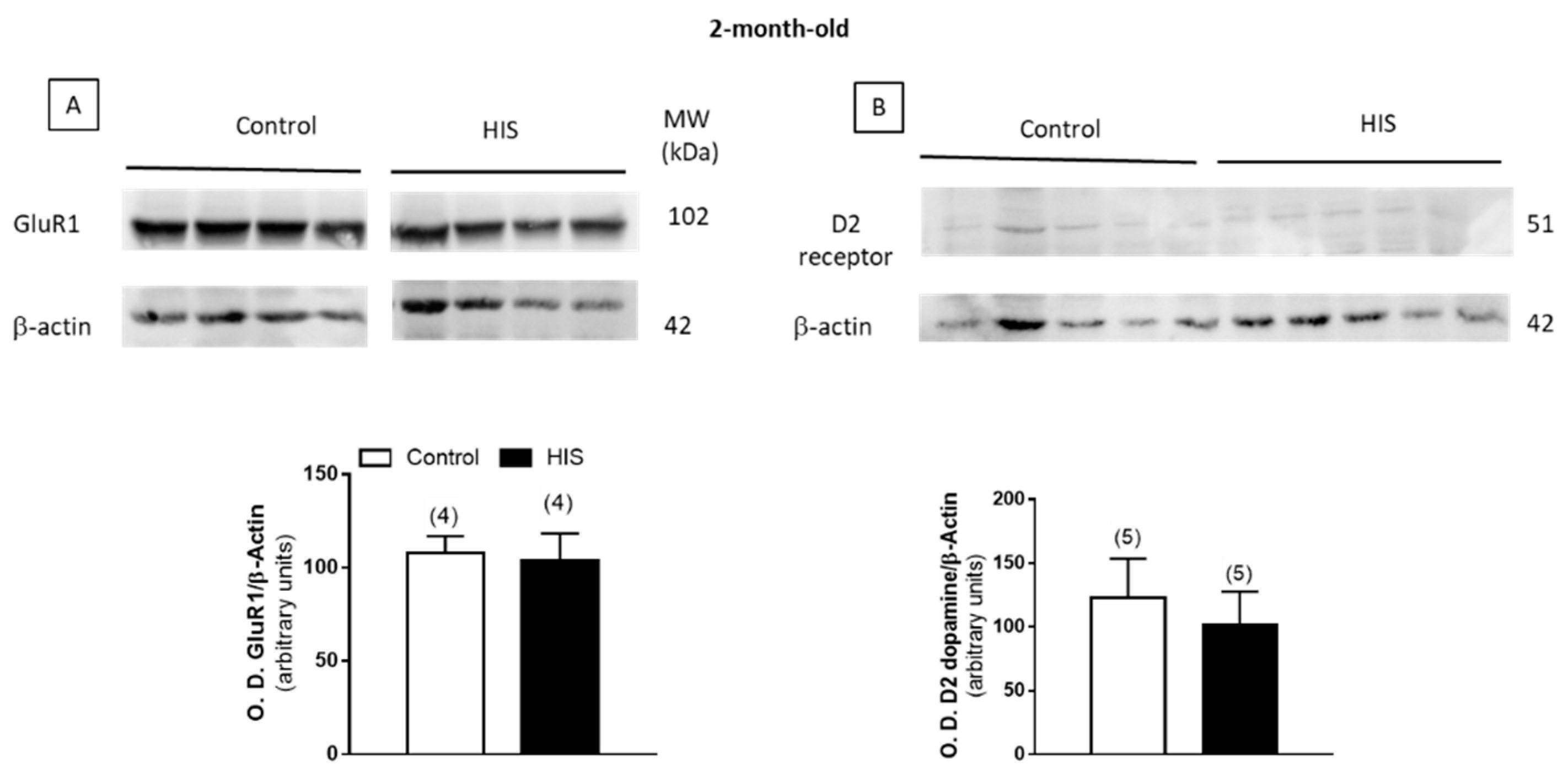
2.8. Protein Level of Na+/K+ATPase and Receptors after HIS

The effect of HIS on the expression of the alpha subunit of Na+/K+-ATPase, dopamine D2 receptors and the subunits NR1 and GluR1 of NMDA and AMPA receptors was studied by a Western-blot analysis. As shown in Figure 8A,B, Figure 9A and Figure 10, no significant variation of the alpha subunit Na+/K+-ATPase, dopamine D2 receptor and NR1 subunit was found immediately after HIS, nor 5 or 20 days after. However, a significant increase in the subunit GluR1 of AMPA receptor was detected after 20 days, whereas it was unaltered 5 days after HIS (0.62 ± 0.06 vs. 1.21 ± 0.13, p < 0.01) (Figure 9B).

3. Discussion

The results obtained in the present work show that Na+/K+-ATPase activity significantly increased 20 days after HIS (PD 32) and in 2-month-old animals (PD 60). This effect was significantly reduced after activation of D2-dopamine and AMPA receptors. When these agonists were studied in 2-month-old animals, only AMPA showed a decrease in Na+/K+-ATPase activity. Furthermore, a significant increase in Mg2+-ATPase activity could be observed 5 days after HIS (PD 17), which was also reduced upon CPA, CGS 21680 and NMDA receptor activation.
Multiple works using animal models of epilepsy have shown changes in Na+/K+-ATPase activity, although the results are discrepant [23,29,30]. Our results agree well with those previously published by Reime Kinjo and coworkers [30], who described that three episodes of pilocarpine-induced status epilepticus at postnatal days 7, 8 and 9 evoked a significant increase in Na+/K+-ATPase activity in the hippocampus 7 or 30 days later. We believe that the increased Na+/K+-ATPase activity observed in cortical plasma membranes 20 days after HIS and maintained until 2 months of age could constitute a compensatory mechanism to reduce the extracellular concentration of potassium and, therefore, reduce the hyperexcitability of the cortex. Supporting this hypothesis, a previous study suggested that the convulsions in febrile seizures are evoked by a malfunction of temperature dependent Kir4.1 channel in astrocytes. This channel, under normal conditions, effectively removes extracellular potassium ions during the neuronal firing process. However, this channel reduced its amplitude at 40º C, being unable to remove extracellular excessive potassium ions, which lead to rapid accumulation and, consequently, the appearance of febrile seizures [31].
Another interesting result obtained in the present work is that the activation of AMPA and D2 dopamine receptors reduced the increase in Na+/K+-ATPase activity evoked by HIS 20 days after, and the AMPA effect was maintained over time up to 2 months of age. However, under control conditions, the activation of these receptors did not induce any significant effect on Na+/K+-ATPase activity. These results can be explained by the ability of Na+/K+-ATPase to associate physically to AMPA and D2 dopamine receptors. The colocalization of Na+/K+-ATPase and AMPA receptor has been shown in cultured cortical neurons, which was also accompanied by a specific association between the alpha subunit of Na+/K+-ATPase and AMPA receptors using coimmunoprecipitation in rat cortex lysates [25]. Finally, Hazelwood and coworkers [32], using coimmunoprecipitation and mass spectrometry, showed that in the brain, D1 and D2 dopamine receptors can form a complex with the Na+/K+-ATPase pump. Furthermore, they suggested that the D2-mediated decrease in Na+/K+-ATPase activity was dependent upon protein–protein interactions, rather than signaling molecules. Therefore, the results obtained in the present work suggest that HIS could promote a physical interaction between Na+/K+-ATPase and AMPA and D2 dopamine receptor. Taking into account that the increase in Na+/K+-ATPase activity detected 20 days after HIS and maintained at 2 months old for AMPA receptor constitute a compensatory mechanism to counterbalance the hyperexcitability of the cortex. The activation of these receptors diminished the efficacy of this mechanism and favored the appearance of epileptic status in adulthood.
The changes observed in the activity of Na+/K+-ATPase 20 days after HIS seems not to be associated to the corresponding increase in the protein level of this enzyme in the plasma membrane, since the densitometric analyses of the bands in the Western blot experiments failed to find any significant variation. A similar lack of significant variation was also observed when the protein levels of D2 dopamine receptor and NR1 subunit of NMDA receptors were studied. Taking into account that NMDA receptors are formed by three different subunits (NR1, NR2 and NR3) and that NR1 is essential for channel formation [33], the results obtained in this work suggest that HIS did not significantly modify the protein level of NMDA in cortical plasma membranes. However, the immunochemical detection of the subunit GluR1 revealed that HIS induced a significant increase in this subunit 20 days after hyperthermic seizures. The AMPA receptor is a tetramer composed of four subunits of GluR1, GluR2, GluR3 and GluR4. An increase in GluR1 subunit’s protein has previously been shown in the hippocampus of epileptic patients [34]. A similar increase in GluR1 has also recently been published in an immature rodent model of temporal lobe epilepsy [35], where the intraperitoneal injection of Kainate in three-week-old mice evoked seizures and an increase in the level of GluR1 expression. Therefore, the increase in GluR1 subunits observed in the present work seems to favor the development of an epileptogenic process.
In 2-month-old animals, we found that D2 dopamine receptor remained unaltered, as at 20 days after HIS. However, GluR1 levels, analyzed for AMPA receptor, that were increased 20 days after HIS, recovered the control values. Therefore, the relationship between Na+/K+-ATPase and AMPA does not seem to depend on the levels of AMPA in the membrane in that case.
Concerning Mg2+-ATPase activity, the results show that HIS significantly increased the activity of this enzyme 5 days after HIS. This enzyme maintains high intracellular concentrations of magnesium, which is important to regulate several biochemical reactions involved in energy metabolism, signal transduction and protein synthesis [36,37]. Thus, magnesium plays a key role in the intracellular signaling process by encouraging protein kinase activity [38]. Moreover, magnesium provides protective effects against free radicals and contributes to genome stability by acting as a cofactor for enzymes involved in DNA repair [39]. Therefore, the increase in Mg2+-ATPase activity observed in the present work suggests the existence of a protective mechanism. Accordingly, de Freitas and coworkers [40] found an increase in Mg2+-ATPase activity in rat hippocampus after pilocarpine-induced seizures, which was attributed to the oxidative stress caused by the seizures. In that sense, previous work carried out in our laboratory has shown that HIS evokes oxidative stress in the cerebellum and cortex of rats [10,41].
Finally, the results in this work show that HIS promotes a negative interaction between NMDA, A1 and A2A receptor and Mg2+-ATPase activity. Thus, the increase in Mg2+-ATPase activity detected in the hyperthermia group was significantly reduced after NMDA, A1 and A2A receptor activation. There are no reports, at least to our knowledge, showing such an interaction. Considering that the increase in Mg2+-ATPase activity following HIS is a protective mechanism, the reduction in this effect could increase the risk of suffering epileptic disorder in adulthood. More work would be helpful to clarify this point.
In summary, after hyperthermia-induced seizures at postnatal day 12, a significant increase in Na+/K+-ATPase 20 days and 2 months later, respectively, and Mg2+-ATPase activity 5 days later, was observed, which agrees well with the existence of a protective mechanism. However, these effects were also accompanied by an increase in the AMPA subunit GluR1 and a negative interaction between Na+/K+-ATPase and Mg2+-ATPase and AMPA and NMDA receptor, respectively, which seem to indicate the loss of such neuroprotective mechanisms increasing the vulnerability and propensity to epileptic events in adults.

4. Materials and Methods

4.1. Materials

N6-cyclopentyladenosine (CPA), adenosine triphosphate (ATP) and malachite green were from Sigma-Aldrich (Madrid, Spain). Ammonium molybdate was from Merck (Madrid, Spain). CGS 21680, NMDA, AMPA and Kainate were from Tocris Bioscience (Madrid, Spain). All other reagents were of analytical grade and obtained from commercial sources.

4.2. Animals

Pregnant rats were obtained from the Autonomous University of Madrid (Spain). The care and use of animals were carried out in accordance with the Spanish laws (RD 53/2013 and Ley 32/2007) governing the use of laboratory animals. All experiments were performed under the guidelines of the Animal Experimental Committee of the University of Castilla-La Mancha. Every effort was made to minimize animal suffering and to reduce the number of animals used. Animals were maintained in a 12 h light/12 h dark cycle (lights on at 07:00 h) in a temperature-controlled room (25 °C) with constant humidity (40–50%) and with free access to food and drinking water. The day of birth was considered day 0. Pups were housed with their mother until weaning at postnatal day (PD) 21.

4.3. Hyperthermia-Induced Seizures

Neonatal rats were submitted to hyperthermia-induced seizures (HIS) at PD 12. This developmental stage is equivalent to a human aged between several months and 3 years, a period especially susceptible to febrile seizures. Hyperthermia was induced using a warmed air stream (45–50 °C) from a hair dryer located 50 cm above a plastic chamber (17 × 12 × 12 cm), using a previously described protocol [10,11]. Rectal temperature was measured at a 2 min interval. In order to judge the occurrence of seizures, two observers monitored the rats’ behavior. The behavioral seizures were stereotyped and previously shown to correlate with electroencephalography (EEG) discharges [5]. These behavioral seizures consisted of the arrest of heat-induced hyperkinesia, followed by body flexion. All pups exhibited these behaviors as well as rearing and falling over associated with hind-limb clonus seizures (stage 5 on Racine scale criteria), at ~10 min after hyperthermia induction when the core temperature was ~42 °C. After 20 min of behavioral seizures, the pups were moved to a cool surface until their rectal temperature reached baseline, and they were then returned to the home cages with their mothers. The duration of heating was 27 ± 3 min. Controls were separated from their mothers for the same duration and placed in a chamber at room temperature.

4.4. Cerebral Cortical Membrane Isolation

Cerebral cortical membranes were isolated following the previously described protocol [42]. Briefly, the cortex brain from pups was homogenized in isolation buffer (50 mM of Tris-HCl, pH 7.4 containing 10 mM of MgCl2 and protease inhibitors) in a Dounce homogenizer. After homogenization, the samples were centrifuged for 5 min at 1000× g in a Beckman JA 21 centrifuge (Coulter, Madrid, Spain). Then, supernatant was centrifuged for 10 min at 27,000× g, and the pellet was finally resuspended in isolation buffer. Protein concentration was measured with the Lowry method, using bovine serum albumin as a standard.

4.5. Na+/K+-ATPase and Mg2+-ATPase Activities Assay

Enzyme activities were analyzed following the method previously described by Schweinberger and coworkers with minor modifications [43].
The reaction mixture contained 5.0 mM of MgCl2, 80.0 mM of NaCl, 20.0 mM of KCl and 40.0 mM of Tris–HCl, pH 7.4, in a final volume of 200 μL. The reaction was initiated by the addition of 3 mM of ATP. The Mg2+-ATPase activity was measured with the addiction of 1.0 mM of ouabain. The Na+/K+-ATPase activity was measured as the difference between the total activity and the activity of the Mg2+-ATPase. The specific activity of the enzyme was expressed as nmol of Pi released per min per mg of protein.
The effect of AMPA, NMDA, Kainate, dopamine D2 and adenosine A1 and A2A receptor activation receptor on these enzymes was measured following the same protocol, but with a 6 min preincubation with AMPA (140 µM), NMDA (140 µM), Kainate (140 µM), sumanirole (140 µM), CPA (140 µM) and CGS 21680 (140 µM) at 30 °C. For each sample, the percentage of action of each ligand on the basal activity of Na+/K+ and Mg2+-ATPase was measured.

4.6. Western-Blotting Assay

Plasma membranes samples (40 μg) were subjected to 7.5% polyacrylamide gel electrophoresis (PAGE) in the presence of sodium dodecyl sulphate (SDS). After electrophoretic separation, the proteins were transferred into nitrocellulose membranes using iBlot™ Dry Blotting System (Invitrogen, Barcelona, Spain) and blocked for 1 h with 5% nonfat- skimmed milk in phosphate-buffered saline. Immunodetection was carried out by incubating the nitrocellulose membranes with the corresponding primary antibody anti-alpha 1 Sodium Potassium ATPase (1:5000, ab7671 from Abcam), anti-NMDAR1 (1:1000, ab134308 from Abcam), recombinant Anti-Glutamate Receptor 1 (AMPA subtype) antibody [EPR19522] (1:1000, ab183797 from Abcam) and Anti-Dopamine D2 Receptor antibody (1:1000, ab88074 from Abcam). β-Actin antibody (1:5000, ab8226 from Abcam) and Ponceau red staining were used as a gel loading controls. After washing, the blots were incubated with horseradish peroxidase-coupled goat anti-rabbit or anti-mouse IgG (1:5000, GARPO 172–1019 and GAMPO 170–6516 from Bio-Rad). Bands were visualized using the ECL chemiluminescence detection kit from GE Healthcare (Madrid, Spain) in a G:Box chamber, and specific bands were quantified by densitometry using Gene-Tools software (Syngene). The Western blot membrane used to detect NMDAR1 was stripped using a stripping solution containing H2O2 30% for 15 min at 37 °C. This method allows the inactivation (quenched) of horseradish peroxidase and, therefore, to label a second protein with a different antibody without any interference. After stripping, the solution membrane was reproved using Anti-Glutamate Receptor 1 (AMPA subtype) antibody.

4.7. Statistical and Data Analysis

Statistical comparisons were carried out using an unpaired two-tailed Student’s t-test and a two-way ANOVA, followed by a Bonferroni comparison post hoc test using GraphPad Prism 5. The results are expressed as mean ± standard error of the mean. Differences between mean values were considered statistically significant at p < 0.05.

Supplementary Materials

The following supporting information can be downloaded at: https://www.mdpi.com/article/10.3390/ijms232314638/s1, Figure S1: Figure 8A, 5 days beta actin; Figure S2: Figure 8A, 5 days Na+/K+ ATPase; Figure S3: Figure 8A, 20 days beta actin; Figure S4: Figure 8A, 20 days Na+/K+ ATPase; Figure S5: Figure 8B, 5 days beta actin; Figure S6: Figure 8B, 5 days D2 dopamine; Figure S7: Figure 8B, 20 days beta actin; Figure S8: Figure 8B, 20 days D2 dopamine; Figure S9: Figure 9A, 5 days beta actin; Figure S10: Figure 9A, 5 days NMDA; Figure S11: Figure 9A, 20 days beta actin; Figure S12: Figure 9A, 20 days NMDA; Figure S13; Figure 9B, 5 days AMPA; Figure S14: Figure 9B, 20 days AMPA; Figure S15: Figure 10A, 2-month-old GluR1; Figure S16: Figure 10A, 2-month-old beta actine; Figure S17: Figure 10B, D2 dopamine; Figure S18: Figure 10B, beta actine.

Author Contributions

Conceptualization, D.A.L.-N. and M.M.; methodology, D.A.L.-N. and M.C.; formal analysis, D.A.L.-N. and M.C.; investigation, M.C.; visualization, D.A.L.-N.; resources, M.M.; data curation, D.A.L.-N. and M.C.; writing—original draft preparation, D.A.L.-N.; writing—review and editing, D.A.L.-N., M.M. and M.C.; supervision, D.A.L.-N. and M.M.; project administration, D.A.L.-N. and M.M.; funding acquisition, M.M. All authors have read and agreed to the published version of the manuscript.

Funding

This research was funded by Ministerio de Ciencia e Innovación (grant PID2019-109206GB-I00), UCLM (grant 2020-GRIN-29108 co-financed with the European Union FEDER), and Junta de Comunidades de Castilla-La Mancha (JCCM) (grant SBPLY/19/180501/000251). M.C. is the recipient of a postdoctoral grant (01150PO906) from JCCM cofounded by FEDER.

Institutional Review Board Statement

The animal study protocol was approved by the Institutional Review Board (or Ethics Committee) of the Animal Experimental Committee of the University of Castilla-La Mancha (EXP 26/2014/13).

Conflicts of Interest

The authors declare no conflict of interest. The funders had no role in the design, execution, interpretation, or writing of the study.

References

  1. Shinnar, S.; Glauser, T.A. Febrile seizures. J. Child Neurol. 2002, 17 (Suppl. 1), S44–S52. [Google Scholar] [CrossRef]
  2. Stafstrom, C.E. Assessing the behavioral and cognitive effects of seizures on the developing brain. Prog. Brain Res. 2002, 135, 377–390. [Google Scholar]
  3. Dubé, C.M.; Brewster, A.L.; Baram, T.Z. Febrile seizures: Mechanisms and relationship to epilepsy. Brain Dev. 2009, 31, 366–371. [Google Scholar] [CrossRef] [PubMed] [Green Version]
  4. McClelland, S.; Dubé, C.M.; Yang, J.; Baram, T.Z. Epileptogenesis after prolonged febrile seizures: Mechanisms, biomarkers and therapeutic opportunities. Neurosci. Lett. 2011, 497, 155–162. [Google Scholar] [CrossRef] [Green Version]
  5. Baram, T.Z.; Gerth, A.; Schultz, L. Febrile seizures: An appropriate-aged model suitable for long-term studies. Brain Res. Dev. Brain Res. 1997, 98, 265–270. [Google Scholar] [CrossRef] [PubMed] [Green Version]
  6. Dubé, C.; Richichi, C.; Bender, R.A.; Chung, G.; Litt, B.; Baram, T.Z. Temporal lobe epilepsy after experimental prolonged febrile seizures: Prospective analysis. Brain 2006, 129, 911–922. [Google Scholar] [CrossRef] [PubMed]
  7. Dubé, C.M.; Brewster, A.L.; Richichi, C.; Zha, Q.; Baram, T.Z. Fever, febrile seizures and epilepsy. Trends Neurosci. 2007, 30, 490–496. [Google Scholar] [CrossRef] [Green Version]
  8. Bender, R.A.; Baram, T.Z. Epileptogenesis in the developing brain: What can we learn from animal models? Epilepsia 2007, 48 (Suppl. 5), 2–6. [Google Scholar] [CrossRef] [PubMed] [Green Version]
  9. León-Navarro, D.A.; Albasanz, J.L.; Martín, M. Hyperthermia-induced seizures alter adenosine A1 and A2A receptors and 5′-nucleotidase activity in rat cerebral cortex. J. Neurochem. 2015, 134, 395–404. [Google Scholar] [CrossRef] [PubMed]
  10. Crespo, M.; León-Navarro, D.A.; Martín, M. Cerebellar oxidative stress and fine motor impairment in adolescent rats exposed to hyperthermia-induced seizures is prevented by maternal caffeine intake during gestation and lactation. Eur. J. Pharmacol. 2018, 822, 186–198. [Google Scholar] [CrossRef]
  11. Crespo, M.; León-Navarro, D.A.; Martín, M. Early-life hyperthermic seizures upregulate adenosine A2A receptors in the cortex and promote depressive-like behavior in adult rats. Epilepsy Behav. 2018, 86, 173–178. [Google Scholar] [CrossRef]
  12. Crespo, M.; León-Navarro, D.A.; Ruíz, M.Á.; Martín, M. Hyperthermia-induced seizures produce long-term effects on the functionality of adenosine A1 receptor in rat cerebral cortex. Int. J. Dev. Neurosci. 2020, 80, 1–12. [Google Scholar] [CrossRef] [Green Version]
  13. Crespo, M.; León-Navarro, D.A.; Martín, M. Hyperthermia-induced seizures during neonatal period alter the functionality of A1 and A2A receptors in the cerebellum and evoke fine motor impairment and gait disturbances in adult rats. Physiol. Behav. 2021, 240, 113543. [Google Scholar] [CrossRef]
  14. Crespo, M.; León-Navarro, D.A.; Martín, M. Glutamatergic System is Affected in Brain from an Hyperthermia-Induced Seizures Rat Model. Cell. Mol. Neurobiol. 2022, 42, 1501–1512. [Google Scholar] [CrossRef] [PubMed]
  15. Tsai, M.-L.; Leung, L.S. Decrease of hippocampal GABA B receptor-mediated inhibition after hyperthermia-induced seizures in immature rats. Epilepsia 2006, 47, 277–287. [Google Scholar] [CrossRef]
  16. Postnikova, T.Y.; Griflyuk, A.V.; Amakhin, D.V.; Kovalenko, A.A.; Soboleva, E.B.; Zubareva, O.E.; Zaitsev, A.V. Early Life Febrile Seizures Impair Hippocampal Synaptic Plasticity in Young Rats. Int. J. Mol. Sci. 2021, 22, 8218. [Google Scholar] [CrossRef]
  17. Chang, Y.-C.; Kuo, Y.-M.; Huang, A.-M.; Huang, C.-C. Repetitive febrile seizures in rat pups cause long-lasting deficits in synaptic plasticity and NR2A tyrosine phosphorylation. Neurobiol. Dis. 2005, 18, 466–475. [Google Scholar] [CrossRef] [PubMed]
  18. Chen, B.; Feng, B.; Tang, Y.; You, Y.; Wang, Y.; Hou, W.; Hu, W.; Chen, Z. Blocking GluN2B subunits reverses the enhanced seizure susceptibility after prolonged febrile seizures with a wide therapeutic time-window. Exp. Neurol. 2016, 283, 29–38. [Google Scholar] [CrossRef]
  19. González Ramírez, M.; Orozco Suárez, S.; Salgado Ceballos, H.; Feria Velasco, A.; Rocha, L. Hyperthermia-induced seizures modify the GABA(A) and benzodiazepine receptor binding in immature rat brain. Cell. Mol. Neurobiol. 2007, 27, 211–227. [Google Scholar] [CrossRef]
  20. Deprez, L.; Weckhuysen, S.; Peeters, K.; Deconinck, T.; Claeys, K.G.; Claes, L.R.F.; Suls, A.; Van Dyck, T.; Palmini, A.; Matthijs, G.; et al. Epilepsy as part of the phenotype associated with ATP1A2 mutations. Epilepsia 2008, 49, 500–508. [Google Scholar] [CrossRef]
  21. Clapcote, S.J.; Duffy, S.; Xie, G.; Kirshenbaum, G.; Bechard, A.R.; Rodacker Schack, V.; Petersen, J.; Sinai, L.; Saab, B.J.; Lerch, J.P.; et al. Mutation I810N in the alpha3 isoform of Na+,K+-ATPase causes impairments in the sodium pump and hyperexcitability in the CNS. Proc. Natl. Acad. Sci. USA 2009, 106, 14085–14090. [Google Scholar] [CrossRef]
  22. Oliveira, M.S.; Furian, A.F.; Royes, L.F.F.; Fighera, M.R.; de Carvalho Myskiw, J.; Fiorenza, N.G.; Mello, C.F. Ascorbate modulates pentylenetetrazol-induced convulsions biphasically. Neuroscience 2004, 128, 721–728. [Google Scholar] [CrossRef]
  23. Marquezan, B.P.; Funck, V.R.; Oliveira, C.V.; Pereira, L.M.; Araújo, S.M.; Zarzecki, M.S.; Royes, L.F.F.; Furian, A.F.; Oliveira, M.S. Pentylenetetrazol-induced seizures are associated with Na+,K+-ATPase activity decrease and alpha subunit phosphorylation state in the mice cerebral cortex. Epilepsy Res. 2013, 105, 396–400. [Google Scholar] [CrossRef]
  24. Akkuratov, E.E.; Lopacheva, O.M.; Kruusmägi, M.; Lopachev, A.V.; Shah, Z.A.; Boldyrev, A.A.; Liu, L. Functional Interaction between Na/K-ATPase and NMDA Receptor in Cerebellar Neurons. Mol. Neurobiol. 2015, 52, 1726–1734. [Google Scholar] [CrossRef]
  25. Zhang, D.; Hou, Q.; Wang, M.; Lin, A.; Jarzylo, L.; Navis, A.; Raissi, A.; Liu, F.; Man, H.-Y. Na, K-ATPase activity regulates AMPA receptor turnover through proteasome-mediated proteolysis. J. Neurosci. 2009, 29, 4498–4511. [Google Scholar] [CrossRef] [PubMed] [Green Version]
  26. Therien, A.G.; Blostein, R. Mechanisms of sodium pump regulation. Am. J. Physiol. Cell Physiol. 2000, 279, C541–C566. [Google Scholar] [CrossRef] [PubMed] [Green Version]
  27. Poulsen, H.; Morth, P.; Egebjerg, J.; Nissen, P. Phosphorylation of the Na+,K+-ATPase and the H+,K+-ATPase. FEBS Lett. 2010, 584, 2589–2595. [Google Scholar] [CrossRef] [Green Version]
  28. Yamanaka, R.; Shindo, Y.; Oka, K. Magnesium Is a Key Player in Neuronal Maturation and Neuropathology. Int. J. Mol. Sci. 2019, 20, 3439. [Google Scholar] [CrossRef] [PubMed] [Green Version]
  29. Fernandes, M.J.; Naffah-Mazzacoratti, M.G.; Cavalheiro, E.A. Na+K+ ATPase activity in the rat hippocampus: A study in the pilocarpine model of epilepsy. Neurochem. Int. 1996, 28, 497–500. [Google Scholar] [CrossRef] [PubMed]
  30. Kinjo, E.R.; Arida, R.M.; de Oliveira, D.M.; da Silva Fernandes, M.J. The Na+/K+ ATPase activity is increased in the hippocampus after multiple status epilepticus induced by pilocarpine in developing rats. Brain Res. 2007, 1138, 203–207. [Google Scholar] [CrossRef]
  31. Du, M.; Li, J.; Ying, W.; Yu, Y. A dynamics model of neuron-astrocyte network accounting for febrile seizures. Cogn. Neurodyn. 2022, 16, 411–423. [Google Scholar] [CrossRef]
  32. Hazelwood, L.A.; Free, R.B.; Cabrera, D.M.; Skinbjerg, M.; Sibley, D.R. Reciprocal modulation of function between the D1 and D2 dopamine receptors and the Na+,K+-ATPase. J. Biol. Chem. 2008, 283, 36441–36453. [Google Scholar] [CrossRef] [Green Version]
  33. Monyer, H.; Sprengel, R.; Schoepfer, R.; Herb, A.; Higuchi, M.; Lomeli, H.; Burnashev, N.; Sakmann, B.; Seeburg, P.H. Heteromeric NMDA receptors: Molecular and functional distinction of subtypes. Science 1992, 256, 1217–1221. [Google Scholar] [CrossRef] [PubMed]
  34. Ying, Z.; Babb, T.L.; Comair, Y.G.; Bushey, M.; Touhalisky, K. Increased densities of AMPA GluR1 subunit proteins and presynaptic mossy fiber sprouting in the fascia dentata of human hippocampal epilepsy. Brain Res. 1998, 798, 239–246. [Google Scholar] [CrossRef]
  35. Wang, T.; Wang, L.; Li, L.; Ma, L.; Liu, X. Effects of perampanel on cognitive behavior and GluR1 expression in immature mice of temporal lobe epilepsy. Biochem. Biophys. Res. Commun. 2022, 588, 68–74. [Google Scholar] [CrossRef]
  36. Stangherlin, A.; O’Neill, J.S. Signal Transduction: Magnesium Manifests as a Second Messenger. Curr. Biol. 2018, 28, R1403–R1405. [Google Scholar] [CrossRef] [PubMed] [Green Version]
  37. Terasaki, M.; Rubin, H. Evidence that intracellular magnesium is present in cells at a regulatory concentration for protein synthesis. Proc. Natl. Acad. Sci. USA 1985, 82, 7324–7326. [Google Scholar] [CrossRef] [Green Version]
  38. Knape, M.J.; Ahuja, L.G.; Bertinetti, D.; Burghardt, N.C.G.; Zimmermann, B.; Taylor, S.S.; Herberg, F.W. Divalent Metal Ions Mg2+ and Ca2+ Have Distinct Effects on Protein Kinase A Activity and Regulation. ACS Chem. Biol. 2015, 10, 2303–2315. [Google Scholar] [CrossRef] [Green Version]
  39. Mathew, A.A.; Panonnummal, R. ’Magnesium’—The master cation-as a drug-possibilities and evidences. Biometals 2021, 34, 955–986. [Google Scholar] [CrossRef] [PubMed]
  40. De Freitas, R.M.; Feng, D.; Jordán, J. Neuropharmacological effects of lipoic acid and ubiquinone on δ-aminolevulinic dehydratase, Na+, K+-ATPase, and Mg2+-ATPase activities in rat hippocampus after pilocarpine-induced seizures. Fundam. Clin. Pharmacol. 2011, 25, 211–216. [Google Scholar] [CrossRef] [PubMed]
  41. Navarro, D.A.L.; Crespo, M.; Martín, M. Oxidative stress in epileptogenesis: Febrile seizures, chemoconvulsant pilocarpine, and electrical stimulation. In Oxidative Stress and Dietary Antioxidants in Neurological Diseases; Elsevier: Amsterdam, The Netherlands, 2020; pp. 81–94. [Google Scholar]
  42. López-Zapata, A.; León-Navarro, D.A.; Crespo, M.; Martín, M. Gender-specific desensitization of group I metabotropic glutamate receptors after maternal l-glutamate intake during lactation. Int. J. Dev. Neurosci. 2018, 68, 10–16. [Google Scholar] [CrossRef] [PubMed]
  43. Schweinberger, B.M.; Schwieder, L.; Scherer, E.; Sitta, A.; Vargas, C.R.; Wyse, A.T.S. Development of an animal model for gestational hypermethioninemia in rat and its effect on brain Na+,K+-ATPase/Mg2+-ATPase activity and oxidative status of the offspring. Metab. Brain Dis. 2014, 29, 153–160. [Google Scholar] [CrossRef] [PubMed]
Figure 1. Status of Na+/K+-ATPase activity in neonatal cortex 5 and 20 days after HIS and in 2-month-old animals. Bar graphs show the effect of HIS on Na+/K+ ATPase activity measured 5 days and 20 days after HIS (PD 17 and PD 32, respectively) and in 2-month-old animals (PD60). Data are mean ± S.E.M. values from 4 to 5 different animals from independent litters. * p < 0.05 and ** p < 0.01 are significantly different from control using unpaired two-tailed Student’s t-test.
Ijms 23 14638 g001
Figure 2. Effects of D2 dopamine (A), adenosine A1 (B) and A2A receptors (C) activation on Na+/K+ ATPase activity in neonatal cortex 20 days after HIS (PD 32). The activity of Na+/K+ ATPase was measured in control rats (PD 32) and after HIS (PD 32), following the protocol described in the Materials and Methods section. To evaluate the influence of D2 dopamine, adenosine A1 and A2A receptor activation on Na+/K+ ATPase activity, the corresponding plasma membranes were preincubated for 6 min at 30 °C with 140 μM of sumanirole, 140 μM of CPA and 140 μM of CGS 21680, respectively. Each bar represents the mean ± SEM for the indicated n value. * p < 0.05, ** p < 0.01 using a two-way ANOVA and Bonferroni’s post hoc test.
Ijms 23 14638 g002
Figure 3. Effects of NMDA (A) and AMPA (B) activation on Na+/K+-ATPase activity in neonatal cortex 20 days after HIS (PD 32). The activity of Na+/K+ ATPase was measured in control rats (PD 32) and after HIS (PD 32), following the protocol described in the Materials and Methods section. To evaluate the influence of NMDA and AMPA receptor activation on Na+/K+ ATPase activity, the corresponding plasma membranes were preincubated for 6 min at 30 °C with 140 μM of NMDA and 140 μM of AMPA, respectively. Each bar represents the mean ± SEM for 4–5 values. * p < 0.05 and ** p < 0.01 using a two-way ANOVA and Bonferroni’s post hoc test.
Ijms 23 14638 g003
Figure 4. Effects of AMPA (A) and D2 dopamine (B) receptors activation on Na+/K+ ATPase activity in cortex after HIS in 2-month-old animals (PD 60). The activity of Na+/K+ ATPase was measured in control rats (PD 60) and after HIS (PD 60), following the protocol described in the Materials and Methods section. To evaluate the influence of AMPA and D2 dopamine receptor activation on Na+/K+ ATPase activity, the corresponding plasma membranes were preincubated for 6 min at 30 °C with 140 μM of AMPA and 140 μM of sumanirole, respectively. Each bar represents the mean ± SEM for for the indicated n value. * p < 0.05 and ** p < 0.01 using a two-way ANOVA and Bonferroni’s post hoc test.
Ijms 23 14638 g004
Figure 5. Status of Mg2+-ATPase activity in neonatal cortex 5 and 20 days after HIS and in 2-month-old animals. Bar graphs show the effect of HIS on Mg2+-ATPase activity measured 5 days and 20 days after HIS (PD 17 and PD 32, respectively) and in 2-month-old animals (PD 60). Data are mean ± SEM. values from 4 to 6 different neonates from independent litters. *** p < 0.001 is significantly different from control using unpaired two-tailed Student’s t-test.
Ijms 23 14638 g005
Figure 6. Effects of D2 dopamine (A), adenosine A1 (B) and A2A receptors (C) activation on Mg2+-ATPase activity in neonatal cortex 5 days after HIS (PD 17). The activity of Mg2+-ATPase was measured in control rats (PD 17) and after HIS (PD 17), following the protocol described in the Materials and Methods section. To evaluate the influence of D2 dopamine, adenosine A1 and A2A receptor activation on Na+/K+ ATPase activity, the corresponding plasma membranes were preincubated for 6 min at 30 °C with 140 μM of sumanirole, 140 μM of CPA and 140 μM of CGS 21680, respectively. Each bar represents the mean ± SEM for n = 5 in all groups analyzed. * p < 0.05 and ** p < 0.01 using a two-way ANOVA and Bonferroni’s post hoc test.
Ijms 23 14638 g006
Figure 7. Effects of NMDA (A) and AMPA (B) receptors activation on Mg2+-ATPase activity in neonatal cortex 5 days after HIS (PD 17). The activity of Mg2+-ATPase was measured in control rats (PD 17) and after HIS (PD 17), following the protocol described in the Materials and Methods section. To evaluate the influence of NMDA and AMPA receptor activation on Mg2+-ATPase activity, the corresponding plasma membranes were preincubated for 6 min at 30 °C with 140 μM of NMDA and 140 μM of AMPA, respectively. Each bar represents the mean ± SEM for n = 5 * p < 0.05 and ** p < 0.01 using a two-way ANOVA and Bonferroni’s post hoc test.
Ijms 23 14638 g007
Figure 8. Immunoblotting analysis of alpha subunit of Na+/K+-ATPase (A) and dopamine D2 receptor (B) in cortical plasma membranes from rats 5 and 20 days after HIS. Plasma membranes (40 μg) were subjected to SDS-PAGE, electrophoretically transferred to nitrocellulose and probed with anti-alpha 1 Sodium Potassium ATPase and anti-Dopamine D2 receptor antibody antisera, as described in Materials and Methods. Data, expressed as arbitrary units, are means ± SEM of experiments performed with five different plasma membrane preparations (Supplementary Material: Figures S1–S8).
Ijms 23 14638 g008
Figure 9. Immunoblotting analysis of alpha subunit of NR1 (A) and GluR1 (B) subunits in cortical plasma membranes from rats 5 and 20 days after HIS. Plasma membranes (40 μg) were subjected to SDS-PAGE, electrophoretically transferred to nitrocellulose and probed with anti-NMDAR1 and recombinant anti-Glutamate Receptor 1 antibody, as described in Materials and Methods. Data, expressed as arbitrary units, are means ± SEM of experiments performed with five different plasma membrane preparations. ** p < 0.01 using unpaired two tailed Student’s t-test (Supplementary Materials: Figures S9–S14).
Ijms 23 14638 g009
Figure 10. Immunoblotting analysis of alpha subunit of GluR1 (A) and dopamine D2 receptor (B) subunits in cortical plasma membranes from 2-month-old rats. Plasma membranes (40 μg) were subjected to SDS-PAGE, electrophoretically transferred to nitrocellulose and probed with recombinant anti-Glutamate Receptor 1 antibody and anti-Dopamine D2 receptor antibody antisera, as described in Materials and Methods. Data, expressed as arbitrary units, are means ± SEM of experiments performed with five different plasma membrane preparations (Supplementary Materials: Figures S15–S18).
Ijms 23 14638 g010
Publisher’s Note: MDPI stays neutral with regard to jurisdictional claims in published maps and institutional affiliations.

