Article

Conjunctival Fluid Secretion Impairment via CaCC-CFTR Dysfunction Is the Key Mechanism in Environmental Dry Eye

1
State Key Laboratory of Ophthalmology, Zhongshan Ophthalmic Center, Sun Yat-sen University, Guangzhou 510060, China
2
School of Life Sciences, Sun Yat-sen University, Guangzhou 510275, China
3
Guangdong Institute for Vision and Eye Research, Guangzhou 510060, China
*
Author to whom correspondence should be addressed.
These authors contributed equally to this work.
Int. J. Mol. Sci. 2022, 23(22), 14399; https://doi.org/10.3390/ijms232214399
Submission received: 22 September 2022 / Revised: 12 November 2022 / Accepted: 15 November 2022 / Published: 19 November 2022

Abstract

:
Dry eye disease (DED) is a multifactorial disease with an incidence of approximately 50% worldwide. DED seriously affects quality of life and work. The prevalence of environmental DED (eDED) ranges from 35 to 48%. Conjunctival fluid secretion dysfunction may be one of the major causes of DED. Notably, the Cl flux corresponds to the conjunctival fluid secretion and could be affected by ATP. Both the cystic fibrosis transmembrane conductance regulator (CFTR) and the Ca2+-activated Cl channel (CaCC) are Cl channels involved in epithelial fluid secretion. Conjunctival fluid secretion could be increased by activating P2Y2R (an ATP receptor) in DED. However, the role of the CaCC and CFTR channels regulated by P2Y2R in eDED remains unclear. In this study, we established a rabbit eDED model using a controlled drying system. A Ussing chamber was used to perform a conjunctival short-circuit current induced by ATP to evaluate the reactivity of the ion channels to the ATP. Our results revealed that eDED accompanied by conjunctival fluid secretion impairment was caused by a P2Y2R dysfunction, which is related to CaCC-CFTR signaling in the conjunctiva epithelium. Notably, the coupling effect of the ATP-induced CaCC-CFTR activation and intracellular Ca2+ may represent a promising therapeutic target for treating eDED.

1. Introduction

More than one billion people suffer from dry eye disease (DED) worldwide. Severe DED can cause visual impairment, which severely affects the quality of life of patients [1,2]. DED is a multifactorial disease of the ocular surface, characterized by a loss of homeostasis of the tear film [3]. The predisposing factors for DED include environmental risk, androgen deficiency, contact lenses, and aging [4]. A mild DED, environmental dry eye disease (eDED), has become increasingly common, with an incidence of 35–48% [1]. Notably, tear production has been shown to be significantly decreased in eDED patients [5]. In tear production, the secretory rate of the conjunctival fluid exceeds the basal tear production [6]. Conjunctival secretion has been shown to contribute approximately one-fourth of the total tear production [7], indicating that conjunctival secretion could play an important role in tear production. Whether conjunctival secretion is impaired in eDED and the role it plays in the pathogenesis of eDED are still unknown.
The secretion of water from conjunctival epithelial cells was evaluated using chloride ion (Cl) fluid. The outflow of the Cl flux corresponded to the conjunctival fluid secretion [8]. The cystic fibrosis transmembrane conductance regulator (CFTR) is a cAMP-dependent Cl channel that facilitates fluid secretion in the apical side of the conjunctiva [9]. The CFTR agonist could increase the tear volume in clinical DED patients [8]. However, no change in the Cl concentration was found in patient tears with cystic fibrosis due to the CFTR mutation [10].
ATP receptors exist in various types of epithelial cells, including the conjunctiva epithelium. P2Y2R is one type of ATP receptor. P2Y2R agonists, such as diquafosol, UTP, and ATP, have been used to increase the tear production in DED patients and rabbits [6,11,12]. Notably, the activation of P2Y2R increased the intracellular calcium ion (Ca2+) [13], which has an obvious effect on the regulation of the Cl flux in the airway epithelium [14]. Moreover, the application of A23187 (a Ca2+ ionophore) apically increased the Cl flux by 58% in the rabbit conjunctiva epithelium [15]. A protein corresponding to the Ca2+-activated Cl channel (CaCC) was detected on the apical side of the conjunctiva [16]. These findings indicate that the CaCC signaling may be involved in conjunctival secretion. However, there are no direct experimental data on the effects of the CaCC on conjunctival secretion. In the kidney inner medullary collecting duct cells and human airway epithelia, the CaCC signaling works with the CFTR [17,18]. Whether P2Y2R contributes to regulating the conjunctival fluid secretion via the combination of the CaCC and CFTR has not been elucidated, especially in eDED.
In this study, the impairment and pathogenesis of conjunctival epithelium secretion in eDED was investigated using an eDED rabbit model. Our study demonstrated that the P2Y2R signaling dysfunction is a key factor in the impairment of conjunctival secretion by the coactivation of the CaCC and CFTR. Our research revealed that a new molecular mechanism underlies the fluid secreted from the conjunctival epithelium, indicating a potential therapeutic target for treating eDED.

2. Results

2.1. eDED Has Ocular Surface Injury and Conjunctival Fluid Secretion Dysfunction

To investigate an ocular surface injury and the tear secretion in eDED, a controlled drying system (CDS) was established. In this study, the disruptions of the conjunctiva and cornea epithelium were stained with lissamine green and fluorescein, respectively. The staining score was positively correlated with the staining area. Compared with the control group, the lissamine green score increased from day 7 to day 14 in the dry eye group (day 7: 0.15 ± 0.08 vs. 1.30 ± 0.15; day 14: 0.15 ± 0.08 vs. 1.90 ± 0.23, all n = 10–11, p < 0.001) (Figure 1a). Similarly, the fluorescein staining score significantly increased from day 7 to day 14 in the dry eye group (day 7: 1.00 ± 0.26 vs. 5.00 ± 0.49; day 14: 1.10 ± 0.18 vs. 7.27 ± 0.65, all n =10–11, p < 0.001) (Figure 1b). The fluid secretion from the conjunctiva is one of the most vital components of tear production. As expected, the tear production also declined from day 7 to day 14 in the dry eye group (9.45 ± 0.47 vs. 5.80 ± 0.39 mm at day 7; 9.63 ± 0.51 vs. 3.20± 0.24 mm at day 14; all n = 10–11, p < 0.001) (Figure 1c). Consistent with the above results, the ocular epithelium combined with the fluid reduction in the conjunctiva was impaired in eDED.

2.2. P2Y2R Plays a Key Role in eDED

Activating ATP receptors increases the intracellular Ca2+ and cAMP [19,20]. To confirm the changes in the intracellular Ca2+ in eDED, we digested the eDED conjunctival epithelium and loaded it with an intracellular calcium fluorescence probe (Fluo-3/AM). The results showed that the intensity of the intracellular Ca2+ was decreased in the eDED conjunctival epithelium (10.80 ± 1.02 vs. 4.85 ± 0.9 AU, n = 6, p < 0.001) (Figure 2a). In addition, the concentration of the cAMP in the conjunctiva epithelium was reduced in the dry eye group (5.08 ± 0.26 vs. 4.30 ± 0.21 nmol/mL, n = 9, p < 0.05) (Figure 2b). Subsequently, the ATP did not evoke an intracellular Ca2+ release of the same amplitude in the conjunctival epithelium in the dry eye group as in the control group (Figure 2c). The ATP-dependent Isc reflects the reactivity of the ion channels to the ATP, which plays a role via P2Y2R [13]. The conjunctival fluid secretion is closely related to the function of the anion channel [8]. Notably, the initial peak (first peak) and sustained peak (second peak) of the conjunctival epithelium Isc were markedly decreased after the ATP stimulation in the dry eye group (first peak: 27.19 ± 3.06 vs. 7.08 ± 1.79, n = 6, p < 0.001; second peak: 28.84 ± 2.92 vs. 6.32 ± 1.28, n = 6, p < 0.05) (Figure 3a). These results demonstrated that the ATP receptor dysfunction in the conjunctiva epithelium plays a key role in eDED.
To elucidate the relationship between the P2Y2R and Isc variation, a P2Y2R inhibitor (suramin) was applied before the conjunctival ATP stimulation. The results showed that treatment with the P2Y2R inhibitor led to a decrease of 50% in the ATP-induced conjunctival Isc (first peak: 24.06 ± 3.24 vs. 11.98 ± 2.34; second peak: 27.27 ± 2.50 vs. 5.14 ± 2.10, all n = 6–8, p < 0.01) (Figure 3b). The ATP-induced conjunctival Isc decreased by more than half in the P2Y2R-siRNA treatment group compared with the control group (first peak: 16.67 ± 2.10 vs. 6.79 ± 3.17; second peak:15.84 ± 2.60 vs. 6.50 ± 2.84, all n = 4–5, p < 0.05) (Figure 3c). The rabbit ocular surfaces were also impaired using the P2Y2R-siRNA eyedrop treatment (Figure 3d). These findings showed that the ATP-induced conjunctival Isc appropriately showed conjunctival fluid secretion. The ATP-induced conjunctival Isc deficiency and subsequent decrease in the conjunctival fluid secretion in eDED contributed to the conjunctival P2Y2R dysfunction.

2.3. ATP-Induced Conjunctival Fluid Secretion Is Related to the Anion Channel

Both the Cl flux and HCO3 flux were secreted from the Cl channel of the cell membrane surface [21]. A conjunctiva buffer solution without Cl or HCO3 was used to determine whether the ATP-induced conjunctival Isc variation is related to the exchange of Cl or/and HCO3. Compared with the normal buffer solution, the ATP-induced conjunctival Isc response decreased by approximately 50% in the Cl-free buffer (first peak: 20.93 ± 2.54 vs. 8.10 ± 2.41; second peak: 23.91 ± 2.31 vs. 8.91 ± 1.85, all n = 5–12, p < 0.01) (Figure 4a,b,e) and in the HCO3-free buffer (first peak: 20.93 ± 2.54 vs. 4.82 ± 0.89; second peak: 23.91 ± 2.32 vs. 6.13 ± 0.92, all n = 6–12, p < 0.001) (Figure 4b,c,e). Moreover, the ATP-induced conjunctival Isc sharply declined by over 90% in the Cl- and HCO3-free buffer (first peak: 20.93 ± 2.54 vs. 2.44 ± 0.72; second peak: 23.91 ± 2.31 vs. 1.97 ± 0.59, all n = 9–12, p < 0.001) (Figure 4a,d,e). These results suggest that the combination of the Cl and HCO3 fluxes contributed to the ATP-induced conjunctival fluid secretion.

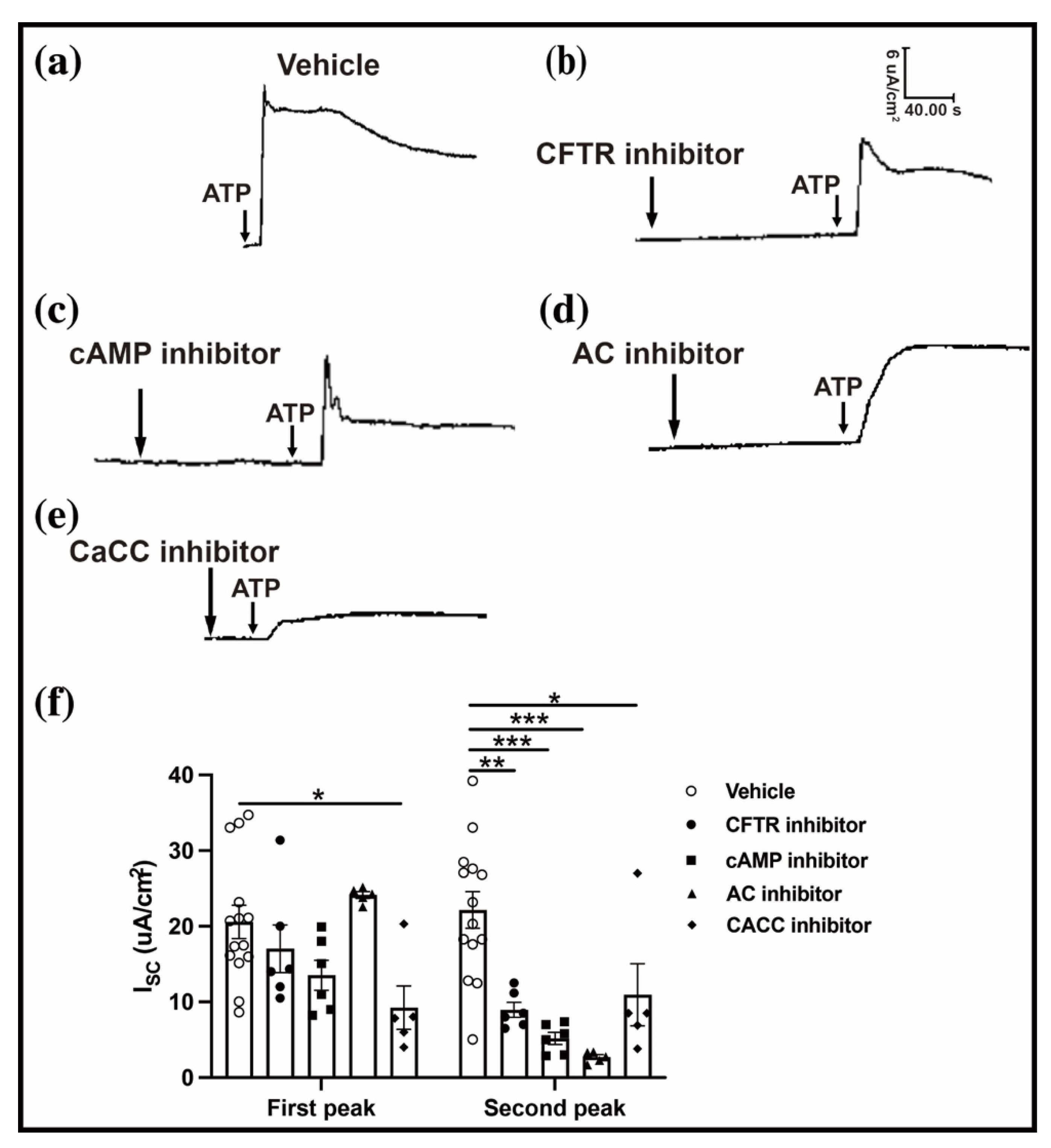
2.4. CaCC-CFTR Pathway Participates in the ATP-Induced Conjunctival Fluid Secretion

The CFTR is a cAMP-dependent Cl channel that is essential for conjunctival fluid secretion [8]. To determine whether the CFTR affects the ATP-induced conjunctival Isc variation, a CFTR inhibitor (CFTRinh-172) and a specific cAMP inhibitor (H89) were applied before the conjunctival ATP simulation. The CFTR inhibitor reduced the second peak of the conjunctival Isc without affecting the first peak (first peak: 20.59 ± 2.19 vs. 17.04 ± 3.15, n = 6–14, p < 0.05; second peak: 22.16 ± 2.40 vs. 8.96 ± 0.98 n = 6–14, p < 0.01) (Figure 5a,b,f). A similar trend was observed with a specific cAMP inhibitor (first peak: 20.59 ± 2.19 vs. 13.54 ± 1.98, n = 6–14, p < 0.05; second peak: 22.16 ± 2.40 vs. 5.18 ± 0.79, n= 6–14, p < 0.001) (Figure 5c,f). Furthermore, an adenylyl cyclase (AC)-specific inhibitor (MDL12330A), which is upstream of the cAMP, was used in the same way, resulting in a similar outcome (first peak: 20.59 ± 2.19 vs. 24.10 ± 0.44, n = 5–14, p < 0.05; second peak: 22.16 ± 2.40 vs. 2.69 ± 0.32, n = 5–14, p < 0.001) (Figure 5d,f). These results demonstrated that the CFTR participates in the conjunctival fluid secretion.
Subsequently, we focused on another Cl channel, CaCC, which is a Ca2+-activated Cl channel on the cell membrane of the conjunctiva epithelium [16]. To investigate the role of the CaCC in the conjunctival Isc, a specific CaCC blocker (T16Ainh-A01) was applied before the conjunctival ATP simulation. Compared with the vehicle group, the CaCC-specific blocker induced a decrease in the ATP-induced conjunctival Isc (first peak: 20.59 ± 2.19 vs. 9.25 ± 2.85, n = 5–14, p < 0.05; second peak: 22.16 ± 2.40 vs. 10.95 ± 4.11, n = 5–14, p < 0.05) (Figure 5e,f). These results indicate that the ATP-induced conjunctival fluid secretion is dependent on the activation of the CaCC-CFTR pathway.

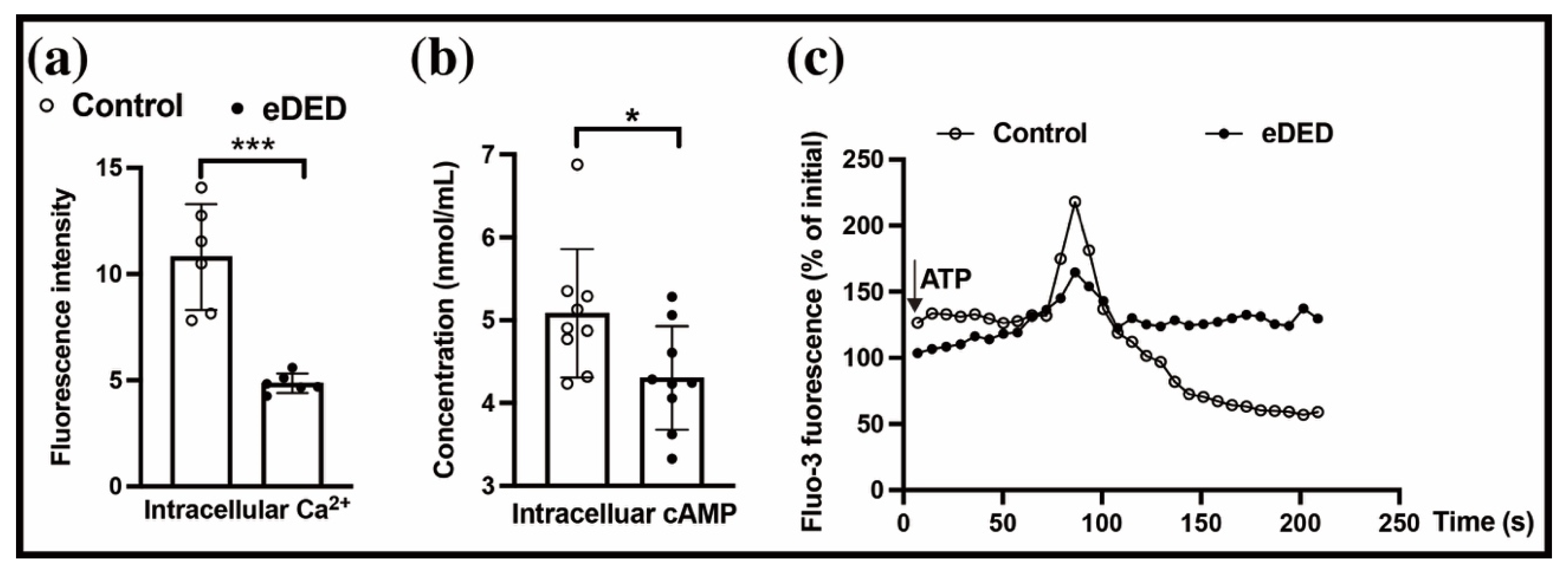
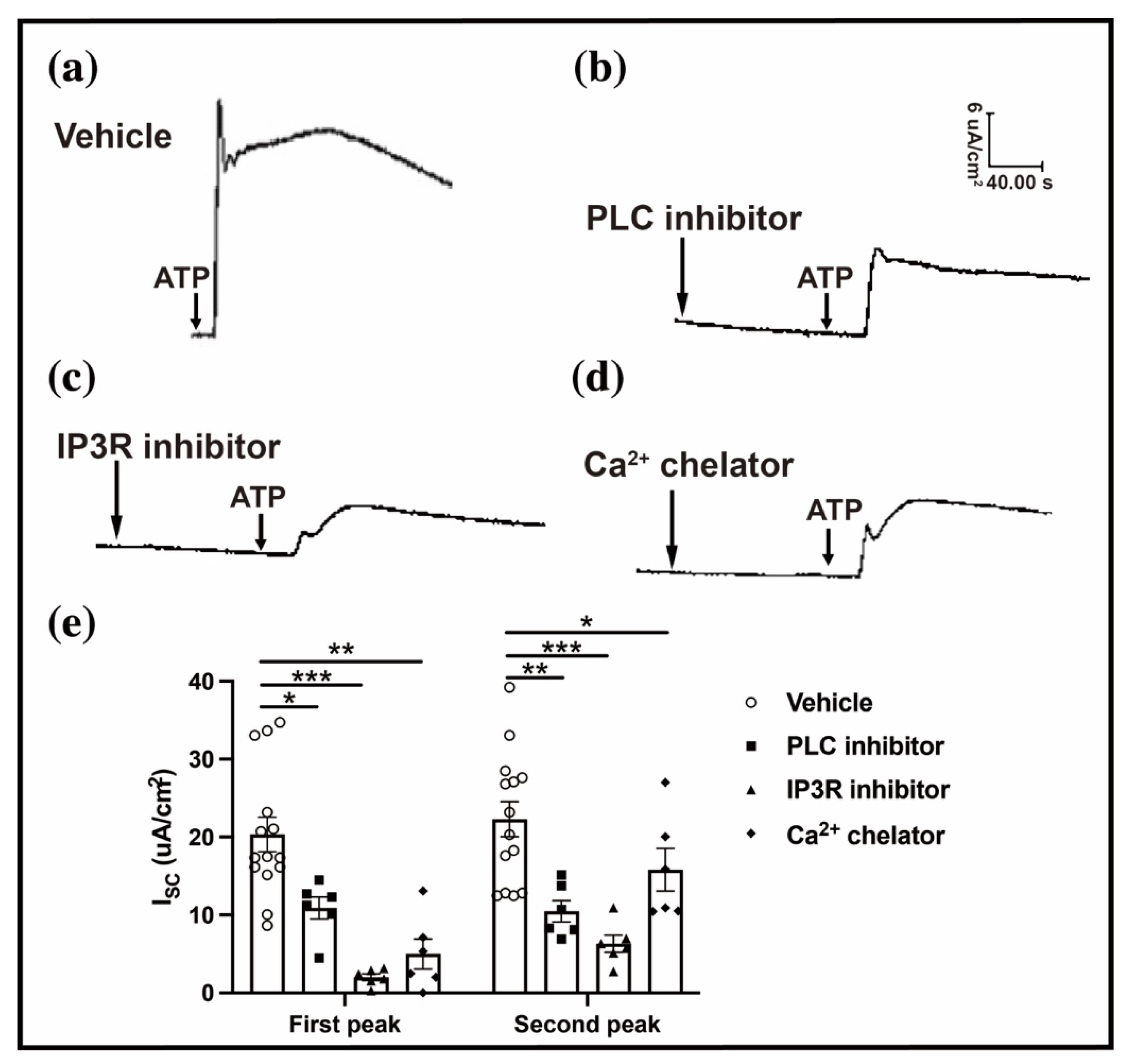
2.5. Intracellular Ca2+ Regulates the ATP-Induced Conjunctival Fluid Secretion

To investigate the role of the intracellular Ca2+ in the ATP-evoked conjunctival Isc, a PLC inhibitor (U73122), an IP3R inhibitor (2-APB), and a Ca2+ chelator (BAPTA/AM) were applied before the ATP simulation. The ATP-induced conjunctival Isc in the PLC inhibitor group decreased to one-half of that in the vehicle group (first peak: 20.34 ± 2.20 vs. 10.90 ± 1.41, n = 6–14, p < 0.05; second peak: 22.30 ± 2.26 vs. 10.49 ± 1.36, n= 6–14, p < 0.01) (Figure 6a,b,e). A same result was found in the treatment with the IP3R inhibitor (first peak: 0.34 ± 2.20 vs. 2.01 ± 0.43; second peak: 22.30 ± 2.26 vs. 6.31 ± 2.68, all n = 6–14, p < 0.001) (Figure 6a,c,e). The results revealed that the intracellular Ca2+ declined in eDED (Figure 2d) as well as several sources of cytosolic Ca2+ after the P2Y2R activation [22]. The ATP-induced conjunctival Isc response was initiated by treatment of the Ca2+ chelator (first peak: 20.34 ± 2.20 vs. 5.00 ± 1.91, n = 6–14, p < 0.01; second peak: 22.30 ± 2.26 vs. 15.80 ± 2.73, n = 6–14, p < 0.05) (Figure 6a,d,e). These results demonstrate that the intracellular Ca2+ is necessary for the ATP-induced conjunctival Isc and subsequent conjunctival fluid secretion.

3. Discussion

In this study, P2Y2R dysfunction combined with the decreased intracellular cAMP and cytosolic Ca2+ in the conjunctival epithelium was demonstrated in an eDED rabbit model. The P2Y2R dysfunction caused a conjunctival fluid secretion impairment, which was derived from the ATP through the CaCC-CFTR signaling. The release of the intracellular Ca2+ plays a core cross-pivotal role in the CaCC-CFTR signaling and is indispensable for the ATP-induced conjunctival fluid secretion. Therefore, the impairment of the conjunctival fluid secretion via the CaCC-CFTR pathway dysfunction could be a key mechanism in eDED.
The previous studies have shown that fluid in the conjunctiva epithelium is transported from the stromal (basolateral) side to the mucosal (apical) side. Conjunctival fluid secretion is dependent on the outflow of the anion flux [8]. There are various ion channels on the conjunctival epithelium, among which aquaporins, CFTR, ENaC, and CaCC are closely related to tear secretion [8,23]. Isc was found to present cellular ion channel reactivity on different activators. The ATP-induced Isc reflects the reactivity of the ion channels to the ATP [24]. As shown in this study, more than 90% of the ATP-induced Isc amplitude responses from the apical side of the conjunctival epithelium were produced by the anion secretion, suggesting that the ATP-induced Isc response could well represent the conjunctival fluid. The secretion of Cl from the conjunctival basal side to the apical side via an exchange with the sodium ions was demonstrated to account for approximately 60% of the Isc response [25,26]. Therefore, the outfluxes of HCO3 and other anions could occupy more than one-third of the ATP-induced Isc response. A similar fluid secretion was also detected in the cortical collecting duct cells [27] and the caudal epididymal epithelium [28].
To date, the activation of the P2Y receptors has been considered to stimulate the fluid secretion across the conjunctiva [24]. The levels of the intracellular cAMP and Ca2+ are elevated after the P2Y receptor activation [19,20]. The intrinsic process of activating P2Y2R is interrelated with the increase in the PLC and the transient increase in the intracellular Ca2+ concentration [11], suggesting that the cytosolic Ca2+ may regulate the conjunctival fluid secretion. Moreover, a release of the intracellular Ca2+ by another way, but not the PLC pathway, could have the same impact [29]. These results indicate that P2Y2R is a key factor in the conjunctival fluid secretion. However, the concentration of the ATP in tears increased in dry eye patients [30]. The concurrent cellular apoptosis of the conjunctiva epithelium, which resulted in P2Y2R dysfunction, was believed to cause increased tear ATP. The results of the present study showed that the P2Y2R dysfunction with decreased intracellular cAMP and Ca2+ existed in the conjunctiva of eDED, suggesting that P2Y2R plays a key role in eDED. In contrast, silencing P2Y2R led to a dry eye condition, and the inhibition of P2Y2R effectively suppressed the ATP-induced conjunctival Isc.
The CFTR is a cAMP-dependent Cl channel existing in both the corneal and conjunctival epithelium, which increased the basal tear volume after activation [8]. The present study also demonstrated that the ATP-induced conjunctival Isc was significantly suppressed by applying a CFTR inhibitor. Hence, the CFTR contributed to the ATP-induced conjunctival fluid secretion. However, the intracellular cAMP in the conjunctiva was decreased in the eDED model, indicating that the CFTR signaling was impaired in the dry eye condition.
In this study, the CaCC inhibitor decreased the ATP-induced conjunctival Isc, indicating that the CaCC signaling may be the main contributor to the ATP-induced conjunctival fluid secretion. The CaCC, another Cl channel, exists in the apical surface of the conjunctiva epithelium [31]. The CaCC is a Ca2+ dependent Cl channel that transports the fluid secretion from the airway epithelium [32]. Our results showed that increased intracellular Ca2+ activated a transient opening of the CaCC and then a persistent CFTR channel for the Cl secretion from the conjunctival epithelial cells. This study is the first in the literature to verify the combination of the CaCC signaling and CFTR in the Cl outflow across the conjunctival epithelium.
Our results showed that the content of the intracellular Ca2+ in the conjunctiva epithelium was decreased in eDED and did not respond to the ATP activation. The level of the intracellular Ca2+ was shown to be closely related to the function of the epithelial secretion, such as in the airway epithelium [13], caudal epididymal epithelium [28], and conjunctiva epithelium [29]. The intracellular Ca2+ in the conjunctiva epithelium can be regulated via different mechanisms. The activation of the transient receptor potential melastatin 8 channel increased the intracellular Ca2+ and then conducted sense information to the higher brain center [33]. In this study, P2Y2R dysfunction was found in the conjunctiva epithelium in eDED, which may be an intrinsic cause of the decreased intracellular Ca2+. Except for P2Y2R, the imbalance in the intracellular Ca2+ could be affected by endoplasmic reticulum stress and a mitochondrial matrix Ca2+ overload, which triggered cell apoptosis [34,35]. Endoplasmic reticulum stress-induced ocular surface epithelial cell apoptosis was shown in the pathogenesis of DED. Thus, further work is needed to explore the detailed mechanism of intracellular Ca2+ in eDED.

4. Materials and Methods

4.1. Animals

Male New Zealand white rabbits weighing 2.0–2.5 kg (purchased from Guangdong Provincial Center for Animal Research, Guangzhou, China) were chosen for the study. The rabbits were quarantined and acclimatized for one week before the experiments. All experimental procedures were approved by the Institutional Animal Care and Use Committee at Zhongshan Ophthalmic Center at Sun Yat-sen University and complied with all relevant ethical regulations for animal testing and research.

4.2. Environmental Dry Eye Disease Model

The rabbit eDED model was established using a controlled drying system (CDS). A detailed system for dry eye models was published in our previous report [36]. In brief, the rabbits in the dry eye group were reared in the CDS at a relative humidity of 22 ± 4%, an air flow of 3–4 m/s, and temperatures from 23 to 25 °C. The control group remained in a normal environment. Each rabbit was lissamine green/fluorescein stained as normal [9,37] and evaluated for tear production on days 0, 7, and 14.

4.3. Intervention of Rabbit Eyes with P2Y2R siRNA

Small interfering RNA (siRNA) sequences for silencing P2Y2R were applied topically four times per day to the ocular surfaces of the rabbits. The nucleotide sequence of the siRNA target site chosen to silence the expression of the rabbit P2Y2R-coding sequence (GenBank EU886321) was as follows: 5′-AAC CTG TAC TGC AGC ATC CTC-3′ [38,39]. The siRNA molecule was synthesized by RiboBio company (Guangzhou, China). The control group was given a normal saline as siRNA vehicle. The measurement of corneal fluorescein staining was in accordance with the above regulations.

4.4. Short-Circuit Current (Isc) Measurements

The Isc measurement was performed following the modified procedure described in our previous reports [40]. The superior conjunctival tissues were carefully isolated from the rabbits and clamped vertically between two halves of a Ussing chamber (EM-CSYS-2 Ussing Chamber Systems, Physiologic Instruments, San Diego, CA, USA) with an inner area of 0.45 cm2. The side of the epithelium was placed in a fixed direction facing the positive electrode. Then, the conjunctiva was bathed on both sides with bicarbonate Ringer’s solution composed of the following (in mM): 116.4 NaCl, 5.4 KCl, 5.4 NaHCO3, 0.78 NaH2PO4, 1.8 CaCl2, 0.81 MgCl2, 5.55 D-glucose, and 10 HEPES. It was then gassed with 95% O2/5% CO2 at 37 °C to maintain a pH of 7.4. The epithelium exhibited a basal transepithelial potential difference, which was measured by Ag/AgCl electrodes with KCl/Agar bridges connected to a voltage-clamp amplifier (VCC MC6, Physiologic Instruments, San Diego, CA, USA). The transepithelial potential difference in the conjunctiva epithelial layer was clamped at 0 mV for the Isc measurement.
The change in Isc was defined as the difference between the value at baseline and that at a peak following compound addition, displayed via a signal collection and analysis system (BL-420E + Systerm, Chengdu TME Technology Co., Ltd., Chengdu, China). When the current changed to the 1 mV pulse, it was used to estimate transepithelial resistance according to Ohm’s law [40]. Ten minutes after the Isc plateau, ATP was applied to the apical side, and then the ATP-induced Isc was recorded.
In the Cl-free BRS, NaCl, KCl, and CaCl2 were replaced by the respective salts of gluconate. In the HCO3-free solution, HCO3 was substituted by 10 mM HEPES and bathed with 100% O2 to maintain pH at 7.4. In the Cl- and HCO3-free solution, the components were the same as in the Cl-free solution, except that NaHCO3 was replaced by 20 mM Na-gluconate and 10 mM HEPES. Inhibitors or a vehicle (0.5% DMSO, D2650, Sigma, Burlington, MA, USA) were added to the buffer medium for 20 min before ATP was applied to the apical side. The dosages of inhibitors were as follows: 100 uM U73122 (U6756, Sigma, USA), a phospholipase C (PLC) inhibitor; 100 uM 2-APB (100065, Sigma, USA), an inositol (1,4,5) trisphosphate (IP3) receptor (IP3R) inhibitor; 100 uM BAPTA/AM (A1076, Sigma, USA), a Ca2+ chelator; 40 uM T16Ainh-A01 (SML0493, Sigma, USA), a specific CaCC inhibitor; and 40 uM CFTR (inh)-172 (C2992, Sigma, USA), a specific CFTR inhibitor; 10uM H89 (B1427, Sigma, USA), a specific cAMP-dependent protein kinase (PKA) inhibitor; and 30 uM MDL12330A (M182, Sigma, USA), an adenylate cyclase (AC) inhibitor. Different inhibitors were individually used in the conjunctival samples.

4.5. Measurement of Intracellular cAMP

The concentration of intracellular cAMP in the conjunctival epithelium was measured using a cAMP assays kit (E-EL-0056c, Elabscience Biotechnology Co., Ltd., Wuhan, China) according to the manufacturer’s protocol. Optical density readings were measured using a SynergyTM H1 multifunctional microplate reader (BioTek Instruments, Inc., Winooski, VT, USA). Concentrations were determined using a standard curve as a reference.

4.6. Measurement of Intracellular Ca2+

Intracellular Ca2+ was measured using the fluorescence dye Fluo-3/AM according to a modified procedure described in a previous report [41]. The conjunctival epithelium from the rabbits was digested and harvested with 1 mg/mL dispase-II solution for approximately 1 h, seeded in DMEM/F12 medium on glass-bottom cell culture dishes (20 mm, NEST, Wuxi, China), and cultured at 37 °C. After 10 min of cell adhesion, the cells were prepared and loaded with the fluorescent dye Fluo-3/AM (Molecular Probes, Eugene, OR, USA). The fluorescence signal was monitored and recorded using a laser-scanning confocal imaging system (TCS SP2; Leica Microsystems, Wetzlar, Germany). The change in fluorescence intensity after ATP treatment was normalized to the initial intensity.

4.7. Statistical Analysis

All statistical analyses were performed using SPSS version 22.0. All results were expressed as mean ± SEM. Statistical significances were determined using a Student’s t-test (two groups), and a repeated measures ANOVA, followed by a Least significant difference post hoc test (groups with repeated measurements). The differences between the groups were considered statistically significant if p was less than 0.05.

5. Conclusions

The results of this study indicate that the Ca2+-activated CaCC-CFTR pathway plays a key role in conjunctival secretion. The impairment of the P2Y2R-initiated CaCC-CFTR may be a key mechanism of the conjunctival secretion dysfunction in eDED.

Author Contributions

Conceptualization, S.Z.; methodology, L.L.; software, Y.W.; validation, L.L., Y.W. and X.C.; formal analysis, J.Z.; data curation, S.Y.; writing—original draft preparation, J.Z. and S.W.; writing—review and editing, W.Z. and S.Z.; supervision, S.Z.; project administration, L.L. and Y.W.; funding acquisition, S.Z. All authors have read and agreed to the published version of the manuscript.

Funding

This research was funded by the National Natural Science Foundation of China, grant number 31671210, and the Natural Science Foundation of Guangdong Province, China, grant number 2022A1515011140.

Institutional Review Board Statement

The animal study protocol was approved by the Ethics Committee of Zhongshan Ophthalmic Center at Sun Yat-sen University (protocol code 2019-165 and approved at 2019.11).

Informed Consent Statement

Not applicable.

Data Availability Statement

Not applicable.

Conflicts of Interest

The authors declare no conflict of interest. The funders had no role in the design of the study; in the collection, analyses, or interpretation of data; in the writing of the manuscript; or in the decision to publish the results.

References

  1. Stapleton, F.; Alves, M.; Bunya, V.Y.; Jalbert, I.; Lekhanont, K.; Malet, F.; Na, K.S.; Schaumberg, D.; Uchino, M.; Vehof, J.; et al. TFOS DEWS II Epidemiology Report. Ocul. Surf. 2017, 15, 334–365. [Google Scholar] [CrossRef] [PubMed]
  2. Bron, A.J.; de Paiva, C.S.; Chauhan, S.K.; Bonini, S.; Gabison, E.E.; Jain, S.; Knop, E.; Markoulli, M.; Ogawa, Y.; Perez, V.; et al. TFOS DEWS II pathophysiology report. Ocul Surf. 2017, 15, 438–510. [Google Scholar] [CrossRef]
  3. Wolffsohn, J.S.; Arita, R.; Chalmers, R.; Djalilian, A.; Dogru, M.; Dumbleton, K.; Gupta, P.K.; Karpecki, P.; Lazreg, S.; Pult, H.; et al. TFOS DEWS II Diagnostic Methodology report. Ocul. Surf. 2017, 15, 539–574. [Google Scholar] [CrossRef]
  4. Uchino, M.; Schaumberg, D.A.; Dogru, M.; Uchino, Y.; Fukagawa, K.; Shimmura, S.; Satoh, T.; Takebayashi, T.; Tsubota, K. Prevalence of dry eye disease among Japanese visual display terminal users. Ophthalmology 2008, 115, 1982–1988. [Google Scholar] [CrossRef] [PubMed]
  5. Abusharha, A.A.; Pearce, E.I. The effect of low humidity on the human tear film. Cornea 2013, 32, 429–434. [Google Scholar] [CrossRef] [PubMed]
  6. LI, Y.; Kuang, K.; Yerxa, B.; Wen, Q.; Rosskothen, H.; Fischbarg, J. Rabbit conjunctival epithelium transports fluid, and P2Y22 receptor agonists stimulate Cl2 and fluid secretion. Am. J. Physiol. Cell Physiol. 2001, 281, C595–C602. [Google Scholar] [CrossRef] [Green Version]
  7. Nandoskar, P.; Wang, Y.; Wei, R.; Liu, Y.; Zhao, P.; Lu, M.; Huang, J. Changes of chloride channels in the lacrimal glands of a rabbit model of Sjgren syndrome. Cornea 2011, 31, 7. [Google Scholar]
  8. Lee, H.K.; Park, J.; Kim, B.R.; Jun, I.; Kim, T.I.; Namkung, W. Isorhamnetin Ameliorates Dry Eye Disease via CFTR Activation in Mice. Int J Mol Sci 2021, 22, 3954. [Google Scholar] [CrossRef]
  9. Flores, A.M.; Casey, S.D.; Felix, C.M.; Phuan, P.W.; Verkman, A.S.; Levin, M.H. Small-molecule CFTR activators increase tear secretion and prevent experimental dry eye disease. FASEB J. 2016, 30, 1789–1797. [Google Scholar]
  10. Botelho, S.Y.; Goldstein, A.M.; Rosenlund, M.L. Tear sodium, potassium, chloride, and calcium at various flow rates: Children with cystic fibrosis and unaffected siblings with and without corneal staining. J Pediatr. 1973, 83, 601–606. [Google Scholar] [CrossRef]
  11. Murakami, T.; Fujihara, T.; Nakamura, M.; Nakata, K. P2Y(2) receptor stimulation increases tear fluid secretion in rabbits. Curr. Eye Res. 2000, 21, 782–787. [Google Scholar] [CrossRef]
  12. Dominguez-Godinez, C.; Carracedo, G.; Pintor, J. Diquafosol Delivery from Silicone Hydrogel Contact Lenses: Improved Effect on Tear Secretion. J. Ocul. Pharmacol. Ther. 2018, 34, 170–176. [Google Scholar] [CrossRef]
  13. Son, M.; Ito, Y.; Sato, S.; Ishikawa, T.; Kondo, M.; Nakayama, S.; Shimokata, K.; Kume, H. Apical and basolateral ATP-induced anion secretion in polarized human airway epithelia. Am. J. Respir. Cell Mol. Biol. 2004, 30, 411–419. [Google Scholar] [CrossRef]
  14. Centeio, R.; Ousingsawat, J.; Schreiber, R.; Kunzelmann, K. CLCA1 Regulates Airway Mucus Production and Ion Secretion Through TMEM16A. Int. J. Mol. Sci. 2021, 22, 5133. [Google Scholar] [CrossRef] [PubMed]
  15. Shiue, M.H.; Kim, K.J.; Lee, V.H. Modulation of chloride secretion across the pigmented rabbit conjunctiva. Exp. Eye Res. 1998, 66, 275–282. [Google Scholar] [CrossRef]
  16. Yu, D.; Thelin, W.R.; Rogers, T.D.; Stutts, M.J.; Randell, S.H.; Grubb, B.R.; Boucher, R.C. Regional differences in rat conjunctival ion transport activities. Am. J. Physiol. Cell Physiol. 2012, 303, C767–C780. [Google Scholar] [CrossRef] [PubMed] [Green Version]
  17. Faria, D.; Schreiber, R.; Kunzelmann, K. CFTR is activated through stimulation of purinergic P2Y2 receptors. Pflugers Arch. 2009, 457, 1373–1380. [Google Scholar] [CrossRef] [PubMed]
  18. Rajagopal, M.; Kathpalia, P.P.; Thomas, S.V.; Pao, A.C. Activation of P2Y1 and P2Y2 receptors induces chloride secretion via calcium-activated chloride channels in kidney inner medullary collecting duct cells. Am. J. Physiol. Renal. Physiol. 2011, 301, F544–F553. [Google Scholar] [CrossRef] [PubMed] [Green Version]
  19. Dutta, A.K.; Woo, K.; Doctor, R.B.; Fitz, J.G.; Feranchak, A.P. Extracellular nucleotides stimulate Cl- currents in biliary epithelia through receptor-mediated IP3 and Ca2+ release. Am. J. Physiol. Gastrointest. Liver Physiol. 2008, 295, G1004–G1015. [Google Scholar] [CrossRef] [Green Version]
  20. Mikolajewicz, N.; Smith, D.; Komarova, S.V.; Khadra, A. High-affinity P2Y2 and low-affinity P2X7 receptor interaction modulates ATP-mediated calcium signaling in murine osteoblasts. PLoS Comput. Biol. 2021, 17, e1008872. [Google Scholar] [CrossRef]
  21. Peng, L.; Gao, D.D.; Xu, J.W.; Xu, J.B.; Ke, L.J.; Qiu, Z.E.; Zhu, Y.X.; Zhang, Y.L.; Zhou, W.L. Cellular mechanisms underlying carbon monoxide stimulated anion secretion in rat epididymal epithelium. Nitric Oxide 2020, 100, 30–37. [Google Scholar] [CrossRef]
  22. Szabadkai, G.; Bianchi, K.; Varnai, P.; De Stefani, D.; Wieckowski, M.R.; Cavagna, D.; Nagy, A.I.; Balla, T.; Rizzuto, R. Chaperone-mediated coupling of endoplasmic reticulum and mitochondrial Ca2+ channels. J. Cell Biol. 2006, 175, 901–911. [Google Scholar] [CrossRef]
  23. Fischbarg, J. Water channels and their roles in some ocular tissues. Mol. Aspects Med. 2012, 33, 638–641. [Google Scholar] [CrossRef]
  24. Kulkarni, A.A.; Trousdale, M.D.; Stevenson, D.; Gukasyan, H.J.; Shiue, M.H.; Kim, K.J.; Read, R.W.; Lee, V.H. Nucleotide-induced restoration of conjunctival chloride and fluid secretion in adenovirus type 5-infected pigmented rabbit eyes. J. Pharmacol. Exp. Ther. 2003, 305, 1206–1211. [Google Scholar] [CrossRef] [Green Version]
  25. Kompella, U.B.; Kim, K.J.; Lee, V.H. Active chloride transport in the pigmented rabbit conjunctiva. Curr. Eye Res. 1993, 12, 1041–1048. [Google Scholar] [CrossRef]
  26. Shi, X.P.; Candia, O.A. Active sodium and chloride transport across the isolated rabbit conjunctiva. Curr. Eye Res. 1995, 14, 927–935. [Google Scholar] [CrossRef]
  27. Rajagopal, M.; Kathpalia, P.P.; Widdicombe, J.H.; Pao, A.C. Differential effects of extracellular ATP on chloride transport in cortical collecting duct cells. Am. J. Physiol. Renal. Physiol. 2012, 303, F483–F491. [Google Scholar] [CrossRef] [Green Version]
  28. Du, J.Y.; Zuo, W.L.; Ruan, Y.C.; Yang, Z.H.; Chen, M.H.; Chen, S.L.; Li, S.; Wu, Z.L.; Xiang, H.; Zhou, W.L. Stimulating effects of dopamine on chloride transport across the rat caudal epididymal epithelium in culture. Biol. Reprod. 2009, 80, 13–23. [Google Scholar] [CrossRef] [Green Version]
  29. Yang, Y.M.; Park, S.; Ji, H.; Kim, T.I.; Kim, E.K.; Kang, K.K.; Shin, D.M. DA-6034 Induces [Ca(2+)]i Increase in Epithelial Cells. Korean J. Physiol. Pharmacol. 2014, 18, 89–94. [Google Scholar] [CrossRef] [Green Version]
  30. Guzman-Aranguez, A.; Perez de Lara, M.J.; Pintor, J. Hyperosmotic stress induces ATP release and changes in P2X7 receptor levels in human corneal and conjunctival epithelial cells. Purinergic Signal. 2017, 13, 249–258. [Google Scholar] [CrossRef] [Green Version]
  31. Pedemonte, N.; Galietta, L.J. Structure and function of TMEM16 proteins (anoctamins). Physiol. Rev. 2014, 94, 419–459. [Google Scholar] [CrossRef] [Green Version]
  32. Guo, H.M.; Gao, J.M.; Luo, Y.L.; Wen, Y.Z.; Zhang, Y.L.; Hide, G.; Zhou, W.L.; Ayala, F.J.; Lun, Z.R. Infection by Toxoplasma gondii, a severe parasite in neonates and AIDS patients, causes impaired anion secretion in airway epithelia. Proc. Natl. Acad. Sci. USA 2015, 112, 4435–4440. [Google Scholar] [CrossRef]
  33. Khajavi, N.; Reinach, P.S.; Slavi, N.; Skrzypski, M.; Lucius, A.; Strauss, O.; Kohrle, J.; Mergler, S. Thyronamine induces TRPM8 channel activation in human conjunctival epithelial cells. Cell Signal 2015, 27, 315–325. [Google Scholar] [CrossRef]
  34. Marchi, S.; Patergnani, S.; Missiroli, S.; Morciano, G.; Rimessi, A.; Wieckowski, M.R.; Giorgi, C.; Pinton, P. Mitochondrial and endoplasmic reticulum calcium homeostasis and cell death. Cell Calcium 2018, 69, 62–72. [Google Scholar] [CrossRef]
  35. Brookes, P.S.; Yoon, Y.; Robotham, J.L.; Anders, M.W.; Sheu, S.S. Calcium, ATP, and ROS: A mitochondrial love-hate triangle. Am. J. Physiol. Cell Physiol. 2004, 287, C817–C833. [Google Scholar] [CrossRef]
  36. Chen, X.M.; Kuang, J.B.; Yu, H.Y.; Wu, Z.N.; Wang, S.Y.; Zhou, S.Y. A Novel Rabbit Dry Eye Model Induced by a Controlled Drying System. Transl. Vis. Sci. Technol. 2021, 10, 32. [Google Scholar] [CrossRef]
  37. Butovich, I.A.; Yuksel, S.; Leonard, B.; Gadek, T.; Polans, A.S.; Albert, D.M. Novel Lipids of the Rabbit Harderian Gland Improve Tear Stability in an Animal Model of Dry Eye Disease. J. Ocul. Pharmacol. Ther. 2021, 37, 545–555. [Google Scholar] [CrossRef]
  38. Crooke, A.; Mediero, A.; Guzman-Aranguez, A.; Pintor, J. Silencing of P2Y2 receptor delays Ap4A-corneal re-epithelialization process. Mol. Vis. 2009, 15, 1169–1178. [Google Scholar]
  39. Martin-Gil, A.; de Lara, M.J.; Crooke, A.; Santano, C.; Peral, A.; Pintor, J. Silencing of P2Y(2) receptors reduces intraocular pressure in New Zealand rabbits. Br. J. Pharmacol. 2012, 165, 1163–1172. [Google Scholar] [CrossRef] [Green Version]
  40. Xu, J.W.; Gao, D.D.; Peng, L.; Qiu, Z.E.; Ke, L.J.; Zhu, Y.X.; Zhang, Y.L.; Zhou, W.L. The gasotransmitter hydrogen sulfide inhibits transepithelial anion secretion of pregnant mouse endometrial epithelium. Nitric Oxide 2019, 90, 37–46. [Google Scholar] [CrossRef]
  41. Shen, Z.Y.; Shen, W.Y.; Chen, M.H.; Shen, J.; Cai, W.J.; Yi, Z. Nitric oxide and calcium ions in apoptotic esophageal carcinoma cells induced by arsenite. World J. Gastroenterol. 2002, 8, 40–43. [Google Scholar] [CrossRef]
Figure 1. Ocular surface injury and tear secretion decreased in eDED. (a) Left, representative images of conjunctiva lissamine green staining in the control and eDED groups. Right, bar graph showing the score of conjunctiva lissamine green in the control (n = 10) and eDED groups (n = 11). (b) Left, representative images of corneal fluorescein staining in the control and eDED groups. Right, bar graph showing the score of cornea fluorescein in the control (n = 10) and eDED groups (n = 11). (c) Bar graph showing the tear volume in the control (n = 11) and eDED groups (n = 10). Data are represented as mean ± SEM. *** p < 0.001.
Ijms 23 14399 g001
Figure 2. Ca2+ and cAMP deficiencies in the conjunctiva epithelium of eDED. (a) Bar graph showing intracellular Ca2+ of conjunctiva epithelium in the control and eDED groups (n = 6 in each group). (b) Bar graph showing the intracellular cAMP of the conjunctiva epithelium in the control and eDED groups (n = 9 in each group). (c) Line graph showing the intracellular Ca2+ of conjunctiva epithelium after ATP treatment in the control and eDED groups (n = 6 in each group). Data are represented as mean ± SEM. * p < 0.05, *** p < 0.001.
Ijms 23 14399 g002
Figure 3. P2Y2R is indispensable for conjunctival fluid secretion. (a) Left, representative images of ATP-induced conjunctival Isc in the control and eDED groups; right, bar graph showing the first and second peaks of ATP-induced conjunctival Isc in the control and eDED groups, n = 6 in each group. (b) Left, representative images of ATP-induced conjunctival Isc of the vehicle and P2Y2R antagonist (suramin) groups; right, bar graph showing the first and second peaks of ATP-induced conjunctival Isc in the vehicle (n = 8) and P2Y2R antagonist groups (n = 6). (c) Top, representative images of ATP-induced conjunctival Isc in the vehicle and P2Y2R-siRNA groups. Bottom, bar graph showing the first and second peak of ATP-induced conjunctival Isc in the vehicle (n = 4) and P2Y2R-siRNA group (n = 5). (d) Representative images of cornea fluorescein in the vehicle and P2Y2R-siRNA group. Data are represented as mean ± SEM. * p < 0.05, ** p < 0.01, *** p < 0.001.
Ijms 23 14399 g003
Figure 4. ATP-induced conjunctival Isc related to Cl channel. (ad) Representative image of ATP-induced conjunctival Isc in the control group (a), Cl-free group (b), HCO3-free group (c), and both Cl-free and HCO3-free groups (d). (e) Bar graph showing the first peak and second peaks of ATP-induced conjunctival Isc in the control (n = 12), Cl-free (n = 5), HCO3-free (n = 6), and both Cl-free and HCO3-free groups (n = 9). Data are represented as mean ± SEM. All data were compared with the control group. ** p < 0.01, *** p < 0.001.
Ijms 23 14399 g004
Figure 5. CaCC-CFTR pathway related to ATP-induced conjunctival Isc. (a) Representative image of ATP-induced conjunctival Isc in the vehicle group. (be) Representative image of ATP-induced conjunctival Isc using a special CFTR inhibitor (CFTRinh−172), a special cAMP blocker (H89), an AC inhibitor (MDL 12330A), and a special CaCC inhibitor (T16Ainh−A01). (f) Bar graph showing the first and second peaks of ATP-induced conjunctival Isc in the vehicle group (n = 14), CFTR special inhibitor group (n = 6), AC inhibitor group (n = 5), and CaCC inhibitor group (n = 5). Data are represented as mean ± SEM. CFTR, cystic fibrosis transmembrane conductance regulator; AC, adenylyl cyclase; CaCC, Ca2+-activated Cl channel. All results were compared with those of the control group. * p < 0.05, ** p < 0.01, *** p < 0.001.
Ijms 23 14399 g005
Figure 6. Ca2+ participates in ATP-induced conjunctival Isc. (a) Representative image of ATP-induced conjunctival Isc in the vehicle group. (bd) Representative image of ATP-induced conjunctival Isc in the PLC inhibitor (U73122) group, IP3R inhibitor (2-APB) group, and Ca2+ chelator (BAPTA/AM) group. (e) Bar graph showing the first and second peaks of ATP-induced conjunctival Isc in the vehicle group (n = 14), PLC inhibitor group, IP3R inhibitor group, and Ca2+ chelator group (n = 6 in each group). Data are represented as mean ± SEM. PLC, phospholipase C; IP3R, inositol (1,4,5) trisphosphate receptor. All results were compared with those of the control group. * p < 0.05, ** p < 0.01, *** p < 0.001.
Ijms 23 14399 g006
Publisher’s Note: MDPI stays neutral with regard to jurisdictional claims in published maps and institutional affiliations.

