Article

The Shared Proteome of the Apomictic Fern Dryopteris affinis ssp. affinis and Its Sexual Relative Dryopteris oreades

by
Sara Ojosnegros
1,
José Manuel Alvarez
1,
Jonas Grossmann
2,3,
Valeria Gagliardini
4,
Luis G. Quintanilla
5,
Ueli Grossniklaus
4 and
Helena Fernández
1,*
1
Area of Plant Physiology, Department of Organisms and Systems Biology, University of Oviedo, 33071 Oviedo, Spain
2
Functional Genomic Center Zurich, University and ETH Zurich, 8092 Zurich, Switzerland
3
SIB Swiss Institute of Bioinformatics, 1015 Lausanne, Switzerland
4
Department of Plant and Microbial Biology & Zurich-Basel Plant Science Center, University of Zurich, 8006 Zurich, Switzerland
5
Department of Biology and Geology, Physics and Inorganic Chemistry, University Rey Juan Carlos, 28933 Móstoles, Spain
*
Author to whom correspondence should be addressed.
Int. J. Mol. Sci. 2022, 23(22), 14027; https://doi.org/10.3390/ijms232214027
Submission received: 8 October 2022 / Revised: 9 November 2022 / Accepted: 10 November 2022 / Published: 14 November 2022
(This article belongs to the Special Issue Molecular Approach to Fern Development)

Abstract

:
Ferns are a diverse evolutionary lineage, sister to the seed plants, which is of great ecological importance and has a high biotechnological potential. Fern gametophytes represent one of the simplest autotrophic, multicellular plant forms and show several experimental advantages, including a simple and space-efficient in vitro culture system. However, the molecular basis of fern growth and development has hardly been studied. Here, we report on a proteomic study that identified 417 proteins shared by gametophytes of the apogamous fern Dryopteris affinis ssp. affinis and its sexual relative Dryopteris oreades. Most proteins are predicted to localize to the cytoplasm, the chloroplast, or the nucleus, and are linked to enzymatic, binding, and structural activities. A subset of 145 proteins are involved in growth, reproduction, phytohormone signaling and biosynthesis, and gene expression, including homologs of SHEPHERD (SHD), HEAT SHOCK PROTEIN 90-5 (CR88), TRP4, BOBBER 1 (BOB1), FLAVONE 3’-O-METHYLTRANSFERASE 1 (OMT1), ZEAXANTHIN EPOXIDASE (ABA1), GLUTAMATE DESCARBOXYLASE 1 (GAD), and dsRNA-BINDING DOMAIN-LIKE SUPERFAMILY PROTEIN (HLY1). Nearly 25% of the annotated proteins are associated with responses to biotic and abiotic stimuli. As for biotic stress, the proteins PROTEIN SGT1 HOMOLOG B (SGT1B), SUPPRESSOR OF SA INSENSITIVE2 (SSI2), PHOSPHOLIPASE D ALPHA 1 (PLDALPHA1), SERINE/THREONINE-PROTEIN KINASE SRK2E (OST1), ACYL CARRIER PROTEIN 4 (ACP4), and NONHOST RESISTANCE TO P. S. PHASEOLICOLA1 (GLPK) are worth mentioning. Regarding abiotic stimuli, we found proteins associated with oxidative stress: SUPEROXIDE DISMUTASE[CU-ZN] 1 (CSD1), and GLUTATHIONE S-TRANSFERASE U19 (GSTU19), light intensity SERINE HYDROXYMETHYLTRANSFERASE 1 (SHM1) and UBIQUITIN-CONJUGATING ENZYME E2 35 (UBC35), salt and heavy metal stress included MITOCHONDRIAL PHOSPHATE CARRIER PROTEIN 3 (PHT3;1), as well as drought and thermotolerance: LEA7, DEAD-BOX ATP-DEPENDENT RNA HELICASE 38 (LOS4), and abundant heat-shock proteins and other chaperones. In addition, we identified interactomes using the STRING platform, revealing protein–protein associations obtained from co-expression, co-occurrence, text mining, homology, databases, and experimental datasets. By focusing on ferns, this proteomic study increases our knowledge on plant development and evolution, and may inspire future applications in crop species.

1. Introduction

Ferns constitute a diverse evolutionary lineage, sister to the seed plants, which is distributed throughout the world and plays an important role in ecosystem functioning. Although research on ferns is very limited compared to that on plants of interest to agroforestry, they have occasionally been considered to study basic developmental processes, such as photomorphogenesis [1], spore germination [2,3], cell polarity [4], cell wall biogenesis [5], etc. Specifically, fern gametophytes are ideal organisms for research on reproduction [6], which is facilitated by a simple in vitro culture system and their small size of a few millimeters [6]. Ferns can also provide some insights into adaptation to environmental changes; for instance, they survived periods with high CO2 levels [7]. In addition, ferns contain multiple secondary metabolites, including flavonoids, alkaloids, phenols, steroids, etc., and produce antibacterial, antidiabetic, anticancer, antioxidant, and other bioactive compounds used in the herbal drinks of traditional medicine or the modern form of nanoparticles [8].
The gametophyte of Dryopteris affinis (Lowe) Fraser-Jenk. ssp. affinis (hereafter referred to as DA) is a preferred model organism for investigating apomixis. This mode of asexual reproduction bypasses meiosis and fertilization to form embryos, and the introduction of apomixis into sexual crops is considered the ‘holy grail of agriculture’ by scientists around the world [9,10,11]. Apomixis is more frequent in ferns than in other plant groups [12]. In DA, spores give rise to gametophytes that form antheridia but not archegonia; thus, sexual reproduction is not feasible and the eggs in the archegonia form embryos without fertilization, the progeny being genetically identical to the mother plant. DA was formed by homoploid hybrid speciation between the sexual diploid D. oreades (hereafter referred to as DO) and an unknown ‘pure’ D. affinis ancestor [13]. Most spores of DA have the same (diploid) constitution as the sporophyte—and have thus not undergone meiosis—and germinate at similar rates to those of related sexual species [14].
The molecular basis of growth and development of DA—and ferns in general—has been elusive until recently because they exhibit higher chromosome numbers and larger genomes than mosses and seed plants, making it more difficult to obtain genomic data. Thanks to the advent of high-throughput technologies, it has been possible to perform transcriptomics by next-generation sequencing (NGS) and shotgun proteomics by tandem mass spectrometry over recent years. However, although there are now almost 100 studies on fern transcriptomes, the proteome has been studied in only a few species: Azolla microphylla [15]; Ceratopteris richardii [16], Cyathea delgadii [17], Diplazium maximum [18], Lygodium japonicum [19], Mohria caffrorum [20], Pteris vittata [21], Pteridium aquilinum [22,23], Struthiopteris spicant [24], and also in the apogamous DA and its sexual relative DO [25,26,27]. In this regard, we previously compared the protein profiles of gametophytes of DA and DO from a total of 879 quantifiable proteins. The goal of the present study was to extend the analyses conducted on the gametophytes of these species and explore potential protein interactions. Specifically, 417 proteins that were equally present in the gametophytes of both species and the protein–protein interactions within them were analyzed using the STRING platform (version 11.5).

2. Results

2.1. Identification of Proteins by LC-MS/MS

Our proteomic analyses involved a total of 417 quantifiable proteins (Table S1) that were equally present in both apogamous (DA) and sexual (DO) gametophytes, exhibiting a total of three assigned spectra and rejecting those with only a few counts. Specifically, a subset of 145 proteins assigned to the functional groups ‘growth’, ‘reproduction’, ‘phytohormone signaling and biosynthesis’, ‘gene expression’, and ‘biotic and abiotic stress’, which are involved in growth, reproduction, phytohormones signaling, and the regulation of gene expression are reported here. The resulting proteome dataset might contribute to insights into the molecular mechanisms governing gametophyte development, a largely overlooked stage of the plant life cycle. Furthermore, the presented dataset increases the general knowledge about this underestimated group of plants.

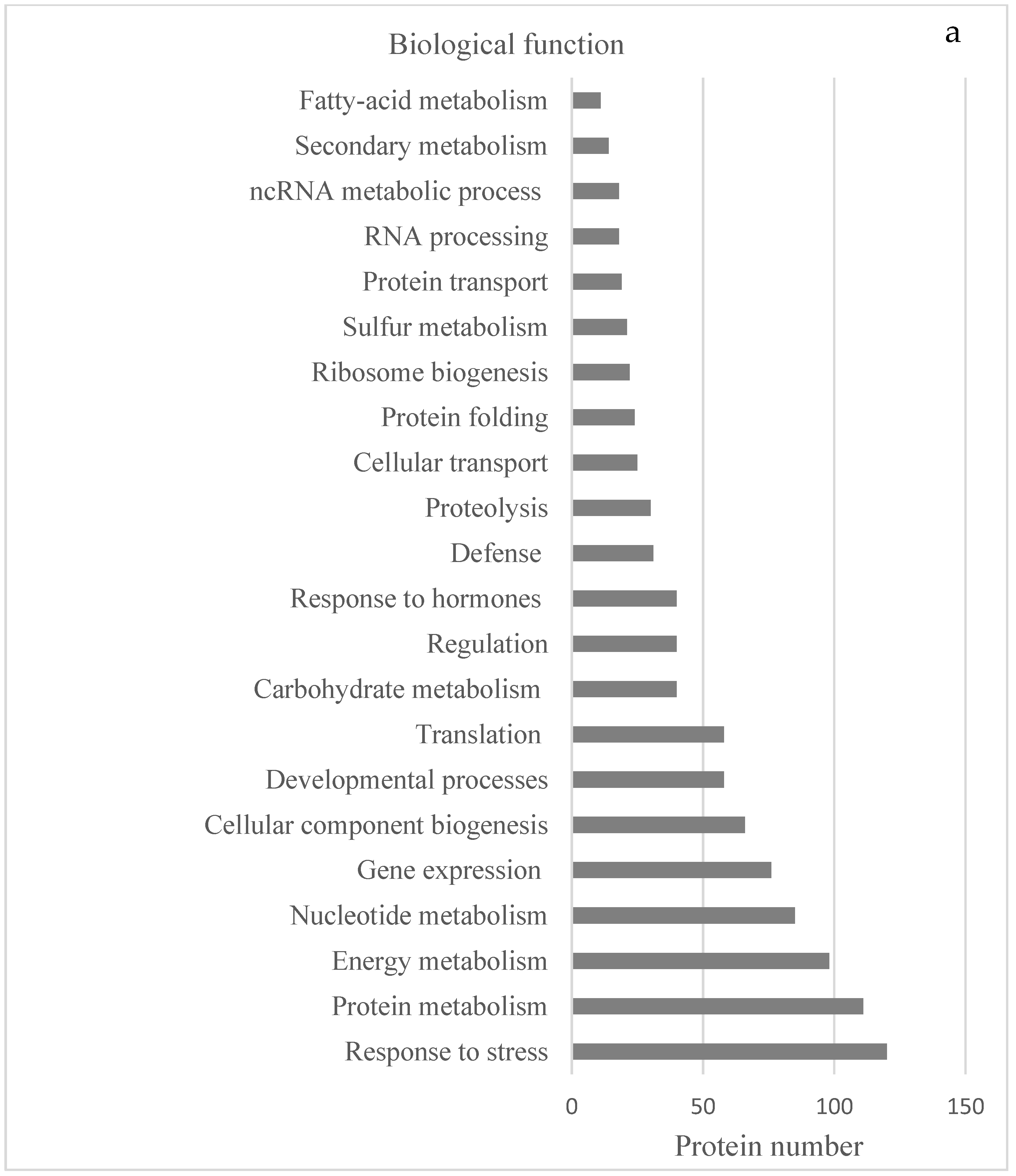
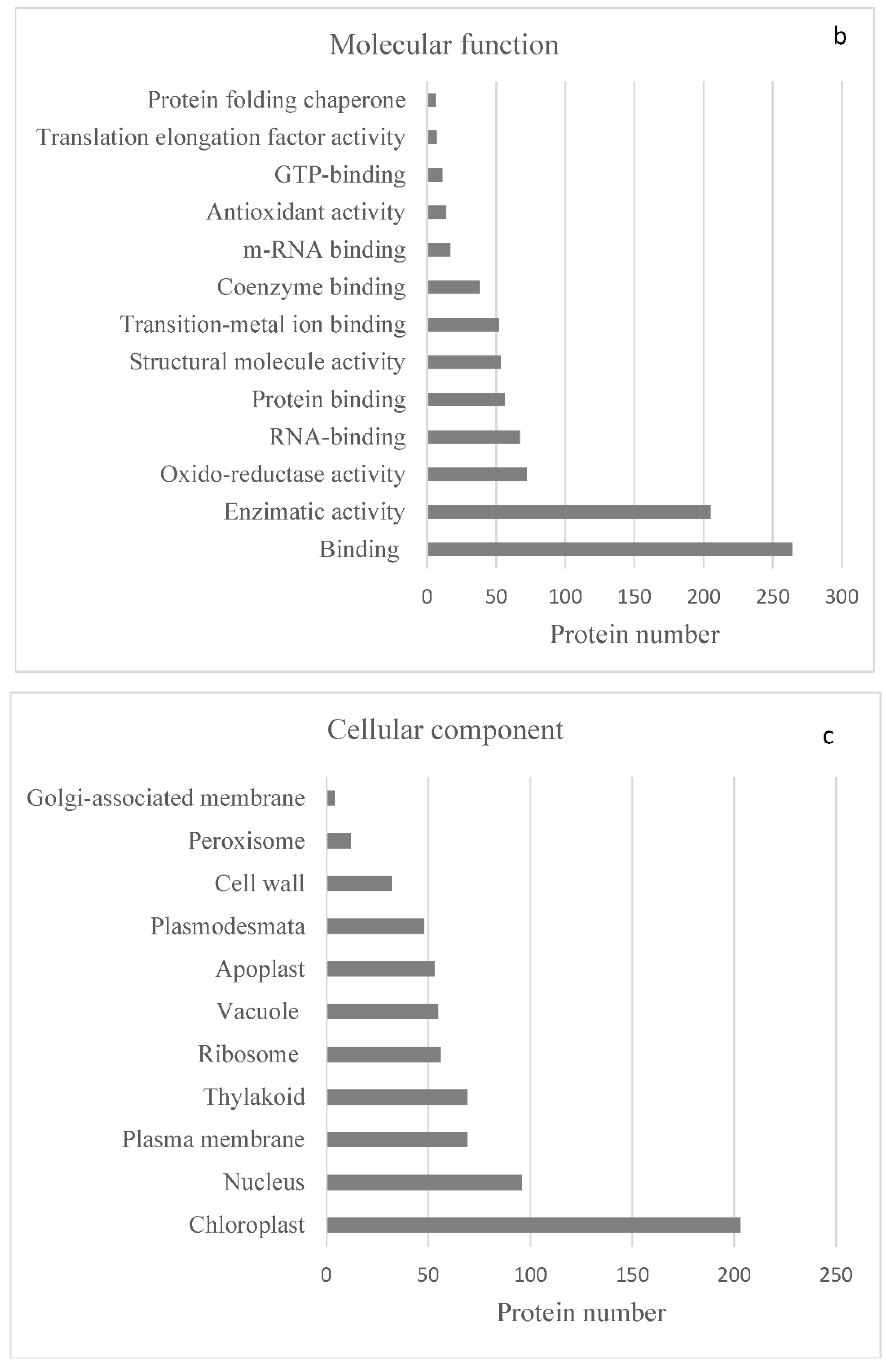
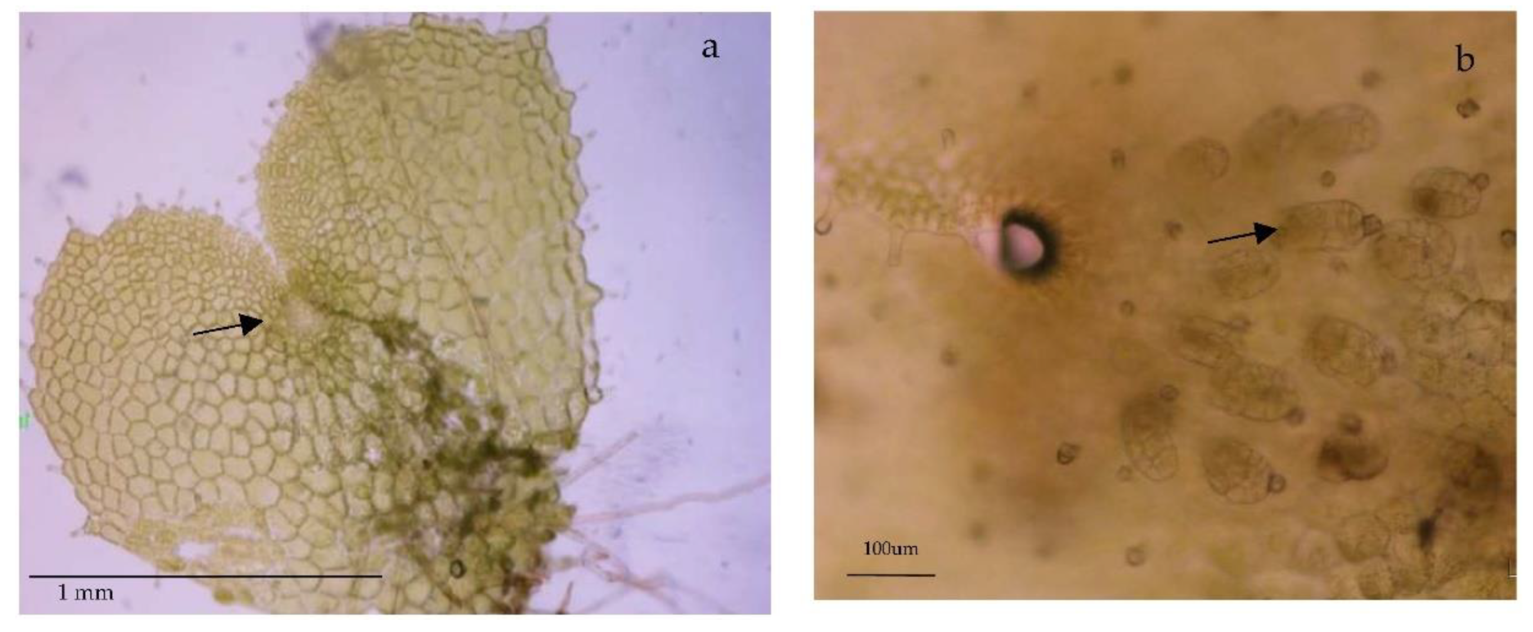
2.2. Protein Analyses in Fern Gametophytes

To obtain better insights into the function of both collections of proteins, Gene Ontology (GO) classification analyses provided by the STRING platform were performed. The set of 417 proteins were checked according to the standard categories ‘biological function’, ‘molecular function’, and ‘cellular component’. In the category ‘biological processes’ (Figure 1a), the largest number of proteins shared by both species have a function in response to stress (about 25%). In addition, there were abundant proteins involved in the primary metabolism of amino acids, peptides, proteins (including folding, transport, and proteolysis), and nucleic acids. Several proteins involved in nitrogen and sulfur metabolism, ribosome biogenesis, gene expression, and translation were also included. We also found a large group of proteins that are involved in the generation of precursor metabolites, energy, and photosynthesis, as well as others involved in plant developmental processes. A smaller number of proteins is associated with translation, transport, toxin catabolism, and chaperone-mediated protein folding.
Under the category ‘molecular function’ (Figure 1b), a very high number of proteins are classified to have ‘binding activity’, mainly to organic cyclic compounds and ions, or to have ‘catalytic activity’. Proteins with oxidoreductase activity are very relevant, as are those related to ‘structural molecule activity’. Noteworthy also are the small number of chaperones detected, of proteins that make up the translation elongation factor, as well as of those with antioxidant activity which, despite being quite important for the detoxification of reactive oxygen species in the cell, represented less than 10% of the total proteins identified and quantified.
Under the category ‘cellular component’ (Figure 1c), most of the proteins were predicted to localize to the cytoplasm and intracellular organelles, especially the chloroplasts. In both species, similar amounts of proteins were found to be associated with the cell wall, plasmodesmata, and thylakoid and plasma membranes, whereas proteins present in the membranes of vesicles of the Golgi apparatus were scarce compared to other subcellular compartments. Likewise, we found quite a few proteins associated with the peroxisome, an organelle that performs very specific functions in plant development and reproduction.
Using the STRING database, an analysis of proteins shared by both species found 417 knots, 3471 interactions, and a probability of enrichment of protein–protein interactions of less than 10−16. These data indicate that proteins have more interactions with each other than might be expected for a random set of proteins of similar size, which means that the proteins are at least partially connected within a group. Here, we focused on a collection of 145 proteins associated with the functional groups for growth, reproduction, phytohormone signaling and biosynthesis, gene expression, response to biotic stress, and response to abiotic stress. The interactome is shown in Figure 2, and only a few proteins lacked any kind of connection. The sources of functional STRING interactions of the DA and DO proteins are detailed in Table S2, and refer to protein–protein associations obtained from co-expression, co-occurrence, text mining, homology, databases, and experimental datasets. The interactions among the proteins are depicted in four groups: those of growth and reproduction, phytohormones, gene expression, and abiotic stress (Figure 3). In this regard, the majority of the proteins associated with growth and reproduction refers to aspects of anatomical structure and systems development. In the case of phytohormones, they are linked to the positive regulation of the abscisic acid (ABA)-activated signaling pathway, endocytosis, and the regulation of biological quality. Concerning gene expression, the proteins found are linked to chromatin organization and translation. Finally, abiotic stress is overrepresented by processes such as the biosynthesis of secondary metabolites, protein folding, glutathione metabolism, superoxide dismutase activity, or by structural components possessing a DnaJ domain.
Paying attention to the major source of STRING interactions in each functional group of proteins, some additional information can be extracted (Figure 4). For instance, the major source for interactions among proteins associated with growth and reproduction is text-mining data, while for phytohormones it is text mining and databases. Concerning gene expression, the major sources are co-expression data (Figure 5), which represents 50% of the score values, and experimental datasets.
The platform also provides a qualitative estimation of the interaction strength that these proteins have in the model species A. thaliana or in other organisms. For example, one can observe a strong interaction between two chloroplast proteins: the homologs of translation factor GUF1 (AT5G08650) and the protein elongation factor EF-G (SCO1) (Figure 6). Finally, in the case of proteins involved in abiotic stress, the interaction score value comes from text mining, homology, and phylogenetic co-occurrence data (Figure 6).
In fact, homology and co-occurrence are reported as the highest sources for the protein interaction scores in all groups of proteins selected for further analysis. Concerning phylogenetic co-occurrence, the taxa include bacteria, fungi, eudicotyledonous plants, the lycopod S. moellendorffii, the moss Phycomitrium patens, and several algae. As was mentioned above, the proteins of our two fern species were analyzed by STRING using the corresponding A. thaliana identifiers. Data in Figure 6 reveal that there are some proteins absent in bacteria: MITOCHONDRIAL PHOSPHATE CARRIER PROTEIN 3 (PHT3.1), LATE EMBRYOGENESIS ABUNDANT 7 (LEA7), EUKARYOTIC TRANSLATION ELONGATION FACTOR 5A-3 (ELF5A-3), RIBOSOMAL PROTEIN L10B (RPL10B), and AT1G05520), while fungi miss TRANSALDOLASE 2 (TRA2), LEA7, STARCH EXCESS1 (SEX1); and FIBRILLIN (FIB), and LEA7 is neither present in S. moellendorffii nor in P. patens. We did not manage to identify GLUTATHIONE S-TRANSFERASE 19 (GSTU19) in moss, and there are some additional gaps for the proteins TRA2, AT2G18340, LEA7, CATALASE (CAT), CBS DOMAIN CONTAINING PROTEIN 3 (CBSX3), COPPER/ZINC SUPEROXIDE DISMUTASE 1 (CSD1), THIAMINE THIAZOLE SYNTHASE 1 (THI1), GSTU19, and AT1G05520.

3. Discussion

Out of 879 quantifiable proteins [27], we detected a large number (417) that are shared by the gametophytes of apomictic DA and its sexual parent DO. These results, together with a previous study using the same two species [27], enlarge the proteomic profile of both ferns. However, unlike for DA [25], transcriptome data for DO are lacking, such that most certainly fewer proteins could be identified in DO than in DA due to sequence divergence. The classification of proteins according to biological processes shows that those associated with responses to abiotic and biotic stresses are predominant in both species (about 25% of all identified proteins). Specifically, we paid attention to a subset of 145 proteins falling into the six functional categories, i.e., growth, reproduction, phytohormone signaling and biosynthesis, gene expression, biotic stress, and abiotic stress (Table 1). We discuss their putative roles in plant development, followed by an analysis of predicted protein interactions.

3.1. Growth

The protein SHEPHERD (SHD) is located in the endoplasmic reticulum and belongs to the HtpG chaperone proteins, which are part of the HSP90 heat shock protein (HSP) family. This protein is involved in regulating the size and organization of the meristem. Single and double mutant analyses suggest that SHD may be required for the correct folding and/or complex formation of CLAVATA (CLV) proteins, as mutations of this family in A. thaliana exhibit expanded shoot meristems, disorganized root meristems, and defective pollen tube elongation [28]. Three proteins are related to the regulation of cell division: CELL DIVISION CONTROL PROTEIN 48 HOMOLOG A (CDC48A) interacts with certain SNAREs as part of specialized membrane fusion events [29]; ACTIN7, an essential component of the cytoskeleton, is a vegetative actin with a role in hormone-induced cell proliferation and callus formation [30]; and the P(1B)-Type ATPase1 (PAA1), a copper-transporting ATPase, has a role in cell proliferation, in this case of glucose signaling-regulated root meristem development [31]. The list also includes the dose-dependent CELL DIVISION PROTEIN FtsZ HOMOLOG 1 (FTSZ1) protein, a component of the plastidial division machinery, which is also involved in blue light-induced chloroplast movement [32].
The gametophytes of both species are characterized by the presence of abundant trichomes, which might be related to the identification of ENOLASE1 (ENO1), an enolase involved in glycolysis, mutants of which produce abnormal trichomes in A. thaliana [33]. The PSBQ-2 protein, a subunit of the oxygen-evolving complex of photosystem II, could help to cope with growth in low-light conditions and/or to assemble and stabilize photosystem II [34]. The GLUTATHIONE S-TRANSFERASE TAU 20 (GSTU20) protein is also involved in—among others processes—regulating the influence of far-red light on development and probably gravitropic signal transduction [35,36,37]. Finally, we identified two chaperones: CHAPERONIN CONTAINING TCP1 SUBUNIT 8 (CCT8), contributing to the maintenance of stem cells [38], and CHLOROPLAST STEM-LOOP BINDING PROTEIN 41 kDa (CSP41B), involved in the regulation of the circadian rhythm [39]. Finally, CYCLOPHILIN-40 (CYP40), a peptidyl-prolyl cis-trans isomerase, promotes the juvenile phase of vegetative development and—to a lesser extent—the positioning of floral buds, floral morphogenesis, and the expression of heat shock proteins [40].

3.2. Reproduction

Although ferns do not form flowers or seeds, we found proteins related to the formation of these reproductive organs in flowering plants. This finding suggests that the genes encoding those proteins arose in evolution before the divergence of the fern and seed plant lineages. This stresses the importance of carrying out genomic and proteomic studies from the perspective of evolutionary developmental biology. Below, we highlight some of the most striking proteins that are associated with gametophyte development, embryogenesis, or flowering.
Among the proteins identified in our fern gametophytes, there are some that impair pollen development in seed plants if defective, either by influencing metabolic or structural processes. Those affecting metabolic processes include the cytosolic isoform of PHOSPHOGLUCOMUTASE 2 (PGM2); EMBRYO SAC DEVELOPMENT ARREST9 (EDA9), encoding a D-3-phosphoglycerate dehydrogenase; the catalytic SUBUNIT A OF VACUOLAR ATP SYNTHASE (VHA-A); PURINE BIOSYNTHESIS4 (PUR4), a probable phosphoribosyl formylglycinamidine synthase; and the REVERSIBLY GLYCOSYLATED POLYPEPTIDE1 (RGP1). Proteins influencing structural processes include those involved in actin filament bundling in pollen tubes, such as PROFILIN5 (PRF5) and VILLIN 2 (VLN2), a Ca2+-regulated actin-binding protein. In addition, two tRNA ligases required for female gametophyte development were found: OVULE ABORTION9 (OVA9) and OVA4, involved in the aminoacylation of tRNAs [41].
Embryogenesis is central to reproduction of any organism. In ferns, gametophytes form the egg cells that, upon fertilization (sexuals) or independent of it (apomicts), produce the sporophytic embryo. Homologs of several proteins we identified are involved in embryogenesis in seed plants, including CHLORATE RESISTANT88 (CR88), encoding the heat shock protein HSP90.5, a chaperone essential for the biogenesis and maintenance of chloroplasts [42]; the CYCLOPHYLLIN 19 (CYP19-4) protein, which interacts in vitro with GNOM, a GDP/GTP exchange factor that plays a central role in embryonic pattern formation [43]; and the PREPHENATE AMINOTRANSFERASE (PAT), a maternally required aspartate aminotransferase necessary for the development of the embryo [44]. In addition, we identified EMBRYO DEFECTIVE2761 (EMB2761), a threonyl-tRNA synthetase found in mitochondria and chloroplasts, that—when affected by a mutation—causes embryonic arrest at the globular stage [41].
As for proteins related to flowering, the fern gametophytes expressed FLOWERING LOCUS D (FLD), a histone demethylase that promotes flowering regardless of photoperiod and vernalization by repressing FLOWERING LOCUS C (FLC) that, in turn, encodes a floral repressor blocking the transition from vegetative to reproductive development [45]. Likewise, we identified EMB14, the pre-mRNA processing-splicing factor 8A, which plays an important role in embryonic development and regulates the splicing efficiency of the COOLAIR non-coding RNA at the FLC locus [46].

3.3. Phytohormone Signaling and Biosynthesis

In the gametophytes of our two fern species, we identified proteins related to the major families of phytohormones, for instance auxins, expanding our previous results in DA [25,26]. The RNA-binding protein AT1G76940, which modulates the effect of auxin on the transcriptome [47], is worth mentioning. We also found SHORT-CHAIN DEHYDROGENASE/REDUCTASE A (SDRA), involved in the β-peroxisomal oxidation of IBA to form IAA. IAA is biologically active and could be involved in the peroxisomal activation of 2,4-D, a precursor of the auxins that inhibit root growth [48]. In line with this, another interesting protein is the chaperone BOBBER1 (BOB1), belonging to the HSP20-like superfamily of non-canonical small heat shock proteins, which is necessary for the establishment of auxin gradients, the patterning of the apical domain of the embryo, and other developmental processes [49]. We also detected three other common proteins related to auxin: CLATHRIN HEAVY CHAIN 2 (CHC2) is required for auxin polar transport through regulating the distribution of transporters of the PIN-FORMED (PIN) family [50]; ADP-RIBOSYLATION FACTOR A1F (ARFA1F), belonging to the family of ARF GTPases, is involved in cell division, cell expansion, and cellulose production [51]; and CHALCONE SYNTHETASE (CHS), a key enzyme involved in the biosynthesis of flavonoids, including the purple anthocyanins that accumulate in leaves and stems, participates in the regulation of auxin transport, and modulates of root gravitropism [52].
We also identified some proteins related to the phytohormones ABA and cytokinin, such as the UDP-GLYCOSYLTRANSFERASE 85A1 (UGT85A1), involved in the O-glycosylation of trans-zeatin and dihydrozeatin, and active in vitro on cis-zeatin. In addition, ABA DEFICIENT1 (ABA1), a zeaxanthin epoxidase, plays an important role in the xanthophyll cycle and ABA biosynthesis by converting zeaxanthin into antheroxanthin and subsequently into violaxanthin. This protein is necessary for many important processes in plants, including resistance to osmotic stress and drought, ABA-dependent closure of stomata, seed development and dormancy, modulation of defense gene expression, as well as resistance to disease and non-photochemical quenching [53,54]. Moreover, we identified the non-specific LIPID-TRANSFER PROTEIN 4 (LTP4), also regulated by ABA. This protein binds to fatty acids and acyl-CoA esters and can transfer several different phospholipids. Its biological function can be linked to stress by salt or water deprivation. In ferns, lipid composition also plays an important role in the integrity and fluidity of membranes and in desiccation tolerance [55].
As for gibberellin-related proteins, TOPLESS-RELATED PROTEIN 4 (TRP4) is a transcriptional co-suppressor involved in the regulation of homeostasis and signaling of these phytohormones. The founding member of the TOPLESS family regulates the polarity of the embryo, preventing the formation of a root meristem in the apical domain [56], and acts redundantly with other TRP proteins.
We found two proteins outside the main families that are relevant for their relationship with important compounds that are little studied in seed plants and even less so in ferns. One is O-METHYLTRANSFERASE 1 (OMT1), involved in methylating flavonols and the biosynthesis of melatonin. The other is GLUTAMATE DECARBOXYLASE 1 (GAD1), which catalyzes γ-aminobutyric acid (GABA) production. The non-protein amino acid GABA has multiple functions under both stress and non-stress conditions and in reproduction [57,58].

3.4. Gene Expression

Undoubtedly, proteins regulating gene expression play a central role in biological processes by participating, in one way or another, in the conversion of genetic into metabolic information. We found many proteins common to DA and DO that are involved in methylation, histone deacetylation, gene silencing, unwinding of nucleic acids, transcription activation or repression, etc. Among the proteins related to gene silencing, we identified HYPONASTIC LEAVES1 (HYL1), which forms a complex with SERRATE and DICER-LIKE1 (DCL1), to promote the precise processing of pre-mRNA by DCL1, and with ARGONAUTE1 (AGO1), which is involved in post-transcriptional gene silencing [59,60,61]. Furthermore, we found HOMOLOGY-DEPENDENT GENE SILENCING1 (HOG1), a S-adenosyl-L-homocysteine hydrolase required for—among other functions—transcriptional gene silencing dependent on DNA methylation.
We also identified some histones, the core components of nucleosomes. DNA is wrapped around nucleosomes, which can compact chromatin, limiting the accessibility of DNA to the cellular machinery that requires DNA as a template. Therefore, histones play a central role in transcriptional regulation, DNA repair, DNA replication, and chromosomal stability. Accessibility to DNA is regulated through a complex set of post-translational histone modifications, also called the ‘histone code’, and nucleosome remodeling, whereby nucleosomes are moved along, unwrapped, or ejected from the DNA. We found HISTONE DEACETYLASE 3 (HDT3), a HD2-type deacetylase, which likely mediates the deacetylation of lysine residues at the amino terminal end of the core of histones (H2A, H2B, H3, H4). Histone deacetylation acts as a label for epigenetic repression and plays an important role in transcriptional regulation, cell cycle progression, and various developmental events. Likewise, this process was suggested to be involved in the modulation of ABA signaling and the regulation of stress-responsive genes [62]. HISTONE H2A PROTEIN 9 (HTA9) is a histone H2A variant that is synthesized throughout the cell cycle in contrast to the canonical S phase-regulated H2A. Another protein worth mentioning is RESISTANCE TO PSEUDOMONAS SYRINGAE PV MACULICOLA INTERACTOR1 (RIN1), a central component of the INO80 chromatin remodeling complex that is involved in transcriptional regulation, DNA replication, and probably DNA repair.
In addition, we found the transcription factor PURIN-RICH ALPHA1 (PURA1), which binds specifically to repeated purine-rich sequences found in cis-regulatory elements called teloboxes. Several helicases, involved in the unwinding of DNA and/or RNA, have also been identified: RNA HELICASE 15 (RH15), a DEAD-box helicase involved in processing of pre-mRNA and nuclear export, and RH37. Finally, we mention UBIQUITIN-CONJUGATING ENZYME 35 (UBC35) that is associated with DNA repair upon damage by UV radiation [63].

3.5. Biotic Stress

Plants must continuously cope with attacks by other macro- and microorganisms. We identified some proteins with a possible role in preventing damage and counteracting, in some way, their vulnerable condition due to sessility. Among others, we detected the protein SUPPRESSOR OF G2 ALLELE OF skp1 HOMOLOG B (SGT1B), involved in the innate immunity of plants [64] and essential for the resistance conferred by multiple resistance (R) genes that recognize different isolates of oomycete pathogens, such as the downy mildew Hyaloperonospora parasitica [65]. Another protein of interest is 3-DEOXY-D-ARABINO-HEPTULOSONATE 7-PHOSPHATE SYNTHASE 1 (DHS1), which is induced by wounding and infections with the pathogenic bacterium Pseudomonas syringae [66]. On the other hand, the SUPPRESSOR OF SA INSENSITIVE2 (SSI2) is a desaturase that can intervene in defense signaling mediated by salicylic acid (SA) and jasmonic acid (JA). The ssi2 mutant has an increased 18:0 and reduced 18:1 fatty acid composition and shows hyper-resistance to the green peach aphid and antibiotic activity in the exudate of the petiole [67]. Membrane lipid biosynthesis is a crucial process, and the ACYL CARRIER PROTEIN 4 (ACP4) plays an essential role in fatty acid biosynthesis in chloroplasts, the formation of cuticular waxes and cutin polymers in leaves, as well as in the establishment of systemic acquired resistance [68]. Moreover, PHOSPHOLIPASE D ALPHA1 (PLDALPHA1) hydrolyzes the terminal phosphodiester bond of glycerol-phospholipids to generate phosphatidic acids, and plays an important role in several cellular processes, including the action of phytohormones and the stress response to cellular acidification. PLDALPHA1 is involved in the induction of JA upon wounding [69] and probably in freezing tolerance by modulating cold-induced gene expression and osmolite accumulation [70]. The protein also mediates the effects of ABA on stomata, on seed aging and deterioration, as well as on microtubule stabilization and salt tolerance [71]. In line with the abovementioned, we found PEROXISOMAL 3-KETOACYL-COA THIOLASE 3 (PKT3), which influences the induction of JA biosynthesis after injury and during senescence [72]. We have already commented on some proteins related to the synthesis of GABA, but we also identified IMPAIRED IN BABA-INDUCED DISEASE IMMUNITY1 (IBI1), an aspartate-tRNA ligase involved in the perception of β-aminobutyric acid (BABA) that is required for the priming effect of BABA in disease resistance [73]. Besides, we identified OPEN STOMATA1 (OST1), a SNF1-related serine/threonine protein kinase that is activated by the ABA signaling pathway and regulates numerous ABA responses, including stomatal closure in response to drought, pathogens, or a decrease in humidity [74]. On the other hand, NONHOST RESISTANCE TO P. S. PHASEOLICOLA1 (GLPK) performs a limiting step in glycerol metabolism and is necessary for both general and specific resistance to bacteria and fungi [75]. Finally, we want to mention the identification of the metallopeptidase ORGANELLAR OLIGOPEPTIDASE (OOP), which was reported to bind SA [76].

3.6. Abiotic Stress

Fern gametophytes harbor a huge biochemical arsenal to cope with abiotic stimuli in order to preserve their reproductive function. Several factors are likely to modify the redox state of this vulnerable generation of the life cycle. In reviewing our proteome profiles, we found numerous proteins involved in “oxidative stress”, such as the cytosolic COPPER/ZINC SUPEROXIDE DISMUTASE 1 (CSD1), the expression of which is affected by mRNA cleavage directed by a microRNA (miR398); the MANGANESE SUPEROXIDE DISMUTASE 1 (MSD1); and two thiol-specific peroxiredoxins: PERIREDOXIN Q (PRXQ) and chloroplastic 2-CYSTEINE PEROXIREDOXIN BAS1-like (2CPB). Other proteins grouped here are DEHYDROASCORBATE REDUCTASE 3 (DHAR3), a key component of the glutathione S-transferase ascorbate recycling system; GLUTATHIONE S-TRANSFERASE PHI 10 (GSTF10) and GSTU19, which detoxify certain herbicides; and a MONODEHYDRO ASCORBATE REDUCTASE (MDAR), which converts monodehydroascorbate to ascorbate, oxidizing nicotinamide adenine dinucleotide hydrogen (NADH) to NAD+ in the process.
Identified proteins involved in the response to “light intensity” include SERINE HYDROXYMETHYLTRANSFERASE 1 (SHM1), which acts in damage caused by sodicity and the hypersensitive defense response [77], and a GroES-like zinc-binding alcohol dehydrogenase that functions as an oxidoreductase encoded by AT5G61510. In addition, we found FIBRILLIN (FIB), which interacts with ABA-INSENSITIVE2 (ABI2) [78] and is involved in ABA-mediated photoprotection. Moreover, we found the RIBOSOMAL PROTEIN L10B (RPL10B), a structural constituent of the ribosome that might participate in the response to UV-B [79].
Regarding stress caused by “salts and heavy metals”, we identified O-ACETYLSERINE (THIOL) LYASE ISOFORM A1 (OASA1), which is induced by high salt and heavy metal concentrations in an ABA-mediated fashion [80]; the transporters PHOSPHATE TRANSPORTER 3;1 (PHT3;1), and the protein cystathionine β-synthase CBS DOMAIN CONTAINING PROTEIN 3 (CBSX3), a cobalt ion binding protein affecting plant growth and reproduction [81].
Associated with “drought and temperature”, we identified the protein LATE EMBRYOGENESIS ABUNDANT7 (LEA7), which might protect plants by avoiding the aggregation of soluble proteins in leaves [82]. Besides, there was LOW EXPRESSION OF OSMOTICALLY RESPONSIVE GENES4 (LOS4), a putative DEAD-box RNA helicase thought to act in temperature sensing [83]. In line with this, the co-chaperone FK506 BINDING PROTEIN 62 (FKBP62) positively modulates thermotolerance by interacting with HSP90.1, as well as increasing HEAT SHOCK TRANSCRIPTION FACTOR A2 (HSFA2)-mediated accumulation of chaperones of the small-HSP family. CHAPERONIN 10 (CPN10) is also upregulated in response to a heat shock treatment. In gametophytes of both DA and DO, several proteins belonging to the HSP20, HSP70, and HSP90 families were identified: HSP 17.6II, HSP 21, MTHSC70-2, HSP 70 KDA, HSP70-15, HSP 81.4, and HSP91, which protect from heat stress damage through their ability to recognize non-native conformations of other proteins. In addition, we detected PEPTIDE METHIONINE SULFOXIDE REDUCTASE (PMSR), which prevents the methionine sulfoxidation of HSP21 and thus its inactivation. The list continues with other chaperones, such as the CASEIN LYTIC PROTEINASE B3 (CLPB3), a HSP101 homolog that confers thermotolerance to chloroplasts during heat stress [84], and DNA J PROTEIN A6 (DJA6), which may function together with the HEAT SHOCK COGNATE PROTEIN 70 (HSC70) chaperone to assist protein folding and prevent protein aggregation during heat stress in the chloroplast [85]. Additionally, two proteins related to cold stress conditions were found: the 31-KDA RNA BINDING PROTEIN (RBP31), which stabilizes transcripts of numerous mRNAs and modulates telomere replication [86], and the alpha-glucan water dikinase STARCH EXCESS1 (SEX1) [87].
Involved in “response to cadmium ions”, gametophytes of both species express some oxidoreductases: a probable N-ACETYL-GAMMA-GLUTAMYL-PHOSPHATE REDUCTASE (AT2G19940), a putative NADPH-DEPENDENT MANNOSE 6-PHOSPHATE REDUCTASE (AT2G21250), and two pyruvate kinases: AT3G52990 and AT5G63680.
We also report proteins related to “vitamin metabolism”. The pyridoxal 5’-phosphate synthase subunit PYRIDOXINE BIOSYNTHESIS 1.3 (PDX1.3) plays several roles in vitamin B6 biosynthesis, development, stress tolerance, and resistance to singlet oxygen-generating photosensitizers in A. thaliana [88]. In addition, the thiazole biosynthetic enzyme THIAMINE1 (THI1), defined as a suicide enzyme because it undergoes only a single turnover, is involved in the biosynthesis of the thiamine precursor thiazole, and the adaptation to various stress conditions [89]. Other noteworthy proteins are the chloroplast protein TRANSKETOLASE 1 (TKL1), linked to the pentose-phosphate shunt, which may also act as a stress sensor [90]; the SULPHURTRANSFERASE 1 (STR1), involved in embryo and seed development [91]; and the EUKARYOTIC ELONGATION FACTOR 5A-3 (ELF5A-3), which acts as a bimodular protein capable of binding to both RNA and proteins and supports growth in response to sublethal osmotic and nutrient stresses [92].

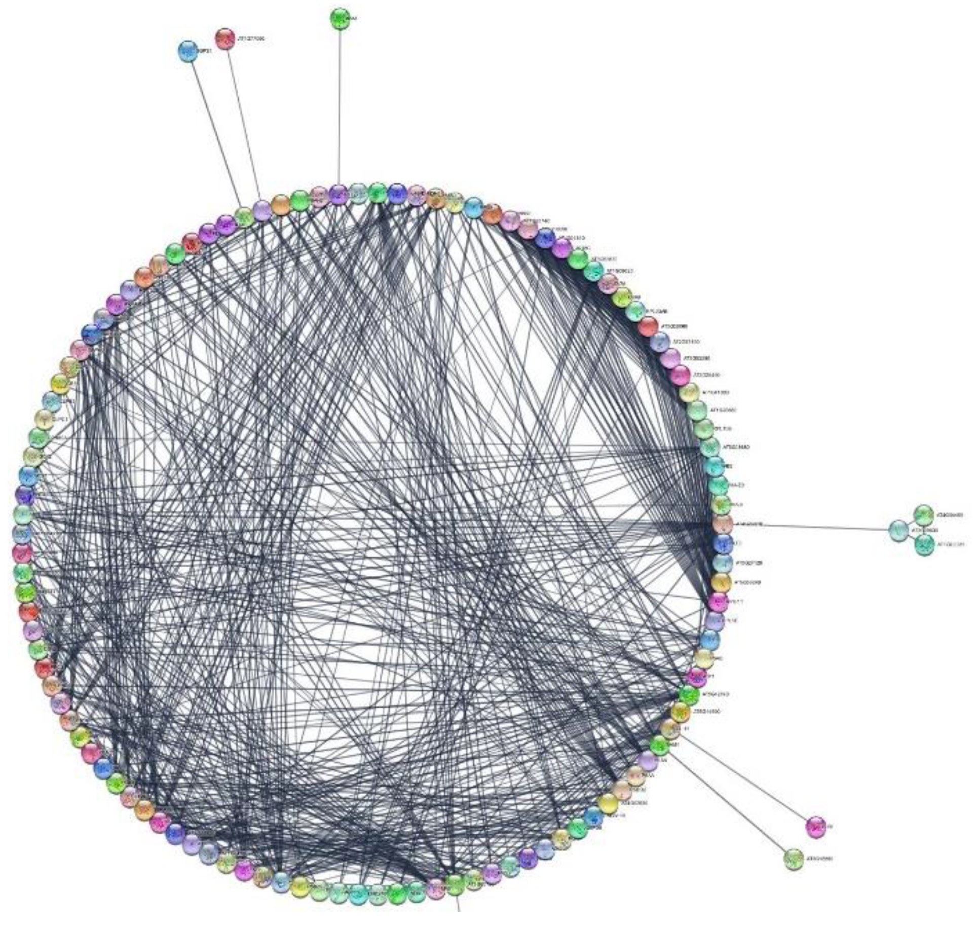
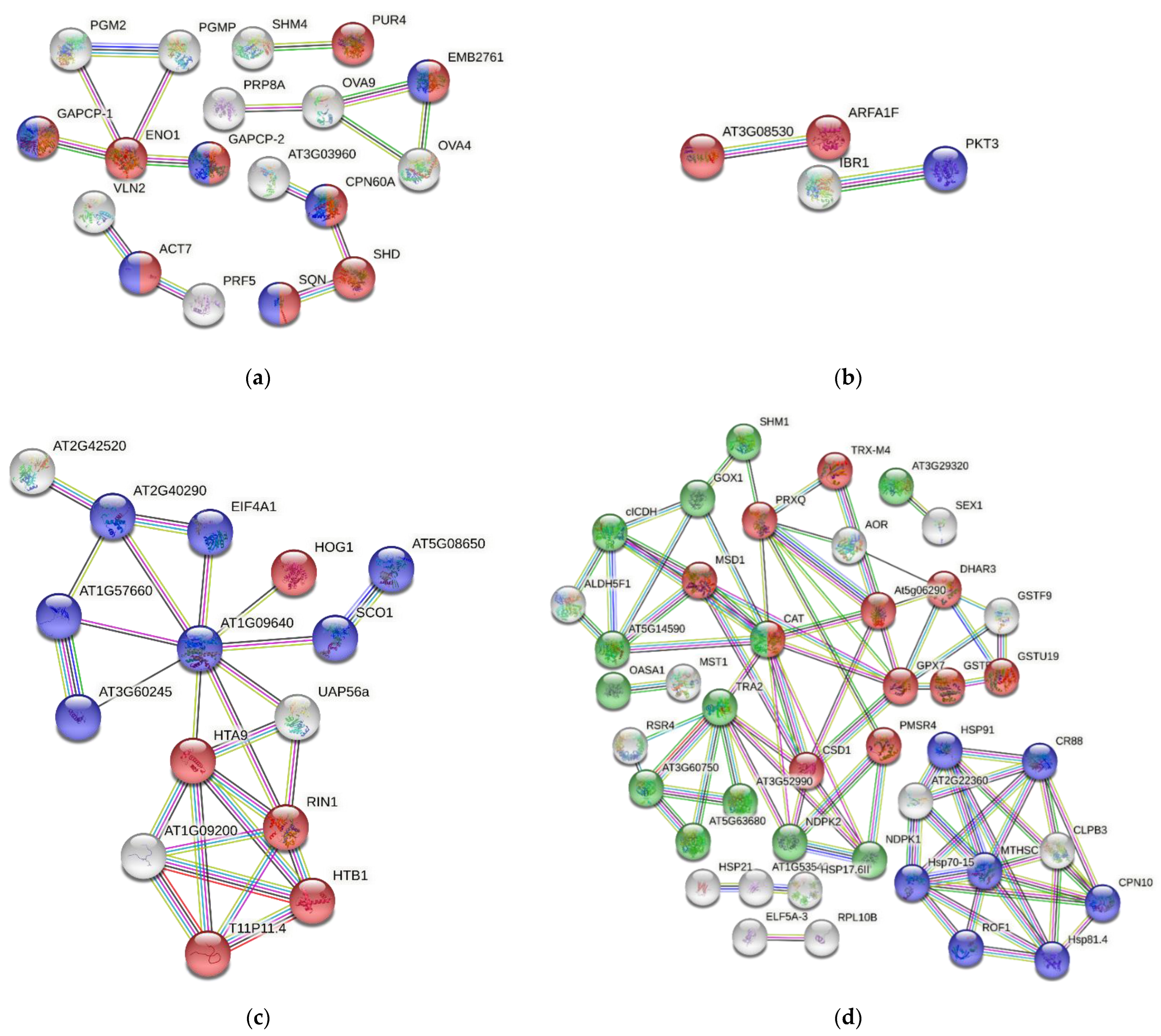
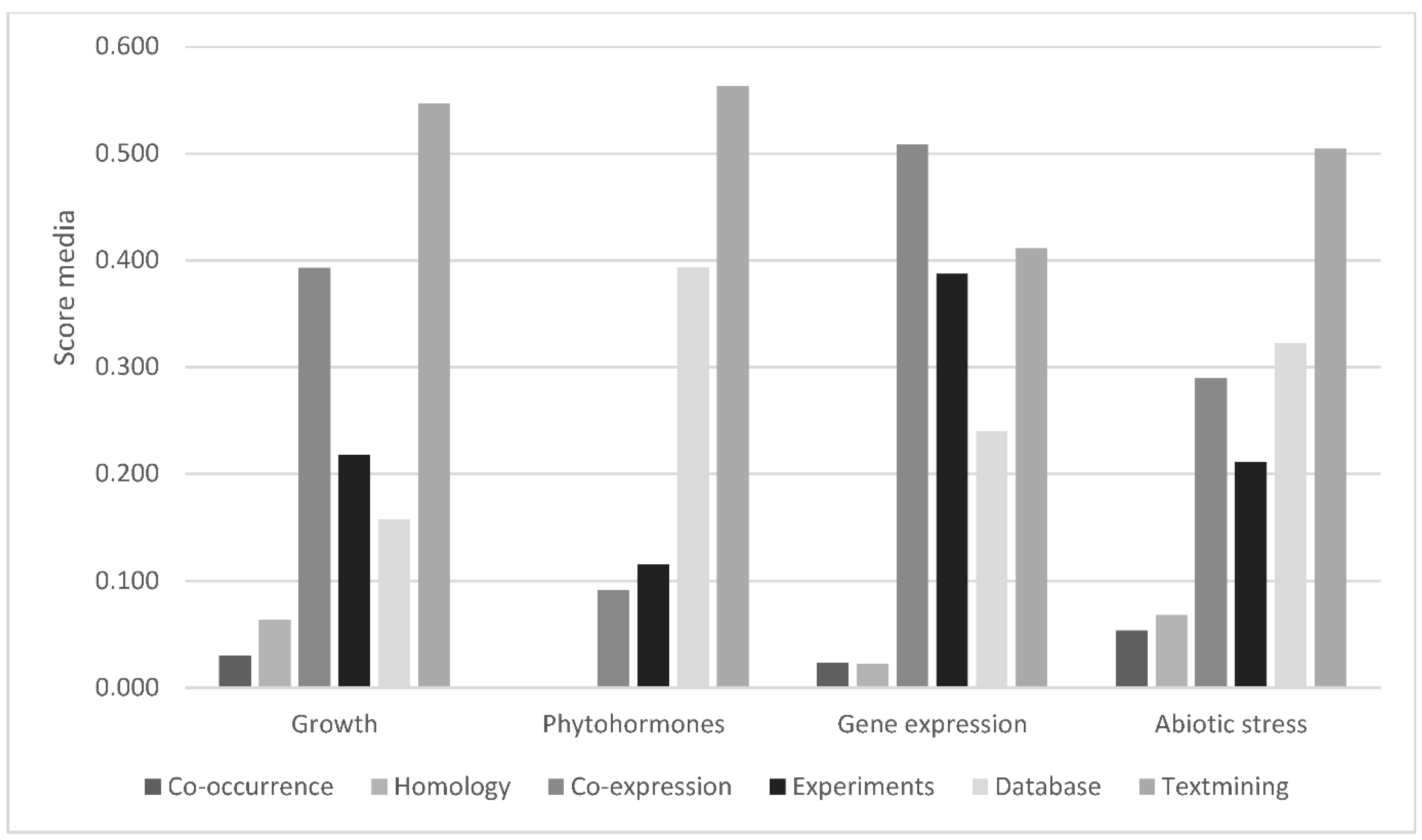
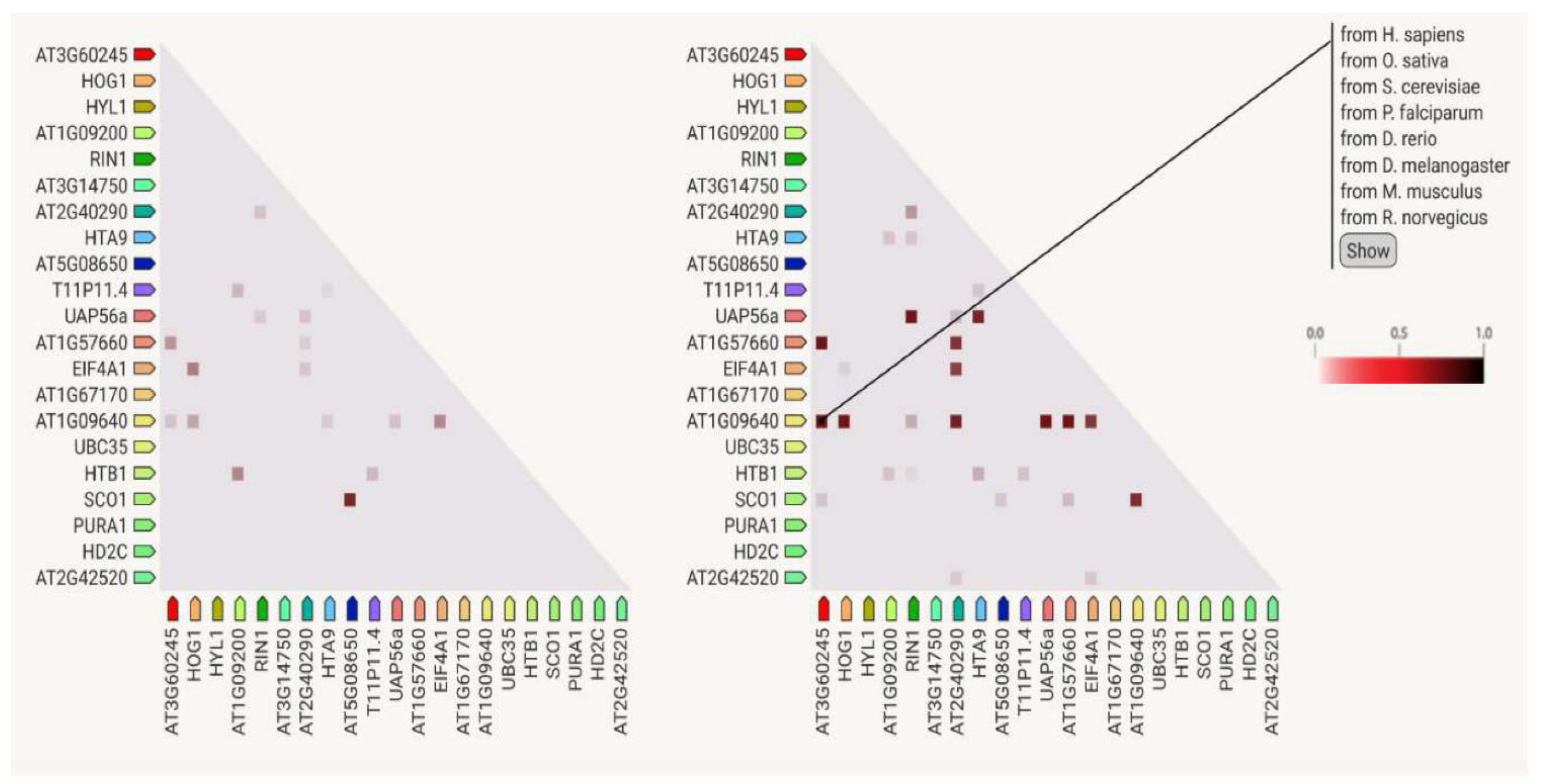
3.7. Protein–Protein Interactions

The network of proteins clustered inside the group “growth and reproduction”, has a PPI enrichment p-value of 5.55 × 10⁻¹⁶, meaning that the proteins are at least partially biologically connected as a group. We focus on those proteins associated with anatomical structure, organelle organization, and systems development, i.e., any process that modulates a qualitative or quantitative biological trait such as an assessable attribute of an organism or part of an organism (size, mass, shape, color, etc.). The proteins RIBULOSE-5-PHOSPHATE-3-EPIMERASE (RPE) and EDA9 exhibit maximum interactions with 12 and 11 nodes, respectively, followed by ENO1, GLYCERALDEHIDE-3-PHOSPHATE-DESHIDROGENASE-1 (GAPCP-1), PHOSPHOGLUCOMUTASE (PGMP), SERINEHYDROXYMETHYLTRANSFERASE 4 (SHM4), GLYCERALDEHYDE-3-PHOSPHATE DESHYDROGENASE (GAPCP-2), PGM2, SHD, OVA4, and PUR4, all of them having more than six nodes. The strength of each type of interaction between proteins is expressed by a scale provided by the STRING platform, ranging from 0 to 1, with 1 meaning maximal and 0 minimal interaction, respectively. According to this scale, the highest interaction score values are those involving the connection between the protein ENO1 and the proteins GAPCP-1, GAPCP-2, PGM2, and PGMP. A very high score is found between PGMP and PGM2, from accurate databases. The values come from co-expression data. Enolases are enzymes that catalyze the reversible conversion of D-2-phosphoglycerate into phosphoenol pyruvate in the glycolytic pathway, which contributes to satisfy the energy demand of the organism. In fern gametophytes, similar to pollen formation and germination in flowering plants, metabolic pathways involving mitochondrial respiration and fermentation are expected to play a very important role [93]. Three subnetworks more are observed in the growth and reproduction set of proteins. First, the interactions among ACT7 with VLN2 and PRF5, important for actin filament bundling and the organization of the actin cytoskeleton, and also among OVA4, OVA 9, and EMB276, which are t-RNA ligases involved in translation and associated with reproduction [41,94]. Second, the protein SHM4, a probable phosphoribosyl-formylglycinamidine-synthase that is essential to male gametophyte development in A. thaliana, and PUR4, a glycine-hydroxymethyl-transferase, have homologs that are neighbors, either co-expressed or mentioned together in other organisms. Third, there are the connections among AT3G03960, CPN60A, SHD, and SQN. Specifically, the interaction between SHD and SQN has already been reported in text-mining datasets [95].
Proteins related to “phytohormones” show two strong interactions: on the one hand, it is the couple ARFA1F-AT3G0830, the first an ARF GTPase essential for vesicle coating and uncoating, and the second mediating endocytosis, which is involved in the correct distribution of PIN auxin transporters. High score values were obtained from co-expression, experimental data, association in curated databases, and text-mining evidence. The other important interaction is between IBRI, a dehydrogenase/reductase involved in the peroxisomal β-oxidation of the auxin IBA to form the biologically active IAA, and PKT3, a 3-ketoacyl-CoA thiolase involved—among other functions—in long chain fatty-acid β-oxidation prior to gluconeogenesis during germination and subsequent seedling growth. Interaction data are provided from co-expression, experimental data, association in curated databases, and text-mining information [96].
The interactions between proteins associated with “gene expression” show 21 nodes, 26 edges, and a PPI enrichment p value of 3.19 × 10⁻⁷. In this list of proteins, we can identify two subnetworks. One of them contains some elongation factors, turning around AT1G09640, such as EIF4A1, AT2G40290, SCO1, and other proteins required for DNA methylation-dependent gene silencing, such as HOG1. The second subnetwork connects proteins related to nucleosome components, such as the histones AT1G09200, T11P11.4, HTB1, RIN1, and the helicase UAP56a. The highest score values come from co-expression, for instance for the following protein pairs: AT1G57660–AT3G60245, HOG1–UAP56a; RIN1–UAP56a; AT1G09640–AT1G57660; AT2G40290–EIF4A1; AT5G08650–SCO1; and HTA9 ̶ UAP56a. All of them have score values above 0.839. These results are expected by the prevalence of biological functions linked to chromatin structure and regulation.
The proteins involved in “response to abiotic factors” were predominant in the studied gametophytes, representing up to 25% of the total proteins. In the corresponding network, there are 59 nodes and 84 edges, and a PPI enrichment p value of <1.0 × 10⁻¹⁶. The proteins are mostly related to protein folding, DnaJ domain and HSP90 proteins, glutathione metabolism, superoxide dismutase activity, redox active center, and the biosynthesis of secondary metabolites. The proteins CAT, GPX7, MTHSC70-2, TRA2, CSD1, PRXQ, AT5G06290, CPN10, CR88, HSP70-15, and HSP81.4 have between 5 and 11 nodes. According to STRING data, there are co-expressed proteins such as AOR–PRXQ; AT3G60750–TRA2; ELF5A-3–RPL10B; AT3G29320–SEX1; Hsp70-15–MTHSC70-2; AT5G06290–PRXQ; MTHSC70-2–ROF1; At1G53540–HSP17.6II; and PRXQ–SHM1. Similarly, the high scores for protein interactions were obtained from information extracted from curated databases. This is also the case for the following pairs: AOR–PRXQ; AT3G60750–TRA2; CAT–GOX1; ALDH5F1–AT5G14590; ALDH5F1–cICDH; RSR4–TRA2; and AT3G60750–RSR4. In addition, strong interactions were obtained from text mining between: CAT–CSD1; AT5G06290–TRX-M4; AT5G06290–PRXQ; GSTF10–GSTU19; GSTF9–GSTU19; AT1G53540–HSP17.6II; and AT5g06290–GPX7, and, finally, from existing gene fusion events, between the aldolase AT3G60750 and the transketolase TRA2 in Proteobateria and Actinobacteria. Both proteins contribute to establish a link between the glycolytic and pentose phosphate pathways. Indeed, transaldolases derived from Actinobacteria are present in land plants, suggesting they supported vascular development and the adaptation to life on land [97]. However, this gene transfer has not been studied in ferns, and it should be given more consideration to gain insights from evo-devo studies in plants. Likewise, co-occurrence data provided by the STRING platform should raise interest in this type of analysis to shed light onto the molecular basis of plant developmental processes. These data show that most proteins involved in the response to abiotic stresses correlate across the phylogenetic tree, as if they were a transversal legacy of genes having been under common selective pressure during evolution and may, therefore, be functionally associated. Further research should be done to clarify the considered interactions and inferences.

4. Materials and Methods

4.1. Plant Material and Growth Conditions

Spores were obtained from two populations in northern Spain: DA in Turón valley (Asturias, 43°12′10″ N−5°43′43″ W) and DO in Neila lagoons (Burgos province, 42°02′48″ N−3°03′44″ W). For each population, ten sporophytes were randomly selected and a frond with sporangia was collected from each individual. Spores were released from sporangia by drying the fronds on sheets of smooth paper for 1 week in the laboratory. Spores from the ten individuals of each population were then pooled, soaked in water for 2 h, and dipped for 10 min in a solution of NaClO (0.5%) and the tensioactive agent Tween 20 (0.1%). Then, the remaining compounds were eliminated with sterile, distilled water. A change of solutions or water was performed by centrifugation at 1300× g for 3 min. Once sterilized, spores were cultured in 500-mL Erlenmeyer flasks containing 100 mL of liquid Murashige and Skoog (MS) medium [98]. Unless otherwise noted, media were supplemented with 2% sucrose (w/v), and the pH adjusted to 5.7 with 1.0 or 0.1 N NaOH. The cultures were kept at 25 °C under cool-white, fluorescent light (70 µmol m−2 s−1) with a 16 h photoperiod and placed on an orbital shaker (75 rpm).
Following spore germination, gametophytes showed filamentous (one-dimensional) growth. Then, they were sub-cultured into 200 mL flasks containing 25 mL of MS medium supplemented with 2% sucrose (w/v) and 0.7% agar. DA gametophytes become two-dimensional, reaching the spatulate and heart stages after 20 or 30 additional days, respectively. DO gametophytes grow slower and needed around six months to become heart-shaped and reach sexual maturity. Reproductively mature gametophytes were collected under a dissection microscope. Gametophytes were considered mature if they had archegonia (DO) or an apogamic center composed of smaller and darker isodiametric cells (DA) (Figure 7). The collected gametophytes were weighed before and after lyophilization for 48 h (Telstar-Cryodos) and stored in Eppendorf tubes in a freezer at −20 °C until used.

4.2. Protein Extraction

From the heart-shaped apogamous or sexual gametophytes (three samples each), 20 mg (dry weight) of gametophytes was ground using a Silamat S5 shaker (Ivoclar Vivadent, Schaan, Liechtenstein). Samples were solubilized with 800 μL of buffer A (0.5 M Tris-HCl (pH 8.0), 5 mM EDTA, 0.1 M HEPES-KOH, 4 mM DTT, 15 mM EGTA, 1 mM PMSF, 0.5% (w/v) PVP, and 1× protease inhibitor cocktail (Roche, Rotkreuz, Switzerland)) were homogenized using a Potter homogenizer (Thermo Fisher Scientific, Bremen, Germany).
Proteins were extracted in two steps: first, the homogenate was subjected to centrifugation at 16,200× g for 10 min at 4 °C on a tabletop centrifuge and, second, the supernatant was subjected to ultracentrifugation at 117–124 kPa (∼100,000× g) for 45 min at 4 °C in an Airfuge (Beckman Coulter, Pasadena, CA, USA), yielding the soluble protein fraction in the supernatant. In parallel, the pellet obtained from the first ultracentrifugation was re-dissolved in 200 μL of buffer B (40 mM Tris-base, 40 mM DTT, 4% (w/v) SDS, 1 × protease inhibitor cocktail (Roche, Rotkreuz, Switzerland)) to extract membrane proteins using the ultracentrifuge as described above, in the supernatant. Protein concentrations were determined using a Qubit Fluorometer (Invitrogen, Carlsbad, CA, USA). The 1D gel electrophoresis was performed, and 1 mg protein per each soluble and membrane fraction was treated with sample loading buffer and 2 M DTT, heated at 99 °C for 5 min, followed by a short cooling period on ice, and then loaded separately onto a 0.75 mm tick, 12% SDS-PAGE mini-gel. Electrophoresis conditions were 150 V and 250 mA for 1 h in 1× running buffer.

4.3. Protein Separation and In-Gel Digestion

Each gel lane was cut into six 0.4 cm wide sections resulting in 48 slices, then fragmented into smaller pieces and subjected to 10 mM DTT (in 25 mM AmBic, pH8) for 45 min at 56 °C and 50 mM iodoacetamide for 1 h at room temperature in the dark, prior to trypsin digestion at 37 °C overnight [99]. Subsequently, they were washed twice with 100 μL of 100 mM NH4HCO3/50% acetonitrile and washed once with 50 μL acetonitrile. At this point, the supernatants were discarded. Peptides were digested with 20 μL trypsin (5 ng/μL in 10 mM Tris/2 mM CaCl2, pH 8.2) and 50 μL buffer (10 mM Tris/2 mM CaCl2, pH 8.2), and after microwave-heating for 30 min at 60 °C, the supernatant was removed, and gel pieces were extracted once with 150 μL 0.1% TFA/50% acetonitrile. All supernatants were put together, then dried and dissolved in 15 μL 0.1% formic acid/3% acetonitrile, and finally transferred to auto-sampler vials for liquid chromatography (LC)-tandem mass spectrometry (MS/MS) for which 5 μL was injected.

4.4. Mass Spectrometry and Peptide Identification (Orbitrap XL)

The samples were analyzed on a LTQ Orbitrap mass spectrometer (Thermo Fisher Scientific, Bremen, Germany) coupled to an Eksigent Nano HPLC system (Eksigent Technologies, Dublin, CA, USA). For this purpose, samples were dissolved in 3% acetonitrile/0.1% formic acid, the solvent composition of buffers being as noted: buffer A was 0.2% formic acid/1% acetonitrile, and buffer B 0.2% formic acid/99.8% acetonitrile. Peptides were loaded onto a self-made tip column (75 μm × 80 mm) packed with reverse phase C18 material (AQ, particle size 3 μm, 200 Å) (Bischoff GmbH, Leonberg, Germany), and eluted at a flow rate of 200 nL per min. after applying the ensuing LC gradient: 0 min.: 5% buffer B, 56 min.: 40% B, 60 min.: 47% B, 64 min.: 97% B, 71 min.: 97% B. Mass spectra were obtained in the m/z range 300–2000 in the Orbitrap mass analyzer at a resolution of 60,000 at m/z 400. MS/MS spectra were acquired in a data-dependent manner from the five most intense signals in the ion trap, using 28% normalized collision energy and an activation time of 30 ms. The precursor ion isolation width was fixed to m/z 3.0. Charge state screening was enabled, and singly charged precursor ions and ions with undefined charge states were omitted. Precursor masses already selected for MS/MS acquisition were left out from further selection for 120 s. MS/MS spectra were transformed to the Mascot generic format (.mgf) using MascotDistiller 2.3.2, and submitted to Mascot (Matrix Science, London, UK; version 2.4.01) for searching, trypsin being selected as the proteolytic enzyme. Mascot was arranged to search against the inhouse generated SSTDB (forward entries: 330,049) combined with the publicly available VPDB (forward entries: 1,031,407, downloaded from uniprot.org in March 2012), and a set of 260 known mass spectrometry contaminants in a target-decoy strategy (using reversed protein sequences). The concatenate dDB is available online (http://fgcz-r-021.uzh.ch/fasta/p1222_combo_NGS_n_Viridi_20160205.fasta (accessed on 9 November 2022)). Data were searched with a fragment ion mass tolerance of ±0.6 Da and a precursor mass tolerance of ±10 ppm, with a maximum of 2 missed cleavages being allowed. Carbamidomethylation of cysteine was specified as a fixed modification, and deamidation (N, Q), Gln->pyro-Glu (N-term Q), and oxidation (M) were specified in Mascot as variable modifications.

4.5. Protein Identification, Verification, and Bioinformatic Downstream Analyses

Mass spectrometry and peptide identification (Orbitrap XL) were performed according to Grossmann and colleagues [36]. Scaffold software (version Scaffold 4.2.1, Proteome Software Inc., Portland, OR, USA) was used to validate MS/MS-based peptide and protein identifications. Mascot results were analyzed together using the MudPIT option. Peptide identifications were accepted if they scored better than 95% probability as specified by the PeptideProphet algorithm [100] with delta mass correction, and protein identifications were accepted if the ProteinProphet [101] probability was above 95%. Proteins that contained the same peptides and could not be differentiated based on MS/MS alone were grouped to satisfy the principles of parsimony using the Scaffolds cluster analysis option. Only proteins that met the above criteria were considered as positively identified and used for further analyses. The number of random matches was evaluated by comparing the Mascot searches against a database containing reversed decoy entries and checking how many decoy entries (proteins or peptides) passed the applied quality filters. The peptide FDR and protein FDR were estimated at 2% and 1%, respectively, indicating the stringency of the analyses. A log2 ratio of the averaged spectral counts from both species was calculated, and proteins were considered to be differentially present if this ratio was above 0.99. In total, 417 proteins were equally present in gametophytes of DA and DO. To gain a functional understanding of the identified proteins, we blasted the whole protein sequences against Selaginella moellendorffii and Arabidopsis thaliana Uniprot sequences, and retrieved the best matching identifier from each of them, along with the corresponding e-value, accepting blast-hits with e-values below 1 × 10⁻⁷. These orthologue identifiers were then used in further downstream analyses.

4.6. Protein Analysis Using the STRING Platform

The identifiers of the proteins from the apogamous (DA) and sexual (DO) gametophyte samples were used as input for STRING version 11.0 and a high threshold (0.700) was chosen for a positive interaction between a pair of proteins.

5. Conclusions

Gametophytes of the apomictic fern DA and its sexual parent DO share 417 proteins, out of a total of 817 identified proteins, most of which localized to the cytoplasm, chloroplasts, and the nucleus. Functions are linked to enzymatic, binding, and structural activities in the cell, and mostly associated with the response to stimuli, primary metabolism, and transcriptional regulation. A set of 145 proteins were assigned to six functional categories: growth, reproduction, phytohormone signaling and biosynthesis, gene expression, and the response to biotic and abiotic stresses. Within these functional groups, abundant protein–protein interactions could be identified. Our results provide information on the functioning and development not only of ferns, but also of plants in general. By looking at the functions of the proteins identified, possible biotechnological applications can be inferred. Specifically, homologs of CSD1, GSTU19, SHM1, UBC35, OASA1, PHT3;1, LEA7, LOS4, etc. could be transferred to plants of agronomic interest using biotechnological approaches. This may increase their ability to tolerate high or low temperatures, a lack or excess of water, high salinity, heavy metal contamination, or high UV radiation; conditions that are increasing in many parts of the world as a consequence of global climate change. Additionally, homologs of SGT1B, SSI2, ACP4, OST1, or GLPK could be transferred to crop plants, since they encode proteins that are involved in the response to infections caused by various microorganisms, such as bacteria and fungi, and could improve resistance to certain diseases. A better resilience towards abiotic stresses and resistance to pathogens would lead to increased crop yields that, in turn, could relieve the pressures currently being put on the environment.

Supplementary Materials

The following supporting information can be downloaded at: https://www.mdpi.com/article/10.3390/ijms232214027/s1.

Author Contributions

Conceptualization, H.F. and U.G.; methodology, H.F., J.G., V.G., J.M.A. and S.O.; formal analysis, J.G. and H.F.; writing—original draft preparation, H.F. and S.O.; writing—review and editing, U.G., J.G., V.G., L.G.Q. and J.M.A.; funding acquisition, H.F. and U.G.; resources, L.G.Q. and U.G. All authors have read and agreed to the published version of the manuscript.

Funding

This research was supported by the University of Zurich, University of Oviedo: Grant CESSTT1819 for International Mobility of Research Staff, and European Union’s 7th Framework Program: PRIME-XS-0002520.

Institutional Review Board Statement

Not applicable.

Informed Consent Statement

Not applicable.

Data Availability Statement

The concatenated dDB is available online at http://fgcz-r-021.uzh.ch/fasta/p1222_combo_NGS_n_Viridi_20160205.fasta (accessed on 9 November 2022).

Acknowledgments

We thank the University of Oviedo for a grant from International Mobility of Research Staff, according to the collaboration agreement CESSTT1819, and the Functional Genomics Center Zurich for access to its excellent infrastructure. We also thank Hanspeter Schöb for his logistic support during visits to the Grossniklaus laboratory.

Conflicts of Interest

The authors declare no conflict of interest.

References

  1. Wada, M. The fern as a model system to study photomorphogenesis. J. Plant Res. 2007, 120, 3–16. [Google Scholar] [CrossRef] [PubMed]
  2. Salmi, M.L.; Bushart, T.; Stout, S.; Roux, S. Profile and analysis of gene expression changes during early development in germinating spores of Ceratopteris richardii. Plant Physiol. 2005, 138, 1734–1745. [Google Scholar] [CrossRef] [PubMed] [Green Version]
  3. Suo, J.; Zhao, Q.; Zhang, Z.; Chen, S.; Cao, J.; Liu, G.; Wei, X.; Wang, T.; Yang, C.; Dai, S. Cytological and proteomic analyses of Osmunda cinnamomea germinating spores reveal characteristics of fern spore germination and rhizoid tip growth. Mol. Cell Proteomics. 2015, 14, 2510–2534. [Google Scholar] [CrossRef] [PubMed] [Green Version]
  4. Salmi, M.L.; Morris, K.E.; Roux, S.J.; Porterfield, D.M. Nitric oxide and cGMP signaling in calcium-dependent development of cell polarity in Ceratopteris richardii. Plant Physiol. 2007, 144, 94–104. [Google Scholar] [CrossRef] [Green Version]
  5. Eeckhout, S.; Leroux, O.; Willats, W.G.T.; Popper, Z.A.; Viane, R.L.L. Comparative glycan profiling of Ceratopteris richardii “C-Fern” gametophytes and sporophytes links cell-wall composition to functional specialization. Ann Bot. 2014, 114, 1295–1307. [Google Scholar] [CrossRef] [PubMed] [Green Version]
  6. Rivera, A.; Cañal, M.J.; Grossniklauss, U.; Fernández, H. The gametophyte of fern: Born to reproducing. In Recent Advances in Fern Research; Helena, F., Ed.; Springer: New York, NY, USA, 2018; pp. 3–19. [Google Scholar]
  7. Rathinasabapathi, B. Ferns represent an untapped biodiversity for improving crops for environmental stress tolerance. New Phytol. 2006, 172, 385–390. [Google Scholar] [CrossRef]
  8. Femi-Adepoju, A.G.; Dada, A.O.; Otun, K.O.; Adepoju, A.O.; Fatoba, O.P. Green synthesis of silver nanoparticles using terrestrial fern Gleichenia pectinata (Willd.) C. Presl.: Characterization and antimicrobial studies. Heliyon 2019, 5, e01543. [Google Scholar] [CrossRef] [Green Version]
  9. Grossniklaus, U.; Nogler, G.A.; van Dijk, P.J. How to avoid Sex: The genetic control of developmental aspects. Plant Cell. 2001, 13, 1491–1497. [Google Scholar]
  10. Spillane, C.; Curtis, M.D.; Grossniklaus, U. Apomixis technology development—Virgin births in farmers’ fields? Nat. Biotechnol. 2004, 22, 687–691. [Google Scholar] [CrossRef]
  11. Underwood, C.J.; Mercier, R. Engineering apomixis: Clonal seeds approaching the fields. Annu. Rev. Plant Biol. 2022, 73, 201–225. [Google Scholar] [CrossRef]
  12. Dyer, R.J.; Savolainen, V.; Schneider, H. Apomixis and reticulate evolution in the Asplenium monanthes fern complex. Ann. Bot. 2012, 110, 1515–1529. [Google Scholar] [CrossRef] [PubMed]
  13. Fraser-Jenkins, C.R. Dryopteris affinis: A new treatment for a complex species in the European Pteridophyte flora. Willdenowia 1980, 10, 107–115. [Google Scholar]
  14. Quintanilla, L.G.; Escudero, A. Spore fitness components do not differ between diploid and allotetraploid species of Dryopteris (Dryopteridaceae). Ann. Bot. 2006, 98, 609–618. [Google Scholar] [CrossRef] [PubMed] [Green Version]
  15. Thagela, P.; Yadav, R.K.; Dahuja, A.; Singh, P.K.; Abraham, G. Physiological and proteomic changes in Azolla microphylla roots upon exposure to salinity. Indian J. Biotechnol. 2016, 15, 101–106. [Google Scholar]
  16. Cordle, A.; Irish, E.; Cheng, C.l.L. Gene expression associated with apogamy commitment in Ceratopteris richardii. Sex Plant Reprod. 2012, 25, 293–304. [Google Scholar] [CrossRef]
  17. Domżalska, L.; Kędracka-Krok, S.; Jankowska, U.; Grzyb, M.; Sobczak, M.; Rybczyński, J.J.; Mikuła, A. Proteomic analysis of stipe explants reveals differentially expressed proteins involved in early direct somatic embryogenesis of the tree fern Cyathea delgadii Sternb. Plant Sci. 2017, 258, 61–76. [Google Scholar] [CrossRef] [PubMed]
  18. Sareen, B.; Thapa, P.; Joshi, R.; Bhattacharya, A. Proteome analysis of the gametophytes of a Western Himalayan fern Diplazium maximum reveals their adaptive responses to changes in their micro-environment. Front. Plant Sci. 2019, 10, 1623. [Google Scholar] [CrossRef]
  19. Aya, K.; Kobayashi, M.; Tanaka, J.; Ohyanagi, H.; Suzuki, T.; Yano, K.; Takano, T.; Yano, K.; Matsuoka, M. De novo transcriptome assembly of a fern, Lygodium japonicum, and a web resource database, Ljtrans DB. Plant Cell Physiol. 2015, 56, e5. [Google Scholar] [CrossRef] [Green Version]
  20. Gill, A.T.; Farrant, J.M.; Rafudeen, M.S. Characterisation of the heat stable proteome of the desiccation tolerant form of the resurrection fern Mohria caffrorum. S. Afr. J. Bot. 2009, 75, 433. [Google Scholar] [CrossRef]
  21. Bona, E.; Cattaneo, C.; Cesaro, P.; Marsano, F.; Lingua, G.; Cavaletto, M.; Berta, G. Proteomic analysis of Pteris vittata fronds: Two arbuscular mycorrhizal fungi differentially modulate protein expression under arsenic contamination. Proteomics 2010, 10, 3811–3834. [Google Scholar] [CrossRef]
  22. Der, J.P.; Barker, M.S.; Wickett, N.J.; dePamphilis, C.W.; Wolf, P.G. De novo characterization of the gametophyte transcriptome in bracken fern, Pteridium aquilinum. BMC Genom. 2011, 12, 99. [Google Scholar] [CrossRef] [PubMed] [Green Version]
  23. Huang, J.S.; Zhang, Y.; Li, J.; Wang, L.B.; Yu, Z.F. Proteomic changes in the stem of wild Pteridium aquilinum during development. J. Plant Growth Regul. 2016, 35, 504–517. [Google Scholar] [CrossRef]
  24. Valledor, L.; Menéndez, V.; Canal, M.J.; Revilla, A.; Fernández, H. Proteomic approaches to sexual development mediated by antheridiogen in the fern Blechnum spicant L. Proteomics 2014, 14, 2061–2071. [Google Scholar] [CrossRef]
  25. Grossmann, J.; Fernández, H.; Chaubey, P.M.; Valdés, A.E.; Gagliardini, V.; Canal-Villanueva, M.J.F.; Russo, G.; Grossniklaus, U. Proteogenomic analysis greatly expands the identification of proteins related to reproduction in the apogamous fern Dryopteris affinis ssp. affinis. Front. Plant Sci. 2017, 8, 336. [Google Scholar] [CrossRef] [Green Version]
  26. Wyder, S.; Rivera, A.; Valdés, A.E.; Cañal, M.J.; Gagliardini, V.; Fernández, H.; Grossniklaus, U. Plant physiology and biochemistry differential gene expression profiling of one- and two-dimensional apogamous gametophytes of the fern Dryopteris affinis ssp. affinis. Plant Physiol. Biochem. 2020, 148, 302–311. [Google Scholar] [CrossRef] [PubMed]
  27. Fernández, H.; Grossmann, J.; Gagliardini, V.; Feito, I.; Rivera, A.; Rodríguez, L.; Quintanilla, L.G.; Quesada, V.; Cañal, M.J.; Grossniklaus, U. Sexual and apogamous species of woodferns show different protein and phytohormone profiles. Front. Plant Sci. 2021, 12, 2326. [Google Scholar] [CrossRef]
  28. Ishiguro, S.; Watanabe, Y.; Ito, N.; Nonaka, H.; Takeda, N.; Sakai, T.; Kanaya, H.; Okada, K. SHEPHERD is the Arabidopsis GRP94 responsible for the formation of functional CLAVATA proteins. EMBO J. 2002, 21, 898–908. [Google Scholar] [CrossRef] [Green Version]
  29. Park, S.; Rancour, D.M.; Bednarek, S.Y. In planta analysis of the cell cycle-dependent localization of AtCDC48A and its critical roles in cell division, expansion, and differentiation. Plant Physiol. 2008, 148, 246–258. [Google Scholar] [CrossRef] [Green Version]
  30. Kandasamy, M.K.; Gilliland, L.U.; McKinney, E.C.; Meagher, R.B. One plant actin isovariant, ACT7, is induced by auxin and required for normal callus formation. Plant Cell. 2001, 13, 1541. [Google Scholar] [CrossRef] [Green Version]
  31. Lee, S.A.; Yoon, E.K.; Heo, J.O.; Lee, M.H.; Hwang, I.; Cheong, H.; Sung Lee, W.; Hwang, Y.-S.; Lim, J. Analysis of Arabidopsis glucose-insensitive growth mutants reveals the involvement of the plastidial copper transporter PAA1 in glucose-induced intracellular signaling. Plant Physiol. 2012, 159, 1001–1012. [Google Scholar] [CrossRef] [Green Version]
  32. Yoder, D.W.; Kadirjan-Kalbach, D.; Olson, B.J.S.C.; Miyagishima, S.-Y.; DeBlasio, S.L.; Hangarter, R.P.; Osteryoung, K.W. Effects of mutations in Arabidopsis FtsZ1 on plastid division, FtsZ ring formation and positioning, and FtsZ filament morphology in vivo. Plant Cell Physiol. 2007, 48, 775–791. [Google Scholar] [CrossRef] [PubMed] [Green Version]
  33. Prabhakar, V.; Löttgert, T.; Gigolashvili, T.; Bell, K.; Flügge, U.-I.; Häusler, R.E. Molecular and functional characterization of the plastid-localized phosphoenolpyruvate enolase (ENO1) from Arabidopsis thaliana. FEBS Lett. 2009, 583, 983–991. [Google Scholar] [CrossRef] [PubMed] [Green Version]
  34. Yi, X.; Hargett, S.R.; Frankel, L.K.; Bricker, T.M. The PsbQ protein is required in Arabidopsis for photosystem II assembly/stability and photoautotrophy under low light conditions. J. Biol. Chem. 2006, 281, 26260–26267. [Google Scholar] [CrossRef]
  35. Chen, I.-C.; Huang, I.-C.; Liu, M.-J.; Wang, Z.-G.; Chung, S.-S.; Hsieh, H.-L. Glutathione S-transferase interacting with FAR-RED INSENSITIVE219 is involved in phytochrome A-mediated signaling in Arabidopsis. Plant Physiol. 2007, 143, 1189–1202. [Google Scholar] [CrossRef] [Green Version]
  36. Schenck, C.A.; Nadella, V.; Clay, S.L.; Lindner, J.; Abrams, Z.; Wyatt, S.E. A proteomics approach identifies novel proteins involved in gravitropic signal transduction. Am. J. Bot. 2013, 100, 194–202. [Google Scholar] [CrossRef]
  37. Chen, C.-Y.; Ho, S.-S.; Kuo, T.-Y.; Hsieh, H.-L.; Cheng, Y.-S. Structural basis of jasmonate-amido synthetase FIN219 in complex with glutathione S-transferase FIP1 during the JA signal regulation. Proc. Natl. Acad. Sci. USA 2017, 114, E1815–E1824. [Google Scholar] [CrossRef] [Green Version]
  38. Lucas, W.J.; Bouché-Pillon, S.; Jackson, D.P.; Nguyen, L.; Baker, L.; Ding, B.; Hake, S. Selective trafficking of KNOTTED1 homeodomain protein and its mRNA through plasmodesmata. Science 1995, 270, 1980–1983. [Google Scholar] [CrossRef] [PubMed]
  39. Hassidim, M.; Yakir, E.; Fradkin, D.; Hilman, D.; Kron, I.; Keren, N.; Harir, Y.; Yerushalmi, S.; Green, R.M. Mutations in CHLOROPLAST RNA BINDING provide evidence for the involvement of the chloroplast in the regulation of the circadian clock in Arabidopsis. Plant J. 2007, 51, 551–562. [Google Scholar] [CrossRef] [PubMed]
  40. Berardini, T.Z.; Bollman, K.; Sun, H.; Poethig, R.S.; Scott Poethig, R. Regulation of vegetative phase change in Arabidopsis thaliana by cyclophilin 40. Science 2001, 291, 2405–2407. [Google Scholar] [CrossRef]
  41. Berg, M.; Rogers, R.; Muralla, R.; Meinke, D. Requirement of aminoacyl-tRNA synthetases for gametogenesis and embryo development in Arabidopsis. Plant J. 2005, 44, 866–878. [Google Scholar] [CrossRef]
  42. Feng, J.; Fan, P.; Jiang, P.; Lv, S.; Chen, X.; Li, Y. Chloroplast-targeted Hsp90 plays essential roles in plastid development and embryogenesis in Arabidopsis possibly linking with VIPP1. Physiol. Plant 2014, 150, 292–307. [Google Scholar] [CrossRef] [PubMed]
  43. Grebe, M.; Gadea, J.; Steinmann, T.; Kientz, M.; Rahfeld, J.-U.; Salchert, K.; Koncz, C.; Jürgensa, G. A conserved domain of the Arabidopsis GNOM protein mediates subunit interaction and cyclophilin 5 binding. Plant Cell 2000, 12, 343. [Google Scholar] [CrossRef] [PubMed]
  44. Pagnussat, G.C.; Yu, H.-J.; Ngo, Q.A.; Rajani, S.; Mayalagu, S.; Johnson, C.S.; Capron, A.; Xie, L.-F.; Ye, D.; Sundaresan, V. Genetic and molecular identification of genes required for female gametophyte development and function in Arabidopsis. Development 2005, 132, 603–614. [Google Scholar] [CrossRef] [PubMed]
  45. He, Y.; Michaels, S.D.; Amasino, R.M. Regulation of flowering time by histone acetylation in Arabidopsis. Science 2003, 302, 1751–1754. [Google Scholar] [CrossRef] [PubMed]
  46. Marquardt, S.; Raitskin, O.; Wu, Z.; Liu, F.; Sun, Q.; Dean, C. Functional consequences of splicing of the antisense transcript COOLAIR on FLC transcription. Mol. Cell 2014, 54, 156–165. [Google Scholar] [CrossRef] [PubMed] [Green Version]
  47. Bardou, F.; Ariel, F.; Simpson, C.G.; Romero-Barrios, N.; Laporte, P.; Balzergue, S.; Brown, J.W.S.; Crespi, M. Long noncoding RNA modulates alternative splicing regulators in Arabidopsis. Dev. Cell 2014, 30, 166–176. [Google Scholar] [CrossRef] [Green Version]
  48. Wiszniewski, A.A.G.; Zhou, W.; Smith, S.M.; Bussell, J.D. Identification of two Arabidopsis genes encoding a peroxisomal oxidoreductase-like protein and an acyl-CoA synthetase-like protein that are required for responses to pro-auxins. Plant Mol. Biol. 2009, 69, 503–515. [Google Scholar] [CrossRef]
  49. Jurkuta, R.J.; Kaplinsky, N.J.; Spindel, J.E.; Kathryn Barton, M.; Barton, M.K. Partitioning the apical domain of the Arabidopsis embryo requires the BOBBER1 NudC domain protein. Plant Cell 2009, 21, 1957–1971. [Google Scholar] [CrossRef] [Green Version]
  50. Kitakura, S.; Vanneste, S.; Robert, S.; Löfke, C.; Teichmann, T.; Tanaka, H.; Friml, J. Clathrin mediates endocytosis and polar distribution of PIN auxin transporters in Arabidopsis. Plant Cell 2011, 23, 1920–1931. [Google Scholar] [CrossRef] [Green Version]
  51. Gebbie, L.K.; Burn, J.E.; Hocart, C.H.; Williamson, R.E. Genes encoding ADP-ribosylation factors in Arabidopsis thaliana L. Heyn.; genome analysis and antisense suppression. J. Exp. Bot. 2005, 56, 1079–1091. [Google Scholar] [CrossRef] [Green Version]
  52. Buer, C.S.; Sukumar, P.; Muday, G.K. Ethylene modulates flavonoid accumulation and gravitropic responses in roots of Arabidopsis. Plant Physiol. 2006, 140, 1384–1396. [Google Scholar] [CrossRef] [PubMed] [Green Version]
  53. Xiong, L.; Lee, H.; Ishitani, M.; Zhu, J.K. Regulation of osmotic stress-responsive gene expression by the LOS6/ABA1 locus in Arabidopsis. J. Biol. Chem. 2002, 277, 8588–8596. [Google Scholar] [CrossRef] [PubMed] [Green Version]
  54. Barrero, J.M.; Piqueras, P.; González-Guzmán, M.; Serrano, R.; Rodríguez, P.L.; Ponce, M.R.; Micol, J.L. A mutational analysis of the ABA1 gene of Arabidopsis thaliana highlights the involvement of ABA in vegetative development. J. Exp. Bot. 2005, 56, 2071–2083. [Google Scholar] [CrossRef] [PubMed]
  55. Rabert, C.; Inostroza, K.; Bravo, S.; Sepúlveda, N.; Bravo, L.A. Exploratory study of fatty acid profile in two filmy ferns with contrasting desiccation tolerance reveal the production of very long chain polyunsaturated omega-3 fatty acids. Plants 2020, 9, 1431. [Google Scholar] [CrossRef]
  56. Long, J.A.; Ohno, C.; Smith, Z.R.; Meyerowitz, E.M. TOPLESS regulates apical embryonic fate in Arabidopsis. Science 2006, 312, 1520–1523. [Google Scholar] [CrossRef] [Green Version]
  57. Palanivelu, R.; Brass, L.; Edlund, A.F.; Preuss, D. Pollen tube growth and guidance is regulated by POP2, an Arabidopsis gene that controls GABA levels. Cell 2003, 114, 47–59. [Google Scholar] [CrossRef] [Green Version]
  58. Ramos-Ruiz, R.; Martinez, F.; Knauf-Beiter, G. The effects of GABA in plants. Tejada Moral, M., editor. Cogent Food Agric. 2019, 5, 1670553. [Google Scholar] [CrossRef]
  59. Lu, C.; Fedoroff, N. A Mutation in the Arabidopsis HYL1 gene encoding a dsRNA binding protein affects responses to abscisic acid, auxin, and cytokinin. Plant Cell 2000, 12, 2351. [Google Scholar] [CrossRef] [Green Version]
  60. Vazquez, F.; Gasciolli, V.; Crété, P.; Vaucheret, H. The nuclear dsRNA binding protein HYL1 is required for microRNA accumulation and plant development, but not posttranscriptional transgene silencing. Curr. Biol. 2004, 14, 346–351. [Google Scholar] [CrossRef]
  61. Yang, L.; Liu, Z.; Lu, F.; Dong, A.; Huang, H. SERRATE is a novel nuclear regulator in primary microRNA processing in Arabidopsis. Plant J. 2006, 47, 841–850. [Google Scholar] [CrossRef]
  62. Sridha, S.; Wu, K. Identification of AtHD2C as a novel regulator of abscisic acid responses in Arabidopsis. Plant J. 2006, 46, 124–133. [Google Scholar] [CrossRef] [PubMed]
  63. Li, W.; Schmidt, W. A lysine-63-linked ubiquitin chain-forming conjugase, UBC13, promotes the developmental responses to iron deficiency in Arabidopsis roots. Plant J. 2010, 62, 330–343. [Google Scholar] [CrossRef] [PubMed]
  64. Azevedo, C.; Betsuyaku, S.; Peart, J.; Takahashi, A.; Noel, L.; Sadanandom, A.; Casais, C.; Parker, J.; Shirasu, K. Role of SGT1 in resistance protein accumulation in plant immunity. EMBO J. 2006, 25, 2007–2016. [Google Scholar] [CrossRef]
  65. Tor, M.; Gordon, P.; Cuzick, A.; Eulgem, T.; Sinapidou, E.; Mert-Türk, F.; Can, C.; Dangl, J.L.; Holub, E.B. Arabidopsis SGT1b Is Required for Defense Signaling Conferred by Several Downy Mildew Resistance Genes. Plant Cell 2002, 14, 993–1003. [Google Scholar] [CrossRef] [PubMed] [Green Version]
  66. Yan, Y.; Stolz, S.; Chételat, A.; Reymond, P.; Pagni, M.; Dubugnon, L.; Farmer, E.E. A Downstream Mediator in the Growth Repression Limb of the Jasmonate Pathway. Plant Cell 2007, 19, 2470–2483. [Google Scholar] [CrossRef] [Green Version]
  67. Kachroo, P.; Venugopal, S.C.; Navarre, D.A.; Lapchyk, L.; Kachroo, A. Role of Salicylic Acid and Fatty Acid Desaturation Pathways in ssi2-Mediated Signaling. Plant Physiol. 2005, 139, 1717–1735. [Google Scholar] [CrossRef] [PubMed] [Green Version]
  68. Xia, Y.; Gao, Q.-M.; Yu, K.; Lapchyk, L.; Navarre, D.; Hildebrand, D.; Kachroo, A.; Kachroo, P. An Intact Cuticle in Distal Tissues Is Essential for the Induction of Systemic Acquired Resistance in Plants. Cell Host Microbe 2009, 5, 151–165. [Google Scholar] [CrossRef] [Green Version]
  69. Wang, C.; Zien, C.A.; Afitlhile, M.; Welti, R.; Hildebrand, D.F.; Wang, X. Involvement of phospholipase D in wound-induced accumulation of jasmonic acid in Arabidopsis. Plant Cell 2000, 12, 2237. [Google Scholar] [CrossRef] [Green Version]
  70. Rajashekar, C.; Zhou, H.-E.; Zhang, Y.; Li, W.; Wang, X. Suppression of phospholipase Dα1 induces freezing tolerance in Arabidopsis: Response of cold-responsive genes and osmolyte accumulation. J. Plant Physiol. 2006, 163, 916–926. [Google Scholar] [CrossRef]
  71. Uraji, M.; Katagiri, T.; Okuma, E.; Ye, W.; Hossain, M.A.; Masuda, C.; Miura, A.; Nakamura, Y.; Mori, I.C.; Shinozaki, K.; et al. Cooperative Function of PLDδ and PLDα1 in Abscisic Acid-Induced Stomatal Closure in Arabidopsis. Plant Physiol. 2012, 159, 450–460. [Google Scholar] [CrossRef] [Green Version]
  72. Afitlhile, M.M.; Fukushige, H.; Nishimura, M.; Hildebrand, D.F. A defect in glyoxysomal fatty acid beta-oxidation reduces jasmonic acid accumulation in Arabidopsis. Plant Physiol. Biochem. 2005, 43, 603–609. [Google Scholar] [CrossRef] [PubMed]
  73. Luna, E.; Van Hulten, M.; Zhang, Y.; Berkowitz, O.; López, A.; Pétriacq, P.; Sellwood, M.A.; Chen, B.; Burrell, M.; van de Meene, A.; et al. Plant perception of β-aminobutyric acid is mediated by an aspartyl-tRNA synthetase. Nat. Chem. Biol. 2014, 10, 450–456. [Google Scholar] [CrossRef] [PubMed] [Green Version]
  74. Mustilli, A.-C.; Merlot, S.; Vavasseur, A.; Fenzi, F.; Giraudat, J. Arabidopsis OST1 Protein Kinase Mediates the Regulation of Stomatal Aperture by Abscisic Acid and Acts Upstream of Reactive Oxygen Species Production. Plant Cell 2002, 14, 3089–3099. [Google Scholar] [CrossRef]
  75. Lu, M.; Tang, X.; Zhou, J. Arabidopsis NHO1 is required for general resistance against Pseudomonas bacteria. Plant Cell 2001, 13, 437–447. [Google Scholar] [CrossRef] [PubMed] [Green Version]
  76. Moreau, M.; Westlake, T.; Zampogna, G.; Popescu, G.; Tian, M.; Noutsos, C.; Popescu, S. The Arabidopsis oligopeptidases TOP1 and TOP2 are salicylic acid targets that modulate SA-mediated signaling and the immune response. Plant J. 2013, 76, 603–614. [Google Scholar] [CrossRef]
  77. Moreno, J.I.; Martín, R.; Castresana, C. Arabidopsis SHMT1, a serine hydroxymethyltransferase that functions in the photorespiratory pathway influences resistance to biotic and abiotic stress. Plant J. 2004, 41, 451–463. [Google Scholar] [CrossRef]
  78. Yang, Y.; Sulpice, R.; Himmelbach, A.; Meinhard, M.; Christmann, A.; Grill, E. Fibrillin expression is regulated by abscisic acid response regulators and is involved in abscisic acid-mediated photoprotection. Proc. Natl. Acad. Sci. USA 2006, 103, 6061–6066. [Google Scholar] [CrossRef] [PubMed] [Green Version]
  79. Ferreyra, M.L.F.; Pezza, A.; Biarc, J.; Burlingame, A.L.; Casati, P. Plant L10 Ribosomal Proteins Have Different Roles during Development and Translation under Ultraviolet-B Stress. Plant Physiol. 2010, 153, 1878–1894. [Google Scholar] [CrossRef] [Green Version]
  80. Barroso, C.; Romero, L.C.; Cejudo, F.J.; Vega, J.M.; Gotor, C. Salt-specific regulation of the cytosolic O-acetylserine(thiol)lyase gene from Arabidopsis thaliana is dependent on abscisic acid. Plant Mol. Biol. 1999, 40, 729–736. [Google Scholar] [CrossRef]
  81. Shin, J.S.; So, W.M.; Kim, S.Y.; Noh, M.; Hyoung, S.; Yoo, K.S.; Shin, J.S. CBSX3-Trxo-2 regulates ROS generation of mitochondrial complex II (succinate dehydrogenase) in Arabidopsis. Plant Sci. 2020, 294, 110458. [Google Scholar] [CrossRef]
  82. Popova, A.V.; Rausch, S.; Hundertmark, M.; Gibon, Y.; Hincha, D.K. The intrinsically disordered protein LEA7 from Arabidopsis thaliana protects the isolated enzyme lactate dehydrogenase and enzymes in a soluble leaf proteome during freezing and drying. Biochim. Biophys. Acta—Proteins Proteom. 2015, 1854, 1517–1525. [Google Scholar] [CrossRef] [PubMed]
  83. Gong, Z.; Lee, H.; Xiong, L.; Jagendorf, A.; Stevenson, B.; Zhu, J.-K. RNA helicase-like protein as an early regulator of transcription factors for plant chilling and freezing tolerance. Proc. Natl. Acad. Sci. USA 2002, 99, 11507–11512. [Google Scholar] [CrossRef] [PubMed] [Green Version]
  84. Myouga, F.; Motohashi, R.; Kuromori, T.; Nagata, N.; Shinozaki, K. An Arabidopsis chloroplast-targeted Hsp101 homologue, APG6, has an essential role in chloroplast development as well as heat-stress response. Plant J. 2006, 48, 249–260. [Google Scholar] [CrossRef] [PubMed]
  85. Chiu, C.-C.; Chen, L.-J.; Su, P.-H.; Li, H.-M. Evolution of Chloroplast J Proteins. PLoS ONE 2013, 8, e70384. [Google Scholar] [CrossRef] [PubMed] [Green Version]
  86. Kupsch, C.; Ruwe, H.; Gusewski, S.; Tillich, M.; Small, I.; Schmitz-Linneweber, C. Arabidopsis Chloroplast RNA Binding Proteins CP31A and CP29A Associate with Large Transcript Pools and Confer Cold Stress Tolerance by Influencing Multiple Chloroplast RNA Processing Steps. Plant Cell 2012, 24, 4266–4280. [Google Scholar] [CrossRef] [PubMed] [Green Version]
  87. Yano, R.; Nakamura, M.; Yoneyama, T.; Nishida, I. Starch-Related α-Glucan/Water Dikinase Is Involved in the Cold-Induced Development of Freezing Tolerance in Arabidopsis. Plant Physiol. 2005, 138, 837–846. [Google Scholar] [CrossRef] [Green Version]
  88. Titiz, O.; Tambasco-Studart, M.; Warzych, E.; Apel, K.; Amrhein, N.; Laloi, C.; Fitzpatrick, T.B. PDX1 is essential for vitamin B6 biosynthesis, development and stress tolerance in Arabidopsis. Plant J. 2006, 48, 933–946. [Google Scholar] [CrossRef]
  89. Machado, C.R.; Costa de Oliveira, R.L.; Boiteux, S.; Praekelt, U.M.; Meacock, P.A.; Menck, C.F.M. THI1, a thiamine biosynthetic gene in Arabidopsis thaliana, complements bacterial defects in DNA repair. Plant Mol Biol. 1996, 31, 585–593. [Google Scholar] [CrossRef]
  90. Rapala-Kozik, M.; Wolak, N.; Kujda, M.; Banas, A.K. The upregulation of thiamine (vitamin B1) biosynthesis in Arabidopsis thaliana seedlings under salt and osmotic stress conditions is mediated by abscisic acid at the early stages of this stress response. BMC Plant Biol. 2012, 12, 2. [Google Scholar] [CrossRef] [Green Version]
  91. Mao, G.; Wang, R.; Guan, Y.; Liu, Y.; Zhang, S. Sulfurtransferases 1 and 2 Play Essential Roles in Embryo and Seed Development in Arabidopsis thaliana. J. Biol. Chem. 2011, 286, 7548–7557. [Google Scholar] [CrossRef] [Green Version]
  92. Ma, F.; Liu, Z.; Wang, T.-W.; Hopkins, M.T.; Peterson, C.A.; Thompson, J.E. Arabidopsis eIF5A3 influences growth and the response to osmotic and nutrient stress. Plant Cell Environ. 2010, 33, 1682–1696. [Google Scholar] [CrossRef] [PubMed]
  93. Selinski, J.; Scheibe, R. Pollen tube growth: Where does the energy come from? Plant Signal Behav. 2014, 9, e977200. [Google Scholar] [CrossRef] [PubMed] [Green Version]
  94. Thierry-Mieg, D.; Thierry-Mieg, J. AceView: A comprehensive cDNA-supported gene and transcripts annotation. Genome Biol. 2006, 7, S12. [Google Scholar] [CrossRef] [PubMed]
  95. Xu, Z.-S.; Li, Z.-Y.; Chen, Y.; Chen, M.; Li, L.-C.; Ma, Y.-Z. Heat Shock Protein 90 in Plants: Molecular Mechanisms and Roles in Stress Responses. Int. J. Mol. Sci. 2012, 13, 15706–15723. [Google Scholar] [CrossRef]
  96. Damodaran, S.; Strader, L.C. Indole 3-butyric acid metabolism and transport in Arabidopsis thaliana. Front. Plant Sci. 2019, 10, 851. [Google Scholar] [CrossRef] [Green Version]
  97. Yang, Z.; Zhou, Y.; Huang, J.; Hu, Y.; Zhang, E.; Xie, Z.; Ma, S.; Gao, Y.; Song, S.; Xu, C.; et al. Ancient horizontal transfer of transaldolase-like protein gene and its role in plant vascular development. New Phytol. 2015, 206, 807–816. [Google Scholar] [CrossRef] [Green Version]
  98. Murashige, T.; Skoog, F. A revised medium for rapid growth and bioassays with tobacco tissue cultures. Plant Physiol. 1962, 15, 473–497. [Google Scholar] [CrossRef]
  99. Baerenfaller, K.; Grossmann, J.; Grobei, M.A.; Hull, R.; Hirsch-Hoffmann, M.; Yalovsky, S.; Zimmermann, P.; Grossniklaus, U.; Gruissem, W.; Baginsky, S. Genome-Scale Proteomics Reveals Arabidopsis thaliana Gene Models and Proteome Dynamics. Science 2008, 320, 938–941. [Google Scholar] [CrossRef]
  100. Keller, A.; Nesvizhskii, A.I.; Kolker, E.; Aebersold, R. Empirical Statistical Model To Estimate the Accuracy of Peptide Identifications Made by MS/MS and Database Search. Anal. Chem. 2002, 74, 5383–5392. [Google Scholar] [CrossRef]
  101. Nesvizhskii, A.I.; Keller, A.; Kolker, E.; Aebersold, R. A Statistical Model for Identifying Proteins by Tandem Mass Spectrometry. Anal. Chem. 2003, 75, 4646–4658. [Google Scholar] [CrossRef]
Figure 1. GO enrichment terms of the proteomes of Dryopteris affinis ssp. affinis and D. oreades according to the three main categories: (a) biological function, (b) molecular function, and (c) cellular component.
Ijms 23 14027 g001aIjms 23 14027 g001b
Figure 2. Interactome corresponding to selected proteins extracted from the gametophytes of the ferns Dryopteris affinis ssp. affinis and D. oreades. Data are displayed in a circular layout using Cytoscape. On the outer circle, the protein reference sets are placed, separated by color. The interactions are drawn as lines inside the circle, among proteins.
Ijms 23 14027 g002
Figure 3. Functional STRING networks of proteins extracted from gametophytes of Dryopteris affinis ssp. affinis and D. oreades. Only high confidence interactions (0.7) are shown. Abbreviations of proteins are reported in Table 1 and Table S1. (a) Growth and reproduction: red color refers to anatomical structure development, and blue to systems development. (b) Phytohormones: blue color refers to positive regulation of abscisic acid-activated signaling pathway, red to endocytosis, and green to regulation of biological quality. (c) Gene expression: red color refers to chromatin organization, and blue to translation. (d) Abiotic stress: blue color refers to protein folding, DnaJ domain and HSP90 proteins; red to glutathione metabolism, superoxide dismutase activity, and redox-active center; and green to biosynthesis of secondary metabolites.
Ijms 23 14027 g003
Figure 4. STRING functional interactions in proteins extracted from gametophytes of D. affinis ssp. affinis and D. oreades involved in biological functions associated with growth and reproduction, phytohormones, gene expression, and abiotic stress. Data express a modified percentage of the score values corresponding to six sources of interaction: co-occurrence, homology, co-expression, experimental data, databases, and text mining.
Ijms 23 14027 g004
Figure 5. Plot of STRING co-expression interaction data among similarly regulated proteins obtained from the gametophytes of Dryopteris affinis ssp. affinis and D. oreades, which are associated with gene expression. Left: co-expression observed in A. thaliana. Right: co-expression observed in other organisms. In the triangle-matrices, the intensity of color indicates the level of confidence that two proteins are functionally connected in the organism.
Ijms 23 14027 g005
Figure 6. Plot of STRING co-occurrence interaction data among similarly regulated proteins obtained from the gametophytes of Dryopteris affinis ssp. affinis and D. oreades, which are associated with abiotic stress. In the triangle-matrices, the intensity of color indicates the level of confidence that two proteins are functionally connected in the organism. Asterisk shows the place of ferns inside Viridiplantaeae, before they split into five organization levels.
Ijms 23 14027 g006
Figure 7. Light microscope images of mature gametophytes of: (a) Dryopteris affinis ssp. affinis, and (b) D. oreades. Arrows indicate an evolving apogamous center in image (a), or female sexual organs (archegonia) in image (b).
Ijms 23 14027 g007
Table 1. Selection of proteins similarly regulated in gametophytes of Dryopteris affinis and D. oreades.
CategoryAccession NumberUniProtKB/Swiss-ProtGene NameProtein NameMW (kDa)Amino AcidsProbability
Growthtr|A7YAU9|A7YAU9_PINTAQ9STX5SHDEndoplasmin homolog951.0300
Growth173702-163_1_ORF1Q9LZF6CDC48ACell division control protein 48 homolog E658940
Growth149166-199_4_ORF2A0A1P8B649HMA6Copper-transporting ATPase PAA1, chloroplastic252461.62 × 10−22
Growthtr|F2DY57|F2DY57_HORVDQ9C9C4ENO1Enolase 1, chloroplastic514770
Growthtr|Q6AV23|Q6AV23_ORYSJQ56XV8CPN60A2Chaperonin 60 subunit alpha 1, chloroplastic578110
Growth3278-1178_2_ORF2Q9C566CYP40Peptidyl-prolyl cis-trans isomerase CYP40405410
Growthtr|O82565|O82565_ANEPHP53492ACT7Actin-7423770
Growth152409-192_5_ORF1Q42545FTSZ1Cell division protein FtsZ homolog 1, chloroplastic474332.99 × 10−165
Growthtr|A9NV11|A9NV11_PICSIQ94K05CCT8T-complex protein 1 subunit theta595490
Growth195-2609_5_ORF2Q41932PSBQ2Oxygen-evolving enhancer protein 3-2, chloroplastic242303.05 × 10−77
Reproduction229414-114_3_ORF1O49485EDA9D-3-phosphoglycerate dehydrogenase 1, chloroplastic689180
Reproduction155382-187_1_ORF2F4I6W4PGM2Phosphoglucomutase (alpha-D-glucose-1,6-bisphosphate-dependent)689150
Reproduction40155-402_5_ORF3Q38905PRF5Profilin-514628.5 × 10−59
Reproduction65126-313_6_ORF2O81644VLN2Villin-21071.0550
Reproduction88715-266_1_ORF2Q8RXE9OVA4Tryptophan--tRNA ligase, chloroplastic/mitochondrial459750
Reproduction12555-680_1_ORF2Q8W4F3OVA9Glutamine--tRNA ligase, cytoplasmic801.0211 × 10−175
Reproduction135880-210_3_ORF2Q8LD94CYP5Glucan endo-1,3-beta-glucosidase, acidic isoform252510
Reproduction152971-191_2_ORF2Q9SIE1PATBifunctional aspartate aminotransferase and glutamate/aspartate-prephenate aminotransferase557780
Reproduction116951-229_1_ORF2F4IFC5EMB2761Threonine--tRNA ligase, chloroplastic/mitochondrial 2456250
Reproduction76060-289_2_ORF2Q9CAE3FLDProtein flowering locus D527420
Reproductiontr|D7MAI5|D7MAI5_ARALLQ9SSD2EMB14Pre-mRNA processing-splicing factor 8A2521.1210
Reproduction1422-1543_1_ORF2O23654VHA-AV-type proton ATPase catalytic subunit A696230
Reproduction50471-358_5_ORF2Q9SAJ6GAPCP1Glyceraldehyde-3-phosphate dehydrogenase GAPCP1, chloroplastic514220
Reproduction35041-428_5_ORF2Q9M8D3PUR4Probable phosphoribosylformylglycinamidine synthase, chloroplastic/mitochondrial1561.4070
Reproductiontr|C5XKU8|C5XKU8_SORBIQ9SRT9RGP1UDP-arabinopyranose mutase 1403570
Reproduction186611-150_1_ORF2Q9SIF2CR88Heat shock protein 90-5, chloroplastic887800
Reproduction135880-210_3_ORF2Q8LDP4CYP19-4Peptidyl-prolyl cis-trans isomerase CYP19-4252019.98 × 10−112
Hormones184149-152_1_ORF2Q9S9W2SDRAShort-chain dehydrogenase/reductase SDRA1932541.35 × 10−115
Hormones167580-170_3_ORF1Q9LV09BOB1Protein BOBBER 1203044.20 × 10−87
Hormones177400-159_3_ORF2Q0WLB5CHC2Clathrin heavy chain 2561.1120
Hormonestr|Q9ZPJ2|Q9ZPJ2_SOYBNQ6ID97ARFA1FADP-ribosylation factor A1F461531.95 × 10−132
Hormones246374-102_1_ORF1Q9SK82UGT85A1UDP-glycosyltransferase 85A1147911.2 × 10−90
Hormones10257-745_6_ORF2Q9FGC7ABA1Zeaxanthin epoxidase, chloroplastic466672.62 × 10−26
Hormones118026-228_6_ORF2Q9LLR6LTP4Non-specific lipid-transferprotein 414562.04 × 10−90
Hormonestr|G7JG11|G7JG11_MEDTRQ27GK7TPR4Topless-related protein 4881.0010
Hormones359046-40_1_ORF1Q9FK25OMT1Flavone 3’-O-methyltransferase 1425621.75 × 10−102
Hormones294468-71_2_ORF2Q42521GAD1Glutamate descarboxylase 1618510
Hormones53416-347_4_ORF2A1A6K6AT1G76940Nuclear speckle RNA-binding protein A292333.86 × 10−28
Hormones114827-231_2_ORF3P13114CHSChalcone synthase513954.93 × 10−124
Gene expression118026-228_6_ORF2A0A1P8AQD8HYL1dsRNA-binding domain-like superfamily protein313673.83 × 10−43
Gene expressiontr|G7JG11|G7JG11_MEDTRQ56XG6UAP56ADEAD-box ATP-dependent RNA helicase 15496880
Gene expression36369-420_5_ORF2Q9LZR5HD2CHistone deacetylase HDT3364783.02 × 10−25
Gene expression57659-333_6_ORF2Q9SKZ1PURA1Transcription factor Pur-alpha 1334221.37 × 10−90
Gene expression135393-211_5_ORF1Q94A97UBC35Ubiquitin-conjugating enzyme E2 35221532.33 × 10−107
Gene expressiontr|A8J568|A8J568_CHLREQ9FMR9RIN1RuvB-like protein 11641.1040
Gene expression239719-107_1_ORF2O23255HOG1Adenosylhomocysteinase 1484850
Gene expressiontr|B9RUZ2|B9RUZ2_RICCOQ9C944HTA9Histone H2A.Z variant141346.67 × 10−76
Biotic stress24640-505_5_ORF2Q940H6OST1Serine/threonine-protein kinase SRK2E413620
Biotic stress59878-327_6_ORF2Q9SW21ACP4Acyl carrier protein 4, chloroplastic141374.47 × 10−29
Biotic stress23981-512_3_ORF1Q38882PLDALPHA1Phospholipase D alpha 1918100
Biotic stress9434-771_1_ORF2Q9SUT5SGT1BProtein SGT1 homolog B393588.74 × 10−118
Biotic stress30827-456_4_ORF1P29976DHS1Phospho-2-dehydro-3-deoxyheptonate aldolase 1, chloroplastic575250
Biotic stresstr|D7MBC8|D7MBC8_ARALLQ9M084IBI1Aspartate-tRNA ligase 2, cytoplasmic625580
Biotic stress3234-1184_1_ORF1Q9M8L4ACP4Glycerol kinase565220
Biotic stress284049-77_6_ORF2Q94AM1OOPOrganellar oligopeptidase A, chloroplastic/mitochondrial887910
Abiotic stress177876-158_1_ORF2Q8LE52DHAR3Glutathione S-transferase DHAR3, chloroplastic282583.08 10−103
Abiotic stress10787-728_3_ORF2P24704CSD1Superoxide dismutase[Cu-Zn] 1151522.09 × 10−77
Abiotic stress180631-155_1_ORF1O81235MSD1Superoxide dismutase[Mn] 1, mitochondrial252311.51 × 10−104
Abiotic stress393073-25_4_ORF2Q9SZJ5SHM1Serine hydroxymethyltransferase 1, mitochondrial575170
Abiotic stress372464-34_1_ORF1Q9LU86PRXQPeroxiredoxine Q, chloroplastic232162.11 × 10−94
Abiotic stress139597-207_6_ORF1Q9FMU6PHT3;1Mitochondrial phosphate carrier protein 3, mitochondrial403754.08 × 10−136
Abiotic stress133552-212_2_ORF2Q96270LEA7Late embryogenesis abundant protein 7181694.76 × 10−5
Abiotic stress145003-202_6_ORF2Q93ZG7LOS4DEAD-box ATP-dependent RNA helicase 38554963.52 × 10−177
Abiotic stress396305-24_6_ORF2Q9C5R82CPB2-Cys peroxiredoxin BAS1-like, chloroplastic312731.94 × 10−129
Abiotic Stress167311-170_6_ORF2Q9LEV3CBSX3CBS domain-containing protein CBSX3, mitochondrial232061.18 × 10−104
Abiotic stress289581-74_6_ORF2Q9ZRW8GSTU19Glutathione S-transferase U19262192.18 × 10−54
Abiotic stress14237-641_3_ORF2Q38931FKBP62Peptidyl-prolyl cis-trans isomerase FKBP62675510
Abiotic stress14173-642_3_ORF2P34893CPN1010 kDa chaperonin, mitochondrial11981.4 × 10−41
Abiotic stress64-3566_3_ORF2Q9LF37CLPB3Chaperone protein ClpB3, chloroplastic1149680
Abiotic stress4712-1020_6_ORF2Q04836RBP3131 kDa ribonucleoprotein, chloroplastic833292.38 × 10−17
Abiotic stress4641-1023_4_ORF2Q38814THI1Thiamine thiazole synthase, chloroplastic423490
Abiotic stress102649-246_6_ORF1O64530STR1Thiosulfate/3-mercaptopyruvate sulfurtransferase 1, mitochondrial433792.01 × 10−141
Publisher’s Note: MDPI stays neutral with regard to jurisdictional claims in published maps and institutional affiliations.

