Article

Neurotrophic and Immunomodulatory Lanostane Triterpenoids from Wood-Inhabiting Basidiomycota

by
Khadija Hassan
1,2,
Blondelle Matio Kemkuignou
1,2,
Marco Kirchenwitz
3,
Kathrin Wittstein
1,2,
Monique Rascher-Albaghdadi
1,4,
Clara Chepkirui
1,2,
Josphat C. Matasyoh
5,
Cony Decock
6,
Reinhard W. Köster
4,
Theresia E. B. Stradal
3 and
Marc Stadler
1,2,*
1
Department of Microbial Drugs, Helmholtz Centre for Infection Research (HZI), German Centre for Infection Research (DZIF), Partner Site Hannover/Braunschweig, Inhoffenstrasse 7, 38124 Braunschweig, Germany
2
Institute of Microbiology, Technische Universität Braunschweig, Spielmannstraße 7, 38106 Braunschweig, Germany
3
Department of Cell Biology, Helmholtz Centre for Infection Research, Inhoffenstrasse 7, 38124 Braunschweig, Germany
4
Department of Cellular and Molecular Neurobiology, Zoological Institute, Technische Universität Braunschweig, Spielmannstraße 7, 38106 Braunschweig, Germany
5
Department of Chemistry, Egerton University, P.O. Box 536, Njoro 20115, Kenya
6
Mycothéque de l’Université Catholique de Louvain (BCCM/MUCL), Place Croix du Sud 3, B-1348 Louvain-la-Neuve, Belgium
*
Author to whom correspondence should be addressed.
Int. J. Mol. Sci. 2022, 23(21), 13593; https://doi.org/10.3390/ijms232113593
Submission received: 29 August 2022 / Revised: 2 November 2022 / Accepted: 3 November 2022 / Published: 6 November 2022
(This article belongs to the Special Issue Secondary Metabolites as Therapeutics of Human Pathogens)

Abstract

:
Neurotrophins such as nerve growth factor (ngf) and brain-derived neurotrophic factor (bdnf) play important roles in the central nervous system. They are potential therapeutic drugs for the treatment of neurodegenerative diseases, including Alzheimer’s disease and Parkinson’s disease. In this study, we investigated the neurotrophic properties of triterpenes isolated from fruiting bodies of Laetiporus sulphureus and a mycelial culture of Antrodia sp. MUCL 56049. The structures of the isolated compounds were elucidated based on nuclear magnetic resonance (NMR) spectroscopy in combination with high-resolution electrospray mass spectrometry (HR-ESIMS). The secondary metabolites were tested for neurotrophin (ngf and bdnf) expression levels on human astrocytoma 1321N1 cells. Neurite outgrowth activity using rat pheochromocytoma (PC-12) cells was also determined. Twelve triterpenoids were isolated, of which several potently stimulated the expression of neurotrophic factors, namely, ngf (sulphurenic acid, 15α-dehydroxytrametenolic acid, fomefficinic acid D, and 16α-hydroxyeburicoic acid) and bdnf (sulphurenic acid and 15α-dehydroxytrametenolic acid), respectively. The triterpenes also potentiated ngf-induced neurite outgrowth in PC-12 cells. This is, to the best of our knowledge, the first report on the compound class of lanostanes in direct relation to bdnf and ngf enhancement. These compounds are widespread in medicinal mushrooms; hence, they appear promising as a starting point for the development of drugs and mycopharmaceuticals to combat neurodegenerative diseases. Interestingly, they do not show any pronounced cytotoxicity and may, therefore, be better suited for therapy than many other neurotrophic compounds that were previously reported.

1. Introduction

Neurodegenerative disease (NDD) is a term used to classify disorders that result in the progressive dysfunction of the nervous system, such as Alzheimer’s disease and Parkinson’s disease [1,2]. These conditions lead to the loss and death of neural structure and function, which finally results in deficits in different brain functions, such as loss of memory and cognition, depending on the neurons affected by the disease. These conditions affect more than 50 million of the global population, with more than $600 billion being required for the management of the condition [3]. Neurodegeneration may occur due to different biological mechanisms, such as inflammation, oxidative stress, or mitochondrial dysfunction, among other factors. Current therapeutic interventions are concerned with relieving the symptoms of the disease and controlling the damage, although interventions that promote regeneration or offer neuron protection and therapies that delay degeneration would be highly desirable [1,3].
Neurotrophins such as nerve growth factor (ngf) and brain-derived neurotrophic factor (bdnf) are a group of proteins that help improve the survival of neurons and enhance their development and function in the central nervous system (CNS) [4,5]. While these proteins hold great promise in therapeutic applications, the challenge faced is that they have suboptimal pharmacological characteristics. For example, they are associated with poor serum stability, low bioavailability, and adverse effects and, thus, limited use in the management of NDD [6,7]. This indicates the need for different approaches, especially the use of neuroprotective strategies in managing NDD. The use of medicinal mushrooms is of great interest due to the potential therapeutic properties of metabolites and bioactive compounds [8].
Over the years, there has been intense exploration and investigation into the benefits of natural products and their bioactive components, especially those that have ngf-potentiating activity with low molecular weight or can mimic neurotrophic action and modulate signaling in the CNS [9,10]. These metabolites are preferred due to their ability to stimulate the production of neurotrophins, in addition to exerting antioxidative and anti-inflammatory properties, which provide neuroprotective action. Several studies showed that metabolites from Hericium erinaceus demonstrated neuritogenic properties in neuronal cells, which may be useful in the management of NDDs [6,11].
In our ongoing search for beneficial fungal metabolites, we have recently tested extracts and pure compounds from a wide range of other Basidiomycota and found some interesting hits. Like the aforementioned terpenoids from Hericium, these metabolites also induce ngf and bdnf mRNA levels on astrocytoma cells in vitro and effected neurite outgrowth activity using rat pheochromocytoma (PC-12) cells. The respective compounds were derived from fruitbodies of a German edible mushroom and the mycelial culture of an African polypore. The current paper is dedicated to describing the identity of the active principles. Since the two species showed similar metabolite profiles, we found it practical to combine the results of their evaluation in a single paper.

2. Results

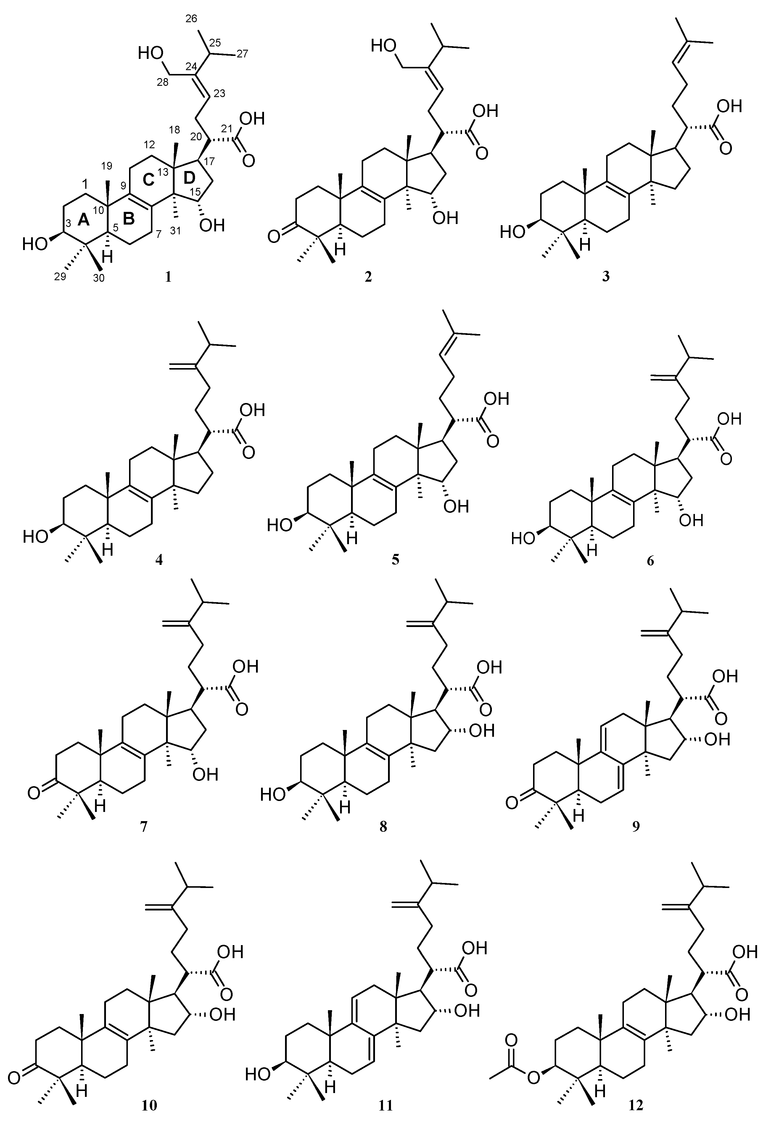
2.1. Isolation of Triterpenoids from L. sulphureus and Antrodia sp. (MUCL 56049)

A thorough analysis of the crude extracts from both L. sulphureus and MUCL 56049 Antrodia sp. by using high-performance liquid chromatography–diode array detector–high-resolution mass spectrometry (HPLC–DAD–HRMS) suggested the presence of lanostane triterpenoids. Consequently, isolation and purification of the crude extracts by using reversed phase preparative RP-HPLC resulted in the elucidation of 12 known triterpenes (Figure 1). The structures were confirmed by comparison of their high-resolution electrospray ionization mass spectrometry (HR-ESIMS), their electrospray ionization mass spectrometry (ESIMS), and NMR spectroscopic data (Supplementary Information Figures S1–S32) with those reported in the literature. The compounds from L. sulphureus were identified as laetiporin C or (3β,15α)-dihydroxy-24-hydroxymethyllanosta-8,23-dien-21-oic acid (1), laetiporin D or 15α-hydroxy-24-hydroxymethyl-3-oxolanosta-8,23-dien-21-oic acid (2) [12], trametenolic acid or 3β-hydroxylanosta-8,24-dien-21-oic acid (3) [13], eburicoic acid or 3β-hydroxy-24-methylene-lanost-8-en-21-oic acid (4) [14], 15α-hydroxytrametenolic acid or (3β,15α)-dihydroxylanosta-8,24-dien-21-oic acid (5) [15], sulphurenic acid or (3β,15α)-dihydroxy-24-methylene-lanost-8-en-21-oic acid (6) [16], and fomefficinic acid D or 15α-hydroxy-24-methylene-3-oxolanosta-8-en21-oic acid (7) [17].
The metabolites from Antrodia sp. MUCL 56049 included tumulosic acid or (3β,16α)-dihydroxy-24-methylenelanost-8-en-21-oic acid (8) [18], polyporenic acid C or 16α-hydroxy-24-methylene-3-oxolanosta-7,9(11)-dien-21-oic acid (9) [19], 16α-hydroxyeburiconic acid or 16α-hydroxy-24-methylene-3-oxolanost-8-en-21-oic acid (10) [19], dehydrotumulosic acid or (3β,16α)-dihydroxy-24-methylene-lanosta-7,9(11)-dien-21-oic acid (11) [20], and pachymic acid or 3β-acetyloxy-16α-hydroxy-24-methylene-lanost-8-en-21-oic acid (12) [21]. Interestingly, according to the literature, including our own previous results [12], none of these triterpenoids has been shown to exhibit any significant cytotoxic effects.

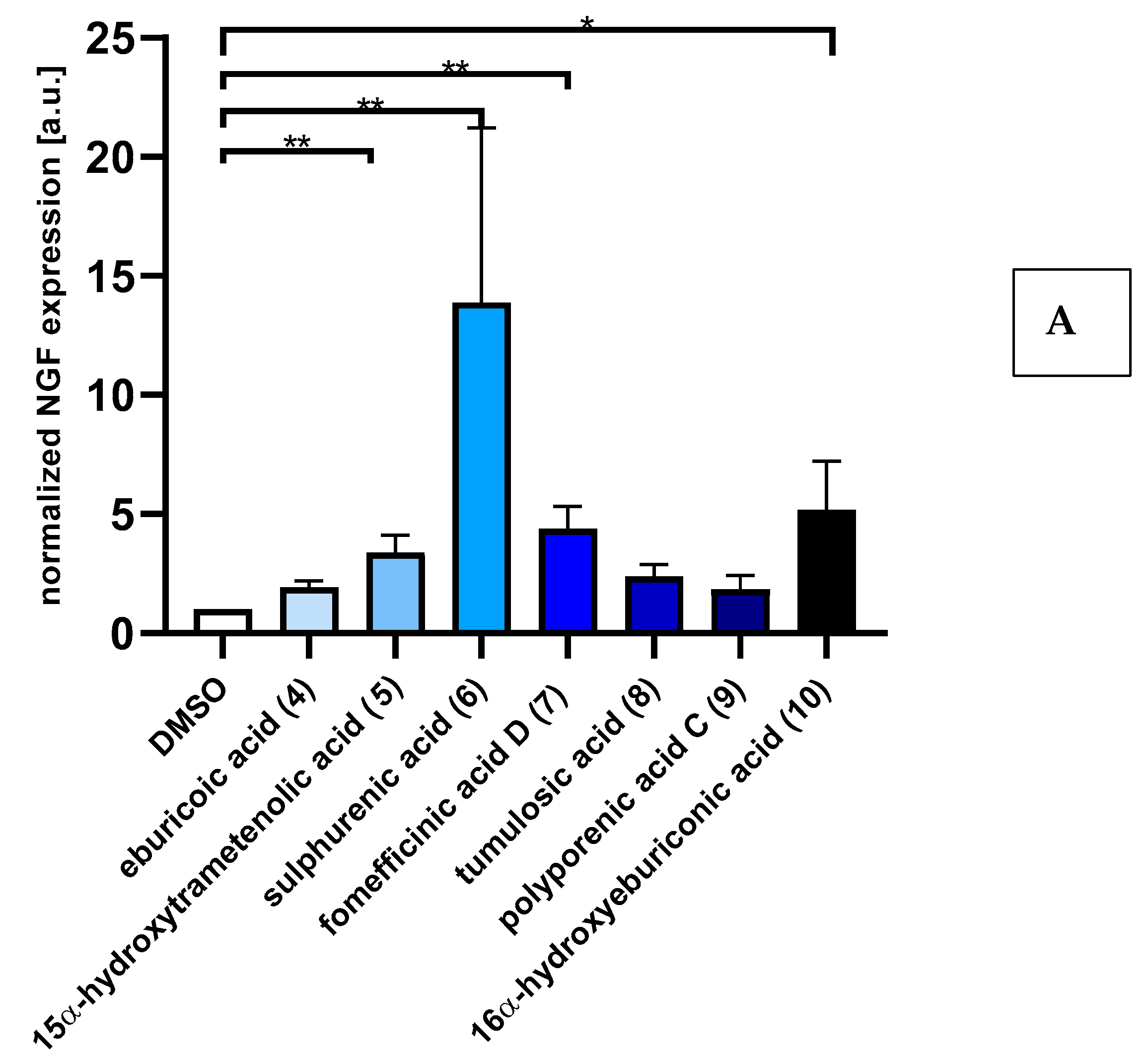
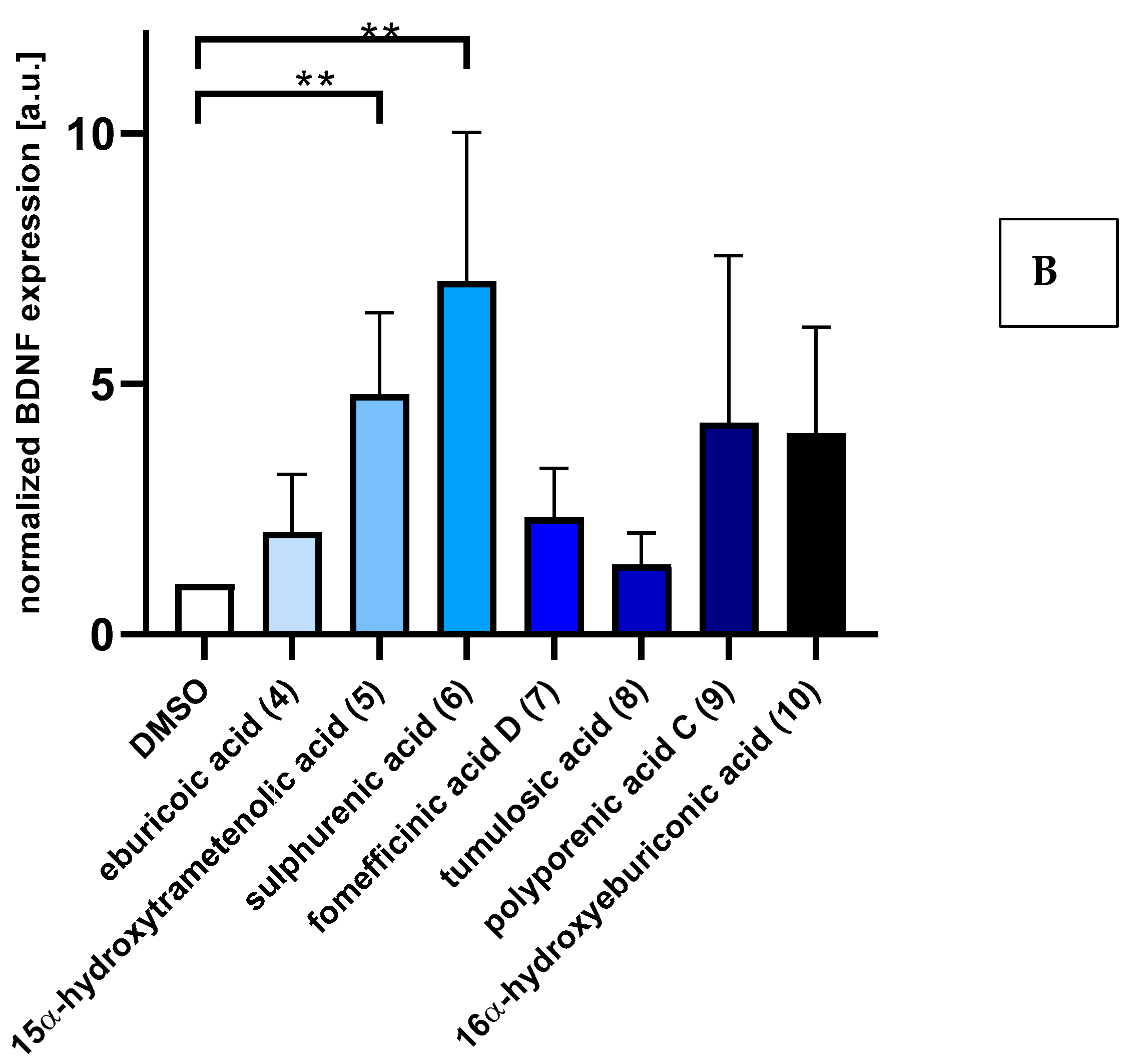
2.2. Triterpenoids Induce Expression of ngf and bdnf in Astrocytoma Cells

Quantitative real-time reverse transcriptase polymerase chain reaction (RT-PCR) analysis revealed that treatments of the astrocytoma cell line 1321N1 cells with triterpenes (5 µg/mL) for 48 h upregulate ngf and bdnf expression compared with DMSO-treated cells (Figure 2). Strikingly, treatments with sulphurenic acid 6 (13-fold), 16α-hydroxyeburiconic acid 10 (5-fold), fomefficinic acid D 7 (4-fold), and 15α-dehydroxytrametenolic acid 5 (3-fold) significantly upregulate ngf mRNA expression levels, while sulphurenic acid 6 (7-fold) and 15α-dehydroxytrametenolic acid 10 (4-fold) significantly upregulate bdnf expression. These results support ngf-induced neurite outgrowth by PC-12 cells treated with isolated triterpenoids.
Using cDNA synthesis and quantitative RT-PCR analysis, human astrocytoma 1321N1 cells treated with triterpenoids were found to upregulate the expression of genes for neurotrophins, including nerve growth factor, as well as brain-derived neurotrophic factor—genes known to regulate neuroprotection [22]. According to [23], cyathane diterpenes isolated from the submerged cultures of H. erinaceus and H. flagellum were found to act on 1321N1 cells by increasing the transcription of either bdnf or ngf. Similarly, a study by [11] reported that erinacine C influenced the expression in these astrocytic cells on the transcriptional level, probably also including factors that regulate ngf expression. In the present study, we clearly demonstrated that the triterpenoids increase ngf and bdnf synthesis in 1321N1 cells. However, the amount of ngf secreted into the culture media after incubation with the compounds was not sufficient to promote the differentiation of PC-12 cells (preliminary work).

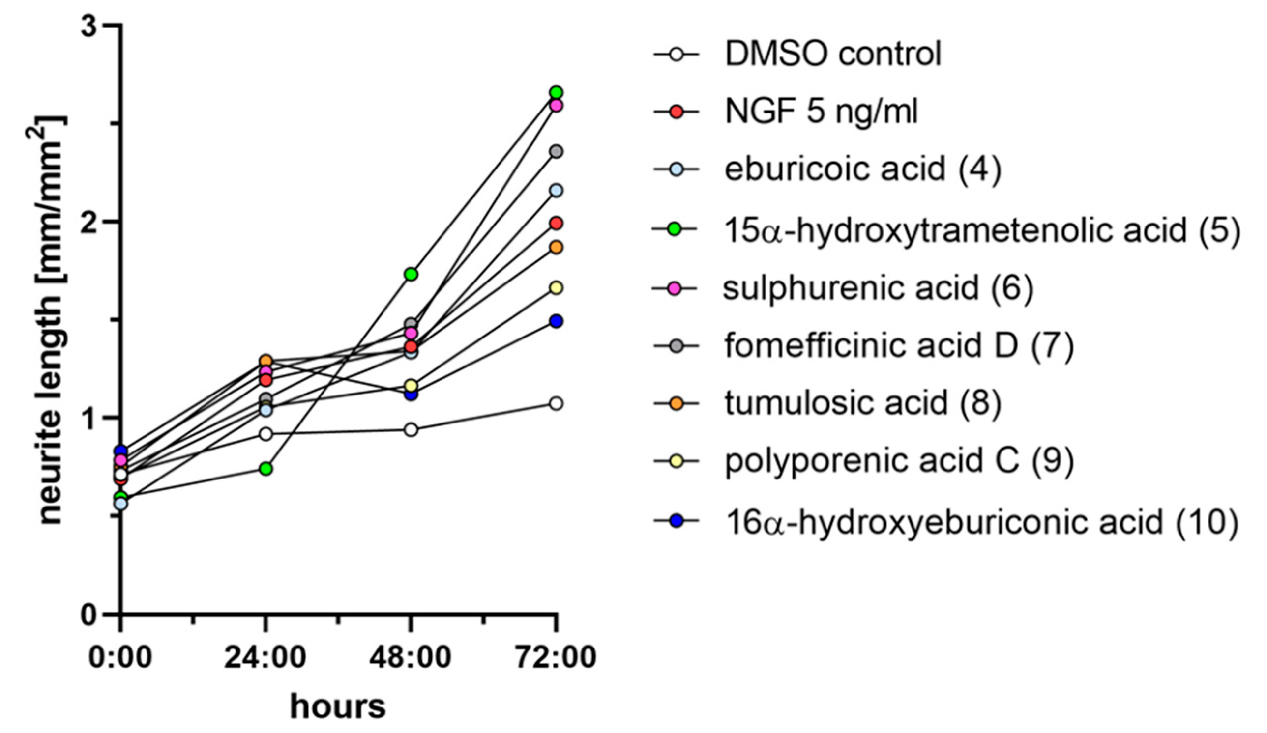
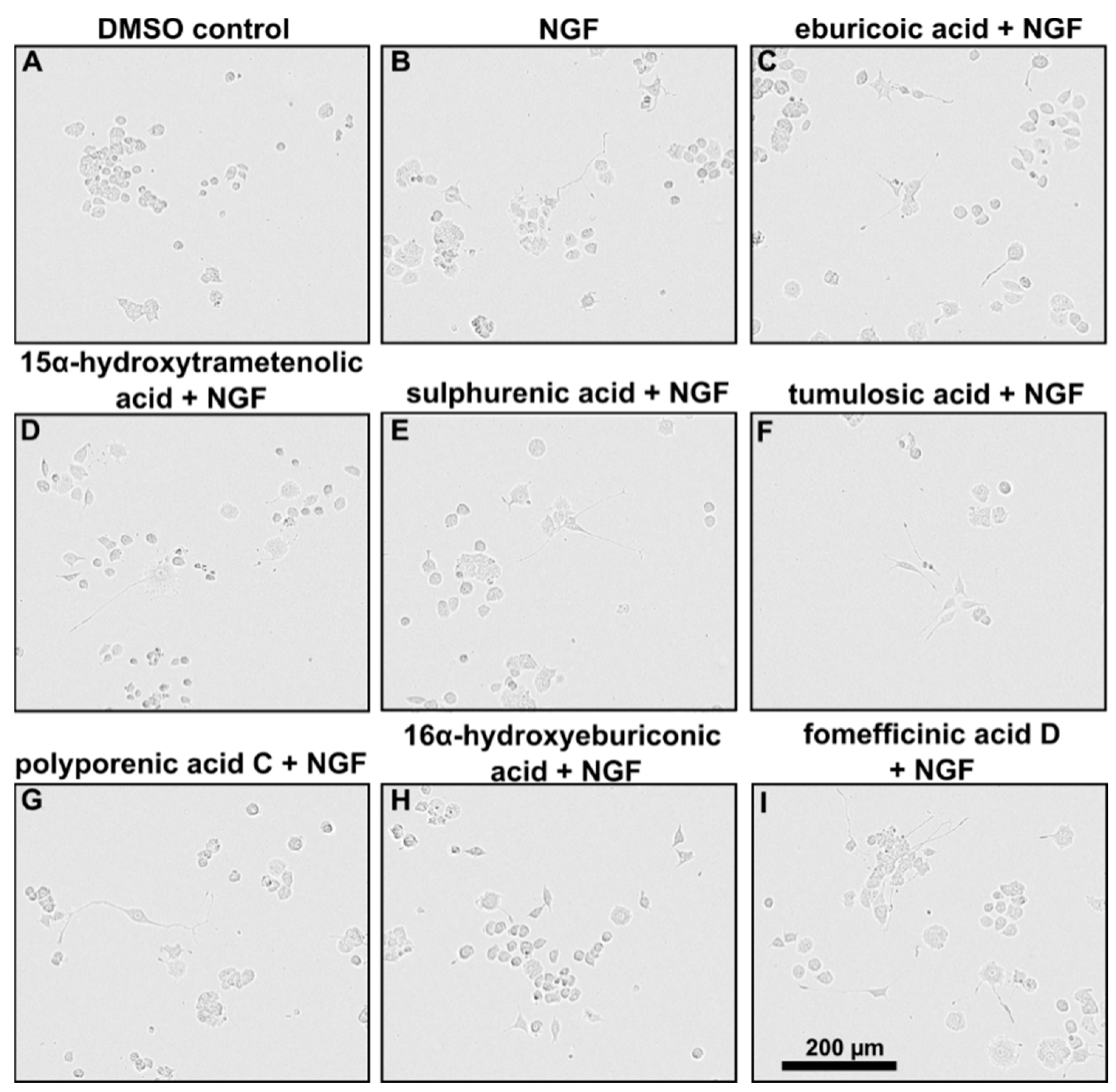
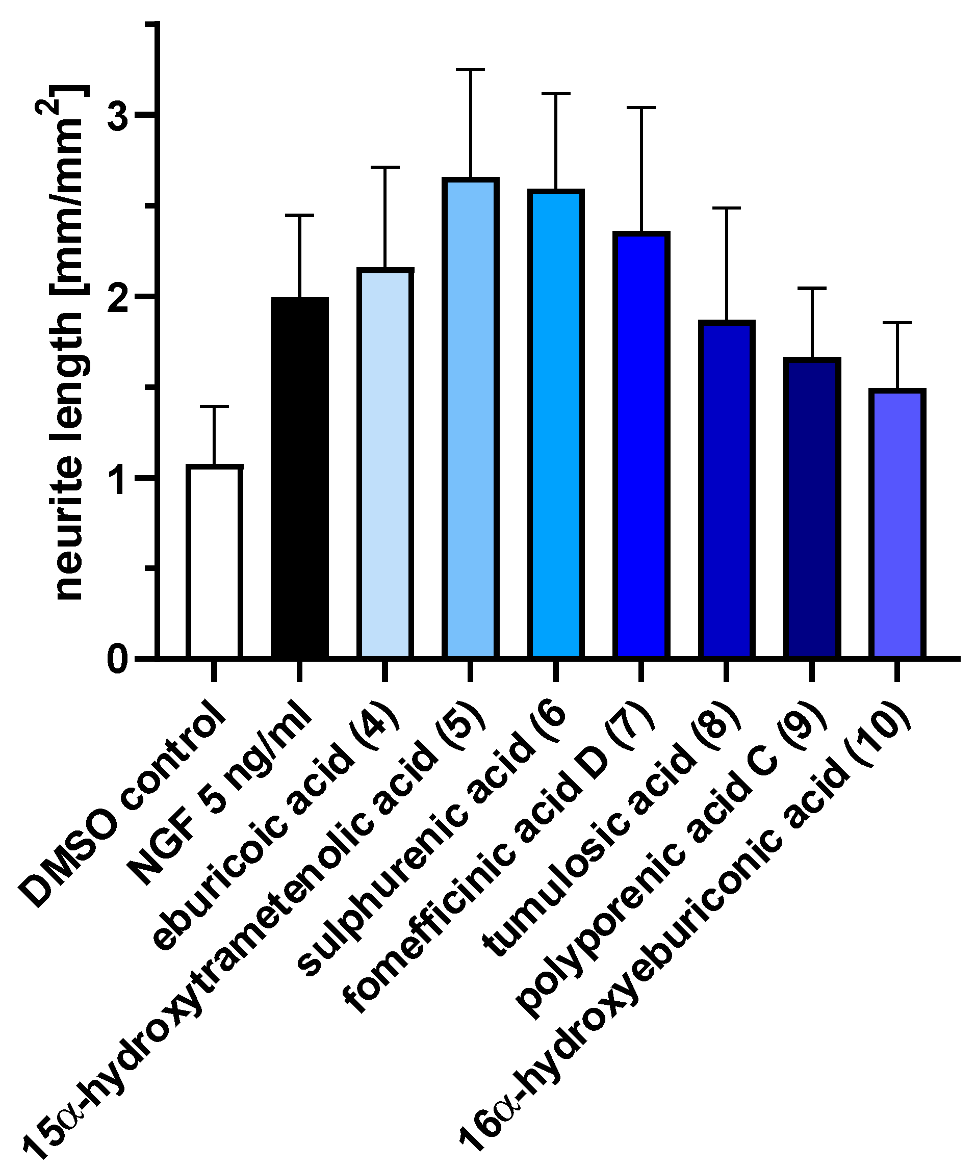
2.3. Triterpenoids Enhance ngf-Induced Neurite Outgrowth in PC-12 Cells

Measuring for neurite outgrowth of PC-12 cells is a well-established model [24], and the same methodology was also used in many previous studies on other fungal metabolites, including our own. Hence, we tested lanostane triterpenes on the efficiency of neurite outgrowth and assessed the mean length of neurite outgrowth by live-cell imaging after 72 h (Figure 3). Neurite outgrowth was not observed when PC-12 cells were treated directly with the triterpenes. However, when PC-12 cells were treated with the triterpenes supplemented with 5 ng/mL NGF, neurite outgrowth was observed (Figure 4). Treatment with sulphurenic acid (6), 15α-dehydroxytrametenolic acid (5), fomefficinic acid D (7), and eburicoic acid (4) caused a stronger differentiation of neurite-bearing cells as compared with the control cells, which had 5 ng/mL of ngf alone (Figure 5) over time. Investigations on whether the abovementioned triterpenes potentiate ngf-induced neurite outgrowth by stimulating ngf synthesis or as substitutes for ngf (ngf-mimicking activity) are ongoing.

3. Discussion

The results of our current study have highly interesting implications for the potential utility of lanostane triterpenoids, which are very commonly found in wood-inhabiting Basidiomycota, including many species of medicinal mushrooms that have been used for millennia in traditional Asian medicine. We will discuss some aspects that relate to the current context.

3.1. Previous Reports of Neurotrophic Activities of Extracts Derived from Other Basidiomycota and Selected Reports on Neurotropic Effects of Plant Metabolites

In our study, several triterpenes supplemented with ngf exhibited enhanced neurite outgrowth of PC-12 cells compared with ngf alone. This observation is in agreement with other studies. A report by [25] showed remarkably increased neurite outgrowth on PC-12 cells treated with hericenones C, D, and E isolated from fruiting bodies of H. erinaceus in the presence of 5 ng/mL ngf, along with further references on Hericium spp. cited earlier in this paper.
Similarly, Ref. [26] observed no cell differentiation or neurite outgrowth in PC-12 cells treated with littorachalcone isolated from Verbena littoralis in the absence of ngf, but markedly enhanced induced neurite outgrowth activity in the presence of ngf (2 ng/mL). A study by [27] reported that a combination of ngf and sclerotium extract of the Malaysian medicinal mushroom Lignosus rhinoceritis had additive effects and enhanced neurite outgrowth. On the other hand, reports on neurite outgrowth on PC-12 cells treated with both crude extracts and pure compounds without the presence of ngf are documented [5,28,29]. However, none of the aforementioned papers has explicitly dealt with a lanostane triterpenoid that was identified as an active principle. These studies either dealt with nondefined crude extracts that were not even characterized analytically, or different metabolites were shown to be responsible for the observed biological effects. The study by [5] reported another plant-derived triterpenoid of a different type (cucurbitane), lindersin B, to be active as an ngf enhancer by activation of the tyrosine kinase A/phosphatidylinositol 3 kinase/extracellular signal-regulated kinase signaling pathways. Ref. [30] also demonstrated the neurite outgrowth-promoting activity of hopane-type triterpenoid 2-acetoxydiplopterol isolated from the leaves of Illicium merrillianum using PC-12 cells. The neurotrophic activities of several other plant-derived triterpenes of different types, including oleanane-type (oleanolic acid) and ursane-type (ursolic acid), have also been reported. The latter compounds showed a markedly enhancing activity of ngf-mediated neurite in PC-12D cells [31]. Oleanolic acid was also proven to ameliorate scopolamine-induced memory impairment by modulating the bdnf-ERK1/2-CREB pathway through TrkB activation in mice [32].

3.2. Known Biological Activities of Compounds 112 and Related Triterpenes

Sulphurenic acid (6) was one of the compounds that showed the most significant effect in our studies and affected both bdnf and ngf enhancement. The compound was first identified by [16] from basidiomata of “Polyporus” (i.e., Laetiporus sulphureus) and has since then been discovered several times from various species of Laetiporus and other members of the Polyporales [33,34], including “Antrodia camphorata” (i.e., Taiwanofungus camphoratus) [35,36]. In their study of the constituents of the latter fungus, Ref. [37] noted only very weak cytotoxic effects, which is in agreement with our own findings reported in [12,14]. As the compound does not apparently affect(eve mammalian cells so strongly but has the strongest neurotrophic effects, it may be one of the most promising candidates for further evaluation. Interestingly, it has also been discovered in Fomitopsis officinalis [38], also known under the synonyms Fomes or Laricifomes, which is one of the most well-known medicinal mushrooms that has been used in Asia as well as in Europe for a very long time [39].
15α-dehydroxytrametenolic acid (5), the second compound that strongly enhanced bdnf and ngf production, as well as fomefficinic acid D (7) and 16α-hydroxyeburicoic acid (10), which only had a significant effect on ngf production, are also known from this group of fungi and have often been encountered concurrently with the aforementioned compounds [17,33,37].
Pachymic acid (12) was first reported by [21] from the basidiomata of the medicinal mushroom “Poria cocos” (currently valid name: Wolfiporia cocos), which also belongs to the Fomitopsidaceae. No bioactivities were initially reported, but the compound was later on found in various studies to have active anticancer and anti-inflammatory properties [40,41]. It was not among the strongest ngf enhancers that we found in the current study. There are also additional derivatives of related metabolites, including some that were isolated and characterized from Laetiporus species. For example, an acetyl derivative of eburicoic acid (4) was also reported to have anti-inflammatory effects on murine macrophages [42].
Previous studies [43,44] reported that triterpenoids isolated from “Ganoderma lucidum” showed neurotropic properties by enhancing the survival and promoting the survival of neurons. These authors, however, did not show these effects in a direct manner but tested their compounds for their ability to initiate TrkA or TrkB-related signal transduction in NIH-3T3/TrkA and NIH-3T3/TrkB cells using an MTT assay. The two derivatives, 4,4,14α-trimethyl-5α-chol-7,9(11)-dien-3-oxo-24-oic acid (which was then reported to be a novel natural derivative) and methyl ganoderic acid B, and six other congeners did not show such effects or were not examined. In addition, triterpenes isolated from “G. lucidum” were also reported to attenuate the production of proinflammatory cytokines in macrophage cells [45]. The fungus that these authors reported on, however, is probably not identical to the European species G. lucidum, which does not occur in China, where these studies were undertaken [46]. It may correspond with G. lingzhi (i.e., the fungus that was confused with G. lucidum and has been used in traditional Asian medicine for centuries) or another native Chinese species (cf.) [47]. Notably, many of the triterpenes from the Ganoderma species are rather cytotoxic (and preparations made from Ganoderma are even in use as anticancer agents). In contrast, the compounds that were identified as neurotrophins in the current study have only weak cytotoxic effects on mammalian cells [12,14] and are also lacking certain structural features, such as the carboxyl group at C-24 of the triterpenoid skeleton. Even though the Ganoderma triterpenoids were apparently not yet tested for ngf-enhancing activities in our bioassays, it seems possible that the ones from Antrodia and Laetiporus exert their neurotrophic effects through a different mode of action. Interestingly, these triterpenoids also have more selective neurotrophic accents than, e.g., the diterpenoids of the erinacine type from Hericium, some of which show cytotoxicity in the low µM range (cf.) [23]. On the other hand, the triterpenoids exhibit their neurotrophic effects at much lower concentrations than the corallocins from Hericium coralloides and related meroterpenoids that are also present in H. erinaceus (cf.) [48].

3.3. Previous Use of Laetiporus and Antrodia Species and Their Relatives in Traditional Medicine and for Other Applications

The genus Laetiporus belongs to the Fomitopsidaceae (Polyporales), and its species form conspicuous basidiomes with bright yellowish colors on wood. They are considered forest pathogens and cause brown rot, but some are edible and medicinal mushrooms [49]. The genus is geographically distributed worldwide, and its species can be found in cold temperate to tropical zones. The type species, Laetiporus sulphureus, goes back to the French mycologist Buillard, who described it as Boletus sulphureus in 1789, and it is very frequently encountered in Europe and other areas of the Northern Hemisphere. It is known as “Chicken of the woods” or “sulphur shelf” in English-speaking countries and as “Schwefelporling” in Germany. Out of the 15 presently accepted species, 11 belong to the L. sulphureus complex, as was recently established by phylogenetic analyses [50,51].
The young fruitbodies are of a soft consistency and are even regarded as a culinary delicacy. In China and Europe, the fruitbodies have been used traditionally to strengthen the human body and as a remedy for various diseases (see summary by [52]). However, according to our literature search, no study has yet been undertaken to link the reported health benefits directly and conclusively to the triterpenoids and other secondary metabolites of L. sulphureus.
Like Laetiporus, the genus Antrodia also belongs to the Fomitopsidaceae and is typified by A. serpens, a fungus that was first described by [53] as Polyporus serpens. The genus presently has more than 50 accepted species [54,55], which are also known to destroy wood by causing brown rot.
The taxonomy of MUCL 56049 is also still unclear. The genus was determined from morphological traits and because the closest matches retrieved from a BLAST search in GenBank based on LSU and ITS sequences were derived from a specimen of “Antrodia aff. albidoides” sensu Ryvarden from Zimbabwe, which has likewise not been formally described. The ITS sequence (GenBank acc. no. KC543176.1) has been generated and uploaded by [55] and also shows high homologies to other species in the family Fomitopsidaceae, to which the genera Laetiporus and Fomitopsis belong. Many of the tropical species of Antrodia and allied genera, especially from Africa, remain widely uncharacterized by modern polyphasic taxonomic methodology, and a recent monograph is not available, which is why we have thus far been unable to classify the specimen MUCL 56049 to species level.
Regarding the practical use of the Antrodia species as food or medicinal mushroom, there is not much information in the literature. The fruitbodies are inedible due to their tough consistency, and reports on medicinal usages of the “Antrodia” species can mostly be related to Taiwanofungus camphoratus, an important Asian medicinal mushroom related to the species of Antrodia, which has in the past been referred to under the synonyms A. camphorata or A. cinnamomea [17]. The economic value of T. camphoratus is mainly due to its medicinal use, and it belongs to the most valued medicinal fungus in Asian countries. This species has already been used as a natural therapeutic ingredient in traditional Chinese medicine (TCM) for its antioxidative, antitumor, anticancer, antihepatitis, vasorelaxation, anti-inflammatory, cytotoxic, and neuroprotective activities [33,54,56].
Because of its use in traditional medicine, the secondary metabolites of T. camphoratus have been studied intensively, and over 70 compounds have been elucidated from this fungus, 39 of which are triterpenoids [54,56]. These triterpenes are of the ergostane and lanostane types (tumulosic acid (8), polyporenic acid C (9), 16α-hydroxyeburicoic acid (10), dehydrotumulosic acid (11), and pachymic acid (12) that were also found in this study from the African Antrodia sp. Neuroprotective effects have been claimed in two patent applications [57,58] in which some of the compounds we studied were mentioned, even though no direct evidence has been provided by these authors, and no ngf- or bndf-enhancing activities were noted in these patent applications. On the other hand, the current study clearly shows that it is worthwhile to further study these fungi, including their secondary metabolism, as this may reveal interesting chemotaxonomic relationships as well as hitherto unprecedented biological activities of their constituents.

4. Materials and Methods

4.1. General Information

HPLC-DAD/MS measurements were performed using an amaZon speed ETD (electron transfer dissociation) ion trap mass spectrometer (Bruker Daltonics, Bremen, Germany) and measured in positive and negative ion modes simultaneously. The HPLC system comprised the following elements: Column C18 Acquity UPLC BEH (Waters, Eschborn, Germany); mobile phase: solvent A (H2O)/solvent B (acetonitrile (ACN)), supplemented with 0.1% formic acid; gradient conditions: 5% B for 0.5 min, increasing to 100% B 20, maintaining isocratic conditions at 100% B for 10 min, flow rate 0.6 mL/min, UV/Vis detection 200−600 nm).
HR-ESIMS (high-resolution electrospray ionization mass spectrometry) data were recorded on a mass spectrometer (Bruker Daltonics) coupled to an Agilent 1260 series HPLC-UV system and equipped with C18 Acquity UPLC BEH (ultraperformance liquid chromatography) (ethylene bridged hybrid) (Waters) column; DAD-UV detection at 200–600 nm; solvent A (H2O) and solvent B (ACN) supplemented with 0.1% formic acid as a modifier; flowrate 0.6 mL/min, 40 °C, gradient elution system with the initial condition 5% B for 0.5 min, increasing to 100% B in 19.5 min and holding at 100% B for 5 min. To determine the molecular formula, Compass DataAnalysis 4.4 SR1 was used using the Smart Formula algorithm (Bruker Daltonics). NMR spectra were recorded on a Bruker 700 MHz Avance III spectrometer equipped with a 5 mm TCI cryoprobe (1H: 700 MHz, 13C: 175 MHz) locked to the respective deuterium signal of the solvent.

4.2. Fungal Material

The species that were studied on the production of neurotrophins were selected out of our ongoing research program for the discovery of antibiotics and other beneficial metabolites from European and tropical Basidiomycota. In this scenario, samples (extracts and pure compounds) that are devoid of cytotoxic and prominent antimicrobial effects were investigated for other potential applications. The current study deals with a fruitbody extract of specimens collected in Germany and an extract derived from a mycelial culture of an African species. Samples derived from these fungi showed interesting neurotrophic effects during preliminary studies on their ability to enhance ngf and bdnf.
Laetiporus sulphureus (Figure 6A) was collected in Braunschweig-Riddagshausen nature reserve on 27 May 2020. Hosts plants were Salix spec., Fagus sylvatica, and Robinia pseudoacacia. The specimens were identified by Harry Andersson based on morphological data, and a voucher specimen was deposited in the fungarium of the HZI, Braunschweig.
Further details, including the isolation of the terpenoids that were tested in this study, have recently been described by [12].
The fruitbodies of Antrodia sp. (Figure 6B) were collected by C. Decock and J. C. Matasyoh from Mount Elgon National Reserve (1°7′6″ N, 34°31′30″ E) in Kenya in April 2016. A dried specimen and a corresponding culture were deposited at Mycothèque de l’Université catholique de Louvain (MUCL, Louvain-la-Neuve, Belgium) under accession number MUCL56049. The genus was identified by morphological studies and comparison of the internal transcribed spacer (ITS)-nrDNA sequences with others from Basidiomycota deposited in GenBank. The sequence is also available in the Supplementary Information.

4.3. Extraction and Isolation of the Secondary Metabolites

The extraction and purification of compounds from L. sulphureus have been described in detail in our recent paper [12] and led to the isolation of seven metabolites (1–7). The same protocols were used for the isolation of the compounds from a batch culture of Antrodia sp. MUCL 56049 that had been grown in twenty 500 mL shake flasks containing 200 mL each of yeast malt (YM 6.3) medium (10 g/L malt extract, 4 g/L yeast extract, 4 g/L d-glucose, and pH = 6.3) for 24 days. The mycelia were harvested 3 days after glucose depletion by filtration, and the culture filtrate was set aside, as it did not contain any target metabolites.
Extraction of the mycelia of Antrodia sp. from the aforementioned four liters of culture media with acetone (200 mL) led to a total of 442 mg crude product. The crude extract (cream solid) was dissolved in methanol (MeOH) and filtered using cotton in a Pasteur pipette. The crude extract was fractionated using preparative reverse phase liquid chromatography (PLC 2020, Gilson, Middleton, WI, USA). VP Nucleodur 100-5C 18 ec column (250 × 40 mm, 7 μm: Macherey-Nagel, Düren, Germany) was used as stationary phase. Deionized water (Milli-Q, Millipore, Schwalbach, Germany) (solvent A) and acetonitrile (solvent B) with 0.05% TFA were used as eluent with flow rate of 40 mL/min. Three runs were performed on the preparative RP-HPLC with an elution gradient of 50–100% solvent B for 60 min and thereafter isocratic condition at 100% solvent B for 5 min. UV/Vis detection was carried out at 210 nm and 300 nm. Eleven fractions were collected according to the selected peaks. Similar fractions from each round of separation were combined, consequently leading to isolation of compounds 8 (4 mg), 9 (1.25 mg), 10 (5.2 mg), 11 (2.2 mg), and 12 (3.5 mg). Their structures were elucidated by using their HR-ESIMS data and their 1D and 2D NMR spectroscopic data, which were then compared with those already reported in the literature. The spectral data for compounds 1–7 are deposited in the Supplementary Information with the preceding studies by [12,14], and the data for compounds 8–12 are given in the Supplementary Information of the current paper.

4.4. Cell Culture

Astrocytoma (1321N1) cells (obtained from Sigma-Aldrich, acc. no. 86030402) were cultured in Gibco DMEM medium (Fisher Scientific, Inc., Waltham, MA, USA) containing 10% heat-inactivated FBS (Capricorn) [11]. Rat pheochromocytoma cells (PC-12, adherent variant) purchased from the European Collection of Authenticated Cell Cultures (ECACC) general collection were grown in Gibco RPMI-1640 (Fisher Scientific, Hampton, NH, USA) medium containing 10% horse serum (Capricorn Scientific GmbH, Ebsdorfergrund, Germany) and 5% heat-inactivated fetal bovine serum-FBS (Capricorn). The media were supplemented with penicillin (0.15 mM), streptomycin (86 µM), and glutamine (2 mM) [23]. The cells were incubated at 37 °C in a humidified environment of 7.5% CO2 and were routinely passaged every 3–4 days. Collagen type IV (Sigma C5533) was coated on 96-well plates and left for 6 h or more before using the plates whenever seeding PC-12 cells.

4.5. cDNA Synthesis and Real-Time Quantitative RT-PCR

Since PC-12 cells do not produce ngf on their own, confirming the induction of neurotrophin expression in 1321N1 cells was tested directly. For real-time quantitative reverse transcriptase PCR, the total RNA was extracted from triterpene-treated 1321N1 cells (2 × 10⁵ cells). For treatment, culture media was replaced by serum-reduced medium (Gibco RPMI with 1% FBS (Capricorn)), and cells were incubated for 24 h. Then, media was replaced with media containing the triterpenes dissolved in 0.5% DMSO. As control, serum-reduced medium supplemented with 0.5% DMSO was used. Cells were incubated for 48 h [11,23]. Total RNA was extracted using the NucleoSpin RNA Plus kit (Macherey-Nagel) followed by further purification (NucleoSpin RNA Clean-up kit) according to the manufacturer’s protocol. To determine the concentration of purified RNA, the corresponding samples were measured using a DS-11+ spectrophotometer Nanodrop (DeNovix Inc., Wilmington, DE, USA). First-strand cDNA synthesis and subsequent real-time PCR were performed using SensiFast SYBR No-Rox One-Step Kit (Cat. No. BIO-72005 (Bioline)). The following PCR primers were used for amplifying specific cDNA fragments: gapdh (sense: 5′-ACCACAGTCCATGCCATCAC-3′; antisense: 5′-TCCACCACCCTGTTGCTGTA-3′ 451 bp), ngf (sense: 5′-CCAAGGGAGCAGTTTCTATCCTGG-3′; antisense: 5′-GGCAGTTGTCAAGGGAATGCTGAAGTT-3′ 189 bp), and bdnf (sense: 5′-TAACGGCGGCAGACAAAAAGA-3′; antisense: 5′-GAAGTATTGCTTCAGTTGGCCT-3′; 101 bp) [11]. The PCR reactions were performed in a 10 µL volume containing cDNA template (2 µL), SensiFast SYBR No-Rox One-step Mix (5 µL), primers (400 nM; 0.4 µL), reverse transcriptase (0.1 µL), RiboSafe RNase Inhibitor (0.2 µL), and Rnase free water (1.9 µL). The amplified cDNAs were analyzed and quantified using the Qiagen (Corbett) Rotor-Gene 3000 and LightCycler 96 (Roche Diagnostics International Ltd., Mannheim, Germany, version 1.1.0.1320) real-time PCR instruments. Amounts of gapdh amplicons value were used as reference and set as 1.

4.6. Neurite Outgrowth Assay

Preliminary studies showed that conditioned media from 1321N1 cells treated with isolated triterpenes did not display PC-12 cell differentiation activity, triggering neurite outgrowth. The neurotrophic activity assay was conducted, as described by Phan et al. (2015) [6]. PC-12 cells were seeded at a density of 1 × 10³ cells per well in growth medium in 96-well culture plates and incubated overnight. Triterpenoids isolated from L. sulphureus and Antrodia sp. were added directly to the cells and further incubated at 37 °C with 5% CO2 for 3 days. Cells treated with nerve growth factor (50 ng/mL) were used as a positive control. After 3 days, the cells were examined using an IncuCyte S3 live-cell analysis system (Sartorius, Göttingen, Germany). Six random fields were examined in each well. The number of neurite outgrowths (axon-like processes), defined as extensions longer than twice the cell body diameter, was recorded. Neurite length was measured using the IncuCyte NeuroTrack Software Module (2019B Rev2 GUI) software. Three independent experiments were conducted for each compound. Neurite outgrowth activity in the presence of both ngf and triterpenoids was also then analyzed. A concentration of ngf (5 ng/mL) was added in addition to the triterpenoids on the PC-12 cells, and the neurite outgrowth was recorded. Three independent experiments were performed in this assay.

4.7. Statistical Analysis

Data are displayed as the mean ± SEM and analyzed by Mann–Whitney rank sum test using the software Prism V8 (Graphpad Software Inc., San Diego, CA, USA).

5. Conclusions

What remains to be performed is to establish a clear-cut structure–activity relationship between the substitution patterns of the lanostane triterpenoids and the corresponding biological effects. Since it was not possible to establish this based on the relatively low number of natural products currently available, it may be feasible to isolate large quantities of one of the most potent derivatives and subject this metabolite to semisynthesis. On the other hand, it could be feasible to use the mixtures of triterpenoids that can be enriched by the development of special extraction techniques as a standardized product that can be used as a “mycopharmaceutical” or, in the case of the edible Laetiporus, as a nutraceutical that can be used as a prophylactic agent for prevention of neuropathic disorders. In any case, the biochemical mode of action of these compounds also needs further study because, so far, it is not evident how the neurotrophic effects observed actually came about. However, the amounts of compounds that became available during the course of this work were too small to conduct follow-up experiments, as described above—and there is a need to follow-up in the near future after further scale-up of production and isolation of the terpenoids. For this purpose, it may even be feasible to study related fungi in order to obtain better yields or to optimize the fermentation process in view of a transfer to larger-scale bioreactors.

Supplementary Materials

The supporting information can be downloaded at: https://www.mdpi.com/article/10.3390/ijms232113593/s1.

Author Contributions

Conceptualization, M.S. and R.W.K.; methodology, M.S., R.W.K. and T.E.B.S.; formal analysis and investigation, K.W., M.R.-A., C.C., K.H., M.K. and B.M.K.; writing—original draft preparation, all authors; writing—review and editing, all authors; funding acquisition, M.S., R.W.K., T.E.B.S., J.C.M. and C.D. All authors have read and agreed to the published version of the manuscript.

Funding

Financial support by a personal PhD stipend from the German Academic Exchange Service (DAAD) and the National Research Fund of Kenya (NRF) to K.H. (program ID—57399475) is greatly acknowledged. B.M.K. is also grateful for financial support by a personal PhD stipend from the DAAD (program ID—57440921). R.W.K. and M.S. are grateful for financial support via the “Drug Discovery and Cheminformatics for New Anti-Infectives (iCA)” program awarded by the Ministry for Science and Culture of the German State of Lower Saxony (MWK No. 21—78904-63-5/19). Furthermore, C.D., J.C.M. and M.S. are grateful for the “ASAFEM” Project (grant no. IC-070) under the ERAfrica Programme. This research also benefited from the European Union’s H2020 Research and Innovation Staff Exchange program (RISE), grant no. 101008129: MYCOBIOMICS, beneficiaries T.E.B.S., M.S. and J.C.M.

Data Availability Statement

All data generated or analyzed during this study are included in this published article and its supplementary information files.

Acknowledgments

We are grateful to Wera Collisi, Christel Kakoschke, Silke Reinecke, and Silvia Pettrin for their technical support, and to Harry Anderson for the collection of the Laetiporus specimen.

Conflicts of Interest

The authors declare no conflict of interest.

References

  1. Chen, X.; Drew, J.; Berney, W.; Lei, W. Neuroprotective natural products for Alzheimer’s disease. Cells 2021, 10, 1309. [Google Scholar] [CrossRef] [PubMed]
  2. Mapook, A.; Hyde, K.D.; Hassan, K.; Kemkuignou, B.M.; Čmoková, A.; Surup, F.; Kuhnert, E.; Paomephan, P.; Cheng, T.; de Hoog, S.; et al. Ten decadal advances in fungal biology leading towards human well-being. Fungal Diversity 2022, 116, 547–614. [Google Scholar] [CrossRef] [PubMed]
  3. Xu, J.; Lacoske, M.H.; Theodorakis, E.A. Neurotrophic natural products: Chemistry and biology. Angew. Chem. Int. Ed. 2014, 53, 956–987. [Google Scholar] [CrossRef] [PubMed] [Green Version]
  4. Mori, K.; Obara, Y.; Hirota, M.; Azumi, T.; Kinugasa, S.; Inatomi, S.; Nakahata, N. Nerve growth factor-inducing activity of Hericium erinaceus in 1321N1 human astrocytoma cells. Biol. Pharm. Bull. 2008, 31, 1727–1732. [Google Scholar] [CrossRef] [PubMed] [Green Version]
  5. Cheng, L.; Ye, Y.; Xiang, L.; Osada, H.; Qi, J. Lindersin B from Lindernia crustacea induces neuritogenesis by activation of tyrosine kinase A/phosphatidylinositol 3 kinase/extracellular signal-regulated kinase signaling pathway. Phytomedicine 2017, 24, 31–38. [Google Scholar] [CrossRef]
  6. Phan, C.W.; David, P.; Naidu, M.; Wong, K.H.; Sabaratnam, V. Therapeutic potential of culinary-medicinal mushrooms for the management of neurodegenerative diseases: Diversity, metabolite, and mechanism. Crit. Rev. Biotechnol. 2015, 35, 355–368. [Google Scholar] [CrossRef]
  7. Zhang, X.Q.; Ip, F.C.; Zhang, D.; Chen, L.-X.; Zhang, W.; Li, Y.-L.; Ip, N.Y.; Ye, W.-C. Triterpenoids from Ganoderma lucidum and their cytotoxic activities. J. Nat. Prod. Res. 2011, 27, 37–41. [Google Scholar]
  8. Thongbai, B.; Rapior, S.; Hyde, K.D.; Wittstein, K.; Stadler, M. Hericium erinaceus, an amazing medicinal mushroom. Mycol. Prog. 2015, 14, 91. [Google Scholar] [CrossRef]
  9. Hyde, K.D.; Xu, J.; Rapior, S.; Jeewon, R.; Lumyong, S.; Niego, A.G.T.; Abeywickrama, P.D.; Aluthmuhandiram, J.V.S.; Brahamanage, R.S.; Brooks, S.; et al. The amazing potential of fungi: 50 ways we can exploit fungi industrially. Fungal Divers. 2019, 97, 1–136. [Google Scholar] [CrossRef] [Green Version]
  10. De Silva, D.D.; Rapior, S.; Sudarman, E.; Stadler, M.; Xu, J.; Aisyah Alias, S.; Hyde, K.D. Bioactive metabolites from macrofungi: Ethnopharmacology, biological activities and chemistry. Fungal Divers. 2013, 62, 1–40. [Google Scholar] [CrossRef]
  11. Rascher, M.; Wittstein, K.; Winter, B.; Rupcic, Z.; Wolf-Asseburg, A.; Stadler, M.; Köster, R.W. Erinacine C activates transcription from a consensus ETS DNA binding site in astrocytic cells in addition to NGF induction. Biomolecules 2020, 10, 1440. [Google Scholar] [CrossRef] [PubMed]
  12. Hassan, K.; Kemkuignou, B.M.; Stadler, M. Two new triterpenes from basidiomata of the medicinal and edible mushroom, Laetiporus sulphureus. Molecules 2021, 26, 7090. [Google Scholar] [CrossRef] [PubMed]
  13. Yoshikawa, K.; Ikuta, M.; Arihara, S.; Matsumura, E.; Katayama, S. Two new steroidal derivatives from the fruit body of Chlorophyllum molybdites. Chem. Pharm. Bull. 2001, 49, 1030–1032. [Google Scholar] [CrossRef] [Green Version]
  14. Chepkirui, C.; Matasyoh, J.C.; Decock, C.; Stadler, M. Two cytotoxic triterpenes from cultures of a Kenyan Laetiporus sp. (Basidiomycota). Phytochem. Lett. 2017, 20, 106–110. [Google Scholar] [CrossRef]
  15. Yang, S.W.; Shen, Y.C.; Chen, C.H. Steroids and triterpenoids of Antrodia cinnamomea—A fungus parasitic on Cinnamomum micranthum. Phytochemistry 1996, 41, 1389–1392. [Google Scholar] [CrossRef]
  16. Fried, J.; Grabowich, P.; Sabo, E.; Cohen, A. The structure of sulphurenic acid: A new triterpenoid from Polyporus sulphureus. Tetrahedron 1964, 20, 2297–2312. [Google Scholar] [CrossRef]
  17. Wu, X.; Yang, J.; Zhou, L.; Dong, Y. New lanostane-type triterpenes from Fomes officinalis. Chem. Pharm. Bull. 2004, 52, 1375–1377. [Google Scholar] [CrossRef] [PubMed] [Green Version]
  18. Cort, L.A.; Gascoigne, R.M.; Holker, J.S.E.; Ralph, B.J.; Robertson, A.; Simes, J.J.H. The chemistry of fungi. Part XXIII. Tumulosic acid. J. Chem Soc. 1954, 3713–3721. [Google Scholar] [CrossRef]
  19. Rösecke, J.; König, W.A. Constituents of the fungi Daedalea quercina and Daedaleopsis confragosa var. tricolor. Phytochemistry 2000, 54, 757–762. [Google Scholar] [CrossRef]
  20. Tai, T.; Shingu, T.; Kikuchi, T.; Tezuka, Y.; Akahori, A. Triterpenes from the surface layer of Poria cocos. Phytochemistry 1995, 39, 1165–1169. [Google Scholar] [CrossRef]
  21. Shingu, T.; Tai, T.; Akahori, A. A lanostane triterpenoid from Poria cocos. Phytochemistry 1992, 31, 2548–2549. [Google Scholar] [CrossRef]
  22. Huang, E.J.; Reichardt, L.F. Neurotrophins: Roles in neuronal development and function. Annu. Rev. Neurosci. 2001, 24, 677–736. [Google Scholar] [CrossRef] [PubMed] [Green Version]
  23. Rupcic, Z.; Rascher, M.; Kanaki, S.; Köster, R.W.; Stadler, M.; Wittstein, K. Two new cyathane diterpenoids from mycelial cultures of the medicinal mushroom Hericium erinaceus and the rare species, Hericium flagellum. Int. J. Mol. Sci. 2018, 19, 740. [Google Scholar] [CrossRef] [PubMed] [Green Version]
  24. Greene, L.A.; Tischler, A.S. Establishment of a noradrenergic clonal line of rat adrenal pheochromocytoma cells which respond to nerve growth factor. Proc. Natl. Acad. Sci. USA 1976, 73, 2424–2428. [Google Scholar] [CrossRef] [PubMed] [Green Version]
  25. Phan, C.W.; Lee, G.S.; Hong, S.L.; Wong, Y.T.; Brkljača, R.; Urban, S.; Abd Malek, S.N.; Sabaratnam, V. Hericium erinaceus (Bull.: Fr) Pers. cultivated under tropical conditions: Isolation of hericenones and demonstration of NGF-mediated neurite outgrowth in PC12 cells via MEK/ERK and PI3K-Akt signaling pathways. Food Funct. 2014, 5, 3160–3169. [Google Scholar] [CrossRef]
  26. Li, Y.; Ishibashi, M.; Chen, X.; Ohizumi, Y. Littorachalcone, a new enhancer of NGF-mediated neurite outgrowth, from Verbena littoralis. Chem. Pharm. Bull. 2003, 51, 872–874. [Google Scholar] [CrossRef]
  27. Eik, L.-F.; Naidu, M.; David, P.; Wong, K.-H.; Tan, Y.-S.; Sabaratnam, V. Lignosus rhinocerus (Cooke) Ryvarden: A medicinal mushroom that stimulates neurite outgrowth in PC-12 cells. Evid.-Based Complement. Altern. Med. 2012, 2012, 320308. [Google Scholar] [CrossRef] [Green Version]
  28. Cheng, L.; Muroi, M.; Cao, S.; Bian, L.; Osada, H.; Xiang, L.; Qi, J. 3β,23,28-Trihydroxy-12-oleanene 3β-Caffeate from Desmodium sambuense-Induced neurogenesis in PC12 cells mediated by ER stress and BDNF-TrkB signaling pathways. Mol. Pharm. 2019, 16, 1423–1432. [Google Scholar] [CrossRef]
  29. Phan, C.-W.; Wong, W.-L.; David, P.; Naidu, M.; Sabaratnam, V. Pleurotus giganteus (Berk.) Karunarathna & K.D. Hyde: Nutritional value and in vitro neurite outgrowth activity in rat pheochromocytoma cells. BMC Complement. Altern. Med. 2012, 12, 102. [Google Scholar] [CrossRef] [Green Version]
  30. Tian, X.-H.; Yue, R.-C.; Fang, X.; Zhang, J.-P.; Wang, G.-W.; Shan, L.; Zhang, W.-D.; Shen, Y.-H. Terpenoids with neurite outgrowth-promoting activity from the branches and leaves of Illicium merrillianum. J. Asian Nat. Prod. Res. 2016, 18, 495–503. [Google Scholar] [CrossRef]
  31. LI, Y.; OHIZUMI, Y. Search for constituents with neurotrophic factor-potentiating activity from the medicinal plants of Paraguay and Thailand. YAKUGAKU ZASSHI 2004, 124, 417–424. [Google Scholar] [CrossRef] [PubMed]
  32. Jeon, S.J.; Lee, H.J.; Lee, H.E.; Park, S.J.; Gwon, Y.; Kim, H.; Zhang, J.; Shin, C.Y.; Kim, D.H.; Ryu, J.H. Oleanolic acid ameliorates cognitive dysfunction caused by cholinergic blockade via TrkB-dependent BDNF signaling. Neuropharmacology 2017, 113, 100–109. [Google Scholar] [CrossRef] [PubMed]
  33. Chen, W.-L.; Ho, Y.-P.; Chou, J.-C. Phenologic variation of major triterpenoids in regular and white Antrodia cinnamomea. Bot. Stud. 2016, 57, 33. [Google Scholar] [CrossRef] [PubMed] [Green Version]
  34. Girometta, C. Antimicrobial properties of Fomitopsis officinalis in the light of its bioactive metabolites: A review. Mycology 2019, 10, 32–39. [Google Scholar] [CrossRef] [PubMed] [Green Version]
  35. Lin, C.H.; Hsiao, L.W.; Kuo, Y.H.; Shih, C.C. Antidiabetic and antihyperlipidemic effects of sulphurenic acid, a triterpenoid compound from Antrodia camphorata, in streptozotocin-induced diabetic mice. Int. J. Mol. Sci. 2019, 20, 4897. [Google Scholar] [CrossRef] [PubMed] [Green Version]
  36. Shen, Y.-C.; Chen, C.-F.; Wang, Y.-H.; Chang, T.-T.; Chou, C.-J. Evaluation of the immuno-modulating activity of some active principles isolated from the fruiting bodies of Antrodia camphorata. Chin. Pharm. J. 2003, 55, 313–318. [Google Scholar] [CrossRef]
  37. Yeh, C.T.; Rao, Y.K.; Yao, C.J.; Yeh, C.F.; Li, C.H.; Chuang, S.E.; Luong, J.H.T.; Lai, G.M.; Tzeng, Y.M. Cytotoxic triterpenes from Antrodia camphorata and their mode of action in HT-29 human colon cancer cells. Cancer Lett. 2009, 285, 73–79. [Google Scholar] [CrossRef] [Green Version]
  38. Wu, X.; Yang, J.; Dong, Y. Chemical Constituents of Fomes officinalis (I) | Chinese Traditional and Herbal Drugs | WPRIM. 1994. Available online: https://pesquisa.bvsalud.org/portal/resource/pt/wpr-573660?lang=en (accessed on 2 March 2022).
  39. Muszyńska, B.; Fijałkowska, A.; Sułkowska-Ziaja, K.; Włodarczyk, A.; Kaczmarczyk, P.; Nogaj, E.; Piętka, J. Fomitopsis officinalis: A Species of arboreal mushroom with promising biological and medicinal properties. Chem. Biodivers. 2020, 17, e2000213. [Google Scholar] [CrossRef]
  40. Gapter, L.; Wang, Z.; Glinski, J.; Ng, K. Induction of apoptosis in prostate cancer cells by pachymic acid from Poria cocos. Biochem. Biophys. Res. Commun. 2005, 332, 1153–1161. [Google Scholar] [CrossRef]
  41. Lee, Y.-H.; Lee, N.-H.; Bhattarai, G.; Kim, G.-E.; Lee, I.-K.; Yun, B.-S.; Hwang, P.-H.; Yi, H.-K. Anti-inflammatory effect of pachymic acid promotes odontoblastic differentiation via HO-1 in dental pulp cells. Oral Dis. 2013, 19, 193–199. [Google Scholar] [CrossRef]
  42. Saba, E.; Son, Y.; Jeon, B.R.; Kim, S.-E.; Lee, I.-K.; Yun, B.-S.; Rhee, M.H. Acetyl Eburicoic Acid from Laetiporus sulphureus var. miniatus suppresses inflammation in murine macrophage RAW 264.7 cells. Mycobiology 2015, 43, 131–136. [Google Scholar] [CrossRef]
  43. Zhang, X.Q.; Ip, F.C.; Zhang, D.M.; Chen, L.X.; Zhang, W.; Li, Y.L.; Ip, N.Y.; Ye, W.C. Triterpenoids with neurotrophic activity from Ganoderma lucidum. Nat. Prod. Res. 2011, 25, 1607–1613. [Google Scholar] [CrossRef] [PubMed]
  44. Wang, Z.-H.; Xiang, J.; Liu, X.; Yu, S.P.; Manfredsson, F.P.; Sandoval, I.M.; Wu, S.; Wang, J.-Z.; Ye, K. Deficiency in BDNF/TrkB neurotrophic activity stimulates δ-secretase by upregulating C/EBPβ in Alzheimer’s disease. Cell Rep. 2019, 28, 655–669. [Google Scholar] [CrossRef] [PubMed] [Green Version]
  45. Wu, Y.-L.; Han, F.; Luan, S.-S.; Ai, R.; Zhang, P.; Li, H.; Chen, L.-X. Triterpenoids from Ganoderma lucidum and their potential anti-inflammatory effects. J. Agric. Food Chem. 2019, 67, 5147–5158. [Google Scholar] [CrossRef] [PubMed]
  46. Richter, C.; Wittstein, K.; Kirk, P.M.; Stadler, M. An assessment of the taxonomy and chemotaxonomy of Ganoderma. Fungal Divers. 2015, 71, 1–15. [Google Scholar] [CrossRef]
  47. Dai, Y.-C.; Zhou, L.-W.; Hattori, T.; Cao, Y.; Stalpers, J.A.; Ryvarden, L.; Buchanan, P.; Oberwinkler, F.; Hallenberg, N.; Liu, P.-G.; et al. Ganoderma lingzhi (Polyporales, Basidiomycota): The scientific binomial for the widely cultivated medicinal fungus Lingzhi. Mycol. Prog. 2017, 16, 1051–1055. [Google Scholar] [CrossRef]
  48. Wittstein, K.; Rascher, M.; Rupcic, Z.; Löwen, E.; Winter, B.; Köster, R.W.; Stadler, M. Corallocins A–C, Nerve growth and brain-derived neurotrophic factor inducing metabolites from the mushroom Hericium coralloides. J. Nat. Prod. 2016, 79, 2264–2269. [Google Scholar] [CrossRef]
  49. Dai, Y.C.; Cui, B.K.; Yuan, H.S.; Li, B.D. Pathogenic wood-decaying fungi in China. For. Pathol. 2007, 37, 105–120. [Google Scholar] [CrossRef]
  50. Song, J.; Cui, B.K. Phylogeny, divergence time and historical biogeography of Laetiporus (Basidiomycota, Polyporales). BMC Evol. Biol. 2017, 17, 102. [Google Scholar] [CrossRef] [Green Version]
  51. Song, J.; Sun, Y.F.; Ji, X.; Dai, Y.C.; Cui, B.K. Phylogeny and taxonomy of Laetiporus (Basidiomycota, polyporales) with descriptions of two new species from western China. MycoKeys 2018, 37, 57–71. [Google Scholar] [CrossRef]
  52. Grienke, U.; Zöll, M.; Peintner, U.; Rollinger, J.M. European medicinal polypores—A modern view on traditional uses. J. Ethnopharmacol. 2014, 154, 564–583. [Google Scholar] [CrossRef] [PubMed]
  53. Fries, E. Observationes Mycologicae; Lund, Sweden, 1818; Volume 2, pp. 1–372. [Google Scholar]
  54. Joshi, R.A. Antrodia camphorata with potential anti- cancerous activities: A review. J. Med. Plants 2017, 5, 284–291. [Google Scholar]
  55. Spirin, V.; Runnel, K.; Vlasák, J.; Miettinen, O.; Põldmaa, K. Species diversity in the Antrodia crassa group (Polyporales, Basidiomycota). Fungal Biol. 2015, 119, 1291–1310. [Google Scholar] [CrossRef] [PubMed] [Green Version]
  56. Geethangili, M.; Tzeng, Y.M. Review of pharmacological effects of Antrodia camphorata and its bioactive compounds. Evid.-Based Complement. Altern. Med. 2011, 2011, 212641. [Google Scholar] [CrossRef]
  57. Wu, Y.; Lo, J.; Shih, Y.; Liang, H.J. Method for Treating Stroke or Reducing Nerve Injury. U.S. Patent US20180353520, 2018. [Google Scholar]
  58. Wu, Y.B.; J, L.; Y, S.; HJ, L.; Lin, P.; Tsay, H. Compositions and Methods for Treating Dementia. World Patent WO2017185073, 2020. [Google Scholar]
Figure 1. Isolated triterpenoids from L. sulphureus (1–7) and Antrodia sp. MUCL 56049 (8–12).
Ijms 23 13593 g001
Figure 2. Triterpenoids induce both ngf and bdnf expression levels in 1321N1 astrocytoma cells. (A) and (B) Quantitative RT-PCR to compare the relative expression levels of ngf and bdnf in 1312N1 cells under triterpenes (5 µg/mL) and 0.5% DMSO treatment for 48 h. Data shown as bar graphs are means ± S.E.M.; n = 3 to 5 independent experiments. * p < 0.05, ** p < 0.01, Mann–Whitney rank sum test.
Ijms 23 13593 g002aIjms 23 13593 g002b
Figure 3. Graph showing the mean neurite length of PC-12 cells treated with different triterpenes supplemented with 5 ng/mL ngf over a time period of 72 h. Negative control is PC-12 cell treated with 0.5% DMSO. Positive control is 5 ng/mL ngf.
Ijms 23 13593 g003
Figure 4. (AI) PC-12 cells treated with 5 ng/mL ngf and either DMSO or different triterpenes (5 µg/mL) were recorded for 3 days. Phase contrast images of cells after 3 days of treatment.
Ijms 23 13593 g004
Figure 5. Graph showing the mean neurite length of PC-12 cells treated with different triterpenes supplemented with 5 ng/mL ngf after 72 h ± S.E.M. Data originate from three independent experiments.
Ijms 23 13593 g005
Figure 6. Fungal species studied in the current paper. (A) Laetiporus sulphureus. (B) Antrodia sp.
Ijms 23 13593 g006
Publisher’s Note: MDPI stays neutral with regard to jurisdictional claims in published maps and institutional affiliations.

