Abstract
The incidence of diabetes mellitus (DM), one of the most common chronic metabolic disorders, has increased dramatically over the past decade and has resulted in higher rates of morbidity and mortality worldwide. The enzyme, α-Glucosidase (α-GLy), is considered a therapeutic target for the treatment of type 2 DM. Herein, we synthesized arylidene, heterocyclic, cyanoetoxy- and propargylated derivatives of quinopimaric acid (levopimaric acid diene adduct with p-benzoquinone) 1–50 and, first, evaluated their ability to inhibit α-GLy. Among the tested compounds, quinopimaric acid 1, 2,3-dihydroquinopimaric acid 8 and its amide and heterocyclic derivatives 9, 30, 33, 39, 44, with IC50 values of 35.57–65.98 μM, emerged as being good inhibitors of α-GLy. Arylidene 1β-hydroxy and 1β,13α-epoxy methyl dihydroquinopimarate derivatives 6, 7, 26–29, thiadiazole 32, 1a,4a-dehydroquinopimaric acid 40 and its indole, nitrile and propargyl hybrids 35–38, 42, 45, 48, and 50 showed excellent inhibitory activities. The most active compounds 38, 45, 48, and 50 displayed IC50 values of 0.15 to 0.68 μM, being 1206 to 266 more active than acarbose (IC50 of 181.02 μM). Kinetic analysis revealed the most active diterpene indole with an alkyne substituent 45 as a competitive inhibitor with Ki of 50.45 μM. Molecular modeling supported this finding and suggested that the indole core plays a key role in the binding. Compound 45 also has favorable pharmacokinetic and safety properties, according to the computational ADMET profiling. The results suggested that quinopimaric acid derivatives should be considered as potential candidates for novel alternative therapies in the treatment of type 2 diabetes.
1. Introduction
Enzymes responsible for breaking down proteins, carbohydrates and lipids into smaller and more readily absorbable molecules are a key component of the digestive system. The main cause of many metabolic diseases is abnormal changes in the activity of these enzymes. Inhibition of metabolic enzymes, such as α-glucosidase (α-GLy), is one of the accepted approaches in the treatment of diabetes mellitus (DM), which is one of the most common chronic endocrine diseases, along with arterial hypertension and obesity [1]. The number of people with disorders of carbohydrate metabolism and the incidence of DM are constantly growing, which is primarily due to an increase in the number of patients with obesity, as well as in average life expectancy [2]. Type II diabetes mellitus (DM2), accounting for about 90% of all cases of diabetes, is caused by a decrease in insulin sensitivity in target organs, such as the liver, muscle, and adipose tissue, as well as a deficiency in insulin secretion [3,4,5]. Medicinal agents with the ability to stimulate glucose uptake in these tissues can be used to improve insulin resistance and, therefore, to treat DM2 [6]. Today, a vast number of synthetic antidiabetic agents, such as acarbose, miglitol, sulfonylurea, metformin, and thiozolidinedione, are readily available on the market [7,8,9]. However, their effectiveness is limited, due to low bioavailability and unwanted side effects [10,11,12]. Therefore, there is a great need to develop alternative and more active antidiabetic drugs from natural sources.
Abietane diterpenoids are classes of compounds which are mainly found in the conifer family and have long been used to treat a variety of ailments [13]. Their derivatives are characterized by a wide range of biological activities, like anticancer, antiviral, antimicrobial, antileishmanial, antiplasmodial, antifungal, antitumour, cytotoxicity, antiulcer, cardiovascular, antioxidant, anti-inflammatory and antidiabetic activities [14,15,16,17,18,19,20,21]. Abietic and dehydroabietic acids have been reported to decrease the activity of glucose-6-phosphatase and to stimulate glycogen synthase [22]. Carnosic acid derivatives are very effective in treating diabetic complications by improving insulin secretion [23] and glucose homeostasis or by stimulating glucose uptake by increasing peripheral glucose clearance in tissues [24]. Abietic and carnosic acids also significantly activate nuclear receptor peroxisome proliferator-activated receptor PPAR-γ by exerting its beneficial effect on lipid and glucose homeostasis through PPAR-γ-mediated pathways [25,26]. Carnosol stimulates glucose uptake [27], improves diabetes and its complications by the regulation of oxidative stress and inflammatory responses [28] and suppresses forskolin-induced luciferase expression, when monitored by the cAMP/response element, and glucose-6-phosphatase gene promoters [29,30,31]. Tanshinones exhibited potent protein tyrosine phosphatase 1B inhibitory activity [32] as well as increased the activity of insulin on the tyrosine phosphorylation of the insulin receptor in addition to the activation of the kinases Akt, ERK1/2, and GSK3beta and may be very useful for developing new anti-diabetic agents as specific insulin receptor activators [33].
Studies evaluating the antidiabetic properties of abietane diterpenoids in an animal model using rats and mice showed that dehydroabietic acid reduces plasma glucose and insulin levels, as well as plasma and hepatic triglyceride levels, by suppressing the production of monocyte chemoattractant protein-1 and tumor necrosis factor-alpha and increasing that of adiponectin, through decrease in macrophage infiltration into adipose tissues [34]. Carnosol and carnosic acid reduced plasma glucose, total cholesterol, and triglycerides in a diabetic group of rats and suppressed inflammation and lipogenesis in mice administered a high-fat diet, through C-kinase substrate regulation [23,28,35]. Tanshinone analogs also demonstrated a significant decrease in blood glucose level, total cholesterol and triglyceride, free fatty acids, and insulin receptor substrate 1 expression, body weight loss and higher insulin resistance when administered to type 2 diabetic rats, with oral administration, resulting in the activation of AMP-activated protein kinase in aortas from ob/ob or db/db mice [35,36]. Data on systematic studies of the enzymatic activity of abietane diterpenoid derivatives, obtained as a result of various modifications of the native core, in particular, on levopimaric acid derivatives as potential inhibitors of α-GLy, are practically absent in the literature. Therefore, herein, we describe the synthesis of abietane type derivatives with arylidene, heterocyclic, nitrile and acetylene fragments. These derivatives were, then, first evaluated for in-vitro α-GLy inhibition. The mechanism of inhibition and enzyme binding were investigated with kinetic and molecular modeling approaches.
2. Results and Discussion
2.1. Chemistry
Since the quinopimaric acid structure (the Diels-Alder reaction product of levopimaric acid and p-benzoquinone) contains major reaction centers at the C-1, C-3, C-4, and C-20 atoms, we planned to functionalize these positions for better understanding of the structure–activity relationship and to reveal new promising molecules with antidiabetic activity (Figure 1).
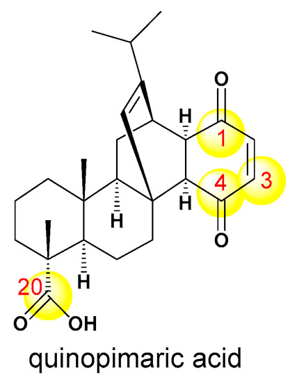
Figure 1.
Quinopimaric acid reaction centers for the SAR studies of the current research.
Modifications of these sites involved the synthesis of arylidene and heterocyclic derivatives and quinopimaric acid cyanoetoxy- and propargylated analogs. Figure 2 shows the structures of quinopimaric acid 1 and its analogs 2–30, modified at position C-1, C-3, C-4, and C-20.
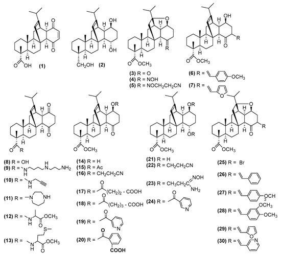
Figure 2.
Structures of quinopimaric acid 1 and its analogs 2–30, modified at position C-1, C-3, C-4, and C-20.
Figure 3 shows the structures of quinopimaric acid heterocyclic derivatives obtained as a result of interaction with hydrazine hydrate 31, thiourea 32, and using the Fischer reaction (indoles 33, 34), Nenitzescu reaction (indoles 35–38) and Beckmann rearrangement (lactam 39).
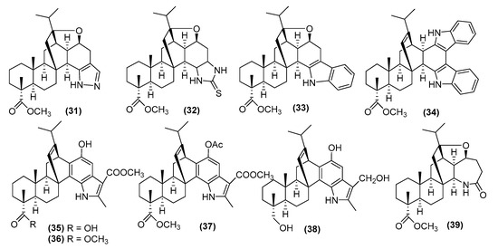
Figure 3.
Structures of quinopimaric acid heterocyclic derivatives 31–39.
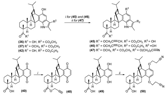
We planned to use the Nenitzescu reaction [37] for the synthesis of new diterpene indoles. In this reaction, 1a,4a-dehydroquinopimaric acid 40, easily formed in two steps from quinopimaric acid 1 [38], was used as the quinone component, as well as ethyl 3-aminocrotonate or 3-aminocrotononitrile being were used as the new enamine components. Under the conditions of the Nenitzescu indole synthesis by the reaction of 1a,4a-dehydroquinopimaric acid 40 with the corresponding enamine in glacial AcOH, at room temperature, diterpene indoles 42, 44 were synthesized in 76 and 69% yields, respectively. Methyl ester of diterpene indole 43 was obtained in quantitative yield by treating compound 42 with methyl iodide during reflux in acetone for 2 h in the presence of potash (Scheme 1), or direct synthesis from 1a,4a-dehydroquinopimaric acid methyl ester 41, similar to the preparation of compounds 42 and 44.
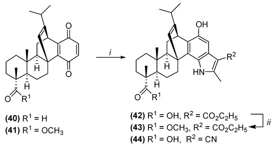
Scheme 1.
Synthesis of new diterpene indoles 42–44 by Nenitzescu reaction. Reagents and conditions: (i) ethyl 3-aminocrotonate for 42, 43 or 3-aminocrotononitrile for 44, AcOH, rt, 20 h; (ii) CH3I, K2CO3, acetone, reflux, 2 h.
Propargyl derivatives 45, 46, 48 were obtained in 79–83% yields by the reaction of diterpene indoles 36, 43 and quinone 40 with propargyl bromide during reflux in dimethylformamide in the presence of K2CO3. Cyanoethyloxy derivatives 47, 50 were prepared by adding acrylonitrile in 1,4-dioxane. at room temperature, to the diterpene indole 37 or aromatic derivative 49 in the occurrence of phase transfer catalyst triethylbenzylammonium chloride in combination with an alkali (30% KOH) (Scheme 2).
Scheme 2.
Synthesis of propargyl 45, 46, 48 and cyanoethyloxy 47, 50 quinopimaric acid derivatives. Reagents and conditions: (i) propargyl bromide, DMF, K2CO3, reflux, 2 h; (ii) acrylonitrile, 1,4-dioxane, KOH, BTAC, rt, 2 h.
The structures of the synthesized compounds were confirmed using mass spectrometry, and one- and two-dimensional (COSY, NOESY, 1H–13C HSQC, 1H–13C HMBC) NMR spectroscopy. Thus, the signal of the C-2 carbon atom of the aromatic ring in the 13C NMR spectra of compound 42–44 appeared at δ 99.7-103.3 ppm, and correlated with the signal of the H-2 proton at δ 6.83–7.28 ppm in the 1H–13C HSQC spectra. The 1H NMR spectra showed characteristic signals of methyl group protons at δ 2.51–2.71 (3′-CH3), as well as broadened signals of the hydroxyl group and NH group at δ 9.12–9.35 and 12.13 ppm, respectively. The 1H NMR spectra of compound 43 contained an additional signal of the protons of the methyl ester group at δ 3.76 ppm, which, in the 1H–13C HSQC spectrum, correlated with the signal of the C-21 atom at δ 15.5 ppm. In the 13C NMR spectra of compound 44, a carbon signal of the CN-group was observed at δ 117.8 ppm. The 1H NMR spectra of propargyl derivatives 45, 46, 48 contained the methylene group proton signal in the region δ 4.67–4.82 ppm, while in the 13C NMR the triple bond carbon signals appeared at δ 74.4–74.9 and 77.9–79.3 ppm, respectively. The signals of the cyanoethyl methylene groups in the 1H NMR spectra of compounds 47, 50 were observed in the region δ 2.80–2.91 and 4.05–4.30 ppm, and the characteristic carbon signal of the nitrile group in the 13C NMR spectra was observed at δ 117.4–117.6 ppm (Figures S1–S18, Supplementary Materials).
2.2. Inhibition of Yeast α-Glucosidase
All the synthesized compounds 1–50 were tested for their inhibitory potential against yeast α-GLy. Acarbose served as a control drug in this experiment. The IC50 values of compounds are provided in Table 1.

Table 1.
α-Glucosidase inhibitory potential of the synthesized compounds 1–50.
Quinopimaric acid derivatives 2–5, 10–25, 27, 31, 34, 41, 43, 46, 49 showed moderate to poor α-GLy inhibition. Quinopimaric acid 1, 2,3-dihydroquinopimaric acid 8 and its amide and heterocyclic derivatives 9, 30, 33, 39, 44, with an IC50 value of 35.24 ± 0.71 Μm—65.98 ± 0.03 μM, emerged as good inhibitors of α-GLy. Arylidene 1β-hydroxy and 1β,13-epoxy methyl dihydroquinopimarate derivatives 6, 7, 26–29, thiadiazole 32, 1a,4a-dehydroquinopimaric acid 40 and its indole, nitrile and propargyl hybrids 35-38, 42, 45, 48, and 50 showed excellent inhibitory activities. The most active compounds, 38, 45, 48, and 50, displayed IC50 values of 0.15 ± 0.008 μM to 0.68 ± 0.045 μM, being 1206 to 266 more potent than acarbose (IC50 of 181.02 ± 3.1 μM).
As shown in Table 1, quinopimaric acid 1 had an activity against α-GLy three times higher than that of acarbose. Its simplest modifications, namely, the reduction of the C2-C3 bond (compound 8), led to an increase in activity, and the introduction of a double bond into the C1a-C4a position (compound 40) further enhanced this. Modifications at positions C-1, C-4, C-20 of dihydroquinopimaric acid (compounds 2–5, 9–14, 15–24) were unsuccessful and led to a complete loss of activity. However, the introduction of arylidene substituents into the C-3 position of 1β,13α-epoxy methyldihydroquinopimarate 3 (compounds 26, 28–30) resulted mainly in active compounds, and the most successful, for this series of compounds, was the presence of a furfural fragment in the molecule. Heterocyclization of dihydroquinopimaric acid (compounds 31–34, 39) led to compounds with high activity against α-GLy only in the case of thiadiazole 32 and indole 34.
The use of acid 40 as a starting compound for heterocyclization provided more active compounds. Indole and its derivatives 35–38 showed excellent activity, especially alcohol 38. Inspired by these results, we carried out the synthesis of new indoles using other enamines. The obtained two new indoles 42 and 44 also had very good activity, and it was better for the indole with an ethyl substituent. Modification of the C-20 position by introducing a triple bond into the molecule (compounds 45, 46), and the C-1 position with a cyanoethyl fragment (compound 47) in the case of indole 36, further enhanced this activity.
Thus, from the studied series of 50 compounds synthesis of indoles based on 1a,4a-dehydroquinopimaric acid 40 proved to be the most successful approach to obtain highly active α-GLy inhibitors. Modification, according to the Nenitzescu reaction, and reduction of carboxyl and methoxycarboxyl groups, with the formation of trihydroxy derivative 38, propargylation of C-20 positions in acid 40 and indole 36, as well as cyanoethylation of the aromatic derivative 49, realized compounds with IC50 values < 1 µM.
For compounds 38, 45, 48 and 50, which showed the highest activity against α-GLy, studies of their anti-antioxidant, antimicrobial and cytotoxic activity were carried out (Tables S1–S3, see Supplementary Materials).
2.3. The Mechanism of α-Glucosidase Inhibition by Compound 45
The mechanism of action for the most active diterpene indole with an alkyne substituent 45 was determined in a kinetic experiment using different 4-nitrophenyl β-D-glucopyranoside (pNPG) substrate concentrations. Nonlinear regression of kinetic curves, using the Michaelis–Menten equation (Figure 4), revealed that higher inhibitor concentrations increased Km, while Vmax remained constant, which rendered compound 45 as a competitive inhibitor. The inhibition constant Ki was estimated as 50.45 µM.
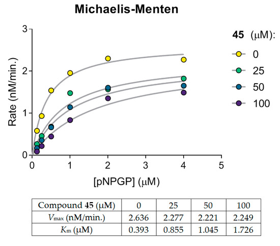
Figure 4.
Michaelis—Menten kinetics for compound 45 indicates a competitive inhibition. The experiment was performed in two independent series.
The literature data regarding the mechanism of α-glucosidase inhibition for diterpenes is scarce. The majority of reported compounds are non-competitive inhibitors, e.g., ent-atisane-3-oxo-16β,17-acetonide [39], (E)-labda-8(17),12-diene-15,16-dial [40], ent-kaurane derivative of chepraecoxin A [41], and bis-labdanic diterpene were reported as mixed-type inhibitors [42]. Notably, these inhibitors share an alicyclic core. In contrast, diterpene carnosol, comprising aromatic catechol moiety, is a competitive inhibitor [43]. Aromatic rings of carnosol and compound 45 favor π-stacking interaction with phenylalanine, tyrosine, or tryptophan side chains [44], which might result in distinct binding patterns and inhibition kinetics.
2.4. Docking Studies for Compound 45
We performed a molecular modeling study to gain insight into the structural basis of interactions between the lead compound 45 and α-GLy enzyme. Since “structure cannot be predicted from kinetics” [45], we avoided preconceived competitive mechanism assumptions and subjected the whole protein surface to a docking procedure (Figure 5).
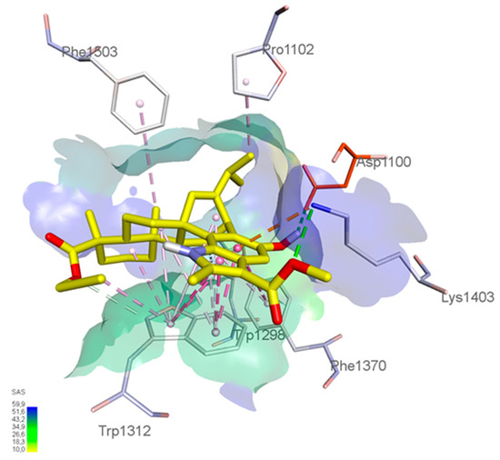
Figure 5.
Proposed binding mode of compound 45 to yeast α-glucosidase. The inhibitor is shown in yellow carbons, catalytic Asp1100 is shown in orange carbons. Surface visualized solvent-accessible area. Dashed lines indicate key interactions with enzyme amino acids.
Nevertheless, docking proposed that diterpene derivative 45 shared a favorable binding site with acarbose. Moreover, the indole hydroxyl group appeared to form a conventional H-bond with carboxyl of the catalytic Asp1100 residue. The indole core itself contributed to the binding the most. It was anchored by strong π-π parallel stacking with a Trp1312 side chain and T-shaped π-stacking with a Phe1370 side chain. The amino group of Lys1403 formed an H-bond with the ester substituent and charged π-cation interaction with the indole aromatic system. The dodecahydrophenanthrene part of the molecule was also stabilized by Van der Waals forces with multiple lipophilic residues (Pro1102, Trp1298, Trp1312, Phe1503). Both ester moieties of compound 45 pointed towards the solvent-accessible area of the pocket, providing an opportunity for the introduction of polar fragments. This modification might improve water solubility without hampering enzyme binding. To sum up, molecular modeling confirmed the competitive mechanism of action revealed in the kinetic experiment and provided guidance for future structural optimization.
2.5. ADMET Profiling of Compound 45
We assessed drug-like, pharmacokinetic and toxicological properties of the lead compound 45 using a consensus of predictive services that took into account different computational strategies (Table 2).
Table 2.
ADMET properties predicted for compound 45.
Compound 45 fulfilled Lipinski’s rule of 5 (with the exception of molecular weight <500) and Pfizer’s rules. At the same time, GSK and “Golden triangle” rules were violated. Mean water solubility was acceptable. Importantly, there was a good consensus on low intestinal absorption and oral bioavailability, which could avoid systemic exposure to the substance. In the case of entering systemic circulation, indole 45 was anticipated to be bound to plasma proteins, likely due to high lipophilicity. Blood–brain barrier penetration was unlikely. Liver metabolism was to be mediated by cytochrome P450 3A4. Acute oral toxicity was predicted to be sufficiently low to achieve a wide therapeutic window. There were no alerts for toxicity to the liver and heart, nor mutagenicity and carcinogenicity. Hence, compound 45’s calculated ADMET profile was favorable for the proposed mechanism of action.
3. Materials and Methods
3.1. General
The spectra were recorded at the Center for the Collective Use “Chemistry” of the UIC UFRC RAS and RCCU “Agidel” of the UFRC RAS. 1H and 13C-NMR spectra were recorded on a “Bruker AM-500” (Bruker, Billerica, MA, USA, 500 and 125.5 MHz respectively, δ, ppm, Hz) in CDCl3, internal standard tetramethylsilane. Melting points were detected on a micro table “Rapido PHMK05“ (Nagema, Dresden, Germany). Optical rotations were measured on a polarimeter “Perkin-Elmer 241 MC” (Perkin Elmer, Waltham, MA, USA) in a tube length of 1 dm. Elemental analysis was performed on a Euro EA-3000 CHNS analyzer (Eurovector, Milan, Italy); the main standard was acetanilide. Thin-layer chromatography analyses were performed on Sorbfil plates (Sorbpolimer, Krasnodar, Russian Federation), using the solvent system chloroform–ethyl acetate, 40:1. Substances were detected by 10% H2SO4 with subsequent heating to 100–120 °C for 2–3 min. All the reagents and solvents were purchased from standard commercial vendors and were used without any further purification. For the synthesis of quinopimaric acid 1 [38] pine resin Pinus silvestris (containing about 25% levopimaric acid) was used. Compounds 2, 14, 15, 17–21, 24 [50], 3 [51], 4 [52], 5 [53], 6, 9, 11, 26–28, 30 [16], 7 [54], 8 [50], 10 [55], 12, 13 [56], 16 [15], 22, 23 [57], 25 [58], 29 [59], 31, 32, 34 [60], 33 [14], 35 [61], 36–39 [38], 40, 49 [62] were obtained according to the methods previously described.
3.2. Synthesis of Compounds 42 and 44
A threefold excess of ethyl 3-aminocrotonate (0.387 g, 3 mmol) or 3-aminocrotononitrile (0.246 g, 3 mmol) was added with stirring to a solution of compound 40 (0.408 g, 1 mmol) in glacial AcOH (20 mL). The reaction mixture was stirred at room temperature for 20 h, and then poured into H2O. The precipitate was filtered off, washed until neutral, and the residue was air-dried. The reaction product was chromatographed on a silica gel column, eluent CHCl3–MeOH, 40: 1.
1-Hydroxy-13-isopropyl-1′-(ethoxycarbonyl)-7,10a,2′-trimethyl-5,6,6b,7,8,9,10,10a, 10b,11,12,13-dodecahydro-12,4b-ethenophenanthro-[2,1-g]indole-7-carboxylic acid (42). Yield 0.395 g (76%), mp 131–133°C, [α]D19 + 12.2° (c 1.5, CHCl3). 1H NMR spectrum, δ, ppm (J, Hz): 0.79 (3H, s, 18-CH3); 0.86–0.99 (1H, m, 10-CH2); 1.18 (3H, d, J = 7.0, 17-CH3); 1.22 (3H, d, J = 7.0, 16-CH3); 1.29 (3H, s, 19-CH3); 1.22–1.69 (10H, m, 6,8,9,10,11-CH2, 10b-CH); 1.77–1.80 (1H, m, 6b-CH); 1.98–2.18 (1H, m, 15-CH); 2.45–2.58 (1H, m, 5-CH2); 2.67 (3H, s, 3′-CH3); 2.82–2.91 (1H, m, 5-CH2); 3.45 (3H, s, 6′-CH3); 4.27 (1H, s, 12-CH); 4.31-4.35 (2H, m, 5′-CH2); 5.68 (1H, s, 14-CH); 7.13 (1H, s, 2-CH); 9.65 (2H, br. s, OH); 9.80 (1H, br. s, NH). 13C NMR spectrum, δ, ppm: 14.9 (C-3′); 16.4 (C-19); 16.7 (C-9), 17.2 (C-18); 20.5 (C-17); 20.8 (C-16); 22.0 (C-6); 28.3 (C-11); 32.0 (C-15); 33.5 (C-5); 36.1 (C-12); 36.8 (C-8); 38.2 (C-4b); 38.9 (C-10); 46.5 (C-7); 46.6 (C-10a); 49.8 (C-6b); 50.3 (C-6′); 54.9 (C-10b); 60.2 (C-5′), 102.9 (C-2); 108.0 (C-1′); 124.5 (C-3); 127.5 (C-13); 131.1 (C-1a); 133.6 (C-4a); 143.3 (C-4); 146.8 (C-1); 153.2 (C-14); 162.8 (C-2′); 165.3 (C-4′); 183.1 (C-20). Mass spectrum, m/z (Irel, %): 520 [M+H]+ (100). Found, %: C 73.95; H 7.92; N 2.68. C32H41NO5. Calculated, %: C 73.96; H 7.95; N 2.70.
1′-Cyano-1-hydroxy-13-isopropyl-7,10a,2′-trimethyl-5,6,6b,7,8,9,10,10a,10b,11,12,13-dodecahydro-12,4b-ethenophenanthro [2,1-g]indole-7-carboxylic acid (44). Yield 0.32 g (69%), mp 127–129°C, [α]D19 +25.5° (c 1.0, CHCl3). 1H NMR spectrum, δ, ppm (J, Hz): 0.78 (3H, s, 18-CH3); 0.82–0.95 (1H, m, 10-CH2); 1.18 (3H, d, J = 7.0, 17-CH3); 1.20 (3H, d, J = 7.0, 16-CH3); 1.25 (3H, s, 19-CH3); 1.22–1.69 (10H, m, 6,8,9,10,11-CH2, 10b-CH); 1.77–1.80 (1H, m, 6b-CH); 1.98–2.18 (1H, m, 15-CH); 2.45–2.58 (1H, m, 5-CH2); 2.51 (3H, s, 3′-CH3); 2.55–2.85 (1H, m, 5-CH2); 4.31 (1H, s, 12-CH); 5.65 (1H, s, 14-CH); 6.55-6.83 (1H, m, 2-CH); 9.35 (3H, br. s, OH, NH). 13C NMR spectrum, δ, ppm: 12.8 (C-3′); 16.4 (C-19); 16.7 (C-9), 17.1 (C-18); 20.1 (C-17); 20.3 (C-16); 22.0 (C-6); 28.1 (C-11); 32.0 (C-15); 34.6 (C-5); 36.8 (C-12); 37.6 (C-8); 38.2 (C-4b); 38.8 (C-10); 46.3 (C-7); 46.8 (C-10a); 49.9 (C-6b); 55.2 (C-10b); 83.3 (C-1′); 99.7 (C-2); 117.8 (C-4′); 124.4 (C-3); 126.8 (C-1a); 127.1 (C-14); 130.9 (C-4); 134.4 (C-4a); 144.3 (C-1); 146.4 (C-13); 153.7 (C-2′); 182.5 (C-20). Mass spectrum, m/z (Irel, %): 473 [M+H]+ (100). Found, %: C 76.22; H 7.65; N 5.90. C30H36N2O3. Calculated, %: C 76.24; H 7.68; N 5.93.
3.3. Synthesis of Methyl 1-Hydroxy-13-Isopropyl-1′-(Ethoxycarbonyl)-7,10a,2′-Trimethyl-5,6,6b,7,8,9,10,10a,10b,11,12,13-Dodecahydro-12,4b-Ethenophenanthro-[2,1-g]indole-7-Carboxylate (43)
Procedure A. Methyl iodide (2 mL) and potassium carbonate (0.21 g) were added to a solution of 40 (0.408 g, 1 mmol) in acetone (15 mL), and the mixture was heated to reflux for 2 h. The reaction mixture was filtered. The filtrate was evaporated under reduced pressure, and the residue was purified on a silica gel column, eluent hexane–ethyl acetate, 5:1.
Procedure B. A threefold excess of 3-aminocrotononitrile (0.246 g, 3 mmol) was added with stirring to a solution of compound 41 (0.422 g, 1 mmol) in glacial AcOH (20 mL). The reaction mixture was stirred at room temperature for 20 h, and then poured into H2O. The precipitate was filtered off, washed until neutral, and the residue was air-dried. The reaction product was chromatographed on a silica gel column, eluent CHCl3–MeOH, 40: 1.
Yield 0.45 g (85%), mp 120–122°C, [α]D19 +12.9° (c 0.75, CHCl3). 1H NMR spectrum, δ, ppm (J, Hz): 0.79 (3H, s, 18-CH3); 0.86–0.99 (1H, m, 10-CH2); 1.18 (3H, d, J = 7.0, 17-CH3); 1.22 (3H, d, J = 7.0, 16-CH3); 1.29 (3H, s, 19-CH3); 1.22–1.69 (10H, m, 6,8,9,10,11-CH2, 10b-CH); 1.40–1.43 (3H, s, 6′-CH3); 1.77–1.80 (1H, m, 6b-CH); 1.98–2.18 (1H, m, 15-CH); 2.45–2.58 (1H, m, 5-CH2); 2.73 (3H, s, 3′-CH3); 2.85–3.15 (1H, m, 5-CH2); 3.76 (3H, s, 21-CH3), 4.27 (1H, s, 12-CH); 4.31–4.39 (2H, m, 5′-CH2); 5.71 (1H, s, 14-CH); 7.28 (1H, s, 2-CH); 5.95 (1H, br. s, NH); 12.13 (1H, br. s, OH). 13C NMR spectrum, δ, ppm: 14.9 (C-3′); 15.5 (C-21), 16.4 (C-19); 16.8 (C-9), 17.2 (C-18); 20.2 (C-17); 20.5 (C-16); 22.1 (C-6); 28.3 (C-11); 32.2 (C-15); 33.2 (C-5); 36.3 (C-12); 36.9 (C-8); 38.4 (C-4b); 38.9 (C-10); 46.6 (C-7); 47.4 (C-10a); 49.9 (C-6b); 52.0 (C-6′); 54.8 (C-10b); 60.3 (C-5′), 103.3 (C-2); 108.2 (C-1′); 124.7 (C-3); 127.6 (C-13); 130.9 (C-1a); 133.6 (C-4a); 143.6 (C-4); 146.4 (C-1); 153.1 (C-14); 162.9 (C-2′); 165.2 (C-4′); 179.7 (C-20). Mass spectrum, m/z (Irel, %): 534 [M] + (100). Found, %: C 74.25; H 8.10; N 2.60. C33H43NO5. Calculated, %: C 74.27; H 8.12; N 2.62.
3.4. Synthesis of Compounds 45, 46 and 49
To a solution containing 1 mmol of compound 36, 37 or 40 in 5 mL of dimethylformamide, 1.2 mmol (0.09 mL) of propargyl bromide and 2.2 mmol (0.30 g) of K2CO3 were added. The reaction mixture was stirred for 18 h and evaporated at room temperature. The residue was diluted with CHCl3, washed with 5% HCl and water, dried over CaCl2, and evaporated in a vacuum. The residue was purified by column chromatography, eluent hexane: ethyl acetate, 5:1
Methyl 7-propynyl 1-hydroxy-13-isopropyl-7,10a,2′-trimethyl- 5,6,6b,7,8,9,10,10a,10b,11,12,13-dodecahydro-12,4b-ethenophenanthro [2,1-g]indole-7,1′-dicarboxylate (45). Yield 0.39 g (72%), mp 110–112°C, [α]D20 + 33.4° (c 0.10, CHCl3). 1H NMR spectrum, δ, ppm (J, Hz): 0.81 (3H, s, 18-CH3); 0.86–0.96 (1H, m, 10-CH2); 1.02 (3H, d, J = 7.0, 17-CH3); 1.05 (3H, d, J = 7.0, 16-CH3); 1.21 (3H, s, 19-CH3); 1.31–1.69 (10H, m, 6,8,9,10,11-CH2, 10b-CH); 1.77–1.80 (1H, m, 6b-CH); 1.98–2.18 (1H, m, 15-CH); 2.41–2.48 (1H, m, 5-CH2); 2.50 (1H, br s., 8′-CH); 2.71 (3H, s, 3′-CH3); 2.99–3.00 (1H, m, 5-CH2); 3.91 (3H, s, 5′-CH3), 4.28 (1H, s, 12-CH); 4.69–4.82 (2H, m, 6′-CH2); 5.71 (1H, s, 14-CH); 7.28 (1H, s, 2-CH); 6.00 (1H, br. s, OH); 12.01 (1H, br. s, NH). 13C NMR spectrum, δ, ppm: 14.9 (C-3′); 16.8 (C-19); 17.1 (C-9), 18.2 (C-18); 20.2 (C-17); 20.5 (C-16); 22.0 (C-6); 28.3 (C-11); 32.2 (C-15); 33.2 (C-5); 36.3 (C-12); 36.9 (C-8); 38.3 (C-4b); 38.8 (C-10); 46.6 (C-7); 47.4 (C-10a); 49.8 (C-6b); 51.4 (C-6′); 52.0 (C-5′); 54.8 (C-10b); 74.4 (C-8′); 78.1 (C-7′); 103.3 (C-2); 108.0 (C-1′); 124.5 (C-3); 127.5 (C-13); 130.9 (C-1a); 133.6 (C-4a); 143.5 (C-4); 146.5 (C-1); 153.2 (C-14); 162.9 (C-2′); 165.6 (C-4′); 178.2 (C-20). Mass spectrum, m/z (Irel, %): 543 [M]+ (100). Found, %: C 75.15; H 7.61; N 2.60. C34H41NO5. Calculated, %: C 75.11; H 7.60; N 2.58.
Ethyl 7-propynyl 1-hydroxy-13-isopropyl-7,10a,2′-trimethyl- 5,6,6b,7,8,9,10,10a,10b, 11,12,13-dodecahydro-12,4b-ethenophenanthro [2,1-g]indole-7,1′-dicarboxylate (46). Yield 0.42 g (75%), mp 98–100°C, [α]D20 +2.9° (c 0.15, CHCl3). 1H NMR spectrum, δ, ppm (J, Hz): 0.72 (3H, s, 18-CH3); 0.80–0.96 (1H, m, 10-CH2); 1.02 (3H, d, J = 7.0, 17-CH3); 1.05 (3H, d, J = 7.0, 16-CH3); 1.21 (3H, s, 19-CH3); 1.31–1.69 (10H, m, 6,8,9,10,11-CH2,10b-CH); 1.77–1.80 (1H, m, 6b-CH); 1.98–2.18 (1H, m, 15-CH); 2.41–2.48 (1H, m, 9′-CH); 2.65–2.70 (1H, m, 5-CH2); 2.73 (3H, s, 3′-CH3); 2.95–3.05 (1H, m, 5-CH2); 3.73 (3H, s, 6′-CH3), 3.95 (1H, s, 12-CH); 4.35-4.42 (2H, m, 5′-CH2); 4.75 (2H, br.s., 6′-CH2); 5.70 (1H, s, 14-CH); 7.49 (1H, s, 2-CH); 9.19 (1H, br. s, OH); 12.01 (1H, br. s, NH). 13C NMR spectrum, δ, ppm: 14.1 (C-3′); 14.2 (C-19); 14.5 (C-9), 15.9 (C-18); 16.5 (C-17); 16.9 (C-16); 20.2 (C-6); 21.4 (C-11); 22.7 (C-15); 28.3 (C-5); 29.7 (C-12); 32.2 (C-8); 33.2 (C-4b); 36.2 (C-10); 36.9 (C-7); 38.8 (C-10a); 49.9 (C-6b); 51.9 (C-7′); 54.9 (C-6′); 57.1 (C-10b); 60.0 (C-5′), 74.9 (C-9′); 79.3 (C-8′); 101.8 (C-2); 108.6 (C-1′); 124.5 (C-3); 127.6 (C-13); 132.8 (C-1a); 133.8 (C-4a); 144.0 (C-4); 148.6 (C-1); 153.2 (C-14); 162.8 (C-2′); 164.8 (C-4′); 179.5 (C-20). Mass spectrum, m/z (Irel, %): 558 [M]+ (100). Found, %: C 75.30; H 7.75; N 2.55. C35H43NO5. Calculated, %: C 75.37; H 7.77; N 2.51.
Propynyl 13-isopropyl-7,10a-dimethyl-1,4-di oxo-4,5,6,6a,7,8,9,10,10a,10b,11,12- dodecahydro-1H-4b,12-ethenochrysene-7-carboxylate (48). Yield 0.33 g (75%), mp 85-89°C, [α]D20 +26.9° (c 0.75, CHCl3). 1H NMR spectrum, δ, ppm (J, Hz): 0.69 (3H, s, 18-CH3); 0.86–0.96 (1H, m, 10-CH2); 1.02 (3H, d, J = 7.0, 17-CH3); 1.05 (3H, d, J = 7.0, 16-CH3); 1.21 (3H, s, 19-CH3); 1.31–1.69 (10H, m, 6,8,9,10,11-CH2, 10b-CH); 1.77–1.80 (1H, m, 6b-CH); 1.98–2.18 (1H, m, 15-CH); 2.41–2.48 (1H, m, 5-CH2); 2.50 (1H, br. s, 3′-CH); 2.85–2.89 (1H, m, 5-CH2); 4.13 (1H, s, 12-CH); 4.67 (2H, br. s, 1′-CH2); 5.63 (1H, s, 14-CH); 6.45-6.56 (2H, m, 2-CH, 3-CH). 13C NMR spectrum, δ, ppm: 16.4 (C-19); 16.8 (C-9), 17.1 (C-18); 20.2 (C-17); 20.6 (C-16); 21.7 (C-6); 27.1 (C-11); 31.5 (C-15); 31.9 (C-5); 36.2 (C-12); 36.4 (C-8); 38.6 (C-4b); 39.3 (C-10); 47.1 (C-7); 49.1 (C-10a); 49.4 (C-6b); 52.1 (C-1′); 54.8 (C-10b); 74.6 (C-3′); 77.9 (C-2′); 127.3 (C-13); 133.6 (C-2); 137.5 (C-3); 150.6 (C-4a); 151.1 (C-14); 152.8 (C-1a); 177.7 (C-20); 184.0 (C-1); 185.3 (C-2). Mass spectrum, m/z (Irel, %): 446 [M]+ (100). Found, %: C 78.05; H 7.65. C29H34O4. Calculated, %: C 78.00; H 7.67.
3.5. Synthesis of Compounds 47 and 50
A mixture of 1 mmol of the compounds 42 or 49, 20 mmol (1.3 mL) of acrylonitrile and 0.5 mL of 30% KOH per one hydroxyl groups, 0.5 mmol (0.11 g) of BTEAC, in 20 mL of dioxane was stirred for 2 h at room temperature. The mixture was poured into a mixture of ice with HCl, the precipitate was filtered off, washed with water until neutral pH, air dried, and extracted with methylene chloride (3 × 80 mL) with heating, the solution was filtered. The filtrate was evaporated under reduced pressure, and the residue was purified on a silica gel column, eluent hexane–ethyl acetate, 10:1.
Methyl 1-(8′-cyanoethoxy)-13-isopropyl-1′-(ethoxycarbonyl)-7,10a,2′-trimethyl -5,6,6b,7,8,9,10,10a,10b,11,12,13-dodecahydro-12,4b-ethenophenanthro [2,1-g]indole-7-carboxylate (47). Yield 0.39 g (68%), mp 127–129 °C, [α]D20 +77.9° (c 0.10, CHCl3). 1H NMR spectrum, δ, ppm (J, Hz): 0.79 (3H, s, 18-CH3); 0.86–0.99 (1H, m, 10-CH2); 1.04 (3H, d, J = 7.0, 17-CH3); 1.07 (3H, d, J = 7.0, 16-CH3); 1.24 (3H, s, 19-CH3); 1.27–1.69 (10H, m, 6,8,9,10,11-CH2, 10b-CH); 1.77–1.80 (1H, m, 6b-CH); 1.98–2.18 (1H, m, 15-CH); 2.45–2.58 (1H, m, 5-CH2); 2.76 (3H, s, 3′-CH3); 2.89–2.91 (2H, m, 7′-CH2); 2.95–3.15 (1H, m, 5-CH2); 3.75 (3H, s, 21-CH3), 3.93 (3H, s, 5′-CH3), 4.28-4.30 (2H, m, 6′-CH2); 4.37 (1H, s, 12-CH); 5.72 (1H, s, 14-CH); 7.28 (1H, s, 2-CH); 12.10 (1H, br. s, NH). 13C NMR spectrum, δ, ppm: 14.9 (C-3′); 15.5 (C-21), 16.9 (C-19); 16.8 (C-9), 17.2 (C-18); 20.2 (C-17); 20.5 (C-16); 22.1 (C-6); 28.3 (C-11); 32.2 (C-15); 33.2 (C-5); 36.3 (C-12); 36.9 (C-8); 38.4 (C-4b); 38.9 (C-10); 46.7 (C-7); 47.4 (C-10a); 49.9 (C-6b); 51.3 (C-7′); 52.0 (C-5′); 54.9 (C-10b); 63.7 (C-6′), 100.9 (C-2); 108.3 (C-1′); 117.4 (C-8′); 124.6 (C-3); 127.6 (C-13); 133.4 (C-1a); 134.1 (C-4a); 144.0 (C-4); 148.4 (C-1); 153.2 (C-14); 163.0 (C-2′); 165.1 (C-4′); 179.6 (C-20). Mass spectrum, m/z (Irel, %): 572 [M]+ (100). Found, %: C 73.45; H 7.75; N 4.91. C35H44N2O5. Calculated, %: C 73.40; H 7.74; N 4.89.
1,4-Bis(2′-cyanoethoxy)-13-isopropyl-7,10a-dimethyl-6,6a,7,8,9,10,10a,10b,11,12-decahydro-5H-4b,12-ethenochrysene-7-carboxylic acid (50). Yield 0.41 g (80%), mp 157–159 °C, [α]D20 + 63.6° (c 0.1, CHCl3). 1H NMR spectrum, δ, ppm (J, Hz): 0.79 (3H, s, 18-CH3); 0.86–0.99 (1H, m, 10-CH2); 1.08 (3H, d, J = 7.0, 17-CH3); 1.10 (3H, d, J = 7.0, 16-CH3); 1.29 (3H, s, 19-CH3); 1.22–1.69 (10H, m, 6,8,9,10,11-CH2, 10b-CH); 1.77–1.80 (1H, m, 6b-CH); 1.98–2.18 (1H, m, 15-CH); 2.45–2.58 (1H, m, 5-CH2); 2.80–2.82 (4H, m, 2′-CH2, 2″-CH2); 2.95–3.00 (1H, m, 5-CH2); 4.05-4.13 (4H, m, 1′-CH2, 1″-CH2); 4.24 (1H, s, 12-CH); 5.71 (1H, s, 14-CH); 6.38-6.47 (2H, m, 2-CH, 3-CH); 9.12 (1H, br. s, OH). 13C NMR spectrum, δ, ppm: 16.4 (C-19); 16.8 (C-9), 17.2 (C-18); 19.9 (C-2′, C-2″); 20.2 (C-17); 20.5 (C-16); 22.1 (C-6); 28.3 (C-11); 32.2 (C-15); 33.2 (C-5); 36.3 (C-12); 36.9 (C-8); 38.4 (C-4b); 38.9 (C-10); 46.6 (C-7); 47.4 (C-10a); 49.9 (C-6b); 54.8 (C-10b); 64.4 (C-1′, C-1″); 111.2 (C-1); 114.4 (C-2); 117.6 (C-3′, C-3″), 128.6 (C-14); 135.2 (C-4a); 138.4 (C-1a); 145.1 (C-4); 146.0 (C-1); 151.7 (C-13); 185.6 (C-20). Mass spectrum, m/z (Irel, %): 517 [M]+H (100). Found, %: C 74.35; H 7.81; N 5.40. C32H40N2O4. Calculated, %: C 74.39; H 7.80; N 5.42.
Data of the study α-Gly inhibition in vitro, kinetic, docking studies and ADMET profiling of compound 45. as well as studies of anti-antioxidant, antimicrobial and cytotoxic activities, can be found in the Supplementary Material (Section S1).
4. Conclusions
The screening of a series of 50 semisynthetic derivatives of levopimaric acid revealed that, in contrast to the majority of previously reported diterpene α-GLy inhibitors, a lead diterpene indole with an alkyne substituent 45 was identified as a competitive inhibitor. As a consequence, one might hope for better translatability to animal and clinical settings, since the active site of yeast α-GLy and intestinal mammalian maltase–glucoamylase are conserved, while allosteric sites are likely to be different. In addition, compound 45 is anticipated to have low intestinal absorption that benefits high concentration of the drug in the target area and helps to avoid systemic exposure. Additional experiments are warranted to confirm antihyperglycemic properties of compound 45 in vivo. In the event of the efficacy and safety being confirmed, novel glucosidase inhibitors open a promising venue to antidiabetic agents able not only to ameliorate postprandial hyperglycemia, but also reduce secretory load on pancreatic beta-cells.
Supplementary Materials
The following supporting information can be downloaded at: https://www.mdpi.com/article/10.3390/ijms232113535/s1. Ref [63,64,65,66,67,68,69,70,71] are cited in Supplementary Materials.
Author Contributions
E.T.—draft preparation; E.T. and I.S. prepared compounds for screening; O.K. brought the idea, managed the research and prepared the manuscript; H.T.T.N., A.S., E.S., D.B. and A.S. (Alina Shevchenko) conducted biological experiments; D.B. and A.S. (Alexander Spasov) prepared the manuscript. All authors have read and agreed to the published version of the manuscript.
Funding
This work was supported by the Federal program (Russian Federation) No. 1021062311392-9-1.4.1. Ha Thi Thu Nguyen thanks the Vietnam Academy of Science and Technology (VAST) for support in the screening of compounds against α-glucosidase (Project No. QTRU01.05/20-21).
Institutional Review Board Statement
Not applicable.
Informed Consent Statement
Not applicable.
Data Availability Statement
Not applicable.
Conflicts of Interest
The authors declare no conflict of interest.
References
- Tang, O.; Matsushita, K.; Coresh, J.; Sharrett, A.R.; McEvoy, J.W.; Windham, B.G.; Ballantyne, C.M.; Selvin, E. Mortality implications of prediabetes and diabetes in older adults. Diabetes Care 2020, 43, 382–388. [Google Scholar] [CrossRef]
- Kitabchi, A.E.; Umpierrez, G.E.; Miles, J.M.; Fisher, J.N. Hyperglycemic crises in adult patients with diabetes. Diabetes Care 2009, 32, 1335–1343. [Google Scholar] [CrossRef]
- Ceriello, A.; Nicolucci, A. Intensive glucose control and type 2 diabetes—15 years on. N. Engl. J. Med. 2019, 381, 1292–1293. [Google Scholar] [CrossRef]
- Zheng, Y.; Ley, S.H.; Hu, F.B. Global aetiology and epidemiology of type 2 diabetes mellitus and its complications. Nat. Rev. Endocrinol. 2018, 14, 88–98. [Google Scholar] [CrossRef]
- Lean, M.; McCombie, L.; McSorely, J. Trends in type 2 diabetes. BMJ 2019, 366, l5407. [Google Scholar] [CrossRef]
- Williams, J.; Loeffler, M. Global trends in type 2 diabetes, 2007–2017. JAMA 2019, 322, 1542. [Google Scholar] [CrossRef]
- Bischoff, H. Pharmacology of alpha-glucosidase inhibition. Eur. J. Clin. Investig. 1994, 24, 3–10. [Google Scholar]
- Toeller, M. α-Glucosidase inhibitors in diabetes: Efficacy in NIDDM subjects. Eur. J. Clin. Investig. 1994, 24, 31–35. [Google Scholar] [CrossRef]
- Soccio, R.E.; Chen, E.R.; Lazar, M.A. Thiazolidinediones and the promise of insulin sensitization in type 2 diabetes. Cell Metab. 2014, 20, 573–591. [Google Scholar] [CrossRef]
- Ullah, A.; Khan, A.; Khan, I. Diabetes mellitus and oxidative stress—A concise review. Pharm. J. 2016, 24, 547–553. [Google Scholar] [CrossRef]
- Lorenzati, B.; Zucco, C.; Miglietta, S.; Lamberti, F.; Bruno, G. Oral hypoglycemic drugs: Pathophysiological basis of their mechanism of action. Pharmaceuticals 2010, 3, 3005–3020. [Google Scholar] [CrossRef] [PubMed]
- Marín-Peñalver, J.J.; Martín-Timón, I.; Sevillano-Collantes, C.; del Cañizo-Gómez, F.J. Type 2 diabetes and cardiovascular disease: Have all risk factors the same strength. World J. Diabetes 2016, 7, 354–395. [Google Scholar] [CrossRef] [PubMed]
- Feliciano, A.S.; Gordaliza, M.; Salinero, M.A.; del Corral, J.M.M. Abietane Acids: Sources, Biological Activities, and Therapeutic Uses. Planta Med. 1993, 59, 485–490. [Google Scholar] [CrossRef] [PubMed]
- Tretyakova, E.V.; Smirnova, I.E.; Kazakova, O.B.; Tolstikov, G.A.; Yavorskaya, N.P.; Golubeva, I.S.; Pugacheva, R.B.; Apryshko, G.N.; Poroikov, V.V. Synthesis and anticancer activity of quinopimaric and maleopimaric acids’ derivatives. Bioorg. Med. Chem. 2014, 22, 6481–6489. [Google Scholar] [CrossRef] [PubMed]
- Tretyakova, E.V.; Smirnova, I.E.; Salimova, E.V.; Odinokov, V.N. Synthesis and antiviral activity of maleopimaric and quinopimaric acids’ derivatives. Bioorg. Med. Chem. 2015, 23, 6543–6550. [Google Scholar] [CrossRef]
- Smirnova, I.E.; Tret’yakova, E.V.; Baev, D.S.; Kazakova, O.B. Synthetic modifications of abietane diterpene acids to potent antimicrobial agents. Nat. Prod. Res. 2021, 1–9. [Google Scholar] [CrossRef]
- Kim, E.; Kang, Y.-G.; Kim, Y.-J.; Lee, T.R.; Yoo, B.C.; Jo, M.; Kim, J.H.; Kim, J.H.; Kim, D.; Cho, J.Y. Dehydroabietic acid suppresses inflammatory response via suppression of Src-, Syk-, and TAK1-mediated pathways. Int. J. Mol. Sci. 2019, 20, 1593. [Google Scholar] [CrossRef]
- Tret’yakova, E.V.; Salimova, E.V.; Parfenova, L.V. Synthesis, modification, and biological activity of propargylated methyl dihydroquinopimarates. Nat. Prod. Res. 2022, 36, 79–86. [Google Scholar] [CrossRef]
- Goncalves, M.D.; Bortoleti, B.T.S.; Tomiotto-Pellissier, F.; Miranda-Sapla, M.M.; Assolini, J.P.; Carloto, A.C.M.; Carvalho, P.G.C.; Tudisco, E.T.; Urbano, A.; Ambrosio, S.R.; et al. Dehydroabietic acid isolated from Pinus elliottii exerts in vitro antileishmanial action by pro-oxidant effect, inducing ROS production in promastigote and downregulating Nrf2/ferritin expression in amastigote forms of Leishmania amazonensis. Fitoterapia 2018, 128, 224–232. [Google Scholar] [CrossRef]
- González, M.A. Aromatic abietane diterpenoids: Their biological activity and synthesis. Nat. Prod. Rep. 2015, 32, 684–704. [Google Scholar] [CrossRef]
- Etsassala, N.G.E.R.; Cupido, C.N.; Iwuoha, I.E.; Hussein, A.A. Abietane Diterpenes as Potential Candidates for the Management of Type 2 Diabetes. Curr. Pharm. Des. 2020, 26, 2885–2891. [Google Scholar] [CrossRef] [PubMed]
- Nachar, A.; Saleem, A.; Arnason, J.T.; Haddad, P.S. Regulation of liver cell glucose homeostasis by dehydroabietic acid, abietic acid and squalene isolated from balsam fir (Abies balsamea (L.) Mill.) a plant of the Eastern James Bay Cree traditional pharmacopeia. Phytochemistry 2015, 117, 373–379. [Google Scholar] [CrossRef] [PubMed]
- Song, H.M.; Li, X.; Liu, Y.Y.; Lu, W.-P.; Cui, Z.-H.; Zhou, L.; Yao, D.; Zhang, H.-M. Carnosic acid protects mice from high-fat diet-induced NAFLD by regulating MARCKS. Int. J. Mol. Med. 2018, 42, 193–207. [Google Scholar] [CrossRef] [PubMed]
- Lipina, C.; Hundal, H.S. Carnosic acid stimulates glucose uptake in skeletal muscle cells via a PME-1/PP2A/PKB signalling axis. Cell. Signal. 2014, 26, 2343–2349. [Google Scholar] [CrossRef] [PubMed]
- Christensen, K.B.; Jørgensen, M.; Kotowska, D.; Petersen, R.K.; Kristiansen, K.; Christensen, L.P. Activation of the nuclear receptor PPARγ by metabolites isolated from sage (Salvia officinalis L.). J. Ethnopharmacol. 2010, 132, 127–133. [Google Scholar] [CrossRef] [PubMed]
- Xie, Z.; Gao, G.; Wang, H.; Lia, E.; Yuan, Y.; Xu, J.; Zhang, Z.; Wang, P.; Fu, Y.; Zeng, H.; et al. Dehydroabietic acid alleviates high fat diet-induced insulin resistance and hepatic steatosis through dual activation of PPAR-γ and PPAR-α. Biomed. Pharmacother. 2020, 127, 110155. [Google Scholar] [CrossRef]
- Vlavcheski, F.; Baron, D.; Vlachogiannis, I.A.; MacPherson, R.E.K.; Tsiani, E. Carnosol increases skeletal muscle cell glucose uptake via AMPK-Dependent GLUT4 glucose transporter translocation. Int. J. Mol. Sci. 2018, 19, 1321. [Google Scholar] [CrossRef]
- Samarghandian, S.; Borji, A.; Farkhondeh, T. Evaluation of antidiabetic activity of carnosol (phenolic diterpene in rosemary) in Streptozotocin-induced diabetic rats. Cardiovasc. Hematol. Disord. Drug Targets 2017, 17, 11–17. [Google Scholar] [CrossRef]
- Cui, L.; Kim, M.O.; Seo, J.H.; Kim, I.S.; Kim, N.Y.; Lee, S.H.; Park, J.; Kim, J.; Lee, H.S. Abietane diterpenoids of Rosmarinus officinalis and their diacylglycerol acyltransferase-inhibitory activity. Food Chem. 2012, 132, 1775–1780. [Google Scholar] [CrossRef]
- Yun, Y.S.; Noda, S.; Shigemori, G.; Kuriyama, R.; Takahashi, S.; Umemura, M.; Takahashi, Y.; Inoue, H. Phenolic diterpenes from rosemary suppress cAMP responsiveness of gluconeogenic gene promoters. Phytother. Res. 2013, 27, 906–910. [Google Scholar] [CrossRef]
- Kubínová, R.; Pořízková, R.; Navrátilová, A.; Farsa, O.; Hanáková, Z.; Bačinská, A.; Cížek, A.; Valentová, M. Antimicrobial and enzyme inhibitory activities of the constituents of Plectranthus madagascariensis (Pers.) Benth. J. Enzym. Inhib. Med. Chem. 2014, 29, 749–752. [Google Scholar] [CrossRef] [PubMed]
- Kim, D.H.; Paudel, P.; Yu, T.; Ngo, T.M.; Kim, J.A.; Jung, H.A.; Yokozawa, T.; Choi, J.S. Characterization of the inhibitory activity of natural tanshinones from Salvia miltiorrhiza roots on protein tyrosine phosphatase 1B. Chem. Biol. Interact. 2017, 278, 65–73. [Google Scholar] [CrossRef] [PubMed]
- Jung, S.H.; Seol, H.J.; Jeon, S.J.; Son, K.H.; Lee, J.R. Insulin-sensitizing activities of tanshinones, diterpene compounds of the root of Salvia miltiorrhiza Bunge. Phytomedicine 2009, 16, 327–335. [Google Scholar] [CrossRef] [PubMed]
- Kang, M.S.; Hirai, S.; Goto, T.; Kuroyanagi, K.; Kim, Y.-I.; Ohyama, K.; Uemura, T.; Lee, J.-Y.; Sakamoto, T.; Ezaki, Y.; et al. Dehydroabietic acid, a diterpene, improves diabetes and hyperlipidemia in obese diabetic KK-Ay mice. Biofactors 2009, 35, 442–448. [Google Scholar] [CrossRef] [PubMed]
- Ou, J.; Huang, J.; Zhao, D.; Du, B.; Wang, M. Protective effect of rosmarinic acid and carnosic acid against streptozotocin-induced oxidation, glycation, inflammation and microbiota imbalance in diabetic rats. Food Funct. 2018, 9, 851–860. [Google Scholar] [CrossRef] [PubMed]
- Wei, Y.; Gao, J.; Qin, L.; Xu, Y.; Wang, D.; Shi, H.; Xu, T.; Liu, T. Tanshinone I alleviates insulin resistance in type 2 diabetes mellitus rats through IRS-1 pathway. Biomed. Pharmacother. 2017, 93, 352–358. [Google Scholar] [CrossRef]
- Allen, G.R., Jr. Organic Reactions; Wiley: New York, NY, USA, 1973; Volume 20, p. 338. [Google Scholar]
- Tretyakova, E.V.; Yarmukhametova, L.R.; Salimova, E.V.; Kukovinets, O.S.; Parfenova, L.V. The Nenitzescu reaction in the synthesis of new abietane diterpene indoles. Chem. Heterocycl. Compd. 2020, 56, 1366–1369. [Google Scholar] [CrossRef]
- Tran, C.-L.; Dao, T.-B.-N.; Tran, T.-N.; Mai, D.-T.; Tran, T.-M.-D.; Tran, N.-M.-A.; Dang, V.-S.; Vo, T.-X.; Duong, T.-H.; Sichaem, J. Alpha-Glucosidase Inhibitory Diterpenes from Euphorbia antiquorum Growing in Vietnam. Molecules 2021, 26, 2257. [Google Scholar] [CrossRef]
- Ghosh, S.; Rangan, L. Molecular Docking and Inhibition Kinetics of α-glucosidase Activity by Labdane Diterpenes Isolated from Tora Seeds (Alpinia nigra B.L. Burtt.). Appl. Biochem. Biotechnol. 2015, 175, 1477–1489. [Google Scholar] [CrossRef]
- Yang, X.-T.; Geng, C.-A.; Li, T.-Z.; Deng, Z.-T.; Chen, J.-J. Synthesis and biological evaluation of chepraecoxin A derivatives as α-glucosidase inhibitors. Bioorg. Med. Chem. Lett. 2020, 30, 127020. [Google Scholar] [CrossRef]
- Loo, K.Y.; Leong, K.H.; Sivasothy, Y.; Ibrahim, H.; Awang, K. Molecular Insight and Mode of Inhibition of α-Glucosidase and α-Amylase by Pahangensin A from Alpinia pahangensis Ridl. Chem. Biodivers. 2019, 16, e1900032. [Google Scholar] [CrossRef] [PubMed]
- Ma, Y.-Y.; Zhao, D.-G.; Zhang, R.; He, X.; Li, B.Q.; Zhang, X.-Z.; Wang, Z.; Zhang, K. Identification of bioactive compounds that contribute to the α-glucosidase inhibitory activity of rosemary. Food Funct. 2020, 11, 1692–1701. [Google Scholar] [CrossRef] [PubMed]
- Bissantz, C.; Kuhn, B.; Stahl, M. A medicinal chemist’s guide to molecular interactions. J. Med. Chem. 2010, 53, 5061–5084. [Google Scholar] [CrossRef] [PubMed]
- Stein, R.L. Kinetics of Enzyme Action: Essential Principles for Drug Hunters; John Wiley & Sons: Hoboken, NJ, USA, 2011. [Google Scholar]
- Dong, J.; Wang, N.-N.; Yao, Z.-J.; Zhang, L.; Cheng, Y.; Ouyang, D.; Lu, A.-P.; Cao, D.-S. ADMETlab: A platform for systematic ADMET evaluation based on a comprehensively collected ADMET database. J. Cheminform. 2018, 10, 29. [Google Scholar] [CrossRef] [PubMed]
- Xiong, G.; Wu, Z.; Yi, J.; Fu, L.; Yang, Z.; Hsieh, C.; Yin, M.; Zeng, X.; Wu, C.; Lu, A.; et al. ADMETlab 2.0: An integrated online platform for accurate and comprehensive predictions of ADMET properties. Nucleic Acids Res. 2021, 49, W5–W14. [Google Scholar] [CrossRef] [PubMed]
- Daina, A.; Michielin, O.; Zoete, V. SwissADME: A free web tool to evaluate pharmacokinetics, drug-likeness and medicinal chemistry friendliness of small molecules. Sci. Rep. 2017, 7, 42717. [Google Scholar] [CrossRef]
- Banerjee, P.; Eckert, A.O.; Schrey, A.K.; Preissner, R. ProTox-II: A webserver for the prediction of toxicity of chemicals. Nucleic Acids Res. 2018, 46, W257–W263. [Google Scholar] [CrossRef]
- Herz, W.; Blackstone, R.C.; Nair, M.G. Resin acids. XI Configuration and transformations of the levopimaric acid-p-benzoquinone adduct. J. Org. Chem. 1967, 32, 2992–2998. [Google Scholar] [CrossRef]
- Smirnova, I.E.; Tret’yakova, E.V.; Flekhter, O.B.; Spirikhin, L.V.; Galin, F.Z.; Tolstikov, G.A.; Starikova, Z.A.; Korlyukov, A.A. Synthesis, structure, and acylation of dihydroquinopimaric acid hydroxyl derivatives. Russ. J. Org. Chem. 2008, 44, 1598–1605. [Google Scholar] [CrossRef]
- Smirnova, I.E.; Tret’yakova, E.V.; Kazakova, O.B.; Starikova, Z.A.; Fedyanin, I.V. Molecular and crystal structure of a new compound methyl-18R-13-isopropyl-10a,7-dimethyl-4-oxo-1-oxahexacyclo 12.4.0.05a,4a.013,120.010a,6a]heneicosane-7-Carboxylate. J. Struct. Chem. 2009, 50, 378–380. [Google Scholar] [CrossRef]
- Smirnova, I.E.; Tret’yakova, E.V.; Kazakova, O.B.; Suponitsky, K.Y. Molecular structure of methyl 20-isopropyl-15(e)-hydroxyimino-5,9-dimethyl-18-oxahexacyclo[12.4.0.22,13.118,20.05,10.04,13]heneicosane-9-carboxylate. J. Struct. Chem. 2010, 51, 1208–1210. [Google Scholar] [CrossRef]
- Tretyakova, E.V.; Salimova, E.V.; Odinokov, V.N.; Dzhemilev, U.M. Synthesis of a Novel 1,2,4-Oxadiazole Diterpene from the Oxime of the Methyl Ester of 1β,13-Epoxydihydroquinopimaric Acid. Nat. Prod. Commun. 2016, 11, 23–24. [Google Scholar] [CrossRef] [PubMed]
- Kazakova, O.B.; Tret’yakova, E.V.; Smirnova, I.E.; Spirikhin, L.V.; Tolstikov, G.A.; Chudov, I.V.; Bazekin, G.V.; Ismagilova, A.F. The synthesis and anti-inflammatory activity of quinopimaric acid derivatives. Russ. J. Bioorg. Chem. 2010, 36, 257–262. [Google Scholar] [CrossRef] [PubMed]
- Tret’yakova, E.V.; Salimova, E.V.; Parfenova, L.V.; Odinokov, V.N. Synthesis and Modifications of Alkyne Derivatives of Dihydroquinopimaric, Maleopimaric, and Fumaropimaric Acids. Russ. J. Org. Chem. 2016, 52, 1496–1502. [Google Scholar] [CrossRef]
- Flekhter, O.B.; Smirnova, I.E.; Tret’yakova, E.V.; Tolstikov, G.A.; Savinova, O.V.; Boreko, E.I. Synthesis of Dihydroquinopymaric Acid Conjugates with Amino Acids. Russ. J. Bioorg. Chem. 2009, 35, 385–390. [Google Scholar] [CrossRef]
- Tretyakova, E.V.; Salimova, E.V.; Parfenova, L.V.; Yunusbaeva, M.M.; Dzhemileva, L.U.; D’yakonov, V.A.; Dzhemilev, U.M. Synthesis of New Dihydroquinopimaric Acid Analogs with Nitrile Groups as Apoptosis-Inducing Anticancer Agents. Anti-Cancer Agents Med. Chem. 2019, 19, 1172–1183. [Google Scholar] [CrossRef]
- Smirnova, I.E.; Kazakova, O.B.; Tret’yakova, E.V.; Spirikhin, L.V.; Glukhov, I.V.; Nelyubina, Y.V. Regioselective Bromination of Quinopimaric Acid Derivatives. Russ. J. Org. Chem. 2010, 46, 1135–1139. [Google Scholar] [CrossRef]
- Kazakova, O.B.; Tret’yakova, E.V.; Smirnova, I.E.; Nazyrov, T.I.; Kukovinets, O.S.; Tolstikov, G.A.; Suponitskii, K.Y. An efficient oxyfunctionalization of quinopimqric acid derivatives with ozone. Nat. Prod. Commun. 2013, 28, 293–296. [Google Scholar]
- Smirnova, I.E.; Kazakova, O.B.; Tret’yakova, E.V.; Tolstikov, G.A.; Spirikhin, L.V. Synthesis of Heterocyclic Derivatives of Dihydroquinopimaric Acid. Russ. J. Org. Chem. 2011, 47, 1576–1580. [Google Scholar] [CrossRef]
- Shul’ts, E.E.; Oleinikov, D.S.; Nechepurenko, I.V.; Shakirov, M.M.; Tolstikov, G.A. Synthetic transformations of higher terpenoids: XVIII. Synthesis of optically active 9,10-anthraquinone derivatives. Russ. J. Org. Chem. 2009, 45, 102–114. [Google Scholar] [CrossRef]
- Ha, N.T.T.; van Cuong, P.; Tra, N.T.; Anh, l.T.; Cham, B.T.; Son, N.T. Chemical constituents from methanolic extract of Garcinia mackeaniana leaves and their antioxydant activity. Vietnam. J. Sci. Technol. 2020, 58, 411–418. [Google Scholar]
- Ha, N.T.T.; van Cuong, P.; Anh, l.T.; Tra, N.T.; Cham, B.T.; Son, N.T. Antimicrobacterial xanthones from Garcinia mackeaniana leaves. Vietnam J. Chem. 2020, 58, 343–348. [Google Scholar] [CrossRef]
- Mosmann, T. Rapid colorimetric assay for cellular growth and survival: Application to proliferation and cytotoxicity assays. J. Immunol. Methods 1983, 65, 55–63. [Google Scholar] [CrossRef]
- Kim, Y.M.; Wang, M.H.; Rhee, H.I. A novel α-glucosidase inhibitor from pine bark. Carbohydr. Res. 2004, 339, 715–717. [Google Scholar] [CrossRef] [PubMed]
- Li, T.; Zhang, X.D.; Song, Y.W.; Liu, J.W. A microplate-based screening method for α-glucosidase inhibitors. Nat. Prod. Res. Dev. 2005, 10, 1128–1134. [Google Scholar]
- MarvinSketch, 18.8.0; ChemAxon Ltd.: Budapest, Hungary, 2018.
- Spasov, A.A.; Babkov, D.A.; Osipov, D.V.; Klochkov, V.G.; Prilepskaya, D.R.; Demidov, M.R.; Osyanin, V.A.; Klimochkin, Y.N. Synthesis, in vitro and in vivo evaluation of 2-aryl-4H-chromene and 3-aryl-1H-benzo[f]chromene derivatives as novel α-glucosidase inhibitors. Bioorg. Med. Chem. Lett. 2019, 29, 119–123. [Google Scholar] [CrossRef]
- Trott, O.; Olson, A.J. AutoDock Vina: Improving the speed and accuracy of docking with a new scoring function, efficient optimization, and multithreading. J. Comput. Chem. 2009, 31, 455–461. [Google Scholar] [CrossRef]
- Discovery Studio Visualizer, 17.2.0.16349; Dassault Systemes Biovia Corp.: San Diego, CA, USA, 2016.
Publisher’s Note: MDPI stays neutral with regard to jurisdictional claims in published maps and institutional affiliations. |
© 2022 by the authors. Licensee MDPI, Basel, Switzerland. This article is an open access article distributed under the terms and conditions of the Creative Commons Attribution (CC BY) license (https://creativecommons.org/licenses/by/4.0/).