Article

Bmp4 Synexpression Gene, Sizzled, Transcription Is Collectively Modulated by Smad1 and Ventx1.1/Ventx2.1 in Early Xenopus Embryos

1
Department of Biochemistry, Institute of Cell Differentiation and Aging, College of Medicine, Hallym University, Chuncheon 24252, Gangwon-Do, Korea
2
Department of Electrical Engineering, Hallym University, Chuncheon 24252, Gangwon-Do, Korea
*
Authors to whom correspondence should be addressed.
Int. J. Mol. Sci. 2022, 23(21), 13335; https://doi.org/10.3390/ijms232113335
Submission received: 8 September 2022 / Revised: 20 October 2022 / Accepted: 28 October 2022 / Published: 1 November 2022
(This article belongs to the Special Issue Signals in Embryonic Germ Layer Specification)

Abstract

:
Sizzled (Szl) is a secreted frizzled protein, having a sequence homology with the extracellular cysteine-rich domain (CRD) of the Wnt receptor, ‘Frizzled’. Contrary to the other secreted frizzled like proteins (Sfrps), szl belongs to the bone morphogenetic protein 4 (Bmp4) synexpression group and is tightly coexpressed with Bmp4. What is not known is how the szl transcription achieves its Bmp4 synexpression pattern. To address the molecular details of szl transcription control, we cloned a promoter of size 1566 base pairs for szl (bps) from the Xenopus laevis genomic DNA. Luciferase and eGFP reporter gene results of this szl promoter (−1566 bp) in its activation and repression patterns by Bmp4/Smad1 and a dominant negative Bmp4 receptor (DNBR) were similar to those of the endogenous szl expression. Reporter gene assays and site-directed mutagenesis of the szl promoter mapped an active Bmp4/Smad1 response element (BRE) and a cis-acting element, which competitively share a direct binding site for Ventx1.1 and Ventx2.1 (a Ventx response element, VRE). Smad1 and ventx2.1 alone increased szl promoter activity; in addition, the binding of each protein component was enhanced with their coexpression. Interestingly, Ventx1.1 repressed this reporter gene activity; however, Ventx1.1 and Ventx2.1 together positively regulated the szl promoter activity. From our analysis, Ventx2.1 binding was enhanced by Ventx1.1, but Ventx1.1 inhibitory binding was inhibited by co-injection of Ventx2.1 for the VRE site. The inhibitory Ventx1.1 co-injection decreased Smad1 binding on the szl promoter. In a triple combination of overexpressed Smad1/Ventx1.1/Ventx2.1, the reduced binding of Smad1 from Ventx1.1 was recovered to that of the Smad1/Ventx2 combination. Collectively, this study provides evidence of Bmp4/Smad1 signaling for a primary immediate early response and its two oppositely behaving target transcription factors, Ventx1.1 and Ventx2.1, for a secondary response, as they together upregulate the szl promoter’s activity to achieve szl expression in a Bmp4 synexpression manner.

1. Introduction

Vertebrate embryogenesis has evolved from a common ancient prototype, emerging around 600 million years ago [1], and all vertebrates share common genes and pathways for early embryogenesis as it is believed to be a recapitulation of a previous common evolutionary track [2]. One of the selected genes in early embryogenesis of both vertebrates and invertebrates is Bmp4 (Dpp) [3]. Bmp4 has an evolutionally conserved signaling that uses the common strategy of fine-tuning its signal intensity via extracellular and intracellular signal effectors, affecting its target transcriptome [4]. In vertebrate embryogenesis, Bmp4 is an early morphogen, which patterns the dorso-ventral embryonic axis and is specifically required for ectoderm and ventral cell fate specification [5,6,7], and loss of Bmp4 signaling leads to expansion of neural tissues [8,9]. Ventral regions of both ectoderm and mesoderm are the major sites of Bmp4 function, in which Bmp4 binds to its receptor to phosphorylate and to activate the receptor-related Smads (Smad1/5/8) [10]. Upon activation by Bmp4, receptor-associated Smads make a heterotetrameric complex with Smad4 and translocate to the nucleus, targeting various sets of genes by binding directly to Bmp4 response elements found in promoters of Bmp4 target genes; these are known as Bmp4 synexpression genes [11,12]. These set of genes include various subgroups from the bambi, szl, ventx, gata, id and msx families [13]. For the Bmp4 synexpression group, however, a mechanistic study is required on the transcriptional regulation for synexpression, especially with respect to the promoter of a given target gene.
Sizzled (Szl) belongs to the family of secreted soluble signaling antagonistic proteins (Sfrps) that are structurally related to the membrane bound Wnt receptor, Frizzled [14]. Different from other Sfrps, szl is a Bmp4 synexpression gene and its levels are dependent on the spatial distribution of the Bmp4 gradient, defined as a high concentration in the ventral region, transitioning to a lower concentration in the dorsal region [15]. The function of Szl has been reported as being a negative feedback regulator of Bmp4 signaling via indirect stabilization of the Chordin protein [16]. For the Xenopus embryo, research has demonstrated that Szl is initially abundant in the animal cap and the ventral marginal zone at the mid-blastula transition stage and during/after gastrulation, with its functional target, chordin, being almost completely repressed [17]. There have been previous reports of Bmp4/Smad1 signaling directly controlling the szl expression via a probable Smad1 binding element [18,19,20]. However, the presence of Smad1 binding and its response elements within the szl promoter have not been reported, although transcriptional regulatory sites for other Bmp4 target genes, including the ventx family members, have been described [21,22]. In addition, for Bmp4 synexpression of szl transcription, involvement of a secondary response for other Bmp4 target synexpression genes, including ventx family genes, has not been addressed. Here, we performed a detailed szl promoter analysis for presence of a Smad1 binding cis-acing element (BRE) as well as a Ventx1.1/2.1 response element (VRE).
In the present study, we isolated the szl promoter containing Bmp4-signaling regulatory cis-acting elements. The isolated promoter of 1566 bps in size had a transcription pattern that was similar to that of the endogenous szl in gastrula of Xenopus embryos. We identified for the first time that the isolated promoter contained a Smad response cis-acting element (BRE) and a Ventx response element (VRE), modulating szl transcription. Additionally, we found that Ventx1.1 alone suppressed szl expression and its promoter activity; however, Ventx2.1 alone had the opposite effect of increasing the relevant expression and promoter activity. Each Ventx1.1 and Ventx2.1 alone recognized the same Vent response element (VRE) located at the proximal region of szl promoter. Interestingly, the combination of both ventx1.1 and ventx2.1 overexpression enhanced szl promoter activity more than that of ventx2.1 overexpression alone. Inhibitory Ventx1.1 coexpression enhanced activatory Ventx2.1 binding on the VRE site. Ventx2.1 also reduced inhibitory Ventx1.1 binding on the same VRE, leading to overall stimulation of szl promoter activity. In addition, inhibitory Ventx1.1 reduced Smad1 binding on the BRE site. The triple combination of Smad1/Ventx1.1/Ventx2.1 recovered the Ventx1.1 mediated reduced binding of Smad1 to the level of Smad1/Ventx2.1 combination. Collectively, these results demonstrate that szl promoter contains direct binding cis-acting elements of Smad1, Ventx2.1 and Ventx1.1 for Bmp4 synexpression, which together positively modulate szl transcription in gastrula Xenopus embryos.

2. Results

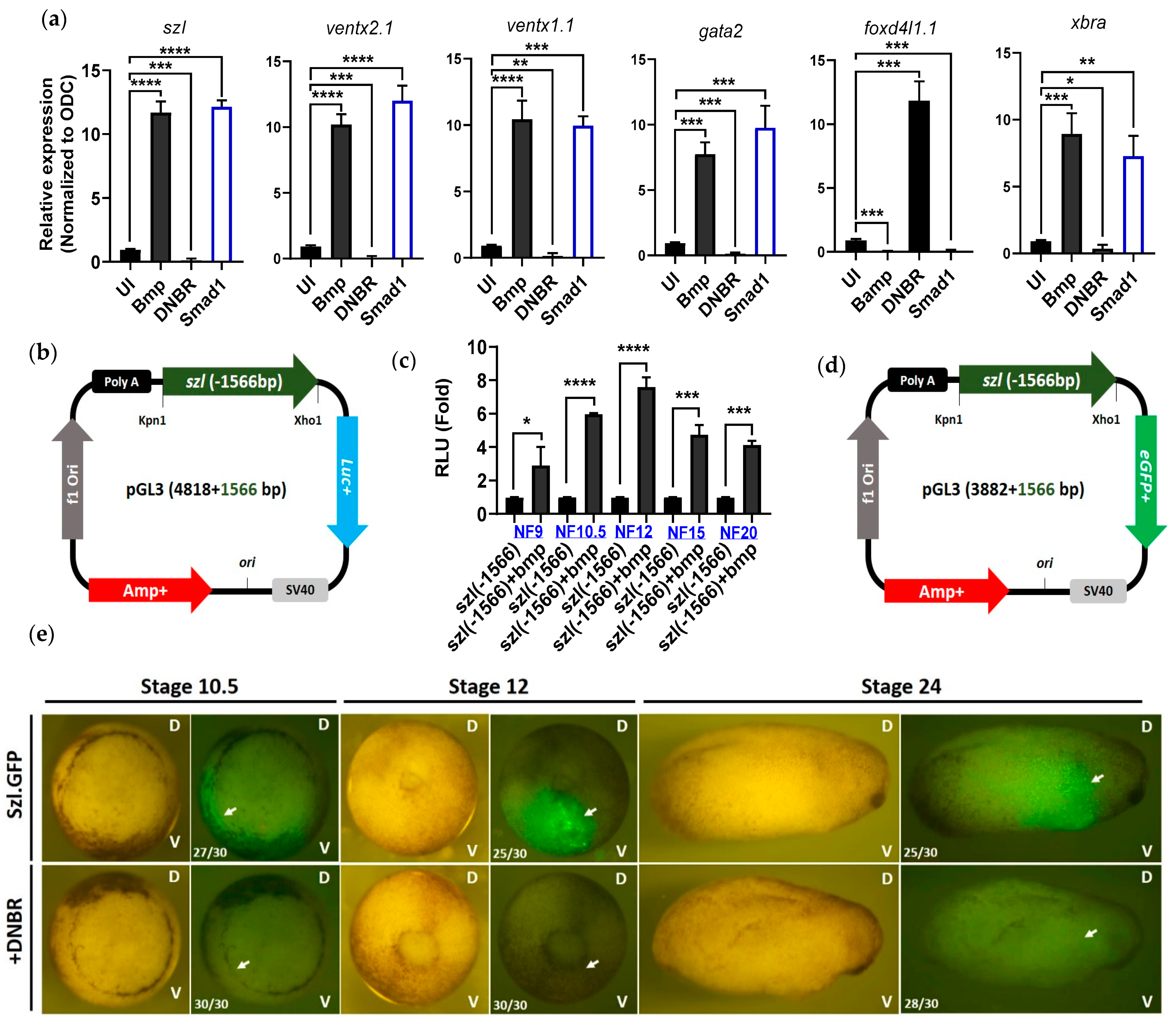
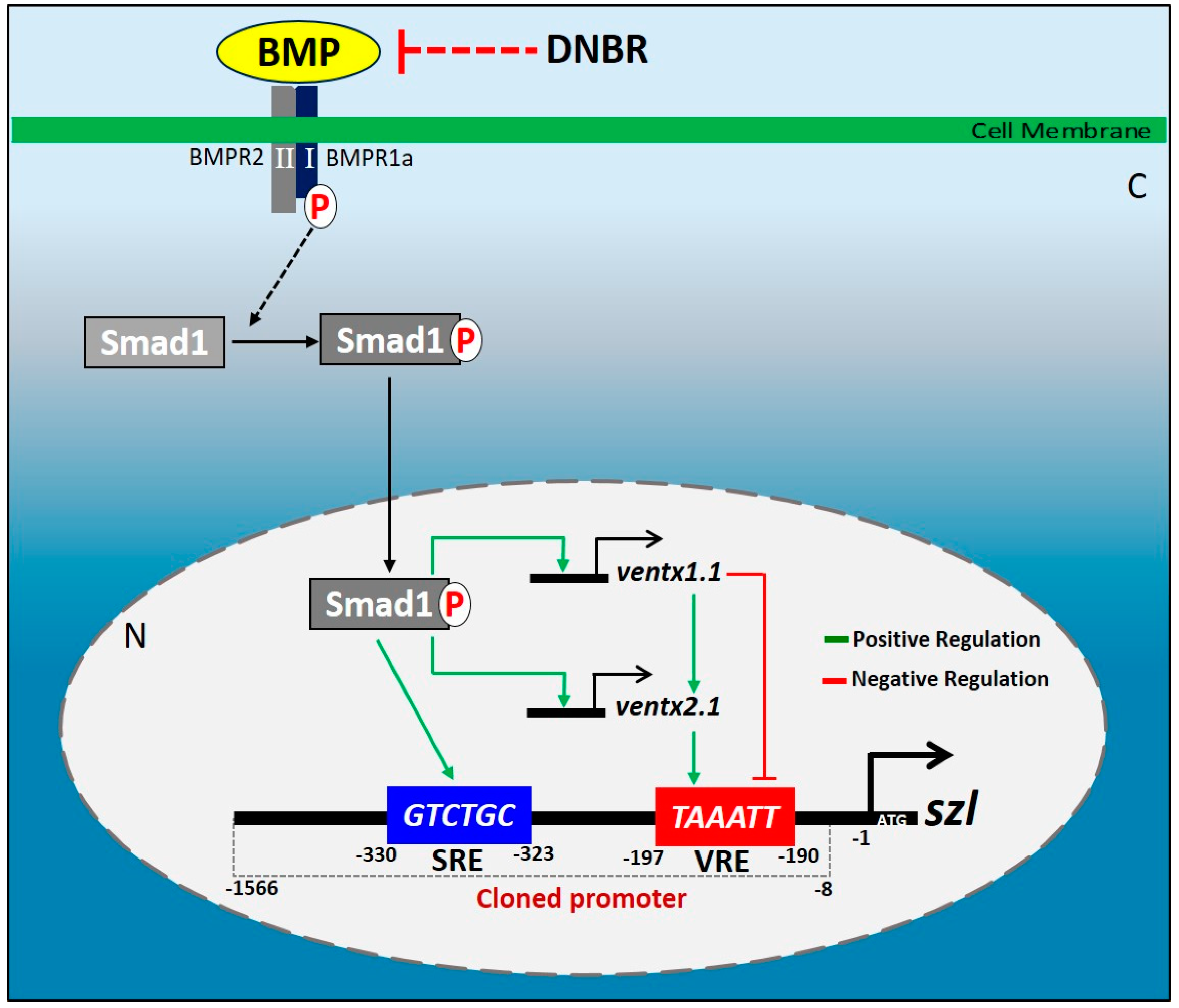
2.1. Isolated Szl(-1566) Promoter Contains the Bmp4 Regulatory Elements Required for Bmp4 Synexpression

Bmp4 is the dominant signaling of the ventral side of embryo initiated from early onset of mid-blastula transition stage [23], and overexpression of Bmp4 profoundly influences the ventral marker genes and downregulates the dorsal genes. Loss of Bmp4 signaling results in upregulated expression of neural genes [24]. The Bmp4 gradient defines the ectoderm and neuroectoderm of the embryo [25]. Reported data suggest that szl is a Bmp4 synexpression gene and its transcription is dependent on the Bmp4 gradient, indicating that szl transcripts accumulate in ectoderm and in the ventral lateral marginal zone [13,26,27]. We analyzed the spatio-temporal expression of Bmp4 synexpression genes including szl, ventx1.1 and ventx2.1 through RT-PCR analysis. We found that szl transcription start from stage 8 at the ventral side of the embryo where Bmp4 signaling is abundant (Supplementary Materials Figure S1a,b). To examine the effects of Bmp4 signaling on szl endogenous expression, bmp4 (1 ng/embryo), dnbr (1 ng/embryo, dominant negative bmp4 type Ia receptor) or smad1 (1 ng/embryo) mRNAs were individually injected to the animal pole region of one-cell stage embryos. The qRT-PCR analysis of Bmp4 target genes including szl, ventx1.1, ventx2.1, gata2 (a Bmp regulated gene, which is expressed in ventral mesoderm and required for normal hematopoiesis [28]), and xbra was performed with the isolated RNA from the embryos or animal cap explants (AC) at stage 11. The isolated RNA samples were balanced with ornithine decarboxylase (odc), a house-keeping gene which is used for equal loading control in RT-PCR experiments. As expected, Bmp4 and its intracellular signal mediator Smad1 overexpression increased szl transcription of the resultant AC (Figure 1a, graph 1). Conversely, the Bmp signaling inhibition reduced the szl mRNA level by treating embryos with dnbr (a dominant negative Bmp-type 1a receptor) mRNA. The dominant negative Bmp-type 1a receptor, which lacks the intracellular kinase domain [29,30], inhibits the Bmpr1a-mediated Bmp signaling pathways in vivo and promotes complete neuralization [15,31,32,33]. The other ventral marker genes, such as ventx1.1, ventx2.1 and gata2 behaved similarly to the szl expression pattern. Bmp/Smad1 overexpression also enhanced xbra, a pan-mesodermal marker [34,35] which was turned out as a secondary Bmp4 response gene in the present study (via CHX experiment; Supplementary Figure S2a). Xbra also plays a cooperative role in the transcription of Bmp target gene ventx1.1 via a physical interaction with Smad1 [5,36].
To examine the transcriptional regulation of szl, a 1566 bp 5′-flanking region of szl.L from translational start site (TLS) was cloned into pGL3-luc+ (Figure 1b) and pGL3-eGFP+ reporter gene vectors (Figure 1d). The cloned szl(-1566) promoter construct was injected (40 pg/embryo) to the one-cell stage embryos, and the reporter assays at the designated stages were performed as shown in Figure 1c,e. The relative luciferase activities indicated that the cloned szl(-1566)-luc+ promoter activity was enhanced by Bmp4 (Figure 1c) and the pattern of eGFP expression mimicked the pattern of endogenous szl expression (Figure 1e, upper panels). The eGFP signal was dramatically reduced by dnbr co-expression with Bmp4 signaling inhibition (Figure 1e, bottom panels). The relative luciferase (Luc) activities maximally responded to Bmp4 treatment at stage 12 (Figure 1c). The reporter gene (eGFP) expression was localized to the ventral half (Figure 1e, bottom side of embryo at stage 10.5) of the embryo and was at its maximum in stage 10.5 and stage 12. Bmp4 response and the eGFP expression levels then gradually were reduced by post stage 12 (Figure 1c,e). Reported research has indicated that bmp7.1 and bambi are also Bmp4 synexpression genes [13]. We then cloned and tested the reporter gene activities of pGL3.luc+(bmp7.1:-3143) (Supplementary Materials, Figure S1c), pGL3.luc+(bambi:-3073) (Supplementary Materials, Figure S1d) and pGL3.eGFP+(bambi:-3073) (Supplementary Materials, Figure S1e) promoters. Both of these promoter luciferase activities and their eGFP fluorescence intensities were reduced in the presence of dnbr mRNA similarly to those of the szl promoter. We next examined the luciferase activities and eGFP signal intensities at different developmental stages to determine whether the szl promoter activity with Bmp4 treatment is replicating its endogenous transcriptional expression patterns. Szl was expressed around the ventral marginal zone (the lower part of the stage 10 embryo). Data from RNA expression patterns (Supplementary Figure S1a,b), eGFP imaging (Figure 1e) and the reporter gene activities of szl in various developmental stages (Figure 1c) indeed indicated a similar pattern of szl expression as shown in the reported szl expression [16,37,38]. Bmp4 signaling responses and the spatio-temporal expression patterns indicated that the cloned szl promoter contained the expected Bmp4 regulatory elements required for synexpression.

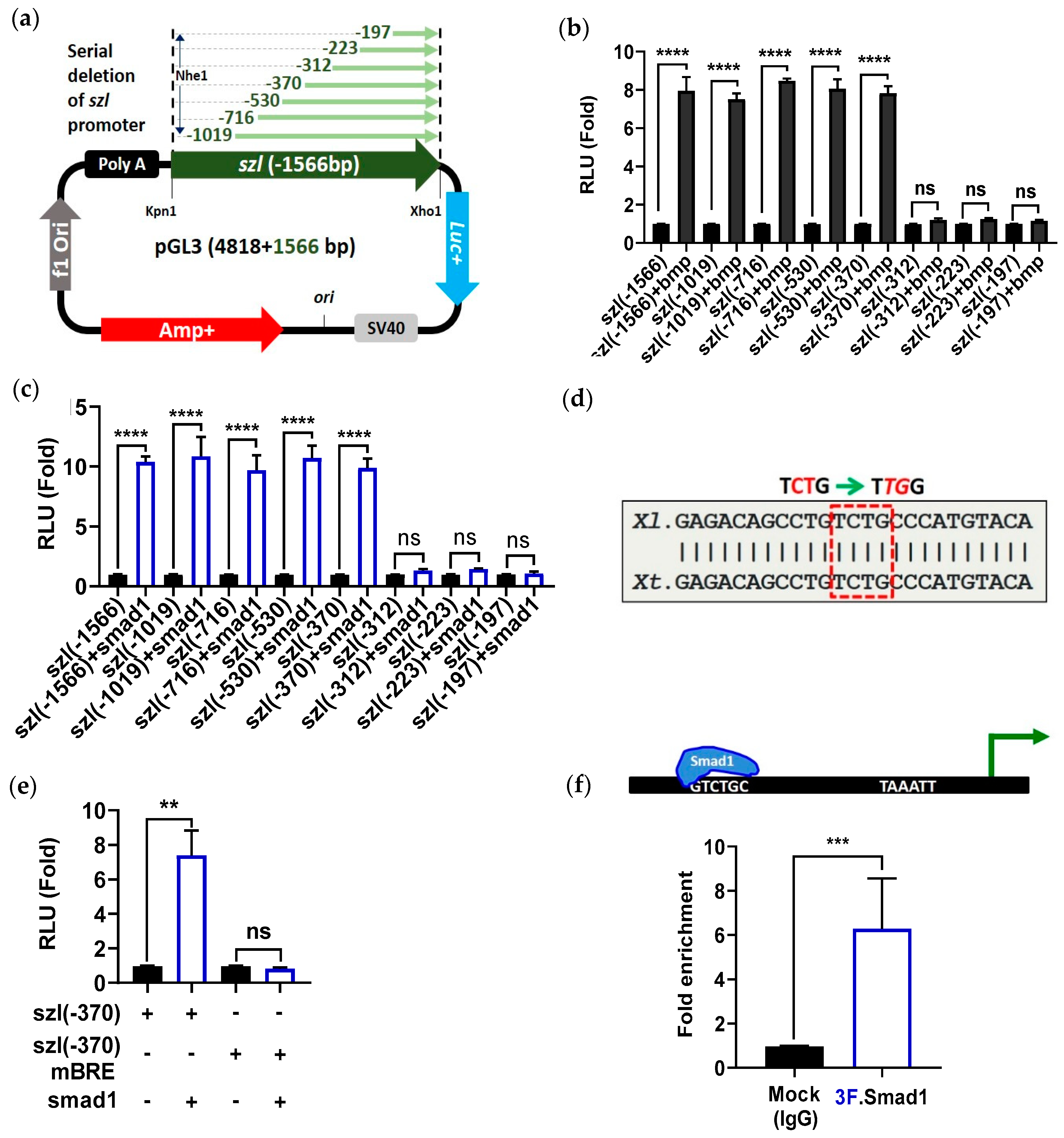
2.2. Direct Smad1 Binding on Bmp4/Smad1 Response Cis-Acting Element (BRE) Site of Szl Promoter Is Essential for Bmp4 Synexpression

It has been known that for Bmp4 synexpression genes, including szl, vent2, bambi and bmp7, their transcription is regulated in a phospho-Smad1 dependent manner [13,39]; however, presence of a direct Smad1 regulatory response element or elements for the szl promoter has not been addressed. To determine whether Bmp4/Smad1 is directly involved in synexpression of szl, a protein synthesis inhibition experiment with cycloheximide (CHX) was performed. Bmp4 mRNA (1 ng/embryo) was injected to the one-cell stage embryos and the animal caps were dissected at stage 8. The animal cap explants (ACs) were then cultured in L-15 media until stage 11 in the presence or the absence of CHX in order to determine whether szl is an immediate early (primary) or a secondary response gene to Bmp4. RT-PCR showed that the expressions of szl and a known Bmp4 direct target gene, ventx2, were maintained in the ACs treated with Bmp4 mRNA in presence of CHX (Supplementary Materials, Figure S2a). By comparison, the expression of xbrachyury (xbra) disappeared in the CHX treated condition.
To determine the location of Bmp4/Smad1 response elements (BREs) within the szl(-1566) promoter, serially-deleted promoter constructs from the cloned szl(-1566) region were made (Figure 2a). The constructs were injected, with or without either bmp4 or smad1 mRNA, to one-cell stage embryos, and reporter gene assays were performed at stage 11. The relative reporter gene activities showed that five serially-deleted promoter constructs, including szl(-1566), (-1019), (-716), (-530) and (-370), were positively up-regulated by either Bmp4 or Smad1 (Figure 2b,c, bar 1–10). Ectopic expression of Bmp4 or Smad1 increased the relative luciferase activities by up to 8- to 10-fold when compared to those without bmp4/smad1 mRNA co-injection (Figure 2b,c, bar 1–8). The shorter constructs, including three constructs (-312) (-223) and (-197), did not respond to either Bmp4 or Smad1 ectopic expression (Figure 2b,c, bar 11–16), indicating that the putative cis-acting response element for Bmp4/Smad1 (BRE; Bmp4 response (or Smad1-binding) cis-acting elements) would be located between the -370 and -312 bps locations of szl TLS. However, the Bmp4 inhibition (DNBR) response remained even in the smallest construct of szl, namely szl(-197) (Supplementary Materials, Figure S2b), suggesting that the downstream region of szl(-197) may contain negative regulatory element(s), which is addressed in Figure 3 and Figure 4. DNA sequence conservation between the -370 and -312 bps of Xenopus laevis with that of Xenopus tropicalis indicated that the promoter region contained a putative conserved consensus sequence (indicated as BRE of TCTG; it is the reverse complement of CAGA; square red dotted box; Figure 2d). A szl(-370) promoter construct containing a mutated BRE from -370 to -312 bps was made and then tested to determine whether the putative BRE was a functioning site for Smad1 (site-directed mutagenesis of BRE TCTG to TTGG with the mutated bases in red italics and underlined in Figure 2d). The wild type and mutated szl(-370)mBRE promoter constructs were injected at the one-cell stage and the reporter gene assays were performed at stage 11. To examine whether the putative BRE was the responding site for Smad1, the constructs with or without Smad1 overexpression were examined in reporter assays at stage 11 (Figure 2e). Smad1 increased relative luciferase activity of wild type szl(-370), but it was completely abolished with the mutated szl(-370)mBRE construct. Direct binding of Smad1 on the 5′-flanking region of endogenous szl promoter was then examined for gastrula embryos with a quantitative ChIP-PCR experiment, using an anti-Flag antibody immunoprecipitation against injected 3Flag-Smad1 mRNA from the total chromatin of gastrula embryos. The results showed that Smad1 directly bound within the 5′-flanking region of the endogenous szl promoter (Figure 2f, 2nd lane) Together, the results showed that the szl promoter contains a direct Smad1 binding site (BRE) required for Bmp4 synexpression.

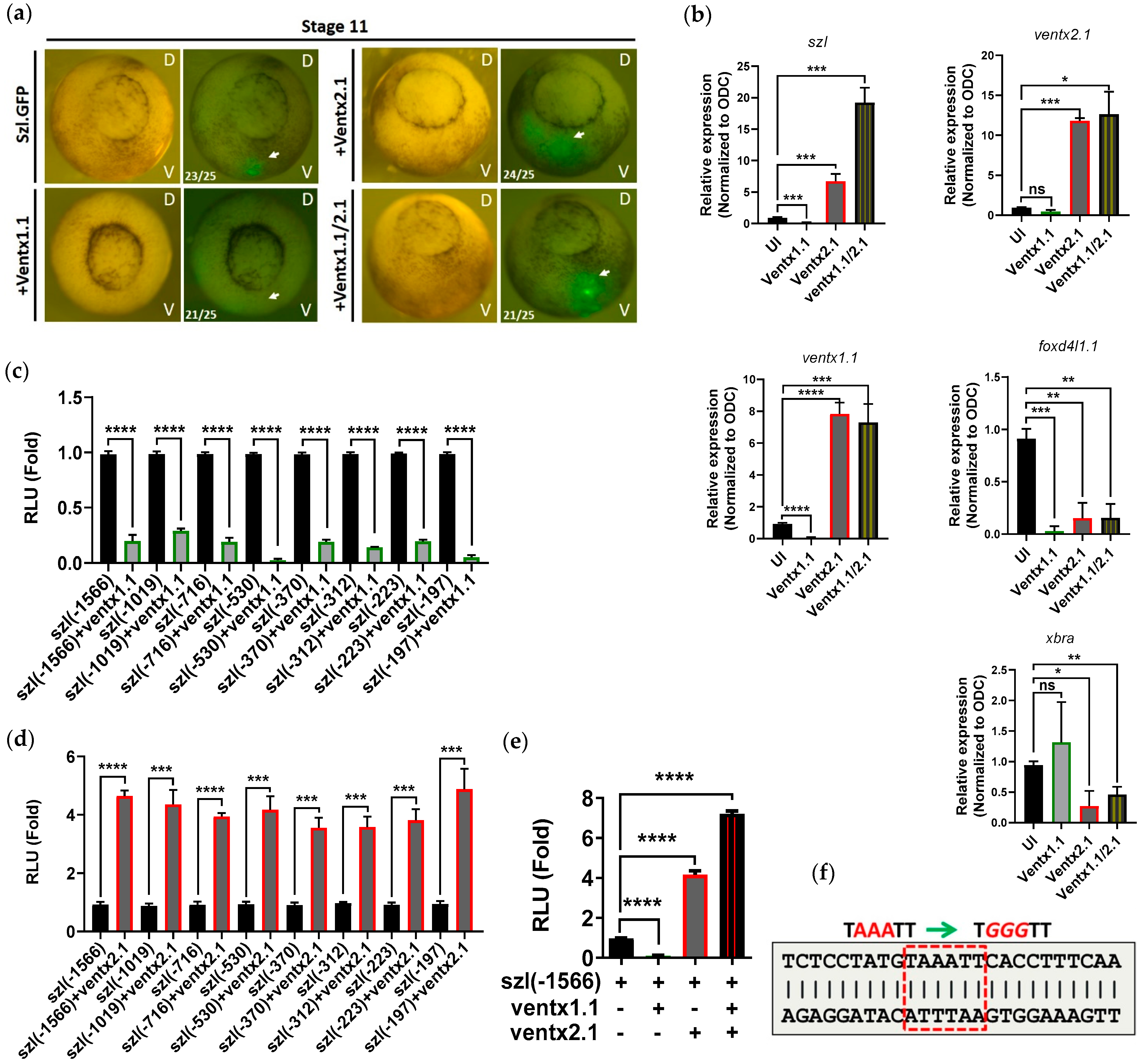
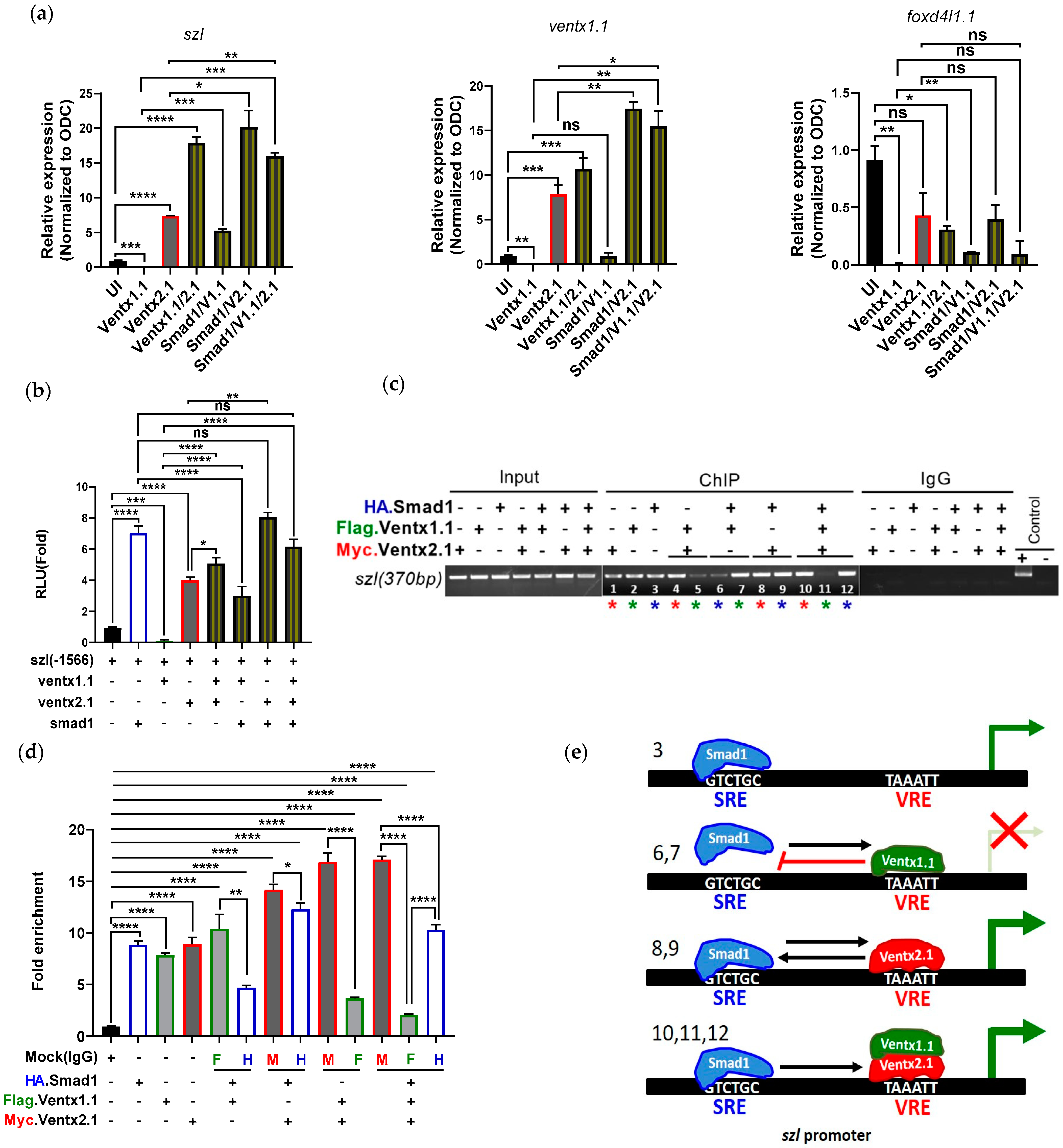
2.3. Two Direct Targets of Bmp4 Signaling, Ventx1.1 and Ventx2.1 Oppositely Modulate but Ventx1.1/2.1 Together Jointly Upregulate Szl Transcription

Szl transcription was positively regulated via direct binding of Smad1 on BRE site of szl promoter, in concordance with the expected results for szl synexpression with Bmp4, as Smad1 is a direct transcription factor mediating Bmp4 signaling. To determine the secondary response elements of Bmp4 signaling on the szl promoter, we examined the effects on szl transcription of two direct targets of Bmp4 signaling, the ventx genes ventx1.1 and ventx2.1. Each promoter of these ventx genes contains a direct binding site of BRE, which is essential for their transcription in response to Bmp4 signaling [21,40]. To examine whether Ventx1.1 and Ventx2.1 transcription factors are involved in szl expression of whole embryos or not, szl(-1566)eGFP (szl-GFP) plasmid (40 pg/embryo) was injected at the one-cell stage with either ventx1.1 or ventx2.1 or ventx1.1/ventx2.1 mRNA together (each at 0.5 ng/embryo). The embryos were then grown until stage 11 to detect their intensity of eGFP fluorescence signal. Ventx1.1 co-injection decreased eGFP intensity (Figure 3a; bottom left panel). On the other hand, ventx2.1 increased eGFP intensity on the ventral side of whole embryos (Figure 3a; Top right panel). Interestingly, the embryos co-injected with ventx1.1/ventx2.1 mRNAs together showed slightly stronger fluorescence intensity than that of the ventx2.1 alone injected group (Figure 3a; bottom right panel). To assess further effects of these Ventxs on szl transcription, ventx1.1 and ventx2.1 mRNAs were injected at the one-cell stage and the animal caps were dissected at stage 8. Quantitative RT-PCR analysis showed that the expression pattern of szl transcript was similar to those of eGFP fluorescence data from whole embryos. Ventx1.1 mRNA injection decreased szl expression, and ventx2.1 increased szl expression. Ventx1.1/ventx2.1 co-injection showed more increased szl expression than that of ventx2.1 alone injected ACs (Figure 3b). We observed similar results in whole embryos injected with the similar set of mRNAs through end point RT-PCR (Supplementary Materials, Figure S3a). To determine the location of Ventx1.1 inhibitory response element (VRE) within the szl(-1566) promoter, the serially-deleted szl promoter constructs were examined for response changes. The constructs were injected with or without ventx1.1 mRNA (0.5 ng/embryo) to the one-cell stage embryos and reporter gene assays were performed at stage 11. The relative reporter gene activities showed that all eight serially-deleted promoter constructs of szl(-1566) to (-197) were more than five-fold down-regulated by ventx1.1 (Figure 3c, bars 1–16). On a similar experiment, the relative reporter activities of all 8 szl(-1566) to (-197) constructs were about four-fold up-regulated by ventx2.1 (Figure 3d, bars 1–16). The largest construct, szl(-1566)luc+, was then selected for examining the combination effects of ventx1.1/ventx2.1. As expected, ventx1.1 injection decreased the relative reporter gene activity of szl(-1566)luc+ (Figure 3e; compare first (control) and second (ventx1.1) bars) and ventx2.1 increased the reporter activity (Figure 3e; compare first (control) and third (ventx2.1) bars). Similar to the results of the eGFP and RT-PCR experiments, ventx1.1/ventx2.1 mRNAs when together injected displayed the highest relative reporter gene activity (about seven-fold increase). Ventx2.1 alone injected embryos showed a four-fold increase when compared to that of control samples (Figure 3e; compare first (control), third (ventx2.1) and fourth (ventx1.1/ventx2.1) bars). These data together indicated that the putative cis-acting response element(s) for both Ventx1.1 and Ventx2.1 (VRE(s); Ventx response cis-acting element(s)) may be located within the shortest -197 bps region of szl TLS. DNA sequence analysis was then performed to find a putative Ventx binding element within the szl(-197) region. We examined whether the promoter region contains the core consensus residues (AAAT or ATTT) between the -196 and -191 bps of the proximal region of szl promoter. The szl promoter region contains a putative conserved consensus sequence (indicated as VRE, TAAATT, marked in square red dotted box; Figure 3f). The szl(-370) promoter construct containing the putative VRE site was mutated to identify whether the putative VRE was a functioning site for Ventx1.1 and/or Ventx2.1, or both. Taken together, the results showed that two direct target transcription factors (Ventx1.1 and Ventx2.1) of Bmp4 signaling oppositely modulate but together upregulate szl transcription, indicating that szl transcription is directly modulated by Bmp4/Smad1 signaling as well as by a secondary response using Bmp4 target transcription factors for Bmp4 synexpression.

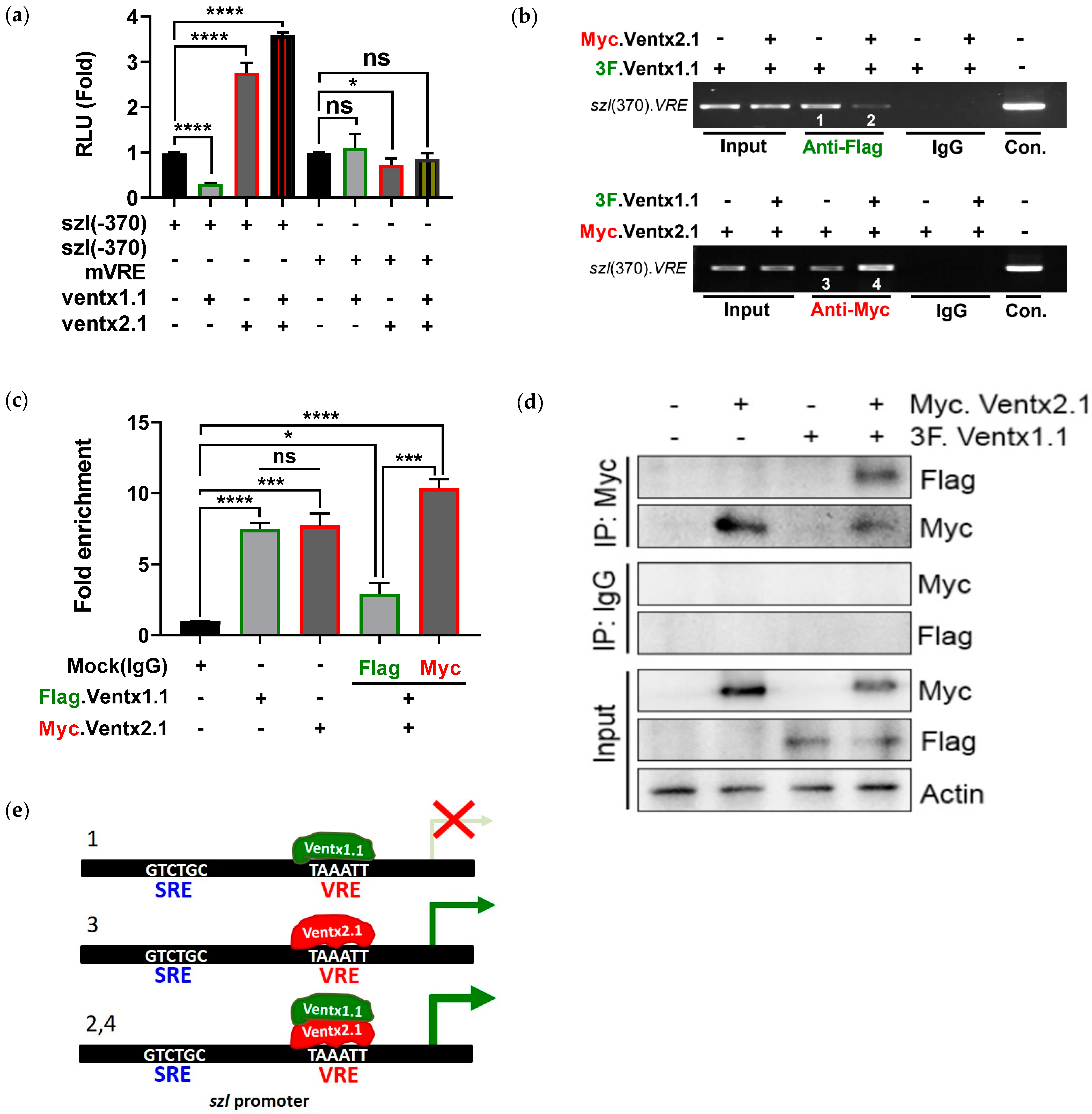
2.4. Site-Directed Mutagenesis of One VRE Site on the Szl Promoter Abolishes Ventx1.1 as Well as Ventx2.1 Mediated Szl Transcriptional Regulation

DNA sequence analysis between the -197 and -1 bps (Supplementary Materials, Figure S3b) region showed that the promoter region contained conserved consensus sequences that belong to a Ventx binding site (marked as VRE, Figure 3f). To identify whether the putative VRE were functioning sites for Ventx1.1 and/or Ventx2.1 or both, site-directed mutagenesis was performed on VRE (TAAATT to TGGGTT) (mutated bases, red italic and underlined; Figure 3f). Wild type szl(-370) or mutated szl(-370)mVRE promoter constructs were injected (40 pg/embryo) with and without ventx1.1 or ventx2.1 mRNA (0.5 ng/embryo) at the one-cell stage. The reporter gene assays were then performed at stage 11. For the mutated szl(-370)mVRE, the Ventx1.1-mediated as well as Ventx2.1- and Ventx1.1/Venx2.1-mediated relative luciferase activities of wild type szl(-370) construct were completely abolished (Figure 4a). The decreased response to Ventx1.1 and the increased responses to Ventx2.1 and Ventx1.1/Venx2.1 of wild-type szl(-370) had all disappeared in the single VRE mutated szl(-370)mVRE construct (Figure 4a, compare first to fourth bars with fifth to eighth bars). The results indicated that the same VRE was the responding site for Ventx1.1 and Ventx2.1. The direct binding of Ventx1.1 and Ventx2.1 on the VRE of endogenous szl promoter was then examined with stage 11 embryos. ChIP-PCR experiments were performed on Flag- or Myc-antibody immunoprecipitates from total chromatin for embryos injected with 3flag-ventx1.1 or myc-ventx2.1 or the 3flag-ventx1.1/myc-ventx2.1 constructs. The ChIP-PCR and ChIP-qPCR results showed that Ventx1.1 and Ventx2.1 directly bound within the 5′-flanking region of endogenous szl promoter (Figure 4b, third lane of upper (Ventx1.1) and bottom (Ventx2.1) panel). In the co-injected 3flag-ventx1.1/myc-ventx2.1 samples, Ventx1.1 binding was reduced in presence of Ventx2.1 (Figure 4b, upper panel; compare third and fourth lanes) (Figure 4c; compare second and fourth bars). On the other hand, Ventx2.1 binding was enhanced by Ventx1.1 co-injection (Figure 4b, bottom panel; compare third and fourth lanes) (Figure 4c, compare third and fifth bars). To check the protein-protein interaction between Ventx1.1 and Ventx2.1, immunoprecipitation assay was performed at the gastrula stage of the embryo. 3F.Ventx1.1 and Myc.Ventx2.1 co-injected sample were pulled down with myc antibody, and through western blotting 3F.Ventx1.1 protein expression was detected, suggesting that Ventx1.1 and Ventx2.1 physically interact with each other and hence Ventx1.1 promotes the binding of Ventx2.1 on VRE site of szl promoter (Figure 4d). These results indicated that the Bmp4 primary as well as secondary response transcription factors Ventx1.1 and Ventx2.1 together are collectively participating in modulation of szl expression by binding to VRE of the szl promoter. For Ventx1.1/Ventx2.1-mediated regulation of szl transcription in a Bmp4 synexpression dependent manner, based on the results of Ventx2.1 and Ventx1.1 co-injection leading to a decreased amount of Ventx1.1 binding and an increased level of Ventx2.1 binding on VRE, the following schematic is proposed (Figure 4e). We hypothesize that Ventx2.1 binding would be assisted by DNA being unbound by Ventx1.1. However, as to which Ventx protein would bind first and how the two Ventx’s cooperate, these remain unknown. Together, these results suggest that one VRE site for Ventx1.1/Ventx2.1 binding is also involved in Bmp4 synexpression of szl.

2.5. Smad1 and Ventx1.1/Ventx2.1 Collectively Modulate Szl Transcription

The above findings show that Bmp4 signaling regulates the szl transcription via primary Bmp4 response mediated by Smad1 and a secondary Bmp4 response mediated by Ventx transcription factors. It has been known that the Smad1 (MH1) directly and specifically interacts with the C-terminal of Ventx2.1 in an additive fashion in transcriptional activation of Ventx2.1 [41,42,43,44]. Our results also showed a Ventx1.1/Ventx2.1-mediated positive regulation of szl transcription. To examine Smad1 and Ventx1.1 and/or Ventx2.1 combined effect on szl transcription, we injected smad1 mRNA alone and in combination of ventx1.1 and/or ventx2.1 at the one-cell stage of Xenopus embryos. Animal caps were then dissected and maintained in L-15 medium. RNAs were isolated from the ACs and the qRT-PCR analysis was then performed. The results showed that szl mRNA expression was enhanced either by smad1 or ventx2.1 and reduced by ventx1.1 (Figure 5a). As expected, smad1 and ventx2.1 showed a certain positive combination effect. However, ventx1.1 retained the inhibitory response in smad1 co-injected group shown as reducing the positive response of Smad1 mediated szl transcription. When smad1 and ventx2.1/ventx1.1 were injected together, ventx1.1 inhibitory effect was not obvious and szl transcription was enhanced as much as that of smad1 and ventx2 co-injected groups. The reporter gene activity of szl(-1566) promoter construct was then measured after injection alone and with smad1, ventx1.1 and ventx2.1 mRNAs. The luciferase activity results were similar to those of quantitative RT-PCR (Figure 5b). To examine the difference in the amount of each protein bound on the szl promoter in the Smad1, Ventx1.1 and Ventx2.1 combined condition, ChIP-PCR analysis was performed with different sample sets for the combined injection groups of ha.smad1, 3flag.ventx1.1 and myc.ventx2.1 mRNA-injected embryos at the gastrula stage. The results showed that Smad1 enhanced Ventx2.1 as well as Ventx1.1 binding on szl promoter (Figure 5c, seventh and ninth lane). On the other hand, Smad1 binding on szl promoter was decreased in presence of Ventx1.1 and increased in presence Ventx2.1 (Figure 5c, sixth and eight lane), indicating that Ventx1.1 inhibited the Smad1 binding on the szl promoter. Ventx2.1 and Smad1 combination enhanced each other’s binding on the szl promoter compared with singly present (Figure 5c, compare Smad1 binding (third (control) vs. Smad1/Ventx2.1 combination (ninth lane) and Ventx2.1 binding (first (control) vs. Smad1/Ventx2.1 combination (eighth lane). This finding is consistent with the previously reported data that Smad1 directly and specifically interacts with Ventx2.1 to activate ventx2.1 transcription [43].
Smad1 and Ventx2.1 are positive regulators of szl transcription and are expected to cooperate with each other to enhance their binding on the szl promoter. Triple combination of Smad1, Ventx1.1/2.1 retained the reduced Ventx1.1 binding (Figure 5c, compare fifth and eleventh lane) found in the Ventx1.1/2.1 combination group. On the other hand, enhanced binding of each Smad1 and Ventx2.1 binding found in combination of Smad1 and Ventx2.1 was also retained in the triple combination group of Smad1, Ventx2.1/Ventx1.1 (Figure 5c, eighth and ninth vs. tenth and twelfth lane) (Figure 5d, ChIP-qPCR analysis). Smad1/Ventx1.1/Ventx2.1 triple action on szl expression is depicted as a schematic (Figure 5e), in which the interaction between Smad1 and Ventx2.1 is presumably maintained in the triple injection experiment including Ventx1.1 (Figure 5e). From Smad1 and Ventx2.1/Ventx1.1 triple injection results, these support a collective regulation for szl expression and maintenance of Smad1 and Ventx2.1 interaction (Figure 5b).

3. Discussion

In the present study, we aimed to uncover how Bmp4 synexpression is achieved by examining the promoter characteristics of szl, a Bmp4 synexpression gene. We found that the proximal region (within -370 from TLS) of the szl promoter contains cis-acting elements responding to Bmp4/Smad1 signaling as a BRE for a primary response. There was also another cis-acting element, one jointly responding to Ventx1.1 and Ventx2.1 as a VRE for a secondary response, confirming the binding of these two Bmp4 targets acting on the szl promoter. Interestingly, Ventx1.1 alone functions as a repressor (Figure 3a, bottom left panel; Figure 3b; Figure 3c, bars 1 to 16) when it binds on the VRE of the szl promoter, but functions as a coactivator enhancing the binding of the activator Ventx2.1 on the same VRE (Figure 4e). The implications of this study are briefly discussed below in light of the reported results.

3.1. Szl Is a Direct Target as Well as a Secondary Response Gene of Bmp4 Signaling

We demonstrated that szl is a direct target of Bmp4/Smad1 signaling in CHX experiments (Supplementary Materials, Figure S2a). Recently, Lee et al. also reported szl as being one of the direct targets of Bmp4/Smad1 signaling with in situ hybridization assays using CHX [26]. They also demonstrated the szl expression pattern in whole embryos using in situ hybridization [10]. In the present study, AC explants were used instead. Notice that the band intensity of szl in presence of CHX was weaker than that of untreated control in Bmp4 treated samples (Supplementary Materials Figure S2a, compare first vs. third and fourth lanes). We observed a similar pattern for ventx1.1 expression, which is a primary as well as a secondary response gene for Bmp4/Smad1 signaling [21]. We speculate that synexpression is more effectively achievable when a gene is both a primary and secondary response gene of Bmp4/Smad1 signaling, since a small change in the morphogen gradient can switch the target gene expression via signal cascade amplification for secondary responses. It has been reported that ventx2.1 expression is also dependent on both a primary and secondary response of Bmp4/Smad1 signaling through Smad1 and the C-terminal domain of Ventx2.1 [41,42,43,44]. Ventx1.1 expression is also positively modulated by smad1, ventx2, gata2 and xbra [44,45]. It is necessary for the primary and secondary genes, including smad1, ventx2.1 and ventx1.1 to be expressed at the same time and in the same cells during normal development. We examined temporal and spatial expression patterns of ventx1.1, ventx2.1 and szl, as shown in Supplementary Materials (Figure S1a,b). RT-PCR results indicate that ventx1.1, ventx2.1 and szl were expressed together for most of the relevant development stages (blastula to neurula) and on the ventral marginal region, but not in dorsal marginal region of normal Xenopus laevis embryos. At the present time, we do not know that Smad1, Ventx2.1 and Ventx1.1 were expressed in the same cells of Xenopus embryos. However, genes of both ventx2.1 and ventx1.1 are the direct targets of Bmp4/Smad1 [21] and are expressed in the ventral region (ventral mesoderm and ventral ectoderm) where szl is also expressed during blastula and gastrula Xenopus embryos. Synexpression genes of Bmp4/Smad1 signaling including ventx2.1 and ventx1.1 may not be exclusively expressed in ventral cells of embryos at least until early gastrula stage since Ventx2.1 has been reported as an activator transcription factor on ventx1.1 promoter working together with Smad1. The regulation differences and synexpression for various Bmp4 synexpression genes in the same cells remain to be investigated; these include the genes whose promoters have been cloned in the present study (bmp7 and bambi1) (Supplementary Materials). On modulation of szl expression, it also remains to be seen whether other secondary (and/or tertiary) transcription factors (TFs) including Gata2 and Xbra are also involved.

3.2. Ventx1.1 Enhances Ventx2.1’s Stimulatory Effect

One interesting observation of this study was the stimulatory transcriptional effect of Ventx1.1 when co-expressed with Ventx2.1 (Figure 4). Ventx1.1 is a well-known suppressive transcription factor and its repressor domain resides in its C-terminal domain [44,46]. Ventx1.1 primarily suppresses organizer gene expression and neural specific genes to protect the ventral mesoderm and ectoderm territory from being dorsal mesoderm and neuroectoderm, respectively [47,48,49]. Ventx1.1 also represses Bmp4 target genes including gata2 [50], ventx2.1 and itself [51]. The inhibitory function of Ventx1.1 was also shown for ventx2.1 and szl (Figure 3b) in the present study. The direct and/or indirect suppressive effects of Ventx1.1 on certain individual genes need more elaboration, although some of the various Ventx1.1 inhibitory cis-acting elements in promoters have been reported [51,52]. In addition, Ventx1.1 competes with the transcriptional activator Xcad2 in binding the common cis-acting elements within the 5′-promoter region of ventx1.1 to negatively regulate its own expression. [51]. In the present study, as a repressor, Ventx1.1 alone inhibits szl transcription. However, Ventx1.1 displays a stimulatory activity for szl expression and reporter activity as well as an increase in the amount of Ventx2.1 binding on VRE site in the szl promoter (according to co-injected group of Ventx1.1 and Ventx2.1). Consistent with this observation, Sander et al. reported that ventx1.1/ventx2.1 double knockdown by morpholinos downregulates szl expression, indicating that szl is the downstream target gene of Ventx transcription factors [45]. Whang et al. also reported Ventx1.1 and Ventx2 physical binding using a yeast two-hybrid assay [44]. However, future work needs to determine whether the attenuation effect of Ventx2.1 on inhibition of Ventx1.1 is general to all Bmp4 synexpression target genes. Additional work needs to elucidate on how Ventx1.1 and Ventx2.1 physical co-binding on the promoter regions occurs with the resultant inhibition of Ventx1.1 suppressor activity. A more complicated picture is one for the Ventx1.1′s own promoter which contains two separate cis-acting elements, one for Ventx2.1 and one for Ventx1.1/Xcad2 competition [21,51]. The present study for the first time shows that Ventx1.1 and Ventx2.1 share one VRE site on the szl promoter and Ventx1.1/Ventx2.1 together positively modulate the expression of szl, a Bmp4 synexpression gene.

3.3. Smad1 and Ventx1.1/Ventx2.1 Collectively Upregulates Szl Expression

Kristine et al. reported that Smad1 directly and specifically interacts with Ventx2.1 and that Smad1 and Ventx2.1 co-injection synergistically activates the BRE on ventx2.1 promoter [43]. Apart from Ventx2.1 as a target for Smad1/Ventx2.1, other target genes of this complex also need to be explored. Our results indicate that szl could be such a target of Smad1/Ventx2.1 complex, with szl mRNA expression and reporter activity assays, as well as Smad1 and Ventx2.1 binding all showing positive combination effects (Figure 5). However, the Ventx1.1 and Smad1 combination did not lead to a positive response in szl mRNA expression, reporter activity as well as Smad1 binding. Ventx1.1 retained the inhibitory response in smad1 co-injection as shown by a reduction in the positive response of Smad1 mediated szl transcription, reporter activity (Figure 5b, compare second vs. sixth bar) and Smad1 binding (Figure 5c, compare third vs. sixth lane). When smad1 and ventx2.1/ventx1.1 were jointly injected, Ventx1.1 inhibitory effect seems to have disappeared as shown in the ventx2.1/ventx1.1 combination group (Figure 5a), indicating that a triple combination of Smad1, Ventx1.1/2.1 retained the attenuation of Ventx1.1 inhibitory effect as shown in Ventx1.1/2.1 combination. Interestingly, Smad1 enhanced Ventx2.1 as well as Ventx1.1 binding on the szl promoter (Figure 5c, seventh and ninth lane). In addition, the enhanced binding of each Smad1 and Ventx2.1 found in Smad1/Ventx2.1 combination group was also retained in the Smad1/Ventx2.1/Ventx1.1 triple combination group (Figure 5c, eighth and ninth vs. tenth and twelfth lane).
Although additional Bmp4 synexpression targets of Smad/Ventx1.1/Ventx2.1 triple combination and their detailed underlying mechanism need to be explored, the present study provides an insight on how the Bmp4 synexpression pattern is achieved with the combinatory target gene effects, making up the primary and secondary responses of Bmp4 signaling and how an inhibitory transcription factor can become stimulatory under the right circumstances (Figure 6). However, it remains to be explored if removing any or all of the Bmp4 target proteins will affect the synexpression response patterns. Taken together, we propose a systematic model of Bmp4 signaling for regulation of szl transcription (Figure 6). This model elaborates the contribution of the Bmp4 response elements (Smad1 (BRE) and Ventx1.1/Ventx2.1 binding (VRE) sites) located on the szl promoter being responsible for Bmp4 synexpression. In summary, the szl promoter is collectively modulated by Smad1/Ventx1.1/Ventx2.1 for its Bmp4 synexpression in early Xenopus embryos.

4. Materials and Methods

4.1. Ethics Statement

This animal study was conducted in accordance with the regulations of the Institutional Animal Care and Use Committee (IACUC) of Hallym University (Hallym 2019-79, 2021-92). All the research members attended both the educational and training courses for the appropriate care and use of experimental animals at our institution in order to receive an animal use permit. Adult X. laevis were grown and tended to in approved containers; these were maintained at a 12 h light/dark (LD 12:12 h) cycle and 18 °C ambient temperature, by authorized personnel and according to the guidelines of the Institute of Laboratory Animal Resources of Hallym University for laboratory animal maintenance.

4.2. Nucleic Acids (DNA, RNA) Preparation

Various constructed cDNA were obtained by transformation of DH5alpha competent bacterial cells. The introduced cDNA clones including pSP64T.bmp4, pSP64T.dnbr, 3F.pCS4.smad1, 3F.pCS4.ventx1.1 and 6Myc.pCS2.ventx2.1 were linearized with restriction enzymes EcoR1, Not1 and Acc65I and mRNA was made through in vitro transcription using a MEGA script kit (Ambion, Austin, TX, USA). All transcribed mRNAs were diluted in DEPC treated water and quantified at 260/280 nm wavelength using a spectrophotometer (SpectraMax, Molecular Devices, San Jose, CA, USA). Aliquots of 0.5, 1 ng/5 nL were made and stored at −80 °C for further use.

4.3. Cloning of Szl, Bmp7.1 and Bambi Promoter Constructs

Isolated genomic DNA from Xenopus blood were used for PCR amplification of szl promoter (−1566 bp), bmp7.1 (−3143 bp) (accession number, NM_001087397.1) and bambi (−3073) (accession number, NM_001094009.1) having a Kpn1 restriction site at the forward and a Xho1 site at the reverse primer for the szl and bmp7.1 promoters and having a Kpn1 restriction site at the forward and a Nhe1 at the reverse primer for bambi promoter. We amplified each promoter region by PCR cloning using the listed primers (Supplementary Table S1). The amplified szl promoter (−1566 bp) was subcloned into pGL3.luc+ and pGL3.eGFP+ (Promega, Madison, WI, USA) basic vectors. The cloned szl promoter (−1566 bp) was generated with the reverse primer sequence starting at −8 from TLS to guarantee including all the regulatory sequences for the transcription start of szl based on the deposited szl.L mRNA sequences (accession number; NM_001088521) which contains 5′UTR of 7 bps [53].
In addition, seven serially deleted constructs of various sizes were amplified from the −1566 bp fragment on the basis of mapping the potent Bmp signaling response elements and were cloned into pGL3.luc+ and pGL3.szl(−1566)eGFP+ constructs for fluorescence detection.

4.4. Xenopus Embryos Microinjection

Korea Xenopus Resource Center for Research was the source of adult Xenopus laevis. Five hundred units of human chorionic gonadotropin hormone (HCG) (Sigma, St. Louis, MO, USA) was injected to female Xenopus leavis in order to induce enough embryos. Freshly chopped male sperm was used for in vitro fertilization of the embryos in 1/20X MMR solution for 30 min. Embryos were de-gelled and subjected to various DNA and mRNA microinjections at the one-cell stage around the sperm entry point into the animal hemisphere. The injected embryos were grown and maintained in 30% MMR solution up to the required specific developmental stage for the experiments.
The szl(−1566)eGFP and bambi(−3073)eGFP constructs were injected (40 pg/embryo) with or without dnbr (1 ng), ventx1.1, ventx2.1 mRNAs (0.5 ng each) per embryo at the one-cell stage and into the animal hemispheres. Green fluorescence was evaluated using a stereo microscope with royal blue light adapter (Stereo Microscope Fluorescence Adapter, NIGHTSEA, Hatfield, PA, USA), and photographs were captured using a Nikon D810 camera (Nikon, Minato City, Tokyo, Japan).

4.5. Embryos Animal Cap Explants Culture

Animal cap explants (10 AC/sample) of injected embryos were dissected at the early blastula stage, cultured in L-15 media in a 96-well plate, and harvested at the specific stage, followed by further experiments.

4.6. RT-PCR Experiment

Total transcribed RNA during a specific required stage was isolated from whole embryos using the TRIzol reagent (Ambion, Austin, TX, USA) and approximately 10 animal cap explants were used. The purified RNA was treated with DNase 1 to remove genomic DNA impurities. SuperScript IV (Invitrogen, Waltham, MA, USA) kit was used for cDNA preparation from 2 µg RNA for each sample.

4.7. Quantitative RT-PCR (qRT-PCR) Analysis

All RT PCR and Chromatin immunoprecipitation assays samples were subjected to qPCR analysis using Biosystems StepOnePlus Real-Time PCR System with Kapa Syber Fast qPCR Master mix. All values were normalized to ornithine decarboxylase (ODC), a house keeping gene of Xenopus embryos which is present all around the embryonic developmental. Graphs were made using threshold cycle (Ct) values, ΔCT, ΔΔCT and relative fold change expression method.

4.8. Luciferase Reporter Gene Assay

Embryos were injected with szl (−1566 bp) or its serially deleted constructs, mutated BRE and VRE constructs, or the bmp7.1 and bambi constructs along with various indicated exogenous mRNA. Five sets of three embryos were collected at a specific stage and homogenized in lysis buffer (10 µL/embryo). Reporter gene activity was quantified using luciferase assay system (Promega) following the manufacturer’s instructions by luminometer (Berthold Technologies, Bad, Wildbad, Germany). Specific RT-PCR primers were used to amplify markers genes. (Supplementary Table S2).

4.9. Site Directed Mutagenesis of Smad1 and Ventx Response Elements (SRE and VRE)

Mutations for Smad1 response element (BRE) and Vent response element (VRE) sites were carried out by using the Muta-Direct site-directed mutagenesis kit (iNtRON Biotechnology, Seoul, Korea), following the manufacturer’s instructions. Mutagenesis PCR was performed from 40 pg/5 nl szl full-length promoter with specific mutagenesis primers (Supplementary Table S3).

4.10. Chromatin Immunoprecipitation (ChIP) Assay

Embryos were injected with HA.pCS4.smad1, 3F.pCS4.ventx1.1 and 6Myc.pCS2.ventX2.1 mRNAs at the one-cell stage either alone or together. Around 200 embryos/ sample at a specific stage were then collected following the reported protocol [54]. Anti-Flag (Sigma, F-1804) monoclonal antibody and anti-Myc (Santa Cruz, SC-789) (Santa Cruz, Dallas, TX, USA) polyclonal antibody were used for immunoprecipitation of crosslinked chromatin fragments. Mouse IgG (Santa Cruz, SC-2025) was used as negative control. ChIP-PCR was performed using specific primers (Supplementary Table S4).

4.11. Immunoprecipitation

Equal amounts of embryos were injected at one-cell stage with 3flag.ventx1.1 and myc.ventx2.1 constructs mRNAs, separately and co-injected. All samples were collected at stage 11, and after removing MMR solution through spin down, embryos were homogenized in IP lysis buffer and followed the protocol as described in Kumar et al. (2018) [36]. The homogenized samples were centrifuged and the cleared lysates were then incubated with respective antibodies overnight at 4 °C. The immunocomplexes were precipitated by protein A/G beads (SC-2003, Santa Cruz Biotechnology, Santa Cruz, CA, USA). Proper amount of precipitated beads-immunocomplexes were heated up in the sample buffer and resolved by 10% SDS-polyacrylamide gel electrophoresis. Western blotting of 3Flag.Ventx1.1 and Myc.Ventx2.1 were performed by using Anti.Myc (Rabbit), Anti.Flag (Mouse) and secondary antibodies. Immune complexes were visualized with the help of ECL detection kit (GE Healthcare).

4.12. Statistical Analysis

GraphPad Prism 8 (GraphPad, San Diego, CA, USA) and MS Excel (Microsoft, Redmond, WA, USA) were used for plotting the graphs. Unpaired two-tailed Student’s t-test or ANOVA were applied for statistical analysis. p ≤ 0.05 for *, p ≤ 0.01 for **, p ≤ 0.001 for ***, p ≤ 0.0001 for ****, ns (not significant) were the assignments for significance.

Supplementary Materials

The following supporting information can be downloaded at: https://www.mdpi.com/article/10.3390/ijms232113335/s1.

Author Contributions

Z.U.R. and F.T. performed all the experiments; Z.U.R. wrote the original draft. J.K. and U.L. designed and supervised the study. All authors have read and agreed to the published version of the manuscript.

Funding

This work was supported by the Basic Science Research Program through the National Research Foundation of Korea (NRF) which is funded by the Ministry of Education, Science, and Technology of Korea (2016R1D1A1B02008770, 2018M3C7A1056285, 2021R1A4A1027355 and 2021M3H9A1097557).

Institutional Review Board Statement

This animal study was conducted in accordance with the regulations of the Institutional Animal Care and Use Committee (IACUC) of Hallym University (Hallym 2019-79, 2021-92).

Conflicts of Interest

The authors declare no conflict of interest. The funders had no role in the design of the study, collection, analyses, or interpretation of data or in the writing of the manuscript and the decision to publish the results.

References

  1. De Robertis, E.M. Evo-devo: Variations on ancestral themes. Cell 2008, 132, 185–195. [Google Scholar] [CrossRef] [PubMed] [Green Version]
  2. Uesaka, M.; Kuratani, S.; Takeda, H.; Irie, N. Recapitulation-like developmental transitions of chromatin accessibility in vertebrates. Zool. Lett. 2019, 5, 33. [Google Scholar] [CrossRef] [PubMed] [Green Version]
  3. Jones, C.M.; Smith, J.C. Inductive signals. Revolving vertebrates. Curr. Biol. 1995, 5, 574–576. [Google Scholar] [CrossRef] [Green Version]
  4. Eivers, E.; Demagny, H.; De Robertis, E.M. Integration of BMP and Wnt signaling via vertebrate Smad1/5/8 and Drosophila Mad. Cytokine Growth Factor Rev. 2009, 20, 357–365. [Google Scholar] [CrossRef] [Green Version]
  5. Wills, A.; Dickinson, K.; Khokha, M.; Baker, J.C. Bmp signaling is necessary and sufficient for ventrolateral endoderm specification in Xenopus. Dev. Dyn. 2008, 237, 2177–2186. [Google Scholar] [CrossRef] [Green Version]
  6. Dosch, R.; Gawantka, V.; Delius, H.; Blumenstock, C.; Niehrs, C. Bmp-4 acts as a morphogen in dorsoventral mesoderm patterning in Xenopus. Development 1997, 124, 2325–2334. [Google Scholar] [CrossRef]
  7. Tucker, J.A.; Mintzer, K.A.; Mullins, M.C. The BMP signaling gradient patterns dorsoventral tissues in a temporally progressive manner along the anteroposterior axis. Dev. Cell 2008, 14, 108–119. [Google Scholar] [CrossRef] [Green Version]
  8. Hawley, S.H.; Wunnenberg-Stapleton, K.; Hashimoto, C.; Laurent, M.N.; Watabe, T.; Blumberg, B.W.; Cho, K.W. Disruption of BMP signals in embryonic Xenopus ectoderm leads to direct neural induction. Genes Dev. 1995, 9, 2923–2935. [Google Scholar] [CrossRef] [Green Version]
  9. Marchal, L.; Luxardi, G.; Thome, V.; Kodjabachian, L. BMP inhibition initiates neural induction via FGF signaling and Zic genes. Proc. Natl. Acad. Sci. USA 2009, 106, 17437–17442. [Google Scholar] [CrossRef] [Green Version]
  10. Madamanchi, A.; Mullins, M.C.; Umulis, D.M. Diversity and robustness of bone morphogenetic protein pattern formation. Development 2021, 148, dev192344. [Google Scholar] [CrossRef]
  11. Miyazono, K.; Kamiya, Y.; Morikawa, M. Bone morphogenetic protein receptors and signal transduction. J. Biochem. 2010, 147, 35–51. [Google Scholar] [CrossRef] [PubMed] [Green Version]
  12. Chen, G.; Deng, C.; Li, Y.P. TGF-beta and BMP signaling in osteoblast differentiation and bone formation. Int. J. Biol. Sci. 2012, 8, 272–288. [Google Scholar] [CrossRef] [PubMed] [Green Version]
  13. Karaulanov, E.; Knochel, W.; Niehrs, C. Transcriptional regulation of BMP4 synexpression in transgenic Xenopus. EMBO J. 2004, 23, 844–856. [Google Scholar] [CrossRef] [PubMed] [Green Version]
  14. Bu, Q.; Li, Z.; Zhang, J.; Xu, F.; Liu, J.; Liu, H. The crystal structure of full-length Sizzled from Xenopus laevis yields insights into Wnt-antagonistic function of secreted Frizzled-related proteins. J. Biol. Chem. 2017, 292, 16055–16069. [Google Scholar] [CrossRef] [Green Version]
  15. Marom, K.; Fainsod, A.; Steinbeisser, H. Patterning of the mesoderm involves several threshold responses to BMP-4 and Xwnt-8. Mech. Dev. 1999, 87, 33–44. [Google Scholar] [CrossRef]
  16. Lee, H.X.; Ambrosio, A.L.; Reversade, B.; De Robertis, E.M. Embryonic dorsal-ventral signaling: Secreted frizzled-related proteins as inhibitors of tolloid proteinases. Cell 2006, 124, 147–159. [Google Scholar] [CrossRef] [Green Version]
  17. Salic, A.N.; Kroll, K.L.; Evans, L.M.; Kirschner, M.W. Sizzled: A secreted Xwnt8 antagonist expressed in the ventral marginal zone of Xenopus embryos. Development 1997, 124, 4739–4748. [Google Scholar] [CrossRef]
  18. Wei, C.Y.; Wang, H.P.; Zhu, Z.Y.; Sun, Y.H. Transcriptional factors smad1 and smad9 act redundantly to mediate zebrafish ventral specification downstream of smad5. J. Biol. Chem. 2014, 289, 6604–6618. [Google Scholar] [CrossRef] [Green Version]
  19. Fuentealba, L.C.; Eivers, E.; Ikeda, A.; Hurtado, C.; Kuroda, H.; Pera, E.M.; De Robertis, E.M. Integrating patterning signals: Wnt/GSK3 regulates the duration of the BMP/Smad1 signal. Cell 2007, 131, 980–993. [Google Scholar] [CrossRef] [Green Version]
  20. Pera, E.M.; Ikeda, A.; Eivers, E.; De Robertis, E.M. Integration of IGF, FGF, and anti-BMP signals via Smad1 phosphorylation in neural induction. Genes Dev 2003, 17, 3023–3028. [Google Scholar] [CrossRef]
  21. Lee, H.S.; Lee, S.Y.; Lee, H.; Hwang, Y.S.; Cha, S.W.; Park, S.; Lee, J.Y.; Park, J.B.; Kim, S.; Park, M.J.; et al. Direct response elements of BMP within the PV.1A promoter are essential for its transcriptional regulation during early Xenopus development. PLoS ONE 2011, 6, e22621. [Google Scholar] [CrossRef] [PubMed] [Green Version]
  22. Stevens, M.L.; Chaturvedi, P.; Rankin, S.A.; Macdonald, M.; Jagannathan, S.; Yukawa, M.; Barski, A.; Zorn, A.M. Genomic integration of Wnt/beta-catenin and BMP/Smad1 signaling coordinates foregut and hindgut transcriptional programs. Development 2017, 144, 1283–1295. [Google Scholar] [PubMed] [Green Version]
  23. Dale, L.; Howes, G.; Price, B.M.; Smith, J.C. Bone morphogenetic protein 4: A ventralizing factor in early Xenopus development. Development 1992, 115, 573–585. [Google Scholar] [CrossRef] [PubMed]
  24. Fainsod, A.; Deissler, K.; Yelin, R.; Marom, K.; Epstein, M.; Pillemer, G.; Steinbeisser, H.; Blum, M. The dorsalizing and neural inducing gene follistatin is an antagonist of BMP-4. Mech. Dev. 1997, 63, 39–50. [Google Scholar] [CrossRef]
  25. Cvekl, A.; Zhang, X. Signaling and Gene Regulatory Networks in Mammalian Lens Development. Trends Genet. 2017, 33, 677–702. [Google Scholar] [CrossRef]
  26. Lee, H.; Seidl, C.; Sun, R.; Glinka, A.; Niehrs, C. R-spondins are BMP receptor antagonists in Xenopus early embryonic development. Nat. Commun. 2020, 11, 5570. [Google Scholar] [CrossRef]
  27. Tuazon, F.B.; Wang, X.; Andrade, J.L.; Umulis, D.; Mullins, M.C. Proteolytic Restriction of Chordin Range Underlies BMP Gradient Formation. Cell Rep. 2020, 32, 108039. [Google Scholar] [CrossRef]
  28. Maeno, M.; Mead, P.E.; Kelley, C.; Xu, R.H.; Kung, H.F.; Suzuki, A.; Ueno, N.; Zon, L.I. The role of BMP-4 and GATA-2 in the induction and differentiation of hematopoietic mesoderm in Xenopus laevis. Blood 1996, 88, 1965–1972. [Google Scholar] [CrossRef] [Green Version]
  29. Suzuki, A.; Thies, R.S.; Yamaji, N.; Song, J.J.; Wozney, J.M.; Murakami, K.; Ueno, N. A truncated bone morphogenetic protein receptor affects dorsal-ventral patterning in the early Xenopus embryo. Proc. Natl. Acad. Sci. USA 1994, 91, 10255–10259. [Google Scholar] [CrossRef] [Green Version]
  30. Xu, R.H.; Kim, J.; Taira, M.; Zhan, S.; Sredni, D.; Kung, H.F. A dominant negative bone morphogenetic protein 4 receptor causes neuralization in Xenopus ectoderm. Biochem. Biophys. Res. Commun. 1995, 212, 212–219. [Google Scholar] [CrossRef]
  31. Inomata, H.; Shibata, T.; Haraguchi, T.; Sasai, Y. Scaling of dorsal-ventral patterning by embryo size-dependent degradation of Spemann’s organizer signals. Cell 2013, 153, 1296–1311. [Google Scholar] [CrossRef] [PubMed] [Green Version]
  32. Itoh, K.; Sokol, S.Y. Axis determination by inhibition of Wnt signaling in Xenopus. Genes. Dev. 1999, 13, 2328–2336. [Google Scholar] [CrossRef] [PubMed] [Green Version]
  33. Saito, H.; Yamamura, K.; Suzuki, N. Reduced bone morphogenetic protein receptor type 1A signaling in neural-crest-derived cells causes facial dysmorphism. Dis. Model Mech. 2012, 5, 948–955. [Google Scholar] [PubMed] [Green Version]
  34. Wilkinson, D.G.; Bhatt, S.; Herrmann, B.G. Expression pattern of the mouse T gene and its role in mesoderm formation. Nature 1990, 343, 657–659. [Google Scholar] [CrossRef] [PubMed]
  35. Zhao, J.; Cao, Y.; Zhao, C.; Postlethwait, J.; Meng, A. An SP1-like transcription factor Spr2 acts downstream of Fgf signaling to mediate mesoderm induction. EMBO J. 2003, 22, 6078–6088. [Google Scholar] [CrossRef] [PubMed] [Green Version]
  36. Kumar, S.; Umair, Z.; Yoon, J.; Lee, U.; Kim, S.C.; Park, J.B.; Lee, J.Y.; Kim, J. Xbra and Smad-1 cooperate to activate the transcription of neural repressor ventx1.1 in Xenopus embryos. Sci. Rep. 2018, 8, 11391. [Google Scholar] [CrossRef] [Green Version]
  37. Pera, E.M.; De Robertis, E.M. A direct screen for secreted proteins in Xenopus embryos identifies distinct activities for the Wnt antagonists Crescent and Frzb-1. Mech. Dev. 2000, 96, 183–195. [Google Scholar] [CrossRef]
  38. Onai, T.; Matsuo-Takasaki, M.; Inomata, H.; Aramaki, T.; Matsumura, M.; Yakura, R.; Sasai, N.; Sasai, Y. XTsh3 is an essential enhancing factor of canonical Wnt signaling in Xenopus axial determination. EMBO J. 2007, 26, 2350–2360. [Google Scholar] [CrossRef] [Green Version]
  39. Eivers, E.; Fuentealba, L.C.; De Robertis, E.M. Integrating positional information at the level of Smad1/5/8. Curr. Opin. Genet. Dev. 2008, 18, 304–310. [Google Scholar] [CrossRef] [Green Version]
  40. Rastegar, S.; Friedle, H.; Frommer, G.; Knochel, W. Transcriptional regulation of Xvent homeobox genes. Mech. Dev. 1999, 81, 139–149. [Google Scholar] [CrossRef]
  41. Friedle, H.; Knochel, W. Cooperative interaction of Xvent-2 and GATA-2 in the activation of the ventral homeobox gene Xvent-1B. J. Biol. Chem. 2002, 277, 23872–23881. [Google Scholar] [CrossRef] [PubMed] [Green Version]
  42. Xu, X.L.; Dai, K.R.; Tang, T.T. The role of Smads and related transcription factors in the signal transduction of bone morphogenetic protein inducing bone formation. Zhongguo Xiu Fu Chong Jian Wai Ke Za Zhi 2003, 17, 359–362. [Google Scholar] [PubMed]
  43. Henningfeld, K.A.; Friedle, H.; Rastegar, S.; Knochel, W. Autoregulation of Xvent-2B; direct interaction and functional cooperation of Xvent-2 and Smad1. J. Biol. Chem. 2002, 277, 2097–2103. [Google Scholar] [CrossRef] [PubMed] [Green Version]
  44. Hwang, Y.-S.; Chae, J.-P.; Kim, D.-S.; Park, K.-M.; Bae, Y.-C.; Park, M.-J. Screening of Interacting Proteins with PV. 1 as Downstream Factors of BMP Signal. Korean J. Anat. 2007, 203–210. [Google Scholar]
  45. Sander, V.; Reversade, B.; De Robertis, E.M. The opposing homeobox genes Goosecoid and Vent1/2 self-regulate Xenopus patterning. EMBO J. 2007, 26, 2955–2965. [Google Scholar] [PubMed] [Green Version]
  46. Hwang, Y.S.; Lee, H.S.; Roh, D.H.; Cha, S.; Lee, S.Y.; Seo, J.J.; Kim, J.; Park, M.J. Active repression of organizer genes by C-terminal domain of PV.1. Biochem. Biophys. Res. Commun. 2003, 308, 79–86. [Google Scholar] [PubMed]
  47. Hwang, Y.S.; Seo, J.J.; Cha, S.W.; Lee, H.S.; Lee, S.Y.; Roh, D.H.; Kung Hf, H.F.; Kim, J.; Ja Park, M. Antimorphic PV.1 causes secondary axis by inducing ectopic organizer. Biochem. Biophys. Res. Commun. 2002, 292, 1081–1086. [Google Scholar]
  48. Umair, Z.; Kumar, S.; Kim, D.H.; Rafiq, K.; Kumar, V.; Kim, S.; Park, J.B.; Lee, J.Y.; Lee, U.; Kim, J. Ventx1.1 as a Direct Repressor of Early Neural Gene zic3 in Xenopus laevis. Mol. Cells 2018, 41, 1061–1071. [Google Scholar]
  49. Yoon, J.; Kim, J.H.; Lee, S.Y.; Kim, S.; Park, J.B.; Lee, J.Y.; Kim, J.P.V. 1 induced by FGF-Xbra functions as a repressor of neurogenesis in Xenopus embryos. BMB Rep. 2014, 47, 673–678. [Google Scholar]
  50. Xu, R.H.; Ault, K.T.; Kim, J.; Park, M.J.; Hwang, Y.S.; Peng, Y.; Sredni, D.; Kung, H. Opposite effects of FGF and BMP-4 on embryonic blood formation: Roles of PV.1 and GATA-2. Dev. Biol. 1999, 208, 352–361. [Google Scholar]
  51. Kumar, S.; Umair, Z.; Kumar, V.; Lee, U.; Choi, S.C.; Kim, J. Ventx1.1 competes with a transcriptional activator Xcad2 to regulate negatively its own expression. BMB Rep. 2019, 52, 403–408. [Google Scholar] [CrossRef] [PubMed] [Green Version]
  52. Kumar, S.; Umair, Z.; Kumar, V.; Kumar, S.; Lee, U.; Kim, J. Foxd4l1.1 negatively regulates transcription of neural repressor ventx1.1 during neuroectoderm formation in Xenopus embryos. Sci. Rep. 2020, 10, 16780. [Google Scholar] [CrossRef] [PubMed]
  53. Bijakowski, C.; Vadon-Le Goff, S.; Delolme, F.; Bourhis, J.M.; Lecorche, P.; Ruggiero, F.; Becker-Pauly, C.; Yiallouros, I.; Stocker, W.; Dive, V.; et al. Sizzled is unique among secreted frizzled-related proteins for its ability to specifically inhibit bone morphogenetic protein-1 (BMP-1)/tolloid-like proteinases. J. Biol. Chem. 2012, 287, 33581–33593. [Google Scholar] [CrossRef] [PubMed] [Green Version]
  54. Blythe, S.A.; Reid, C.D.; Kessler, D.S.; Klein, P.S. Chromatin immunoprecipitation in early Xenopus laevis embryos. Dev. Dyn. 2009, 238, 1422–1432. [Google Scholar] [CrossRef] [PubMed]
Figure 1. Bmp4 signaling modulates the isolated promoter activity of szl(-1566bp) in a stage-dependent manner: (a) Xenopus leavis embryos were injected with bmp4, dominant negative Bmp4 receptor (dnbr) and smad1 mRNAs (1 ng/embryo) at the one-cell stage and animal caps (10 AC/sample) were dissected at stage 8 and grown up to stage 11. Ventral marker genes, including szl mRNA expression, were assayed through quantitative RT-PCR analysis. (UI: uninjected.) The qPCR values were normalized to ODC. (b) The Szl(-1566) promoter was cloned into pGL3-luc+ vector. (c) Analysis of szl expression at various developmental stages in the presence of Bmp4, the reporter gene activity of the szl(-1566) promoter construct injected with bmp4 mRNA was assayed. Five sets of three, three embryos were used for each sample in each developmental stage. (a,c) Unpaired two-tailed Student’s t-test or ANOVA were applied for statistical analysis. p ≤ 0.05 for *, p ≤ 0.01 for **, p ≤ 0.001 for ***, p ≤ 0.0001 for ****, ns (non-significant) were the assignments for significance. (d) Szl(-1566) promoter was cloned into the pGL3-eGFP+ vector (luc was replaced with eGFP). (e) Embryos (30 embryos/sample) were injected with the reporter construct of szl(-1566)-eGFP+ with or without dnbr mRNA and their eGFP fluorescence was observed at stages 10.5, 12 and 24. (D: dorsal, V: ventral.). The arrows indicate the regions of eGFP fluorescence appeared with szl(-1566)-eGFP+ without dnbr mRNA and decreased with dnbr mRNA.
Ijms 23 13335 g001
Figure 2. Bmp4 signaling upregulates szl reporter gene activity via Smad1 regulatory response elements within the szl promoter: (a) Schematic diagram of serially deleted constructs of szl promoter. (b) Reporter gene activities of the serially deleted constructs of szl promoter injected with or without bmp4 and (c) smad1 mRNAs. (d) Conserved region of szl promoter consisting of a Bmp4 response element (BRE) (outlined within the dashed red box). (e) Luciferase reporter gene activity of szl(-370) wild type and mutated BRE constructs injected with or without smad1 mRNA were examined. (b,c,e) p ≤ 0.01 for **, p ≤ 0.001 for ***, p ≤ 0.0001 for ****, ns (non-significant) were the assignments for significance. (f) Embryos were injected with 3flag-smad1 mRNA and quantitative ChIP-PCR was performed with anti-Flag immunoprecipitated genomic DNA using specific primers for the szl promoter fragment having the Smad1 binding site. Fold enrichment method used to normalize ChIP-qPCR. Smad1 binding scheme is given in the upper part of figure. (SRE: Smad1 response elements).
Ijms 23 13335 g002
Figure 3. Ventx1.1 and Ventx2.1 mediated szl transcriptional modulation acts as a secondary Bmp4 signaling response: (a) Green fluorescence of embryos injected with szl(-1566)-eGFP+ in combination of ventx1.1 and/or ventx2.1 mRNAs at the one-cell stage was observed at stage 11. (25 embryos were injected per sample) (b) Embryos were injected with, without or with some combination of ventx1.1/ventx2.1 mRNAs. Animal cap explants (10 AC/sample) were dissected at stage eight and relative expression of ventral specific marker genes, including szl at gastrula stage, through quantitative RT-PCR was performed. Ct values were normalized to ODC, a housekeeping gene. (c) Serially deleted constructs of the szl promoter were injected with or without ventx1.1 and (d) ventx2.1 mRNAs. Reporter gene activities were analyzed. (e) Szl (-1566)-expressing embryos were injected with, without or with some combination of ventx1.1/ventx2.1 mRNAs and analyzed for luciferase activity. (be) p ≤ 0.05 for *, p ≤ 0.01 for **, p ≤ 0.001 for ***, p ≤ 0.0001 for ****, ns (non-significant) were the assignments for significance. (f) Fragment of szl promoter having the putative Ventx response elements (VRE, highlighted within the dashed red box).
Ijms 23 13335 g003
Figure 4. One VRE (ventx cis-acting response element) site for Ventx1.1/2.1 binding in szl promoter is required for szl transcription: (a) Szl(-370) wild type or szl(-370)mVRE were injected with the indicated mRNAs and their combination. Luciferase activities of the samples were analyzed. (b) Embryos (300 embryos/sample) were injected alone or with the combination of 3flag-ventx1.1 and myc-ventx2.1 mRNAs (1 ng/embryo) and cultured to the gastrula stage. ChIP-PCR analysis using specific primers for szl promoter region having the VRE site was performed. (c) Graph represents the relative band intensities of ChIP-PCR bands. (a,c) p ≤ 0.05 for *, p ≤ 0.001 for ***, p ≤ 0.0001 for ****, ns (non-significant) were the assignments for significance. (d) Immunoprecipitation assay were performed for 3F.Ventx1.1 and Myc.Ventx2.1 protein-protein interaction during szl promoter binding. (30 embryos/sample) (e) A model of Ventx1.1/2.1 alone and combination mediated regulation of szl transcription. The numbers indicate the number of lane shown on (e), indicating that each schematic model diagram is related with and based on the result(s) of lane(s) shown on (b).
Ijms 23 13335 g004
Figure 5. Smad1 enhances Ventx2.1-mediated szl activation and rescues Ventx1.1 mediated szl suppression. (a) Embryos were injected with smad1, ventx1.1 and ventx2.1 mRNAs individually and in different combinations. Animal cap explants (10 AC/sample) were dissected at stage eight and quantitative RT-PCR of ventral specific marker genes including szl at gastrula stage were analyzed. V1.1 = Ventx1.1, V2.1 = Ventx2.1 (b) Szl(-1566) promoter construct were injected with smad1, ventx1.1 and ventx2.1 mRNA alone and possible combinations and reporter gene activities were analyzed. (c) Xenopus leavis embryos (300 embryos/sample) were injected with ha.smad1, 3flag.ventx1.1, myc.ventx2.1 mRNAs alone and their possible combinations (0.5 ng/embryo each) and cultured to the gastrula stage. ChIP-PCR analysis using specific primers for the szl promoter region having the VRE site was performed. The asterisks of different colors indicate the difference of antibodies used for ChIP; red (*) for Myc.Ventx2.1, green (*) for Flag.Ventx1.1 and blue (*) for HA.Smad1. (d) Quantitative PCR analysis was performed with fold enrichment method and the experiment was repeated three times. (a,b,d) p ≤ 0.05 for *, p ≤ 0.01 for **, p ≤ 0.001 for ***, p ≤ 0.0001 for ****, ns (non-significant) were the assignments for significance. (e) A model of Smad1 and Ventx1.1/2.1 alone and in combinations mediated regulation of szl transcription. The numbers indicate the number of lane shown on (c), indicating that each schematic model diagram is related with and based on the result(s) of lane(s) shown on (c).
Ijms 23 13335 g005
Figure 6. Proposed model of the szl transcriptional regulation by Bmp4 signaling via Smad1/Ventx1.1/Ventx2.1 axis. This schematic overview of szl transcriptional regulation during Bmp4 synexpression fashion is based on our findings above. Szl promoter has two types of Bmp4 response elements required for synexpression; they are the Smad1 binding response element (SRE) and the Ventx binding response element (VRE). Smad1 binds to SRE (reported here as TCTG) while Ventx1.1 and Ventx2.1 bind to one VRE (described herein as TAAATT) to regulate szl expression. Ventx1.1 reduces szl expression, while Ventx2.1 induces it, and its combination further enhances it, compared to Ventx2.1 presence alone.
Ijms 23 13335 g006
Publisher’s Note: MDPI stays neutral with regard to jurisdictional claims in published maps and institutional affiliations.

