Article

NEAT1 Confers Radioresistance to Hepatocellular Carcinoma Cells by Inducing Autophagy through GABARAP

1
Department of Radiation Oncology, Tottori University Hospital, 86 Nishi-cho, Yonago 683-8503, Japan
2
Division of Medical Genetics and Regenerative Medicine, Department of Genomic Medicine and Regenerative Therapeutics, Graduate School of Medicine, Tottori University, 86 Nishi-cho, Yonago 683-8503, Japan
3
Radiation Therapy, Tokyo Saiseikai Central Hospital, 1-4-17 Mita, Minato-ku, Tokyo 108-0073, Japan
*
Author to whom correspondence should be addressed.
Int. J. Mol. Sci. 2022, 23(2), 711; https://doi.org/10.3390/ijms23020711
Submission received: 29 December 2021 / Accepted: 6 January 2022 / Published: 10 January 2022
(This article belongs to the Collection Feature Papers in Molecular Oncology)

Abstract

:
A long noncoding RNA (lncRNA), nuclear enriched abundant transcript 1 (NEAT1) variant 1 (NEAT1v1), is involved in the maintenance of cancer stem cells (CSCs) in hepatocellular carcinoma (HCC). CSCs are suggested to play important roles in therapeutic resistance. Therefore, we investigated whether NEAT1v1 is involved in the sensitivity to radiation therapy in HCC. Gene knockdown was performed using short hairpin RNAs, and NEAT1v1-overexpressing HCC cell lines were generated by stable transfection with a NEAT1v1-expressing plasmid DNA. Cells were irradiated using an X-ray generator. We found that NEAT1 knockdown enhanced the radiosensitivity of HCC cell lines and concomitantly inhibited autophagy. NEAT1v1 overexpression enhanced autophagy in the irradiated cells and conferred radioresistance. Gamma-aminobutyric acid receptor-associated protein (GABARAP) expression was downregulated by NEAT1 knockdown, whereas it was upregulated in NEAT1v1-overexpressing cells. Moreover, GABARAP was required for NEAT1v1-induced autophagy and radioresistance as its knockdown significantly inhibited autophagy and sensitized the cells to radiation. Since GABARAP is a crucial protein for the autophagosome-lysosome fusion, our results suggest that NEAT1v1 confers radioresistance to HCC by promoting autophagy through GABARAP.

1. Introduction

Primary liver cancer is a malignant tumor with a poor prognosis and the third leading cause of cancer death worldwide [1]. In particular, hepatocellular carcinoma (HCC) accounts for 80% of primary liver cancers and is the most common cause of liver cancer death [1]. Curative treatments for early-stage HCC are surgical resection, liver transplantation, and radiofrequency ablation [2]. However, most HCC patients are diagnosed at intermediate and advanced stages and do not benefit from these treatments [3]. Therefore, patients with locally advanced HCC are mainly treated with transcatheter arterial chemoembolization. Recently, stereotactic body radiation therapy has emerged as another local ablative noninvasive treatment approach with high rates of local control [4]. However, the emergence of radioresistant tumor cells is still a major obstacle for the clinical application of radiotherapy as a treatment against HCC. Therefore, it is essential to improve our understanding of the mechanisms underlying HCC radioresistance to increase the efficacy of radiotherapy.
Cancer stem cells (CSCs) constitute a subpopulation of cancer cells capable of self-renewal and differentiation into non-CSCs [5,6]. Moreover, CSCs are likely resistant to chemotherapy and radiation therapy and might consequently be involved in tumor recurrence and metastasis [5,7,8]. In HCC, CD44, CD133, and epithelial adhesion molecules have been identified as CSC markers. The expression levels of these markers were associated with poor prognosis [5].
Nuclear enriched abundant transcript 1 (NEAT1) is a long noncoding RNA (lncRNA) required for the formation of paraspeckles, which are nuclear substructures found in most cultured cells [9]. The NEAT1 gene is expressed as two variant isoforms: NEAT1v1 (3.8 kb in length in humans) and NEAT1v2 (22.7 kb). We previously reported that NEAT1v1 plays a critical role in the maintenance of CSC properties in HCC [10]. We also demonstrated that NEAT1v1-overexpressing HCC cells were resistant to 5-fluorouracil and cisplatin, whereas the knockout (KO) of NEAT1 increased the cell sensitivity to those drugs [10]. However, the association between radiosensitivity of HCC and NEAT1v1 remains to be determined.
In the present study, we investigated the effect of NEAT1v1 on HCC cell radiosensitivity. Our data revealed that NEAT1v1 induced radioresistance in HCC cells. We also demonstrated that the radioresistance was mediated by autophagy via gamma-aminobutyric acid A receptor type A-associated protein (GABARAP) and that NEAT1v1 induced the expression of GABARAP at the transcription level in HCC cells. These results suggest that NEAT1v1 contributes to the establishment of HCC resistance to radiotherapy.

2. Results

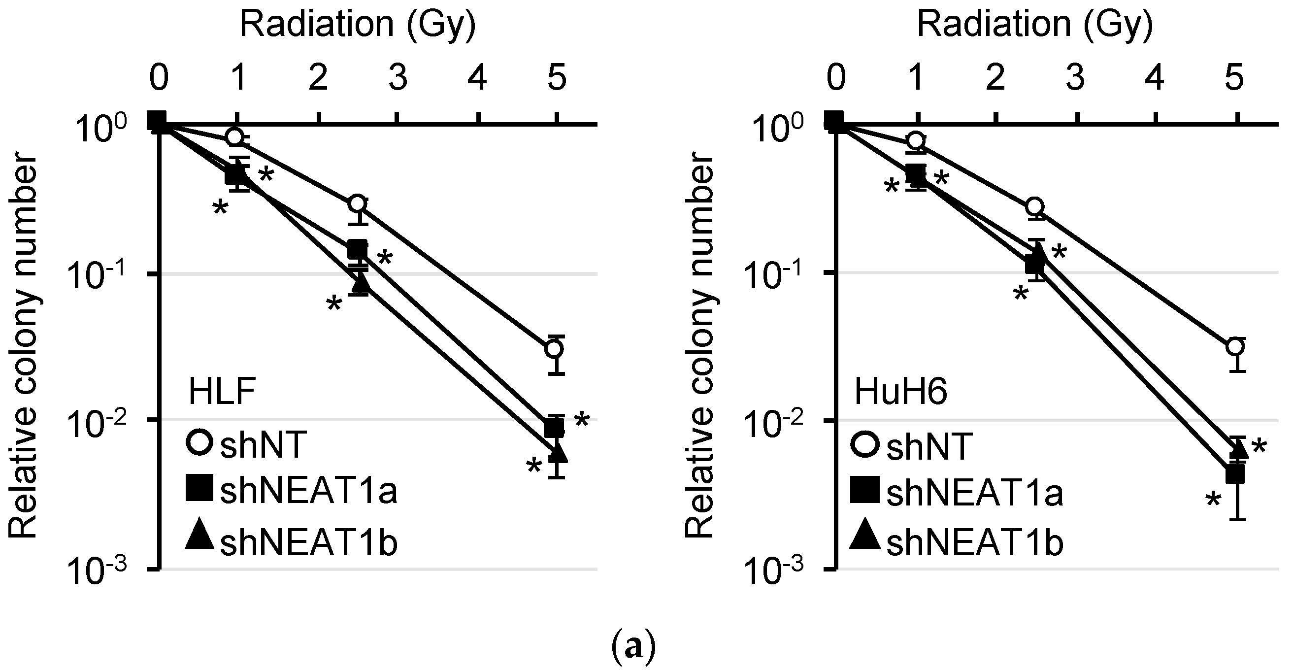
2.1. Radiosensitization of HCC Cell Lines by NEAT1 Knockdown

NEAT1v1 and NEAT1v2 have the same transcription start site, but their transcription terminates at different positions without splicing (Figure S1A). The expression of total NEAT1 (NEAT1v1 and NEAT1v2) and NEAT1v2 was significantly knocked down by shRNAs targeting NEAT1 (shNEAT1a and shNEAT1b) in HLF and HuH6 cell lines (Figure S1B). To evaluate the radiosensitivity of these cells, we performed a colony formation assay. It showed that the number of colonies was significantly decreased by NEAT1 knockdown (Figure 1a).

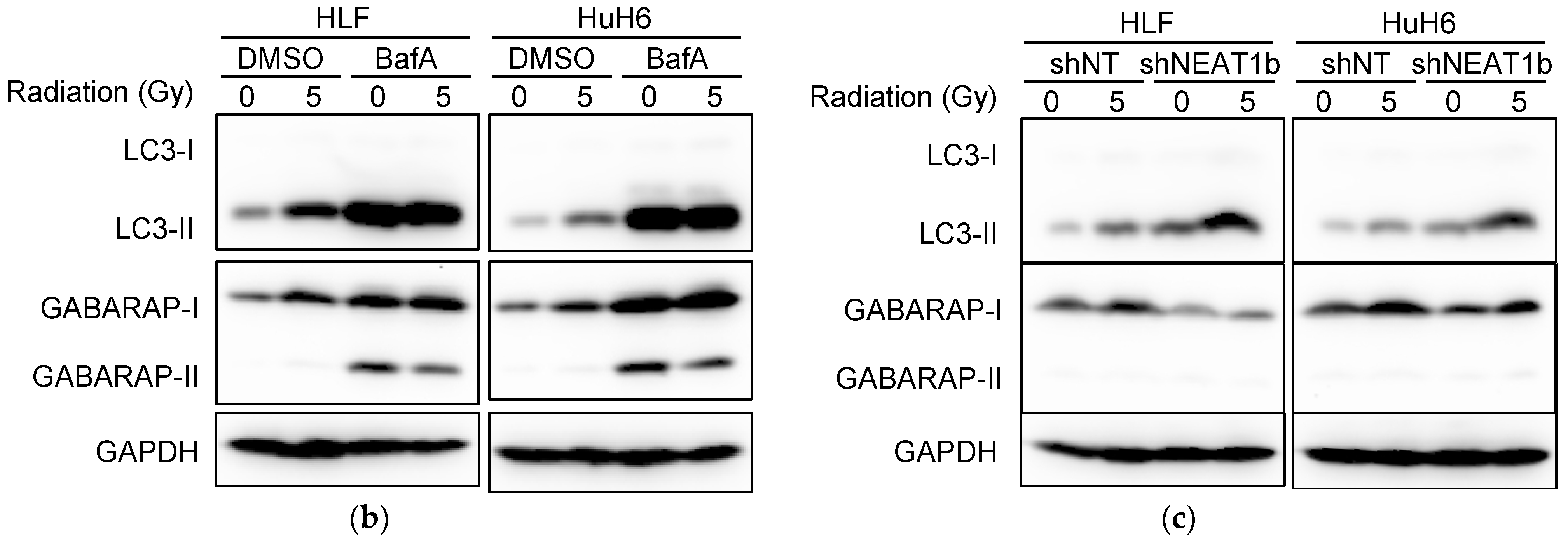
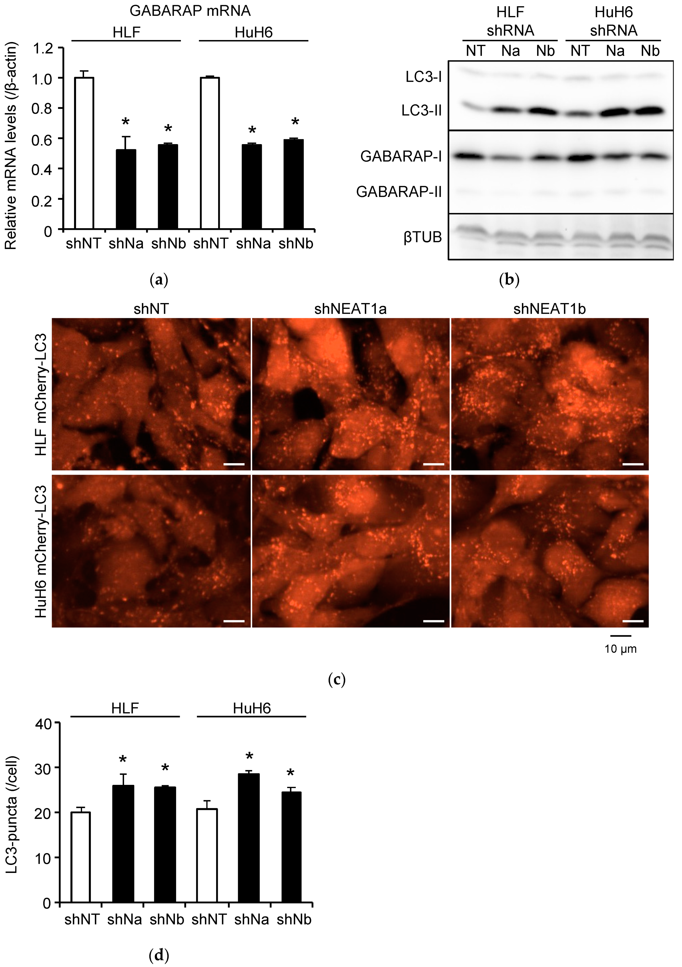
2.2. Stronger Suppression of Autophagy Induced by Radiation after NEAT1 Knockdown

Autophagy is one of the factors determining radiosensitivity [11]. LC3-II levels were increased in HLF and HuH6 cells after 5-Gy irradiation. However, they were not affected by the presence of bafilomycin A1 (BafA) (Figure 1b and Figure S2A), suggesting that the autophagy flux was inhibited by radiations. GABARAP, similar to LC3-II, is incorporated into autophagosomes and degraded by autophagy. GABARAP expression was also upregulated by radiation (Figure 1b and Figure S2A). NEAT1 knockdown further increased LC3-II levels in irradiated cells, suggesting that it enhanced the suppression of autophagy induced by radiations (Figure 1c and Figure S2B). The expression of GABARAP was decreased by NEAT1 knockdown in irradiated cells (Figure 1c and Figure S2B).

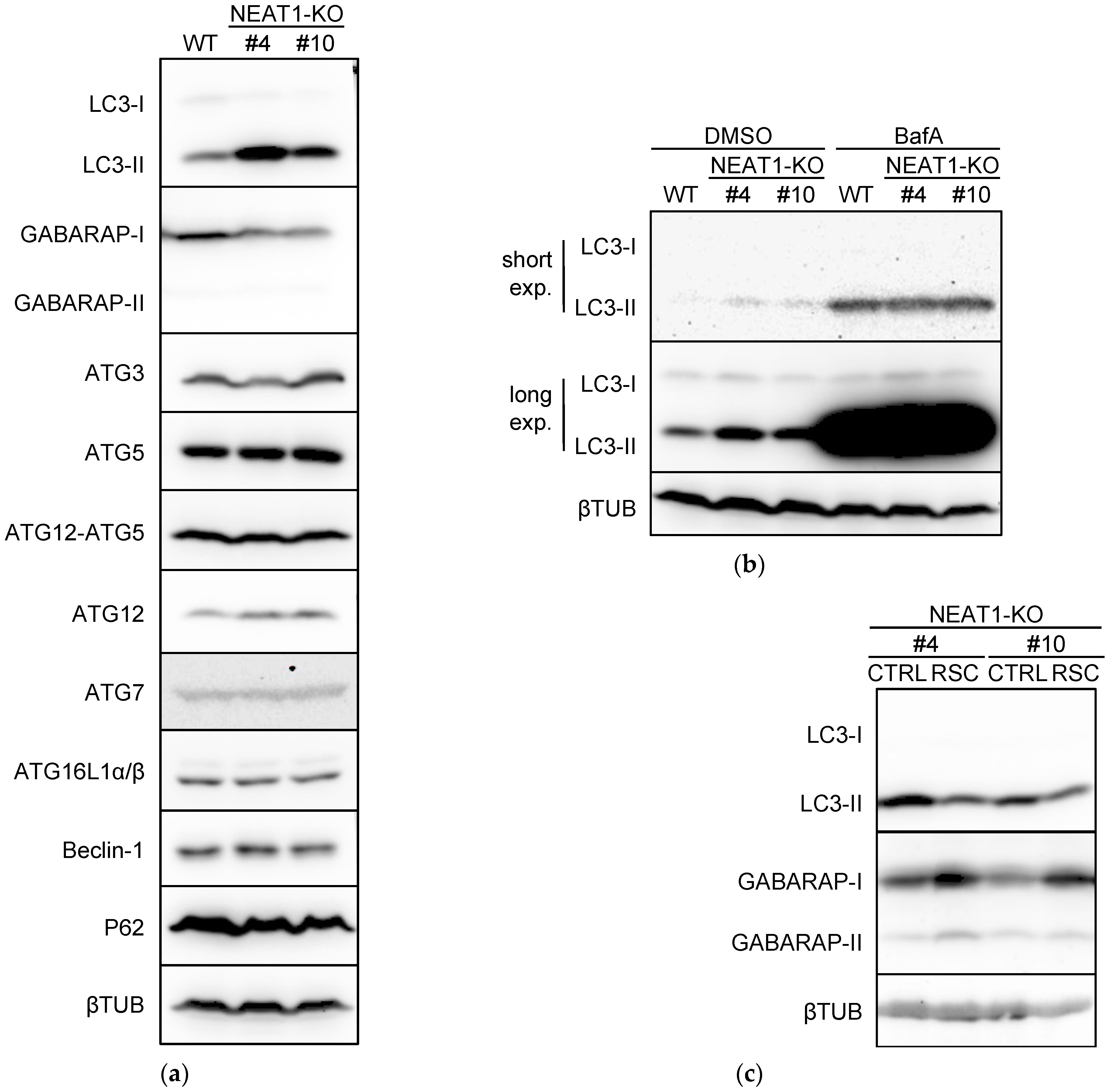
2.3. Suppression of Autophagy by NEAT1 Knockout

Because NEAT1 knockdown affected the autophagy flux (Figure 1), we investigated the expression of autophagy-related proteins in NEAT1-KO cells (clones #4 and #10) [10]. The expression of LC3-II and autophagy-related gene (ATG) 12 was upregulated in NEAT1-KO cells, whereas that of GABARAP was downregulated (Figure 2a and Figure S2C). Although ATG12 exerts its function by conjugating with ATG5 [12], the expression of the ATG12-ATG5 complex was not affected by NEAT1-KO (Figure 2a and Figure S2C). The autophagic flux assay revealed that the difference in LC3-II expression between wild-type (WT) and NEAT1-KO cells disappeared in the presence of BafA (Figure 2b and Figure S2D), suggesting that autophagy was suppressed in NEAT1-KO cells. This notion is consistent with the fact that GABARAP is a protein essential for the formation of autolysosomes and that its downregulation suppresses autophagy [13,14]. Next, we examined the expression levels of LC3-II and GABARAP in RSC cells generated from NEAT1-KO cells by stable transfection with NEAT1v1-expressing plasmid DNA. As shown in Figure 2c and Figure S2E, LC3-II levels were decreased by NEAT1v1 overexpression in NEAT1-KO cells, whereas GABARAP expression was increased. These results suggest that GABARAP protein expression is regulated by NEAT1v1.
As NEAT1 is required for the maintenance of CSCs in HCC cell lines [10], the constitutive lack of this lncRNA might cause unexpected phenotypic changes. Thus, we investigated the effects of NEAT1 deficiency on autophagy by performing transient knockdown of NEAT1. As shown in Figure 3a,b and Figure S2F, NEAT1 knockdown decreased the expression levels of GABARAP in HLF and HuH6 cells at both mRNA and protein levels. A reporter assay measuring the activity of the GABARAP promoter indicated that NEAT1v1 induced GABARAP expression by activating its promoter (Figure S3), while the intracellular stability of GABARAP protein was not affected by NEAT1v1 (Figure S4), suggesting that NEAT1v1 regulates GABARAP expression at the transcriptional level. Moreover, NEAT1 knockdown significantly increased LC3-II expression level and the number of LC3 puncta (Figure 3b–d and Figure S2F). These results suggest that NEAT1 is involved in maintaining the autophagy flux in HCC cells under nonirradiated conditions.

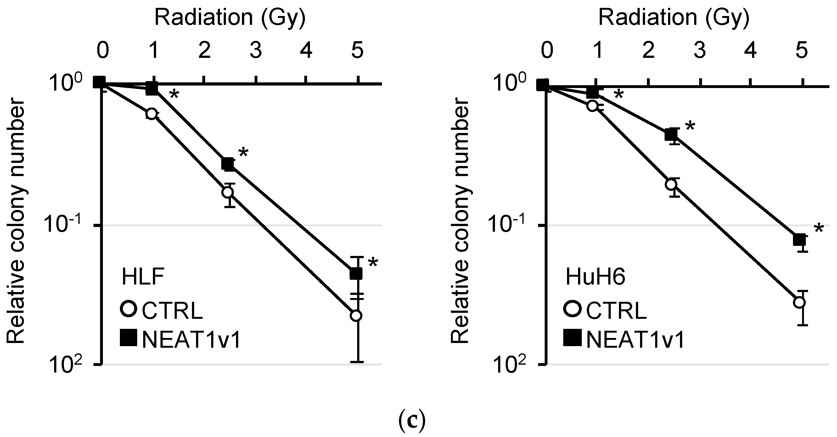
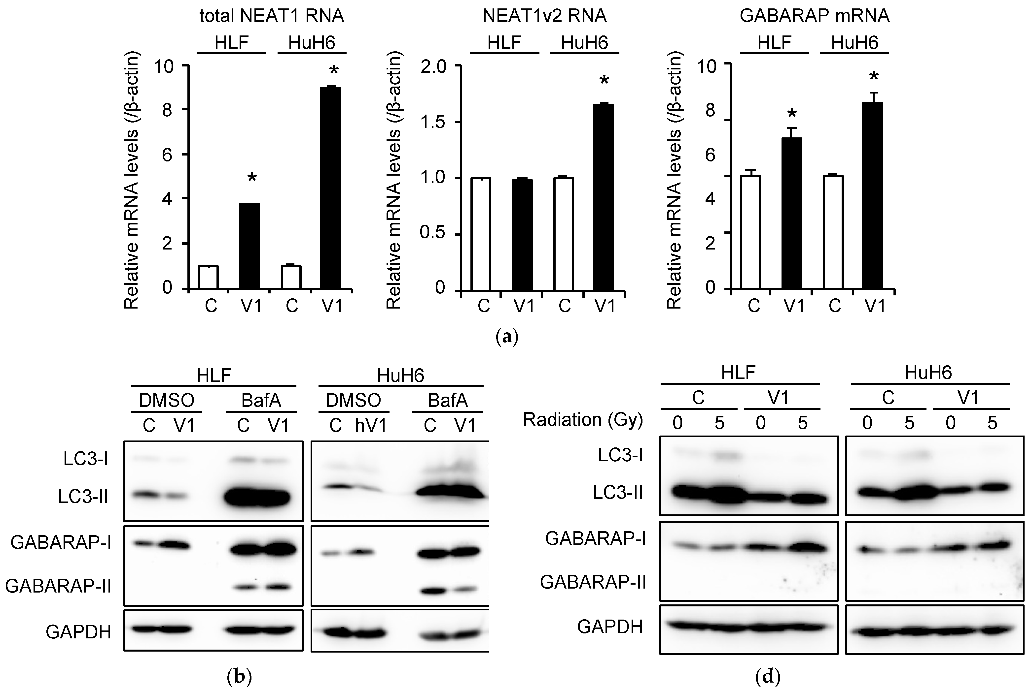
2.4. Induction of Autophagy and Radioresistance by NEAT1v1

We previously reported that NEAT1v1, but not NEAT1v2, is required for the maintenance of CSCs in HCC cell lines [10]. The rescue experiment showed that the overexpression of NEAT1v1 increased GABARAP levels in NEAT1-KO cells (Figure 2c and Figure S2E). Thus, HLF and HuH6 cell lines stably expressing NEAT1v1 were generated. In these cells, the mRNA expression levels of total NEAT1 and GABARAP were significantly increased (Figure 4a). Although no significant difference in NEAT1v2 expression was observed in HLF cells, it was approximately 1.6-fold upregulated by NEAT1v1 overexpression in HuH6 cells (Figure 4a). However, in both cell lines, GABARAP protein expression was upregulated, whereas that of LC3-II was downregulated, suggesting that autophagy was promoted by NEAT1v1 overexpression (Figure 4b and Figure S2G). Radioresistance was also significantly increased by NEAT1v1 overexpression in both cell lines (Figure 4c). Moreover, as shown in Figure 4d and Figure S2H, LC3-II protein expression was downregulated even after irradiation, suggesting that the autophagy flux inhibited by irradiation was restored by NEAT1v1 overexpression.

2.5. Induction of CSC Marker Expression by NEAT1v1 and Radiation

Because NEAT1v1 is involved in the maintenance of CSCs and was shown to regulate CD44, a CSC marker, expression in HuH7 and HepG2 cell lines [10], we determined the expression of CSC markers in HLF and HuH6 cell lines stably expressing NEAT1v1 after irradiation. As shown in Figure S5, the expression of total NEAT1, NEAT1v2, and GABARAP was not changed by the radiation. CD13 mRNA was upregulated after irradiation in both cell lines overexpressing NEAT1v1 (Figure S5). However, the expression of CD44 was increased only in NEAT1v1-overexpressing HLF cells, while radiation upregulated its expression in both cell lines (Figure S5). By contrast, CD90 was downregulated in NEAT1v1-overexpressing HLF cells, while it was significantly upregulated in NEAT1v1-overexpressing HuH6 cells after irradiation (Figure S5). The expression of the epithelial cell adhesion molecule (EPCAM) was induced by radiation in HLF cells but not affected by NEAT1v1 (Figure S5). Of note, CD133 expression could not be detected because of its low expression in the cell lines (data not shown). These results suggest that NEAT1v1 induces CSCs expressing different CSC markers depending on the HCC cell lines.

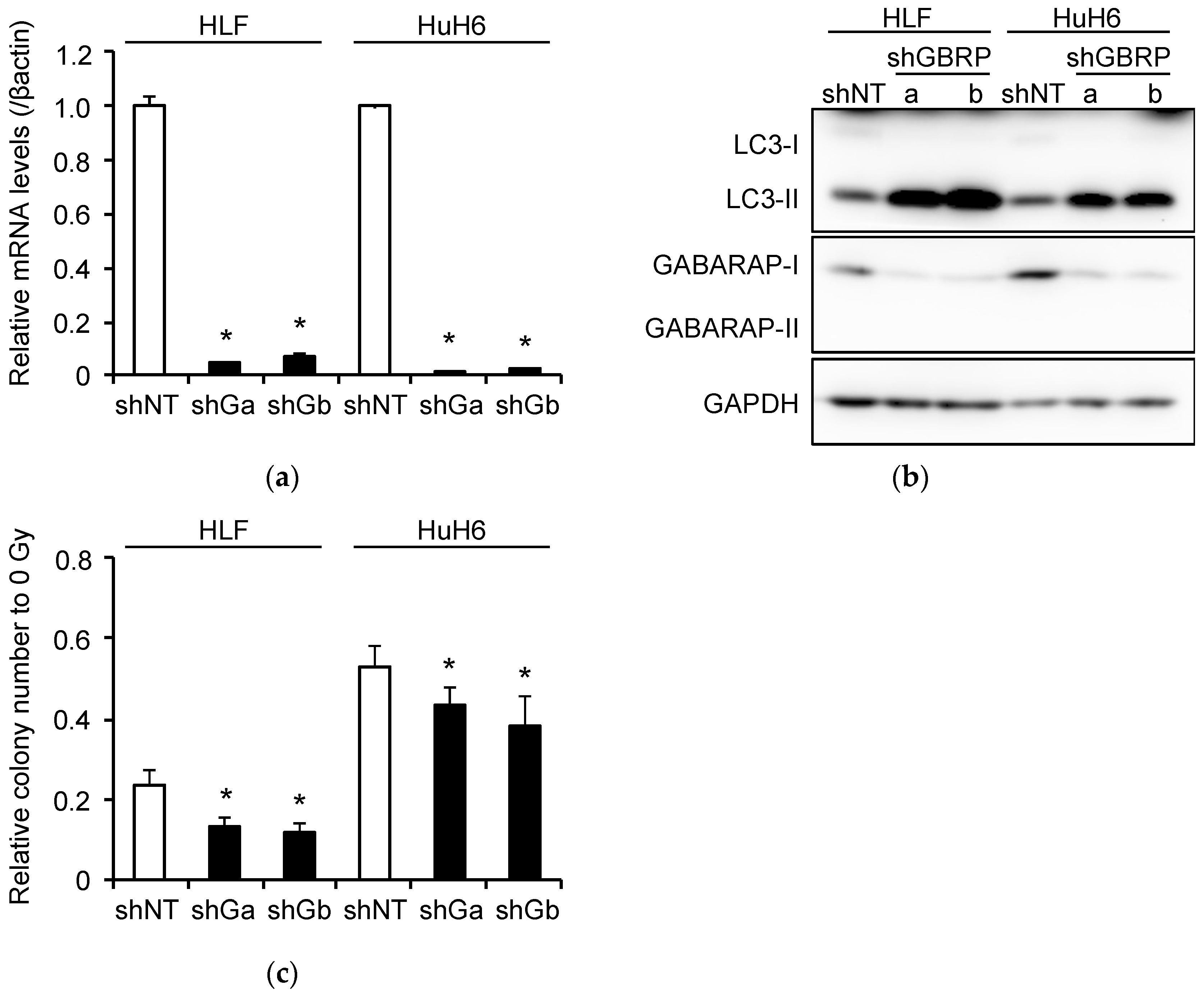
2.6. Induction of Radioresistance by NEAT1 via GABARAP

GABARAP expression was significantly knocked down by shRNAs targeting GABARAP (shGBRPa and shGBRPb) in HLF and HuH6 cells overexpressing NEAT1v1 (Figure 5a,b and Figure S2I). Concomitantly, LC3-II protein levels were markedly increased, suggesting the suppression of autophagy flux (Figure 5b and Figure S2I). Moreover, the clonogenicity of NEAT1v1-overexpressing HLF and HuH6 cells after irradiation was significantly decreased by GABARAP knockdown (Figure 5c). These results suggest that GABARAP is a critical mediator for NEAT1v1-induced radioresistance.

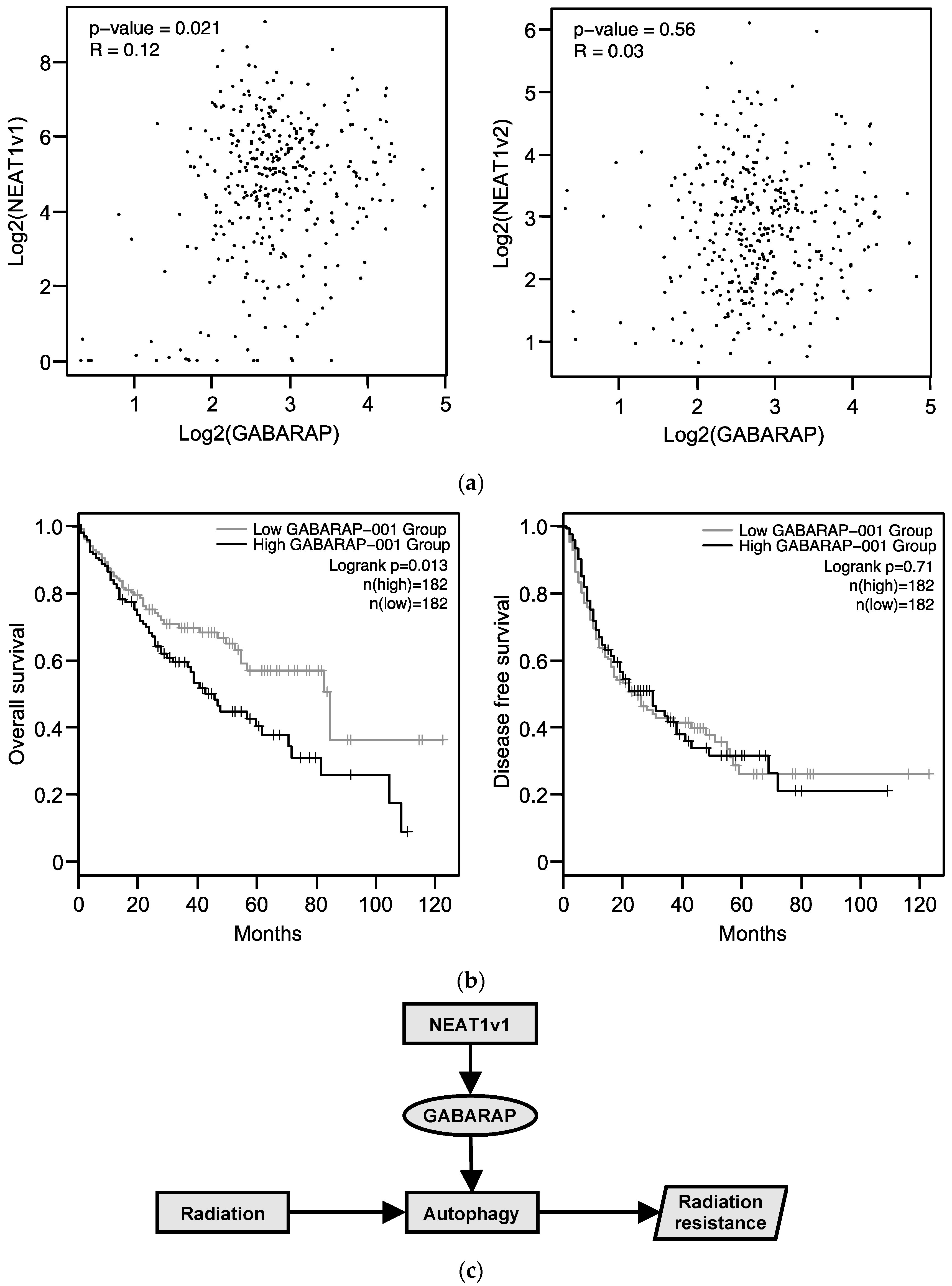
2.7. Relationship between GABARAP Expression in Tissues from HCC Patients and Prognosis

We analyzed RNA-seq data from HCC tissues obtained from TCGA. The expression of NEAT1v1, but not that of NEATv2, was significantly correlated with GABARAP expression (Figure 6a). Moreover, GABARAP expression was significantly correlated with the overall survival but not with the disease-free survival (Figure 6b). The patients registered in TCGA appear to have received various treatments, although the exact treatments are not clearly listed. However, these data suggest that autophagic dysfunctions are present in HCC tissues and might be associated with clinical outcomes in patients with HCC.

3. Discussion

Although radiation therapy is effective and noninvasive, it has so far played a limited role in HCC treatment because of the low tolerance of normal hepatic tissue. Recent advancements in radiation therapy, such as SBRT, allow efficiently delivering an ablative dose of radiation to tumors while sparing normal hepatocytes. Consequently, the number of HCC patients treated with radiation therapy is increasing [15]. However, radioresistance still remains a challenging issue, and its underlying mechanism needs to be clarified to improve the clinical efficacy of radiation therapy. The present study reveals that NEAT1v1 induces radioresistance in HCC cells. Moreover, we demonstrated that GABARAP expression is induced by NEAT1v1 and is involved in radioresistance by promoting autophagy (Figure 6c).
GABARAP, similar to LC3, belongs to the ATG8 family and is conjugated with phosphatidylethanolamine to bind the autophagosome membrane [16,17]. The ATG8 family consists of the LC3 subfamily, which includes LC3A, LC3B, and LC3C, and the GABARAP subfamily, constituted by GABARAP, GABARAPL1, and GABARAPL2 [16,17]. ATG8 family members are involved in the autophagosome formation and the autophagosome-lysosome fusion by binding proteins harboring an LC3-interacting region, including autophagy core proteins, cargo receptors, transport proteins, and proteins from the fusion machinery [16]. LC3 subfamily members are involved in the incorporation of autophagy cargos, whereas the GABARAP subfamily members are required for the autophagosome–lysosome fusion [17,18]. Our data demonstrate that the knockdown of GABARAP led to the inhibition of autophagy assessed by the accumulation of LC3-II (Figure 5b and Figure S2I). Therefore, and in agreement with previous studies [18], GABARAP is an indispensable protein for the formation of autolysosome.
Autophagy is an essential cellular process involving multiple factors in addition to GABARAP and LC-3. Beclin-1 is a component of class III phosphatidylinositol 3-kinase complex I and initiates autophagosome formation followed by phagophore expansion [19]. ATG3, ATG5, ATG7, ATG12, and ATG16L1 are involved in the sequential process of the conjugation of ATG8 family proteins with phosphatidylethanolamine [20,21]. P62, a representative autophagy substrate, plays a role as a cargo receptor that incorporates proteins and organelles into autophagosomes by binding to LC-3II [17]. The function of beclin-1 is inhibited by B-cell CLL/lymphoma 2, and thus, it has been demonstrated that beclin-1 acts as a tumor suppressor [22,23]. ATG proteins, such as ATG3, ATG12, and ATG16L1, are also demonstrated to have anti-oncogenic properties [24,25,26,27]. The impairment of autophagy causes the accumulation of P62, leading to the development of HCC [28]. Moreover, mice lacking Atg5 or Atg7 have been reported to develop liver cancers [29]. This suggests that autophagy plays a pivotal role in suppressing hepatocarcinogenesis. Moreover, GABARAP and LC3 were reported to have an anti-oncogenic function [16]. However, because autophagy plays a variety of pathophysiological roles, it contributes to both the survival and death of cancer cells [30,31]. Indeed, the expression of GABARAP and LC3 in breast cancer is significantly correlated with tumor malignancy and poor prognosis [32]. Moreover, the suppression of FOXO3 expression confers sorafenib resistance to HCC cells by accelerating autophagy [33], whereas the inhibition of autophagy enhanced the sensitivity to sorafenib [34]. Accumulating evidence suggests that autophagy has inhibitory functions against early carcinogenic processes in normal cells, whereas, in cancer cells, it contributes to tumor growth, malignant transformation, and therapeutic resistance [21]. This notion is consistent with our analysis of the TCGA dataset showing that the expression of GABARAP was correlated to the shortening of the overall survival of patients with HCC (Figure 5b and Figure S2I). Therefore, GABARAP-induced autophagy might promote the malignant transformation of HCC.
Autophagy is associated with the radiosensitivity of cancer cells. In esophageal cancer, autophagy induced by liver kinase B1 through the AMP-activated protein kinase pathway was shown to enhance radioresistance [35]. Additionally, cancer cells knocked down for ATG5 or beclin-1 exhibited an enhanced radiosensitivity as a consequence of the inhibition of autophagy [36]. In agreement with these reports, the present study demonstrated that autophagy in HCC cells was suppressed after irradiation (Figure 1b and Figure S2A). In contrast, NEAT1v1 induced the expression of GABARAP and promoted autophagy (Figure 4d and Figure S2H). Moreover, we showed that GABARAP knockdown inhibited autophagy in HCC cells overexpressing NEAT1v1 and enhanced their radiosensitivity (Figure 5b,c and Figure S2I). These results suggest that autophagy-mediated degradation and regeneration of organelles injured by radiation constitute possible mechanisms underlying the radioresistance of HCC. Our findings suggest that NEAT1 and GABARAP play important roles in organelle regeneration in HCC cells and that the inhibition of NEAT1v1 and GABARAP would improve the efficacy of radiation therapy. However, it was shown that autophagy accelerates radiation-induced cell death and enhances radiosensitivity [37,38,39]. The inhibition of autophagy also reduces radioresponse in vivo by suppressing the anti-tumor immunity [36]. In addition, autophagy is required to maintain homeostasis in cancer cells and in normal cells. Therefore, further studies are required to investigate the clinical benefit of radiosensitization provided by the inhibition of NEAT1v1 and GABARAP.
Several studies showed that NEAT1 is involved in the regulation of autophagy. NEAT1 accelerates lipopolysaccharide-induced autophagy to enhance the inflammatory response in renal fibroblast cells and osteoblast cells [40,41]. Conversely, NEAT1 inhibits PTEN-induced kinase 1dependent autophagy by facilitating the proteasomal degradation of NEDD4-like E3 ubiquitin-protein ligase. Consequently, NEAT1 inhibits the degradation and regeneration of impaired mitochondria and promotes pathophysiological processes involved in Alzheimer’s disease [42]. There are controversial reports regarding the regulation of autophagy by NEAT1 [43,44] in cardiac myocytes. The discrepancies might be attributed to the different pathologies and experimental models and require further investigation. In contrast, NEAT1 was reported to induce autophagy to enhance drug resistance of colon cancer and HCC [45,46]. Here, we also found that NEAT1v1 induced autophagy via GABARAP. Moreover, we demonstrated that NEAT1v1 activated the promoter of the GABARAP gene (Figure S3). The precise mechanisms by which NEAT1v1 induces GABARAP expression remain to be elucidated. The identification of the binding partners of NEAT1v1 by RNA pull-down assay would facilitate accomplishing it. Other additional NEAT1v1 target genes in HCC would also be useful to identify the transcription factors that regulate NEAT1v1 target genes. Conversely, in the above-mentioned studies [40,43,45,46], NEAT1 promoted autophagy by acting as a competing endogenous RNA (ceRNA) and preventing miRNAs (miR-22-3p, miR-34a, miR-204, and miR-378a-3p) to target autophagy-related proteins. Therefore, NEAT1 might regulate GABARAP expression at the pre-and post-transcriptional levels.
NEAT1 might influence the radiosensitivity of various cancer cells including HCC. In many cases, NEAT1, as a ceRNA, inhibited miR-27b-3p [47], miR-101-3p [48,49], miR-193-3p [50], and miR-204 [51] to augment [47,48,50,51] or attenuate [49] the radioresistance of cancer cells. However, these miRNAs are involved in the cell cycle and epithelial-mesenchymal transition. Autophagy-related miRNAs have not been implicated in the radiosensitivity regulated by NEAT1. On the other hand, although the involvement of miRNAs is not clear, NEAT1 induces radioresistance in triple-negative breast cancer (TNBC) cells by upregulating the translation of NAD(P)H:quinone oxidoreductase rather than its transcription [52]. Intriguingly, NEAT1 concomitantly increased the cancer stemness of TNBC cells and upregulated the expression of BMI1, OCT4, and SOX2 [52]. In agreement with this, we demonstrated that NEAT1v1 increased CSC makers, such as CD13, CD44 and CD90, in irradiated cells, suggesting that NEAT1v1 might induce cancer stemness to protect HCC cells from radiation. Because CSCs are more dependent on autophagy than normal stem cells [53], NEAT1v1-induced autophagy via GABARAP might be a possible mechanism underlying the maintenance of cancer stemness.
In conclusion, we show that NEAT1v1 confers radioresistance to HCC cells by inducing autophagy through GABARAP (Figure 6c). Thus, treatments targeting NEAT1v1 and GABARAP are expected to improve the efficacy of radiation therapy. Moreover, since NEAT1v1 plays a pivotal role in maintaining cancer stemness, the suppression of NEAT1v1 might lead to effective eradication of HCC.

4. Materials and Methods

4.1. Cell Culture

Human HCC cell lines, HuH7, HLF, and HuH6, were purchased from the Japanese Collection of Research Bioresources Cell Bank (Osaka, Japan) and were maintained in Dulbecco’s Modified Eagle Medium (Nissui Pharmaceutical, Tokyo, Japan) supplemented with 10% inactivated fetal bovine serum (Sigma-Aldrich, St. Louis, MO, USA). NEAT1-KO HuH7 cells and rescue (RSC) cells were previously reported [10]. HLF and HuH6 cells overexpressing human NEAT1v1 were constructed by transfection of pcDNA6-hNEAT1v1-AcGFP [10] with LipofectAMINE2000 (Thermo Fisher Scientific, Waltham, MA, USA). Following blasticidin selection, AcGFP-positive cells were sorted by flow cytometry. HLF and HuH6 cells overexpressing mCherry-LC3 were constructed by transfection of pmCherry-LC3 [54], which was provided by the RIKEN BRC through the National Bio-Resource Project of the MEXT, Japan, with LipofectAMINE2000. Following G418 selection, an mCherry-positive clone was manually picked.

4.2. Adenovirus Construction

All oligo DNAs and primers used are shown in Table S1. A BsaI-linker was ligated into pENTR/U6 (Thermo Fisher Scientific), resulting in pENTR/U6-Bgl2. The pAmCyan1-C1 plasmid (Takara Bio, Shiga, Japan) was digested with BglII and BamHI and then self-ligated to remove the multi-cloning site, resulting in pAmCyan1-noMCS. The AmCyan1-expressing cassette was amplified by KOD-neo-plus (Toyobo, Osaka, Japan) with AmCyan primers. Following BamHI digestion, it was inserted into the BglII site of pENTR/U6-Bgl2, resulting in pENTR/U6-AmCyan1. After BsaI digestion of pENTR/U6-AmCyan1, non-targeting (NT) short hairpin RNAs (shRNAs) (shNT), NEAT1-targeting shRNAs (shNEAT1a/b), or GABARAP-targeting shRNAs (shGBRPa/b) were ligated with Ligation High ver.2 (Toyobo). The shRNA and AmCyan1-expressing cassettes were transferred by LR reaction to pAd/BLOCK-iT-DEST (Thermo Fisher Scientific). Adenovirus vectors were constructed by transfection of adenovirus plasmid DNAs into 293T cells with LipofectAMINE2000 (Thermo Fisher Scientific) according to the manufacturer’s protocol. The adenovirus titer was determined using the infectious genome titration protocol [55].

4.3. Reverse-Transcription Quantitative PCR (RT-qPCR) and Western Blot Analyses

RT-qPCR and Western blot analyses were performed as reported previously [10]. Protein and mRNA samples were prepared 48 h after seeding or irradiation. The primers used for RT-qPCR are found in Table S1. Relative mRNA expression levels were calculated using β-actin as the internal control. For Western blot analyses, antibodies recognizing GABARAP (sc-377300, Santa Cruz Biotechnology, Santa Cruz, CA, USA), glyceraldehyde-3-phosphate dehydrogenase (GAPDH; sc-365062, Santa Cruz Biotechnology), and β-tubulin (βTUB; sc-55529, Santa Cruz Biotechnology), as well as the Autophagy Antibody Sampler Kit (#4445, Cell Signaling Technology, Danvers, MA, USA), were used.

4.4. Colony Formation Assay

Cells were seeded into a 24-well plate and were allowed to attach overnight. The cells were irradiated (0, 1, 2.5, or 5 Gy) using an X-ray generator (MX-160Labo, mediXtec Japan, Chiba, Japan). Afterward, the medium was changed, or cells were transduced with adenovirus at a multiplicity of infection of 200. After 24 h in culture, 100–10,000 viable cells were quantified and seeded into a new plate. After 10 days, the cells were fixed and stained with a crystal violet solution (0.5% crystal violet in 10% methanol), and colonies (>50 cells) were counted.

4.5. Autophagic Flux Assay

Twenty-four hours after seeding or irradiation, cells were treated with 50 nM bafilomycin A1 (BafA; Cayman Chemical, Ann Arbor, MI, USA) for 24 h. We have confirmed that this treatment does not affect cell viabilities by a WST assay using Cell Counting Kit-8 ((Dojindo, Kumamoto, Japan) (data not shown). An equal volume of dimethyl sulfoxide (DMSO) was added to untreated control cells. LC3 puncta were assessed in HLF and HuH6 cells overexpressing mCherry-LC3. Cells were seeded onto a 3.5-cm glass-bottom dish (Matsunami Glass, Osaka, Japan) and were allowed to attach overnight. Cells were transduced with adenovirus vectors expressing shNT, shNEAT1a, or shNEAT1b. Confocal images of LC3-puncta (4 pictures for each shRNA) were obtained 48 h post-transduction using an LCV110CSU microscope (Olympus, Tokyo, Japan). The number of LC3-puncta was counted by ImageJ software (Bethesda, MD, USA).

4.6. Analysis of Gene Expression in HCC Tissues

The expression of NEAT1v1 (ENST00000499732.2), NEAT1v2 (ENST00000501122.2), and GABARAP (ENST00000302386.9) was analyzed in the Cancer Genome Atlas (TCGA) dataset for HCC (LIHC) by GEPIA2 [56].

4.7. Statistical Analysis

Three or more independent samples were analyzed in each experiment. All experimental values were expressed as means ± standard deviations. The differences between two groups were assessed by two-tailed unpaired Student’s t-test. Multiple comparisons were made using the two-tailed Dunnett’s or Tukey’s test. The differences were considered significant for a p value less than 0.05.

Supplementary Materials

The following are available online at https://www.mdpi.com/article/10.3390/ijms23020711/s1.

Author Contributions

Conceptualization, H.S., H.T., Y.K. and T.T.; methodology, H.T.; investigation, H.S. and H.T.; data curation, H.S. and H.T.; writing—original draft preparation, H.S. and H.T.; writing—review and editing, Y.K., T.T., K.Y., N.U. and G.S.; supervision, H.T., K.Y., N.U. and G.S.; project administration, K.Y., N.U. and G.S.; funding acquisition, H.S. and H.T. All authors have read and agreed to the published version of the manuscript.

Funding

This work was funded by JSPS KAKENHI Grant Number JP19K08469 (HT) and JP19K17460 (HS).

Institutional Review Board Statement

This article does not contain any studies involving human participants performed by any of the authors.

Informed Consent Statement

Not applicable.

Data Availability Statement

The raw data are available upon request, please contact the corresponding author.

Acknowledgments

The authors are sincerely grateful to A. Fox (University of Western Australia) for the gift of pCRII_TOPO_hNEAT1.

Conflicts of Interest

We declare the following conflict of interest: GS holds more than 5% of the total shares of KanonCure Inc., and receives compensation as a member of KanonCure Inc. The other authors have no competing interests.

References

  1. Sung, H.; Ferlay, J.; Siegel, R.L.; Laversanne, M.; Soerjomataram, I.; Jemal, A.; Bray, F. Global cancer statistics 2020: Globocan estimates of incidence and mortality worldwide for 36 cancers in 185 countries. CA Cancer J. Clin. 2021, 71, 209–249. [Google Scholar] [CrossRef] [PubMed]
  2. European Association for the Study of the Liver. EASL Clinical Practice Guidelines: Management of hepatocellular carcinoma. J. Hepatol. 2018, 69, 182–236. [Google Scholar] [CrossRef] [Green Version]
  3. Peng, W.X.; Wan, Y.Y.; Gong, A.H.; Ge, L.; Jin, J.; Xu, M.; Wu, C.Y. Egr-1 regulates irradiation-induced autophagy through Atg4B to promote radioresistance in hepatocellular carcinoma cells. Oncogenesis 2017, 6, e292. [Google Scholar] [CrossRef] [Green Version]
  4. Su, T.S.; Liang, P.; Lu, H.Z.; Liang, J.; Gao, Y.C.; Zhou, Y.; Huang, Y.; Tang, M.Y.; Liang, J.N. Stereotactic body radiation therapy for small primary or recurrent hepatocellular carcinoma in 132 Chinese patients. J. Surg. Oncol. 2016, 113, 181–187. [Google Scholar] [CrossRef]
  5. Tsuchiya, H.; Shiota, G. Clinical and Biological Implications of Cancer Stem Cells in Hepatocellular Carcinoma. Yonago Acta Med. 2021, 64, 1–11. [Google Scholar] [CrossRef]
  6. Shindoh, J.; Makuuchi, M.; Matsuyama, Y.; Mise, Y.; Arita, J.; Sakamoto, Y.; Hasegawa, K.; Kokudo, N. Complete removal of the tumor-bearing portal territory decreases local tumor recurrence and improves disease-specific survival of patients with hepatocellular carcinoma. J. Hepatol. 2016, 64, 594–600. [Google Scholar] [CrossRef] [PubMed]
  7. Gao, W.; Chen, L.; Ma, Z.; Du, Z.; Zhao, Z.; Hu, Z.; Li, Q. Isolation and phenotypic characterization of colorectal cancer stem cells with organ-specific metastatic potential. Gastroenterology 2013, 145, 636–646. [Google Scholar] [CrossRef] [PubMed]
  8. Ayob, A.Z.; Ramasamy, T.S. Cancer stem cells as key drivers of tumour progression. J. Biomed. Sci. 2018, 25, 20. [Google Scholar] [CrossRef] [PubMed]
  9. Nakagawa, S.; Naganuma, T.; Shioi, G.; Hirose, T. Paraspeckles are subpopulation-specific nuclear bodies that are not essential in mice. J. Cell Biol. 2011, 193, 31–39. [Google Scholar] [CrossRef]
  10. Koyama, S.; Tsuchiya, H.; Amisaki, M.; Sakaguchi, H.; Honjo, S.; Fujiwara, Y.; Shiota, G. NEAT1 is Required for the Expression of the Liver Cancer Stem Cell Marker CD44. Int. J. Mol. Sci. 2020, 21, 1927. [Google Scholar] [CrossRef] [Green Version]
  11. Kim, J.; Lee, S.; Kim, H.; Lee, H.; Seong, K.M.; Youn, H.; Youn, B. Autophagic Organelles in DNA Damage Response. Front. Cell Dev. Biol. 2021, 9, 668735. [Google Scholar] [CrossRef]
  12. Sakoh-Nakatogawa, M.; Matoba, K.; Asai, E.; Kirisako, H.; Ishii, J.; Noda, N.N.; Inagaki, F.; Nakatogawa, H.; Ohsumi, Y. Atg12-Atg5 conjugate enhances E2 activity of Atg3 by rearranging its catalytic site. Nat. Struct. Mol. Biol. 2013, 20, 433–439. [Google Scholar] [CrossRef] [PubMed]
  13. Nakamura, S.; Yoshimori, T. New insights into autophagosome-lysosome fusion. J. Cell Sci. 2017, 130, 1209–1216. [Google Scholar] [CrossRef] [PubMed] [Green Version]
  14. Wang, H.; Sun, H.Q.; Zhu, X.; Zhang, L.; Albanesi, J.; Levine, B.; Yin, H. GABARAPs regulate PI4P-dependent autophagosome:lysosome fusion. Proc. Natl. Acad. Sci. USA 2015, 112, 7015–7020. [Google Scholar] [CrossRef] [PubMed] [Green Version]
  15. Kamimura, K.; Terai, S. The promise of radiotherapy for hepatocellular carcinoma. Hepatol. Res. 2021, 51, 837–838. [Google Scholar] [CrossRef] [PubMed]
  16. Schaaf, M.B.; Keulers, T.G.; Vooijs, M.A.; Rouschop, K.M. LC3/GABARAP family proteins: Autophagy-(un)related functions. FASEB J. 2016, 30, 3961–3978. [Google Scholar] [CrossRef] [Green Version]
  17. Johansen, T.; Lamark, T. Selective Autophagy: ATG8 Family Proteins, LIR Motifs and Cargo Receptors. J. Mol. Biol. 2020, 432, 80–103. [Google Scholar] [CrossRef]
  18. Nguyen, T.N.; Padman, B.S.; Usher, J.; Oorschot, V.; Ramm, G.; Lazarou, M. Atg8 family LC3/GABARAP proteins are crucial for autophagosome-lysosome fusion but not autophagosome formation during PINK1/Parkin mitophagy and starvation. J. Cell Biol. 2016, 215, 857–874. [Google Scholar] [CrossRef]
  19. Itakura, E.; Kishi, C.; Inoue, K.; Mizushima, N. Beclin 1 forms two distinct phosphatidylinositol 3-kinase complexes with mammalian Atg14 and UVRAG. Mol. Biol. Cell 2008, 19, 5360–5372. [Google Scholar] [CrossRef] [PubMed] [Green Version]
  20. Klionsky, D.J.; Petroni, G.; Amaravadi, R.K.; Baehrecke, E.H.; Ballabio, A.; Boya, P.; Bravo-San Pedro, J.M.; Cadwell, K.; Cecconi, F.; Choi, A.; et al. Autophagy in major human diseases. EMBO J. 2021, 40, e108863. [Google Scholar] [CrossRef] [PubMed]
  21. Amaravadi, R.K.; Kimmelman, A.C.; Debnath, J. Targeting Autophagy in Cancer: Recent Advances and Future Directions. Cancer Discov. 2019, 9, 1167–1181. [Google Scholar] [CrossRef] [Green Version]
  22. Pattingre, S.; Tassa, A.; Qu, X.; Garuti, R.; Liang, X.H.; Mizushima, N.; Packer, M.; Schneider, M.D.; Levine, B. Bcl-2 antiapoptotic proteins inhibit Beclin 1-dependent autophagy. Cell 2005, 122, 927–939. [Google Scholar] [CrossRef] [Green Version]
  23. Yue, Z.; Jin, S.; Yang, C.; Levine, A.J.; Heintz, N. Beclin 1, an autophagy gene essential for early embryonic development, is a haploinsufficient tumor suppressor. Proc. Natl. Acad. Sci. USA 2003, 100, 15077–15082. [Google Scholar] [CrossRef] [PubMed] [Green Version]
  24. Altman, B.J.; Jacobs, S.R.; Mason, E.F.; Michalek, R.D.; MacIntyre, A.N.; Coloff, J.L.; Ilkayeva, O.; Jia, W.; He, Y.W.; Rathmell, J.C. Autophagy is essential to suppress cell stress and to allow BCR-Abl-mediated leukemogenesis. Oncogene 2011, 30, 1855–1867. [Google Scholar] [CrossRef] [PubMed] [Green Version]
  25. Keulers, T.G.; Koch, A.; van Gisbergen, M.W.; Barbeau, L.; Zonneveld, M.I.; de Jong, M.C.; Savelkouls, K.; Wanders, R.G.; Bussink, J.; Melotte, V.; et al. ATG12 deficiency results in intracellular glutamine depletion, abrogation of tumor hypoxia and a favorable prognosis in cancer. Autophagy 2021, 1–17, Advance online publication. [Google Scholar] [CrossRef] [PubMed]
  26. Reuken, P.A.; Lutz, P.; Casper, M.; Al-Herwi, E.; Stengel, S.; Spengler, U.; Stallmach, A.; Lammert, F.; Nischalke, H.D.; Bruns, T. The ATG16L1 gene variant rs2241880 (p.T300A) is associated with susceptibility to HCC in patients with cirrhosis. Liver Int. 2019, 39, 2360–2367. [Google Scholar] [CrossRef] [Green Version]
  27. Capparelli, C.; Guido, C.; Whitaker-Menezes, D.; Bonuccelli, G.; Balliet, R.; Pestell, T.G.; Goldberg, A.F.; Pestell, R.G.; Howell, A.; Sneddon, S.; et al. Autophagy and senescence in cancer-associated fibroblasts metabolically supports tumor growth and metastasis via glycolysis and ketone production. Cell Cycle 2012, 11, 2285–2302. [Google Scholar] [CrossRef] [Green Version]
  28. Ichimura, Y.; Komatsu, M. Activation of p62/SQSTM1-Keap1-Nuclear Factor Erythroid 2-Related Factor 2 Pathway in Cancer. Front. Oncol. 2018, 8, 210. [Google Scholar] [CrossRef]
  29. Takamura, A.; Komatsu, M.; Hara, T.; Sakamoto, A.; Kishi, C.; Waguri, S.; Eishi, Y.; Hino, O.; Tanaka, K.; Mizushima, N. Autophagy-deficient mice develop multiple liver tumors. Genes Dev. 2011, 25, 795–800. [Google Scholar] [CrossRef] [Green Version]
  30. White, E.; DiPaola, R.S. The double-edged sword of autophagy modulation in cancer. Clin. Cancer Res. 2009, 15, 5308–5316. [Google Scholar] [CrossRef] [PubMed] [Green Version]
  31. Singh, S.S.; Vats, S.; Chia, A.Y.; Tan, T.Z.; Deng, S.; Ong, M.S.; Arfuso, F.; Yap, C.T.; Goh, B.C.; Sethi, G.; et al. Dual role of autophagy in hallmarks of cancer. Oncogene 2018, 37, 1142–1158. [Google Scholar] [CrossRef]
  32. Bortnik, S.; Tessier-Cloutier, B.; Leung, S.; Xu, J.; Asleh, K.; Burugu, S.; Magrill, J.; Greening, K.; Derakhshan, F.; Yip, S.; et al. Differential expression and prognostic relevance of autophagy-related markers ATG4B, GABARAP, and LC3B in breast cancer. Breast Cancer Res. Treat. 2020, 183, 525–547. [Google Scholar] [CrossRef]
  33. Lin, Z.; Niu, Y.; Wan, A.; Chen, D.; Liang, H.; Chen, X.; Sun, L.; Zhan, S.; Chen, L.; Cheng, C.; et al. RNA m6 A methylation regulates sorafenib resistance in liver cancer through FOXO3-mediated autophagy. EMBO J. 2020, 39, e103181. [Google Scholar] [CrossRef]
  34. Shimizu, S.; Takehara, T.; Hikita, H.; Kodama, T.; Tsunematsu, H.; Miyagi, T.; Hosui, A.; Ishida, H.; Tatsumi, T.; Kanto, T.; et al. Inhibition of autophagy potentiates the antitumor effect of the multikinase inhibitor sorafenib in hepatocellular carcinoma. Int. J. Cancer. 2012, 131, 548–557. [Google Scholar] [CrossRef] [PubMed]
  35. He, Q.; Li, J.; Dong, F.; Cai, C.; Zou, X. LKB1 promotes radioresistance in esophageal cancer cells exposed to radiation, by suppression of apoptosis and activation of autophagy via the AMPK pathway. Mol. Med. Rep. 2017, 16, 2205–2210. [Google Scholar] [CrossRef] [PubMed] [Green Version]
  36. Ko, A.; Kanehisa, A.; Martins, I.; Senovilla, L.; Chargari, C.; Dugue, D.; Mariño, G.; Kepp, O.; Michaud, M.; Perfettini, J.L.; et al. Autophagy inhibition radiosensitizes in vitro, yet reduces radioresponses in vivo due to deficient immunogenic signalling. Cell Death Differ. 2014, 21, 92–99. [Google Scholar] [CrossRef]
  37. Peng, P.L.; Kuo, W.H.; Tseng, H.C.; Chou, F.P. Synergistic tumor-killing effect of radiation and berberine combined treatment in lung cancer: The contribution of autophagic cell death. Int. J. Radiat. Oncol. Biol. Phys. 2008, 70, 529–542. [Google Scholar] [CrossRef] [PubMed]
  38. Chen, Z.; Wang, B.; Yu, F.; Chen, Q.; Tian, Y.; Ma, S.; Liu, X. The roles of mitochondria in radiation-induced autophagic cell death in cervical cancer cells. Tumor Biol. 2016, 37, 4083–4091. [Google Scholar] [CrossRef] [PubMed]
  39. Cui, L.; Song, Z.; Liang, B.; Jia, L.; Ma, S.; Liu, X. Radiation induces autophagic cell death via the p53/DRAM signaling pathway in breast cancer cells. Oncol. Rep. 2016, 35, 3639–3647. [Google Scholar] [CrossRef] [Green Version]
  40. Feng, Y.; Liu, J.; Wu, R.; Yang, P.; Ye, Z.; Song, F. NEAT1 aggravates sepsis-induced acute kidney injury by sponging miR-22-3p. Open Med. 2020, 15, 333–342. [Google Scholar] [CrossRef] [PubMed]
  41. Dai, W.; Wang, M.; Wang, P.; Wen, J.; Wang, J.; Cha, S.; Xiao, X.; He, Y.; Shu, R.; Bai, D. lncRNA NEAT1 ameliorates LPS-induced inflammation in MG63 cells by activating autophagy and suppressing the NLRP3 inflammasome. Int. J. Mol. Med. 2021, 47, 607–620. [Google Scholar] [CrossRef] [PubMed]
  42. Huang, Z.; Zhao, J.; Wang, W.; Zhou, J.; Zhang, J. Depletion of LncRNA NEAT1 Rescues Mitochondrial Dysfunction Through NEDD4L-Dependent PINK1 Degradation in Animal Models of Alzheimer’s Disease. Front. Cell. Neurosci. 2020, 14, 28. [Google Scholar] [CrossRef] [Green Version]
  43. Zhao, J.; Chen, F.; Ma, W.; Zhang, P. Suppression of long noncoding RNA NEAT1 attenuates hypoxia-induced cardiomyocytes injury by targeting miR-378a-3p. Gene 2020, 731, 144324. [Google Scholar] [CrossRef] [PubMed]
  44. Lv, Y.; Liu, Z.; Huang, J.; Yu, J.; Dong, Y.; Wang, J. LncRNA nuclear-enriched abundant transcript 1 regulates hypoxia-evoked apoptosis and autophagy via mediation of microRNA-181b. Mol. Cell. Biochem. 2020, 464, 193–203. [Google Scholar] [CrossRef] [PubMed]
  45. Liu, F.; Ai, F.Y.; Zhang, D.C.; Tian, L.; Yang, Z.Y.; Liu, S.J. LncRNA NEAT1 knockdown attenuates autophagy to elevate 5-FU sensitivity in colorectal cancer via targeting miR-34a. Cancer Med. 2020, 9, 1079–1091. [Google Scholar] [CrossRef] [Green Version]
  46. Li, X.; Zhou, Y.; Yang, L.; Ma, Y.; Peng, X.; Yang, S.; Li, H.; Liu, J. LncRNA NEAT1 promotes autophagy via regulating miR-204/ATG3 and enhanced cell resistance to sorafenib in hepatocellular carcinoma. J. Cell. Physiol. 2020, 235, 3402–3413. [Google Scholar] [CrossRef]
  47. Jiang, Y.; Jin, S.; Tan, S.; Xue, Y.; Cao, X. Long noncoding RNA NEAT1 regulates radio-sensitivity via microRNA-27b-3p in gastric cancer. Cancer Cell Int. 2020, 20, 581. [Google Scholar] [CrossRef]
  48. Chen, X.; Zhang, N. Downregulation of lncRNA NEAT1_2 radiosensitizes hepatocellular carcinoma cells through regulation of miR-101-3p/WEE1 axis. Cell Biol. Int. 2019, 43, 44–55. [Google Scholar] [CrossRef] [Green Version]
  49. Wang, Y.; Wang, C.; Chen, C.; Wu, F.; Shen, P.; Zhang, P.; He, G.; Li, X. Long non-coding RNA NEAT1 regulates epithelial membrane protein 2 expression to repress nasopharyngeal carcinoma migration and irradiation-resistance through miR-101-3p as a competing endogenous RNA mechanism. Oncotarget 2017, 8, 70156–70171. [Google Scholar] [CrossRef] [Green Version]
  50. Han, D.; Wang, J.; Cheng, G. LncRNA NEAT1 enhances the radio-resistance of cervical cancer via miR-193b-3p/CCND1 axis. Oncotarget 2017, 9, 2395–2409. [Google Scholar] [CrossRef] [Green Version]
  51. Lu, Y.; Li, T.; Wei, G.; Liu, L.; Chen, Q.; Xu, L.; Zhang, K.; Zeng, D.; Liao, R. The long non-coding RNA NEAT1 regulates epithelial to mesenchymal transition and radioresistance in through miR-204/ZEB1 axis in nasopharyngeal carcinoma. Tumor Biol. 2016, 37, 11733–11741. [Google Scholar] [CrossRef] [PubMed]
  52. Lin, L.C.; Lee, H.T.; Chien, P.J.; Huang, Y.H.; Chang, M.Y.; Lee, Y.C.; Chang, W.W. NAD(P)H:quinone oxidoreductase 1 determines radiosensitivity of triple negative breast cancer cells and is controlled by long non-coding RNA NEAT1. Int. J. Med. Sci. 2020, 17, 2214–2224. [Google Scholar] [CrossRef] [PubMed]
  53. Folkerts, H.; Hilgendorf, S.; Vellenga, E.; Bremer, E.; Wiersma, V.R. The multifaceted role of autophagy in cancer and the microenvironment. Med. Res. Rev. 2019, 39, 517–560. [Google Scholar] [CrossRef]
  54. Miki, T.; Awa, M.; Nishikawa, Y.; Kiyonaka, S.; Wakabayashi, M.; Ishihama, Y.; Hamachi, I. A conditional proteomics approach to identify proteins involved in zinc homeostasis. Nat. Methods 2016, 13, 931–937. [Google Scholar] [CrossRef] [PubMed]
  55. Gallaher, S.D.; Berk, A.J. A rapid Q-PCR titration protocol for adenovirus and helper-dependent adenovirus vectors that produces biologically relevant results. J. Virol. Methods 2013, 192, 28–38. [Google Scholar] [CrossRef] [Green Version]
  56. Tang, Z.; Kang, B.; Li, C.; Chen, T.; Zhang, Z. GEPIA2: An enhanced web server for large-scale expression profiling and interactive analysis. Nucleic Acids Res. 2019, 47, W556–W560. [Google Scholar] [CrossRef] [Green Version]
Figure 1. Radiosensitization induced by NEAT1 knockdown in hepatocellular carcinoma (HCC) cell lines. (a) Clonogenicity of HLF (left) and HuH6 (right) after X-ray irradiation. Following irradiation, cells were transduced with adenovirus vectors expressing non-targeting (shNT) (open circles) or NEAT1-targeting shRNAs (shNEAT1a and shNEAT1b; closed rectangles and triangles, respectively). * p < 0.05 vs. shNT; Dunnett’s test (n = 4). (b,c) LC3 protein expression in HLF and HuH6 cells irradiated at 5 Gy. (b) Twenty-four hours after irradiation, cells were treated with dimethyl sulfoxide (DMSO) or 50 nM bafilomycin A1 (BafA) for 24 h. (c) Following irradiation, cells were immediately transduced with shNT or shNEAT1b adenovirus vectors for 48 h. GAPDH was used as an internal control.
Ijms 23 00711 g001aIjms 23 00711 g001b
Figure 2. Impaired autophagy in NEAT1 knockout (NEAT1-KO) HuH7 cells. (a) Expression of autophagy-related proteins in parental HuH7 wild-type (WT) or NEAT1-KO (#4 and #10) cells. (b) LC3 protein expression (upper; short exposure; lower; long exposure) in NEAT1-KO cells treated with dimethyl sulfoxide (DMSO) or 50 nM bafilomycin A1 (BafA) for 24 h. (c) LC3 and GABARAP expression in NEAT1-KO (CTRL) and rescued (RSC) cells. The internal control was βTUB.
Ijms 23 00711 g002
Figure 3. Inhibition of autophagy by NEAT1 knockdown in HCC cell lines. (a) GABARAP mRNA expression levels in HLF and HuH6 cells transduced with shNT, shNEAT1a (shNa), or shNEAT1b (shNb) for 48 h. * p < 0.05 vs. shNT; Dunnett’s test (n = 3). (b) LC3 and GABARAP protein expression in HLF and HuH6 cells transduced with shNT (NT), shNEAT1a (Na), or shNEAT1b (Nb) for 48 h. The internal control was βTUB. (c,d) Representative images (c) and relative number (d) of LC3 puncta in mCherry-LC3-expressing HLF and HuH6 cells transduced with shNT, shNEAT1a (shNa), or shNEAT1b (shNb) for 48 h. * p < 0.05 vs. shNT; Dunnett’s test (n = 4).
Ijms 23 00711 g003
Figure 4. Promotion of autophagy and radioresistance by NEAT1v1 overexpression. (a) Expression levels of total NEAT1, NEAT1v2, and GABARAP mRNA in HLF and HuH6 cells stably transfected with NEAT1v1 (V1) or empty vectors (C). * p < 0.05 vs. C; Students’ t-test (n = 4). (b) LC3 and GABARAP protein expression in NEAT1v1-expressing HLF and HuH6 cells treated with DMSO or 50 nM bafilomycin A1(BafA) for 24 h. (c) Clonogenicity of HLF (left) and HuH6 (right) cells after X-ray irradiation. Cells were stably transfected with NEAT1v1 (closed rectangles) or empty (CTRL; open circles) vectors. * p < 0.05 vs. CTRL; Students’ t-test (n = 3). (d) LC3 and GABARAP protein expression in NEAT1v1-expressing HLF and HuH6 cells 48 h after 5-Gy irradiation.
Ijms 23 00711 g004aIjms 23 00711 g004b
Figure 5. Radiosensitization and autophagy inhibition induced by GABARAP knockdown in HCC cell lines. (a) GABARAP mRNA expression levels in HLF and HuH6 cells transduced with shNT or GABARAP-targeting shRNAs (shGa and shGb) for 48 h. * p < 0.05 vs. shNT; Dunnett’s test (n = 3). (b) LC3 and GABARAP protein expression in HLF and HuH6 cells transduced with shNT or GABARAP-targeting shRNAs (shGBRPa and shGBRPb) for 48 h. (c) Clonogenicity of HLF and HuH6 overexpressing NEAT1v1 after X-ray irradiation. Following irradiation, the cells were immediately transduced with shNT, shGa, or shGb. * p < 0.05 vs. shNT; Dunnett’s test (n = 4).
Ijms 23 00711 g005
Figure 6. NEAT1 and GABARAP expression in HCC tissues of the TCGA dataset. (a) Spearman correlation analysis between GABARAP (ENST00000302386.9) and NEAT1v1 (ENST00000499732.2) (left) or NEAT1v2 (ENST00000501122.2) (right). (b) Overall and recurrence-free survival of HCC patients according to GABARAP (ENST00000302386.9) expression. (c) Schematic representation of the mechanism underlying NEAT1v1-induced radioresistance via GABARAP.
Ijms 23 00711 g006
Publisher’s Note: MDPI stays neutral with regard to jurisdictional claims in published maps and institutional affiliations.

