
Preconcentration and Separation of Gold Nanoparticles from Environmental Waters Using Extraction Techniques Followed by Spectrometric Quantification

 ,
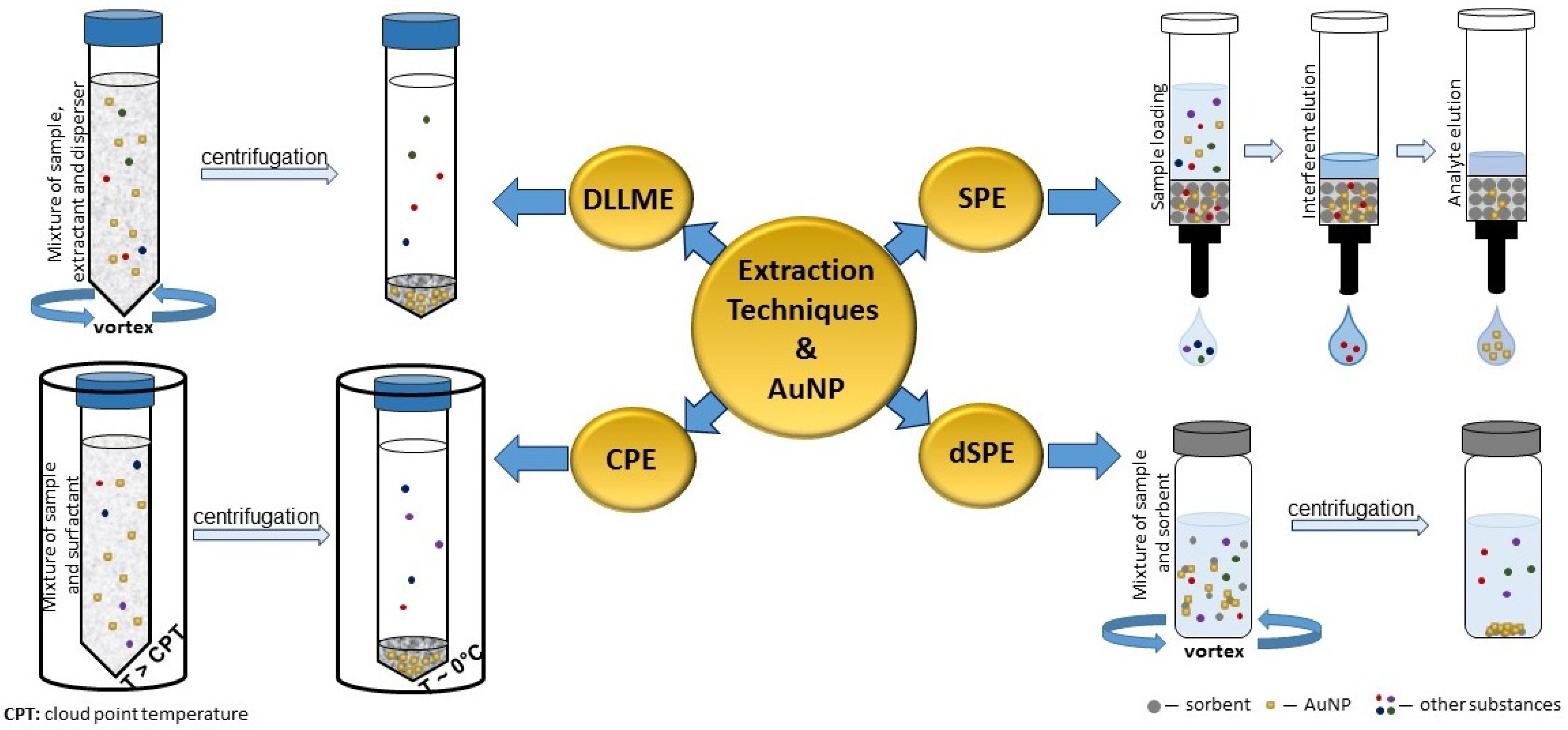
, .png) ,
,  ,
,  and
and
Abstract
1. Introduction
2. Solvent Extraction Procedures
2.1. Liquid-Phase Microextraction
2.2. Dispersive Liquid–Liquid Microextraction
2.3. Cloud Point Extraction
2.4. Suspended Aggregate Microextraction
3. Sorbent Extraction Procedures
3.1. Solid-Phase Extraction
3.2. Dispersive Solid-Phase Extraction
3.3. Magnetic Solid-Phase Extraction
4. Summary
5. Conclusions
Author Contributions
Funding
Institutional Review Board Statement
Informed Consent Statement
Data Availability Statement
Conflicts of Interest
Abbreviations
| 1-DDT | 1-dodecanethiol |
| A4F | asymmetric flow field-flow fractionation |
| AFM | atomic force microscopy |
| AgNP | silver nanoparticles |
| AuNP | gold nanoparticles |
| BMIM PF6 | 1-butyl-3-methylimidazolium hexafluorophosphate |
| CL | chemiluminescence |
| CME | capillary microextraction |
| CPE | cloud point extraction |
| CRM | certified reference material |
| CTAB | cetyltrimethylammonium bromide |
| CTAC | cetyltrimethylammonium chloride |
| dSPE | dispersive solid-phase extraction |
| DLLME | dispersive liquid-liquid microextraction |
| EDTA | ethylenediaminetetraacetic acid |
| EF | enrichment factor |
| ETAAS | electrothermal atomic absorption spectrometry |
| ETV-ICP-MS | electrothermal vaporization inductively coupled plasma mass spectrometry |
| Fe3O4@SiO2@IDA–Al3+ | Al3+-immobilized on core-shell structure of Fe3O4 and SiO2 microspheres functionalized with iminodiacetic acid |
| Fe3O4NP | iron oxide nanoparticles |
| FIA | flow injection analysis |
| FTIR | Fourier-transform infrared spectroscopy |
| HA | humic acid |
| HF-LPME | hollow-fiber liquid-phase microextraction |
| ICP-MS | inductively coupled plasma mass spectrometry |
| IL-µLLE | micro liquid-liquid extraction in an ionic liquid |
| iSAME | in situ suspended aggregate microextraction |
| IT-SPME | in-tube solid-phase microextraction |
| LDH | layered double hydroxides |
| LLE | liquid–liquid extraction |
| LOD | limit of detection |
| LPME | liquid-phase microextraction |
| MSA | mercaptosuccinic acid |
| MSPE | magnetic solid-phase extraction |
| MUA | 11-mercaptoundecanoic acid |
| NOM | natural organic matter |
| OILS | optical incoherent light scattering |
| PdNP | palladium nanoparticles |
| PVA | polyvinylalcohol |
| PVP | polyvinylpyrrolidone |
| poly(AA-VP-Bis) | poly(acrylamide-vinylpyridine-methylene bis-(acrylamide) |
| PR-C18 | C-18 reversed-phase silica gel |
| RSD | relative standard deviation |
| SA-DLLME | surfactant-assisted dispersive liquid-liquid microextraction |
| SBME | solvent bar microextraction |
| SBSE | stir bar sorptive extraction |
| SDME | single drop microextraction |
| SEM | scanning electron microscopy |
| s-NC | sulfonated nanocellulose |
| SPE | solid-phase extraction |
| (sp)ICP-MS | single particle inductively coupled plasma mass spectrometry |
| SPME | solid-phase microextraction |
| SPR | surface plasmon resonance |
| SSA | sulfosalicylic acid |
| TEM | transmission electron microscopy |
| TFME | thin-film microextraction |
| TiO2NP | titanium dioxide nanoparticles |
| TS | thiosulphate |
| TXRF | total reflection X-ray fluorescence spectrometry |
| UV-Vis | UV/Vis spectrophotometry |
| XPS | X-ray photoelectron spectroscopy |
| XRF | X-ray fluorescence |
| ZnONP | zinc oxide nanoparticles |
References
- Pryshchepa, O.; Pomastowski, P.; Buszewski, B. Silver nanoparticles: Synthesis, investigation techniques, and properties. Adv. Colloid Interface Sci. 2020, 284, 102246. [Google Scholar] [CrossRef]
- Definition—Nanomaterials—Environment—European Commission. Available online: https://ec.europa.eu/environment/chemicals/nanotech/faq/definition_en.htm (accessed on 20 February 2022).
- Alfieri, A.; Anantharaman, S.B.; Zhang, H.Q.; Jariwala, D. Nanomaterials for quantum information science and engineering. Adv. Mater. 2022, 2109621. [Google Scholar] [CrossRef]
- Bilal, M.; Rasheed, T.; Mehmood, S.; Tang, H.Z.; Ferreira, L.F.R.; Bharagava, R.N.; Iqbal, H.M.N. Mitigation of environmentally-related hazardous pollutants from water matrices using nanostructured materials—A review. Chemosphere 2020, 253, 126770. [Google Scholar] [CrossRef]
- Hagarová, I. Separation and quantification of metallic nanoparticles using cloud point extraction and spectrometric methods: A brief review of latest applications. Anal. Methods 2017, 9, 3594–3601. [Google Scholar] [CrossRef]
- Majedi, S.M.; Lee, H.K. Recent advances in the separation and quantification of metallic nanoparticles and ions in the environment. TrAC Trends Anal. Chem. 2016, 75, 183–196. [Google Scholar] [CrossRef]
- Yaqoob, A.A.; Ahmad, H.; Parveen, T.; Ahmad, A.; Oves, M.; Ismail, I.M.I.; Qari, H.A.; Umar, K.; Ibrahim, M.N.M. Recent advances in metal decorated nanomaterials and their various biological applications: A review. Front. Chem. 2020, 8, 341. [Google Scholar] [CrossRef]
- López-Serrano, A.; Olivas, R.M.; Landaluze, J.S.; Cámara, C. Nanoparticles: A global vision. Characterization, separation, and quantification methods. Potential environmental and health impact. Anal. Methods 2014, 6, 38–56. [Google Scholar] [CrossRef]
- Ogarev, V.A.; Rudoi, V.M.; Dementeva, O.V. Gold nanoparticles: Synthesis, optical properties, and application. Inorg. Mater. Appl. Res. 2018, 9, 134–140. [Google Scholar] [CrossRef]
- Guo, S.J.; Wang, E.K. Noble metal nanomaterials: Controllable synthesis and application in fuel cells and analytical sensors. Nano Today 2011, 6, 240–264. [Google Scholar] [CrossRef]
- Saravanan, A.; Kumar, P.S.; Karishma, S.; Vo, D.V.N.; Jeevanantham, S.; Yaashikaa, P.R.; George, C.S. A review on biosynthesis of metal nanoparticles and its environmental applications. Chemosphere 2021, 264, 128580. [Google Scholar] [CrossRef]
- Cao, S.W.; Tao, F.; Tang, Y.; Li, Y.T.; Yu, J.G. Size- and shape-dependent catalytic performances of oxidation and reduction reactions on nanocatalysts. Chem. Soc. Rev. 2016, 45, 4747–4765. [Google Scholar] [CrossRef]
- Saha, K.; Agasti, S.S.; Kim, C.; Li, X.N.; Rotello, V.M. Gold nanoparticles in chemical and biological sensing. Chem. Rev. 2012, 112, 2739–2779. [Google Scholar] [CrossRef]
- Shah, M.; Fawcett, D.; Sharma, S.; Tripathy, S.K.; Poinern, G.E.J. Green synthesis of metallic nanoparticles via biological entities. Materials 2015, 8, 7278–7308. [Google Scholar] [CrossRef]
- Bai, X.; Wang, Y.Y.; Song, Z.Y.; Feng, Y.M.; Chen, Y.Y.; Zhang, D.Y. The basic properties of gold nanoparticles and their applications in tumor diagnosis and treatment. Int. J. Mol. Sci. 2020, 21, 2480. [Google Scholar] [CrossRef]
- Das, M.; Shim, K.H.; An, S.S.A.; Yi, D.K. Review on gold nanoparticles and their applications. Toxicol. Environ. Health Sci. 2011, 3, 193–205. [Google Scholar] [CrossRef]
- Nooranian, S.; Mohammadinejad, A.; Mohajeri, T.; Aleyaghoob, G.; Kazemi Oskuee, R. Biosensors based on aptamer-conjugated gold nanoparticles: A review. Biotechnol. Appl. Biochem. 2021, 69, 1517–1534. [Google Scholar] [CrossRef]
- Paidari, S.; Ibrahim, S.A. Potential application of gold nanoparticles in food packaging: A mini review. Gold Bull. 2021, 54, 31–36. [Google Scholar] [CrossRef]
- Chen, F.; Si, P.; de la Zerda, A.; Jokerst, J.V.; Myung, D. Gold nanoparticles to enhance ophthalmic imaging. Biomater. Sci. 2021, 9, 367–390. [Google Scholar] [CrossRef]
- Lipińska, W.; Grochowska, K.; Siuzdak, K. Enzyme immobilization on gold nanoparticles for electrochemical glucose biosensors. Nanomaterials 2021, 11, 1156. [Google Scholar] [CrossRef]
- Luo, D.; Wang, X.; Burda, C.; Basilion, J.P. Recent development of gold nanoparticles as contrast agents for cancer diagnosis. Cancers 2021, 13, 1825. [Google Scholar] [CrossRef]
- Saleh, T.A. Trends in the sample preparation and analysis of nanomaterials as environmental contaminants. Trends Environ. Anal. Chem. 2020, 28, e00101. [Google Scholar] [CrossRef]
- Šebesta, M.; Kolenčík, M.; Matúš, P.; Kořenková, L. Transport and distribution of engineered nanoparticles in soils and sediments. Chem. Listy 2017, 111, 322–328. [Google Scholar]
- Ma, L.Y.; Li, Q.Y.; Yu, X.; Jiang, M.; Xu, L. Recent developments in the removal of metal-based engineered nanoparticles from the aquatic environments by adsorption. Chemosphere 2022, 291, 133089. [Google Scholar] [CrossRef] [PubMed]
- Besha, A.T.; Liu, Y.; Fang, C.; Bekele, D.N.; Naidu, R. Assessing the interactions between micropollutants and nanoparticles in engineered and natural aquatic environments. Crit. Rev. Environ. Sci. Technol. 2020, 50, 135–215. [Google Scholar] [CrossRef]
- Sung, H.K.; Jo, E.; Kim, E.; Yoo, S.K.; Lee, J.W.; Kim, P.J.; Kim, Y.; Eom, I.C. Analysis of gold and silver nanoparticles internalized by zebrafish (Danio rerio) using single particle-inductively coupled plasma-mass spectrometry. Chemosphere 2018, 209, 815–822. [Google Scholar] [CrossRef]
- Sani, A.; Cao, C.; Cui, D. Toxicity of gold nanoparticles (AuNPs): A review. Biochem. Biophys. Rep. 2021, 26, 100991. [Google Scholar] [CrossRef] [PubMed]
- Liu, Y.; He, M.; Chen, B.B.; Hu, B. Ultra-trace determination of gold nanoparticles in environmental water by surfactant assisted dispersive liquid liquid microextraction coupled with electrothermal vaporization-inductively coupled plasma-mass spectrometry. Spectrochim. Acta Part B At. Spectrosc. 2016, 122, 94–102. [Google Scholar] [CrossRef]
- Laborda, F.; Bolea, E.; Cepriá, G.; Gómez, M.T.; Jiménez, M.S.; Pérez-Arantegui, J.; Castillo, J.R. Detection, characterization and quantification of inorganic engineered nanomaterials: A review of techniques and methodological approaches for the analysis of complex samples. Anal. Chim. Acta 2016, 904, 10–32. [Google Scholar] [CrossRef]
- Sýkora, D.; Kašička, V.; Mikšík, I.; Řezanka, P.; Záruba, K.; Matějka, P.; Král, V. Application of gold nanoparticles in separation sciences. J. Sep. Sci. 2010, 33, 372–387. [Google Scholar] [CrossRef]
- Šebesta, M.; Matúš, P. Separation, determination, and characterization of inorganic engineered nanoparticles in complex environmental samples. Chem. Listy 2018, 112, 583–589. [Google Scholar]
- Von der Kammer, F.; Ferguson, P.L.; Holden, P.A.; Masion, A.; Rogers, K.R.; Klaine, S.J.; Koelmans, A.A.; Horne, N.; Unrine, J.M. Analysis of engineered nanomaterials in complex matrices (environment and biota): General considerations and conceptual case studies. Environ. Toxicol. Chem. 2012, 31, 32–49. [Google Scholar] [CrossRef]
- Nemček, L.; Hagarová, I. The recent strategies employed in chemical analysis of contaminated waters, sediments and soils as a part of the remediation process: Extraction. In Environmental Pollution and Remediation. Environmental and Microbial Biotechnology; Prasad, R., Ed.; Springer: Singapore, 2021; pp. 131–173. [Google Scholar]
- Che, D.; Cheng, J.; Ji, Z.; Zhang, S.; Li, G.; Sun, Z.; You, J. Recent advances and applications of polydopamine-derived adsorbents for sample pretreatment. TrAC Trends Anal. Chem. 2017, 97, 1–14. [Google Scholar] [CrossRef]
- Ibrahim, A.S.A.; Al-Farawati, R.; Hawas, U.; Shaban, Y. Recent microextraction techniques for determination and chemical speciation of selenium. Open Chem. 2017, 15, 103–122. [Google Scholar] [CrossRef]
- Werner, J.; Grześkowiak, T.; Zgoła-Grześkowiak, A.; Stanisz, E. Recent trends in microextraction techniques used in determination of arsenic species. TrAC Trends Anal. Chem. 2018, 105, 121–136. [Google Scholar] [CrossRef]
- Campillo, N.; Gavazov, K.; Viñas, P.; Hagarová, I.; Andruch, V. Liquid-phase microextraction: Update May 2016 to December 2018. Appl. Spectrosc. Rev. 2020, 55, 307–326. [Google Scholar] [CrossRef]
- Gavazov, K.B.; Hagarová, I.; Halko, R.; Andruch, V. Recent advances in the application of nanoparticles in cloud point extraction. J. Mol. Liq. 2019, 281, 93–99. [Google Scholar] [CrossRef]
- Câmara, J.S.; Perestrelo, R.; Olayanju, B.; Berenguer, C.V.; Kabir, A.; Pereira, J.A.M. Overview of different modes and applications of liquid phase-based microextraction techniques. Processes 2022, 10, 1347. [Google Scholar] [CrossRef]
- López-Lorente, A.I.; Simonet, B.M.; Valcárcel, M. Rapid analysis of gold nanoparticles in liver and river water samples. Analyst 2012, 137, 3528–3534. [Google Scholar] [CrossRef]
- He, Z.; Alexandridis, P. Ionic liquid and nanoparticle hybrid systems: Emerging applications. Adv. Colloid Interface Sci. 2017, 244, 54–70. [Google Scholar] [CrossRef]
- Sajid, M. Dispersive liquid-liquid microextraction: Evolution in design, application areas, and green aspects. Trac-Trends Anal. Chem. 2022, 152, 116636. [Google Scholar] [CrossRef]
- Gharanjik, R.; Nassiri, M.; Hashemi, H. Spectrophotometric determination of copper and nickel in marine brown algae after preconcentration with surfactant assisted dispersive liquid-liquid microextraction. Iran. J. Chem. Chem. Eng. 2020, 39, 117–126. [Google Scholar]
- Sobhi, H.R.; Azadikhah, E.; Behbahani, M.; Esrafili, A.; Ghambarian, M. Application of a surfactant-assisted dispersive liquid-liquid microextraction method along with central composite design for micro-volume based spectrophotometric determination of low level of Cr(VI) ions in aquatic samples. Spectrochim. Acta Part A Mol. Biomol. Spectrosc. 2018, 202, 36–40. [Google Scholar] [CrossRef] [PubMed]
- Dokpikul, N.; Chaiyasith, W.C.; Sananmuang, R.; Ampiah-Bonney, R.J. Surfactant-assisted emulsification dispersive liquid-liquid microextraction using 2-thenoyltrifluoroacetone as a chelating agent coupled with electrothermal atomic absorption spectrometry for the speciation of chromium in water and rice samples. Food Chem. 2018, 246, 379–385. [Google Scholar] [CrossRef]
- Timofeeva, I.; Stepanova, K.; Bulatov, A. In-a-syringe surfactant-assisted dispersive liquid-liquid microextraction of polycyclic aromatic hydrocarbons in supramolecular solvent from tea infusion. Talanta 2021, 224, 121888. [Google Scholar] [CrossRef] [PubMed]
- Mammana, S.B.; Abraham, E.D.C.; Camargo, A.B.; Vázquez, Á.; Altamirano, J.C. Enzymatic digestion coupled to surfactant-assisted dispersive liquid-liquid microextraction: A mild approach for determining polybrominated diphenyl ethers in human hair sample. Chemistryselect 2020, 5, 2179–2184. [Google Scholar] [CrossRef]
- Liu, C.; Liu, Z.; Wang, M.; Yang, Y. Surfactant-assisted dispersive liquid–liquid micro-extraction combined with magnetic solid-phase extraction for analysis of polyphenols in tobacco samples. J. Iran. Chem. Soc. 2018, 15, 1561–1568. [Google Scholar] [CrossRef]
- Hagarová, I.; Urík, M. New approaches to the cloud point extraction: Utilizable for separation and preconcentration of trace metals. Curr. Anal. Chem. 2016, 12, 87–93. [Google Scholar] [CrossRef]
- Mandal, S.; Lahiri, S. A review on extraction, preconcentration and speciation of metal ions by sustainable cloud point extraction. Microchem. J. 2022, 175, 107150. [Google Scholar] [CrossRef]
- Liu, J.; Liu, R.; Yin, Y.; Jiang, G. Triton X-114 based cloud point extraction: A thermoreversible approach for separation/concentration and dispersion of nanomaterials in the aqueous phase. Chem. Commun. 2009, 1514–1516. [Google Scholar] [CrossRef]
- Hartmann, G.; Schuster, M. Species selective preconcentration and quantification of gold nanoparticles using cloud point extraction and electrothermal atomic absorption spectrometry. Anal. Chim. Acta 2013, 761, 27–33. [Google Scholar] [CrossRef]
- Tsogas, G.Z.; Giokas, D.L.; Vlessidis, A.G. Ultratrace determination of silver, gold, and iron oxide nanoparticles by micelle mediated preconcentration/selective back-extraction coupled with flow injection chemiluminescence detection. Anal. Chem. 2014, 86, 3484–3492. [Google Scholar] [CrossRef] [PubMed]
- Bahadir, Z.; Torrent, L.; Hidalgo, M.; Marguí, E. Simultaneous determination of silver and gold nanoparticles by cloud point extraction and total reflection X-ray fluorescence analysis. Spectrochim. Acta Part B At. Spectrosc. 2018, 149, 22–29. [Google Scholar] [CrossRef]
- El Hadri, H.; Hackley, V.A. Investigation of cloud point extraction for the analysis of metallic nanoparticles in a soil matrix. Environ. Sci.–Nano 2017, 4, 105–116. [Google Scholar] [CrossRef]
- Mandyla, S.P.; Tsogas, G.Z.; Vlessidis, A.G.; Giokas, D.L. Determination of gold nanoparticles in environmental water samples by second-order optical scattering using dithiotreitol-functionalized CdS quantum dots after cloud point extraction. J. Hazard. Mater. 2017, 323, 67–74. [Google Scholar] [CrossRef] [PubMed]
- Benedé, J.L.; Giokas, D.L.; Chisvert, A.; Salvador, A. In-situ suspended aggregate microextraction: A sample preparation approach for the enrichment of organic compounds in aqueous solutions. J. Chromatogr. A 2015, 1408, 63–71. [Google Scholar] [CrossRef]
- Choleva, T.G.; Kappi, F.A.; Tsogas, G.Z.; Vlessidis, A.G.; Giokas, D.L. In-situ suspended aggregate microextraction of gold nanoparticles from water samples and determination by electrothermal atomic absorption spectrometry. Talanta 2016, 151, 91–99. [Google Scholar] [CrossRef]
- Medveď, J.; Bujdoš, M.; Matúš, P.; Kubová, J. Determination of trace amounts of gold in acid-attacked environmental samples by atomic absorption spectrometry with electrothermal atomization after preconcentration. Anal. Bioanal. Chem. 2004, 379, 60–65. [Google Scholar] [CrossRef]
- Medveď, J.; Matúš, P.; Bujdoš, M.; Kubová, J. Gold and silver determination in waters by SPHERON® thiol 1000 preconcentration and ETAAS. Chem. Pap. 2006, 60, 27–31. [Google Scholar] [CrossRef]
- Hagarová, I.; Nemček, L. Application of metallic nanoparticles and their hybrids as innovative sorbents for separation and pre-concentration of trace elements by dispersive micro-solid phase extraction: A minireview. Front. Chem. 2021, 9, 672755. [Google Scholar] [CrossRef]
- Zhang, C.; Xing, H.F.; Yang, L.R.; Fei, P.F.; Liu, H.Z. Development trend and prospect of solid phase extraction technology. Chin. J. Chem. Eng. 2022, 42, 245–255. [Google Scholar] [CrossRef]
- Li, L.; Leopold, K.; Schuster, M. Effective and selective extraction of noble metal nanoparticles from environmental water through a noncovalent reversible reaction on an ionic exchange resin. Chem. Commun. 2012, 48, 9165–9167. [Google Scholar] [CrossRef] [PubMed]
- Li, L.; Leopold, K. Ligand-assisted extraction for separation and preconcentration of gold nanoparticles from waters. Anal. Chem. 2012, 84, 4340–4349. [Google Scholar] [CrossRef] [PubMed]
- Zhang, L.; Chen, B.; He, M.; Liu, X.; Hu, B. Hydrophilic polymer monolithic capillary microextraction online coupled to ICPMS for the determination of carboxyl group-containing gold nanoparticles in environmental waters. Anal. Chem. 2015, 87, 1789–1796. [Google Scholar] [CrossRef] [PubMed]
- Ścigalski, P.; Kosobucki, P. Recent materials developed for dispersive solid phase extraction. Molecules 2020, 25, 4869. [Google Scholar] [CrossRef]
- Choleva, T.G.; Giokas, D.L. Application of dissolvable Mg/Al layered double hydroxides as an adsorbent for the dispersive solid phase extraction of gold nanoparticles prior to their determination by atomic absorption spectrometry. Anal. Methods 2020, 12, 368–375. [Google Scholar] [CrossRef]
- Jesús Dueñas-Mas, M.; Laura Soriano, M.; Ruiz-Palomero, C.; Valcárcel, M. Modified nanocellulose as promising material for the extraction of gold nanoparticles. Microchem. J. 2018, 138, 379–383. [Google Scholar] [CrossRef]
- Hagarová, I. Magnetic solid phase extraction as a promising technique for fast separation of metallic nanoparticles and their ionic species: A review of recent advances. J. Anal. Methods Chem. 2020, 2020, 8847565. [Google Scholar] [CrossRef]
- Ricardo, A.I.C.; Abujaber, F.; Bernardo, F.J.G.; Martin-Doimeadios, R.C.R.; Rios, A. Magnetic solid phase extraction as a valuable tool for elemental speciation analysis. Trends Environ. Anal. Chem. 2020, 27, e00097. [Google Scholar] [CrossRef]
- Su, S.W.; Chen, B.B.; He, M.; Xiao, Z.W.; Hu, B. A novel strategy for sequential analysis of gold nanoparticles and gold ions in water samples by combining magnetic solid phase extraction with inductively coupled plasma mass spectrometry. J. Anal. At. Spectrom. 2014, 29, 444–453. [Google Scholar] [CrossRef]
- García-Figueroa, A.; Pena-Pereira, F.; Lavilla, I.; Bendicho, C. Speciation of gold nanoparticles and total gold in natural waters: A novel approach based on naked magnetite nanoparticles in combination with ascorbic acid. Talanta 2019, 193, 176–183. [Google Scholar] [CrossRef]
- Application Note, Particle Size Analysis of Gold Nanoparticles. Available online: https://static.horiba.com/fileadmin/Horiba/Application/Materials/Chemical_Manufacturing/AN194_Particle_Size_Analysis_of_Gold_Nanoparticles.pdf (accessed on 8 February 2022).


| Extraction Technique | Water Samples | Detection Method | EF | LOD (ng L−1) | RSD (%) | Recovery (%) | Reference |
|---|---|---|---|---|---|---|---|
| IL-µLLE | River water | UV-Vis | NR | 0.335 | 18.0 | 79–103 | [40] |
| SA-DLLME | Tap, lake, river water | ETV-ICP-MS | 152 | 2.20 | 9.3 | 90–102 | [28] |
| CPE | River water, influent and effluent wastewater | ETAAS | 80 | 5.00 | 9.5 | 91–103 | [52] |
| CPE | Lake, river water, influent wastewater | CL | NR | 0.217 | 2.3–12.4 | 79–114 | [53] |
| CPE | Tap, river, sea, mineral water | TXRF | NR | 200 | 9.6–16.0 | 90–102 | [54] |
| CPE | River, lake water, effluent wastewater | OILS | NR | 0.114 | 9.3 | 79–110 | [56] |
| iSAME | Tap, river water, effluent wastewater | ETAAS | 8 | 0.015 | 5.4–12.0 | 81–93 | [58] |
| SPE | River, lake, brook water | ETAAS | 132 | NR | NR | 62–69 | [63] |
| SPE | Tap, river, lake, brook water, effluent wastewater | UV-Vis | 250 | NR | NR | 68–99 | [64] |
| CME | Tap, river, lake water | ICP-MS | 10 | 0.005 | 5.6 | 77–103 | [65] |
| dSPE | River, lake water, effluent wastewater | ETAAS | NR | 0.004 | 7.8–8.9 | 71–92 | [67] |
| MSPE | Sea water, lake, river, sewage water | ICP-MS | 50 | 0.31 | 4.9 | 73–100 | [71] |
| MSPE | Sea water, surface, ground water, artificial wastewater | ETAAS | 199 | 19.5 | 5.3 | 85–98 | [72] |
Publisher’s Note: MDPI stays neutral with regard to jurisdictional claims in published maps and institutional affiliations. |
© 2022 by the authors. Licensee MDPI, Basel, Switzerland. This article is an open access article distributed under the terms and conditions of the Creative Commons Attribution (CC BY) license (https://creativecommons.org/licenses/by/4.0/).
Share and Cite
Hagarová, I.; Nemček, L.; Šebesta, M.; Zvěřina, O.; Kasak, P.; Urík, M. Preconcentration and Separation of Gold Nanoparticles from Environmental Waters Using Extraction Techniques Followed by Spectrometric Quantification. Int. J. Mol. Sci. 2022, 23, 11465. https://doi.org/10.3390/ijms231911465
Hagarová I, Nemček L, Šebesta M, Zvěřina O, Kasak P, Urík M. Preconcentration and Separation of Gold Nanoparticles from Environmental Waters Using Extraction Techniques Followed by Spectrometric Quantification. International Journal of Molecular Sciences. 2022; 23(19):11465. https://doi.org/10.3390/ijms231911465
Chicago/Turabian StyleHagarová, Ingrid, Lucia Nemček, Martin Šebesta, Ondřej Zvěřina, Peter Kasak, and Martin Urík. 2022. "Preconcentration and Separation of Gold Nanoparticles from Environmental Waters Using Extraction Techniques Followed by Spectrometric Quantification" International Journal of Molecular Sciences 23, no. 19: 11465. https://doi.org/10.3390/ijms231911465
APA StyleHagarová, I., Nemček, L., Šebesta, M., Zvěřina, O., Kasak, P., & Urík, M. (2022). Preconcentration and Separation of Gold Nanoparticles from Environmental Waters Using Extraction Techniques Followed by Spectrometric Quantification. International Journal of Molecular Sciences, 23(19), 11465. https://doi.org/10.3390/ijms231911465