Review

Organoids: A New Chapter in Sarcoma Diagnosis and Treatment

1
Department of Gynecology, Charité-Universitätsmedizin Berlin, Corporate Member of Freie Universität Berlin and Humboldt–Universität zu Berlin, Augustenburger Platz 1, 13353 Berlin, Germany
2
First Department of Pathology, Medical School, National and Kapodistrian University of Athens, 75 Mikras Asias Street, Bld 10, Goudi, 11527 Athens, Greece
3
Section of Medical Oncology, 2nd Department of Medicine and Laboratory, National and Kapodistrian University of Athens, Hippocratio General Hospital of Athens, V. Sofias 108, 11526 Athens, Greece
4
Department of Oncology, Lausanne University Hospital and University of Lausanne, CH-1011 Lausanne, Switzerland
*
Author to whom correspondence should be addressed.
Int. J. Mol. Sci. 2022, 23(19), 11271; https://doi.org/10.3390/ijms231911271
Submission received: 28 July 2022 / Revised: 15 September 2022 / Accepted: 20 September 2022 / Published: 24 September 2022

Abstract

:
Sarcomas are malignant tumors of mesenchymal origin that can occur at any age. The rarity of these tumors in combination with the vast number of histological subtypes render the study of sarcomas challenging. Organoids represent complex three-dimensional cell culture systems, deriving from stem cells and preserving the capacity to differentiate into the cell types of their tissue of origin. The aim of the present review is to study the current status of patient-derived organoids, as well as their potential to model tumorigenesis and perform drug screenings for sarcomas. In order to identify relevant studies, a literature review was conducted and we were able to identify 16 studies published between 2019 and 2022. The current manuscript represents the first comprehensive review of the literature focusing on the use of organoids for disease modelling and drug sensitivity testing in diverse sarcoma subtypes.

1. Introduction

Sarcomas comprise a heterogeneous group of connective tissue malignancies of mesenchymal origin, including numerous distinct diagnostic entities [1]. Despite the large number of subtypes, sarcomas can be divided into soft tissue sarcomas (STS) and Primary bone sarcomas (PBS) [2]. Rhabdomyosarcoma and non-rhabdomyosarcoma STS represent the two major histological classes of STS, whereas osteosarcoma and Ewing sarcoma are the most common histological subtypes of PBS [3]. According to the American Cancer Society, about 13,190 new cases of STS and 3910 cases of primary cancer of the bones and joints will be diagnosed in the United States in 2022, leading to about 5130 and 2100 deaths, respectively [4,5]. STS usually arise in the extremities or the retroperitoneum, and most patients notice an eventually painful growing tumor mass [6]. The 5-year survival rate for people diagnosed with STS drops to 15% when a distant metastasis is present [7]. PBS most commonly cause pain, swelling and pathological bone fractures [8]. Even though most PBS are found at an early stage [9], the 5-year survival rate for patients diagnosed at a distant surveillance, epidemiology, and end results (SEER) stage amounts to 23–39%, depending on the histological subtype [10,11,12]. Diagnostic evaluation of sarcoma includes, in addition to a physical examination, a plain X-ray, computed tomography (CT) scans, and magnetic resonance imaging (MRI), eventually combined with a positron emission tomography (PET) scan. Nevertheless, definite diagnosis always requires biopsy of the tumor mass [13,14]. For patients with early stage resectable sarcoma, surgical excision represents the mainstay of treatment, combined with chemotherapy for chemosensitive histotypes (mainly Ewing sarcoma and osteosarcoma, whereas in STS, it may be administered in certain cases of high-risk tumors). Patients with advanced disease are mainly treated with systemic therapy [15,16].
Organoids represent three-dimensional in vitro growing systems that derive from self-organizing cells, capable of recapitulating the in vivo structural and functional features of an organ [17]. Organoids may originate from embryonic, induced pluripotent, neonatal, or adult stem cells [18]. Their establishment requires scaffold or scaffold-free techniques to avert direct physical contact to the plastic dish [19]. The most commonly used scaffold is Matrigel, a heterogeneous and gelatinous protein mixture purified from Engelbreth–Holm–Swarm mouse sarcoma cells that resembles the natural extracellular matrix (ECM) [20]. The use of Matrigel or similar hydrogels has successfully enabled the generation of gastrointestinal, salivary gland, hepatic, pancreatic, brain, retinal, renal, pulmonary, or gynecological organoids [21]. The application of organoids ranges from human developmental biology, disease modeling, and tissue engineering to regenerative medicine, personalized medicine, and drug screening [22]. Importantly, given that organoids are expandable, cryopreservable, and genetically modifiable, they allow for various applications in cancer research as well, particularly through the development of tumor organoids [23,24].
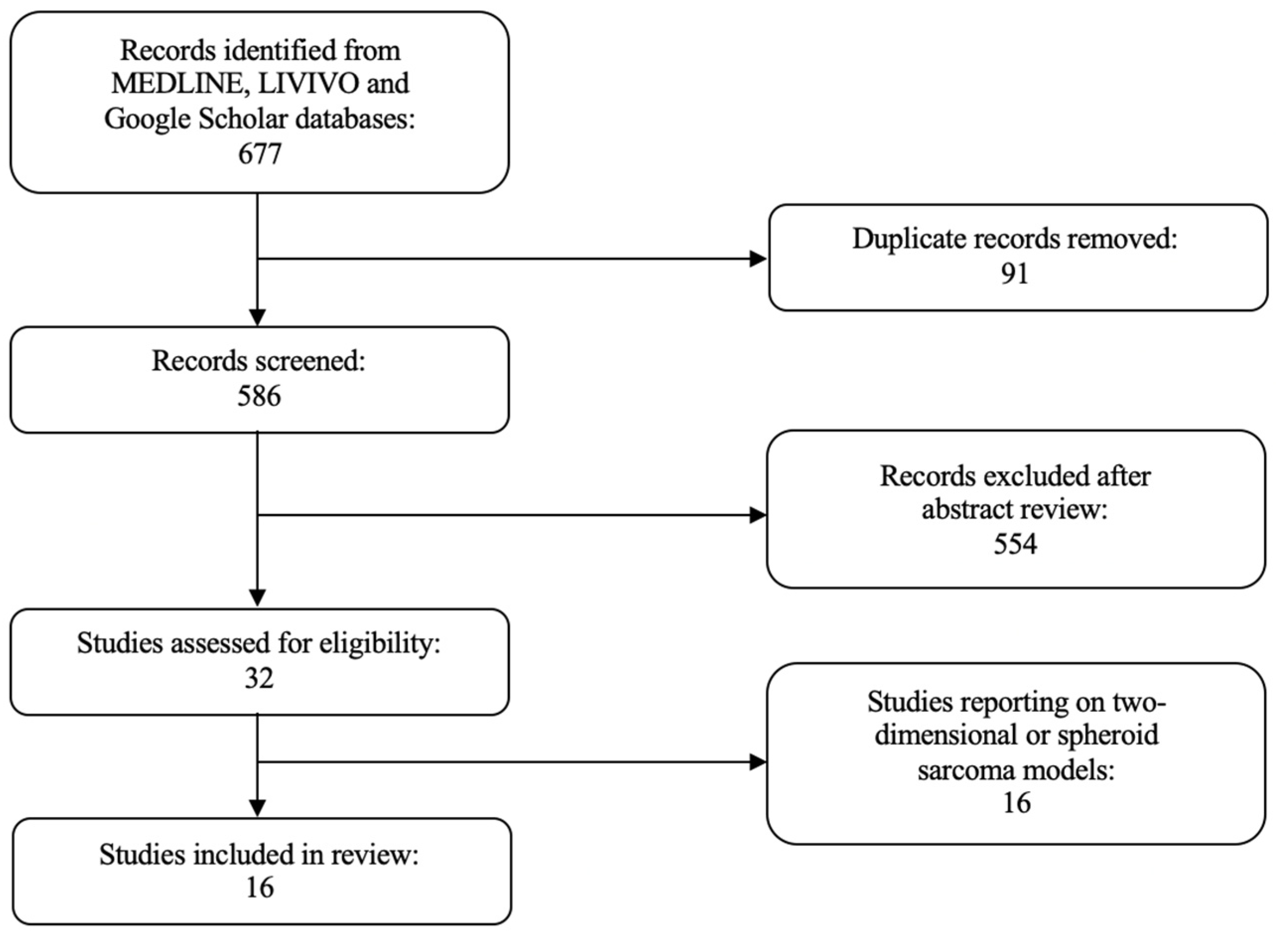
In this review, we extensively investigated the use of organoids for disease modeling and drug sensitivity testing in sarcoma (Figure 1). The literature review was conducted using the MEDLINE, LIVIVO, and Google Scholar databases. We included only original research articles and scientific abstracts written in the English language, that explicitly reported on the development of three-dimensional sarcoma organoid models. Studies incorporating two-dimensional or spheroid sarcoma models, as well as studies not clearly stating the use of organoid sarcoma models were excluded. The search terms sarcoma, rhabdomyosarcoma, fibrosarcoma, carcinosarcoma, osteosarcoma, chondrosarcoma, and organoid were employed and we were able to identify a total of 586 articles published between 1953 and April 2022, after the exclusion of duplicates. A total of 554 were discarded in the initial selection process after abstract review. The full texts of the remaining 32 publications were evaluated, and after detailed analysis, a total of 16 relevant studies published between 2019 and April 2022, that met the inclusion criteria, were selected for the literature review. Figure 2 presents an overview of the selection process.

2. Soft Tissue Sarcoma

2.1. Rhabdomyosarcoma

Two study groups have recently described their initial data on rhabdomyosarcoma organoids. Gatzweiler et al. used tumor samples from the INFORM pediatric precision oncology program (individualized therapy for relapsed malignancies in childhood) to study the molecular tumor profile and the drug-screening results of long-term embryonal rhabdomyosarcoma organoid-like cultures, and concluded that these organoids not only preserve the molecular characteristics of the original tumor, but also yield a sufficient amount of viable cells for the evaluation of drug combinations [25]. Meister et al. generated pediatric-patient-derived rhabdomyosarcoma organoid models comprising all major subtypes, and found that rhabdomyosarcoma organoids retain marker protein expression, represent the diverse clinical presentation of the different histopathological subtypes, as well as molecularly resemble the tumor of origin. Moreover, with 5/7 tested lines reaching passage 40, the models remain genetically and transcriptionally stable after culture over time, while rhabdomyosarcoma organoid drug screening reflects established drug sensitivities. Of note, the study group succeeded in genetically editing the organoid models using CRISPR/Cas9 and described that p53-deficient embryonal rhabdomyosarcoma tumoroid cells show a more sensitive response to the checkpoint kinase inhibitor prexasertib [26].

2.2. Non-Rhabdomyosarcoma

Several studies have highlighted the usefulness of non-rhabdomyosarcoma organoid models. Gaebler et al. managed to create long-term non-rhabdomyosarcoma organoids deriving from patients with myxoid liposarcoma, undifferentiated pleomorphic sarcoma or biphasic synovial sarcoma. This organoid platform allowed for drug screening of a set of compounds resembling first-line chemotherapy and novel compounds, whereas a multiplexed protein-profiling assay provided an insight into (phospho)-proteomics [27]. Boulay et al. generated patient-derived synovial sarcoma organoids and performed genome-wide epigenomic profiling, with a view to studying specific chromatin-remodeling mechanisms and dependencies. Primary synovial sarcoma organoids were shown to display distinctive patterns of BRG1/BRM-associated factor (BAF) complex distribution, while broad BAF complex domains correlated with active chromatin states, as well as the expression of a tumor-specific gene signature. Additionally, the presence of polycomb repressive complex 1 (PRC1) and the related H2AK119ub histone mark at broad BAF domains was confirmed, and synovial sarcoma cells were more sensitive to incremental doses of the ubiquitin-specific protease 7 (USP7) inhibitor FT827 than Ewing sarcoma cells [28]. Maloney et al. developed patient-derived skin fibrosarcoma organoids, which were subjected to the tyrosine kinase inhibitor imatinib or the antibiotic anthracycline chemotherapy agent doxorubicin. Interestingly, a significant decrease in adenosine 5′-triphosphate (ATP) activity was reported only after the organoid cultures had been subjected to a high concentration of imatinib, whereas low concentrations of doxorubicin generated a significant reduction in ATP activity [29]. Maru et al. investigated the transformation potential of the combination of Kirsten rat sarcoma virus (Kras) activation and Phosphatase and tensin homolog (Pten) inactivation in murine endometrial organoids in the subcutis of immunodeficient mice, and reported that Cyclin-Dependent Kinase Inhibitor 2A (CDKN2A) knockdown or transformation-related protein 53 (Trp53) deletion led to the induction of sarcomatous differentiation and, consequently, to the development of uterine carcinosarcoma [30]. The Japanese study group also developed Trp53 wildtype fallopian tube organoids expressing the mutant Kras, that were found to develop ovarian carcinosarcoma upon CDKN2A suppression [31]. McCorkle et al. successfully established in vitro monolayer and ovarian carcinosarcoma organoid cell lines to investigate the effects of diverse chemotherapeutic agents. Notably, dose–response curves revealed a relatively high resistance to carboplatin, gemcitabine, and topotecan, but sensitivity to paclitaxel, doxorubicin, and artesunate [32].
Table 1 summarizes the results of the different studies on STS organoids.

3. Primary Bone Sarcoma

3.1. Osteosarcoma

A handful of studies have investigated the use of organoids for disease modeling and drug sensitivity testing in osteosarcoma. Wang et al. harvested early passage osteosarcoma cells from mice tumors to develop osteosarcoma organoids and proved that the inhibition of p27 degradation by S-Phase Kinase Associated Protein 2 (SKP2) significantly delays osteosarcoma development and progression, induces apoptosis, and diminishes tumor-initiating properties in the organoid model [33]. Subramaniam et al. generated multicell-type lung organoid models with osteosarcoma cells and reported a significant reduction in osteosarcoma cell growth after treatment with pimozide [34]. In her study, Johansson created patient-derived osteosarcoma organoids displaying rounded structure in microscopy images and secreting Vascular Endothelial Growth Factor (VEGF) under the cultivation. By performing cell viability assays on both the organoids and the cryopreserved cancer cells from the original tumor, the author described similar resistance profiles [35]. Last but not least, He et al. firstly generated a patient-derived organoid platform for lung metastatic osteosarcoma that preserved the cellular morphology and expression of the osteosarcoma markers Vimentin and SRY-Box Transcription Factor 9 (Sox9). Interestingly, given that the primary lung metastatic osteosarcoma organoids retained the T-cell distribution of the parental tumors, anti-programmed cell death protein 1 (PD1) treatment was found to activate CD8+ T-cells in the organoid cultures [36].

3.2. Chondrosarcoma

Only one study group has, to date, reported on the experience with a three-dimensional chondrosarcoma organoid model. Veys et al. created SW1353-cell-derived chondrosarcoma organoids to test the anti-tumor activity of microRNA-342-5p and microRNA-491-5p, and concluded that microRNA-342-5p significantly promotes apoptosis, especially in hypoxia [37].

3.3. Ewing Sarcoma

Two study groups have recently described their initial experiences with Ewing sarcoma organoids. Maurer et al. developed Ewing sarcoma organoids and monolayers from a metastatic pulmonary lesion from a patient with an inherited BRCA1 Associated RING Domain 1 (BARD1) mutation. The organoids surprisingly demonstrated high sensitivity to poly (ADP-ribose) polymerase (PARP) inhibitors [38]. Two years after their initial publication, the same study group published the results of their second study, stating that the loss of BARD1 increases Ewing sarcoma sensitivity to DNA damage, and that Guanylate-binding protein 1 (GBP1) expression contributes to DNA damage response in Ewing sarcoma organoids [39]. Komatsu et al., on the other hand, were the first to use patient-derived cell lines of CIC-DUX4 sarcoma to generate Ewing-like small round cell sarcoma organoids. Notably, drug sensitivity assays revealed a dose-dependent decrease in organoid size after treatment with two different concentrations of gemcitabine [40].
Table 2 summarizes the results of the different studies on PBS organoids.

4. Conclusions

Organoids, in general, offer a unique opportunity, since they are translatable, reproducible, and scalable. Their generation from pluripotent or adult stem cells renders them an exceptional three-dimensional culture system, capable of closely mimicking the architecture and physiology of the tissue of origin [41]. Nonetheless, organoids show a relatively random growth nature, do not support interorgan communication, and lack vasculature and immune cells [42]. As such, the development of standardized protocols for routine organoid handling is of utmost importance, in order to increase the success rate of organoid generation and research [43].
Sarcoma is a challenging disease, and the lack of reliable treatments in this field renders paramount the development of new in vivo assays that can support drug discovery. However, for the time being, there has been limited success in cell culture, and great difficulty in identifying pure sarcoma organoids. Formulation of optimal culture media and maintenance processes has been difficult, as it depends on each subtype, yet sarcomas, unlike epithelial-based tumors, are notorious for their heterogeneity, defined as the existence of distinct cell subpopulations with varying inter- and intra-tumor morphological, genotypic, and phenotypic features [44].
One other challenge to overcome is the limited available amount of tumor tissue to be used for organoid culture. It has been shown that increasing the amount of starting material, e.g., by taking multiple biopsies from epithelial-based tumors, improves the outgrowth of tumor organoids. In sarcoma, however, the risk of contamination to other tissues during biopsy and the large quantity of tissue required for complex diagnosis render the available material from biopsies to be used for organoids often restricted. Consequently, these limitations also hinder the study of inter- and intra-patient heterogeneity in sarcomas.
Several literature reviews have been recently published on three-dimensional sarcoma models [45,46,47]. These publications mostly report on sarcoma-derived three-dimensional cultures or spheroids and only underline the importance of the generation of advanced sarcoma organoids. Notably, both Kapalczynska et al. and Jensen et al. recently stated that, compared to two-dimensional cell cultures, three-dimensional models mimic the in vivo cell environment, and may, thus, offer greater opportunities to study cellular signaling or drug sensitivity [48,49]. Moreover, Gunti et al. compared spheroids with organoids, and concluded that, although spheroid establishment is simpler, organoids represent long-term, cryopreservable culture models, histologically and genetically resembling the tissue of origin [50]. A major advantage of 3D systems such as organoids is the preservation of intra-tumor heterogeneity, that allows them to recapitulate human tumors much more closely, compared to their more conventional 2D cell culture methods. Carcinogenesis is a multi-step process that evolves through the progressive emergence of neoplastic clones, characterized by distinct genetic and epigenetic alteration. Malignant tumors are, therefore, collections of molecularly divergent cell populations which, in conditions of selection pressure, such as those encountered during treatment, provide the fuel for therapy resistance. Thus, the potential of organoids to capture the heterogeneity of original tumors makes them an ideal tool for the evaluation of novel drugs and the clarification of drug-resistance mechanisms.
To our knowledge, this is the first comprehensive literature review focusing on the use of organoids for disease modeling and drug sensitivity testing in different sarcoma subtypes. We were able to identify sixteen original articles on the successful development of mostly patient-derived sarcoma organoids, with a total of six studies presenting novel results in terms of non-rhabdomyosarcoma pathogenesis and/or treatment [27,28,29,30,31,32], followed by four manuscripts providing valuable information on osteosarcoma treatment options [33,34,35,36]. These organoid cultures represented useful preclinical models for the identification of molecular mechanisms correlating with sarcoma genesis and progression, and, most importantly, allowed for drug screening assays, thus paving the way for the establishment of novel potent treatment tools. Given that STSs alone are further subdivided into approximately 70, partly extremely rare, morphologically distinct subtypes [51], the development of patient-derived sarcoma organoids undoubtedly marks the beginning of a new era in sarcoma diagnosis and treatment. Of note, future studies should not put their focus only on main sarcoma subtypes, but also try to establish rare sarcoma organoids, in order to shed light on the countless questions of both involved clinicians and affected patients. All in all, the establishment of next-generation organoids requires a reduction in high organoid diversity, organoid maturation promotion, generation of larger organoids, as well as high-throughput live imaging [52].

Author Contributions

Literature analysis and conceptualization, I.P. and S.T.; original draft preparation and writing, I.P.; artwork, I.P.; review and supervision, S.K., K.P., A.D., K.V. and S.T. All authors have read and agreed to the published version of the manuscript.

Funding

This study was supported by the Open Access Publication Fund of Charité—Universitätsmedizin Berlin and the German Research Foundation (DFG).

Institutional Review Board Statement

Not applicable.

Informed Consent Statement

Not applicable.

Data Availability Statement

Not applicable.

Conflicts of Interest

The authors declare no conflict of interest.

References

  1. Hui, J.Y. Epidemiology and Etiology of Sarcomas. Surg. Clin. N. Am. 2016, 96, 901–914. [Google Scholar] [CrossRef]
  2. Skubitz, K.M.; D’Adamo, D.R. Sarcoma. Mayo. Clin. Proc. 2007, 82, 1409–1432. [Google Scholar] [CrossRef] [PubMed]
  3. Lupo, P.J.; Spector, L.G.; O’Brien, S.; Schiffman, J.D.; Hettmer, S. Epidemiology of Bone and Soft Tissue Sarcomas. In Sarcomas of Bone and Soft Tissues in Children and Adolescents; Arnd, C.A.S., Ed.; Pediatric Oncology; Springer: Cham, Switzerland, 2020. [Google Scholar]
  4. American Cancer Society. Key Statistics for Soft Tissue Sarcomas. Available online: https://www.cancer.org/cancer/soft-tissue-sarcoma/about/key-statistics.html (accessed on 17 June 2022).
  5. American Cancer Society. Key Statistics about Bone Cancer. Available online: https://www.cancer.org/cancer/bone-cancer/about/key-statistics.html (accessed on 17 June 2022).
  6. American Cancer Society. Signs and Symptoms of Soft Tissue Sarcomas. Available online: https://www.cancer.org/cancer/soft-tissue-sarcoma/detection-diagnosis-staging/signs-symptoms.html (accessed on 17 June 2022).
  7. American Cancer Society. Survival Rates for Soft Tissue Sarcoma. Available online: https://www.cancer.org/cancer/soft-tissue-sarcoma/detection-diagnosis-staging/survival-rates.html (accessed on 17 June 2022).
  8. American Cancer Society. Signs and Symptoms of Bone Cancer. Available online: https://www.cancer.org/cancer/bone-cancer/detection-diagnosis-staging/signs-symptoms.html (accessed on 17 June 2022).
  9. American Cancer Society. Can Bone Cancer Be Found Early? Available online: https://www.cancer.org/cancer/bone-cancer/detection-diagnosis-staging/detection.html (accessed on 17 June 2022).
  10. American Cancer Society. Survival Rates for Osteosarcoma. Available online: https://www.cancer.org/cancer/osteosarcoma/detection-diagnosis-staging/survival-rates.html (accessed on 17 June 2022).
  11. American Cancer Society. Survival Rates for Ewing Tumors. Available online: https://www.cancer.org/cancer/ewing-tumor/detection-diagnosis-staging/survival-rates.html (accessed on 17 June 2022).
  12. American Cancer Society. Survival Rates for Bone Cancer. Available online: https://www.cancer.org/cancer/bone-cancer/detection-diagnosis-staging/survival-statistics.html (accessed on 17 June 2022).
  13. American Cancer Society. Tests for Soft Tissue Sarcomas. Available online: https://www.cancer.org/cancer/soft-tissue-sarcoma/detection-diagnosis-staging/how-diagnosed.html (accessed on 17 June 2022).
  14. American Cancer Society. Tests for Bone Cancer. Available online: https://www.cancer.org/cancer/bone-cancer/detection-diagnosis-staging/how-diagnosed.html (accessed on 17 June 2022).
  15. American Cancer Society. Treatment of Soft Tissue Sarcomas, by Stage. Available online: https://www.cancer.org/cancer/soft-tissue-sarcoma/treating/by-stage.html (accessed on 17 June 2022).
  16. American Cancer Society. Treating Specific Types of Bone Cancer. Available online: https://www.cancer.org/cancer/bone-cancer/treating/treating-specific-bone-cancers.html (accessed on 17 June 2022).
  17. Lancaster, M.A.; Knoblich, J.A. Organogenesis in a dish: Modeling development and disease using organoid technologies. Science 2014, 345, 1247125. [Google Scholar] [CrossRef]
  18. Huch, M.; Koo, B.K. Modeling mouse and human development using organoid cultures. Development 2015, 142, 3113–3125. [Google Scholar] [CrossRef] [PubMed]
  19. Corro, C.; Novellasdemunt, L.; Li, V.S.W. A brief history of organoids. Am. J. Physiol. Cell Physiol. 2020, 319, C151–C165. [Google Scholar] [CrossRef]
  20. Orkin, R.W.; Gehron, P.; McGoodwin, E.B.; Martin, G.R.; Valentine, T.; Swarm, R. A murine tumor producing a matrix of basement membrane. J. Exp. Med. 1977, 145, 204–220. [Google Scholar] [CrossRef]
  21. Clevers, H. Modeling Development and Disease with Organoids. Cell 2016, 165, 1586–1597. [Google Scholar] [CrossRef]
  22. Lehmann, R.; Lee, C.M.; Shugart, E.C.; Benedetti, M.; Charo, R.A.; Gartner, Z.; Hogan, B.; Knoblich, J.; Nelson, C.M.; Wilson, K.M. Human organoids: A new dimension in cell biology. Mol. Biol. Cell 2019, 30, 1129–1137. [Google Scholar] [CrossRef]
  23. Azar, J.; Bahmad, H.F.; Daher, D.; Moubarak, M.M.; Hadadeh, O.; Monzer, A.; Al Bitar, S.; Jamal, M.; Al-Sayegh, M.; Abou-Kheir, W. The Use of Stem Cell-Derived Organoids in Disease Modeling: An Update. Int. J. Mol. Sci. 2021, 22, 7667. [Google Scholar] [CrossRef]
  24. Drost, J.; Clevers, H. Organoids in cancer research. Nat. Rev. Cancer 2018, 18, 407–418. [Google Scholar] [CrossRef]
  25. Gatzweiler, C.; Ridinger, J.; Herter, S.; Gerloff, X.F.; ElHarouni, D.; Berker, Y.; Imle, R.; Schmitt, L.; Kreth, S.; Stainczyk, S.; et al. Functional Therapeutic Target Validation Using Pediatric Zebrafish Xenograft Models. Cancers 2022, 14, 849. [Google Scholar] [CrossRef]
  26. Meister, M.T.; Groot Koerkamp, M.J.A.; de Souza, T.; Breunis, W.B.; Frazer-Mendelewska, E.; Brok, M.; DeMartino, J.; Manders, F.; Calandrini, C.; Kerstens, H.H.D.; et al. Mesenchymal tumor organoid models recapitulate rhabdomyosarcoma subtypes. bioRxiv 2022. [Google Scholar] [CrossRef]
  27. Gaebler, M.; Silvestri, A.; Reichardt, P.; Wardelmann, E.; Gambara, G.; Haybaeck, J.; Stroebel, P.; Niethard, M.; Erdmann, G.; Regenbrecht, C.R. Abstract 469: Patient-derived sarcoma models: First results from the SARQMA study. Cancer Res. 2019, 79, 469. [Google Scholar] [CrossRef]
  28. Boulay, G.; Cironi, L.; Garcia, S.P.; Rengarajan, S.; Xing, Y.H.; Lee, L.; Awad, M.E.; Naigles, B.; Iyer, S.; Broye, L.C.; et al. The chromatin landscape of primary synovial sarcoma organoids is linked to specific epigenetic mechanisms and dependencies. Life Sci. Alliance 2021, 4, 808. [Google Scholar] [CrossRef] [PubMed]
  29. Maloney, E.; Clark, C.; Sivakumar, H.; Yoo, K.; Aleman, J.; Rajan, S.A.P.; Forsythe, S.; Mazzocchi, A.; Laxton, A.W.; Tatter, S.B.; et al. Immersion Bioprinting of Tumor Organoids in Multi-Well Plates for Increasing Chemotherapy Screening Throughput. Micromachines 2020, 11, 208. [Google Scholar] [CrossRef] [PubMed]
  30. Maru, Y.; Tanaka, N.; Tatsumi, Y.; Nakamura, Y.; Itami, M.; Hippo, Y. Kras activation in endometrial organoids drives cellular transformation and epithelial-mesenchymal transition. Oncogenesis 2021, 10, 46. [Google Scholar] [CrossRef] [PubMed]
  31. Maru, Y.; Tanaka, N.; Tatsumi, Y.; Nakamura, Y.; Yao, R.; Noda, T.; Itami, M.; Hippo, Y. Probing the tumorigenic potential of genetic interactions reconstituted in murine fallopian tube organoids. J. Pathol. 2021, 255, 177–189. [Google Scholar] [CrossRef] [PubMed]
  32. McCorkle, J.R.; Burgess, B.T.; McDowell, A.B.; DeJohn, J.; DeSimone, C.P.; Ueland, F.R.; Kolesar, J.M.; Gorski, J.W. Abstract B22: Development of the first ovarian carcinosarcoma patient-derived xenograft and tissue organoid model to predict clinical response to chemotherapy. Clin. Cancer Res. 2020, 26, B22. [Google Scholar] [CrossRef]
  33. Wang, J.; Aldahamsheh, O.; Ferrena, A.; Singh, S.; Singla, A.; Borjihan, H.; Yaguare, S.; Viscarret, V.; Tingling, J.; Zi, X.; et al. Abstract 2008: Targeting SKP2 by p27 Inhibits stemness and prolong the survival in osteosarcoma. Cancer Res. 2021, 81, 2008. [Google Scholar] [CrossRef]
  34. Subramaniam, D.; Angulo, P.; Ponnurangam, S.; Dandawate, P.; Ramamoorthy, P.; Srinivasan, P.; Iwakuma, T.; Weir, S.J.; Chastain, K.; Anant, S. Suppressing STAT5 signaling affects osteosarcoma growth and stemness. Cell Death Dis. 2020, 11, 149. [Google Scholar] [CrossRef] [Green Version]
  35. Johansson, S. Patient-Derived Organoid Culture for 3D Culture of Colorectal Cancer, Renal Cancer and Osteosarcoma; Uppsala University: Uppsala, Sweden, 2019. [Google Scholar]
  36. He, A.; Huang, Y.; Cheng, W.; Zhang, D.; He, W.; Bai, Y.; Gu, C.; Ma, Z.; He, Z.; Si, G.; et al. Organoid culture system for patient-derived lung metastatic osteosarcoma. Med. Oncol. 2020, 37, 105. [Google Scholar] [CrossRef] [PubMed]
  37. Veys, C.; Benmoussa, A.; Contentin, R.; Duchemin, A.; Brotin, E.; Lafont, J.E.; Saintigny, Y.; Poulain, L.; Denoyelle, C.; Demoor, M.; et al. Tumor Suppressive Role of miR-342-5p in Human Chondrosarcoma Cells and 3D Organoids. Int. J. Mol. Sci. 2021, 22, 5590. [Google Scholar] [CrossRef] [PubMed]
  38. Maurer, L.M.; Venier, R.M.; Julian, C.; Bailey, K.M. Abstract A14: Exploiting DNA damage repair defects to enhance PD-L1 expression in Ewing sarcoma. Cancer Res. 2020, 80, A14. [Google Scholar] [CrossRef]
  39. Maurer, L.M.; Daley, J.D.; Mukherjee, E.; Venier, R.M.; Julian, C.M.; Bailey, N.G.; Jacobs, M.F.; Kumar-Sinha, C.; Raphael, H.; Periyapatna, N.; et al. BRCA1-Associated RING Domain-1 (BARD1) Loss and GBP1 Expression Enhance Sensitivity to DNA Damage in Ewing Sarcoma. Cancer Res. Commun. 2022, 2, 220–232. [Google Scholar] [CrossRef]
  40. Komatsu, A.; Matsumoto, K.; Yoshimatsu, Y.; Sin, Y.; Kubota, A.; Saito, T.; Mizumoto, A.; Ohashi, S.; Muto, M.; Noguchi, R.; et al. The CAM Model for CIC-DUX4 Sarcoma and Its Potential Use for Precision Medicine. Cells 2021, 10, 2613. [Google Scholar] [CrossRef]
  41. Kim, J.; Koo, B.K.; Knoblich, J.A. Human organoids: Model systems for human biology and medicine. Nat. Rev. Mol. Cell Biol. 2020, 21, 571–584. [Google Scholar] [CrossRef]
  42. Shankaran, A.; Prasad, K.; Chaudhari, S.; Brand, A.; Satyamoorthy, K. Advances in development and application of human organoids. 3 Biotech 2021, 11, 257. [Google Scholar] [CrossRef]
  43. Clinton, J.; McWilliams-Koeppen, P. Initiation, Expansion, and Cryopreservation of Human Primary Tissue-Derived Normal and Diseased Organoids in Embedded Three-Dimensional Culture. Curr. Protoc. Cell Biol. 2019, 82, e66. [Google Scholar] [CrossRef]
  44. Fisher, R.; Pusztai, L.; Swanton, C. Cancer heterogeneity: Implications for targeted therapeutics. Br. J. Cancer 2013, 108, 479–485. [Google Scholar] [CrossRef]
  45. Colella, G.; Fazioli, F.; Gallo, M.; De Chiara, A.; Apice, G.; Ruosi, C.; Cimmino, A.; de Nigris, F. Sarcoma Spheroids and Organoids-Promising Tools in the Era of Personalized Medicine. Int. J. Mol. Sci. 2018, 19, 615. [Google Scholar] [CrossRef] [Green Version]
  46. Kondo, T. Current status and perspectives of patient-derived rare cancer models. Hum. Cell 2020, 33, 919–929. [Google Scholar] [CrossRef]
  47. Zanoni, M.; Cortesi, M.; Zamagni, A.; Arienti, C.; Pignatta, S.; Tesei, A. Modeling neoplastic disease with spheroids and organoids. J. Hematol. Oncol. 2020, 13, 97. [Google Scholar] [CrossRef]
  48. Jensen, C.; Teng, Y. Is It Time to Start Transitioning From 2D to 3D Cell Culture? Front. Mol. Biosci. 2020, 7, 33. [Google Scholar] [CrossRef]
  49. Kapalczynska, M.; Kolenda, T.; Przybyla, W.; Zajaczkowska, M.; Teresiak, A.; Filas, V.; Ibbs, M.; Blizniak, R.; Luczewski, L.; Lamperska, K. 2D and 3D cell cultures—A comparison of different types of cancer cell cultures. Arch. Med. Sci. 2018, 14, 910–919. [Google Scholar] [CrossRef]
  50. Gunti, S.; Hoke, A.T.K.; Vu, K.P.; London, N.R., Jr. Organoid and Spheroid Tumor Models: Techniques and Applications. Cancers 2021, 13, 874. [Google Scholar] [CrossRef]
  51. Sbaraglia, M.; Bellan, E.; Dei Tos, A.P. The 2020 WHO Classification of Soft Tissue Tumours: News and perspectives. Pathologica 2021, 113, 70–84. [Google Scholar] [CrossRef]
  52. Shariati, L.; Esmaeili, Y.; Haghjooy Javanmard, S.; Bidram, E.; Amini, A. Organoid technology: Current standing and future perspectives. Stem Cells 2021, 39, 1625–1649. [Google Scholar] [CrossRef]
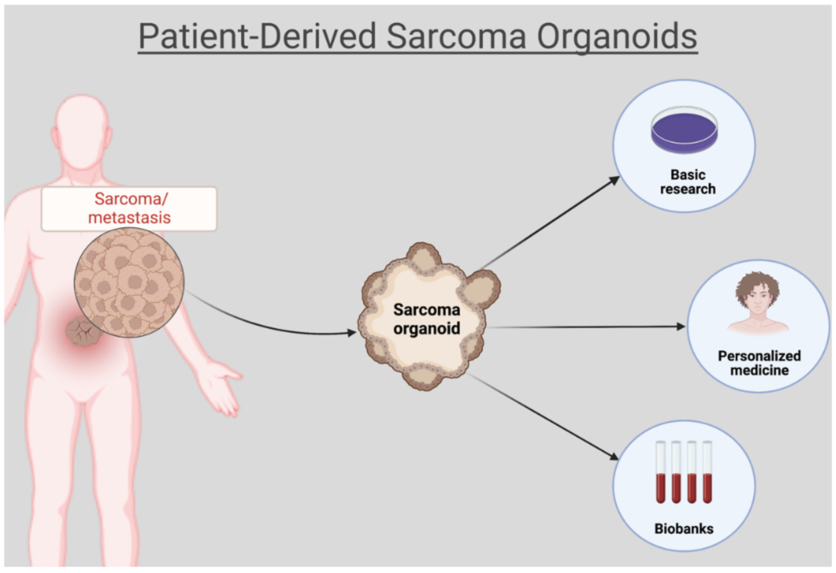
Figure 1. Patient-derived sarcoma organoids for disease modeling and drug sensitivity testing. Created with BioRender.com (accessed on 26 July 2022).
Ijms 23 11271 g001
Figure 2. PRISMA flow diagram visually summarizing the screening process.
Ijms 23 11271 g002
Table 1. Use of organoids in different types of soft tissue sarcoma.
STS TypeOrganoidsCulture ConditionsMain ResultsReferences
Embryonal
rhabdomyosarcoma
Patient-derived
long-term organoid-like cultures
  • Fresh patients’ surgical specimens or mouse patient derived xenografts dissociated mechanically and enzymatically (collagenase and trypsin)
  • Sub-culture of free-floating semi-adherent spheroids by dissociation with TrypLE
  • Seeding in a 1:2 to 1:5 ratio in fresh TSM complete medium, containing antibiotics and growth factors (EGF, FGF, PDGF)
  • Six-times
    passaged cultures
  • Preservation of the main molecular
    characteristics of the embryonal
    rhabdomyosarcoma
  • Shift in mean drug sensitivity
  • Zebrafish embryonal rhabdomyosarcoma
    model showed most sensitive response to
    idasanutlin and navitoclax
[25]
RhabdomyosarcomaPatient-derived
rhabdomyosarcoma organoids
  • Needle biopsies, surgical specimens or bone marrow aspirates
  • Minced pieces in extracellular matrix or single-cell suspensions in extracellular matrix (ECM) substitute
  • Basement-membrane-extract-supplemented medium, that contained antibiotics and growth factors (EGF, FGF, and IGF)
  • Tumor cell outgrowth to two-dimensional monolayers
  • Further propagation and expansion
  • Preservation of original tumor
    characteristics and clinical presentation
  • Genetic and transcriptional stability of
    tumoroid models over time
  • Drug screening reflects established drug sensitivities, with higher sensitivity of p53 deficient organoid cells to prexasertib
[26]
Myxoid liposarcoma, undifferentiated
pleomorphic
sarcoma,
biphasic
synovial sarcoma
Patient-derived non-rhabdomyosarcoma organoids
  • Sample dissociation
  • Seeding into 24w plates in matrix-like scaffolds
  • Growth until >100 μm and harvesting
  • Pharmacokinetic properties profiling through high-throughput drug screening
  • (Phospho)-proteomics analysis via
    multiplexed protein-profiling assay
[27]
Synovial sarcomaSynovial sarcoma and Ewing sarcoma patient-derived
tumor organoids
  • Fresh patients’
    surgical specimens
    dissociated and
    cultured in IMDM
  • Supplementation with 20% KO serum, 10 ng/mL human recombinant EGF and basic fibroblast growth factor, and 1% Pen/Strep in ultra-low attachment flasks
  • Correlation of broad BAF complex
    domains with active chromatin states
    and the expression of a tumor-specific
    gene signature
  • Reversible BAF complex retargeting through SS18-SSX expression and
    consequent functional PRC1–PRC2
    complex uncoupling
  • USP7 depletion as an epigenetic
    vulnerability in synovial sarcoma
[28]
Skin fibrosarcomaPatient-derived
skin fibrosarcoma
organoids
  • Immersion
    bioprinting of collagen–hyaluronic acid bioinks in 96-well plates
  • Mechanical and enzymatic (collagenase, protease, and hyaluronidase) dissociation of human surgical specimens
  • Maintenance in DMEM-HG supplemented with 10% FBS, 1% L-glutamine and 1% penicillin/streptomycin with 5% CO2
  • Successful employment of bioprinted
    patient-derived sarcoma organoids for
    chemotherapy screening studies
  • Significant decrease in ATP activity after imatinib and doxorubicin increase
[29]
Uterine
carcinosarcoma
Murine endometrial organoids
  • Mechanical and enzymatic (collagenase, dispase II) dissociation of mouse fresh tissues
  • Resuspended in solidified Matrigel and cultured in medium supplemented with R-spondin1, Noggin, Jagged-1, Y27632) (MBOC: Matrigel bilayer organoid culture protocol)
  • Carcinosarcoma development in KrasG12D organoids with Cdkn2a knockdown or Trp53 deletion
  • Epithelial–mesenchymal transition (EMT) state preservation, as well as presentation of heterogeneous statuses in the Kras loci in tumor-derived organoids
[30]
Ovarian
carcinosarcoma
Murine fallopian tube organoids
  • Dissociation of fresh mouse tissues into single cells, which are then suspended in Matrigel (MBOC: Matrigel bilayer organoid culture protocol)
  • Cooperation of Kras activation with p53 loss in terms of carcinosarcoma genesis
  • Resistance of KrasG12D-driven carcinosarcoma-derived organoids to paclitaxel and cisplatin
[31]
Ovarian
carcinosarcoma
Ovarian
carcinosarcoma
organoid
cell lines
  • Tumor tissue
    harvesting
  • Digestion with highly purified
    collagenase I and II
  • High resistance to carboplatin, gemcitabine, and topotecan
  • Sensitivity to paclitaxel, doxorubicin, and artesunate
[32]
Table 2. Use of organoids in different types of primary bone sarcoma.
PBS TypeOrganoidsCulture ConditionsMain ResultsReferences
OsteosarcomaOsteosarcoma
organoid culture
  • Not specified
  • Slower proliferation of Osx1-Cre; Rb1lox/lox; Trp53lox/lox; p27T187A/T187A tumors both in vivo and in vitro organoid
  • C1 and Pevenodistat showed selective inhibition in Osx1-Cre; Rb1lox/lox; Trp53lox/lox in osteosarcoma
    organoid
[33]
OsteosarcomaMulticell-type lung organoid model
  • Cell mix with KHOS/NP GFP
    positive cells
  • Growth in an ultra-low attachment using specific spheroid media
  • Significant growth reduction in osteosarcoma cells after treatment with 20 μM pimozide
[34]
OsteosarcomaPatient-derived osteosarcoma
organoids
  • Primary cell-line-derived osteosarcoma patient
  • Single-cell suspensions in Basement-Menbrane Extract-supplemented medium, with the addition of FGF and EGF 6-well plate incubation
  • Cell expansion and cryopreservation
  • Gradual increase in VEGF level in
    osteosarcoma organoids
  • Same tendency of survival index toward
    chemotherapy, yet different resistance toward oxaliplatin compared to cryopreserved and
    original cancer cells
[35]
OsteosarcomaPatient-derived
lung metastatic
osteosarcoma
organoids
  • Surgical specimens of primary or metastatic osteosarcomas
  • Cut/EnBloc protocol
  • Single-cell model
  • Histological feature and T-cell distribution maintenance of parental osteosarcoma lung
    metastatic tumors
  • CD8+ T-cell activation through PD-1 immune checkpoint blockade
[36]
ChondrosarcomaThree-
dimensional
chondrosarcoma
organoid model
  • Chondrosarcoma cell line
  • Cell growth in collagen I/III scaffolds
  • Seeding in 96-well culture plates
  • Incubation at 5% CO2 in HG-DMEM supplemented with 10% FCS and antibiotics
  • Transfer to 24-well
    plates
  • Incubation in the same medium pre-equilibrated with 3% O2
  • Cell death induction via microRNA-342-5p on a three-dimensional chondrosarcoma organoid model under hypoxia
[37]
Ewing sarcomaPatient-derived Ewing-sarcoma organoids
  • Not specified
  • High sensitivity to PARP inhibitors
  • Sensitization of PARP inhibitor-resistant Ewing cell lines to PARP inhibition via BARD1 small-interfering-RNA
  • Upregulation of tumor cell surface expression of PD-L1 after PARP inhibition
  • Early tumor cell death after PD-1 blocking
    antibody addition to T-cell/tumor cell cocultures
[38]
Ewing sarcomaPatient-derived Ewing-sarcoma organoids
  • Embedding of tumor pieces in growth-factor-reduced Matrigel
  • Incubation at 5% CO2, with media exchange every 3 days
  • Upregulation of immunoregulatory pathways upon relapse
  • High Ewing sarcoma sensitivity to DNA
    damage, talazoparib treatment and radiation
    after BARD1 loss
  • GBP1 expression promotes DNA damage
    response in Ewing sarcoma organoid cells
[39]
Ewing-like small
round cell
sarcoma
Tumor
organoids from chorioallantoic membrane tumor
  • Enzymatic digestion with Trypsin-EDTA and Liberase
  • Cell inoculation to a 96-well plate
  • Incubation at CO2
  • Tumor organoid formation from the
    chorioallantoic membrane tumor
  • CIC-DUX4 gene retention in chorioallantoic membrane tumor organoids
  • Dose-dependent effect of gemcitabine on
    organoid size
[40]
Publisher’s Note: MDPI stays neutral with regard to jurisdictional claims in published maps and institutional affiliations.