Communication

Identification, Pathogenicity, and Genetic Diversity of Fusarium spp. Associated with Maize Sheath Rot in Heilongjiang Province, China

1
Key Laboratory of Agricultural Microbiology of Heilongjiang Province, School of Life Sciences, Northeast Agricultural University, No. 600 Changjiang Road, Harbin 150030, China
2
State Key Laboratory for Biology of Plant Diseases and Insect Pests, Institute of Plant Protection, Chinese Academy of Agricultural Sciences, Beijing 100097, China
*
Authors to whom correspondence should be addressed.
Int. J. Mol. Sci. 2022, 23(18), 10821; https://doi.org/10.3390/ijms231810821
Submission received: 15 August 2022 / Revised: 31 August 2022 / Accepted: 14 September 2022 / Published: 16 September 2022
(This article belongs to the Section Molecular Plant Sciences)

Abstract

:
Maize sheath rot is a prevalent maize disease in China. From 2020 to 2021, symptomatic samples were collected from the main maize-growing regions of Heilongjiang province. To clarify the population and genetic diversity, as well as the virulence of pathogens responsible for maize sheath rot, a total of 132 Fusarium isolates were obtained and used for follow-up studies. Ten Fusarium species were identified based on morphological characteristics, and phylogenetic analysis was conducted using the TEF-1α gene sequences, including F. verticillioides (50.00%), F. subglutinans (18.94%), the Fusarium incarnatum-equiseti species complex (14.39%), F. temperatum (5.30%), F. acuminatum (3.03%), F. solani (2.27%), F. sporotrichioides (2.27%), F. tricinctum (1.52%), F. asiaticum (1.52%), and F. proliferatum (0.76%). All 10 Fusarium species could produce oval-to-annular lesions on maize sheath, and the lesions were grayish yellow to dark brown in the center and surrounded by a dark gray-to-dark brown halo. Of these, F. tricinctum and F. proliferatum showed significantly higher virulence than the other Fusarium species. In addition, haplotype analysis based on the concatenated sequences of the ITS and TEF-1a genes showed that 99 Fusarium isolates which belonged to the Fusarium fujikuroi species complex—consisting of F. verticillioides isolates, F. subglutinans isolates, F. temperatum isolates, and F. proliferatum isolates—could be grouped into 10 haplotypes, including 5 shared haplotypes (Haps 1, 2, 4, 5, and 6) and 5 private haplotypes (Haps 3, 7, 8, 9, and 10). Furthermore, the F. verticillioides clade in the haplotype network was radial with the center of Hap 2, suggesting that population expansion occurred. This research showed that Fusarium species associated with maize sheath rot in Heilongjiang province are more diverse than previously reported, and this is the first time that F. subglutinans, F. temperatum, F. solani, F. sporotrichioides, F. tricinctum, and F. acuminatum have been confirmed as the causal agents of maize sheath rot in Heilongjiang province.

1. Introduction

Maize is one of the three major food crops worldwide and the main raw feed material. It is also widely used in medicine, energy, and other fields. In China, maize is an important food crop, and the planting area was 41,264 million hectares in 2020 [1]. Meanwhile, maize is also the most important crop in Northeast China, accounting for more than 50% of the planting area of crops [2]. Maize sheath rot was first recorded in 2008 in Northeast China and has been reported to cause varying degrees of maize yield loss [3,4,5,6,7]. In addition, studies also showed that maize sheath rot has occurred in more than 11 provinces of China, including Liaoning, Jilin, Heilongjiang, Hebei, Shandong, Shanxi, Jiangsu, Hunan, Sichuan, Shaanxi, and Gansu, as well as the Ningxia Hui Autonomous Region [8]. The most prevalent pathogens of maize sheath rot are Fusarium species, and the diversity of Fusarium species causing maize sheath rot is gradually increasing [8].
The genus Fusarium was first described by Link in 1809, based on the distinctive banana- or canoe-shaped conidia [9]. Many Fusarium species could infect maize and cause a series of diseases, and could also contaminate agricultural products through mycotoxins [10]. As far as we know, maize production can be affected by several diseases caused by Fusarium species, such as maize leaf blight, maize stalk rot, maize ear rot, maize seedling blight, maize root rot, maize crown rot, maize storage molds, and maize sheath rot [4,11,12,13,14,15,16]. The Fusarium fujikuroi species complex (FFSC) was first established in 1925 by Wollenweber et al. [17] and contained more than 60 phylogenetic species, including F. proliferatum, F. subglutinans, F. verticillioides, F. temperatum, F. fujikuroi, etc., which were well-known for their ability to cause plant diseases. Members of the FFSC could infect many plants, including mango, Amaranthus cruentus, rice, soybean, Lilium lancifolium, banana, melon, pineapple, sugarcane, maize, peach, sorghum, Clivia miniate, etc. [18,19,20,21,22,23,24,25,26,27,28,29,30,31,32].
F. proliferatum was first reported as the causal agent of maize sheath rot in Northeast China [3]. Additionally, F. graminearum, F. verticillioides, F. fujikuroi, F. meridionale, F. asiaticum, and F. equiseti have also been identified as causal agents of maize sheath rot [8]. Symptomatic maize leaf sheaths initially showed small yellow, grayish-brown, brown, or black spots, and then, the lesions gradually expanded into circular, oval, or irregular spots with dark reddish or brown margins. Multiple spots converged to form irregular lesions in the late stage and extended to the whole leaf sheath, resulting in rot of the leaf sheath [3,5,8]. However, current studies on maize sheath rot mainly focus on the identification and pathogenicity of pathogens, and the diversity of Fusarium species associated with maize sheath rot is still unclear. Haplotype analysis was used to analyze population genetic data, visualize genealogical relationships, and provide information about the biogeography and history of populations [33]. Identifying the composition of specific haplotypes in particular individuals is very important to understand the phenotype, genetic diversity, and recombination mechanism. In addition, haplotype analysis could also play an important role in mycology research [34].
So far, there have been few studies on Fusarium causing maize sheath rot worldwide, and the phylogenetic relationship, phenotypic characteristics, and haplotypes of pathogens are still undefined. Therefore, the purposes of this study were to: (i) isolate and identify pathogens associated with maize sheath rot in different maize-growing regions of Heilongjiang province, (ii) evaluate the pathogenicity of the Fusarium isolates obtained in this study, and (iii) determine the genetic diversity of the FFSC based on haplotype analysis.

2. Results

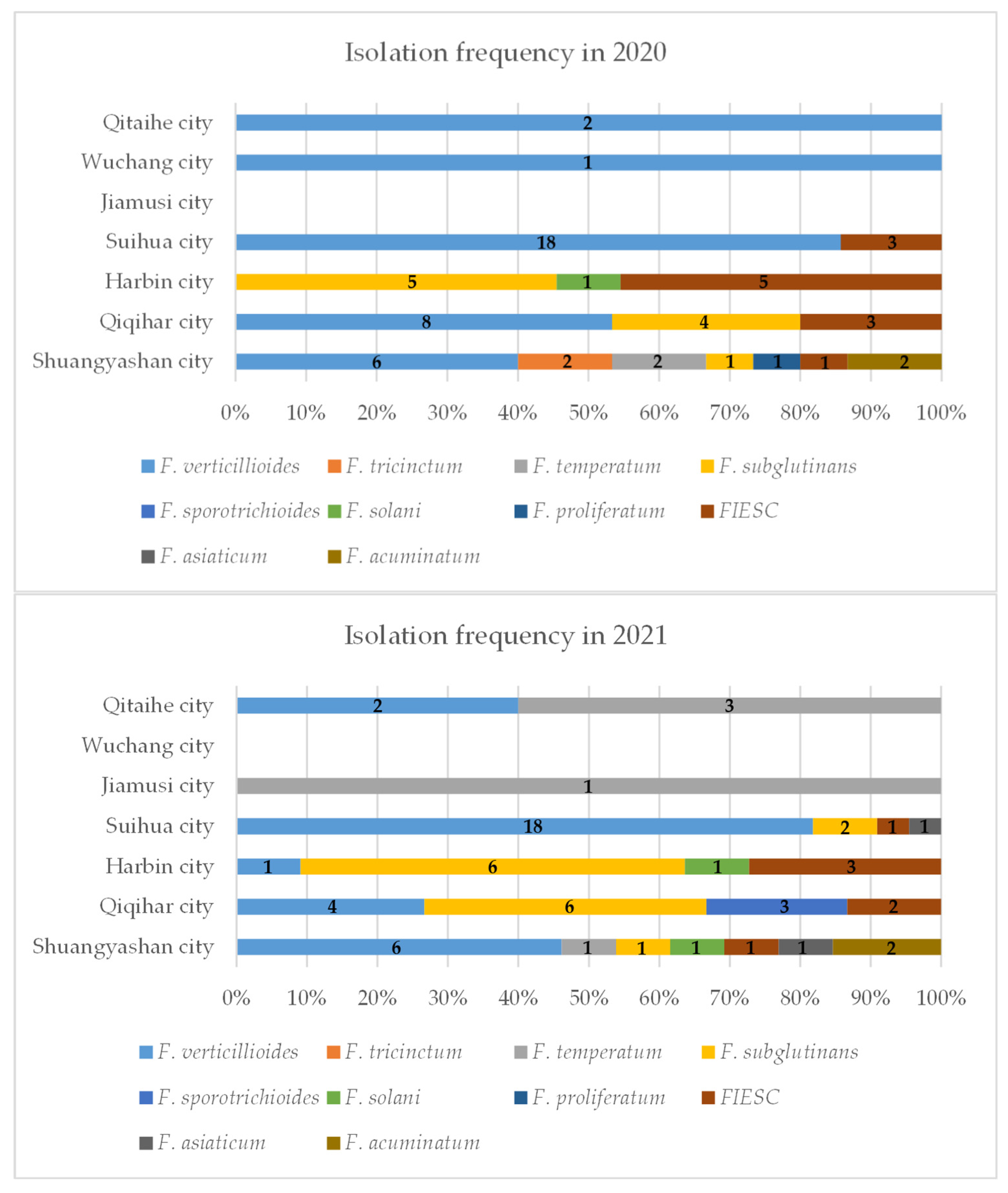
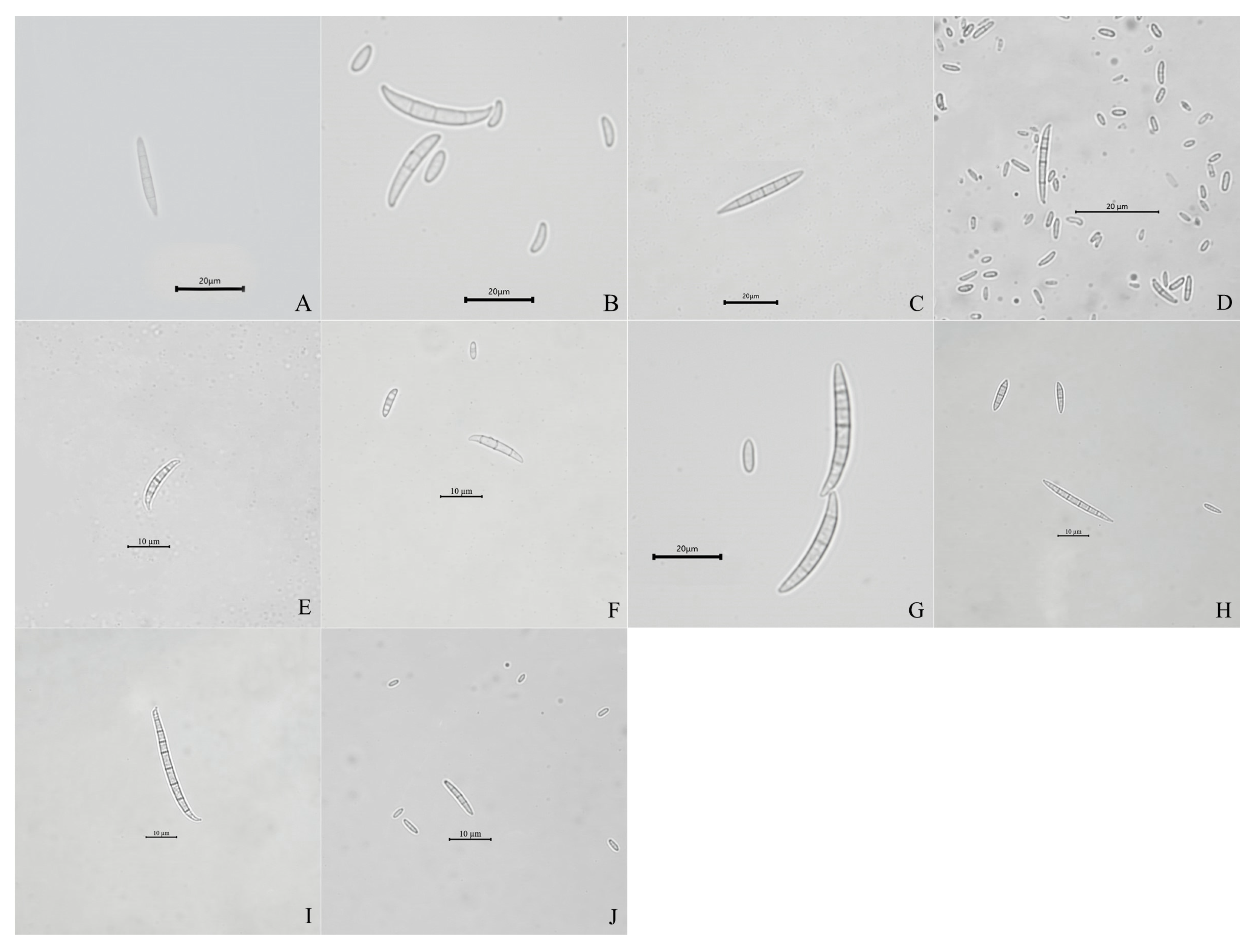
2.1. Isolation and Identification of Fusarium Isolates

In 2020, 53 samples of maize sheath rot were collected from six maize-growing areas of Heilongjiang Province (including Qitaihe city, Wuchang city, Suihua city, Harbin city, Qiqihar city, and Shuangyashan city); In 2021, 45 samples were collected from maize-growing areas of Heilongjiang Province (including Qitaihe city, Jiamusi city, Suihua city, Harbin city, Qiqihar city, and Shuangyashan city). A total of 98 symptomatic maize leaf sheath samples were collected from seven cities in the main maize-growing areas of Heilongjiang province during 2020–2021. Additionally, Shuangyashan city had the highest frequency of isolation (Figure 1).
A total of 132 Fusarium isolates obtained from symptomatic maize leaf sheaths, collected from seven cities in the main maize-growing areas of Heilongjiang province (Tables S1 and S2), were tentatively classified into 10 groups according to their morphological characteristics (Table 1, Figure 2 and Figure 3), including F. verticillioides, F. subglutinans, F. proliferatum, F. temperatum, F. tricinctum, F. solani, F. sporotrichioides, F. asiaticum, F. acuminatum, and the Fusarium incarnatum-equiseti species complex (FIESC).
Of the isolates, 66 were identified as the F. verticillioides morphological group, which could produce white to greyish-purple colonies with dark yellow to purple–gray reverse. The macroconidia were slightly falcate-to-almost straight, with three to four transverse septa, and in a size range of 16.6–23.4 × 2.3–3.4 μm (n = 30).
Twenty-five isolates initially produced yellow mycelia, and then, turned lavender; these were identified as the F. subglutinans morphological group. The macroconidia were slightly falcate and septate, with three to five transverse septa, in a size range of 13.6–21.1 × 1.6–2.3 μm (n = 30).
Nineteen isolates forming white to light beige with loosely floccose mycelia and producing light yellow pigmentation were classified into the FIESC morphological group. The macroconidia were falcate, with three to six transverse septa and in a size range of 16–18 × 1.5–3 μm (n = 30).
Seven isolates forming white to peach-colored mycelia were identified as the F. temperatum morphological group. Typical macroconidia were falcate, with three to five septa, and in a size range of 13.6–23.1 × 1.6–3.3 μm (n = 30).
Four isolates forming abundant and partly carmine mycelia were identified as the F. acuminatum morphological group. The macroconidia were slender, equilaterally curved, septate with three to five transverse septa, and in a size range of 13.2–24.2 × 1.9–3.7 μm (n = 30). Three isolates forming dense and white mycelia were classified into the F. solani morphological group. The macroconidia were abundant and sickle-shaped, with two to four transverse septa, and in a size range of 16–26 × 1.5–2.8 μm (n = 30).
Three isolates initially produced white mycelia, and then, turned pink; these were identified as the F. sporotrichioides morphological group. The macroconidia were sickle-shaped, with three to five septa, moderately curved-to-straight, and measured 12.5–24.3 × 3.1–4.2 μm (n = 30).
Two isolates forming white-to-canary yellow, abundant, and dense mycelia were classified into the F. tricinctum morphological group. The macroconidia were slightly curved-to-falcate, with three to five septa, and in a size range of 12.5–26.0 × 1.5–3.0 µm (n = 30).
Two isolates forming white mycelia with pink pigmentation were classified into the F. asiaticum morphological group. The macroconidia were falcate, with four to eight transverse septa, and in a size range of 13–36 × 1.6–3.8 μm (n = 30).
One isolate forming white mycelia with dark violet pigmentation was identified as the F. proliferatum morphological group. The macroconidia were sparse, slender, with three to four septa, and measured 13.6–22.3 × 3.1–4.2 μm (n = 30).

2.2. Phylogenetic Analysis of PCR-Generated DNA Sequences

For further molecular verification, a maximum likelihood tree based on the TEF-1α gene sequences of 132 Fusarium isolates and 11 reference strains (Table S1) was constructed, and these isolates were also grouped into 10 species, including F. verticillioides (n = 66), F. subglutinans (n = 25), the FIESC (n = 19), F. temperatum (n = 7), F. acuminatum (n = 4), F. solani (n = 3), F. sporotrichioides (n = 3), F. tricinctum (n = 2), F. asiaticum (n = 2), and F. proliferatum (n = 1) (Figure 4).

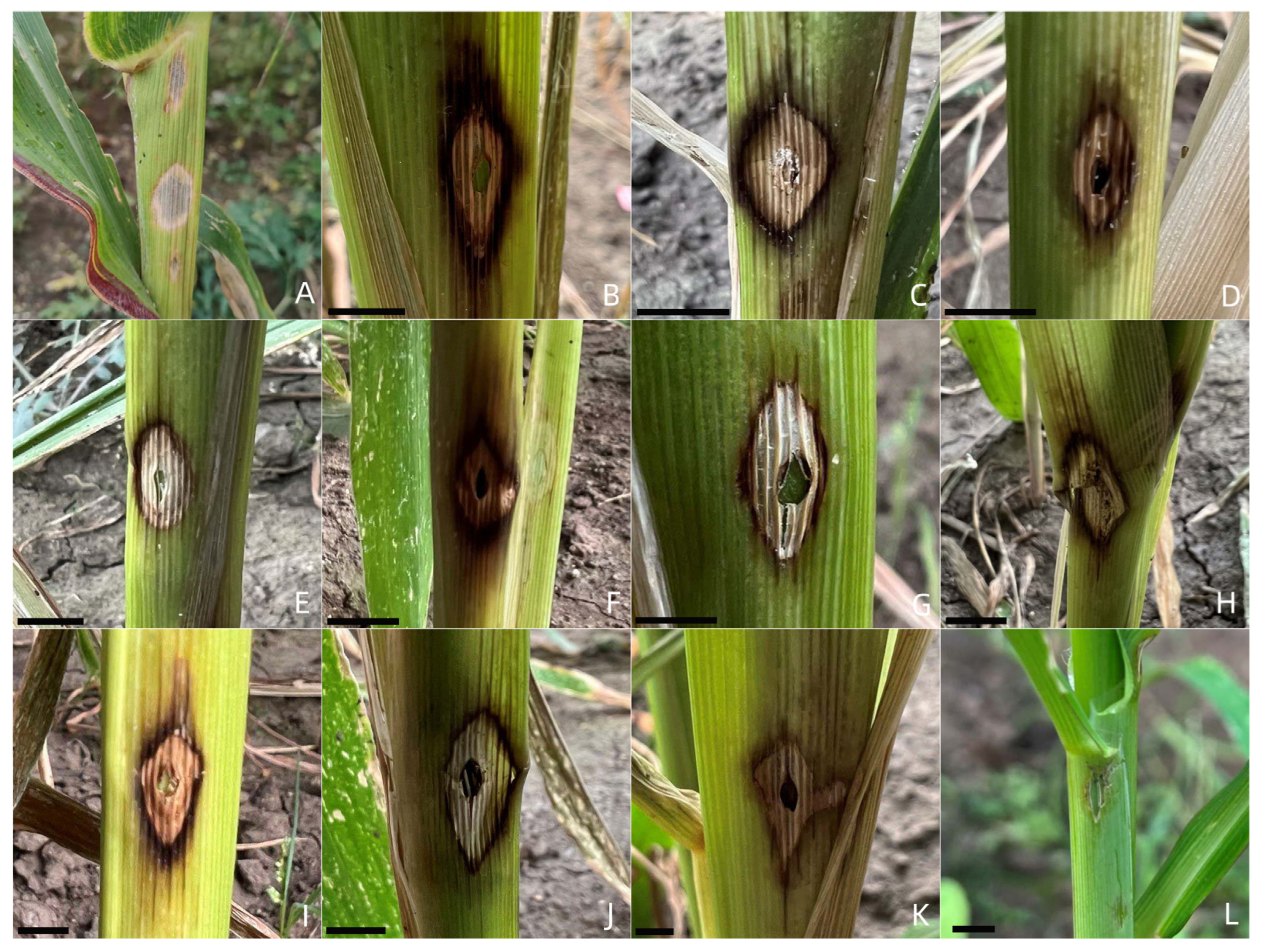
2.3. Pathogenicity Tests

All Fusarium isolates could cause maize sheath rot in this study (Table 2 and Table S3). The symptoms caused by the 10 Fusarium species were similar between species and initially formed grayish-white spots, the spots were gradually enlarged and surrounded by dark brown halos, and the center of the lesions turned to grayish yellow or dark gray with time (Figure 5). The symptoms of maize sheath rot presented in this study were similar to the field symptoms (Figure 5), while no symptoms appeared in the control group (Figure 5). To fulfill Koch’s postulates, the Fusarium isolates were all re-isolated from the symptomatic maize leaf sheaths and confirmed according to morphological and molecular methods, whereas no Fusarium isolates were obtained from the control group. The disease incidence and disease index caused by Fusarium species were 100% and 11.1–100 (Table 2), respectively. Of which F. tricinctum and F. proliferatum showed significantly higher virulence than the other Fusarium species, with an average disease index of 94.4; this was followed by F. asiaticum, F. acuminatum, and F. solani, which showed similar disease indexes of 73.6, 68.1, and 61.1, respectively. Moreover, F. sporotrichioides, F. temperatum, F. verticillioides, F. subglutinans, and the FIESC exhibited somewhat lower virulence, with average disease indexes ranging from 42.3 to 58.3.

2.4. Haplotype Network

The haplotype network was constructed based on the concatenated sequences of rDNA ITS and TEF-1α genes, and 10 haplotypes were identified among the 99 FFSC isolates, of which five haplotypes (Haps 1, 2, 4, 5, and 6) were the shared haplotypes (Figure 6). Haplotype 2 was the most abundant haplotype and distributed in five locations (Suihua city, Shuangyashan city, Qitaihe city, Qiqihar city, and Wuchang city). Haplotype 4 was found in Harbin city, Qiqihar city, Suihua city, and Shuangyashan city. Haplotype 1 was detected in Qiqihar city, Suihua city, and Shuangyashan city. Haplotype 6 was present in Qiqihar city, Suihua city, and Jiamusi city. Haplotype 5 was only found in two locations (Qiqihar city and Harbin city). The other five haplotypes were all private haplotypes. In addition, the haplotypes detected in Suihua city and Shuangyashan city were the most abundant and included five haplotypes, followed by Qiqihar city (four haplotypes), Harbin city (three haplotypes), Qitaihe city (three haplotypes), Wuchang city (one haplotype), and Jiamusi city (one haplotype).

3. Discussion

There are few records about maize sheath rot in the world. White et al. (1999; 2005) first recorded the occurrence of purple sheath blight disease on maize in the United States, which began to occur at the silking stage of maize, and only harmed the leaf sheath without infecting the leaves and stalks. It was considered that it may be caused by Fusarium spp. Purple sheath blight is common in the U.S. Corn Belt, but it does not result in yield loss. Since then, there have been almost no reports on maize sheath rot abroad. However, maize sheath rot was first recorded in 2008 in northeast China and has been reported to cause varying degrees of maize yield loss. In recent years, maize sheath rot has frequently occurred, affecting the yield of maize; for example, Li et al. [5] reported that sheath rot occurred in several maize fields of Liaoning, Jilin, and Heilongjiang provinces, causing an approximately 30% yield loss; Sun et al. [7] found sheath rot presented on approximately 40% of maize leaf sheaths in three 7 ha commercial fields in Heilongjiang province. Moreover, it has also been reported that serious infection with sheath rot could reduce the lodging resistance of maize [6,8].
Maize sheath rot occurs during the reproductive stage of maize, and the silking stage shows the strongest infection. The occurrence of maize sheath rot is closely related to many factors, including the maize variety, climate conditions, and the feeding of aphids [2]. A previous study showed that the disease incidence at the silking stage of maize was significantly higher than that of the tasseling stage, grain filling stage, and physiological maturity. The activities of defense enzymes at the silking stage of maize were also significantly higher than at the other stages. In addition, hybrid maize varieties were relatively more resistant than the inbred lines, and maize grown in a humid environment and a warm climate was more susceptible to Fusarium infection [35]. In the survey conducted in Heilongjiang province from 2021 to 2022 in this study, during the reproductive stage of maize, the disease incidence of maize sheath rot was 56.8–76.6% and the diversity of Fusarium species causing maize sheath rot gradually increased.
In the present study, 132 isolates belonging to 10 Fusarium species were identified as the causal agents of maize sheath rot in the main maize-growing areas of Heilongjiang province, and F. subglutinans, F. temperatum, F. solani, F. sporotrichioides, F. tricinctum, and F. acuminatum were first reported as the causal agents of maize sheath rot worldwide. Moreover, four Fusarium species—F. verticillioides (n = 66), F. subglutinans (n = 25), F. temperatum (n = 7), and F. proliferatum (n = 1), including 99 isolates—were classified into the Fusarium fujikuroi species complex (FFSC), of which F. verticillioides was the most prevalent fungal pathogen on maize and has been reported to cause serious stalk rot, ear rot, sheath rot, and leaf blight in maize [8,11,36,37]. F. subglutinans could cause seedling disease and ear rot in maize, resulting in serious loss of yield [38]. F. temperatum is a common maize pathogen which could produce mycotoxins and cause plant disease [39]. F. proliferatum is the major pathogen of maize sheath rot and was first reported in 2008 by Xu et al.; it could produce enniatins, beauvericin, fumonisins, fusaproliferin, fusaric acid, fusarins and moniliformin [40], contaminating mainly maize and maize products [41]. However, only one F. proliferatum isolate was obtained in this study, suggesting that the diversity and population structure of Fusarium species associated with maize sheath rot may have changed and the changes may be related to natural environmental conditions, maize varieties, tillage patterns, and other objective factors [42]. These results require us to pay attention to the genetic evolution and population composition of pathogens associated with plant diseases and reasonably select fungicides and biocontrol agents, as well as appropriate cultivars.
Moreover, different Fusarium species showed differentiated aggressiveness on maize sheath. The average disease indexes of F. tricinctum, F. proliferatum, and F. asiaticum were higher than those of the other Fusarium species, which was different from the previous conclusion; the average disease index of F. proliferatum was the highest, while the average disease index of F. asiaticum was relatively low [8], indicating that the disease index may be affected by the inoculation method [43]. This study is the first to clarify and compare the pathogenicity of F. subglutinans, F. temperatum, F. solani, F. sporotrichioides, F. tricinctum, and F. acuminatum associated with maize sheath rot in Heilongjiang province. These Fusarium species with different pathogenicities could also be used to evaluate the resistance of different maize varieties in future and will be helpful in breeding new resistant maize varieties [44].
In addition, the haplotypes of 99 FFSC isolates were identified, and 10 haplotypes were detected. Haplotypes 1, 2, 3, 7, 8, and 9 belonged to the F. verticillioides clade, and the haplotype network was radial with the center of haplotype 2, indicating that the population expanded rapidly after reaching the bottleneck [45]. Furthermore, haplotypes 4 and 5 belonged to the F. subglutinans clade, haplotype 6 belonged to the F. temperatum clade, and haplotype 10 belonged to the F. proliferatum clade. These members of the FFSC were assigned to different clades in the haplotype network, suggesting that the haplotype network could effectively distinguish Fusarium species in the complex, and also confirming our classification results. Moreover, the major haplotypes (Haps 1, 2, 4, and 6) presented in multiple locations of Heilongjiang province, and no significant correlation was found between geographic origin and haplotype distribution. Spores of the Fusarium species may be transmitted by wind, insects, or the trade of agricultural products [46,47,48,49,50], which might explain the wide geographic distribution of FFSC isolates in Heilongjiang province.

4. Materials and Methods

4.1. Sample Collection and Fusarium Isolation

A total of 98 symptomatic maize leaf sheath samples were collected from seven cities in the main maize-growing areas of Heilongjiang province during 2020–2021. The samples were kept in paper bags and stored in a 4 °C refrigerator. The maize leaf sheaths were cut into 0.5 cm × 0.5 cm pieces at the junction between diseased and healthy tissue prior to isolation. The leaf sheath pieces were soaked in 1% sodium hypochlorite for 1 min, rinsed twice with sterile distilled water, and placed on potato dextrose agar (PDA) plates supplemented with 100 μg/mL nalidixic acid sodium salt, for 3 days at 25 °C, for fungal isolation. All fungal colonies were picked out from the plates; however, only isolates identified as Fusarium species using morphological observation were selected for further study. Colony purification was carried out by picking a single spore using a stereomicroscope and culturing it on PDA at 25 °C for 7 days. In total, 132 Fusarium isolates were obtained (Table S1) and preserved on PDA slants at 4 °C [51]. In addition, several other fungal colonies (including 4 Didymella americana isolates and 2 Nigrospora musae isolates) were also obtained in addition to these Fusarium isolates, but those fungi were all not pathogenic to maize leaf sheath.

4.2. Morphological Characterization

All isolates were inoculated on PDA plates and incubated at 25 °C in the dark for 7 days. Each isolate was assessed based on colony texture and colony color. In addition, these isolates were cultured on PDA at 25 °C for 14 days with a light/dark cycle of 8/16 h to observe the well-developed macroconidia. The macroconidia were evaluated using light microscopy (Zeiss Axiolab 5 equipped with an Axiocam 208 color industrial digital camera) [52].

4.3. DNA Extraction and Sequencing

Fungal DNA was extracted using 2% cetyltrimethylammonium bromide as Leslie and Summerell described [53]. The primers ITS1 (TCCGTAGGTGAACCTGCGG)/ITS4 (GCTGCGTTCTTCATCGATGC) and EF1-728F (CATCGAGAAGTTCGAGAAGG)/EF4-986R (TACTTGAAGGAACCCTTACC) were used to amplify the partial rDNA-ITS and TEF1-α genes, respectively [54,55]. The PCR products were purified and sequenced at Beijing Biootech Co. Ltd. The obtained gene sequences of 132 Fusarium isolates were aligned in GenBank and the Fusarium ID-database, and then, deposited into the NCBI GenBank.

4.4. Phylogenetic Analysis of Fusarium Isolates

In the present study, MEGA 7 software with default parameters was used for phylogenetic analysis, and the sequences were aligned based on the Clustal W algorithm. A phylogenetic tree based on the TEF-1α gene sequences was constructed using 1000 bootstrap repeated with the maximum likelihood (ML) method. Alternaria junci-acuti IRAN 3512C was selected as the outgroup.

4.5. Pathogenicity Tests

Forty-seven representative Fusarium isolates were selected for the pathogenicity test and the selection method and number are shown in Table S2. All 47 Fusarium isolates were inoculated into 50 mL carboxymethyl cellulose (CMC) liquid medium (7.5 g sodium carboxymethyl cellulose, 0.5 g yeast extract, 2.5 g K2HPO4, and 0.25 g MgSO4·7H2O were added to 1000 mL water) and cultured for 3 days on a rotary shaker at 25 °C, 160 r.p.m. [56]. The conidia suspension was filtered and adjusted to 1 × 10⁶ conidia/mL. Healthy maize plants (var. Zhengdan 958; nine maize plants for each isolate) grown in the greenhouse at the heading stage and were selected for the pathogenicity test to fulfill Koch’s postulates. The maize leaf sheaths were surface-disinfected and wounded with a sterile needle (1mm diameter), and then, 10 μL of conidia suspension was injected into the maize sheaths. Three control maize leaf sheaths were inoculated with 10 μL sterile water. Symptoms were observed after 25 days post inoculation, and disease severity was scored according to the modified method described by Zhang et al. [57]. The experiment was repeated twice with three maize leaf sheaths per replication.
The disease index was assessed based on a 0–3 scale—0 (no obvious lesion), 1 (diameter of lesion between 0–0.3 cm), 2 (diameter of lesion between 0.3–0.5 cm), and 3 (diameter of lesion above 0.5 cm). The disease index (DI) was calculated by following the formula: DI = [100 × ∑ (n × corresponding DS)]/(N × 3), where n is the number of infected inoculation sheaths corresponding to each disease rating, and N is the total number of inoculation sheaths. SPSS software (v. 20.0; SPSS Inc., Wacker Drive, Chicago, IL, USA. IBM Corp., 2012. IBM) was used for statistical analysis using a least significant difference (LSD) test at a significance level of p < 0.05. All Fusarium isolates were re-isolated from the symptomatic maize leaf sheaths and identified based on morphological and molecular methods.

4.6. Nucleotide Diversity and Haplotype Assignment within Species

The ITS and TEF-1α gene sequences of 99 Fusarium fujikuroi species complex (FFSC) isolates were individually concatenated for haplotype analysis. PopART software version 1.7 (Population Analysis with Reticulate Trees is free, open-source population genetics software that was developed as part of the Allan Wilson Centre Imaging Evolution Initiative) was used for generating a genealogical network based on the concatenated sequences [58]. Haplotype identification based on parsimony probability computed for pairwise comparisons was carried out using a TCS network [59,60].

4.7. Data Analysis

Differences in the field survey, growth rate, and pathogenicity were analyzed using the Statistical Package for Social Sciences (SPSS) (v. 20.0; SPSS Inc., Wacker Drive, Chicago, Illinois.IBM Corp., 2012. IBM). An analysis of variance was performed using the general linear model, and the means were compared using Duncan’s New Multiple Range test in SPSS, with differences considered significant at p ≤ 0.05.

5. Conclusions

In conclusion, 10 Fusarium species were isolated from seven major maize-growing areas in Heilongjiang province, China. F. verticillioides was the dominant species. F. subglutinans, F. temperatum, F. solani, F. sporotrichioides, F. tricinctum, and F. acuminatum were first reported as the casual agents of maize sheath rot. Clarifying the population structure and pathogenicity of Fusarium spp. associated with maize sheath rot in Heilongjiang province will be helpful in studying resistant maize varieties, and in selecting the control agents and providing guidelines for the comprehensive control of maize disease in Heilongjiang province.

Supplementary Materials

The following supporting information can be downloaded at: https://www.mdpi.com/article/10.3390/ijms231810821/s1.

Author Contributions

X.Y., X.X. and S.W. performed the experiments. L.Z. and G.S. prepared the figures and tables. H.T., C.S. and C.Y. analyzed the data. W.X., X.W. and J.Z. designed the experiments and reviewed the manuscript. All authors have read and agreed to the published version of the manuscript.

Funding

This work was supported, in part, by grants from the National Natural Science Foundation of China (No. 31972291), the Outstanding Youth Project of Natural Science Foundation of Heilongjiang Province (YQ2021C012), the postdoctoral research fund of Heilongjiang Province (LBH-Q21072), the Academic Backbone Project of the Northeast Agricultural University (20XG33), and the National Natural Youth Science Foundation of China (No. 31701858).

Institutional Review Board Statement

Not applicable.

Informed Consent Statement

Not applicable.

Data Availability Statement

Sequences have been deposited in GenBank. The data presented in this study are openly available in NCBI. Publicly available datasets were analyzed in this study. These data can be found here: https://www.ncbi.nlm.nih.gov/.

Conflicts of Interest

The authors declare that there are no conflict of interest.

References

  1. Li, S. Review of maize market in 2020–2021 and prospect in 2021–2022. China Pig Ind. 2022, 17, 29–33. [Google Scholar]
  2. Wang, Q.L.; Zhang, N.N.; Liu, H. An empirical study on Maize supply response in China under the background of Market-oriented Reform-based on Provincial Panel data of three Northeast provinces from 2008 to 2019. Agric. Resour. Reg. China 2021, 42, 145–152. [Google Scholar]
  3. Xu, X.D.; Jiang, Y.; Wang, L.J.; Dong, H.Y.; Hu, L.; Lu, G.Z.; Liu, Z.H. Corn Sheath Rot—A New Disease Found in China. Sci. Agric. Sin. 2008, 41, 3083–3087. [Google Scholar]
  4. Hu, L.; Xu, X.D.; Jiang, Y.; Wang, L.J.; Xu, J.; Zhao, Y. Study on biological characteristics of corn sheath rot causal agent. Maize Sci. 2008, 16, 131–134. [Google Scholar]
  5. Li, P.P.; Cao, Z.Y.; Wang, K.; Zhai, H.; Jia, H.; Liu, N.; Li, S.H.; Hao, Z.M.; Gu, S.Q.; Dong, J.G. First report of Fusarium equiseti causing a sheath rot of corn in China. Plant Dis. 2014, 98, 998. [Google Scholar] [CrossRef]
  6. Wang, K.; Cao, Z.; Li, P.; Yin, H.; Liu, J.; Dong, J. Analysis of the correlation between diseased-degree of corn sheath rot and corn lodging and yield loss. Acta Phytophylacica Sin. 2015, 42, 949–956. [Google Scholar]
  7. Sun, L.; Wang, S.; Zhang, W.; Chi, F.; Hao, X.Y.; Bian, J.Y.; Li, Y. First report of sheath rot of corn caused by Fusarium verticillioides in northeast China. J. Plant Pathol. 2020, 102, 1301–1302. [Google Scholar] [CrossRef]
  8. Wang, W.; Wang, B.; Sun, X.; Qi, X.; Gong, G. Symptoms and pathogens diversity of maize Fusarium sheath rot in Sichuan province, China. Sci. Rep. 2021, 11, 2835. [Google Scholar] [CrossRef]
  9. Abdel-Azeem, A.M.; Azeem, M.A.; Darwish, A.G.; Nafady, N.A.; Ibrahim, N.A. Fusarium: Biodiversity, Ecological Significances, and Industrial Applications. In Proceedings of the 3rd ISNPS, Avignon, France, 11–16 June 2016. [Google Scholar]
  10. Pfordt, A.; Romero, L.R.; Schiwek, S.; Karlovsky, P.; Tiedemann, A.V. Impact of environmental conditions and agronomic practices on the prevalence of Fusarium species associated with ear and stalk rot in maize. Pathogens 2020, 9, 236. [Google Scholar] [CrossRef]
  11. Schieber, R. A leaf blight of corn (Zea mays) incited by Fusarium moniliforme. Phytopathology 1968, 58, 554. [Google Scholar]
  12. Gai, X.T.; Yang, R.X.; Pan, X.J.; Yuan, Y.; Wang, S.N.; Liang, B.B.; Gao, Z.G. First Report of Fusarium incarnatum Causing Stalk Rot on Maize in China. Plant Dis. 2016, 100, 1010. [Google Scholar] [CrossRef]
  13. Wang, J.H.; Li, H.P.; Zhang, J.B.; Wang, B.T.; Liao, Y.C. First report of Fusarium maize ear rot caused by Fusarium meridionale in China. Plant Dis. 2014, 98, 279. [Google Scholar] [CrossRef] [PubMed]
  14. Dong, H.; Qin, P.; Gao, Z.; Xu, J.; Xu, X. First report of seedling blight of maize caused by Fusarium asiaticum in Northeast China. Plant Dis. 2020, 105, 1206. [Google Scholar] [CrossRef] [PubMed]
  15. Mezzalama, M.; Guarnaccia, V.; Martino, I.; Tabome, G.; Gullino, M.L. First report of Fusarium commune causing root and crown rot on maize in Italy. Plant Dis. 2021, 105, 4156. [Google Scholar] [CrossRef]
  16. Kabeere, F.; Hampton, J.G.; Hill, M.J. Transmission of Fusarium graminearum (Schwabe) from maize seeds to seedlings. Seed Sci. Technol. 1997, 25, 245–252. [Google Scholar]
  17. Wollenweber, H.W.; Sherbakoff, C.D.; Reinking, O.A.; Johann, H.; Bailey, A.A. Fundamentals for taxonomic studies of Fusarium. J. Agric. Res. 1925, 30, 833–843. [Google Scholar]
  18. Lima, C.S.; Pfenning, L.H.; Costa, S.S.; Campos, M.A.; Leslie, J.F. A new Fusarium lineage within the Gibberella fujikuroi species complex is the main causal agent of mango malformation disease in Brazil. Plant Pathol. 2010, 58, 33–42. [Google Scholar] [CrossRef]
  19. Vermeulen, M.; Rothmann, L.A.; Swart, W.J.; Gryzenhout, M. Fusarium casha sp. nov. and F. curculicola sp. nov. in the Fusarium fujikuroi Species Complex Isolated from Amaranthus cruentus and Three Weevil Species in South Africa. Diversity 2021, 10, 472. [Google Scholar] [CrossRef]
  20. Bashyal, B.M.; Aggarwal, R. Molecular identification of Fusarium species associated with bakanae disease of rice (Oryza sativa) in India. Indian J. Agric. Sci. 2013, 83, 71–76. [Google Scholar]
  21. Qiu, J.; Lu, Y.; He, D.; Lee, Y.W.; Ji, F.; Xu, J.; Shi, J. Fusarium fujikuroi Species Complex Associated with Rice, Maize, and Soybean from Jiangsu Province, China: Phylogenetic, Pathogenic, and Toxigenic Analysis. Plant Dis. 2020, 104, 2193–2201. [Google Scholar] [CrossRef]
  22. Prabhukarthikeyan, S.R.; Keerthana, U.; Nagendran, K.; Yadav, M.K.; Parameswaran, C.; Panneerselvam, P.; Rath, P.C. First Report of Fusarium proliferatum Causing Sheath Rot Disease of Rice in Eastern India. Plant Dis. 2021, 105, 704. [Google Scholar] [CrossRef] [PubMed]
  23. Fang, D.; Chen, J.; Yi, C.; Li, Z.; Gao, C. First report of Fusarium fujikuroi causing bulb rot on Lilium lancifolium in China. Plant Dis. 2021, 105, 2254. [Google Scholar] [CrossRef] [PubMed]
  24. Tushar, B.; Rakholiya, K. Environment friendly way to management of Fusarium fruit rot disease of banana in vivo by essential oils. Int. J. Genet. 2020, 12, 798–800. [Google Scholar]
  25. Huang, S.P.; Wei, J.G.; Guo, T.X.; Li, Q.L.; Tang, L.H.; Mo, J.Y.; Wei, J.F.; Yang, X.B. First report of sheath rot caused by Fusarium proliferatum on Pisang Awak Banana (Musa ABB) in China. Plant Pathol. 2019, 101, 1271–1272. [Google Scholar] [CrossRef]
  26. Araújo, M.B.M.; Moreira, G.M.; Nascimento, L.V.; Nogueira, G.D.A.; Nascimento, S.R.D.; Pfenning, L.H.; Ambrósio, M.M.D. Fusarium rot of melon is caused by several Fusarium species. Plant Pathol. 2020, 70, 712–721. [Google Scholar] [CrossRef]
  27. Jacobs, A.; Van Wyk, P.S.; Marasas, W.F.O.; Wingfield, B.D.; Wingfield, M.J.; Coutinho, T.A. Fusarium ananatum sp. nov. in the Gibberella fujikuroi species complex from pineapples with fruit rot in south Africa. Fungal Biol. 2010, 114, 515–527. [Google Scholar] [CrossRef]
  28. Hsuan, H.M.; Salleh, B.; Zakaria, L. Molecular identification of Fusarium species in Gibberella fujikuroi species complex from rice, sugarcane and maize from Peninsular Malaysia. Int. J. Mol. Sci. 2011, 12, 6722–6732. [Google Scholar] [CrossRef]
  29. Donát, M.; Csaba, Z.; Zsuzsanna, K.; Árpád, S.; János, B. Identification of airborne propagules of the Gibberella fujikuroi species complex during maize production. Aerobiologia 2012, 28, 263–271. [Google Scholar] [CrossRef]
  30. Jarek, T.M.; dos Santos, Á.F.; Tessmann, D.J.; Vieira, E. Inoculation methods and aggressiveness of five Fusarium species against peach palm. Ciênc. Rural 2018, 48, e20170462. [Google Scholar] [CrossRef]
  31. Félix-Gastélum, R.; Mora-Carlón, B.A.; Leyva-Madrigal, K.Y.; Solano-Báez, A.R.; Pérez-Mora, J.L.; Guerra-Meza, O.; Mora-Romero, G.A. Sorghum Sheath Blight Caused by Fusarium spp. in Sinaloa, Mexico. Plant Dis. 2022, 106, 1454–1461. [Google Scholar] [CrossRef]
  32. Li, Y.L.; Yan, Z.B.; Wang, Y.H.; Zhou, Z. First report of Fusarium proliferatum causing leaf sheath rot on Clivia miniata in Henan province, China. Plant Dis. 2020, 104, 1552. [Google Scholar] [CrossRef]
  33. Librado, P.; Rozas, J. DnaSP v5: A software for comprehensive analysis of DNA polymorphism data. Bioinformatics 2009, 25, 1451–1452. [Google Scholar] [CrossRef] [PubMed]
  34. Berman, J.; Forche, A. Haplotyping a Non-meiotic Diploid Fungal Pathogen Using Induced Aneuploidies and SNP/CGH Microarray Analysis. Methods Mol. Biol. 2017, 1551, 131–146. [Google Scholar] [PubMed]
  35. Liu, J.; Xu, M.M.; Wang, A.P.; Yin, H.F.; Wang, K.; Cao, Z.Y.; Dong, J.G. Resistant Evaluation and Yield Loss Common Corn Varieties to Corn Sheath Rot. Maize Sci. 2018, 26, 29–36. [Google Scholar]
  36. Chalivendra, S.; Huang, F.N.; Busman, M.; Williams, W.P.; Ham, J.H. Low aflatoxin levels in aspergillus flavus-resistant maize are correlated with increased corn earworm damage and enhanced seed fumonisin. Front. Plant Sci. 2020, 11, 565323. [Google Scholar] [CrossRef]
  37. Kaur, H.; Mohan, C.; Vika, Y.; Singh, M. Pathogenic and molecular characterization of Fusarium moniliforme Sheld, the incitant of Fusarium maize stalk rot in the Punjab State of India. Maydica 2008, 59, 290–297. [Google Scholar]
  38. Li, L.; Qu, Q.; Cao, Z.; Guo, Z.; Dong, J. The relationship analysis on corn stalk rot and ear rot according to Fusarium species and fumonisin contamination in kernels. Toxins 2019, 11, 320. [Google Scholar] [CrossRef]
  39. Fumero, M.V.; Villani, A.; Susca, A.; Haidukowski, M.; Cimmarusti, M.T.; Toomajian, C.; Leslie, J.F.; Chulze, S.N.; Moretti, A. Fumonisin and beauvericin chemotypes and genotypes of the sister species Fusarium subglutinans and Fusarium temperatum. Appl. Environ. Microbiol. 2020, 86, e00133-20. [Google Scholar] [CrossRef]
  40. Munkvold, G.P. Fusarium Species and their Associated Mycotoxins. Methods Mol. Biol. 2017, 1542, 51–106. [Google Scholar]
  41. Sheng, W.; Wu, H.; Ji, W.; Li, Z.; Chu, F.; Wang, S. Visual Non-Instrumental On-Site Detection of Fumonisin B1, B2, and B3 in Cereal Samples Using a Clean-Up Combined with Gel-Based Immunoaffinity Test Column Assay. Toxins 2018, 10, 165. [Google Scholar] [CrossRef]
  42. Koné, D.; Badou, O.J.; Bomisso, E.L.; Camara, B.; Ake, S. In vitro activity of different fungicides on the growth in Mycosphaerella fijiensis var. difformis Stover and Dickson, Cladosporium musae Morelet and Deightoniella torulosa (Syd.) Ellis, isolated parasites of the banana phyllosphere in the Ivory Coast. Comptes Rendus Biol. 2009, 332, 448–455. [Google Scholar]
  43. Yi, R.H.; Zhu, X.R.; Zhou, E.X. On the evaluation of different methods by artificially inoculating with rice sheath blight fungus (Rhizoctonia solani). J. Guangzhou Univ. 2003, 3, 224–227. [Google Scholar]
  44. Endo, T.; Fujii, H.; Omura, M.; Shimada, T. Fast-track breeding system to introduce CTV resistance of trifoliate orange into citrus germplasm, by integrating early flowering transgenic plants with marker-assisted selection. Plant Biol. 2020, 20, 224. [Google Scholar] [CrossRef] [PubMed]
  45. Matić, S.; Tabome, G.; Garibaldi, A.; Gullino, M.L. Alternaria leaf spot caused by Alternaria species: An emerging problem on ornamental plants in Italy. Plant Dis. 2020, 104, 2275–2287. [Google Scholar] [CrossRef]
  46. Hoffmann, A.; Funk, R.; Müller, M.E.H. Blowin’ in the Wind: Wind Dispersal Ability of Phytopathogenic Fusarium in a Wind Tunnel Experiment. Atmosphere 2021, 12, 1653. [Google Scholar] [CrossRef]
  47. Feldman, T.S.; O’Brien, H.E.; Arnold, A.E. Moths that Vector a Plant Pathogen also Transport Endophytic Fungi and Mycoparasitic Antagonists. Microb. Ecol. 2008, 56, 742–750. [Google Scholar] [CrossRef]
  48. Guo, Z.Q.; Pfohl, K.; Karlovsky, P.; Dehne, H.W.; Altincicek, B. Dissemination of Fusarium proliferatum by mealworm beetle Tenebrio molitor. PLoS ONE 2018, 13, e0204602. [Google Scholar] [CrossRef]
  49. Yin, H.F.; Cao, Z.Y.; Wang, K.; Li, P.P.; Jia, H.; Dong, J.G. The influence of aphids occurrence on corn sheath rot. J. Agric. Univ. Hebei 2015, 38, 86–91. [Google Scholar]
  50. Arseniuk, E.; Scharen, A.L.; Czembor, H.J. Pathogenicity of seed transmitted Fusarium spp. to triticale seedlings. Mycotoxin Res. 1991, 7, 121–127. [Google Scholar] [CrossRef]
  51. Xu, X.; Yang, X.L.; Zhang, L.; Cao, H.S.; Cao, P.; Wang, X.J.; Xiang, W.S.; Zhao, J.W. First report of leaf blight caused by Limonomyces roseipellis on maize (Zea mays L.) in China. Plant Dis. 2021, 105, 2730. [Google Scholar] [CrossRef]
  52. Xu, X.; Zhang, L.; Yang, X.L.; Cao, H.S.; Li, J.J.; Cao, P.; Guo, L.F.; Wang, X.J.; Zhao, J.W.; Xiang, W.S. Alternaria spp. associated with leaf blight of maize in Heilongjiang Province, China. Plant Dis. 2022, 106, 572–584. [Google Scholar] [CrossRef] [PubMed]
  53. Leslie, J.F.; Summerell, B.A. The Fusarium Laboratory Manual; Blackwell Professional: Ames, IA, USA, 2006; Volume 56, p. 1037. [Google Scholar]
  54. O’Donnell, K.; Cigelnik, E. Two divergent intragenomic rDNA ITS2 types within a monophyletic lineage of the fungus Fusarium are nonorthologous. Mol. Phylogenet. Evol. 1997, 7, 103–116. [Google Scholar] [CrossRef] [PubMed]
  55. Glass, N.L.; Donaldson, G.C. Development of primer sets designed for use with the PCR to amplify conserved genes from filamentous ascomycetes. Appl. Environ. Microbiol. 1995, 61, 1323–1330. [Google Scholar] [CrossRef] [PubMed]
  56. Chang, X.L.; Dai, H.; Wang, D.P.; Zhou, H.H.; He, W.Q.; Fu, Y.; Ibrahim, F.; Zhou, Y.; Gong, G.; Shang, J.; et al. Identification of Fusarium species associated with soybean root rot in Sichuan Province, China. Eur. J. Plant Pathol. 2018, 151, 563–577. [Google Scholar] [CrossRef]
  57. Zhang, Y.; Xing, Y.X.; Wang, Z.Y.; Wen, J.; Hao, D.Y.; Li, S.J.; Zhang, Y. Study on Inoculation Methods of Ear Rot Caused by Fusarium verticilloides and Fusarium graminearum in Maize. J. Northeast. Agric. Sci. 2021, 46, 64–69. [Google Scholar]
  58. Leigh, J.W.; Bryant, D. Popart: Full-feature software for haplotype network construction. Methods Ecol. Evol. 2015, 6, 1110–1116. [Google Scholar] [CrossRef]
  59. Clement, M.; Posada, D.; Crandall, K.A. TCS: A computer program to estimate gene genealogies. Mol. Ecol. 2000, 9, 1657–1659. [Google Scholar] [CrossRef] [Green Version]
  60. Templeton, A.R.; Crandall, K.A.; Sing, C.F. A cladistics analysis of phenotypic associations with haplotypes inferred from restriction endo nuclease mapping and DNA sequence data III. Cladogram estimation. Genetics 1992, 132, 619–633. [Google Scholar] [CrossRef]
Figure 1. Isolation frequency of Fusarium isolates collected from 7 locations of Heilongjiang Province in 2020 and 2021, respectively.
Ijms 23 10821 g001
Figure 2. Colony appearance of representative isolates of 10 Fusarium species. (AJ) Colonies of the representative isolates of F. verticillioides, F. subglutinans, Fusarium incarnatum-equiseti species complex, F. temperatum, F. acuminatum, F. solani, F. sporotrichioides, F. tricinctum, F. asiaticum, and F. proliferatum, respectively.
Ijms 23 10821 g002
Figure 3. Conidia morphology of representative isolates of 10 Fusarium species. (AJ) Macroconidia or microconidia of representative isolates of F. verticillioides, F. subglutinans, Fusarium incarnatum-equiseti species complex, F. temperatum, F. acuminatum, F. solani, F. sporotrichioides, F. tricinctum, F. asiaticum, and F. proliferatum, respectively.
Ijms 23 10821 g003
Figure 4. Phylogenetic tree obtained from maximum likelihood analysis based on the TEF-1α gene sequences.
Ijms 23 10821 g004
Figure 5. Inoculation of maize leaf sheaths with different Fusarium species (var. Zhengdan 958). (A) Maize sheath rot symptoms observed in the field; (BK) typical symptoms on maize sheaths caused by F. verticillioides, F. subglutinans, Fusarium incarnatum-equiseti species complex, F. temperatum, F. acuminatum, F. solani, F. sporotrichioides, F. tricinctum, F. asiaticum, and F. proliferatum, respectively; (L) CK. Bar = 5 mm.
Ijms 23 10821 g005
Figure 6. Haplotype distribution and TCS analyses based on the concatenated sequences of ITS and TEF-1α genes of 99 FFSC isolates obtained from different locations of Heilongjiang province, China. Each haplotype is represented by a circle, the size of which is proportional to the haplotype frequency.
Ijms 23 10821 g006
Table 1. The morphological characteristics of Fusarium species obtained in this study.
GroupsColonies AppearanceConidia
Length (μm)Width (μm)SeptumShape
F. verticillioidesWhite to greyish-purple mycelia16.6–23.42.3–3.43–4Slightly falcate to almost straight
F. subglutinansYellow mycelia, and then, turned lavender13.6–21.11.6–2.33–5Slightly falcate
FIESCWhite to light beige with loosely floccose mycelia16.0–18.01.5–33-6Falcate
F. temperatumWhite to peach-colored mycelia13.6–23.11.6–3.33-5Falcate
F. acuminatumAbundant and partly carmine mycelia13.2–24.21.9–3.73-5Slender, equilaterally curved
F. solaniDense and white mycelia16.0–26.01.5–2.82-4Sickle-shaped
F. sporotrichioidesWhite mycelia, and then, turned pink12.5–24.33.1–4.23-5Sickle-shaped
F. tricinctumWhite to canary yellow, and dense mycelia12.5–26.01.5–3.03-5Falcate
F. asiaticumWhite mycelia with pink pigmentation13.0–36.01.6–3.84-8Falcate
F. proliferatumWhite mycelia with dark violet pigmentation13.6–22.33.1–4.23-4Slender
Table 2. Disease index and severity degree of maize leaves inoculated with 10 Fusarium species.
Fusarium SpeciesDisease Index a
F. tricinctum88.9–100 (94.4 ± 6.4) a
F. proliferatum88.9–100 (94.4 ± 5.6) a
F. asiaticum33.3–100 (73.6 ± 20.5) ab
F. acuminatum33.3–88.9 (68.1 ± 17.3) ab
F. solani22.2–100 (61.1 ± 39.0) ab
F. sporotrichioides33.3–100 (58.3 ± 29.2) b
F. temperatum44.4–77.8 (58.3 ± 16.7) b
F. verticillioides33.3–100 (57.4 ± 26.7) b
F. subglutinans22.2–100 (53.7 ± 32.5) b
FIESC11.1–77.8 (42.3 ± 22.6) b
a Maize sheaths were surface-disinfected and wounded with a sterile needle, and then 10 μL of conidia suspension (1 × 10⁶ spores/mL) was injected into maize sheaths (var. Zhengdan 958). Disease severity (DS) was scored after 25 days incubation at 25 °C and 90% relative humidity using a 0–3 scale. Numbers outside the parentheses are the range of disease incidence and disease index on sheaths of maize inoculated with corresponding Fusarium species. Values in parentheses are the mean ± standard deviation based on the data of each tested Fusarium isolate of the corresponding species. Values followed by different lowercase letters within a column are significantly different according to the least significant difference test (p < 0.05).
Publisher’s Note: MDPI stays neutral with regard to jurisdictional claims in published maps and institutional affiliations.

