Review

Diverse Roles of the Exon Junction Complex Factors in the Cell Cycle, Cancer, and Neurodevelopmental Disorders-Potential for Therapeutic Targeting

1
Department of Biology, Pennsylvania State University, University Park, State College, PA 16802, USA
2
Feinberg School of Medicine, Northwestern University, 303 East Superior Street, Chicago, IL 60611, USA
*
Author to whom correspondence should be addressed.
Int. J. Mol. Sci. 2022, 23(18), 10375; https://doi.org/10.3390/ijms231810375
Submission received: 10 August 2022 / Revised: 1 September 2022 / Accepted: 5 September 2022 / Published: 8 September 2022

Abstract

:
The exon junction complex (EJC) plays a crucial role in regulating gene expression at the levels of alternative splicing, translation, mRNA localization, and nonsense-mediated decay (NMD). The EJC is comprised of three core proteins: RNA-binding motif 8A (RBM8A), Mago homolog (MAGOH), eukaryotic initiation factor 4A3 (eIF4A3), and a peripheral EJC factor, metastatic lymph node 51 (MLN51), in addition to other peripheral factors whose structural integration is activity-dependent. The physiological and mechanistic roles of the EJC in contribution to molecular, cellular, and organismal level function continue to be explored for potential insights into genetic or pathological dysfunction. The EJC’s specific role in the cell cycle and its implications in cancer and neurodevelopmental disorders prompt enhanced investigation of the EJC as a potential target for these diseases. In this review, we highlight the current understanding of the EJC’s position in the cell cycle, its relation to cancer and developmental diseases, and potential avenues for therapeutic targeting.
Keywords:
EJC; NMD; RBM8A; MAGOH; eIF4A3; MLN51

1. Introduction

Processing of mRNA is crucially regulated in the mammalian cells via differential alternative splicing, translation, mRNA localization, and NMD [1]. The EJC has emerged as a significant component and regulator of mRNA processing [2,3,4]. The EJC is composed of three core components and a peripheral factor, although the auxiliary factors are integrated dependent on physiological EJC function or cell context as shown in Table 1 [5]. EJC assembly (Figure 1) follows a sequential order and is dependent on splicing activity [6,7]. Spliceosome protein and EJC core component eIF4A3 binding partner CWC22 provide this direct link between EJC deposition and splicing activity [8]. Due to their tight adherence to mRNAs after binding, EJCs participate in the majority of an mRNA’s lifecycle [9]. Therefore, the EJC’s influence on mRNA metabolism places it in an important regulatory position over various gene expression stages.
NMD, a highly conserved pathway of RNA surveillance and degradation, pinpoints abnormal mRNAs according to the presence of a premature termination codon (PTC) or other termination signatures [22]. This mechanism ensures that faulty mRNAs are not fully translated into nonfunctional proteins. This mechanism relies on EJC to help identify mRNAs with PTCs. NMD is an mRNA post-transcriptional quality control mechanism affecting about 10–25% of the mammalian genome because of its regulatory function in governing the fate of alternative splicing products [23]. As the mRNA is being translated, the ribosome senses EJCs downstream from the stop codon. Many genes have no exons after the stop codon. When the ribosome reaches a stop codon, it should not sense any more EJCs downstream. In the presence of a PTC, when the ribosome reaches the stop codon, EJCs will remain downstream of the ribosome. This signals to the ribosome that the stop codon is premature and the ribosome then recruits the SMG-1-Upf1-eRF1-eRF3 (SURF) complex to interact with the EJC to tag the mRNA via UPF1 mediated phosphorylation for degradation [24,25,26,27,28].
NMD also regulates a subset of RNAs that display special characteristics and lack PTCs. These characteristics include a long 3′UTR, an exon-exon junction >50 nucleotides downstream of a stop codon, a long upstream open reading frame (ORF), and introns in the 3′ UTR. RNAs with introns in the 3′UTR usually introduce a stop codon, or a frame-shift, which leads to a PTC downstream, causing the RNA to be tagged for degradation. Typically, NMD is identified when the ribosome reaches a stop codon and there is still an EJC downstream. In the case of an intron in the 3′UTR, this occurs when in some cases not all the exons are translated. This leaves both exon(s) and intron(s) downstream, which can also trigger NMD [25,29,30,31]. The physiological role of NMD and its corresponding dysfunction being implicated in genetic disease and cancer have resulted in tremendous interest in targeting the pathway [32,33,34,35]. Investigations of NMD pathway mechanisms indicate both EJC-dependent and EJC-independent branches that occur across differential cofactor requirements [16,36]. The EJC peripheral components involved in NMD are summarized in Table 1. The detailed mechanism of EJC-dependent NMD, associated branch pathways, and co-functional steps is complex and out of the scope of this review; although these details have been summarized further in other reviews [2,37].
In this review, we aim to discuss the EJC’s increasingly vital roles in the cell cycle and its corresponding implications on cancers and genetic disorders. Accordingly, we suggest NMD inhibition at both EJC-dependent and independent branches to be a viable target for further investigation for therapeutic potential.

2. Core EJC Components Influence Normal Cell Cycle Progression

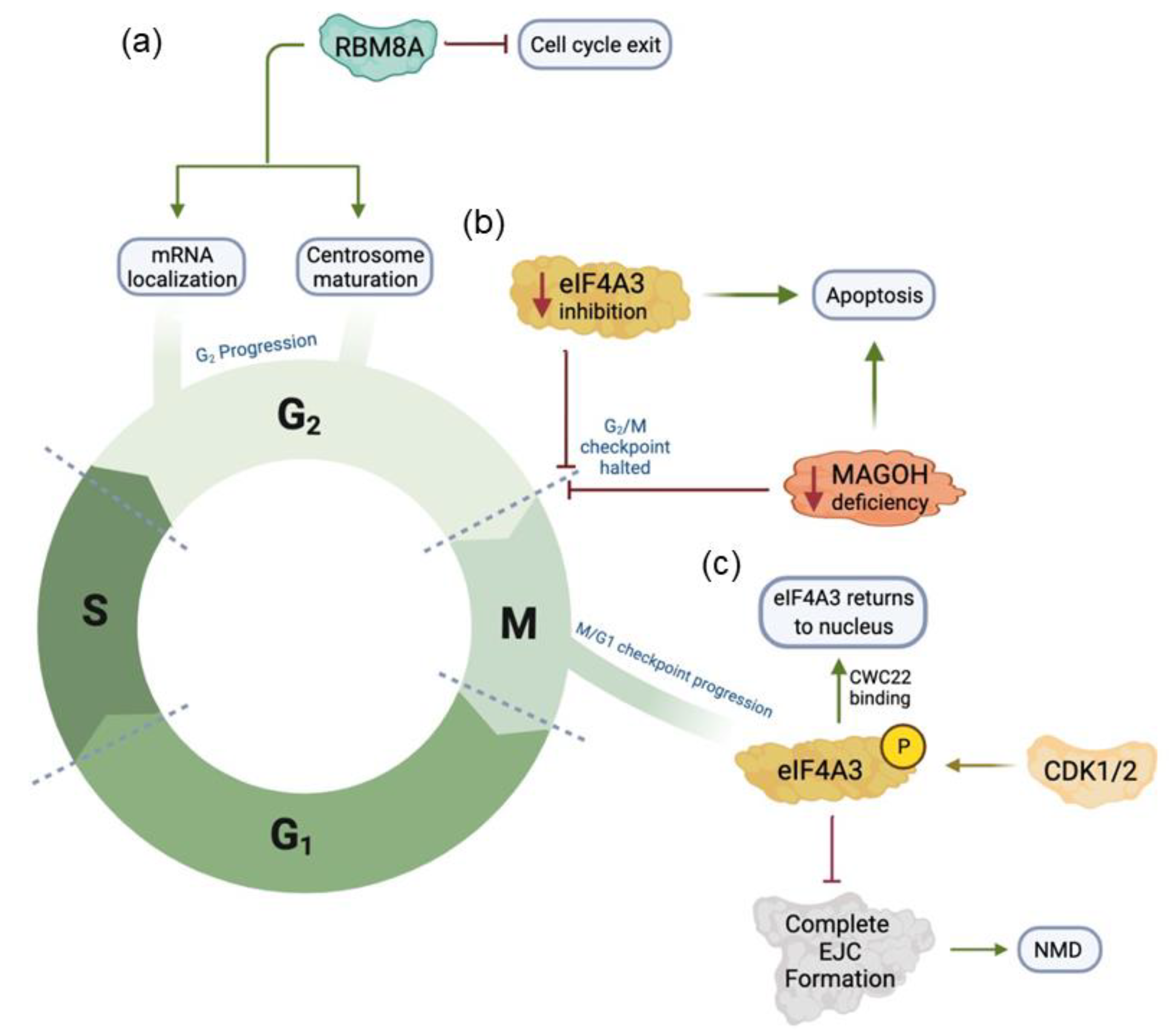
The cell cycle is a fine-tuned process with little leeway for errors. Dysregulation in proteins that control this process can result in pathologies like cancer. Recent studies have examined the roles of EJC core proteins in the cell cycle and cancer. Mazloomian et al. showed that eIF4A3 inhibition in mammalian HCT116 and HeLa cell lines causes the G2/M checkpoint arrest, chromosome segregation abnormalities, and increased apoptosis (Figure 2) [38]. These results indicate normal eIF4A3 expression is necessary for normal cell cycle progression. Mechanistically, eIF4A3 is phosphorylated in a cell cycle-dependent manner by CDK1 and CDK2 [39]. Phosphorylation of eIF4A3 on Threonine 163 residue during the M phase inhibits the function of NMD as phosphorylated eIF4A3 decreases binding affinity to mRNA transcripts (Figure 2). Particularly, phosphorylation of eIF4A3 prevents the formation of a complete EJC complex with other EJC factors, but prompts CWC22 binding, which facilitates eIF4A3 back to the nucleus to form an active spliceosome. Dephosphorylation of eIF4A3 via an unknown phosphatase influences the M/G1 checkpoint. Overexpression of eIF4A3 is associated with poor prognosis of patients with various cancers and systematic analysis of co-expression reveals an association between eIF4A3 and known proteins involved in the cell cycle and apoptosis [39]. Interestingly, the researchers determine that the tumor necrosis factor-α (TNF-α) pathway is specifically impacted by eIF4A3 mutations (Table 2) [40].
A study of RBM8A knockdown in HeLa cells demonstrates that genes encoding for splicing in mitosis and genome stability are dependent on the EJC complex [41]. Knockdown of RBM8A decreases MAGOH expression but not eIF4A3 expression [41]. RBM8A knockdown alters p53 splicing and causes the G2/M arrest (Figure 2) [41]. Multinucleated cells with abnormal nuclear structure and overall genome instability emerging under RBM8A knockdown support that RBM8A is required for normal cell cycle function [41]. Furthermore, another study found the cell cycle stalling at the G2/M checkpoint in human tumor cells deficient for RBM8A [42]. Specifically, centrosome maturation is negatively impacted, preventing the cells from going from mitosis to G1 [42]. The cells at M-phase subsequently undergo apoptosis via caspase activation. A properly assembled centrosome is a necessity for mitosis as its associated spindle fibers function to separate sister chromatids. A recent study has found that mRNAs are colocalized at the centrosome in a cell cycle-dependent manner [43]. The high-throughput single-molecule fluorescence in situ hybridization (smFISH) detected that eight mRNAs behave in this way: ASPM, BICD2, CCDC88C, CEP350, HMMR, NIN, NUMA1, and PCNT [43]. The centrosome-mRNA colocalization requires nascent protein translation as puromycin but not cycloheximide prevents the centrosomal mRNA localization. The polysomes with mRNA transcripts are actively transported towards centrosomes through microtubule during prophase and prometaphase [43]. Therefore, improper localization of mRNA to a mature centrosome could stand as one of the possible downstream effects of abnormal RBM8A levels in cells.
Multiple studies have investigated the effects of MAGOH deficiency in the context of neurodevelopment. Mitosis is essential in neurodevelopment in the context of cell proliferation and differentiation. In the context of melanoblast development, this process is associated with normal pigmentation of dorsal and ventral regions in adult mice (Table 2) [44]. Magoh depletion, namely haploinsufficiency, inhibits melanoblast proliferation [44]. While the cells are not undergoing apoptosis, the mitosis is arrested at the G2/M phase, preventing cell proliferation [44]. Another study also found that low levels of MAGOH in interneuron progenitors result in mitotic staling [45]. Consequently, this reduces the critical number of cortical interneurons. The interneuron progenitors display upregulated p53 signaling, aligning with observed apoptotic divisions [45]. Magoh deficiency in radial glial cell progenitors results in apoptosis, via p53 activation, upon mitosis delay [46]. In this study, changes to the microtubules that compose the mitotic spindle are evident. In addition to MAGOH, RBM8A expression regulates neurodevelopment. Consistent with the essential roles of EJCs in neurodevelopment, in a study of neural progenitor cells (NPCs), RBM8A is deemed necessary for cell cycle regulation, NPCs and interneuron progenitor proliferation [47,48]. Elevated levels of RBM8A inhibit cell cycle exit, leading to more proliferation, whereas low levels were associated with cell cycle exit and subsequent differentiation [47].

3. The EJC Is Implicated in a Diverse Set of Cancers

Recently, the major core and peripheral components of the EJC have been implicated in various carcinogenic pathways. Several forms of cancers have been associated with the EJC as shown in Table 3. These associations vary greatly in both the experimental paradigms utilized to establish these associations and in the exact role the implicated EJC component plays. Additionally, mechanistic insight thus far has demonstrated several pathways differentially affected such as NOTCH1, STAT3, mTOR, RAS, or MAPK among many other cellular signaling networks [42,49,50,51,52,53,54].
While a detailed summary of each of these associations is out of the scope of this review, we will highlight some of these EJC-associated cancer mechanisms below.

3.1. RBM8A Regulates Glioblastoma Progression

A recent study on the effect of RBM8A knockdown in glioblastoma cells shows that it inhibits glioblastoma cell proliferation and invasion. RBM8A increases cell proliferation and migration in glioblastoma tissues through the Notch/STAT3 pathway [56]. As RBM8A mediates DNA binding and regulates the downstream genes by interacting with a transcriptional factor STAT3 [58], the protein levels of Notch, phospho-STAT3, and phospho-H3 are significantly decreased by RBM8A knockdown [56]. The opposite effects have been detected in RBM8A overexpression that transwell analysis confirms an increase in cell migration and invasion of glioblastoma cells when RBM8A is overexpressed but a dramatic decrease when RBM8A is knocked down. To further examine RBM8A function in vivo, authors demonstrated that RBM8A knockdown leads to a reduction of Notch1 protein level in tissues and a slow growth of the tumor cells in a xenograft mouse model. Furthermore, higher expression of RBM8A correlates to a worse prognosis in patients. These findings provide a possible avenue for effective diagnosis and treatment of glioblastoma and other cancers [56,59,60].

3.2. RBM8A Affects Apoptosis in Human Cell Models of Adenocarcinoma

RBM8A functions as an important apoptotic modulator. Knockdown studies of RBM8A in human cervical carcinoma cell line (HeLa) and human lung adenocarcinoma cell line (A549) further established the role of RBM8A in cell cycle regulation. RBM8A knockdown inhibits tumor cell activity, arrests the cell cycle at the M phase, and promotes apoptosis [42,59,60]. The apoptotic defining characteristics featured abnormal centrosomes in cancer cells, supporting the importance of RBM8A in apoptosis regulation [61]. RBM8A depletion regulates the expression of pro-apoptotic genes, such as Bcl-Xs and Bim, and tumor development associated with mRNA splicing regulation [59,60,62]. Results also show that RBM8A expression affects tumor diameter, degree of pathological differentiation and clinical stage, overall survival, and progression-free survival. Interestingly, the knockdown of different EJC/NMD components gives different effects on Bcl-Xs splicing. Downregulation of RBM8A and eIF4A3 or auxiliary (RNPS1, Acinus, and SAP18) components of the EJC, promotes the production of the proapoptotic Bcl-xS splice variant. However, UAP56, Aly/Ref, and TAP or NMD factors (MNL51, Upf1, Upf2, and Upf3b), do not produce the same effect [62].

3.3. RBM8A and eIF4A3 Contribute to the Pathogenesis of Hepatocellular Carcinoma (HCC)

RBM8A is important in cell development, differentiation, metabolism, and regulation of the cell cycle. Elevated RBM8A expression has been observed in hepatocellular cellular carcinoma [57]. This overexpression has been linked to poor patient prognosis [59]. In HCC, overexpression of RBM8A increases cell proliferation, migration, and invasion into surrounding tissues. KEGG analysis has revealed that the following cellular functions are impacted in these patients: DNA replication, cell cycle, ribosomal activity, and spliceosomal activity [63]. In line with the role of cell cycle regulation, cell cycle-related kinases: CDK1, CDK2, and CHEK1, are impacted [63]. The transcription factor, E2F1DP1RB, which modulates mitosis at the G1/S transition, is also affected [63]. Overexpression of RBM8A upregulates the epithelial-mesenchymal transition (EMT) signaling pathway. The EMT is implicated in tumor cell metastasis and the resistance to chemotherapy treatments [57]. The activation of the EMT via RBM8A promotes HCC growth, which plays a significant role in the regulation of oxaliplatin (OXA)-based systemic chemotherapy resistance [57]. A high level of RBM8A is associated with a higher HDAC9 level, resulting in the resistance effect of OXA, a key chemotherapy drug [57]. Moreover, higher RBM8A expression was detected in advanced HCC than in early-stage HCC, suggesting a good correlation between RBM8A level and carcinogenesis [59,60]. These results indicate the possibility of RBM8A as an HCC prognostic tool.
Besides RBM8A, eIF4A3 is overexpressed in HCC tissues compared to normal tissues, which is indicative of a poor prognosis. Functional gene network analyses revealed a connection between eIF4A3 and various chemokine pathways, cell cycle and spliceosome pathways, several cell cycle regulatory kinases, and tumor-associated transcription factors [38]. Through antagonistic binding to WDR66 with miR-2113, eIF4A3 promotes HCC cell proliferation and EMT function. Downregulation of eIF4A3 leads to G2/M phase arrest, increased apoptosis, as well as abnormalities in centrosome-spindle formation [38]. Moreover, eIF4A3 has been associated with RNA stress granule formation and maintenance, indicating another possible mechanistic link between the EJC and cancer [38]. Together, these findings provide another possible mechanism for therapeutic development through manipulating the EJC level.

4. The EJC Plays a Crucial Role in Neurodevelopmental Disorders

The EJC components have been studied widely for their role in gene regulation, as well as their significant effects on downstream regulation of cell cycle and splicing. In addition to cancer, abnormal expression of RBM8A plays a role in thrombocytopenia and neurodevelopmental diseases, such as autism and schizophrenia. However, the pathologic mechanisms behind the role of RBM8A in these diseases are not yet fully understood.

4.1. The EJC Controls Cell Division of Neural Stem Cells (NSCs)

The development of the nervous system requires a balance between the proliferation and maintenance of a pool of NSCs and the differentiation/migration of NSCs to the cortical and subcortical regions of the brain. Cortical development begins with the generation of radial glial cells around embryonic day 9/10 (E9/10) in mice. NSCs undergo symmetric division or asymmetric division to ensure that the stem cell pool is maintained, while enough cells differentiate into neurons and glial in the brain. In the development of the cortex, neurogenesis relies on asymmetric division, which produces a stem cell and a cell that will terminally differentiate. This is in contrast to symmetric mitotic division, which results in two identical daughter cells. Asymmetric division is regulated by the orientation of the mitotic spindles that determine the position and identity of the daughter cells [64,65,66]. During asymmetric division, cleavage planes are usually within 30° of the apicobasal axis. This orientation is consistent through both proliferative and neurogenic phases [67].
Previous studies have shown that EJC components are highly associated with neurogenesis. Magoh haploinsufficiency is a prime example of how changes in the orientation of mitotic spindles can result in deficits in the asymmetric division. Magoh haploinsufficiency results in microcephaly and leads to increased cell division along the horizontal plane [68]. Cells that divide horizontally, are destined to become basal daughter cells and post-mitotic neurons. Thus, Magoh haploinsufficiency significantly prolongs mitosis of NSCs and leads to increased ectopic neuron differentiation [46,68]. Consistently, haploinsufficiency for each core EJC component causes microcephaly, depletion of NSCs, and extensive apoptosis in both neurons and NSCs in mice [69]. The microcephaly phenotypes are significantly more severe in Rbm8a and Eif4a3 mutant mice, with an average reduction of 70%, compared to Magoh in postnatal brains. However, this is thought to be due to a second homolog of Magoh. Together, NSCs and neurogenesis analyses in embryonic brains demonstrate that deletions in all three EJCs contribute to NSC division defects and induction of apoptosis to a similar extent, leading to microcephaly [69].
Three EJC genes are closely related in pathophysiology as each mutant alters each other’s overlapping transcription levels. Upregulation of p53 transcript observed in all three EJC mutants suggests that p53 activation may be involved in pathogenesis. Combined with a previous finding in clinical studies that RBM8A deletion is closely related to human microcephaly, attenuation of p53 in Rbm8a mutants shows a partial but significant rescue of microcephaly and apoptosis [70,71]. This suggests that p53 activation plays a role in neuronal loss, thus leading to microcephaly. However, it is unclear whether p53 activation leads to induced apoptosis or delayed mitosis. Further experiments are needed to assess this relationship.

4.2. EJC Modulates RNA Splicing during Development

The EJC factors assembly at 20–24 nucleotides upstream of mRNA exon-exon junctions stepwise in an orderly pattern during RNA splicing [72], and provide a critical link between pre-mRNA splicing and subsequent events including mRNA export, localization, translation, and NMD [73]. Clearly, RNA splicing modulates gene expression and plays a critical role in various developmental stages, particularly in the nervous system [74].
EJC factors can regulate mRNA splicing of key genes in multiple essential signal pathways to modulate development. In Drosophila, Magoh mutations cause specific defects in photoreceptor differentiation due to altered MAPK splicing [75]. Interestingly, other EJC factors like RBM8A and eIF4A3 are also required for MAPK splicing, supporting a nuclear role of EJC in the splicing of a subset of transcripts [75]. Another splicing substrate of EJC in Drosophila is dlg1, a cell polarity gene [76]. Dlg1 protein promotes Dvl protein stabilization and stimulates the Wnt signaling pathway. Thus, EJC can modulate the activation of the Wnt pathway via Dlg1 splicing [76]. A genome-wide approach, isolation of protein complexes and associated RNA targets (ipaRt), has been used to identify EJC-bound RNAs that are enriched with genes regulating differentiation and development in adult Drosophila [77]. Moreover, EJC binds to strong splicing sites, CG-rich hexamers [77]. In addition to large introns, EJC can also regulate the splicing of many mini-introns in P. tetraurelia, suggesting that a fine-tuning expression of EJC genes is required for steady intron removal [78].
Core EJC factors are essential for neurodevelopment [69]. Deletion of each core EJC factor leads to microcephaly in mice. Consistent with the notion, spliceosome dysregulation is implicated in RNAseq analyses for all EJC mutant mice [69]. Alternative pre-mRNA splicing is normally coupled with NMD to selectively degrade some splicing isoforms while permitting the expression of other isoforms. This mechanism induces developmental expression of the synaptic scaffold protein PSD-95 in mammals but not in invertebrates, suggesting an evolutionary mechanism regulating neural-specific expression of PSD-95 [79]. Interestingly, exon 5 splicing of a proapoptotic gene Bak1 is subject to developmental regulation [80]. In NPCs, splicing regulator PTBP1 is high and promotes exon 5 skipping of Bak1, which allows Bak1 to escape from NMD and produce functional BAK1 protein for activating apoptosis. Upon differentiation into neurons, PTBP1 level goes down, which leads to Bak1 exon 5 inclusion and trigger NMD mechanism to break down pro-apoptotic Bak1 transcripts and enable neuronal survival. Consistently, germline deletion of Bak1 exon 5 increases neuronal apoptosis and leads to postnatal mortality [80], supporting a critical function of splicing and NMD in controlling brain development.

4.3. RBM8A Contributes to Thrombocytopenia and Absent Radius (TAR) Syndrome and Neurodevelopmental Disorders

The proximal region of 1q21 copy number variation (CNV) is often referred to as the TAR region, as a microdeletion of this region has been found in patients with the TAR syndrome in previous clinical studies [63,71,72]. Heterozygosity of Rbm8a deletion resulted in platelet deficiency and increased tail bleeding in knockout mice [81]. Further immunohistochemical staining (IHC) demonstrated that Rbm8a knockout reduces the average size of megakaryocytes and impairs its differentiation as it forms clusters in the spleen, resulting in splenomegaly in mice [81]. Flow cytometry of human erythroleukemia (HEL) cells stained with megakaryocyte markers shows that RBM8A depletion causes cell-cycle arrest and attenuates platelet differentiation. Consistent with the finding of an inverse relationship between RBM8A expression and p53 protein levels in HEL cells, p53 inhibitor and p53 knockout not only increase the transcriptional levels of Rbm8a but also partially rescue both megakaryocyte differentiation and platelet counts [81].
A dosage effect of RBM8A has been found in many clinical manifestations, followed by altered cell proliferation and differentiation. Overexpression of RBM8A promotes embryonic NSC proliferation and suppresses neuronal differentiation, whereas the knockdown of Rbm8a has the opposite effect [46,47]. Thus, abnormal expression of RBM8A has been studied in neurodegenerative, neurodevelopmental, and neuropsychiatric research regarding Alzheimer’s disease, autism spectrum disorder, and schizophrenia [82]. Rbm8a deficits lead to prolonged mitosis in NSCs and intermediate progenitors, as well as the downregulation of Lis1 during neurogenesis together with MAGOH heterodimers [69]. The unstable MAGOH heterodimer complex is highly associated with microcephaly due to apoptosis.

5. Targeting the EJC and NMD in Diseases

In 2001, it was proposed to utilize drugs or siRNA to inhibit NMD for cancer treatment [60,83,84]. The mRNA stability has been associated with the prevalence of malignant tumors, which supports the need to study NMD in different cancers [60]. Three cellular pathways, Ras/MAPK, JAK/STAT3 and NF-κB, have been associated with RBM8A’s role in liver cancer [59,60]. Depletion of RBM8A inhibits all three pathways. MAPK protein synthesis is declined, thereby inhibiting the Ras/MAPK pathway [60]. RBM8A depletion also inhibits the JAK/STAT3 pathway and decreases STAT3 DNA-binding activity [60]. The effects of RBM8A on these signaling pathways suggest a critical role of RBM8A in tumorigenesis. Increasing NMD efficiency has recently been proposed to target carcinogenesis [60,85]. Quantification of NMD-triggering versus NMD-evading PTC leads to the classification of tumor-suppressor genes [85].
As previously found that Eif4a3 knockdown induces apoptosis in cancer cells, allosteric inhibitors have been investigated as a potential NMD suppression tool [86]. Frameshift insertion/deletions (fs-indels) are normally targeted and degraded by NMD. However, some fs-indels are in the genomic regions that can escape NMD. Studies of T cell reactivity have shown that antigens derived from fs-indels, specifically those with elongated ORFs, can elicit an immune response [87]. These NMD-escape mutations could be possible targets for immunotherapy because these mutations can stimulate anti-tumor response through checkpoint inhibitors (CPI) with clear clinical benefits [87].
Several other conserved regulatory pathways can modulate NMD efficacy. First, miR-128 inhibits NMD by targeting UPF1 and CASC3 during neuronal differentiation [88]. Second, the cytoskeleton is responsible for the transportation of mRNAs carrying PTCs and ribonucleoproteins (RNPs) to the processing body (P-body) for degradation. Disruptions of different cytoskeletons exert different effects. Interruption of actin function by polymerization inhibitors blocks NMD and activates PTC readthrough, whereas microtubule disruption only inhibits NMD. PTC readthrough requires UPF proteins’ presence in P-bodies, suggesting that different cytoskeletons involve in various NMD stages [89]. Third, mRNA itself contains a wide variety of features to escape NMD. The eukaryotic release factor 3a (eRF3a) close to an upstream termination codon inhibits NMD in a manner that is poly-A binding protein C1 (PABPC1) dependent, while eIF4G does so in a PABPC1-independent manner [90]. A cis-element positioned downstream of the termination codon in the 3′ UTR has been identified to inhibit NMD and is proposed to do so through efficient translation termination. The same study identifies another cis-element in the 3′ UTR that inhibits NMD through a different but unclear mechanism [91]. Fourth, multiple RNPs regulate NMD efficacy. Heterogeneous nuclear ribonucleoprotein L (hnRNP-L) protects oncogenic BCL2 mRNA from NMD. HnRNP-L shares structural similarities with polypyrimidine tract binding protein 1 (PTBP1), which is also known to protect mRNA from NMD. Both hnRNP-L and PTBP1 aid in NMD evasion by attenuating UPF1 binding to mRNA [92]. Poly-A binding protein 1 (PABPC1) and other peptides containing the PABP-interacting motif 2 (PAM2) have been shown to inhibit NMD degradation by binding to 3′ UTRs [93]. FUS mutations in patients with amyotrophic lateral sclerosis (ALS) cause protein inclusion and sequester translation, thereby affecting NMD. In cells derived from ALS patients, UPF3A is decreased but UPF1 and UPF3B are increased [94]. Thus, abnormal NMD function could contribute to neurodegenerative diseases like ALS.
Several NMD inhibitors have been discovered or synthesized to explore the therapeutic potential of NMD inhibition [95,96,97,98]. A high-throughput screen that monitors the RNA-dependent ATPase activity of the eIF4A3−MLN51 complex, has identified 1,4-Diacylpiperazines as a selective NMD inhibitor [86] and the authors further optimize the prototype compound into several highly-selective noncompetitive inhibitors of eIF4A3 [99]. Antisense oligonucleotides have been generated to reduce the expression of different NMD factors. Knockdown of Upf1, Upf2, Smg1, and Smg6 suppresses NMD, and downregulation of Upf3b, Smg5, and Smg7 moderately affect NMD. However, Smg8 and Smg9 knockdown demonstrates no significant effect [100]. Upf3b inhibition minimally affects the overall transcriptome while stabilizing both PTC-containing dystrophin mRNA and coagulation factor IX mRNA in mouse models [100].
Unbiased high-throughput chemical screens using a multi-colored bioluminescence NMD reporter system and FDA-approved drugs have revealed some interesting hits, including a group of cardiac glycosides, including ouabain and digoxin, as potent inhibitors of NMD [101]. Cardiac glycosides target Na-K ATPase on the cell membrane to increase intracellular Ca2+ levels and inhibit NMD [101]. Through profiling multiple endogenous NMD targets, Zhao et al. identified several NMD modulators, including the anticancer drug homoharringtonine [102]. Given the diversity of methods that can be used to suppress NMD, NMD inhibition holds a promising therapeutic potential for ameliorating a variety of disease phenotypes.

6. Conclusions

The scientific literature clearly evinces the powerful influence the EJC exerts over many cellular processes. Alongside the NMD pathway, the EJC regulates the cell cycle and therefore may be associated with several cell cycle dysfunctions, carcinogenic pathways, and neurodevelopmental and neurodegenerative diseases. NMD emerges as a target of inhibition in both an EJC-dependent and independent manner for future therapeutic potential. Although progress has been made in the discovery of the roles of the EJC and NMD in cell cycle dysfunction and associated pathologies, further research is still necessary to reveal key mechanistic links within the cell cycle in normal developmental and pathological conditions.

Author Contributions

Section 3 and leading writer: H.M.; Section 2 and Table 2: J.R.; Abstract, introduction, conclusion, editing: S.A.; Section 4: J.Y.; Figure 1 and Figure 2 and Tables: S.P.; citations, formatting, writing—review and editing: J.M., Z.P. and Y.M., supervision and funding acquisition: Y.M. All authors have read and agreed to the published version of the manuscript.

Funding

This work was supported by the National Institute of Mental Health of the National Institutes of Health under Award Number R01MH122556, the National Center for Advancing Translational Sciences, National Institutes of Health, through Grant UL1TR002014, and PSU IEE SEED funding. The content is solely the responsibility of the authors and does not necessarily represent the official views of the NIH.

Institutional Review Board Statement

Not applicable.

Informed Consent Statement

Not applicable.

Data Availability Statement

Not applicable.

Acknowledgments

We thank all lab members for their suggestions for the paper.

Conflicts of Interest

The authors declare no conflict of interest.

References

  1. Schlautmann, L.P.; Gehring, N.H. A Day in the Life of the Exon Junction Complex. Biomolecules 2020, 10, 866. [Google Scholar] [CrossRef]
  2. Leung, C.S.; Johnson, T.L. The exon junction complex: A multitasking guardian of the transcriptome. Mol. Cell 2018, 72, 799–801. [Google Scholar] [CrossRef]
  3. Boehm, V.; Gehring, N.H. Exon junction complexes: Supervising the gene expression assembly line. Trends Genet. 2016, 32, 724–735. [Google Scholar] [CrossRef]
  4. Le Hir, H.; Seraphin, B. EJCs at the heart of translational control. Cell 2008, 133, 213–216. [Google Scholar] [CrossRef]
  5. Le Hir, H.; Andersen, G.R. Structural insights into the exon junction complex. Curr. Opin. Struct. Biol. 2008, 18, 112–119. [Google Scholar] [CrossRef]
  6. Le Hir, H.; Izaurralde, E.; Maquat, L.E.; Moore, M.J. The spliceosome deposits multiple proteins 20–24 nucleotides upstream of mRNA exon–exon junctions. EMBO J. 2000, 19, 6860–6869. [Google Scholar] [CrossRef]
  7. Andersen, C.B.F.; Ballut, L.; Johansen, J.S.; Chamieh, H.; Nielsen, K.H.; Oliveira, C.L.P.; Pedersen, J.S.; Seraphin, B.; Le Hir, H.; Andersen, G.R. Structure of the exon junction core complex with a trapped DEAD-box ATPase bound to RNA. Science 2006, 313, 1968–1972. [Google Scholar] [CrossRef]
  8. Buchwald, G.; Schussler, S.; Basquin, C.; Le Hir, H.; Conti, E. Crystal structure of the human eIF4AIII-CWC22 complex shows how a DEAD-box protein is inhibited by a MIF4G domain. Proc. Natl. Acad. Sci. USA 2013, 110, E4611–E4618. [Google Scholar] [CrossRef]
  9. Noble, C.G.; Song, H.W. MLN51 Stimulates the RNA-Helicase Activity of eIF4AIII. PLoS ONE 2007, 2, e303. [Google Scholar] [CrossRef]
  10. Wang, Z.; Ballut, L.; Barbosa, I.; Le Hir, H. Exon Junction Complexes can have distinct functional flavours to regulate specific splicing events. Sci. Rep. 2018, 8, 9509. [Google Scholar] [CrossRef] [Green Version]
  11. Murachelli, A.G.; Ebert, J.; Basquin, C.; Le Hir, H.; Conti, E. The structure of the ASAP core complex reveals the existence of a Pinin-containing PSAP complex. Nat. Struct. Mol. Biol. 2012, 19, 378–386. [Google Scholar] [CrossRef]
  12. Viphakone, N.; Sudbery, I.; Griffith, L.; Heath, C.G.; Sims, D.; Wilson, S.A. Co-transcriptional Loading of RNA Export Factors Shapes the Human Transcriptome. Mol. Cell 2019, 75, 310–323. [Google Scholar] [CrossRef]
  13. Ballut, L.; Marchadier, B.; Baguet, A.; Tomasetto, C.; Seraphin, B.; Le Hir, H. The exon junction core complex is locked onto RNA by inhibition of eIF4AIII ATPase activity. Nat. Struct. Mol. Biol. 2005, 12, 861–869. [Google Scholar] [CrossRef]
  14. Bono, F.; Cook, A.G.; Grunwald, M.; Ebert, J.; Conti, E. Nuclear Import Mechanism of the EJC Component Mago-Y14 Revealed by Structural Studies of Importin 13. Mol. Cell 2010, 37, 211–222. [Google Scholar] [CrossRef]
  15. Kataoka, N.; Diem, M.D.; Kim, V.N.; Yong, J.; Dreyfuss, G. Magoh, a human homolog of Drosophila mago nashi protein, is a component of the splicing-dependent exon–exon junction complex. EMBO J. 2001, 20, 6424–6433. [Google Scholar] [CrossRef]
  16. Gehring, N.H.; Kunz, J.B.; Neu-Yilik, G.; Breit, S.; Viegas, M.H.; Hentze, M.W.; Kulozik, A.E. Exon-junction complex components specify distinct routes of nonsense-mediated mRNA decay with differential cofactor requirements. Mol. Cell 2005, 20, 65–75. [Google Scholar] [CrossRef]
  17. Diem, M.D.; Chan, C.C.; Younis, I.; Dreyfuss, G. PYM binds the cytoplasmic exon-junction complex and ribosomes to enhance translation of spliced mRNAs. Nat. Struct. Mol. Biol. 2007, 14, 1173–1179. [Google Scholar] [CrossRef]
  18. Kim, V.N.; Yong, J.; Kataoka, N.; Abel, L.; Diem, M.D.; Dreyfuss, G. The Y14 protein communicates to the cytoplasm the position of exon–exon junctions. EMBO J. 2001, 20, 2062–2068. [Google Scholar] [CrossRef]
  19. Tange, T.O.; Shibuya, T.; Jurica, M.S.; Moore, M.J. Biochemical analysis of the EJC reveals two new factors and a stable tetrameric protein core. RNA 2005, 11, 1869–1883. [Google Scholar] [CrossRef]
  20. Karousis, E.D.; Nasif, S.; Mühlemann, O. Nonsense-mediated mRNA decay: Novel mechanistic insights and biological impact. Wiley Interdiscip. Rev. RNA 2016, 7, 661–682. [Google Scholar] [CrossRef] [Green Version]
  21. Kunz, J.B.; Neu-Yilik, G.; Hentze, M.W.; Kulozik, A.E.; Gehring, N.H. Functions of hUpf3a and hUpf3b in nonsense-mediated mRNA decay and translation. RNA 2006, 12, 1015–1022. [Google Scholar] [CrossRef]
  22. Hug, N.; Longman, D.; Cáceres, J.F. Mechanism and regulation of the nonsense-mediated decay pathway. Nucleic Acids Res. 2016, 44, 1483–1495. [Google Scholar] [CrossRef]
  23. Kishor, A.; Fritz, S.E.; Hogg, J.R. Nonsense-mediated mRNA decay: The challenge of telling right from wrong in a complex transcriptome. Wiley Interdiscip. Rev. RNA 2019, 10, e1548. [Google Scholar] [CrossRef]
  24. Hosoda, N.; Kim, Y.K.; Lejeune, F.; Maquat, L.E. CBP80 promotes interaction of Upf1 with Upf2 during nonsense-mediated mRNA decay in mammalian cells. Nat. Struct. Mol. Biol. 2005, 12, 893–901. [Google Scholar] [CrossRef]
  25. Isken, O.; Maquat, L.E. The multiple lives of NMD factors: Balancing roles in gene and genome regulation. Nat. Rev. Genet. 2008, 9, 699–712. [Google Scholar] [CrossRef]
  26. Woeller, C.F.; Gaspari, M.; Isken, O.; Maquat, L.E. NMD resulting from encephalomyocarditis virus IRES-directed translation initiation seems to be restricted to CBP80/20-bound mRNA. EMBO Rep. 2008, 9, 446–451. [Google Scholar] [CrossRef]
  27. Kashima, I.; Yamashita, A.; Izumi, N.; Kataoka, N.; Morishita, R.; Hoshino, S.; Ohno, M.; Dreyfuss, G.; Ohno, S. Binding of a novel SMG-1-Upf1-eRF1-eRF3 complex (SURF) to the exon junction complex triggers Upf1 phosphorylation and nonsense-mediated mRNA decay. Genes Dev. 2006, 20, 355–367. [Google Scholar] [CrossRef]
  28. Lopez-Perrote, A.; Castano, R.; Melero, R.; Zamarro, T.; Kurosawa, H.; Ohnishi, T.; Uchiyama, A.; Aoyagi, K.; Buchwald, G.; Kataoka, N.; et al. Human nonsense-mediated mRNA decay factor UPF2 interacts directly with eRF3 and the SURF complex. Nucleic Acids Res. 2016, 44, 1909–1923. [Google Scholar] [CrossRef]
  29. Behm-Ansmant, I.; Izaurralde, E. Quality control of gene expression: A stepwise assembly pathway for the surveillance complex that triggers nonsense-mediated mRNA decay. Genes Dev. 2006, 20, 391–398. [Google Scholar] [CrossRef]
  30. Kervestin, S.; Jacobson, A. NMD: A multifaceted response to premature translational termination. Nat. Rev. Mol. Cell Biol. 2012, 13, 700–712. [Google Scholar] [CrossRef] [Green Version]
  31. Peccarelli, M.; Kebaara, B.W. Regulation of natural mRNAs by the nonsense-mediated mRNA decay pathway. Eukaryot. Cell 2014, 13, 1126–1135. [Google Scholar] [CrossRef] [PubMed]
  32. Holbrook, J.A.; Neu-Yilik, G.; Hentze, M.W.; Kulozik, A.E. Nonsense-mediated decay approaches the clinic. Nat. Genet. 2004, 36, 801–808. [Google Scholar] [CrossRef] [PubMed]
  33. Khajavi, M.; Inoue, K.; Lupski, J.R. Nonsense-mediated mRNA decay modulates clinical outcome of genetic disease. Eur. J. Hum. Genet. 2006, 14, 1074–1081. [Google Scholar] [CrossRef] [PubMed]
  34. Bhuvanagiri, M.; Schlitter, A.M.; Hentze, M.W.; Kulozik, A.E. NMD: RNA biology meets human genetic medicine. Biochem. J. 2010, 430, 365–377. [Google Scholar] [CrossRef]
  35. Gardner, L.B. Nonsense-mediated RNA decay regulation by cellular stress: Implications for tumorigenesis. Mol. Cancer Res. 2010, 8, 295–308. [Google Scholar] [CrossRef]
  36. Gehring, N.H.; Lamprinaki, S.; Hentze, M.W.; Kulozik, A.E. The Hierarchy of Exon-Junction Complex Assembly by the Spliceosome Explains Key Features of Mammalian Nonsense-Mediated mRNA Decay. PLoS Biol. 2009, 7, e1000120. [Google Scholar] [CrossRef]
  37. Woodward, L.A.; Mabin, J.W.; Gangras, P.; Singh, G. The exon junction complex: A lifelong guardian of mRNA fate. Wiley Interdiscip. Rev. RNA 2017, 8, e1411. [Google Scholar] [CrossRef]
  38. Mazloomian, A.; Araki, S.; Ohori, M.; El-Naggar, A.M.; Yap, D.; Bashashati, A.; Nakao, S.; Sorensen, P.H.; Nakanishi, A.; Shah, S.; et al. Pharmacological systems analysis defines EIF4A3 functions in cell-cycle and RNA stress granule formation. Commun. Biol. 2019, 2, 165. [Google Scholar] [CrossRef]
  39. Ryu, I.; Won, Y.S.; Ha, H.; Kim, E.; Park, Y.; Kim, M.K.; Kwon, D.H.; Choe, J.; Song, H.K.; Jung, H.; et al. eIF4A3 Phosphorylation by CDKs Affects NMD during the Cell Cycle. Cell Rep. 2019, 26, 2126–2139.e9. [Google Scholar] [CrossRef]
  40. Lin, Y.; Zhang, J.Y.; Cai, J.Y.; Liang, R.; Chen, G.Y.; Qin, G.; Han, X.Q.; Yuan, C.L.; Liu, Z.; Li, Y.Q.; et al. Systematic Analysis of Gene Expression Alteration and Co-Expression Network of Eukaryotic Initiation Factor 4A-3 in Cancer. J. Cancer 2018, 9, 4568–4577. [Google Scholar] [CrossRef]
  41. Lu, C.C.; Lee, C.C.; Tseng, C.T.; Tarn, W.Y. Y14 governs p53 expression and modulates DNA damage sensitivity. Sci. Rep. 2017, 7, srep45558. [Google Scholar] [CrossRef] [PubMed]
  42. Ishigaki, Y.; Nakamura, Y.; Tatsuno, T.; Hashimoto, M.; Shimasaki, T.; Iwabuchi, K.; Tomosugi, N. Depletion of RNA-binding protein RBM8A (Y14) causes cell cycle deficiency and apoptosis in human cells. Exp. Biol. Med. 2013, 238, 889–897. [Google Scholar] [CrossRef] [PubMed]
  43. Safieddine, A.; Coleno, E.; Salloum, S.; Imbert, A.; Traboulsi, A.M.; Kwon, O.S.; Lionneton, F.; Georget, V.; Robert, M.C.; Gostan, T.; et al. A choreography of centrosomal mRNAs reveals a conserved localization mechanism involving active polysome transport. Nat. Commun. 2021, 12, 21. [Google Scholar] [CrossRef]
  44. Silver, D.L.; Leeds, K.E.; Hwang, H.W.; Miller, E.E.; Pavan, W.J. The EJC component Magoh regulates proliferation and expansion of neural crest-derived melanocytes. Dev. Biol. 2013, 375, 172–181. [Google Scholar] [CrossRef] [PubMed]
  45. Sheehan, C.J.; McMahon, J.J.; Serdar, L.D.; Silver, D.L. Dosage-dependent requirements of Magoh for cortical interneuron generation and survival. Development 2020, 147, dev182295. [Google Scholar] [CrossRef]
  46. Pilaz, L.J.; McMahon, J.J.; Miller, E.E.; Lennox, A.L.; Suzuki, A.; Salmon, E.; Silver, D.L. Prolonged Mitosis of Neural Progenitors Alters Cell Fate in the Developing Brain. Neuron 2016, 89, 83–99. [Google Scholar] [CrossRef]
  47. Zou, D.H.; McSweeney, C.; Sebastian, A.; Reynolds, D.J.; Dong, F.P.; Zhou, Y.J.; Deng, D.Z.; Wang, Y.G.; Liu, L.; Zhu, J.; et al. A critical role of RBM8a in proliferation and differentiation of embryonic neural progenitors. Neural Dev. 2015, 10, 16. [Google Scholar] [CrossRef]
  48. McSweeney, C.; Dong, F.; Chen, M.; Vitale, J.; Xu, L.; Crowley, N.; Luscher, B.; Zou, D.; Mao, Y. Full function of exon junction complex factor, Rbm8a, is critical for interneuron development. Transl. Psychiatry 2020, 10, 379. [Google Scholar] [CrossRef]
  49. Kim, T.J.; Choi, J.J.; Kim, W.Y.; Choi, C.H.; Lee, J.W.; Bae, D.S.; Son, D.S.; Kim, J.; Park, B.K.; Ahn, G. Gene expression profiling for the prediction of lymph node metastasis in patients with cervical cancer. Cancer Sci. 2008, 99, 31–38. [Google Scholar] [CrossRef]
  50. Petroziello, J.; Yamane, A.; Westendorf, L.; Thompson, M.; McDonagh, C.; Cerveny, C.; Law, C.L.; Wahl, A.; Carter, P. Suppression subtractive hybridization and expression profiling identifies a unique set of genes overexpressed in non-small-cell lung cancer. Oncogene 2004, 23, 7734–7745. [Google Scholar] [CrossRef] [Green Version]
  51. Degot, S.; Le Hir, H.; Alpy, F.; Kedinger, V.; Stoll, I.; Wendling, C.; Seraphin, B.; Rio, M.C.; Tomasetto, C. Association of the breast cancer protein MLN51 with the Exon junction complex via its speckle localizer and RNA binding module. J. Biol. Chem. 2004, 279, 33702–33715. [Google Scholar] [CrossRef] [PubMed]
  52. Zheng, X.Y.; Huang, M.G.; Xing, L.; Yang, R.; Wang, X.S.; Jiang, R.; Zhang, L.Y.; Chen, J.X. The circRNA circSEPT9 mediated by E2F1 and EIF4A3 facilitates the carcinogenesis and development of triple-negative breast cancer. Mol. Cancer 2020, 19, 73. [Google Scholar] [CrossRef] [PubMed]
  53. Han, D.; Gao, X.; Wang, M.; Qiao, Y.; Xu, Y.; Yang, J.; Dong, N.; He, J.; Sun, Q.; Lv, G.; et al. Long noncoding RNA H19 indicates a poor prognosis of colorectal cancer and promotes tumor growth by recruiting and binding to eIF4A3. Oncotarget 2016, 7, 22159–22173. [Google Scholar] [CrossRef]
  54. Zhang, S.; Leng, T.; Zhang, Q.; Zhao, Q.; Nie, X.; Yang, L. Sanguinarine inhibits epithelial ovarian cancer development via regulating long non-coding RNA CASC2-EIF4A3 axis and/or inhibiting NF-κB signaling or PI3K/AKT/mTOR pathway. Biomed. Pharmacother. 2018, 102, 302–308. [Google Scholar] [CrossRef] [PubMed]
  55. Zhou, Y.; Li, Z.; Wu, X.; Tou, L.; Zheng, J.; Zhou, D. MAGOH/MAGOHB Inhibits the Tumorigenesis of Gastric Cancer via Inactivation of b-RAF/MEK/ERK Signaling. OncoTargets Ther. 2020, 13, 12723–12735. [Google Scholar] [CrossRef] [PubMed]
  56. Lin, Y.; Wei, L.; Hu, B.; Zhang, J.; Wei, J.; Qian, Z.; Zou, D. RBM8A Promotes Glioblastoma Growth and Invasion Through the Notch/STAT3 Pathway. Front. Oncol. 2021, 11, 736941. [Google Scholar] [CrossRef]
  57. Liang, R.; Zhang, J.; Liu, Z.; Liu, Z.; Li, Q.; Luo, X.; Li, Y.; Ye, J.; Lin, Y. Mechanism and molecular network of RBM8A-mediated regulation of oxaliplatin resistance in hepatocellular carcinoma. Front. Oncol. 2020, 10, 585452. [Google Scholar] [CrossRef]
  58. Muromoto, R.; Taira, N.; Ikeda, O.; Shiga, K.; Kamitani, S.; Togi, S.; Kawakami, S.; Sekine, Y.; Nanbo, A.; Oritani, K. The exon-junction complex proteins, Y14 and MAGOH regulate STAT3 activation. Biochem. Biophys. Res. Commun. 2009, 382, 63–68. [Google Scholar] [CrossRef]
  59. Liang, R.; Lin, Y.; Ye, J.Z.; Yan, X.X.; Liu, Z.H.; Li, Y.Q.; Luo, X.L.; Ye, H.H. High expression of RBM8A predicts poor patient prognosis and promotes tumor progression in hepatocellular carcinoma. Oncol. Rep. 2017, 37, 2167–2176. [Google Scholar] [CrossRef]
  60. Liang, R.; Lin, Y.; Yan, X.-X.; Ye, J.-Z.; Li, Y.-Q.; Ye, H.-H. Enhanced RBM8A expression in human hepatocellular carcinoma. Int. J. Clin. Exp. Med. 2017, 10, 598–607. [Google Scholar]
  61. Ishigaki, Y.; Nakamura, Y.; Tatsuno, T.; Hashimoto, M.; Iwabuchi, K.; Tomosugi, N. RNA-binding protein RBM8A (Y14) and MAGOH localize to centrosome in human A549 cells. Histochem. Cell Biol. 2014, 141, 101–109. [Google Scholar] [CrossRef]
  62. Michelle, L.; Cloutier, A.; Toutant, J.; Shkreta, L.; Thibault, P.; Durand, M.; Garneau, D.; Gendron, D.; Lapointe, E.; Couture, S.; et al. Proteins Associated with the Exon Junction Complex Also Control the Alternative Splicing of Apoptotic Regulators. Mol. Cell. Biol. 2012, 32, 954–967. [Google Scholar] [CrossRef]
  63. Lin, Y.; Liang, R.; Qiu, Y.F.; Lv, Y.F.; Zhang, J.Y.; Qin, G.; Yuan, C.L.; Liu, Z.H.; Li, Y.Q.; Zou, D.H.; et al. Expression and gene regulation network of RBM8A in hepatocellular carcinoma based on data mining. Aging 2019, 11, 423–447. [Google Scholar] [CrossRef] [PubMed]
  64. Kaltschmidt, J.A.; Davidson, C.M.; Brown, N.H.; Brand, A.H. Rotation and asymmetry of the mitotic spindle direct asymmetric cell division in the developing central nervous system. Nat. Cell Biol. 2000, 2, 7–12. [Google Scholar] [CrossRef]
  65. Morin, X.; Bellaïche, Y. Mitotic spindle orientation in asymmetric and symmetric cell divisions during animal development. Dev. Cell 2011, 21, 102–119. [Google Scholar] [CrossRef] [PubMed]
  66. Morrison, S.J.; Kimble, J. Asymmetric and symmetric stem-cell divisions in development and cancer. Nature 2006, 441, 1068–1074. [Google Scholar] [CrossRef] [PubMed]
  67. Konno, D.; Shioi, G.; Shitamukai, A.; Mori, A.; Kiyonari, H.; Miyata, T.; Matsuzaki, F. Neuroepithelial progenitors undergo LGN-dependent planar divisions to maintain self-renewability during mammalian neurogenesis. Nat. Cell Biol. 2008, 10, 93–101. [Google Scholar] [CrossRef]
  68. Silver, D.L.; Watkins-Chow, D.E.; Schreck, K.C.; Pierfelice, T.J.; Larson, D.M.; Burnetti, A.J.; Liaw, H.J.; Myung, K.; Walsh, C.A.; Gaiano, N.; et al. The exon junction complex component Magoh controls brain size by regulating neural stem cell division. Nat. Neurosci. 2010, 13, 551–558. [Google Scholar] [CrossRef]
  69. Mao, H.Q.; McMahon, J.J.; Tsai, Y.H.; Wang, Z.F.; Silver, D.L. Haploinsufficiency for Core Exon Junction Complex Components Disrupts Embryonic Neurogenesis and Causes p53-Mediated Microcephaly. PLoS Genet. 2016, 12, e1006282. [Google Scholar] [CrossRef]
  70. Brunetti-Pierri, N.; Berg, J.S.; Scaglia, F.; Belmont, J.; Bacino, C.A.; Sahoo, T.; Lalani, S.R.; Graham, B.; Lee, B.; Shinawi, M. Recurrent reciprocal 1q21. 1 deletions and duplications associated with microcephaly or macrocephaly and developmental and behavioral abnormalities. Nat. Genet. 2008, 40, 1466–1471. [Google Scholar] [CrossRef]
  71. Mefford, H.C.; Sharp, A.J.; Baker, C.; Itsara, A.; Jiang, Z.; Buysse, K.; Huang, S.; Maloney, V.K.; Crolla, J.A.; Baralle, D. Recurrent rearrangements of chromosome 1q21. 1 and variable pediatric phenotypes. N. Engl. J. Med. 2008, 359, 1685–1699. [Google Scholar] [CrossRef] [Green Version]
  72. Kataoka, N.; Dreyfuss, G. A simple whole cell lysate system for in vitro splicing reveals a stepwise assembly of the exon-exon junction complex. J. Biol. Chem. 2004, 279, 7009–7013. [Google Scholar] [CrossRef] [PubMed]
  73. Le Hir, H.; Gatfield, D.; Izaurralde, E.; Moore, M.J. The exon-exon junction complex provides a binding platform for factors involved in mRNA export and nonsense-mediated mRNA decay. EMBO J. 2001, 20, 4987–4997. [Google Scholar] [CrossRef] [PubMed]
  74. Vuong, C.K.; Black, D.L.; Zheng, S. The neurogenetics of alternative splicing. Nat. Rev. Neurosci. 2016, 17, 265–281. [Google Scholar] [CrossRef] [PubMed]
  75. Roignant, J.Y.; Treisman, J.E. Exon Junction Complex Subunits Are Required to Splice Drosophila MAP Kinase, a Large Heterochromatic Gene. Cell 2010, 143, 238–250. [Google Scholar] [CrossRef] [PubMed]
  76. Liu, M.; Li, Y.; Liu, A.; Li, R.; Su, Y.; Du, J.; Li, C.; Zhu, A.J. The exon junction complex regulates the splicing of cell polarity gene dlg1 to control Wingless signaling in development. eLife 2016, 5, e17200. [Google Scholar] [CrossRef]
  77. Obrdlik, A.; Lin, G.; Haberman, N.; Ule, J.; Ephrussi, A. The Transcriptome-wide Landscape and Modalities of EJC Binding in Adult Drosophila. Cell Rep. 2019, 28, 1219–1236.e11. [Google Scholar] [CrossRef]
  78. Contreras, J.; Begley, V.; Marsella, L.; Villalobo, E. The splicing of tiny introns of Paramecium is controlled by MAGO. Gene 2018, 663, 101–109. [Google Scholar] [CrossRef]
  79. Zheng, S. Alternative splicing and nonsense-mediated mRNA decay enforce neural specific gene expression. Int. J. Dev. Neurosci. 2016, 55, 102–108. [Google Scholar] [CrossRef]
  80. Lin, L.; Zhang, M.; Stoilov, P.; Chen, L.; Zheng, S. Developmental Attenuation of Neuronal Apoptosis by Neural-Specific Splicing of Bak1 Microexon. Neuron 2020, 107, 1180–1196.e8. [Google Scholar] [CrossRef]
  81. Su, C.-H.; Liao, W.-J.; Ke, W.-C.; Yang, R.-B.; Tarn, W.-Y. The Y14-p53 regulatory circuit in megakaryocyte differentiation and thrombocytopenia. iScience 2021, 24, 103368. [Google Scholar] [CrossRef] [PubMed]
  82. Zou, D.H.; Li, R.J.; Huang, X.H.; Chen, G.Y.; Liu, Y.; Meng, Y.S.; Wang, Y.M.; Wu, Y.; Mao, Y.W. Identification of molecular correlations of RBM8A with autophagy in Alzheimer’s disease. Aging 2019, 11, 11673–11685. [Google Scholar] [CrossRef]
  83. Pastor, F.; Kolonias, D.; Giangrande, P.H.; Gilboa, E. Induction of tumour immunity by targeted inhibition of nonsense-mediated mRNA decay. Nature 2010, 465, 227–230. [Google Scholar] [CrossRef]
  84. Tan, K.; Stupack, D.G.; Wilkinson, M.F. Nonsense-mediated RNA decay: An emerging modulator of malignancy. Nat. Rev. Cancer 2022, 22, 437–451. [Google Scholar] [CrossRef] [PubMed]
  85. Lindeboom, R.G.; Supek, F.; Lehner, B. The rules and impact of nonsense-mediated mRNA decay in human cancers. Nat. Genet. 2016, 48, 1112–1118. [Google Scholar] [CrossRef]
  86. Ito, M.; Tanaka, T.; Cary, D.R.; Iwatani-Yoshihara, M.; Kamada, Y.; Kawamoto, T.; Aparicio, S.; Nakanishi, A.; Imaeda, Y. Discovery of Novel 1,4-Diacylpiperazines as Selective and Cell-Active eIF4A3 Inhibitors. J. Med. Chem. 2017, 60, 3335–3351. [Google Scholar] [CrossRef]
  87. Litchfield, K.; Reading, J.L.; Lim, E.L.; Xu, H.; Liu, P.; Al-Bakir, M.; Wong, Y.N.S.; Rowan, A.; Funt, S.A.; Merghoub, T. Escape from nonsense-mediated decay associates with anti-tumor immunogenicity. Nat. Commun. 2020, 11, 3800. [Google Scholar] [CrossRef]
  88. Bruno, I.G.; Karam, R.; Huang, L.L.; Bhardwaj, A.; Lou, C.H.; Shum, E.Y.; Song, H.W.; Corbett, M.A.; Gifford, W.D.; Gecz, J.; et al. Identification of a MicroRNA that Activates Gene Expression by Repressing Nonsense-Mediated RNA Decay. Mol. Cell 2011, 42, 500–510. [Google Scholar] [CrossRef] [PubMed]
  89. Jia, J.S.; Werkmeister, E.; Gonzalez-Hilarion, S.; Leroy, C.; Gruenert, D.C.; Lafont, F.; Tulasne, D.; Lejeune, F. Premature termination codon readthrough in human cells occurs in novel cytoplasmic foci and requires UPF proteins. J. Cell Sci. 2017, 130, 3009–3022. [Google Scholar] [CrossRef]
  90. Fatscher, T.; Boehm, V.; Weiche, B.; Gehring, N.H. The interaction of cytoplasmic poly(A)-binding protein with eukaryotic initiation factor 4G suppresses nonsense-mediated mRNA decay. RNA 2014, 20, 1579–1592. [Google Scholar] [CrossRef]
  91. Toma, K.G.; Rebbapragada, I.; Durand, S.; Lykke-Andersen, J. Identification of elements in human long 3′ UTRs that inhibit nonsense-mediated decay. RNA 2015, 21, 887–897. [Google Scholar] [CrossRef] [Green Version]
  92. Kishor, A.; Ge, Z.; Hogg, J.R. hnRNP L-dependent protection of normal mRNAs from NMD subverts quality control in B cell lymphoma. EMBO J. 2019, 38, e99128. [Google Scholar] [CrossRef]
  93. Fatscher, T.; Gehring, N.H. Harnessing short poly(A)-binding protein-interacting peptides for the suppression of nonsense-mediated mRNA decay. Sci. Rep. 2016, 6, 37311. [Google Scholar] [CrossRef]
  94. Kamelgarn, M.; Chen, J.; Kuang, L.; Jin, H.; Kasarskis, E.J.; Zhu, H. ALS mutations of FUS suppress protein translation and disrupt the regulation of nonsense-mediated decay. Proc. Natl. Acad. Sci. USA 2018, 115, E11904–E11913. [Google Scholar] [CrossRef]
  95. Dang, Y.; Low, W.-K.; Xu, J.; Gehring, N.H.; Dietz, H.C.; Romo, D.; Liu, J.O. Inhibition of Nonsense-mediated mRNA Decay by the Natural Product Pateamine A through Eukaryotic Initiation Factor 4AIII*. J. Biol. Chem. 2009, 284, 23613–23621. [Google Scholar] [CrossRef]
  96. Durand, S.; Cougot, N.; Mahuteau-Betzer, F.; Nguyen, C.-H.; Grierson, D.S.; Bertrand, E.; Tazi, J.; Lejeune, F. Inhibition of nonsense-mediated mRNA decay (NMD) by a new chemical molecule reveals the dynamic of NMD factors in P-bodies. J. Cell Biol. 2007, 178, 1145–1160. [Google Scholar] [CrossRef]
  97. Martin, L.; Grigoryan, A.; Wang, D.; Wang, J.; Breda, L.; Rivella, S.; Cardozo, T.; Gardner, L.B. Identification and Characterization of Small Molecules That Inhibit Nonsense-Mediated RNA Decay and Suppress Nonsense p53 Mutations. Cancer Res. 2014, 74, 3104–3113. [Google Scholar] [CrossRef]
  98. Bhuvanagiri, M.; Lewis, J.; Putzker, K.; Becker, J.P.; Leicht, S.; Krijgsveld, J.; Batra, R.; Turnwald, B.; Jovanovic, B.; Hauer, C.; et al. 5-azacytidine inhibits nonsense-mediated decay in a MYC-dependent fashion. EMBO Mol. Med. 2014, 6, 1593–1609. [Google Scholar] [CrossRef]
  99. Iwatani-Yoshihara, M.; Ito, M.; Ishibashi, Y.; Oki, H.; Tanaka, T.; Morishita, D.; Ito, T.; Kimura, H.; Imaeda, Y.; Aparicio, S.; et al. Discovery and Characterization of a Eukaryotic Initiation Factor 4A-3-Selective Inhibitor That Suppresses Nonsense-Mediated mRNA Decay. ACS Chem. Biol. 2017, 12, 1760–1768. [Google Scholar] [CrossRef]
  100. Huang, L.; Low, A.; Damle, S.S.; Keenan, M.M.; Kuntz, S.; Murray, S.F.; Monia, B.P.; Guo, S. Antisense suppression of the nonsense mediated decay factor Upf3b as a potential treatment for diseases caused by nonsense mutations. Genome Biol. 2018, 19, 4. [Google Scholar] [CrossRef]
  101. Nickless, A.; Jackson, E.; Marasa, J.; Nugent, P.; Mercer, R.W.; Piwnica-Worms, D.; You, Z. Intracellular calcium regulates nonsense-mediated mRNA decay. Nat. Med. 2014, 20, 961–966. [Google Scholar] [CrossRef] [PubMed] [Green Version]
  102. Zhao, J.; Li, Z.; Puri, R.; Liu, K.; Nunez, I.; Chen, L.; Zheng, S. Molecular profiling of individual FDA-approved clinical drugs identifies modulators of nonsense-mediated mRNA decay. Mol. Ther. Nucleic Acids 2022, 27, 304–318. [Google Scholar] [CrossRef] [PubMed]
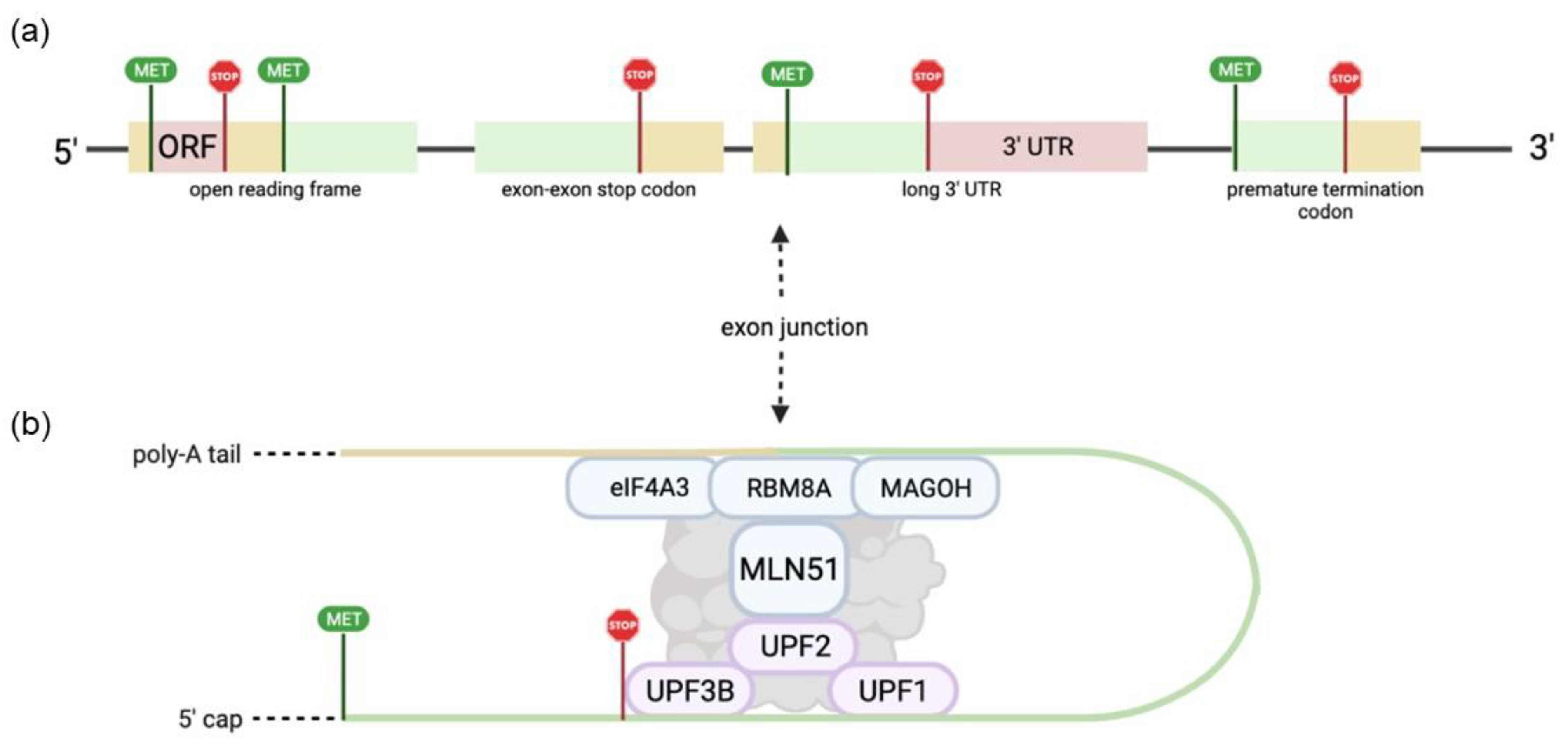
Figure 1. Graphical representation of EJC binding in mRNA during NMD. (a) NMD features that often trigger mRNA degradation, include upstream open reading frames, exon-exon stop codons, a long 3′ UTR, and a premature termination codon. (b) NMD cofactors UPF3B, UPF2, and UPF1 bind upstream of the exon junction, triggering exon-junction complex binding and NMD. Only core EJC factors are shown.
Ijms 23 10375 g001
Figure 2. Graphical representation of the function of EJC components in the cell cycle. (a) RBM8A inhibits cell cycle exit and is essential for progression past the G2/M checkpoint through mRNA localization and/or centrosome formation. (b) Inhibition of eIF4A3 has been shown to cause apoptosis at the G2/M checkpoint. MAGOH deficiency in interneuron and glial progenitors has been shown to cause apoptosis through p53 activation. (c) Phosphorylation eIF4A3 by CDK1/2 results in NMD inhibition and progression past the M/G1 boundary.
Ijms 23 10375 g002
Table 1. EJC core and major peripheral components associated with functional roles in context of various EJC functions. Included are key references in which EJC component structural integration is detailed.
EJC and NMD FactorsFunctional RoleReferences
ACIN1Alternative splicing[10,11]
ALYREFmRNA localization[12]
CWC22Alternative splicing[8,13]
eIF4A3Invariable EJC core component ubiquitous in all EJC functions[8,13]
IPO13mRNA localization[14]
MAGOHInvariable EJC core component ubiquitous in all EJC functions[15]
MLN51Alternative splicing, mRNA localization, mRNA translation, and NMD[10,16]
PNNAlternative Splicing[10,11]
PYMmRNA translation and EJC disassembly[17]
RBM8AInvariable EJC core component ubiquitous in all EJC functions[18]
RNPS1Alternative splicing and mRNA translation[19]
SAP18Alternative Splicing[10,11]
SMG6NMD[20]
UPF1NMD[20]
UPF2NMD[20]
UPF3AmRNA translation and NMD[21]
UPF3BmRNA translation and NMD[21]
Table 2. Summary of cell cycle abnormalities caused by dysfunction of core EJC components. Included are key references in which EJC cell cycle abnormality is detailed.
EJC ComponentDysfunctionReferences
eIF4A3Deficiency results in G2/M cell cycle arrest, chromosome segregation abnormalities, and apoptosis. Mutations of eIF4A3 specifically impact Tumor Factor-α pathway.[38,40]
RBM8ADeficiency results in G2/M cell cycle arrest, multinucleated cells with abnormal nuclear structure, and genome instability. Improper mRNA localization to mature centrosome has also been observed.[41,42,43]
MAGOHDeficiency inhibits melanoblast proliferation via arrest of cell cycle at G2/M and stalled mitosis in interneuron progenitors. Also results in apoptosis of radial glial cell progenitors.[44,45,46]
Table 3. EJC-associated cancers with implicated EJC components and references.
EJC Associated CancersEJC Component(s) ImplicatedReferences
Breast CancerMLN51, eIF4A3[51,52]
Cervical cancerRBM8A[49]
Colorectal CancereIF4A3[53]
Gastric CancerMAGOH[55]
GlioblastomaRBM8A[56]
Hepatocellular carcinomaRBM8A, MAGOH, eIF4A3[38,57]
Lung AdenocarcinomaRBM8A[42,50]
Non-small-cell lung carcinomaRBM8A[50]
Ovarian CancereIF4A3[54]
Publisher’s Note: MDPI stays neutral with regard to jurisdictional claims in published maps and institutional affiliations.