Article

Physiological and Molecular Response Modifications by Ultraviolet-C Radiation in Plutella xylostella and Its Compatibility with Cordyceps fumosorosea

1
Chongqing Key Laboratory of Vector Insects, College of Life Sciences, Chongqing Normal University, Chongqing 401331, China
2
Key Laboratory of Bio-Pesticide Innovation and Application of Guangdong Province, South China Agricultural University, Guangzhou 510640, China
3
School of Agriculture and Biology, Shanghai Jiao Tong University, Shanghai 200240, China
4
State Key Laboratory Breeding Base for Zhejiang Sustainable Pest and Disease Control, Institute of Plant Protection and Microbiology, Zhejiang Academy of Agricultural Sciences, Hangzhou 310021, China
*
Author to whom correspondence should be addressed.
Int. J. Mol. Sci. 2022, 23(17), 9800; https://doi.org/10.3390/ijms23179800
Submission received: 30 June 2022 / Revised: 19 August 2022 / Accepted: 24 August 2022 / Published: 29 August 2022
(This article belongs to the Section Molecular Biology)

Abstract

:
Ultraviolet-C (UV-C) radiation significantly impacts living organisms. UV-C radiation can also be used as a pest management tool. Therefore, this study was designed to investigate the effect of UV-C radiation on the physiology and gene expression level of Plutella xylostella, a destructive vegetable pest. Results showed that, after exposure to UV-C radiation for 3, 6, 12, and 24 h, the activity of SOD (superoxide dismutase) and CAT (catalase) of P. xylostella increased, while the activity of PPO (polyphenol oxidase), POD (peroxidase), AChE (acetylcholinesterase), CarE (carboxylesterase), and ACP (acid phosphatase) decreased with increased exposure time. Correlation coefficient analyses indicated that the activity of CAT correlated positively, while PPO and CarE correlated negatively, with exposure time. Gene regulation analysis via qRT-PCR confirmed a significant increase in regulation in CAT, CarE, and PPO-related genes. We also investigated the effect of UV-C exposure on the virulence of Cordyceps fumosorosea against P. xylostella. Here, results indicated that when the fungal treatment was applied to larvae before UV-C radiation, the virulence of C. fumosorosea was significantly reduced. However, this decline in virulence of C. fumosorosea due to UV-C exposure remained only for one generation, and no effect was observed on secondary infection. On the other hand, when larvae were exposed to UV-C radiation before fungal application, the mortality rate significantly increased as the exposure time to UV-C radiation increased. From the current study, it could be concluded that UV-C exposure suppressed the immunity to P. xylostella, which later enhanced the virulence of entomopathogenic fungi. Moreover, the study also suggested that UV irradiation is an effective pest management tool that could be incorporated into pest management strategies, which could help reduce pesticide application, be economically beneficial for the farmer, and be environmentally safe.

1. Introduction

Stress factors, both abiotic and biotic, play important roles in the development and growth of an insect. Environmental stresses such as ozone exposure, ultraviolet irradiation, and temperature stress significantly impact insects. For many organisms, ultraviolet (UV) radiation is a significant source of stress [1]. UV irradiation is an important ecological stressor for biological entities [2]. Ultraviolet-C longwave (UV-C) is a wavelength of electromagnetic radiation between 100 and 280 nm. Most moths can tolerate UV irradiation; however, blacklight, a synthetic form of UV-light, has been extensively used to control nocturnal moths [3,4]. Previous studies reported a direct effect of UV light on insect behavior [5], development, biology, and physiology [6,7], and biochemistry [8], but there is no study available reporting direct mortality of moths caused by UV light.
Insects and microorganisms are reported to have detrimental effects from UV-C radiation exposure [9,10,11]. Tribolium castaneum (Herbst, 1797), Tribolium confusum (DuVal, 1863), Cadra cautella (Walker, 1863), and Trogoderma granarium (Everts, 1899) are a few grain storage pests that have been examined for the use of UV-C radiation to manage them [11]. The silkworm Bombyx mori (Linnaeus, 1758) larvae are particularly reported to be vulnerable to the lethal effects of UV-C radiation [12]. Studies have reported that exposure to UV-C radiation could kill nymphs of Periplaneta americana (Linnaeus, 1785) and other pests [13,14]. UV-light exposure affects insect biology and is also known to negatively influence the virulence of entomopathogenic fungi [15], which may provide an alternative to synthetic insecticidal control. Most fungi can be killed by direct solar radiation exposure because UV radiation is the most dangerous and mutagenic light form of the solar spectrum [16].
Under normal conditions, antioxidants are stable within insects and help maintain regular metabolic activity. However, UV light can cause oxidative stress and damage insect protein functional activity [17,18]. Recent studies have proposed that UV light can significantly change antioxidant enzyme activity [19,20,21,22] and detoxification enzyme activity [21]. In addition, UV light exerts strong genotoxic effects via DNA damage, induces mutations, and could also be carcinogenic [23]. Several studies [24,25,26,27] reported that enzyme activity in any organism depends on gene regulation. Therefore, small changes in gene expression may have a significant impact on enzyme activity. As UV-C radiation can cause gene mutation, it is also possible that variation in enzymatic activity could result from changes in gene expression.
The diamondback moth, Plutella xylostella (Linnaeus, 1758) (Lepidoptera: Plutellidae), is the most important cruciferous vegetable pest in the world [28]. Due to short generation time, high fecundity, and indiscriminate use of insecticides, P. xylostella has become resistant to 81 insecticides to date [29,30], including spinosad [31,32], avermectins (abamectin and emamectin benzoate) [33], indoxacarb [34] and Bacillus thuringiensis [31,35]. High insecticide resistance development in P. xylostella made it a difficult pest to manage [36,37,38]. Therefore, it is necessary to promote alternative strategies such as entomopathogenic fungi to control P. xylostella and minimize synthetic insecticides [39].
In the 1990s, Cordyceps fumosorosea (Wize, 1904) (formerly known as Isaria fumosorosea Wize) [40] was reportedly the most common entomopathogenic fungi in Asia, Russia, the USA, South America, and India. Under favorable conditions, C. fumosorosea can cause a significant reduction in insect pest populations [41]. Apart from its efficacy and low cost of production, the use of C. fumosorosea has other advantages, including broader insecticidal activity, a diversified host range, and safety for humans and other non−target organisms [42]. C. fumosorosea is a highly effective antagonist of P. xylostella [43,44,45]. Several studies have confirmed the potential of C. fumosorosea, such as Bemisia tabaci (Gennadius) [46,47], Empoasca vitis (Gothe, 1875) [48], Aphis gossypii (Glover) [49] and Corythucha ciliata (Say) [50]. The corpses of host insects loaded with conidia generate cadavers and play an important role in the secondary spread of infection [51]. Furthermore, C. fumosorosea is commercially available worldwide [52].
The current study was designed to test the following hypothesis: (1) UV-C radiation affects the activity of the following antioxidant enzymes: Superoxide dismutase (SOD), catalase (CAT), peroxidase (POD), polyphenol oxidase (PPO), and glutathione S-transferase (GST) and the following detoxifying enzymes: Acetylcholinesterase (AChE), carboxylesterase (CarE), alkaline phosphatase (ALP) and acidic phosphatase (ACP); (2) The alteration in enzyme activity is due to alteration in gene expression; (3) The alteration in physiology will affect the virulence of C. fumosorosea; (4) UV-C exposure to the P. xylostella is effective before or after the application of C. fumosorosea.

2. Results

2.1. Effect of UV-C Radiation on the Physiology of Plutella xylostella

2.1.1. Antioxidant Enzyme Activities

The P. xylostella larvae were exposed to UV-C irradiation for 3 h, 6 h, 12 h, and 24 h, and the activity of enzymes was measured. SOD activity was significantly lower after 3 h of exposure to UV-C radiation compared to the control, 12 h, and 24 h, while the activity of SOD in 3 and 6 h of exposure was not affected compared to each other (F4,14 = 13.4; P < 0.01) (Figure 1A). The CAT was higher in larvae exposed to UV-C radiation for 6, 12, and 24 h compared to control (F4,14 = 12.9; P < 0.01) (Figure 1B). POD activity in P. xylostella third instar larvae exposed to UV-C for 3 h was not changed compared to the control, while the activity was significantly lower after 6, 12, and 24 h of exposure to UV-C radiation (F4,14 = 18.6; P < 0.01) (Figure 1C). The activity of PPO was significantly lower in treatments exposed to UV-C radiation for 12 and 24 h as compared to control and to 3 h of exposure (F4,14 = 14.8; P < 0.01) (Figure 1D). The GST activity was highest when third instar larvae were exposed to UV-C for 3 h compared to the control and other treatments. As exposure time increased, a gradual decline in GST was observed (F4,14 = 6.64; P < 0.01) (Figure 1E).

2.1.2. Detoxification Enzyme Activity

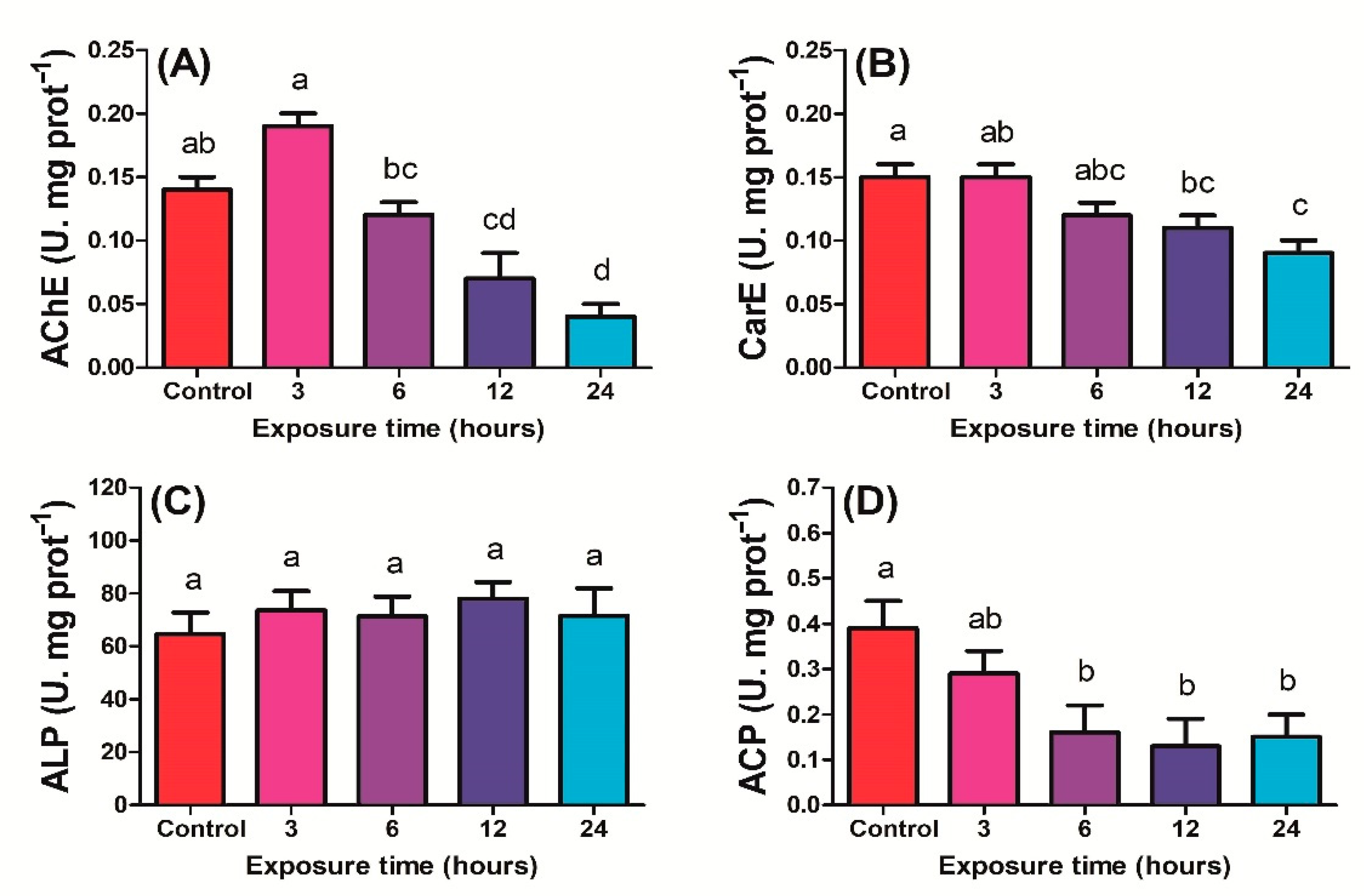
After 3 and 6 h of exposure to UV-C, no significant change in AChE activity of P. xylostella third instar larvae was observed compared to control, while 12 and 24 h activity was significantly decreased compared to control (F4,14 = 25.7; P < 0.01) (Figure 2A). The CarE activity decreased significantly when P. xylostella was exposed to UV-C for 12 and 24 h compared to the control, while no significant change in CarE activity was observed after control, 3 h or 6 h of exposure (F4,14 = 8.96; P < 0.01) (Figure 2B). ALP activity variations in response to exposure time were found to be non-significant (F4,14 = 0.36; P > 0.05) (Figure 2C). ACP activity was considerably higher in control compared to 6, 12 and 24 h exposed to UV-C radiation. Variation in ACP activity was non-significant when P. xylostella was exposed to UV-C radiation for 6, 12, and 24 h (F4,14 = 4.01; P < 0.05) (Figure 2D).

2.1.3. Correlation of Enzyme Activity with UV Exposure Time

The main objective of conducting Pearson correlation between exposure time and enzyme activity was to determine whether the enzyme has a significant correlation with time so the genes regulating enzyme could be selected to determine their gene expression. The results demonstrated that SOD and ALP had insignificant positive correlations, while POD, AChE, and ACP showed insignificant negative correlations with UV-C radiation exposure time. Alternatively, CAT correlated significantly (r = 0.905; P < 0.05) positively, while PPO (r = −0.908; P < 0.05) and CarE (r = −0.940; P < 0.05) correlated significantly negatively with time under UV-C radiation exposure. Correlation coefficients between exposure time and enzyme activity and between enzymes are shown in Figure 3. The results also showed the association between the activities of different enzymes. The enzymes with P < 0.05 were picked for gene expression analysis.

2.2. Effect of UV Radiation on Gene Expression of Plutella xylostella

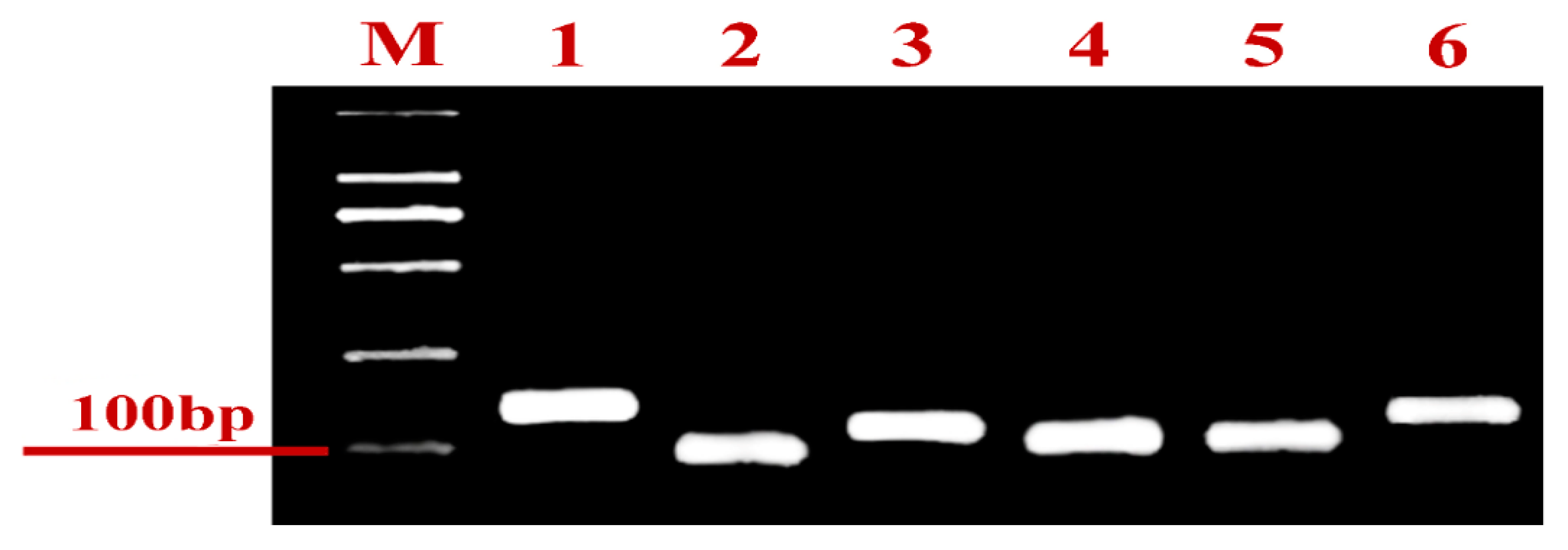
Two genes of each enzyme showing a significant correlation with UV-C exposure time (CAT, PPO, and CarE) were selected and subjected to gene expression analysis. Gene expression was analyzed after 0, 3, 6, 12, and 24 h exposure to UV-C radiation, and primer pairs were tested before gene expression analysis (Figure 4).
Results showed that UV-C radiation exposure significantly increased the gene expression of CAT1 compared with the control (Figure 5). The gene expression was 3.02-fold higher after 24 h than in control (F4,14 = 32.2; P < 0.01). Gene expression of the CAT2 gene increased; however, gene expression had stabilized after 6, 12, and 24 h. Nonetheless, gene expression was significantly elevated in all treatments compared with the control (F4,14 = 85.8; P < 0.01). The maximum fold change was observed after 24 h of exposure to UV-C radiation (3.20-fold). Expression levels of the genes CarE1 (F4,14 = 30.6; P < 0.01) and CarE2 (F4,14 = 26.6; P < 0.01), which regulate carboxylesterase activity in P. xylostella, were increased in treatments exposed to UV-C for 6, 12 and 24 h compared to control. The gene expression of CarE1 increased 2.66-fold, while CarE2 increased 2.31-fold following 24 h of UV-C exposure. PPO1 and PPO2 genes responsible for protoporphyrinogen oxidase activity in P. xylostella PPO1 were also significantly increased with UV-C radiation exposure time (F4,14 = 33.3; P < 0.01) after 24 h of exposure, PPO1 expression had increased 3.47-fold. PPO2 had increased in all UV-C radiation treatments compared to control but had no statistical difference from each other. The maximum fold change in PPO2 was observed after 12 h of exposure (2.34-fold).

2.3. Effect of UV-C Radiation on the Virulence of Cordyceps fumosorosea

Virulence of C. fumosorosea towards P. xylostella third instar larvae when UV-C radiation exposure preceded the application of the conidial suspension is shown in Figure 6A. Results revealed that the virulence of C. fumosorosea increased. The mortalities in all UV-C-exposed treatments were non-significantly different than control, but there was a significant increase in virulence observed after 24 h (F4,19 = 20.2; P < 0.01), 48 h (F4,19 = 9.31; P < 0.01) and 72 h (F4,19 = 4.04; P < 0.01) due to 24 h of exposure to UV-C radiation.
The mortality of P. xylostella third instar larvae, first exposed to C. fumosorosea and then to UV-C, is shown in Figure 6B. Results indicated that exposure to UV-C radiation reduced the virulence of C. fumosorosea against P. xylostella. As the exposure time increased, the virulence of C. fumosorosea significantly decreased. There was a significant decrease of virulence observed when C. fumosorosea-treated P. xylostella larvae were exposed to UV-C radiation for 24 h (F4,19 = 7.8; P < 0.01), 48 h (F4,19 = 9.92; P < 0.01) and 72 h (F4,19 = 11.90; P < 0.01).
The effect of UV-C in the F1 generation of C. fumosorosea was also assessed by collecting dead larvae and isolating C. fumosorosea from the third instar larvae of P. xylostella, which were firstly treated with C. fumosorosea and then exposed to UV-C radiation. The results are shown in Figure 6C. There was no significant difference in the mortalities of P. xylostella third instar larvae in F1 generation fungi, in which the F0 generation was exposed to UV-C radiation fungi or not (control). Mortality rates among P. xylostella third instar larvae was non-significant after 24 h (F4,19 = 1.23; P > 0.05), 48 h (F4,19 = 2.75; P > 0.05) or 72 h (F4,19 = 0.32; P > 0.86).

3. Discussion

Antioxidant enzymes are an important component of the insect immune system, protecting cellular networks from oxidative damage caused by xenobiotics [53]. SOD is an essential antioxidant protein used to alleviate superoxide radicals present in cells. In the present study, SOD activity increased with increasing exposure time, suggesting that UV-light irradiation induced the formation of superoxide radicals in P. xylostella larvae, which has been observed previously in the Antarctic midge Belgica antarctica and Dendrolimus tabulaeformis (Tsai & Liu, 1962) [21,54]. When insects were exposed to UV-C radiation for 24 h, SOD activity increased substantially, implying that SOD was stimulated to forage for superoxide radicals and protect larvae from UV stress. However, a significant decrease was found at 3 h, followed by increased SOD activity and UV-C radiation. These results contradict the previous findings that high doses of UV irradiation suppressed protective enzyme activities in cells [55,56].
The current study demonstrated that CAT enzyme activity in P. xylostella third instar larvae significantly increased with increasing exposure time. Additionally, several previous studies also found that UV exposure increased CAT activity. For example, Zhou et al. [22] reported that UV-light stress significantly increased CAT activity in Sitobion avenae (Fabricius, 1775). Moreover, Wang et al. [21] reported that CAT activity correlated positively with UV exposure time in D. tabulaeformis (Tsai & Liu, 1962) females. These results support the findings of the current study.
When third instar larvae of P. xylostella were exposed to UV-C radiation for 3 h, a substantial increase in POD activity was observed. However, previous studies showed that oxidative stress or excess substrate negatively affected POD enzyme activity [57]. Conversely, PPO activity decreased with increasing UV exposure time in the present study. Previous studies reported that PPO catalyzes the breakdown of phenolic compounds in organisms. UV exposure-induced suppression of PPO activity has resulted in elevated phenolic compound concentration, which hinders normal cell functions [58,59].
GST is a primary antioxidant enzyme that metabolizes lipid peroxides [60]. In the current study, following 3 h of exposure, a sudden spike was observed in GST activity and declined with increasing exposure time. Meng et al. [61] reported that UV-light exposure significantly reduced GST activity. Thus, P. xylostella exposed to UV-C radiation appeared capable of removing lipid peroxidation products generated during UV-C radiation stress, suggesting a protective effect from elevated GST levels.
AChE, a common detoxification enzyme, plays a critical role in regulating the normal transmission of nerve impulses along synapses and preserves normal physiological functioning in different organisms [62]. AChE is commonly believed to be the target of organophosphorus and carbamate pesticides, and changes to its amino acid sequence are likely to cause insecticide resistance [63]. Additionally, AChE plays a vital role in insect growth and development. Previous studies showed that the ploidy of Helicoverpa armigera (Hubner) was reduced significantly with gene silencing of AChE [64]. In our study, the AChE activities in the third-instar larvae of P. xylostella were reduced after 24 h of UV-C radiation. CarE in P. xylostella showed the same proclivity as AChE, while ALP activity with exposure time was not changed. We assume that UV-C radiation can deter the transmission of neurotransmitters in insects. ALP and ACP belong to nonspecific phosphohydrolases involved in phosphate group transmission and metabolism. For ALP, the optimum pH is >7 and ACP is <7 [65]. These attributes also concluded that UV-C radiation exposure increased ROS (reactive oxygen species) production and distorted the equilibrium between acid and base within P. xylostella.
Previous studies [24,25,26,27] reported that genes regulate enzyme activities in organisms. Thus, small changes in gene expression may have a significant impact on enzyme activity. This appears to be the case in the current study, as the enzymes with activity levels most correlated with exposure time showed significantly altered gene expression.
Cordyceps fumosorosea is an important entomopathogenic fungus used to manage various insect pests [66,67,68]. Previous studies reported that C. fumosorosea is an effective biological control agent against P. xylostella as measured by affected survival, fecundity, enzyme activity, and gene expression of P. xylostella [69,70,71]. The current study evaluates the virulence of C. fumosorosea against P. xylostella before and after exposure to UV-C radiation. Results revealed that the virulence of C. fumosorosea was low if insects were exposed to UV light after treatment with conidial suspension, which may be due to damage inflicted on conidia by UV-C exposure. Indeed, several studies have shown that exposure to UV-C radiation could inhibit fungal virulence and cause molecular and physiological changes in fungal conidia [15,16,72,73]. The current study reported that exposure to UV-C radiation reduces the immunity of P. xylostella, which increase the effectiveness of C. fumosorosea. Khan et al. [7] also reported that if Bemisia tabaci was exposed to UV-A before application of C. fumosorosea, the LC50 decreases with an increase in exposure time, which implies that the B. tabaci become less and less immune against fungal infection due to UV-A exposure.
Many natural abiotic factors are known to limit the ability of a fungal agent to control pests biologically, but solar ultraviolet (UV) radiation (UV-A and UV-B) is probably the most detrimental environmental factor affecting the viability of fungi applied for pest control [74,75]. Most UV-tolerant isolates can survive a few hours of direct exposure to solar UV radiation, but UV-susceptible isolates succumb. In addition, the exposure of fungi to UV-B [76,77,78,79] or UV-A [80,81] may delay conidial germination of survivors and reduce fungal development, which decreases the persistence and efficacy of infective propagules in the field [82,83].

4. Materials and Methods

4.1. Plutella xylostella Rearing

Plutella xylostella larvae were collected from the Engineering Research Center of Biological Control at South China Agricultural University (SCAU) and reared on Brassica rapa L. (Chinese cabbage). Third instar larvae (emerged < 24 h) were used in all experiments. Adults were reared on a 10% honey solution in an iron-framed, plastic sieve cage (60 × 60 × 60 cm). A piece of cotton was soaked in the honey solution, placed into a 2-inch diameter petri dish, and placed in a cage for feeding. The cotton was replaced after 24 h. The culture was maintained under controlled conditions (25 ± 1 °C, 70 ± 5% RH, and 16:8 h (L:D) photoperiod), and experiments were conducted under identical conditions.

4.2. UV-Irradiation Exposure

During testing, P. xylostella was irradiated at 15 W by UV-C radiation (X-series, peak emission 254 nm; Spectronics, Westbury, NY, USA). All larvae were placed for two hours in darkness before UV-C exposure. Five treatments were established: 0 (control), 3 h, 6 h, 12 h, and 24 h UV-C exposure under dark conditions. Each treatment contained ten third instar P. xylostella larvae per replication, and three replications per treatment were established. After exposure to UV-C irradiation for different time durations, the insects were immediately frozen with liquid nitrogen and stored at −80 °C until the enzymatic activity was assessed.

4.3. Effect of UV-C Radiation on the Physiology of Plutella xylostella

4.3.1. Sample Preparation

The five third instar larvae of P. xylostella were first weighed, and then samples were homogenized in an iced buffer (0.1 M phosphate buffer, 0.1 mM EDTA-2Na, 10 mM saccharose, 0.9% NaCl, pH = 7.4) with a bodyweight ratio of 0.1 g to 1 mL buffer. Next, homogenates were centrifuged for 20 min at 2500 RPM at 4 °C, and the supernatant was used for subsequent analyzes.

4.3.2. Antioxidant Enzyme Activity Assay

ELISA kits (Jianglai Biotechnology Co., Ltd., Shanghai, China) were used to determine antioxidant enzyme activity. The double-antibody sandwiching system was adopted to determine the enzyme activity and related substance content. The activity of SOD, POD, PPO, CAT, and GST was assessed. Using SOD as an example, a microplate was covered with pure SOD antibody to form a solid phase antibody, and SOD was simultaneously applied to the coated monoclonal microcapsule. Then, SOD was combined with horseradish peroxidase (HRP)-labelled SOD antibody to form an antibody-antigen-enzyme-labelled antibody complex. After thorough washing, substrates 3, 3′, 5, 5′-tetramethylbenzidine (TMB) were used for color development. TMB was transformed to blue under HRP enzyme catalysis and converted to its final yellow color through acid activity. The depth of color in the test sample was positively correlated with SOD. The absorbance (OD value) was measured at a wavelength of 450 nm using an enzyme-labelling instrument (SpectraMax Plus 384, Molecular Devices Co., Ltd., Silicon Valley, CA, USA), and the sample concentration of SOD activity was determined from a standard curve.

4.3.3. Detoxifying Enzyme Activity Assay

ELISA kits (Jianglai Biotechnology Co., Ltd., Shanghai, China) were used to test the key detoxifying enzymes, including ACP, CarE, ALP, and AChE, as per the procedures outlined in the user manual.

4.4. Effect of UV-C Radiation on Gene Regulation of Plutella xylostella

To determine the effect of UV-C radiation on gene regulation, two genes of each enzyme significantly correlated with UV-C radiation exposure time were selected and subjected to gene regulation quantification via qRT-PCR (Table 1).

4.4.1. RNA Extraction and cDNA Synthesis

Total RNA was isolated using TRIzol Reagent (Invitrogen, Waltham, MA, USA); 1 mL TRIzol was added to a ground sample on ice and left open at room temperature for 5 min. Next, 200 μL chloroform/1 mL TRIzol was added and, after thorough shaking, left at room temperature for 15 min. The mixture was centrifuged at 4 °C at 12,000 rpm for 15 min. The upper aqueous phase was moved into a new centrifuge tube. Then, 0.5 mL isopropanol/1 mL TRIzol was added, and after shaking, left on ice for 10 min. The mixture was again centrifuged at 4 °C at 12,000 rpm for 10 min. The supernatant was removed from the tube, leaving only RNA pellets. Next, 75% ethanol 1 mL/1 mL of TRIzol was added to wash the pellet via centrifuging at 4 °C at 8000 rpm for 5 min. The washed liquid was discarded, and the RNA pellet was air-dried for 5–10 min. The RNA pellet was then re-suspended in RNase-free water (20–50 μL) and stored at –80 °C.
To prepare first-strand cDNA, 1 μg of total RNA was used. PrimeScript™ RT reagent Kit with gDNA Eraser (Perfect Real Time) (Takara, Japan) was used according to the manufacturer’s instructions. The cDNAs were diluted tenfold before the following quantitative real-time RT-PCR reactions (qRT-PCR).

4.4.2. Primer Design and Testing

The gene sequences were obtained from NCBI, and the primers were designed using the CDS region. A 25 µL reaction mixture containing LA Taq DNA polymerase was used (Takara, Japan). The PCR conditions used in this study were previously explained by Guo et al. (2020). The pair of primers expressed as a single band was used in RT-qPCR.

4.4.3. RT-qPCR with SYBR Green

A total 50 μL reaction volume contained 2.5 μL of each primer, 25 μL SYBR Premix (Takara, Japan), 2.5 μL diluted cDNA template, and 17.5 μL of RNase-free water. The above reaction solution was separated into three technical repeats containing 15 µL of the reaction mixture. All reactions were performed using the CFX96 real-time PCR system (Bio-Rad, Hercules, CA, USA). The qPCR program included an initial denaturation for 3 min at 95 °C followed by 40 cycles of 95 °C for 10 s and 55 °C for 30 s. Dissociation curve analysis was performed for each reaction to confirm the amplification specificity; a dissociation step cycle (55 °C for 10 s, and then 0.5 °C for 10 s until 95 °C) was added. RPS13 was used as the reference gene. The relative gene expression was computed using the 2-ΔΔCt method [84,85,86]. Details concerning the primers used are given in Table 1.

4.5. Preparation of the Conidial Suspension

Cordyceps fumosorosea fungal culture (Sp535) originally isolated from soil was obtained from the repository of the Key Laboratory of Biopesticides Innovation and Application of Guangdong Province, SCAU, and cultured on potato dextrose agar (PDA) in Petri dishes (9 cm in diameter) for ten days in an incubator at 25 ± 1 °C. Conidia were collected under sterile conditions in deionized water with 0.1% v/v Tween-80. The suspension was shaken for 20 min on a magnetized stirrer to break conidial clumps and then filtered to remove debris via four-layer medical gauze. The conidial concentration was determined and diluted up to 107 conidia/mL with a hemocytometer. Conidial viability was assessed by spraying 0.1 mL of the diluted spore suspension onto PDA and then testing the amount of germinating conidia at 25 °C after 24 h incubation [87,88]. In all tests, conidial suspension demonstrated viability of >95%.

4.6. Virulence Assessment Bioassay

Virulence of the fungi was assessed in two ways; (1) Larvae were treated with fungal suspension before exposure to UV-C radiation; (2) Larvae were treated with fungal suspension after UV-C radiation exposure. Four replications were set for five exposure times (0, 3, 6, 12, and 24 h). Each replicate contained fifteen third instar larvae of P. xylostella kept in a single Petri plate. Larvae were kept in 9 cm Ø Petri dishes with sufficient food (Chinese cabbage leaves).
For the bioassay of larvae treated with fungal suspension before UV-C exposure, the larvae were dipped into the conidial suspension for 5 s. They were then sieved out from suspension and kept at room temperature for 5 min to dry. The larvae were then transferred onto cabbage leaves in a Petri dish. The Petri dish was covered with 60 nylon mesh to prevent the larvae from escaping. Larvae were kept in the dark for 2 h before UV-C radiation exposure. Petri dishes were placed 50 cm below the UV-C radiation source. After 3, 6, 12, and 24 h, the Petri dishes were removed from the UV-C radiation source and placed in a climate-controlled chamber (25 ± 1 °C, 70–80% RH and 16:8 h (L:D) photoperiod). Mortality data were collected after 24, 48, and 72 h. The same methodology was adopted for other bioassays in which larvae were treated with C. fumosorosea before exposure to UV-C radiation.
To assess whether the UV-C radiation affected the virulence of C. fumosorosea, dead larvae from the bioassay in which fungal treatment was applied to larvae before UV-C radiation were kept on PDA plates. The grown fungi from larvae were recultured and subjected to the virulence test with the method outlined above.

4.7. Statistical Analysis

The corrected mortality (%) was calculated using Henderson-Tilton’s formula [89]. Variation of larval mortality among different fungal treatments, enzyme activity, and relative gene expression was computed using One-Way ANOVA, and significance among the mean treatment values was assessed with Tukey post hoc test at P < 0.05 using SPSS software. Replications were used to calculate the standard deviation of the mean. Mortality rate, enzymatic activity, and relative gene expression graphs were constructed by Sigmaplot 12.0. The Pearson correlation analysis was conducted to determine the relation between UV exposure time and the activity of different enzymes. The correlation matrix graph was designed with R software × 64 3.6.3 using the GGally package.

5. Conclusions

In conclusion, UV-C light works as an immunosuppressor by interfering with the antioxidant and detoxification enzyme activity, which leads to a reduction in the immunity of P. xylostella and an increase in virulence of the C. fumosorosea, if UV-C is applied to larvae before application of fungi. However, if UV-C irradiation is applied after the application of fungi, due to its germicidal effect, the virulence of C. fumosorosea could be reduced. This study is applicable under laboratory and greenhouse conditions, where synthetic pesticides cannot be used and quick and effective pest management is required. But before applying in the greenhouse, the effect of UV-C on plants must be studied. The molecular mechanism of an insect under UV stress after exposure to UV-C irradiation was found to interfere with the virulence of the entomopathogenic fungus. However, these effects were not permanent and were limited to a single generation. Furthermore, UV-light exposure disrupted antioxidant and detoxifying enzyme activity in P. xylostella; this change in enzyme activity may be due to altered gene expression in P. xylostella. This current study provides additional information regarding the efficacy of UV-C radiation against insect pests, and with further research, UV-C radiation may be employed as a pest management strategy.

Author Contributions

Conceptualization, M.M.K. and B.-L.Q.; methodology, M.M.K.; software, M.M.K. and Z.-Y.F.; validation, M.M.K. and S.W.; formal analysis, M.M.K.; investigation, M.M.K., M.H. and Z.-Y.F.; resources, J.-H.W.; data curation, I.A.S.; writing—original draft preparation, M.M.K.; writing—review and editing, B.-L.Q. and J.-H.W.; supervision, B.-L.Q.; project administration, B.-L.Q.; funding acquisition, B.-L.Q. All authors have read and agreed to the published version of the manuscript.

Funding

The work was supported by the Laboratory of Lingnan Modern Agriculture Project (NT2021003) and the China National Key Research and Development Program (2019YFD1002100) to Bao-Li Qiu.

Institutional Review Board Statement

Not applicable.

Informed Consent Statement

Not applicable.

Data Availability Statement

All the data are available in the manuscript file.

Acknowledgments

The authors sincerely thank Andrew G.S. Cuthbertson for reviewing and editing the paper.

Conflicts of Interest

The authors declare no conflict of interest.

References

  1. Meyer-Rochow, V.B. Risks, especially for the eye, emanating from the rise of solar UV-radiation in the Arctic and Antarctic regions. Int. J. Clin. Health 2000, 59, 38–51. [Google Scholar]
  2. Schauena, M.; Hornig-Doa, H.-T.; Schomberg, S.; Herrmann, G.; Wiesner, R.J. Mitochondrial electron transport chain activity is not involved in ultraviolet A (UVA)-induced cell death. Free Radic. Biol. Med. 2007, 42, 499–509. [Google Scholar] [CrossRef] [PubMed]
  3. Antignus, Y. Manipulation of wavelength-dependent behaviour of insects: An IPM tool to impede insects and restrict epidemics of insect-borne viruses. Virus Res. 2000, 71, 213–220. [Google Scholar] [CrossRef]
  4. Kojima, Y.; Aoyagi, K.; Yasue, T. Effect of lithium ion addition on afterglow time of green-emitting Ce3+ and Pr3+ codoped CaS phosphor by black light irradiation. J. Lumin. 2005, 115, 13–18. [Google Scholar] [CrossRef]
  5. Mazza, C.A.; Izaguirre, M.M.; Zavala, J.; Scopel, A.L.; Ballaré, C.L. Insect perception of ambient ultraviolet-B radiation. Ecol. Lett. 2002, 5, 722–726. [Google Scholar] [CrossRef]
  6. Gunn, A. The determination of larval phase coloration in the African armyworm, Spodoptera exempta and its consequences for thermoregulation and protection from UV light. Entomol. Exp. Appl. 1998, 86, 125–133. [Google Scholar] [CrossRef]
  7. Khan, M.M.; Fan, Z.; Rothenberg, D.O.N.; Peng, J.; Hafeez, M.; Chen, X.; Pan, H.; Wu, J.; Qiu, B. Phototoxicity of ultraviolet-A against the whitefly Bemisia tabaci and its compatibility with an entomopathogenic fungus and whitefly parasitoid. Oxidative Med. Cell. Longev. 2021, 2021, 2060288. [Google Scholar] [CrossRef]
  8. Mackerness, A.H.S.; Surplus, S.L.; Blake, P.; John, C.F.; Buchanan-Wollaston, V.; Jordan, B.R.; Thomas, B. Ultraviolet-B-induced stress and changes in gene expression in Arabidopsis thaliana: Role of signalling pathways controlled by jasmonic acid, ethylene and reactive oxygen species. Plant Cell Environ. 1999, 22, 1413–1423. [Google Scholar] [CrossRef]
  9. Beard, R.L. Lethal action of UV irradiation on insects. J. Econ. Entomol. 1972, 65, 650–654. [Google Scholar] [CrossRef]
  10. Reed, N.G. The history of ultraviolet germicidal irradiation for air disinfection. Public Health Rep. 2000, 115, 581. [Google Scholar] [CrossRef]
  11. Hori, M.; Shibuya, K.; Sato, M.; Saito, Y. Lethal effects of short-wavelength visible light on insects. Sci. Rep. 2014, 4, 7383. [Google Scholar] [CrossRef] [PubMed] [Green Version]
  12. Nakajima, M.; Yoshida, H. Studies on ultraviolet sensitivity in the silkworm, with Special Reference to the Effect of UV-irradiation on melanin formation in the cuticle of the striped Silkworm. Jpn. J. Appl. Entomol. Zool. 1971, 15, 51–55. [Google Scholar] [CrossRef]
  13. Wharton, D.R.A. Ultraviolet repellent and lethal action on the American Cockroach. J. Econ. Entomol. 1971, 64, 252–255. [Google Scholar] [CrossRef]
  14. Lah, E.F.C.; Musa, R.N.A.R.; Ming, H.T. Effect of germicidal UV-C light (254 nm) on eggs and adult of house dustmites, Dermatophagoides pteronyssinus and Dermatophagoides farinae (Astigmata: Pyroglyhidae). Asian Pac. J. Trop. Biomed. 2012, 2, 679–683. [Google Scholar] [CrossRef]
  15. Cagan, L.; Svercel, M. The Influence of ultraviolet light on pathogenicity of entomopathogenic fungus Beauveria bassiana (Balsamo) Vuillemin to the European corn borer, Ostrinia Nubilalis Hbn. (Lepidoptera: Crambidae). J. Cent. Eur. Agric. 2002, 2, 227–234. [Google Scholar]
  16. Braga, G.U.L.; Rangel, D.E.N.; Fernandes, É.K.K.; Flint, S.D.; Roberts, D.W. Molecular and physiological effects of environmental UV radiation on fungal conidia. Curr. Genet. 2015, 61, 405–425. [Google Scholar] [CrossRef]
  17. Dahms, H.U.; Lee, J.S. UV radiation in marine ectotherms: Molecular effects and responses. Aquat. Toxicol. 2010, 97, 3–14. [Google Scholar] [CrossRef]
  18. Wang, Y.; Oberley, L.W.; Murhammer, D.W. Antioxidant defence system of two lepidopteran insect cell lines. Free Radic. Biol. Med. 2001, 30, 1254–1262. [Google Scholar] [CrossRef]
  19. Ali, A.; Rashid, M.A.; Huang, Q.Y.; Lei, C.L. Influence of UV-A radiation on oxidative stress and antioxidant enzymes in Mythimna separata (Lepidoptera: Noctuidae). Environ. Sci. Pollut. Res. 2017, 24, 8392–8398. [Google Scholar] [CrossRef]
  20. Cui, H.; Zeng, Y.; Singh, G.V.P.; Gao, F.; Li, Z.; Zhao, Z. UV radiation increases mortality and decreases the antioxidant activity in a tephritid fly. Food Energy Secur. 2021, 10, e297. [Google Scholar] [CrossRef]
  21. Wang, W.; Gao, C.; Ren, L.; Luo, Y. The effect of longwave ultraviolet light radiation on Dendrolimus tabulaeformis antioxidant and detoxifying enzymes. Insects 2019, 11, 1. [Google Scholar] [CrossRef] [PubMed]
  22. Zhou, D.; Du, Y.; Yang, J.; Zhang, L.; Zhao, H.; Hu, Z.; Hu, X. Effect of UV-B radiation in successive generation on the activity of protective enzymes in the grain aphid, Sitobion avenae (Hemiptera: Aphididae). Acta Entomol. Sin. 2014, 57, 762–768. [Google Scholar]
  23. Ikehata, H.; Ono, T. The mechanisms of UV mutagenesis. J. Radiat. Res. 2011, 52, 115–125. [Google Scholar] [CrossRef] [Green Version]
  24. Mao, T.; Li, F.; Fang, Y.; Wang, H.; Chen, J.; Li, M.; Lu, Z.; Qu, J.; Li, J.; Hu, J.; et al. Effects of chlorantraniliprole exposure on detoxification enzyme activities and detoxification-related gene expression in the fat body of the silkworm, Bombyx mori. Ecotoxicol. Environ. Saf. 2019, 176, 58–63. [Google Scholar] [CrossRef] [PubMed]
  25. Udomsinprasert, R.; Pongjaroenkit, S.; Wongsantichon, J.; Oakley, A.J.; Prapanthadara, L.A.; Wilce, M.C.J.; Ketterman, A.J. Identification, characterization and structure of a new Delta class glutathione transferase isoenzyme. Biochem. J. 2005, 388, 763–771. [Google Scholar] [CrossRef]
  26. Yuan, J.; Wang, X.; Gu, Z.; Zhang, Y.; Wang, Z. Activity and transcriptional responses of hepatopancreatic biotransformation and antioxidant enzymes in the oriental river prawn Macrobrachium nipponense exposed to microcystin-LR. Toxins 2015, 7, 4006–4022. [Google Scholar] [CrossRef]
  27. Zanger, U.M.; Schwab, M. Cytochrome P450 enzymes in drug metabolism: Regulation of gene expression, enzyme activities, and impact of genetic variation. Pharmacol. Ther. 2013, 138, 103–141. [Google Scholar] [CrossRef]
  28. Talekar, N.S.; Shelton, A.M. Biology, ecology, and management of the diamondback moth. Annu. Rev. Entomol. 1993, 38, 275–301. [Google Scholar] [CrossRef]
  29. Yang, J.; Tian, L.; Xu, B.; Xie, W.; Wang, S.; Zhang, Y.; Wang, X.; Wu, Q. Insight into the migration routes of Plutella xylostella in China using mtCOI and ISSR markers. PLoS ONE 2015, 10, e0130905. [Google Scholar] [CrossRef]
  30. Zhu, B.; Xu, M.; Shi, H.; Gao, X.; Liang, P. Genome-wide identification of lncRNAs associated with chlorantraniliprole resistance in diamondback moth Plutella xylostella (L.). BMC Genom. 2017, 18, 380. [Google Scholar] [CrossRef]
  31. Sarfraz, M.; Keddie, B.A. Conserving the efficacy of insecticides against Plutella xylostella (L.) (Lep., Plutellidae). J. Appl. Entomol. 2005, 129, 149–157. [Google Scholar] [CrossRef]
  32. Zhao, J.-Z.; Li, Y.-X.; Collins, H.L.; Gusukuma-Minuto, L.; Mau, R.F.L.; Thompson, G.D.; Shelton, A.M. Monitoring and characterization of diamondback moth (Lepidoptera: Plutellidae) resistance to spinosad. J. Econ. Entomol. 2009, 95, 430–436. [Google Scholar] [CrossRef] [PubMed]
  33. Pu, X.; Yang, Y.; Wu, S.; Wu, Y. Characterisation of abamectin resistance in a field-evolved multiresistant population of Plutella xylostella. Pest Manag. Sci. 2010, 66, 371–378. [Google Scholar] [CrossRef] [PubMed]
  34. Sayyed, A.H.; Wright, D.J. Genetics and evidence for an esterase-associated mechanism of resistance to indoxacarb in a field population of diamondback moth (Lepidoptera: Plutellidae). Pest Manag. Sci. 2006, 62, 1045–1051. [Google Scholar] [CrossRef]
  35. Liao, J.; Xue, Y.; Xiao, G.; Xie, M.; Huang, S.; You, S.; Wyckhuys, K.A.G.; You, M. Inheritance and fitness costs of resistance to Bacillus thuringiensis toxin Cry2Ad in laboratory strains of the diamondback moth, Plutella xylostella (L.). Sci. Rep. 2019, 9, 6113. [Google Scholar] [CrossRef] [PubMed]
  36. Sayyed, A.H.; Omar, D.; Wright, D.J. Genetics of spinosad resistance in a multi-resistant field-selected population of Plutella xylostella. Pest Manag. Sci. 2004, 60, 827–832. [Google Scholar] [CrossRef]
  37. Sarfraz, M.; Dosdall, L.M.; Keddie, B.A. Diamondback moth-host plant interactions: Implications for pest management. Crop Prot. 2006, 25, 625–639. [Google Scholar] [CrossRef]
  38. Zhao, J.-Z.; Collins, H.L.; Li, Y.-X.; Mau, R.F.L.; Thompson, G.D.; Hertlein, M.; Andaloro, J.T.; Boykin, R.; Shelton, A.M. Monitoring of diamondback moth (Lepidoptera: Plutellidae) resistance to spinosad, indoxacarb, and emamectin benzoate. J. Econ. Entomol. 2006, 99, 176–181. [Google Scholar] [CrossRef]
  39. Nian, X.G.; He, Y.R.; Lu, L.H.; Zhao, R. Evaluation of the time-concentration-mortality responses of Plutella xylostella larvae to the interaction of Isaria fumosorosea with the insecticides beta-cypermethrin and Bacillus thuringiensis. Pest Manag. Sci. 2015, 71, 216–224. [Google Scholar] [CrossRef]
  40. Kepler, R.M.; Luangsa-Ard, J.J.; Hywel-Jones, N.L.; Quandt, C.A.; Sung, G.H.; Rehner, S.A.; Aime, M.C.; Henkel, T.W.; Sanjuan, T.; Zare, R.; et al. A phylogenetically-based nomenclature for Cordycipitaceae (Hypocreales). IMA Fungus 2017, 8, 335–353. [Google Scholar] [CrossRef]
  41. Zhang, C.; Wang, X.; Ashraf, U.; Qiu, B.; Ali, S. Transfer of lead (Pb) in the soil-plant-mealybug-ladybird beetle food chain, a comparison between two host plants. Ecotoxicol. Environ. Saf. 2017, 143, 289–295. [Google Scholar] [CrossRef] [PubMed]
  42. Ali, S.; Zhang, C.; Wang, Z.; Wang, X.M.; Wu, J.H.; Cuthbertson, A.G.S.; Shao, Z.; Qiu, B.L. Toxicological and biochemical basis of synergism between the entomopathogenic fungus Lecanicillium muscarium and the insecticide matrine against Bemisia tabaci (Gennadius). Sci. Rep. 2017, 7, 46558. [Google Scholar] [CrossRef] [PubMed]
  43. Altre, J.A.; Vandenberg, J.D. Factors influencing the infectivity of isolates of Paecilomyces fumosoroseus against diamondback moth, Plutella xylostella. J. Invertebr. Pathol. 2001, 78, 31–36. [Google Scholar] [CrossRef] [PubMed]
  44. Altre, J.A.; Vandenberg, J.D.; Cantone, F.A. Pathogenicity of Paecilomyces fumosoroseus isolates to diamondback Moth, Plutella xylostella: Correlation with spore size, germination speed, and Attachment to Cuticle. J. Invertebr. Pathol. 1999, 73, 332–338. [Google Scholar] [CrossRef]
  45. Li, H.U.; Yu, R.H.; Ya, J.W.; Xia, F.; Huan, Y.C. The time-dose-mortality model of a Paecilomyces fumosoroseus isolate on the diamondback moth, Plutella xylostella. Acta Entomol. Sin. 2007, 50, 567–573. [Google Scholar]
  46. Boopathi, T.; Karuppuchamy, P.; Singh, S.B.; Kalyanasundaram, M.; Mohankumar, S.; Ravi, M. Microbial control of the invasive spiraling whitefly on cassava with entomopathogenic fungi. Braz. J. Microbiol. 2015, 46, 1077–1085. [Google Scholar] [CrossRef]
  47. Gao, T.; Wang, Z.; Huang, Y.; Keyhani, N.O.; Huang, Z. Lack of resistance development in Bemisia tabaci to Isaria fumosorosea after multiple generations of selection. Sci. Rep. 2017, 7, 42727. [Google Scholar] [CrossRef]
  48. Chen, M.; Zhang, D.; Peng, F.; Li, Z. Wettable powder development of Isaria javanica for control of the lesser green leafhopper, Empoasca vitis. Chin. J. Biol. Control 2014, 30, 51–57. [Google Scholar]
  49. Jandricic, S.E.; Filotas, M.; Sanderson, J.P.; Wraight, S.P. Pathogenicity of conidia-based preparations of entomopathogenic fungi against the greenhouse pest aphids Myzus persicae, Aphis gossypii, and Aulacorthum solani (Hemiptera: Aphididae). J. Invertebr. Pathol. 2014, 118, 34–46. [Google Scholar] [CrossRef]
  50. Sevim, A.; Demir, I.; Sönmez, E.; Kocaçevik, S.; Demirbaǧ, Z. Evaluation of entomopathogenic fungi against the sycamore lace bug, Corythucha ciliata (Say) (Hemiptera: Tingidae). Turk. J. Agric. For. 2013, 37, 595–603. [Google Scholar] [CrossRef]
  51. Bugti, G.A.; Na, C.; Bin, W.; Lin, H.F. Control of plant sap-sucking insects using entomopathogenic fungi Isaria fumosorosea strain (Ifu13a). Plant Prot. Sci. 2018, 54, 258–264. [Google Scholar]
  52. de Faria, M.R.; Wraight, S.P. Mycoinsecticides and Mycoacaricides: A comprehensive list with worldwide coverage and international classification of formulation types. Biol. Control 2007, 43, 237–256. [Google Scholar] [CrossRef]
  53. Ahmad, S. Oxidative stress from environmental pollutants. Arch. Insect Biochem. Physiol. 1995, 29, 135–157. [Google Scholar] [CrossRef]
  54. Lopez-Martinez, G.; Elnitsky, M.A.; Benoit, J.B.; Lee, R.E.; Denlinger, D.L. High resistance to oxidative damage in the Antarctic midge Belgica antarctica, and developmentally linked expression of genes encoding superoxide dismutase, catalase and heat shock proteins. Insect Biochem. Mol. Biol. 2008, 38, 796–804. [Google Scholar] [CrossRef]
  55. Heck, D.E.; Vetrano, A.M.; Mariano, T.M.; Laskin, J.D. UVB light stimulates production of reactive oxygen species: Unexpected role for catalase. J. Biol. Chem. 2003, 278, 22432–22436. [Google Scholar] [CrossRef] [PubMed]
  56. Polte, T.; Tyrrell, R.M. Involvement of lipid peroxidation and organic peroxides in UVA-induced matrix metalloproteinase-1 expression. Free Radic. Biol. Med. 2004, 36, 1566–1574. [Google Scholar] [CrossRef]
  57. Tabatabaie, T.; Floyd, R.A. Susceptibility of glutathione peroxidase and glutathione reductase to oxidative damage and the protective effect of spin trapping agents. Arch. Biochem. Biophys. 1994, 314, 112–119. [Google Scholar] [CrossRef]
  58. Czerniewicz, P.; Sytykiewicz, H.; Durak, R.; Borowiak-Sobkowiak, B.; Chrzanowski, G. Role of phenolic compounds during antioxidative responses of winter triticale to aphid and beetle attack. Plant Physiol. Biochem. 2017, 118, 529–540. [Google Scholar] [CrossRef]
  59. Haddouche, L.; Phalak, A.; Tikekar, R.V. Inactivation of polyphenol oxidase using 254nm ultraviolet light in a model system. LWT Food Sci. Technol. 2015, 62, 97–103. [Google Scholar] [CrossRef]
  60. Ahmad, S.; Duval, D.L.; Weinhold, L.C.; Pardini, R.S. Cabbage looper antioxidant enzymes: Tissue specificity. Insect Biochem. 1991, 21, 563–572. [Google Scholar] [CrossRef]
  61. Meng, J.Y.; Zhang, C.Y.; Zhu, F.; Wang, X.P.; Lei, C.L. Ultraviolet light-induced oxidative stress: Effects on antioxidant response of Helicoverpa armigera adults. J. Insect Physiol. 2009, 55, 588–592. [Google Scholar] [CrossRef] [PubMed]
  62. Meng, X.; Miao, L.; Dong, F.; Wang, J. Advances in the research on invertebrate acetylcholinesterase. J. Environ. Entomol. 2019, 41, 508–519. [Google Scholar]
  63. Kim, Y.H.; Lee, S.H. Which acetylcholinesterase functions as the main catalytic enzyme in the Class Insecta? Insect Biochem. Mol. Biol. 2013, 43, 47–53. [Google Scholar] [CrossRef]
  64. Kumar, M.; Gupta, G.P.; Rajam, M.V. Silencing of acetylcholinesterase gene of Helicoverpa armigera by siRNA affects larval growth and its life cycle. J. Insect Physiol. 2009, 55, 273–278. [Google Scholar] [CrossRef] [PubMed]
  65. Vincent, J.B.; Crowder, M.W.; Averill, B.A. Hydrolysis of phosphate monoesters: A biological problem with multiple chemical solutions. Trends Biochem. Sci. 1992, 17, 105–110. [Google Scholar] [CrossRef]
  66. Hu, Q.; Liu, S.; Yin, F.; Cai, S.; Zhong, G.; Ren, S. Diversity and virulence of soil-dwelling fungi Isaria spp. and Paecilomyces spp. against Solenopsis invicta (Hymenoptera: Formicidae). Biocontrol Sci. Technol. 2011, 21, 225–234. [Google Scholar] [CrossRef]
  67. da Silva Lopes, R.; de Lima, G.; dos Santos Correia, M.T.; da Costa, A.F.; de Luna Alves Lima, E.Á.; de Menezes Lima, V.L. The potential of Isaria spp. as a bioinsecticide for the biological control of Nasutitermes corniger. Biocontrol Sci. Technol. 2017, 27, 1038–1048. [Google Scholar] [CrossRef]
  68. Cuthbertson, A.G.S.; Audsley, N. Further screening of entomopathogenic fungi and nematodes as control agents for Drosophila suzukii. Insects 2016, 7, 24. [Google Scholar] [CrossRef]
  69. Ali, S.; Huang, Z.; Ren, S. Production of cuticle degrading enzymes by Isaria fumosorosea and their evaluation as a biocontrol agent against diamondback moth. J. Pest Sci. 2010, 83, 361–370. [Google Scholar] [CrossRef]
  70. Huang, Z.; Sahar, F.; Ren, S.; Ali, S. Effect of Isaria fumosoroseus on eretmocerus sp. nr. furuhashii (hymenoptera: Aphelinidae), a parasitoid of Bemisia tabaci (hemiptera: Aleyrodidae). Pak. J. Zool. 2010, 42, 121–127. [Google Scholar]
  71. Xu, J.; Xu, X.; Li, S.; Wang, S.; Xu, X.; Zhou, X.; Yu, J.; Yu, X.; Shakeel, M.; Jin, F. Genome-wide profiling of Plutella xylostella immunity-related miRNAs after Isaria fumosorosea infection. Front. Physiol. 2017, 8, 1054. [Google Scholar] [CrossRef] [PubMed]
  72. Fang, W.; St. Leger, R.J. Enhanced UV resistance and improved killing of malaria mosquitoes by photolyase transgenic entomopathogenic fungi. PLoS ONE 2012, 7, e43069. [Google Scholar] [CrossRef] [PubMed]
  73. Fernandes, É.K.K.; Rangel, D.E.N.; Braga, G.U.L.; Roberts, D.W. Tolerance of entomopathogenic fungi to ultraviolet radiation: A review on screening of strains and their formulation. Curr. Genet. 2015, 61, 427–440. [Google Scholar] [CrossRef]
  74. Ignoffo, C.; Garcia, C. Influence of conidial colour on inactivation of several entomogenous fungi (hyphomycetes) by simulated sunlight. Environ. Entomol. 1992, 21, 913–917. [Google Scholar] [CrossRef]
  75. Moore, D.; Bridge, P.; Higgins, P.; Bateman, R.; Prior, C. Ultraviolet radiation damage to Metarhizium flavoviride conidia and the protection given by vegetable and mineral oils and chemical sunscreens. Ann. Appl. Biol. 1993, 122, 605–616. [Google Scholar] [CrossRef]
  76. Fernandes, É.; Rangel, D.; Moraes, A.; Bittencourt, V.; Roberts, D. Variability in tolerance to UV-B radiation among Beauveria spp. isolates. J. Invertebr. Pathol. 2007, 96, 237–243. [Google Scholar] [CrossRef]
  77. Fargues, J.; Goettel, M.S.; Smits, N.; Ouedraogo, A.; Vidal, C.; Lacey, L.A.; Lomer, C.J.; Rougier, M. Variability in susceptibility to simulated sunlight of conidia among isolates of entomopathogenic Hyphomycetes. Mycopathologia 1996, 135, 171–181. [Google Scholar] [CrossRef]
  78. Braga, G.U.L.; Flint, S.D.; Messias, C.L.; Anderson, A.J.; Roberts, D.W. Effects of UVB irradiance on conidia and germinants of the entomopathogenic hyphomycete Metarhizium anisopliae: A Study of reciprocity and recovery. Photochem. Photobiol. 2001, 73, 140. [Google Scholar] [CrossRef]
  79. Nascimento, É.; da Silva, S.; Marques, E.; Roberts, D.; Braga, G. Quantification of cyclobutane pyrimidine dimers induced by UVB radiation in conidia of the fungi Aspergillus fumigatus, Aspergillus nidulans, Metarhizium acridum and Metarhizium robertsii. Photochem. Photobiol. 2010, 86, 1256–1266. [Google Scholar] [CrossRef]
  80. Fargues, J.; Rougier, M.; Goujet, R.; Smits, N.; Coustere, C.; Itier, B. Inactivation of conidia of Paecilomyces fumosoroseus by near-ultraviolet (UVB and UVA) and visible radiation. J. Invertebr. Pathol. 1997, 69, 70–78. [Google Scholar] [CrossRef]
  81. Braga, G.U.L.; Flint, S.D.; Miller, C.D.; Anderson, A.J.; Roberts, D.W. Both Solar UVA and UVB Radiation impair conidial culturability and delay germination in the entomopathogenic fungus Metarhizium anisopliae. Photochem. Photobiol. 2001, 74, 734. [Google Scholar] [CrossRef]
  82. Zimmermann, G. Effect of high temperatures and artificial sunlight on the viability of conidia of Metarhizium anisopliae. J. Invertebr. Pathol. 1982, 40, 36–40. [Google Scholar] [CrossRef]
  83. Roberts, D.W.; Campbell, A.S. Stability of entomopathogenic fungi. Entomol. Soc. Am. 1977, 10, 19–76. [Google Scholar]
  84. Guo, C.F.; Pan, H.P.; Zhang, L.H.; Ou, D.; Lu, Z.T.; Khan, M.M.; Qiu, B.L. Comprehensive assessment of candidate reference genes for gene expression studies using RT-qPCR in Tamarixia radiata, a predominant parasitoid of Diaphorina citri. Genes 2020, 11, 1178. [Google Scholar] [CrossRef]
  85. Pinheiro, D.H.; Siegfried, B.D. Selection of reference genes for normalization of RT-qPCR data in gene expression studies in Anthonomus eugenii Cano (Coleoptera: Curculionidae). Sci. Rep. 2020, 10, 5070. [Google Scholar] [CrossRef]
  86. Livak, K.J.; Schmittgen, T.D. Analysis of relative gene expression data using real-time quantitative PCR and the 2−ΔΔCT method. Methods 2001, 25, 402–408. [Google Scholar] [CrossRef]
  87. Tian, J.; Diao, H.; Liang, L.; Hao, C.; Arthurs, S.; Ma, R. Pathogenicity of Isaria fumosorosea to Bemisia tabaci, with some observations on the fungal infection process and host immune response. J. Invertebr. Pathol. 2015, 130, 147–153. [Google Scholar] [CrossRef] [PubMed]
  88. Gökçe, A.; Er, M.K. Pathogenicity of Paecilomyces spp. to the glasshouse whitefly, Trialeurodes vaporariorum, with some observations on the fungal infection process. Turk. J. Agric. For. 2005, 29, 331–339. [Google Scholar]
  89. Henderson, C.F.; Tilton, E.W. Tests with acaricides against the brown wheat mite. J. Econ. Entomol. 1955, 48, 157–161. [Google Scholar] [CrossRef]
Figure 1. Activities of different antioxidant enzymes (A) SOD, (B) CAT, (C) POD, (D) PPO, and (E) GST of Plutella xylostella larvae exposed to UV−light for control (0 h), 3, 6, 12, and 24 h. The bars indicate the mean values of four replications. Standard error bars indicate the standard deviation of the mean. Lowercase letters indicate the significant difference among the treatments at P < 0.05. Similar letters have no significant difference among treatments.
Ijms 23 09800 g001
Figure 2. Activities of detoxification enzymes (A) AChE, (B) CarE, (C) ALP, and (D) ACP of Plutella xylostella larvae exposed to UV−light for control (0 h), 3, 6, 12, and 24 h. The bars indicate the mean value of four replications, and standard error bars indicate the standard deviation of the mean. Lowercase letters indicate a significant difference among the treatments at P < 0.05, and similar letters have no significant difference among treatments.
Ijms 23 09800 g002
Figure 3. Pearson correlation matrix of UV−light exposure time and Plutella xylostella larvae enzyme activity. The distribution of each variable is shown on the diagonal. The bivariate scatter plots with a fitted line are displayed on the bottom of the diagonal. The correlation value and the significance level as stars are on the top of the diagonal. Each significance level is associated to a symbol: P-values (0, 0.001, 0.01, 0.05, 0.1) < = > symbols (“***”, “**”, “*”, “.”, “ ”).
Ijms 23 09800 g003
Figure 4. Agarose gel electrophoresis of six genes regulating enzyme of significant correlation with UV exposure. M, Molecular marker. Templates in the polymerase chain reactions (PCRs) were as follows: (1) CAT1; (2) CAT2; (3) CarE1; (4) CarE2; (5) PPO1; and (6) PPO2.
Ijms 23 09800 g004
Figure 5. The gene regulation of Catalase-related (CAT1 and CAT2), Carboxylesterase-related (CarE1 and CarE2), and Prophenoloxidase-related (PPO1 and PPO2) genes of Plutella xylostella larvae after 0 (control), 3, 6, 12, and 24 h exposures to UV−light. The bars indicate the mean values of four replications; standard error bars indicate the standard deviation of the mean, and error bars indicate the mean deviation. Small letters indicate a significant difference among the treatments at P < 0.05, and similar letters have no significant difference among treatments.
Ijms 23 09800 g005
Figure 6. Mortality percentage of third instar larvae of Plutella xylostella exposed to UV-C radiation (A) First UV−C radiation then Cordyceps fumosorosea treatment; (B) First C. fumosorosea treatment then UV-C radiation; (C) Dead insects from (B) collected, F1 fungal generation regrown and virulence checked again. Each bar shows the mean value of four replications, and error bars indicate the mean deviation. Lowercase letters indicate a significant difference among the treatments at P < 0.05, and similar letters have no significant difference among treatments.
Ijms 23 09800 g006
Table 1. The list of genes and primers of genes used on qRT-PCR.
Gene NameNCBI Reference Sequence NumberGene NameSequence (5′ to 3′)Sequence
CAT1XM_011560295.1PREDICTED: Plutella xylostella catalase-like (LOC105389213), mRNAFggctcaacgacaacctcatccg
Rcgtgcgtgacctcgaagtagc
CAT2XM_011561829.1PREDICTED: Plutella xylostella catalase-like (LOC105390515), mRNAFcaccaagtattccgccgccaag
Rtccgcccaccgtcgagaatc
CarE1XM_011552809.1PREDICTED: Plutella xylostella carboxylesterase 1C-like (LOC105382842), mRNAFcatgggaaagtatccgggaacacg
Rtggttgtggtggcagaaatctcag
CarE2XM_011558702.1PREDICTED: Plutella xylostella carboxylesterase 1E-like (LOC105387900), mRNAFactgcccatgccaagaccaaac
Ragacgctcgccttagctccag
PPO1NW_011952494.1Plutella xylostella strain DBM-FZ-S unplaced genomic scaffold, DBM_FJ_V1.1 scaffold_467, whole genome shotgun sequenceFagccataggaagcctgacctcatc
Rgctgacgacaccgaccacaat
PPO2XM_011565232.1PREDICTED: Plutella xylostella uncharacterized LOC105393465 (LOC105393465), mRNAFggagtgaagccgccgaaagc
Rtgttgccaccgataatccgatcag
RPS13NM_001017.3Ribosomal protein S13Ftcaggcttattctcgtcg
Rgctgtgctggattcgtac
Publisher’s Note: MDPI stays neutral with regard to jurisdictional claims in published maps and institutional affiliations.