Article

Cysteine Regulates Oxidative Stress and Glutathione-Related Antioxidative Capacity before and after Colorectal Tumor Resection

1
Division of Colorectal Surgery, Department of Surgery, Taichung Veterans General Hospital, Taichung 40705, Taiwan
2
Department of Food and Nutrition, Providence University, Taichung 43301, Taiwan
3
Chiayi & Wanqiao Branch, Taichung Veterans General Hospital, Chiayi 60090, Taiwan
4
Department of Nutrition, Chung Shan Medical University, Taichung 40201, Taiwan
5
Department of Nutrition, Chung Shan Medical University Hospital, Taichung 40201, Taiwan
6
Department of Health Industry Technology Management, Chung Shan Medical University, Taichung 40201, Taiwan
7
Department of Respiratory Therapy, Taichung Veterans General Hospital, Taichung 40705, Taiwan
*
Author to whom correspondence should be addressed.
Int. J. Mol. Sci. 2022, 23(17), 9581; https://doi.org/10.3390/ijms23179581
Submission received: 9 August 2022 / Revised: 19 August 2022 / Accepted: 22 August 2022 / Published: 24 August 2022

Abstract

:
Cysteine might scavenge free radicals and is a limiting substrate for the cellular synthesis of glutathione (GSH). We investigated the association of cysteine with oxidative stress and GSH-related antioxidant capacity in colorectal cancer (CRC) patients. Plasma samples were drawn from 66 patients 1 day before (pre-resection) and 4 weeks after resection (post-resection). Tumor and adjacent normal tissues were collected. We measured levels of plasma and tissue cysteine, homocysteine, oxidative stress indicators (malondialdehyde, MDA; advanced oxidation protein products, AOPP), GSH, and antioxidant enzyme activities. After tumor resection, patients had significantly higher levels of plasma cysteine, homocysteine, MDA, AOPP, and GSH-related antioxidant enzyme activities when compared with pre-resection. Levels of cysteine, homocysteine, AOPP and all antioxidant capacity indicators in tumor tissue were significantly higher than those levels in the adjacent normal tissue. Plasma cysteine levels measured at pre-resection were positively associated with MDA levels in the tumor and in the adjacent normal tissues. Cysteine levels in tumor and adjacent normal tissues were significantly associated with tissue levels of homocysteine, almost as indicators of oxidative stress and antioxidant capacities. Cysteine in the circulation was likely utilized to mediate GSH-related antioxidant capacity and further cope with increased oxidative stress in tumor and adjacent normal tissues.

1. Introduction

Colorectal cancer (CRC) is one of the most common types of cancer, with rising mortality worldwide over the past decade [1]. Risk factors for CRC are obesity; male gender; high red meat intake, saturated fat and alcohol consumption; low fruit, vegetable, and fiber intake; prior diseases (i.e., diabetic mellitus, inflammatory bowel disease); smoking; and family history of CRC [2]. Greater oxidative stress or reduced antioxidant capacity leads to redox imbalance, resulting in tumor initiation and progression [3]. An imbalance between oxidative stress and antioxidant capacity has been suggested to cause CRC. The evidence is that healthy subjects have lower oxidative stress and higher antioxidant capacity in their plasma compared with CRC patients [4,5,6,7,8,9,10,11]. Tumor tissues also show higher oxidative stress and higher antioxidant capacity compared with adjacent normal tissues [6,12,13,14,15].
Adequate antioxidant defense can prevent threats of high oxidative stress to lower disease incidence or slow down disease progression. Regarding antioxidant defense mechanisms in the human body, glutathione peroxidase (GPx) and superoxide dismutase (SOD) situate at the first line, while glutathione (GSH, γ-L-glutamyl-L-cysteinylglycine) situates at the second line [16]. Under conditions of high oxidative stress, GSH offers electrons via GPx to reduce hydroperoxides, and then it self-oxidizes to glutathione disulfide (GSSG, an oxidized form of GSH). GSSG is further reduced back to GSH through catalyzation by glutathione reductase (GR). The thiol group (-SH) of cysteine is not only a major yet limiting substrate for the synthesis of GSH within cells, but it also directly scavenges free radicals; cysteine thiols thus play a crucial role in oxidative stress [17]. Studies indicated that CRC cells likely take up cysteine to protect themselves against high oxidative stress to maintain growth [18,19,20]. If tumor cells capture cysteine from circulation to protect themselves against high oxidative stress, theoretically, plasma cysteine levels in CRC patients might be associated with the risk of CRC. However, no significant association between plasma cysteine levels and CRC risk has been reported in previous studies [21,22,23,24] except one showing that high plasma cysteine levels significantly lower the risk of rectal tumors in postmenopausal women [25]. On the other hand, we observed in our previous study that CRC patients had higher levels of plasma cysteine and GSH compared with healthy subjects [22]. We could not rule out the possibility that cysteine mediates oxidative stress and antioxidant capacity through GSH-related defense systems to affect CRC risk indirectly.
The role that cysteine plays in regulating oxidative stress and antioxidant defenses in CRC patients has not been thoroughly investigated. Here, we aimed to investigate the association of cysteine with oxidative stress and GSH-related antioxidant capacity before and after tumor resection in CRC patients.

2. Results

Table 1 shows CRC patients’ demographic and clinical characteristics at pre- and post-resection times. Of the 66 CRC patients (34 men, 32 women), 32 had colon cancer and 34 had rectal cancer. More than half of the patients were diagnosed to be in CRC stages II and III. Patients during post-resection time had significantly lower body mass index (BMI), serum carcinoembryonic antigen (CEA), and carbohydrate antigen 19-9 (CA 19-9) after tumor resection compared with pre-resection.
Table 2 and Table 3 show the cysteine and homocysteine concentrations, oxidative stress indicators, and antioxidant capacity indicators in plasma, tumor, and adjacent normal tissues. Patients during post-resection not only had significantly higher plasma cysteine and homocysteine concentrations but also had significantly higher levels of malondialdehyde (MDA), advanced oxidation protein products (AOPP), trolox equivalent antioxidant capacity (TEAC), GSH, GSSG, GPx, GR, and glutathione S-transferase (GST) when compared with pre-resection. Their cysteine and homocysteine concentrations, levels of AOPP, and all antioxidant capacity indicators in tumor tissue were significantly higher than those in the adjacent normal tissue. The only exception was the significantly lower MDA levels in tumor tissue compared with the adjacent normal tissue.
Partial Spearman’s correlation was performed whether cysteine levels at pre-resection, tumor and adjacent normal tissues were associated with indicators of oxidative stress and antioxidant capacities (Table 4). More than half of our CRC patients were in stages II and III, and serum C-reactive protein (CRP) level slightly increased after tumor resection. We therefore considered the CRC stage and serum CRP level to be potential confounders for oxidative stress, and we adjusted for age, sex, smoking and drinking habits, cancer stage, and CRP levels when we performed the partial correlation analyses. Plasma cysteine level at pre-resection was positively associated with plasma pre-resection levels of homocysteine and TEAC levels, and it was also associated with plasma cysteine and homocysteine levels of post-resection. Plasma cysteine level was associated with homocysteine, GSH, and GSSG level at post-resection. Plasma cysteine level at pre-resection was not associated with cysteine and homocysteine levels in either tumor or adjacent normal tissues. Instead, it was positively associated with MDA, TEAC, and GSSG levels in tumor tissue and was further associated with MDA level in the adjacent normal tissue. Cysteine levels in tumor and adjacent normal tissues were significantly associated with tissue levels of homocysteine, which were likely indicators of oxidative stress and antioxidant capacity.

3. Discussion

CRC patients are well-known to have higher oxidative stress and lower antioxidative capacity compared with healthy subjects [4,5,6,7,8,9,10,11]. MDA, a byproduct of polyunsaturated fatty acid peroxidation, is considered a biological marker of oxidative stress [26]. AOPP are products of protein oxidation, likely reliable markers of oxidant-mediated protein damage [27]. To study oxidative stress and antioxidative capacity, we used levels of MDA and AOPP as oxidative stress indicators. Previous studies reported that plasma MDA and AOPP levels are elevated in CRC patients when compared with healthy controls [9]. These two plasma levels are simultaneously reduced at 24 h after tumor resection compared with pre-resection levels [11]. We wondered why our CRC patients had significantly higher plasma levels of MDA and AOPP after tumor resection compared with those before resection. It is worth noting that our CRC patients’ serum CRP and neutrophil-to-lymphocyte ratio (NRL) levels indicated that they were still in an inflammatory status after tumor resection. Ongoing inflammatory response might lead to elevated oxidative stress. Although surgery per se could induce oxidative stress [28], redox status is known to be regulated, returning to normal within 24 h after surgery [29]. Apart from surgery itself, inflammatory response, medication uses, or illness stress might all be possible causes of elevated oxidative stress in our CRC patients after tumor resection.
Regarding oxidative stress status in CRC tumor and in adjacent normal tissue, the colorectal tumor tissue has MDA levels higher than the adjacent normal tissue [13,14,15]. Veljković et al. [30] found that both MDA and AOPP are significantly higher in CRC tumor tissue than in normal tissue. In our present study, more surprisingly, such findings were opposite. Regarding tissue AOPP levels, we found oxidative stress status in tumor tissue was higher than in adjacent normal tissue. The level of MDA, other than AOPP, might be a potential non-invasive biomarker to differentiate tumor invasion depth or the presence of lymph node metastasis [9]. We thus speculated that one marker might be more sensitive than the other to reflect oxidative stress. Further study is warranted to confirm our speculation. Regardless of the status of oxidative stress in the tumor and adjacent normal tissues, colorectal tumor tissue had higher cysteine levels and antioxidant capacity compared with the adjacent normal tissue. Consistent with previous reports [6,13,14,15], malignant colorectal tissue likely utilizes cysteine and GSH-related antioxidant capacities to protect tumor against elevated oxidative stress.
Cysteine and GSH are essential antioxidants. Inadequate cysteine status may further disrupt cysteine and GSH metabolism to affect redox homeostasis. In our study, we did not recruit healthy controls. Total plasma cysteine concentration in healthy individuals is known to be 200 to 300 μmol/L [31]. Our CRC patients had mean plasma cysteine concentrations below 200 μmol/L, even though their mean levels were significantly higher, but still did not reach normal values after tumor resection. Our CRC patients’ deficient cysteine status might well be due to inadequate cysteine intake. As their dietary intake data were not recorded, this possibility was not assessed. In addition to dietary intake, cysteine is generated through the transsulfuration pathway. During transsulfuration, homocysteine transfers sulfur to form cysteine via cystathionine [32]. Approximately 50% of cysteine so generated can be utilized to synthesize GSH in hepatic cells [33]. Enzymes (e.g., cystathionine β-synthase and cystathionine γ-lyase) and coenzymes (e.g., vitamin B-6) are involved in the transsulfuration pathway [32]. Although we did not measure these enzyme activities and vitamin B-6 status in our study, cysteine concentrations were closely associated with homocysteine levels in both plasma and tissue. The generation of cysteine via the transsulfuration pathway was likely normal in our CRC patients. The transsulfuration pathway can be activated through enhanced de novo synthesis of cysteine during tumorigenesis, and cysteine deprivation in the transsulfuration pathway might be cytoprotective [20,34]. Thus, we propose that cysteine generation is likely enhanced through the transsulfuration pathway and that cysteine is used by tumor tissue to maintain its growth. This speculation is based on the changes of cysteine level in plasma between pre- and post-resection and the difference between tumor and adjacent normal tissues.
Yang et al. [35] observed that cysteine is likely more oxidized in CRC tissue than in non-tumor tissues, while cysteine is usually maintained in the reduced form in non-tumor tissue. If cysteinyl oxidation fosters free radical production and increases oxidative stress in tumor tissue, tumor cells can protect themselves against higher oxidative stress. We found that plasma cysteine levels at the pre-resection time were closely related to MDA levels in tumor and adjacent normal tissues, and cysteine levels were significantly associated with GSH-related antioxidant enzyme activities in tumor and adjacent normal tissues. Although we did not measure reactive oxygen species (ROS) levels in plasma and tissues, it is possible that plasma cysteine can move to tissue directly via its thiol group or that it indirectly mediates antioxidative capacity through a GSH-related defense system to cope with elevated oxidative stress in both tumor and adjacent normal tissues. The regulation of redox state by cysteine in the tumor and its adjacent normal tissue is likely complex during the tumorigenesis, and that needs to be further investigated.
The strength of our study was that tissue and plasma samples at the pre- and post-resection times were simultaneously collected. Study limitations existed. First, we did not collect healthy controls for reference. Second, we did not record dietary intake data. Third, the levels of ROS in plasma and tissues were not measured. Although we calculated sample size, 66 patients might not have adequate statistical power to reveal a more significant effect of cysteine on oxidative stress and GSH-related antioxidant capacities. Otherwise, the picture of cysteine status could be completely analyzed in our CRC patients.

4. Materials and Methods

4.1. Study Design and Sample Size Calculation

This was a cross-sectional study. To detect a significant correlation (r = 0.3) between the level of plasma cysteine and oxidative stress indicator (i.e., MDA), we needed 62 CRC patients to reach 80% statistical power (1 − β) and a two-sided α of less than 0.05.

4.2. Subjects

Consecutive patients were recruited from the division of colorectal surgery of Taichung Veterans General Hospital, Taiwan, with confirmed diagnosis of either colon or rectal cancer (International Classification of Diseases, Tenth Revision, Clinical Modification ICD-10-CM, codes C18–C20, respectively). All patients were scheduled to undergo tumor resection. Patients were excluded if they were younger than 20 years old or older than 80 years, pregnant or lactating, or had cardiovascular, liver, or renal diseases. Patients signed the informed consent form prior to enrolling the study. Our study was approved by the Institutional Review Board of Taichung Veterans General Hospital, Taichung, Taiwan (No. CF 19330A).

4.3. Data Collection and Biochemical Measurements

We recorded patients’ essential characteristics, such as age, sex, smoking and drinking habits, medications, and family history of colorectal cancer. Height, weight, systolic and diastolic blood pressures were measured one day before tumor resection (pre-resection) and 4 weeks after tumor resection (post-resection). BMI (kg/m2) was calculated as weight (kg) divided by height squared (m2).
Patients’ fasting blood samples were drawn and stored in vacutainer tubes (Becton Dickinson, Rutherford, NJ, USA) that contained appropriate anticoagulant or no anticoagulant as required at pre- and post-resection times. Serum levels of white blood cell, neutrophil count, and lymphocyte count, albumin, alanine aminotransferase, and creatinine were determined using an Automated Biochemical analyzer. We used the NRL as a reliable marker of ongoing cancer-related inflammation, and a valid indicator of prognosis of solid tumors, NRL ≥ 2.5–5.0, was adopted to serve as an independent prognostic index for CRC [36]. Therefore, this ratio was calculated using neutrophil and lymphocyte counts. Serum C-reactive protein was analyzed by particle-enhanced immunonephelometry using an Image analyzer, with a normal level at <0.3 mg/dL. Serum levels of CEA and CA 19-9, are widely used as tumor markers for CRC in clinical settings. We therefore determined these levels using an automated chemiluminescent immunoassay. The aforementioned analyses were carried out at our hospital’s department of pathology and laboratory medicine.
Colorectal tumor tissue and its adjacent normal tissue were obtained at the time of surgical resection. Patients’ diagnosis and cancer staging, histological type, and pathological grade of CRC were confirmed jointly by a pathologist and an oncologist. Tumor size and location were recorded accordingly. Resected tissues were homogenized on ice in 1 mL phosphate saline buffer (pH = 7.4). Homogenates were centrifuged at 13,000 rpm for 10 min at 4 °C. Supernatants were stored at −80 °C until analysis.
The following measurements were performed in the laboratory. Plasma and tissue cysteine levels and homocysteine levels were carried out under yellow lighting to minimize photodestruction and quantified by high performance liquid chromatography using fluorescence [37,38]. Plasma and tissue MDA and AOPP were determined to reflect oxidative stress. MDA was measured by the reaction of thiobarbituric acid at an excitation wavelength of 515 nm, and an emission wavelength of 555 nm using a fluorescence spectrophotometer [39]. AOPP were measured at a wavelength of 340 nm using commercial kits (Abcam company, Cambridge, UK). Plasma and tissue TEAC, GSH and GSSG levels, and activities of GPx, GR, GST, and SOD were used to reflect antioxidative capacity. TEAC was determined following the method of Arnao et al. [40]. GR activity was measured according to a published method [41]. GSH and GSSG concentrations (BioVision Incorporated, Milpitas, CA, USA), activities of GPx, GST, and SOD (Cayman Chemical Company, Ann Arbor, MI, USA) were analyzed using commercial kits.

4.4. Statistical Analyses

We used the SAS statistical software package (version 9.4, Statistical Analysis System Institute Inc., Cary, NC, USA). Shapiro–Wilk test was used to assess normality of distributions. Paired t-test or Wilcoxon signed-rank test was used to compare differences between pre- and post-resections. Categorical data were compared between groups using Chi-square or Fisher’s exact tests. The partial Spearman’s correlation was used to assess the associations between plasma or tissue cysteine concentrations and indicators of oxidative stress and also antioxidative capacity. These associations were analyzed after adjusting for age, sex, smoking and drinking habits, cancer stage, and CRP levels. Statistical significance was set at a two-sided p < 0.05.

5. Conclusions

CRC patients likely utilize cysteine in the circulation to mediate GSH-related antioxidative capacity and further cope with increased oxidative stress in tumor and adjacent normal tissues (Figure 1). Longer follow-up times (i.e., one or three years after tumor resection) would improve the monitoring clinical outcomes and the understanding of the roles of cysteine and GSH-related antioxidant capacity in the convalescence after tumor resection.

Author Contributions

F.-F.C. was responsible for subjects’ screening and monitoring, research funding, interpretation of the results, and manuscript rafting. T.-H.C. was responsible for the screening of subjects and research funding. S.-C.H. was responsible for hematological measurements and the interpretation of the results. C.-H.C. was responsible for the screening of subjects and statistical analyses. Y.-Y.T. was responsible for the sample collection and biochemical measurements. Y.-C.H. was responsible for the development of intellectual content and the study design, interpretation of the results, and manuscript drafting. All authors have read and agreed to the published version of the manuscript.

Funding

This study was supported by a grant from Taichung Veterans General Hospital, and Taichung Veterans General Hospital, Chiayi Branch, Taiwan (TCVGH-VHCY1098606, TCVGH-VHCY 1108601).

Institutional Review Board Statement

The study was conducted according to the guidelines of the Declaration of Helsinki and approved by the Institutional Review Board of Taichung Veterans General Hospital, Taichung, Taiwan (No. CF 19330A).

Informed Consent Statement

Informed consent was obtained from all subjects who participated in the study.

Data Availability Statement

The data are contained within the article.

Conflicts of Interest

The authors declare no conflict of interest.

References

  1. Sung, H.; Ferlay, J.; Siegel, R.L.; Laversanne, M.; Soerjomataram, I.; Jemal, A.; Bray, F. Global Cancer Statistics 2020: GLOBOCAN estimates of incidence and mortality worldwide for 36 cancers in 185 countries. CA Cancer J. Clin. 2021, 71, 209–249. [Google Scholar] [CrossRef] [PubMed]
  2. Gandomani, H.S.; Yousefi, S.M.; Aghajani, M.; Mohammadian-Hafshejani, A.; Tarazoj, A.A.; Pouyesh, V.; Salehiniya, H. Colorectal cancer in the world: Incidence, mortality and risk factors. Biomed. Res. Ther. 2017, 4, 1656–1675. [Google Scholar] [CrossRef] [Green Version]
  3. Poprac, P.; Jomova, K.; Simunkova, M.; Kollar, V.; Rhodes, C.J.; Valko, M. Targeting Free Radicals in Oxidative Stress-Related Human Diseases. Trends Pharmacol. Sci. 2017, 38, 592–607. [Google Scholar] [CrossRef] [PubMed]
  4. Chang, D.; Wang, F.; Zhao, Y.S.; Pan, H.Z. Evaluation of oxidative stress in colorectal cancer patients. Biomed. Environ. Sci. 2008, 21, 286–289. [Google Scholar] [CrossRef]
  5. Leufkens, A.M.; van Duijnhoven, F.J.; Woudt, S.H.; Siersema, P.D.; Jenab, M.; Jansen, E.H.J.M.; Pischon, T.; Tjønneland, A.; Olsen, A.; Overvad, K.; et al. Biomarkers of oxidative stress and risk of developing colorectal cancer: A cohort-nested case-control study in the European Prospective Investigation into Cancer and Nutrition. Am. J. Epidemiol. 2012, 175, 653–663. [Google Scholar] [CrossRef] [PubMed] [Green Version]
  6. Strzelczyk, J.K.; Wielkoszyński, T.; Krakowczyk, Ł.; Adamek, B.; Zalewska-Ziob, M.; Gawron, K.; Kasperczyk, J.; Wiczkowski, A. The activity of antioxidant enzymes in colorectal adenocarcinoma and corresponding normal mucosa. Acta Biochim. Pol. 2012, 59, 549–556. [Google Scholar] [CrossRef] [PubMed] [Green Version]
  7. Gopčević, K.R.; Rovčanin, B.R.; Tatić, S.B.; Krivokapić, Z.V.; Gajić, M.M.; Dragutinović, V.V. Activity of superoxide dismutase, catalase, glutathione peroxidase, and glutathione reductase in different stages of colorectal carcinoma. Dig. Dis. Sci. 2013, 58, 2646–2652. [Google Scholar] [CrossRef] [PubMed]
  8. Perše, M. Oxidative stress in the pathogenesis of colorectal cancer: Cause or consequence? Biomed. Res. Int. 2013, 2013, 725710. [Google Scholar] [CrossRef] [Green Version]
  9. Zińczuk, J.; Maciejczyk, M.; Zaręba, K.; Romaniuk, W.; Markowski, A.; Kędra, B.; Zalewska, A.; Pryczynicz, A.; Matowicka-Karna, J.; Guzińnska-Ustymowicz, K. Antioxidant barrier, redox status, and oxidative damage to biomolecules in patients with colorectal cancer. Can malondialdehyde and catalase be markers of colorectal cancer advancement? Biomolecules 2019, 9, 637. [Google Scholar] [CrossRef] [Green Version]
  10. Kundaktepe, B.P.; Sozer, V.; Durmus, S.; Kocael, P.C.; Kundaktepe, F.O.; Papila, C.; Gelisgen, R.; Uzun, H. The evaluation of oxidative stress parameters in breast and colon cancer. Medicine 2021, 100, e25104. [Google Scholar] [CrossRef]
  11. Salehi, S.S.; Mirmiranpour, H.; Rabizadeh, S.; Esteghamati, A.; Tomasello, G.; Alibakhshi, A.; Najafi, N.; Rajab, A.; Nakhjavani, M. Improvement in redox homeostasis after cytoreductive surgery in colorectal adenocarcinoma. Oxid. Med. Cell Longev. 2021, 2021, 8864905. [Google Scholar] [CrossRef]
  12. Özdemirler, G.; Pabuççuoğlu, H.; Bulut, T.; Buğra, D.; Uysal, M.; Toker, G. Increased lipoperoxide levels and antioxidant system in colorectal cancer. J. Cancer Res. Clin. Oncol. 1998, 124, 555–559. [Google Scholar] [CrossRef]
  13. Skrzydlewska, E.; Sulkowski, S.; Koda, M.; Zalewski, B.; Kanczuga-Koda, L.; Sulkowska, M. Lipid peroxidation and antioxidant status in colorectal cancer. World J. Gastroenterol. 2005, 11, 403–406. [Google Scholar] [CrossRef]
  14. Rainis, T.; Maor, I.; Lanir, A.; Shnizer, S.; Lavy, A. Enhanced oxidative stress and leucocyte activation in neoplastic tissues of the colon. Dig. Dis. Sci. 2007, 52, 526–530. [Google Scholar] [CrossRef]
  15. Kekec, Y.; Paydas, S.; Tuli, A.; Zorludemir, S.; Sakman, G.; Seydaoglu, G. Antioxidant enzyme levels in cases with gastrointestinal cancer. Eur. J. Intern. Med. 2009, 20, 403–406. [Google Scholar] [CrossRef]
  16. Ighodar, O.M.; Akinoloye, O.A. First line defence antioxidants-superoxide dismutase (SOD), catalase (CAT) and glutathione peroxidase (GPX); The fundamental role in the entire antioxidant defence grid. Alex. J. Med. 2018, 54, 287–293. [Google Scholar] [CrossRef] [Green Version]
  17. Fra, A.; Yoboue, E.D.; Sitia, R. Cysteine as redox molecular switches and targets of disease. Front. Mol. Neurosci. 2017, 10, 167. [Google Scholar] [CrossRef] [Green Version]
  18. Xu, X.; Zhang, X.; Wei, C.; Zheng, D.; Lu, X.; Yang, Y.; Luo, A.; Zhang, K.; Duan, X.; Wang, Y. Targeting SLC7A11 specifically suppresses the progression of colorectal cancer stem cells via inducing ferroptosis. Eur. J. Pharm. Sci. 2020, 152, 105450. [Google Scholar] [CrossRef]
  19. Wu, J.; Yeung, S.C.J.; Liu, S.; Qdaisat, A.; Jiang, D.; Liu, W.; Cheng, Z.; Liu, W.; Wang, H.; Li, L.; et al. Cyst(e)ine in nutrition formulation promotes colon cancer growth and chemoresistance by activating mTORC1 and scavenging ROS. Sig. Transduct. Target Ther. 2021, 6, 188. [Google Scholar] [CrossRef]
  20. Zhang, H.F.; Klein Geltink, R.I.; Parker, S.J.; Sorensen, P.H. Transsulfuration, minor player or crucial for cysteine homeostasis in cancer. Trends Cell Biol. 2022, 32, 800–814. [Google Scholar] [CrossRef]
  21. Le Marchand, L.; White, K.K.; Nomura, A.M.; Wilkens, L.R.; Selhub, J.S.; Tiirikainen, M.; Goodman, M.T.; Murphy, S.P.; Henderson, B.E.; Kolonel, L.N. Plasma levels of B vitamins and colorectal cancer risk: The multiethnic cohort study. Cancer Epidemiol. Biomark. Prev. 2009, 18, 2195–2201. [Google Scholar] [CrossRef] [PubMed] [Green Version]
  22. Chiang, F.F.; Wang, H.M.; Lan, Y.C.; Yang, M.H.; Huang, S.C.; Huang, Y.C. High homocysteine is associated with an increased risk of colorectal cancer independently of oxidative stress and antioxidant capacities. Clin. Nutr. 2014, 33, 1054–1060. [Google Scholar] [CrossRef] [PubMed]
  23. Myte, R.; Gylling, B.; Haggstrom, J.; Schneede, J.; Ueland, P.M.; Hallmans, G.; Johansson, I.; Palmqvist, R.; Van Guelpen, B. Untangling the role of one-carbon metabolism in colorectal cancer risk: A comprehensive Bayesian network analysis. Sci. Rep. 2017, 7, 43434. [Google Scholar] [CrossRef] [PubMed] [Green Version]
  24. Gylling, B.; Myte, R.; Ulvik, A.; Ueland, P.M.; Middttun, Ø.; Schnede, J.; Hallmans, G.; Häggstrőm, J.; Hohansson, I.; Van Guelpen, B.; et al. One-carbon metabolite ratios as functional vitamin markers and in relation to colorectal cancer risk. Int. J. Cancer 2019, 144, 947–956. [Google Scholar] [CrossRef] [Green Version]
  25. Miller, J.W.; Beresford, S.A.A.; Neuhouser, M.L.; Cheng, T.Y.D.; Song, X.; Brown, E.C.; Zheng, Y.; Rodriguez, B.; Green, R.; Ulrich, C.M. Homocysteine, cysteine, and risk of incident colorectal cancer in the women’s health initiative observational cohort. Am. J. Clin. Nutr. 2013, 97, 827–834. [Google Scholar] [CrossRef]
  26. Rio, D.D.; Stwart, A.J.; Pellegrini, N. A review of recent studies on malondialdehyde as toxic molecule and biological marker of oxidative stress. Nutr. Metab. Cardiovas. Dis. 2005, 15, 316–328. [Google Scholar]
  27. Witko-Sarsat, V.; Friedlander, M.; Capeillère-Blandin, C.; Nguyen-Khoa, T.; Nguyen, A.T.; Zingraff, J.; Jungers, P.; Descamps-Latscha, B. Advanced oxidation protein products as a novel marker of oxidative stress in uremia. Kidney Int. 1996, 49, 1304–1313. [Google Scholar] [CrossRef] [Green Version]
  28. Rosenfeldt, F.; Wilson, M.; Lee, G.; Kure, C.; Ou, R.; Braun, L.; de Haan, J. Oxidative stress in surgery in an ageing population: Pathophysiology and therapy. Exp. Gerontol. 2013, 48, 45–54. [Google Scholar] [CrossRef]
  29. Stipancic, I.; Zarkovic, N.; Servis, D.; Sabolović, S.; Tatzber, F.; Busic, Z. Oxidative stress markers after laparoscopic and open cholecystectomy. J. Laparoendosc. Adv. Surg. Tech. 2005, 15, 347–352. [Google Scholar] [CrossRef]
  30. Veljković, A.; Stanojević, G.; Branković, B.; Pavlović, D.; Stojanović, I.; Cvetković, T. Parameters of oxidative stress in colon cancer tissue. Acta Med. Median. 2016, 55, 32–37. [Google Scholar] [CrossRef]
  31. Bonifácio, V.D.B.; Pereira, S.A.; Serpa, J.; Vicente, J.B. Cysteine metabolic circuitries: Druggable targets in cancer. Brit. J. Cancer 2021, 124, 862–879. [Google Scholar] [CrossRef]
  32. Sbodio, J.I.; Synder, S.H.; Paul, B.D. Regulators of the transsulfuration pathway. Brit. J. Pharmacol. 2019, 176, 583–593. [Google Scholar] [CrossRef]
  33. Mosharov, E.; Cranford, M.R.; Banerjee, R. The quantitatively important relationship between homocysteine metabolism and glutathione synthesis by the transsulfuration pathway and its regulation by redox changes. Biochemistry 2000, 39, 13005–13011. [Google Scholar] [CrossRef]
  34. Chombs, J.A.; DeNicola, G.M. The non-essential amino acid cysteine becomes essential for tumor proliferation and survival. Cancers 2019, 11, 678. [Google Scholar] [CrossRef] [Green Version]
  35. Yang, H.Y.; Chay, K.O.; Kwon, J.; Kwon, S.O.; Park, Y.K.; Lee, T.H. Comparative proteomic analysis of cysteine oxidation in colorectal cancer patients. Mol. Cells 2013, 35, 533–542. [Google Scholar] [CrossRef] [Green Version]
  36. Zahorec, R. Neutrophil-to-lymphocyte ratio, past, present and future perspectives. Bratisl. Lek. List. 2021, 122, 474–488. [Google Scholar] [CrossRef]
  37. Araki, A.; Sako, Y. Determination of free and total homocysteine in human plasma by high-performance liquid chromatography with fluorescence detection. J. Chromatogr. 1987, 422, 43–52. [Google Scholar] [CrossRef]
  38. Radha Rama Devi, A.; Naushad, S.M.; Prasad, K.C. Evaluation of total plasma homocysteine in Indian newborns using heel-prick samples. Indian J. Pediatr. 2006, 73, 503–508. [Google Scholar] [CrossRef]
  39. Lapenna, D.; Ciofani, G.; Pierdomenico, S.D.; Giamberardino, M.A.; Cuccurullo, F. Reaction conditions affecting the relationship between thiobarbituric acid reactivity and lipid peroxides in human plasma. Free Radic. Biol. Med. 2001, 31, 331–335. [Google Scholar] [CrossRef]
  40. Arnao, M.B.; Cano, A.; Hernandez-Ruiz, J.; Garcia-Canovas, F.; Acosta, M. Inhibition by L-ascorbic acid and other antioxidants of the 2.2′ -azino-bis[3-ethylbenzthiazoline-6-sulfonic acid] oxidation catalyzed by peroxidase: A new approach for determining total antioxidant status of foods. Anal. Biochem. 1996, 236, 255–261. [Google Scholar] [CrossRef]
  41. Carlberg, I.; Mannervik, B. Purification and characterization of the flavoenzyme glutathione reductase from rat liver. J. Biol. Chem. 1975, 250, 5475–5480. [Google Scholar] [CrossRef]
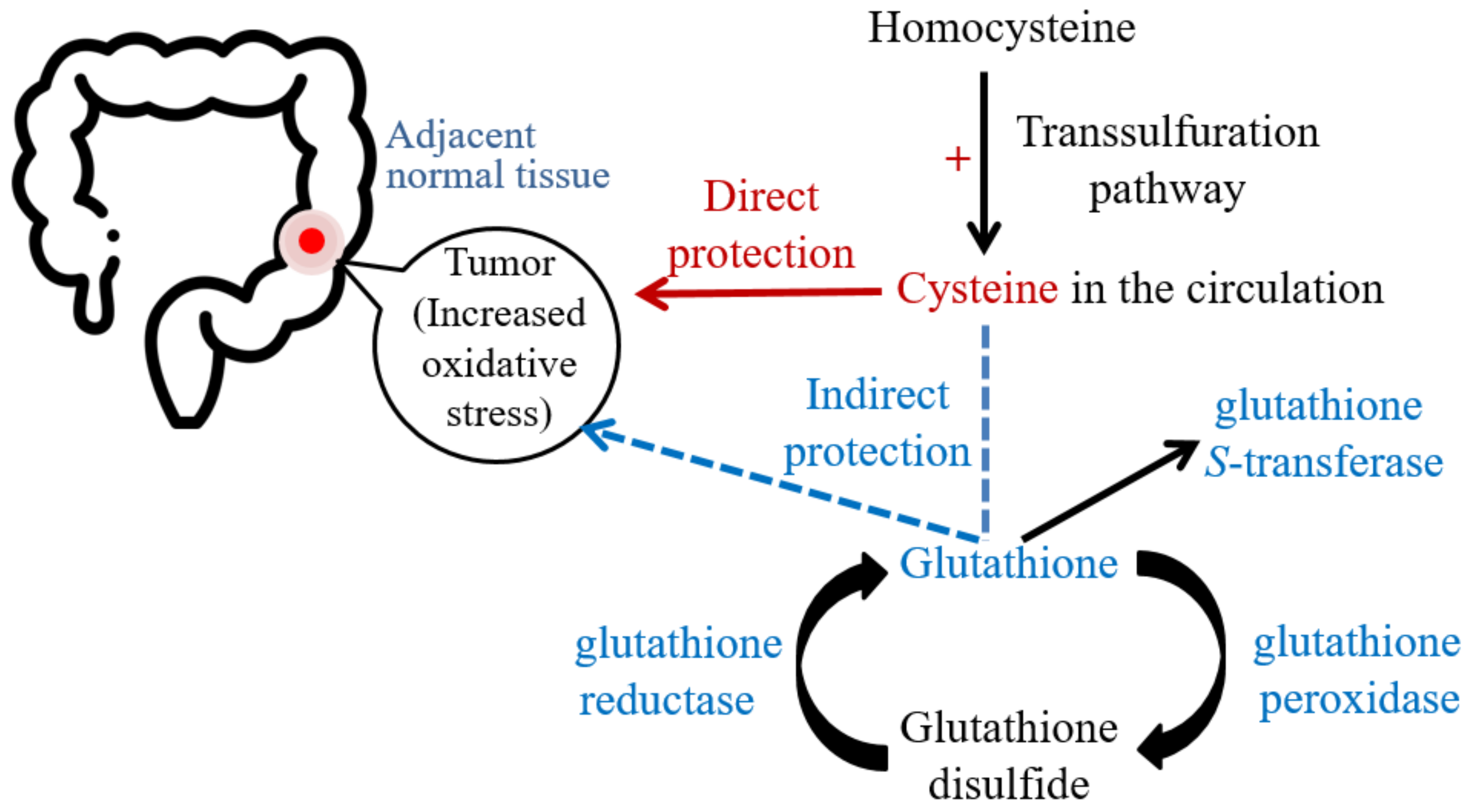
Figure 1. The possible effects of cysteine on oxidative stress and glutathione-related antioxidant capacities during the existence of colorectal cancer.
Ijms 23 09581 g001
Table 1. Demographic and clinical characteristics of patients with colorectal cancer pre- and post-resection.
 ParametersPre-ResectionPost-Resection
 Age (y)61.8 ± 11.1
(64.5)
 Male/Female34/32
 BMI (kg/m2)24.6 ± 4.6
(24)
23.3 ± 4.5
(23.2)
 Blood pressure (mmHg)
  systolic133.8 ± 14.3 (135)135.2 ± 17.8 (135)
  diastolic77.1 ± 10.9 (75)79.4 ± 11.4 (79.5)
 White blood cell (103/μL)7.6 ± 3.7
(6.8)
7.1 ± 2.4
(6.5)
 Neutrophil-to-lymphocyte ratio4.5 ± 4.4
(3.3)
3.5 ± 3.4
(2.5)
 Serum C-reactive protein (mg/dL)1.0 ± 1.8
(0.3)
1.2 ± 2.7
(0.3)
 Serum albumin (g/dL)4.1 ± 0.5
(4.1)
4.2 ± 0.4 **
(4.3)
 Serum ALT (U/L)21.8 ± 22.0
(16)
30.9 ± 36.8 *
(20)
 Serum creatinine (mg/dL)0.9 ± 0.3
(0.8)
0.9 ± 0.3
(0.9)
 Serum carcinoembryonic antigen (ng/mL)56.8 ± 264.1
(4.7)
52.0 ± 353.3
(2.3)
 Serum carbohydrate antigen 19-9 (U/mL)66.7 ± 294.5
(12.4)
36.3 ± 110.9 **
(10.3)
 Family history of colorectal cancer (n, %)11, 16.7%
 Stage at diagnosis (n, %)
  Stage 02, 3%
  Stage I8, 12.1%
  Stage II15, 22.7%
  Stage III27, 40.9%
  Stage IV14, 21.2%
 Cancer location
  Colon32 (48.5%)
  Rectum34 (51.5%)
 Smoking habit (n, %)
  Current6, 9.1%
  Past8, 12.1%
  No52, 78.8%
 Drinking habit (n, %)
  Current5, 7.6%
  Past5, 7.6%
  No56, 84.8%
n = 66. Values are means ± standard deviation with median in the parentheses. BMI, body mass index; SBP, systolic blood pressure; DBP, diastolic blood pressure; ALT, alanine aminotransferase. * Values are significantly different from pre-resection; * p < 0.05; ** p < 0.01; p < 0.001.
Table 2. Plasma cysteine, homocysteine, indicators of oxidative stress, and antioxidant capacities in patients with colorectal cancer.
Pre-ResectionPost-Resectionp Value
 Cysteine (μmol/L)167.42 ± 35.07186.09 ± 40.50<0.001
 Homocysteine (μmol/L)9.94 ± 3.6611.48 ± 3.88<0.001
 Oxidative stress indicators
  MDA (μmol/L)0.97 ± 0.271.03 ± 0.300.044
  AOPP (μmol/L)385.06 ± 157.71479.40 ± 244.000.012
 Antioxidant capacities
  TEAC (μmol/L)3909.91 ± 326.454486.69 ± 395.27<0.001
  GSH (μmol/L)79.28 ± 45.11114.69 ± 47.99<0.001
  GSSG (μmol/L)482.33 ± 64.16578.331 ± 76.43<0.001
  GSH/GSSG ratio0.16 ± 0.070.20 ± 0.07<0.001
  GPx (nmol/mL/min)166.74 ± 55.28232.44 ± 66.93<0.001
  GR (nmol/mL/min)55.96 ± 18.4367.34 ± 29.07<0.001
  GST (nmol/mL/min)26.01 ± 19.6537.79 ± 21.62<0.001
  SOD (U/mL/min)3.47 ± 1.763.61 ± 1.410.220
n = 66. Values are mean ± standard deviation. GSH, glutathione; GSSG, glutathione disulfide; MDA, malondialdehyde; AOPP, advanced oxidation protein products; TEAC, trolox equivalent antioxidant capacity; GPx, glutathione peroxidase; GR, glutathione reductase; GST, glutathione S-transferase; SOD, superoxide dismutase.
Table 3. Cysteine, homocysteine, indicators of oxidative stress, and antioxidant capacities in colorectal tumor tissues and adjacent normal tissues.
Tumor TissueAdjacent Normal Tissuep Value
 Homocysteine (μmol/g protein)1.00 ± 0.440.65 ± 0.28<0.001
 Cysteine (μmol/g protein)35.03 ± 20.2618.95 ± 10.01<0.001
 Oxidative stress indicators
  MDA (μmol/g protein)0.20 ± 0.200.28 ± 0.510.017
  AOPP (μmol/protein)130.03 ± 42.3486.42 ± 29.57<0.001
 Antioxidant capacities
  TEAC (μmol/g protein)445.00 ± 177.28394.59 ± 156.990.020
  GSH (μmol/g protein)10.90 ± 12.507.55 ± 4.090.001
  GSSG (μmol/g protein)187.96 ± 83.87141.00 ± 42.97<0.001
  GSH/GSSG ratio5.72 ± 4.085.33 ± 2.200.448
  GPx (nmol/min/g protein)136.01 ± 53.2488.21 ± 35.71<0.001
  GR (nmol/min/g protein)157.71 ± 76.98134.20 ± 59.250.012
  GST (nmol/min/g protein)32.75 ± 11.4428.49 ± 10.030.019
  SOD (U/min/g protein)17.10 ± 6.4614.62 ± 4.150.003
Values are means ± standard deviation. GSH, glutathione; GSSG, glutathione disfluide; MDA, malondialdehyde; AOPP, advanced oxidation protein products; TEAC, trolox equivalent antioxidant capacity; GSH-Px, glutathione peroxidase; GSH-Rd, glutathione reductase; GSH-ST, glutathione S-transferase; SOD, superoxide dismutase.
Table 4. Partial Spearman’s correlations (rs) of cysteine level with indicators of oxidative stress and antioxidant capacities in patients with colorectal cancer at pre-resection, tumor tissues, and adjacent normal tissues after adjusting for potential confounders.
Plasma Cysteine at Pre-ResectionPlasma Cysteine at Post-ResectionCysteine in Tumor TissueCysteine in Adjacent Normal Tissue
Plasma Level at Pre-ResectionPlasma Level at Post-ResectionTumor TissueAdjacent Normal TissuePlasma Level at Post-ResectionTumor TissueAdjacent Normal TissueTumor TissueAdjacent
Normal Tissue
 Homocysteine0.61 0.36 **0.180.070.39 **0.74 0.35 **0.34 **0.63
 Cysteine0.45 0.210.190.59 0.59
 MDA−0.23−0.150.36 **0.37 **0.170.200.32 *0.34 **0.14
 AOPP0.24−0.090.15−0.010.030.050.32 *−0.180.44
 TEAC0.35 **0.080.42 **0.210.030.62 0.42 **0.48 0.57
 GSH0.000.000.29 *0.090.31 *−0.050.110.050.37 **
 GSSG0.04−0.29 *0.50 0.230.34 **0.56 0.31 *0.42 **0.71
 GSH/GSSG ratio−0.01−0.06−0.02−0.140.25−0.40 −0.23−0.19−0.20
 GPx−0.070.090.240.150.250.48 0.36 **0.41 **0.49
 GR0.11−0.08−0.000.010.060.63 0.51 0.46 0.80
 GST0.22−0.020.140.010.020.46 0.33 *0.230.52
 SOD0.16−0.030.260.07−0.110.27 *0.33 *0.190.56
n = 66. Values are partial Spearman’s correlation coefficients (rs). Adjusting for age, sex, C-reactive protein, smoking and drinking habits and cancer stage. MDA, malondialdehyde; AOPP, advanced oxidation protein products; TEAC, trolox equivalent antioxidant capacity; GSH, glutathione; GSSG, glutathione disulfide; GR, glutathione reductase; GPx, glutathione peroxidase. * p < 0.05; ** p < 0.01; p < 0.001.
Publisher’s Note: MDPI stays neutral with regard to jurisdictional claims in published maps and institutional affiliations.