Review

Multiple Cell Cultures for MRI Analysis

by
Zuzanna Bober
1,
David Aebisher
1,
Marcin Olek
2,
Aleksandra Kawczyk-Krupka
3,* and
Dorota Bartusik-Aebisher
4,*
1
Department of Photomedicine and Physical Chemistry, Medical College of Rzeszów University, University of Rzeszów, 35-310 Rzeszów, Poland
2
Department of Orthodontics, Faculty of Medical Sciences in Zabrze, Medical University of Silesia, 40-055 Katowice, Poland
3
Center for Laser Diagnostics and Therapy, Department of Internal Medicine, Angiology and Physical Medicine, Medical University of Silesia in Katowice, 41-902 Bytom, Poland
4
Department of Biochemistry and General Chemistry, Medical College of Rzeszów University, University of Rzeszów, 35-310 Rzeszów, Poland
*
Authors to whom correspondence should be addressed.
Int. J. Mol. Sci. 2022, 23(17), 10109; https://doi.org/10.3390/ijms231710109
Submission received: 5 August 2022 / Revised: 27 August 2022 / Accepted: 29 August 2022 / Published: 3 September 2022
(This article belongs to the Special Issue 23rd Anniversary of IJMS: Advances in Biochemistry)

Abstract

:
Magnetic resonance imaging (MRI) is an imaging method that enables diagnostics. In recent years, this technique has been widely used for research using cell cultures used in pharmaceutical science to understand the distribution of various drugs in a variety of biological samples, from cellular models to tissues. MRI’s dynamic development in recent years, in addition to diagnostics, has allowed the method to be implemented to assess response to applied therapies. Conventional MRI imaging provides anatomical and pathological information. Due to advanced technology, MRI provides physiological information. The use of cell cultures is very important in the process of testing new synthesized drugs, cancer research, and stem cell research, among others. Two-dimensional (2D) cell cultures conducted under laboratory conditions, although they provide a lot of information, do not reflect the basic characteristics of the tumor. To replicate the tumor microenvironment in science, a three-dimensional (3D) culture of tumor cells was developed. This makes it possible to reproduce in vivo conditions where, in addition, there is a complex and dynamic process of cell-to-cell communication and cell–matrix interaction. In this work, we reviewed current research in 2D and 3D cultures and their use in MRI studies. Articles for each section were collected from PubMed, ScienceDirect, Web of Science, and Google Scholar.

1. Introduction

Molecular imaging is used to improve diagnosis, prognosis, and monitoring of therapy in patients. Among molecular imaging methods, we distinguish optical imaging, magnetic resonance imaging (MRI), computed tomography (CT), positron emission tomography (PET), and single-photon emission computed tomography (SPECT). These methods allow for the visualization of anatomical, genetic, biochemical, and physiological changes in vivo. The development of MRI in recent years, in addition to diagnostics, has made possible implementing this method to assess the response to applied therapies. Conventional MRI provides anatomical information and, due to advances in technology, provides physiological information. Recent MRI research has focused on single cell imaging in vivo [1]. Molecular imaging technology in brain research can aid in diagnosis and treatment but can also be used to study brain function. At the molecular level, optical imaging has also been used with the support of reporter gene technology [2]. Machine learning (ML) has been implemented for cancer classification, diagnosis, genomic biomarker identification, progression detection, and survival prediction. The use of ML-based radiomic analysis allows quantification of tumor heterogeneity. It may be used in the future as a non-invasive marker for diagnosis and the monitoring of response to applied treatment as well as predicting patient prognosis. An attempt has been made to combine MRI and ML as a new tool for assessing treatment response and the prognosis of patients with high-grade gliomas (HGG) [3]. MRI is also being used to visualize stem cells (SCs) in vivo [4]. An extremely valuable advantage of this method is its non-invasive nature and the possibility for multiple studies. The implementation of contrast agents and tracers into the study allows for the detection of cells in MRI. Recent studies may also find reports from the evaluation of cellular immunotherapy using preclinical cell tracking technologies as well as the possibility of clinical translation [5]. The development of nanotherapeutics eliminates the limitations of traditional drugs and may contribute to more effective cancer therapies. Imaging modalities such as MRI, CT, photoacoustic imaging (PAI), PET, and electron microscopy [6] play key roles in monitoring drug delivery as well as controlling the therapeutic effect in real time. MR imaging can be used to decipher the delivery of albumin-targeted drugs. Many drugs inextricably bind to albumin; imaging techniques enable quantification at the molecular level and allow the assessment of mechanisms, guiding drug development and personalizing treatment [7]. A recent study evaluated the effect of a preoperative drug (POA) in pregnant women undergoing elective cesarean section. Drug levels and neonatal outcomes were evaluated, with levels of TNF-α, IL-6, IL-8, and pro-inflammatory cytokines in cord blood. Based on the analysis of the results of 66 volunteers, there was a significant correlation among POA, fetal birth weight, and the parameters of fetal cord blood TNF-α, IL-6, and IL-8. The analysis showed an increase in the levels of cytokines TNF-α, IL-6, and IL-8 in cord blood in women with high levels of POA, with negative consequences for the newborn, i.e., increased levels of inflammatory markers in fetal cord blood and significantly lower birth weight [8]. Another study presented research on the detection of severe acute respiratory syndrome coronavirus 2 (SARS-CoV-2). Researchers have developed a rapid response and quantitative capacitive aptasensor for detecting nucleocapsid protein ultrastructures based on a microelectrode array (MEA) system. Researchers have developed an MEA-based aptasensor using a specific aptamer to recognize N-protein from SARS-CoV-2 utilizing the capacity of the solid–liquid interface with picofarad-level sensitivity for detection at ultra-low femtogram per milliliter levels in various matrices. Recognition occurs at N-protein concentrations of about 10-1 ng/mL or much lower. The potential of using this method is very high for screening asymptomatic patients, which is a very good alternative to the existing method, i.e., PRC-based RNA, which is time consuming, complicated to operate, and expensive [9]. The applications of ultrasound have been widely studied in biomedical engineering. The design of a new ultrasonic transducer for future biomedical applications has great potential. The use of ultrasound transducers can be used, among other things, to disrupt the blood–brain barrier to improve drug delivery. Improving the performance of the ultrasound transducer could enable a wider use in ultrasound diagnostics, ultrasound therapy, particle/cell manipulation, drug delivery, and nerve stimulation. The invented Sm-PMN-PT monocrystal features giant piezoelectricity and a transparent PMN-PT monocrystal with ultra-high piezoelectricity. The optoacoustic transducer has the ability to precisely control the operation of the optoacoustic transducer [10]. A new, non-invasive neuromodulation integrating focused transcranial ultrasound stimulation (tFUS) offers a wide range of applications for both understanding and treating the brain. In their work, Zhang et al. described the detectability of tFUS in the application of analgesia. Based on their rat study, they deduced that tFUS stimulation with PAG can effectively suppress formalin-generated nociceptive activity. This confirms that tFUS PAG stimulation can achieve the effect of analgesia, which, in further studies, can help develop non-invasive analgesic technology [11]. Several virtual screening models for small molecules targeting primary miRNAs have been presented in research studies. Mature miRNAs and their specific target mRNAs during miRNA–mRNA interactions can form unique Argonaute-mediated functional loops (AGOs). These loops can serve as potential targets for small-molecule drug discovery. In their work, Zhuo et al. first used a loop-based and incorporated (AGO) virtual screening model to target loops. The potential of the created loop by targeting the AGO-mediated miR-214–mRNA interaction can be used to rescue the bone phenotype in genetically engineered mice [12]. The first targeted glutathione (GSH)-responsive thermostatic system ([email protected]) for tuberculosis with a rifampicin (IF)-loaded near-infrared emission carrier was also presented. The hydrophobic–hydrophobic interaction induced by the photocleavage reaction was exploited, enabling the early diagnosis of TB by tracking granulomas. This allowed the selective imaging and precise inhibition of localized TB by released RIF. A gSH-activated hyaluronic acid (HA) system loaded with rifampicin was shown to achieve positive effects in TB therapy [13].
Cell culture research has a key role in scientific research including the development of imaging methods. Cell cultures enable the development of such fields of science as biotechnology, biopharmacy, or toxicology. Cell cultures are increasingly being used in pharmaceutical research for drug absorption studies and for assessing toxicity and predicting properties. The isolation and cultivation of various types of cells enables the development of various types of scientific research due to the real resemblance to a living organism. Currently, two methods of cell culture are used: two-dimensional (2D) and three-dimensional (3D) (Figure 1). In classic 2D cell culture, cells multiply in monolayers on a previously prepared medium. The 2D culture method has many limitations and does not allow for full control of cell growth and differentiation. The medium used plays an extremely important role in cell culture because it provides the cells with the necessary nutrients, growth factors, and hormones and the antibiotic content protects the cells and regulates the pH. Growth rate is usually logarithmic. To achieve this growth phase, pass the cells regularly. After the culture reaches about 80% confluence, transfer them to sterile bottles with fresh medium. In the case of divided adherent cells, after the removal of the culture medium, most often the cell layer is “trypsinized”. After washing the monolayer with, 0.9% NaCl, 0.25% EDTA trypsin solution is added in an appropriate amount. The cells are then incubated under the same conditions as used for culture after about 3.5 min under microscopic control. The cell suspension is then added to the culture medium in an appropriate volume, which, after mixing, can be transferred to new and sterile culture vessels.
Cell cultures conducted in laboratories require special conditions: appropriate humidity, temperature, and gas concentration in the incubator must be maintained. In laboratory conditions, it is possible to control the conditions and create an optimal microenvironment for breeding. In addition, we have the option of applying active substances to breeding, such as, for example, growth factor, hormones, or proteins. In addition, cell culture should always be carried out under sterile conditions, maintaining full sterility of the room and all tools and substances used for the cultivation. The culture vessels must also be sterile and disposable. All materials used for the cell culture should be non-toxic to cells and allow for their growth and gas exchange with the environment. A 2D cell culture media can be made, for example, of polystyrene (PS) [14] or glass [15]. Growth media are used for breeding; the complete medium is most often enriched with heat-inactivated fetal bovine serum (FBS) and an antibiotic. The cultivation is normally carried out in an incubator with the following parameters: temperature 37 °C, 95% air, 5% CO2, and 100% relative humidity. The appropriate growth medium is selected depending on the cultured cell line. The 2D cell cultures ensure a low cell concentration and, moreover, do not correspond to the tumor morphology in vivo. The implementation of a 3D culture enables the reconstruction of the tumor microenvironment and the development of a procedure for conducting a 3D culture with high repeatability and preserving the tumor features and allows for the implementation of preclinical tests. The historical development of 3D breeding methods is presented in Table 1. Many physicochemical and biochemical factors influence the cells in the natural environment, which, in turn, influences the proliferation, reproduction, and development. The implementation of 3D breeding enables the re-creation of natural conditions. The 3D cultures involve many methods; for example, the use of 3D cell cultures in a bioreactor allows for an extremely high density of cells that grow in a controlled environment. These types of cultures enable high repeatability and the possibility of carrying out research, e.g., on medicinal substances. Compared to the monolayer culture, the 3D cell culture better reflects the tumor structure. In the 3D model, we have an interaction between adjacent cells and the extracellular matrix (ECM). While these interactions affect the biochemical and mechanical signals of cell physiology, there is no such interaction for cells grown in 2D. In contrast, in the case of a 3D culture, we can create an environment by mimicking in vivo conditions. With the use of 3D methods, we obtain more information about the interactions between cells: the model that creates a tumor reflects its metabolism and characteristics.
On the other hand, in 3D cultures, various methods are used, including those based on hydrogels, polymers, and glass fibers, forming the so-called “scaffolds”. The years of publications on 3D matrices based on a hydrogel for cultivation are shown in Figure 2.
However, in the case of scaffold-free techniques, we can find microplates with hanging droplets, magnetic levitation, and spheroidal microplates with a coating with a very low level of adhesion. Nodular models are widely used as avascular tumor models for metastasis and for screening. Breeding techniques using different types of scaffolding have many advantages. They can be used to build spheroids [34], which we prepare depending on the needs and the experiment being conducted. Spheroids are a type of three-dimensional cell cultures of many cell lines that have the ability to self-organize [35]. An exemplary model is the multicellular spheroid (MCS) model, which reflects the similarity to real tissues in many respects.

1.1. Nodular Cell Culture

One example of a 3D cell culture is the nodular culture. This type of culture shows limited adhesion to the substrate, which allows the clusters of cells to form into three-dimensional clusters of many cells, which allows the three-dimensional contact of the cells. Moreover, it should be noted that these types of cultures have a limited diffusion of nutrients into the inner part of the spheroid; therefore, the spheroids in their volume are varied. Inside, there are cells with necrotic changes, followed by cells in the resting phase, and, in the last few layers, cells that are proliferating. Due to their structure, spheroids are used as models of non-vascularized neoplastic tumors used, for example, to assess the effectiveness of anticancer drugs. For example, Foxall presented a 3D nodular culture model of diffuse large B-cell lymphoma (DLBCL), with the replication of tumor microenvironment (TME) components consisting of malignant and benign cells, including CAF and tumor-associated macrophages (TAMs) that drive tumor initiation and tumor growth. Based on the research and analysis, it was found that the viability of DLBCL cells grown in the 3D system, compared to 2D, is characterized by a higher viability [36]. Haro and Orsulic, in their work, demonstrated that the DLBCL stromal gene signature shows a greater chance of survival in DLBCL and several other B-cell lymphomas, in contrast to ovarian cancer [37]. Diffuse DLBCL rebuilds the fibroblastic mesh network, reprogramming the HLFs into CAFs, which acquire the ability to modulate TME. CAF activation increases PD-L1 expression, which results in abnormal immunosuppressive abilities [38]. In addition, the humoral factor pyruvate, which is secreted, inter alia, by the patient’s cancer-associated fibroblasts, promoted cell survival for primary lymphoma cells [39]. Kuen et al. presented a 3D model of a pancreatic cancer (PC) cell culture with CAF and monocytes to evaluate cellular mechanisms in the tumor immunosuppressive microenvironment. Immunosuppressive cytokines were produced that induce M2 polarization and also demonstrated the ability to inhibit CD4+ and CD8+ T cell activation and proliferation in in vitro studies [40]. In their research, the group of Dolznig et al. grew human colon tumor cells as multicellular spheroids and then cultured them together with normal fibroblasts or CAF in collagen I gels [41]. In addition, CAF mediated inflammation in human breast and ovarian tumors through the pro-inflammatory factors IL-6, COX-2, and CXCL1 [42].

1.2. The 3D Culture Matrices Based on a Hydrogel

The 3D cell cultures using hydrogels are increasingly being carried out. These are highly hydrated hydrophilic polymers. Hydrogels form a network of fibers with a specific pore size to reflect the extracellular matrix. Due to the fact that they are transparent, hydrogenation makes it possible to observe the cell culture. Moreover, their great advantage is the fact that they have a good diffusion of nutrients. Hydrogels used in 3D breeding can be natural or artificial [43]. The natural ones are most often based on natural polymers, for example, fibrinogen [44], hyaluronic acid [45], collagen [46], Matrigel [47], gelatin [48], chitosan [49], and alginate [50]. In addition, hydrogels based on the extracellular matrix (ECM), due to soluble factors (growth and cytokines) implanted into the stroke cavity, can attract endogenous cells; the migration of cells to the ECM hydrogel enables tissue regeneration [51]. Hydrogels are more and more often superior to 2D cultures due to the fact that they turn out to be more physiological. In stem cell research, 3D hydrogel-based cultures of cells grow in a manner similar to the in vivo situation. Depending on the composition, the hydrogels may be more or less hydrated, and, thus, may have a different flexibility, which also affects the mechanical properties of the scaffold. Another advantage is that by changing polymer concentrations or cross-linking methods, we can change the physical properties of the 3D model. In addition, in vitro studies on 3D models improve mapping to the tumor in vivo, due to the fact that cells do not have a homogeneous growth environment and are exposed to a lack of sufficient oxygen and nutrients inside the tumor, which may affect tumor progression [52]. Moreover, it may be of importance in the case of the supply of pharmaceutical substances to cell cultures. In a 2D culture, pharmaceuticals such as anticancer agents reach cells without barriers. However, the situation in the in vivo environment is different. Therefore, research on 3D cell cultures turns out to be important. Tumor cells grown on 3D matrices show significant features of native tumor tissues. It turns out that the tumor structure significantly changes the diffusion profile of the drug. In the literature, we can read many reports on the description of the physicochemical properties of native micro-neoplastic environments. Various types of interactions play an important role: cell–cell interactions and cell matrix [53]. Various types of 3D models have the features of solid tumor tissues with a corresponding morphology. Xu et al., in their work, presented the mutual interactions between the tumor and its stroma. A two-layer hydrogel system based on hyaluronic acid (HA) for tumor culture from LNCaP (PCa) prostate cancer cells was developed and characterized. There was a significant increase in the expression of two pro-angiogenic factors, vascular endothelial growth factor-165 (VEGF (165)) and interleukin-8 (IL-8), both at the mRNA and protein levels [54]. In addition, in the literature, we can find many reports on the creation of matrices for the 3D breeding of natural and synthetic origins. Among the natural matrices, we can distinguish matrices derived from fibroblasts, Matrigel™ or Cultrex® [55], type I collagen, silk fibroin, alginate, or hyaluronic acid. On the other hand, among the synthetic matrices, we most often find hydrogels based on poly (ethylene glycol) (PEG) [56], poly (lactide-co-glycolide) (PLGA) [57], scaffolds made of electrospun poly (ε-caprolactone) (PCL) [58], or synthetic peptides [59].

2. Natural Matrices

2.1. Culture Based on Matrigel™ or Cultrex®

On the other hand, Matrigel™ and Cultrex® are extracts from basement membranes, consisting of type IV collagen, laminin, perlecan, etc., used, inter alia, for human colon adenocarcinoma SW480 [60], human breast adenocarcinoma MDA-MB-231 [61], and pancreatic adenocarcinoma Capan-1 [62]. Wessels et al., on the basis of a 3D breast cancer cell culture, demonstrated the complex signaling between fibroblasts and breast cancer cells, which coordinates aggregation and coalescence, which is a specific response of 3D cells [63]. The study presented a 3D model of an MCF-7 breast cancer cell culture based on Matrigel and an assessment of the survival of MCF-7 transfectants expressing wild-type or catalytically inactive ST3 (ST3wt or ST3cat-) [64]. Kasper et al. found that the overexpression of stromelysin-3 exacerbated the tumor in MCF-7 and MDA-MB-231 cell cultures in an orthotopic model of human tumor xenograft in nude mice and in a Matrigel-based 3D culture system [65]. On the other hand, Song et al., in their research, used 3D collagen gel and Matrigel to evaluate the expression and reorganization of structural proteins of rabbit aortic smooth muscle cells [66]. Matrigel hydrogel in combination with a synthetic polymer of polylactic and co-glycolic acid (PLGA) and polylactic acid (PLLA) was also used for 3D lung tissue engineering [67]. Li et al. described the positive effect of the 3D Matrigel environment on pig tooth enamel cells [68]. In other studies, researchers demonstrated 3D culturing of stem cells under pulsed conditions [69].

2.2. Collagen-Based Matrices

Collagen biomaterials are widely used as 3D scaffolds for cell culture [70]. Due to the fact that it is the main component of the extracellular matrix (ECM), type I collagen is used for the cultivation of human breast cancer MCF-7 [71], human hepatocellular carcinoma HepG2 [72], and human epithelium of the small intestine [73]. A recent study by Quarta et al. presented the composition of agarose–collagen hydrogels and spheroid cells of three different breast cancer cell lines, MCF-7, MDA-MB-361, and MDA-MB-231 [74]. Rossi et al. assessed the effect of HSF fibroblasts on the growth of MCF-7 cells and their sensitivity to radiation in collagen gels. Growth inhibition and increased sensitivity to radiation were found [75]. Moreover, the models of the cultures on 3D collagen scaffolds caused a differentiated morphology of cells, which is related to the porosity of the scaffold, and also a prolonged cell proliferation. Additionally, the use of 3D collagen scaffolds can generate a population of cells with cancer stem cell (CSC) properties [76].

2.3. Matrices Derived from Fibroblasts

In the case of matrices derived from fibroblasts, they are used as a microenvironment in advanced cancers of, for example, the colon [77] or pancreas [78]. Models based on fibroblasts show a different cell morphology compared to 2D cultures. However, despite many advantages, it should be noted that this type of breeding is associated with a longer preparation and differences in the composition of individual batches. Moreover, matrices based on fibroblasts may not fully reproduce the structure and composition of the tumor microenvironment [79]. In addition, fibroblasts have an influence on the tumor initiation capacity. In contrast, cancer-associated fibroblasts (CAFs) play a role in the adhesion and motility of tumor cells. Moreover, they are considered to be the most abundant type of stromal cells in various cancers and constitute a heterogeneous population of cells. They stimulate tumor growth and progression in many types of cancer. In addition, CAFs secrete H2O2, stimulating cancer through the stroma and transformation of primary epithelial cells and intensification of the aggressiveness of cancer cells. Liao et al., in their work, presented the effect of cancer-associated fibroblasts on tumor growth and metastasis in a 4T1 model of murine breast cancer. Studies have shown that modulating the immune microenvironment influences both tumor growth and metastasis [80]. Additionally, Yavuz et al. emphasized in their work that CAFs play a key role in carving the tumor microenvironment in breast cancer. Fibroblasts help induce immunosuppressive PD-1 + TAM [81]. CAFs are also responsible for the induction of FGF4 expression in ovarian cancer stem cells [82]. Fibroblasts are considered to be the most abundant cells in the connective tissue proper. They cause the production of collagen, various growth factors, cytokines, and chemokines and the degradation of the extracellular matrix. It should be emphasized that cancer-related fibroblasts cause the migration and invasion of cancer cells [83] and contribute to driving tumor growth [84]. Human fibroblasts are also involved in carcinogenesis, proliferation, and metastasis of A-549 non-small cell lung cancer. Studies have shown that CAF, compared to normal fibroblasts (NF), increases the invasion of A549 cells, as demonstrated by Horie et al. based on a 3D model of in vitro culture [85]. Additionally, Nakamura et al. conducted a 3D model to evaluate the effect of podoplanin-positive fibroblasts on tumor cell proliferation. Studies have shown that proliferation increases with lung cancer [86]. In contrast, immunofibroblasts support the earliest stages of the formation of tertiary lymphoid structures (TLS) [87]. Recent studies by Cribaro et al. analyzed the complexity of the microenvironment of glioblastoma (GBM). The group presented a 3D visualization showing the exact composition of human GBM in the form of a three-dimensional landscape of the GBM vascular microenvironment [88]. On the other hand, the group of Chhetri et al., in their work, presented an important step in cancer research based on the evaluation of a 3D cell culture. The influence of the microenvironment of a 3D culture on the cell nucleus and its influence on the stimulation mechanisms of the nucleus were presented [89]. An important aspect in cell culture is also the influence of electrical biostimulation and silver ions on fibroblast cells. The group of Zhao et al., in their latest publication, assessed porcine fibroblast cells and changes in the dynamics of the cell transcriptome after electrical biostimulation in the presence of silver ions. After analysis, no increase in the expression of pluripotency genes was found [90].

2.4. Matrices Based on Calcium Alginate

An equally frequently used biodegradable hydrogel is alginate [91], which has valuable properties because it has a controlled pore size and does not adhere to cells. Alginate-based hydrogels are used as 3D scaffolds [92,93]. In addition, a covalently cross-linked hydrogel provides chemical stability [94]. Alginate is useful in the culture of human hepatocellular carcinoma MHCC97L, HCCLM3 [95], and oral squamous cell carcinoma OSCC-3 [96]. Cavo et al. developed a new alginate–Matrigel hydrogel for use in the 3D culture of aggressive human breast cancer cells (MDA-MB-231), which perfectly mirrors the tumor in vivo [97]. Chen et al. presented porous 3D scaffolds made of Ca alginate in the form of a human osteoblast (hOB) platform and calcium alginate scaffolds. In addition, bioreactors were constructed for the transplantation of human osteoblast clusters [98]. Using the MCF-7 breast cancer cell line and a 3D cell culture model based on alginate hydrogels, the assessment of cell activity depending on the flexibility of the medium was made. Studies have shown that the number of MCF-7 cells decreased as the flexibility of the hydrogel-based medium increased. It was noted that in the most delicate hydrogels, cell proliferation was highest after 2 weeks [99]. Karimpoor et al. used alginate foam for a 3D culture; it allowed them to obtain a porous scaffold that perfectly reflects the structure of the bone marrow (BM). Moreover, it increased bone marrow differentiation in both leukemia and hematopoietic cells [100]. In other studies, Shakibaei et al., using a 3D model of an alginate-based scaffold, investigated the effects of curcumin on HCT116R cells and chemotherapy based on the drug 5-fluorouracil (5-FU), which confirmed the use of the model, which was also used in the study of drug effects [101]. Additionally, Utech et al., using hydrogels, developed mesenchymal stem cells (MSCs) in the microspheres, which allowed them to obtain a continuous supply of nutrients that caused the growth and proliferation of aspirated cells [102]. Sidhu et al. presented research in which they used a 3D platform based on alginate microcapsules. It was shown to allow the study of hESC proliferation and differentiation into different lines [103]. Xu et al., using a 3D dynamic culture, investigated the effect on the regeneration of cartilage tissue in rabbit joints using encapsulated cells [104].

2.5. Matrices Based on Fibrinogen

Almany and Seliktar, in their work, described a hydrogel scaffold based on fibrinogen and polyethylene glycol. The advantages of this type of scaffolding were presented, including high plasticity while maintaining biological functionality [32]. Additionally, Dikovsky et al. described the use of a synthetic polyethylene glycol (PEG) scaffold and endogenous fibrinogen precursor molecules, demonstrating the preservation of quality between the molecular architecture of the matrix and the cell morphology [105]. On the other hand, Kim et al., due to the fact that the mechanical properties of PEG–fibrinogen-based hydrogels decrease significantly over time in PBS, found a positive effect of ascorbic acid on the improvement of matrix properties [106]. On the other hand, Shachaf et al. developed a biomimetic material based on fibrinogen and PluronicF127 [107]. In contrast, Pradhan et al., in their work, presented PEG–fibrinogen hydrogels for the 3D culture of breast cancer of three cell lines: MCF-7, SK-BR-3, and MDA-MB-231 [108].

2.6. Matrices Based on Hyaluronic Acid

In addition, there are reports in the literature on the use of hyaluronic acid (HA) for the cultivation of three-dimensional tumor models, which is structurally an ideal equivalent of in vivo conditions. Hyaluronic acid is an essential component of ECM in the tumor microenvironment used in human prostate cancer [109] and human glioblastoma U87MG [110,111]. It has the following properties: it is involved in the formation of blood vessels within the tumor and its metastasis and also interacts with cell surface receptors such as CD44 or RHAMM. Recent studies by Harris et al. presented biocompatible 3D scaffolds decellularized from plant material using carbon dioxide. The cultivation of human fibroblast cells on the surface of a spinach leaf demonstrated the biocompatibility of decellularized scCO2 scaffolds [112]. On the other hand, in their research, Feng et al. cultured MCF-7 breast cancer cells in 3D using fibrous polycaprolactone (PCL) scaffolds, showing an increased proportion of CSC [113].

2.7. Gelatin-Based Matrices

Jiang et al. presented, in their work, an alginate/gelatin composite hydrogel with adjustable mechanical and adhesive properties. As a result, it is possible to obtain mechanically soft gels with a larger number of cell adhesion groups or stiffer with a smaller number [114]. Additionally, Jiang et al., in another study, presented the use of an alginate/gelatin hydrogel with breast cancer cells and fibroblasts, forming a 3D model [115]. On the other hand, Wang et al. used bacterial cellulose–gelatin hydrogels for the breast cancer cell line (MDA-MD-231) as a 3D scaffold. Studies showed that cells exhibited significant adhesion, proliferation, ingrowth, and differentiation [116].

2.8. Chitosan-Based Matrices

Dhiman et al. presented a chitosan-based matrix for the cultivation of MCF-7 breast cancer cells. The results showed that glucose consumption and melate production were similar to cell growth in tissue culture flasks, which indicated that it can be used for the 3D cell culture of the MCF-7 line [117]. Additionally, Dhiman et al., in their previous studies, used these matrices to determine the cytotoxicity of tamoxifen. Studies showed the effect of tamoxifen on estrogen-positive cancer cells and the inhibition of cathepsin D uptake from the culture medium [118]. In the latest published studies, the group of Mohseni et al. described chitosan–alginate polymer nanoparticles that were synthesized as a clinical Dotarem® carrier based on (Gd 3+) used for labeling mesenchymal stem cells for MRI imaging in in vitro studies [119]. In their work, Huang et al. assessed the influence of spatial architecture on the behavior of cells in 2D and 3D cultures based on chitosan scaffolds [120].

2.9. Alginate-Based Matrices

Another example of a natural matrix is an alginate-based matrix. Lan and Starly presented a platform for the 3D culture of liver cells with high density and single-layer growth of the breast cancer cell line (MCF-7), which allowed for the assessment of the drug dose concentration and the effect on breast cancer cells [121]. On the other hand, the group of Yu et al. used alginate core-shell beads to grow MCF-7 breast cancer 3D cells [122]. Conversely, Lee et al. investigated human breast cancer cells expressing GFP (GFP-MCF-7) encapsulated in an alginate hydrogel in a 3D model. The culture was assessed using a capacitive sensor, which made it possible to monitor the migration of human mesenchymal stem cells (hMSC) [123].

2.10. Matrix Based on Silk Fibroin

Another natural material is silk fibroin, which has good biocompatibility and unique mechanical properties, which allows it to be used in 3D models [124]. It is a matrix used, inter alia, for the cultivation of human breast adenocarcinoma MDA-MB-231 [125] and breast cancer EMT6 [126]. Chaturvedi et al. used 3D silk fibroin scaffolds to differentiate human skeletal muscle cells’ myoblasts in vitro [127]. Ashari et al. investigated silk fibroin for the properties of improving the condition of mouse islets in vitro against inflammatory stress during islet cell transplantation [128]. On the other hand, Lovett et al. used silk fibroin for microvessels on a microvascular scale (ID < 6 mm). Research has shown that the use of silk fibroin-based microducts can be used as a biomaterial for microvascular transplants [129]. In other studies, Zhao et al. used apatite-coated silk fibroin scaffolds in conjunction with bMSC to repair jaw injuries in dogs [130]. In contrast, other studies by Wang et al. presented hybrid nanofiber scaffolds made of silk fibroin (SF) and poly (lactide-co-ε-caprolactone) (PLCL) with improved properties for bone regeneration [131]. Niu et al. presented silk fibroin labeled with quantum dots and their interactions with cells used for controlled drug delivery [132]. Other studies presented an in vitro 3D tumor model of breast cancer from the MDA-MB-231 cell line on a silk fibroin scaffold to evaluate the efficacy of cyclosaplin; studies showed a significant decrease in MMP-9 activity in the tumor [133]. The group of Bhardwaj et al. used scaffolds made of a mixture of silk fibroin and chitosan for the engineering of cartilage in in vitro studies [134]. An earlier study by Mauney et al. used silk fiber biomaterials together with collagen and polylactic acid scaffolds for 3D scaffolds in adipose tissue engineering. They presented 3D silk fibroin scaffolds with human bone marrow and adipose-derived mesenchymal stem cells [135]. In addition, silk fibroin was used to create 3D scaffolds to repair damage to articular cartilage. It turns out that during cultivation, the initial density of cell seeding is important: the denser the cell seeding is, the faster the rate of formation of functional cartilage tissue can be [136]. Additionally, the team of Li et al. used silk fibroin as well as chitosan and gelatin to culture 3D MC3T3-E1 cells in bone tissue engineering. Four types of scaffolds were prepared: SF, SF/C, SF/gel, and SF/Cs/gel. After analysis, they concluded that the best choice would be the SF/Cs/gel scaffold [137].

3. Synthetic Matrices

3.1. Poly (Lactic-Co-Glycolic Acid) PLGA Matrices

PLGA porous, biodegradable synthetic scaffolds are also used on a large scale, mainly for oral squamous cell carcinoma OSCC-3 [138] and human glioblastoma U251 [139] but also for bone marrow mesenchymal stem cells [140]. By contrast, Sahoo et al., in their research, presented the characteristics of the porous, biodegradable PLGA/PLA microparticles used for the 3D culture of breast cancer [141]. Priwitaningrum et al. presented a 3D nodular matrix reflecting the tumor stroma and performed penetration tests of silica and PLGA nanoparticles [142]. Published papers also include reports on 3D PLGA scaffolds used for tissue engineering [143].

3.2. Synthetic Peptides

Synthetic peptides with a defined amino acid composition for easy incorporation of specific biological ligands are also used for 3D culture, for example, in SK-OV-3 human ovarian cancer [144] or MCF-7 breast cancer [145]. Synthetic polymers, on the other hand, are used for 3D printing of organs. Biodegradable synthetic polymers have very good mechanical properties and are made of non-toxic products [146]. Fong et al. created a 3D model of Ewing’s sarcoma ex vivo based on porous three-dimensional scaffolds made of electrospun poly (ε-caprolactone). Studies have shown that this type of scaffold can be used to evaluate new anticancer drugs for mechanistic bone sarcomas [147].

3.3. Dies Based on Electrospun Poly (ε-Caprolactone) PCL Scaffolding

Biologically neutral synthetic PCL scaffolds [148] are also popular, used, inter alia, for breeding Ewing’s sarcoma TC-71 [134], prostate cancer PC3 [149], and LNCaP [150]. In a recent study by Zhao et al., they presented the culture of human gingival fibroblasts (HGF) on poly (lactide and glycolide) (PLGA) scaffolds and assessed the effect of Smad4 on caspase-3 and Bcl-2 expression [151]. On the other hand, Hamedani et al., in their work, presented an innovative PLGA-based electrospun polymer fiber scaffold for renal cell carcinoma (RCC). The scaffold was loaded with a natural compound showing a strong anticancer and anti-inflammatory effect, which is Honokiol, which showed an inhibitory effect on the proliferation and migration of kidney cancer cells [152]. In turn, Zheng et al., in the latest research, presented the 3D scaffold HA15 bone tissue, printed in 3D β-TCP/PLGA technology with the drug promoting osteogenesis HA15 in in vivo studies in rabbits. This type of scaffold has been shown to be a good substitute material for the treatment of bone defects [153].

3.4. Matrices Based on Poly (Ethylene Glycol) (PEG)

In the case of synthetic matrices, PEGs with controlled biochemical and mechanical properties are most often used. They are used, inter alia, for the cultivation of human ovarian epithelial carcinoma OV-MZ-6 [154] and human pancreatic ductal adenocarcinoma PANC-1 [155]. Balion et al., instead, presented studies on human HROG36 glioma, C6 glioma cells in rats, and human A37 melanoma cells [156]. Lee et al., on the basis of their research, found that semi-penetrating network compositions based on PEG–diacrylate and hyaluronic acid can support the survival, spread, and migration of 3D cells [157]. Additionally, in their latest research, McKee and his group analyzed naive human embryonic stem cells (ESCs) grown on PEG-based 3D scaffolds. Based on the research, the possible mechanisms of self-renewal of naive ESCs were presented for the first time [158]. On the other hand, viscoelastic hydrogels enable the regulation of stress relaxation, which, as it turns out, after studies by Nam et al., has an important role in cell differentiation, spread, and proliferation. Alginate–PEG hydrogels coupled with RGD are characterized by a faster relaxation, which enables the spread and proliferation of fibroblasts and increased osteogenic differentiation of MSCs [159]. Su et al., in their work, presented an overview of applications of scaffolding printed in various 3D technologies made of various raw materials. The presented scaffolds can be used in regenerative medicine using stem cells for bone tissue engineering [160]. On the other hand, the group of Hassan et al. presented 3D cultures of human adipose tissue-derived stem cells encapsulated in a hydrogel composed of a hyperbranched copolymer based on PEG and hyaluronic acid. Based on the research, the secretion of growth factors for wound healing in the PEG–hyaluronic acid (HA) hybrid hydrogel was assessed [161]. Urrios et al. presented the developed PEG-DA-250 print on a transparent, low-molecular-weight poly (ethylene glycol) diacrylate resin for cell culture, which can be used for culture for several days [162]. Kutikov et al., in their work, presented biodegradable scaffolds based on PEG for tissue engineering [163]. On the other hand, Nemeth et al. used nano-sighted PEG-GelMA-HA hydrogels for three-dimensional chondrogenic dental pulp stem cell (DPSC) spheroids, which enabled chondrogenic differentiation in in vitro studies [164]. Cruz-Acuña et al. used PEG–4MAL hydrogels to encapsulate human pluripotent stem cells’ (hPSC) HO into damaged mouse colon in in vivo studies. Research confirmed that PEG–4MAL promotes HO implantation and accelerates colon wound healing [165]. Fernandes-Cunha et al. used a type I collagen hydrogel of cross-linked polyethylene glycol (PEG)-N-hydroxysuccinimide (NHS) and assessed its regenerative capacity in the case of corneal defects in in vitro and in vivo tests [166].

4. The 3D Cell Culture

The use of a 3D cell culture in research allows the mimicking of physiological vascular networks and the engineering of organs and tissues in vitro [31]. As pre-presented in Table 1, this is one of the bioreactors that allow a 3D culture. Special attention should be paid to the features of this type of bioreactor, mainly, the excellent mass transport properties. HTB bioreactors allow the transport of nutrients without restriction by diffusion, which is often a limitation in the creation of large in vitro constructs.

4.1. Hollow Fiber Bioreactors

Hollow fiber bioreactor-type bioreactors are also used for cell cultures, which are used in numerous preclinical applications, including the cultivation of monoclonal antibodies and growth hormones, and also in 3D cell cultures. It is a bioreactor that perfectly reflects the in vivo environment. In these types of bioreactors, nutrients are delivered to the cells in a controlled manner, while the metabolic components are utilized. These types of bioreactors are suitable for culturing for an extended period of time, depending on the selected cell line. The main components that make up this type of bioreactor are the intragranular (IC) and extra-spherical (EC) spaces. Cells are seeded into the EC space of the fiber bioreactor and grow there. The cell culture medium is pumped through the IC space to provide oxygen and nutrients. The use of a bioreactor with capillary fibers allows us to obtain a very large number of cells in a small volume. The cell culture can fill the EC space to a density >108 cells/mL. Compared to the 2D culture in the bioreactor, we have a 10-fold increase in the number of cells. In addition, this type of bioreactor enables the continuous exchange of nutrients and the exchange of metabolic products. In addition, the fiber in this type of bioreactor is porous and it turns out to be a very good substrate for cell attachment. In addition, the culture parameters are strictly controlled and constant; we do not have any changes in temperature, pH, or other biochemical changes and the influence of mechanical stress, which may also be important in a cell culture. A recent study by Gobin et al. presented a culture in a hollow fiber bioreactor with extracellular vesicles (EV) produced by bone marrow-derived mesenchymal stromal cells (hBM-MSC). The production of EV from hBM-MSC with immunoregulatory components was carried out in the same quantity and with the same quality for a longer period [167]. It should also be noted that EVs contain many bioactive molecules that can affect intercellular communication, including stromal cell re-education, changing the microenvironment, and tumor metabolism and metastasis, and may even increase drug resistance. Tai et al., in an article, presented the role of extracellular vesicles in prostate cancer [168].

4.2. Stirred-Tank Bioreactors (STBR)

Another type of bioreactor is the stirred-tank, which is used in chemical and environmental engineering. A work by Rodriguez-Granrose et al. demonstrated the transition from static culture to a stirred-tank bioreactor. The study assessed the production of beads in the Erlenmeyer system and of discogenic cells for the development of cell therapy for the treatment of lumbar disc degeneration [169]. On the other hand, Manstein et al. combined STBR with the power of in silico process modeling for the best results in a human pluripotent stem cell (hPSC) culture [170].

4.3. Rotary Cell Culture System (RCCS) Bioreactors

Rotary cell culture system bioreactors are also used for three-dimensional microgravity cultures. Zheng et al., in their work, assessed the proliferation of MCF-7 cells cultured in rotary cell culture systems (RCCS). The studies showed that this type of culture, by inducing the ERK1/2 pathway, can promote the proliferation of MCF-7 cells [171]. In contrast, Fournier and Harrison used RCCS to study MLO-Y4 osteocytes in collagen–hydroxyapatite scaffolds [172]. Cui et al., in their work, said that RCCS increases NTRK3 expression in an NSC cell culture grown in the RCCS system on a collagen sponge [173].

5. Conclusions

Carrying out cell cultures requires special precautions, as they are very often pathogenic, which is why biological safety is so important. In addition, it is important to maintain aseptic conditions so that the culture is not contaminated with microorganisms. Cell cultures are grown in an optimally selected medium. The emergence of three-dimensional cell cultures in in vitro studies is closer to in vivo structures. It is possible to conduct, e.g., drug interaction studies and applied therapies that were not possible in the case of single-layer cultures. The 3D cell culture enables many experimental studies such as drug research, cell physiology evaluation, therapy evaluation, and tissue engineering.

Funding

This research received no external funding.

Institutional Review Board Statement

Not applicable.

Informed Consent Statement

Not applicable.

Conflicts of Interest

The authors declare no conflict of interest.

References

  1. Hong, S.; Rhee, S.; Jung, K.O. In vivo molecular and single cell imaging. BMB Rep. 2022, 55, 267–274. [Google Scholar] [CrossRef] [PubMed]
  2. Gao, T.; Wang, P.; Gong, T.; Zhou, Y.; Wang, A.; Tang, X.; Song, X.; Fan, Y. Reporter Genes for Brain Imaging Using MRI, SPECT and PET. Int. J. Mol. Sci. 2022, 23, 8443. [Google Scholar] [CrossRef] [PubMed]
  3. Zhou, Q.; Xue, C.; Ke, X.; Zhou, J. Treatment Response and Prognosis Evaluation in High-Grade Glioma: An Imaging Review Based on MRI. J. Magn. Reson. Imaging 2022, 56, 325–340. [Google Scholar] [CrossRef] [PubMed]
  4. Moonshi, S.S.; Wu, Y.; Ta, H.T. Visualizing stem cells in vivo using magnetic resonance imaging. Wiley Interdiscip. Rev. Nanomed. Nanobiotechnol. 2022, 14, 1760. [Google Scholar]
  5. Sato, N.; Choyke, P.L. Whole-Body Imaging to Assess Cell-Based Immunotherapy: Preclinical Studies with an Update on Clinical Translation. Mol. Imaging Biol. 2022, 24, 235–248. [Google Scholar] [CrossRef]
  6. Li, P.; Wang, D.; Hu, J.; Yang, X. The role of imaging in targeted delivery of nanomedicine for cancer therapy. Adv. Drug Deliv. Rev. 2022, 189, 114447. [Google Scholar] [CrossRef]
  7. Hu, H.; Quintana, J.; Weissleder, R.; Parangi, S.; Miller, M. Deciphering albumin-directed drug delivery by imaging. Adv. Drug Deliv. Rev. 2022, 185, 114237. [Google Scholar] [CrossRef]
  8. Miniksar, Ö.H.; Yıldız Miniksar, D.; Honca, M.; Onat, T.; Gocmen, A.Y.; Öz, H. The Effect of Preoperative Anxiety on Fetal Cord Blood Tumor Necrosis Factor-Alpha, Interleukin-6, and Neonatal Outcomes in Pregnant Women. Psychiatr. Danub. 2021, 33, 321–326. [Google Scholar]
  9. Qi, H.; Hu, Z.; Yang, Z.; Zhang, J.; Wu, J.J.; Cheng, C.; Wang, C.; Zheng, L. Capacitive Aptasensor Coupled with Microfluidic Enrichment for Real-Time Detection of Trace SARS-CoV-2 Nucleocapsid Protein. Anal. Chem. 2022, 94, 2812–2819. [Google Scholar] [CrossRef]
  10. Li, J.; Ma, Y.; Zhang, T.; Shung, K.K.; Zhu, B. Recent Advancements in Ultrasound Transducer: From Material Strategies to Biomedical Applications. BME Frontiers 2022, 2022, 9764501. [Google Scholar] [CrossRef]
  11. Zhang, T.; Wang, Z.; Liang, H.; Wu, Z.; Li, J.; Ou-Yang, J.; Yang, X.; Peng, Y.B.; Zhu, B. Transcranial Focused Ultrasound Stimulation of Periaqueductal Gray for Analgesia; IEEE Transactions on Biomedical Engineering: Piscataway, NJ, USA, 2022. [Google Scholar] [CrossRef]
  12. Zhuo, Z.; Wan, Y.; Guan, D.; Ni, S.; Wang, L.; Zhang, Z.; Liu, J.; Liang, C.; Yu, Y.; Lu, A.; et al. A Loop-Based and AGO-Incorporated Virtual Screening Model Targeting AGO-Mediated miRNA-mRNA Interactions for Drug Discovery to Rescue Bone Phenotype in Genetically Modified Mice. Adv. Sci. 2020, 7, 1903451. [Google Scholar]
  13. Zheng, J.; Long, X.; Chen, H.; Ji, Z.; Shu, B.; Yue, R.; Liao, Y.; Ma, S.; Qiao, K.; Liu, Y.; et al. Photoclick Reaction Constructs Glutathione-Responsive Theranostic System for Anti-Tuberculosis. Front. Mol. Biosci. 2022, 9, 845179. [Google Scholar] [CrossRef] [PubMed]
  14. Hegre, O.D.; Marshall, S.; Schulte, B.A.; Hickey, G.E.; Williams, F.; Sorenson, R.L.; Serie, J.R. Nonenzymic in vitro isolation of perinatal islets of Langerhans. In Vitro 1983, 19, 611–620. [Google Scholar] [CrossRef]
  15. Cukierman, E. Cell migration analyses within fibroblast-derived 3-D matrices. Methods Mol. Biol. 2005, 294, 79–93. [Google Scholar] [PubMed]
  16. Xu, S.; Gao, J. Invasiveness and metastasis of tumor spheroid aggregates of human giant cell carcinoma (lung clone strain PLA801-95D) in vitro and in vivo. Zhongguo Yi Xue Ke Xue Yuan Xue Bao 1991, 13, 353–358. [Google Scholar] [PubMed]
  17. Kleinman, H.K.; McGarvey, M.L.; Hassell, J.R.; Star, V.L.; Cannon, F.B.; Laurie, G.W.; Martin, G.R. Basement membrane complexes with biological activity. Biochemistry 1986, 25, 312–318. [Google Scholar] [CrossRef]
  18. Bilozur, M.; Hay, E.D. Neural crest cell migration in 3D extracellular matrix utilizes laminin, fibronectin or collagen. Dev. Biol. 1988, 125, 19–33. [Google Scholar] [CrossRef]
  19. Woessner, J.F., Jr.; Dannenberg, A.M., Jr.; Pula, P.J.; Selzer, M.G.; Ruppert, C.L.; Higuchi, K.; Kajiki, A.; Nakamura, M.; Dahms, N.M.; Kerr, J.S.; et al. Extracellular collagenase, proteoglycanase and products of their activity, released in organ culture by intact dermal inflammatory lesions produced by sulfur mustard. J. Investig. Dermatol. 1990, 95, 717–726. [Google Scholar] [CrossRef]
  20. Decker, M.L.; Behnke-Barclay, M.; Cook, M.G.; La Pres, J.J.; Clark, W.A.; Decker, R.S. Cell shape and organization of the contractile apparatus in cultured adult cardiac myocytes. J. Mol. Cell Cardiol. 1991, 23, 817–832. [Google Scholar] [CrossRef]
  21. Talbot, N.C.; Rexroad, C.E., Jr.; Powell, A.M.; Pursel, V.G.; Caperna, T.J.; Ogg, S.L.; Nel, N.D. A continuous culture of pluripotent fetal hepatocytes derived from the 8-day epiblast of the pig. In Vitro Cell Dev. Biol. Anim. 1994, 30, 843–850. [Google Scholar] [CrossRef]
  22. Hanthamrongwit, M.; Reid, W.H.; Grant, M.H. Chondroitin-6-sulphate incorporated into collagen gels for the growth of human keratinocytes: The effect of cross-linking agents and diamines. Biomaterials 1996, 17, 775–780. [Google Scholar] [CrossRef]
  23. Schor, S.L.; Ellis, I.; Dolman, C.; Banyard, J.; Humphries, M.J.; Mosher, D.F.; Grey, A.M.; Mould, A.P.; Sottile, J.; Schor, A.M. Substratum-dependent stimulation of fibroblast migration by the gelatin-binding domain of fibronectin. J. Cell Sci. 1996, 109, 2581–2590. [Google Scholar] [CrossRef] [PubMed]
  24. Dillon, G.P.; Yu, X.; Sridharan, A.; Ranieri, J.P.; Bellamkonda, R.V. The influence of physical structure and charge on neurite extension in a 3D hydrogel scaffold. J. Biomater. Sci. Polym. Ed. 1998, 9, 1049–1069. [Google Scholar] [CrossRef] [PubMed]
  25. Selden, C.; Shariat, A.; McCloskey, P.; Ryder, T.; Roberts, E.; Hodgson, H. Three-dimensional in vitro cell culture leads to a marked upregulation of cell function in human hepatocyte cell lines--an important tool for the development of a bioartificial liver machine. Ann. N. Y. Acad. Sci. 1999, 875, 353–363. [Google Scholar] [CrossRef]
  26. Kim, H.J.; Kim, U.J.; Vunjak-Novakovic, G.; Min, B.H.; Kaplan, D.L. Influence of macroporous protein scaffolds on bone tissue engineering from bone marrow stem cells. Biomaterials 2005, 26, 4442–4452. [Google Scholar] [CrossRef]
  27. Rivard, C.H.; Chaput, C.; Rhalmi, S.; Selmani, A. Polyesters biosynthétiques absorbables et régénération tissulaire. Etude de la prolifération tridimensionnelle de chondrocytes et ostéoblastes ovins [Bio-absorbable synthetic polyesters and tissue regeneration. A study of three-dimensional proliferation of ovine chondrocytes and osteoblasts]. Ann. Chir. 1996, 50, 651–658. [Google Scholar]
  28. Bhatnagar, R.S.; Qian, J.J.; Wedrychowska, A.; Sadeghi, M.; Wu, Y.M.; Smith, N. Design of biomimetic habitats for tissue engineering with P-15, a synthetic peptide analogue of collagen. Tissue Eng. 1999, 5, 53–65. [Google Scholar] [CrossRef]
  29. Schantz, J.T.; Teoh, S.H.; Lim, T.C.; Endres, M.; Lam, C.X.; Hutmacher, D.W. Repair of calvarial defects with customized tissue-engineered bone grafts I. Evaluation of osteogenesis in a three-dimensional culture system. Tissue Eng. 2003, 9, S113–S126. [Google Scholar] [CrossRef]
  30. Almany, L.; Seliktar, D. Biosynthetic hydrogel scaffolds made from fibrinogen and polyethylene glycol for 3D cell cultures. Biomaterials 2005, 26, 2467–2477. [Google Scholar] [CrossRef]
  31. Shatford, R.A.; Nyberg, S.L.; Meier, S.J.; White, J.G.; Payne, W.D.; Hu, W.S.; Cerra, F.B. Hepatocyte function in a hollow fiber bioreactor: A potential bioartificial liver. J. Surg. Res. 1992, 53, 549–557. [Google Scholar] [CrossRef]
  32. Margaritis, A.; Bajpai, P. Continuous ethanol production from Jerusalem artichoke tubers. I. Use of free cells of Kluyveromyces marxianus. Biotechnol. Bioeng. 1982, 24, 1473–1482. [Google Scholar] [CrossRef] [PubMed]
  33. Mitteregger, R.; Vogt, G.; Rossmanith, E.; Falkenhagen, D. Rotary cell culture system (RCCS): A new method for cultivating hepatocytes on microcarriers. Int. J. Artif. Organs 1999, 22, 816–822. [Google Scholar] [CrossRef] [PubMed]
  34. Pinto, B.; Henriques, A.C.; Silva, P.M.A.; Bousbaa, H. Three-Dimensional Spheroids as In Vitro Preclinical Models for Cancer Research. Pharmaceutics 2020, 12, 1186. [Google Scholar] [CrossRef] [PubMed]
  35. Białkowska, K.; Komorowski, P.; Bryszewska, M.; Miłowska, K. Spheroids as a Type of Three-Dimensional Cell Cultures—Examples of Methods of Preparation and the Most Important Application. Int. J. Mol. Sci. 2020, 21, 6225. [Google Scholar] [CrossRef]
  36. Foxall, R.; Narang, P.; Glaysher, B.; Hub, E.; Teal, E.; Coles, M.C.; Ashton-Key, M.; Beers, S.A.; Cragg, M.S. Developing a 3D B Cell Lymphoma Culture System to Model Antibody Therapy. Front. Immunol. 2020, 11, 605231. [Google Scholar] [CrossRef]
  37. Haro, M.; Orsulic, S. A Paradoxical Correlation of Cancer-Associated Fibroblasts With Survival Outcomes in B-Cell Lymphomas and Carcinomas. Front. Cell Dev. Biol. 2018, 6, 98. [Google Scholar] [CrossRef]
  38. Apollonio, B.; Jarvis, P.; Phillips, B.; Kuhnl, A.; Salisbury, J.; Zacharioudakis, G.; Sutton, L.A.; Rosenquist, R.; Jarrett, R.; Amini, R.M.; et al. Diffuse Large B-Cell Lymphoma Remodels the Fibroblastic Reticular Network that Acquires Aberrant Immunosuppressive Capabilities; Implications for the Regulation of Anti-Tumor Immunity in the Immuno-Oncology Era. Blood 2018, 132, 675. [Google Scholar] [CrossRef]
  39. Sakamoto, A.; Kunou, S.; Shimada, K.; Tsunoda, M.; Aoki, T.; Iriyama, C.; Tomita, A.; Nakamura, S.; Hayakawa, F.; Kiyoi, H. Pyruvate secreted from patient-derived cancer-associated fibroblasts supports survival of primary lymphoma cells. Cancer Sci. 2019, 110, 269–278. [Google Scholar] [CrossRef]
  40. Kuen, J.; Darowski, D.; Kluge, T.; Majety, M. Pancreatic cancer cell/fibroblast co-culture induces M2 like macrophages that influence therapeutic response in a 3D model. PLoS ONE 2017, 12, e0182039. [Google Scholar] [CrossRef]
  41. Dolznig, H.; Rupp, C.; Puri, C.; Haslinger, C.; Schweifer, N.; Wieser, E.; Kerjaschki, D.; Garin-Chesa, P. Modeling Colon Adenocarcinomas in Vitro. A 3D Co-Culture System Induces Cancer-Relevant Pathways upon Tumor Cell and Stromal Fibroblast Interaction. Am. J. Pathol. 2011, 179, 487–501. [Google Scholar] [CrossRef]
  42. Erez, N.; Glanz, S.; Raz, Y.; Avivi, C.; Barshack, I. Cancer associated fibroblasts express pro-inflammatory factors in human breast and ovarian tumors. Biochem. Biophys. Res. Commun. 2013, 437, 397–402. [Google Scholar] [CrossRef] [PubMed]
  43. Caliari, S.R.; Burdick, J.A. A Practical Guide to Hydrogels for Cell Culture. Nat. Methods 2016, 13, 405–414. [Google Scholar] [CrossRef] [PubMed]
  44. Laidmäe, I.; Ērglis, K.; Cēbers, A.; Janmey, P.A.; Uibo, R. Salmon fibrinogen and chitosan scaffold for tissue engineering: In vitro and in vivo evaluation. J. Mater. Sci. Mater. Med. 2018, 29, 182. [Google Scholar] [CrossRef] [PubMed] [Green Version]
  45. Ekerdt, B.L.; Fuentes, C.M.; Lei, Y.; Adil, M.M.; Ramasubramanian, A.; Segalman, R.A.; Schaffer, D.V. Thermoreversible Hyaluronic Acid-PNIPAAm Hydrogel Systems for 3D Stem Cell Culture. Adv. Healthc. Mater. 2018, 7, e1800225. [Google Scholar] [CrossRef]
  46. Lv, D.; Yu, S.C.; Ping, Y.F.; Wu, H.; Zhao, X.; Zhang, H.; Cui, Y.; Chen, B.; Zhang, X.; Dai, J.; et al. A three-dimensional collagen scaffold cell culture system for screening anti-glioma therapeutics. Oncotarget 2016, 7, 56904–56914. [Google Scholar] [CrossRef]
  47. Fan, R.; Piou, M.; Darling, E.; Cormier, D.; Sun, J.; Wan, J. Bio-printing cell-laden Matrigel–agarose constructs. J. Biomater. Appl. 2016, 31, 684–692. [Google Scholar] [CrossRef]
  48. Afewerki, S.; Sheikhi, A.; Kannan, S.; Ahadian, S.; Khademhosseini, A. Gelatin-polysaccharide composite scaffolds for 3D cell culture and tissue engineering: Towards natural therapeutics. Bioeng. Transl. Med. 2019, 4, 96–115. [Google Scholar] [CrossRef]
  49. Lu, T.J.; Chiu, F.Y.; Chiu, H.Y.; Chang, M.C.; Hung, S.C. Chondrogenic Differentiation of Mesenchymal Stem Cells in Three-Dimensional Chitosan Film Culture. Cell Transpl. 2017, 26, 417–427. [Google Scholar] [CrossRef]
  50. Andersen, T.; Markussen, C.; Dornish, M.; Heier-Baardson, H.; Melvik, J.E.; Alsberg, E.; Christensen, B.E. In Situ Gelation for Cell Immobilization and Culture in Alginate Foam Scaffolds. Tissue Eng. Part A. 2014, 20, 600–610. [Google Scholar]
  51. Ghuman, H.; Mauney, C.; Donnelly, J.; Massensini, A.R.; Badylak, S.F.; Modo, M. Biodegradation of ECM hydrogel promotes endogenous brain tissue restoration in a rat model of stroke. Acta Biomater. 2018, 80, 66–84. [Google Scholar] [CrossRef]
  52. Mehta, G.; Hsiao, A.Y.; Ingram, M.; Luker, G.D.; Takayama, S. Opportunities and challenges for use of tumor spheroids as models to test drug delivery and efficacy. J. Control Release 2012, 164, 192–204. [Google Scholar] [CrossRef] [PubMed]
  53. Infanger, D.W.; Lynch, M.E.; Fischbach, C. Engineered culture models for studies of tumor-microenvironment interactions. Annu. Rev. Biomed. Eng. 2013, 15, 29–53. [Google Scholar] [CrossRef] [PubMed]
  54. Xu, X.; Gurski, L.A.; Zhang, C.; Harrington, D.A.; Farach-Carson, M.C.; Jia, X. Recreating the tumor microenvironment in a bilayer, hyaluronic acid hydrogel construct for the growth of prostate cancer spheroids. Biomaterials 2012, 33, 9049–9060. [Google Scholar] [CrossRef] [PubMed]
  55. Marchini, A.; Favoino, C.; Gelain, F. Multi-Functionalized Self-Assembling Peptides as Reproducible 3D Cell Culture Systems Enabling Differentiation and Survival of Various Human Neural Stem Cell Lines. Front. Neurosci. 2020, 14, 413. [Google Scholar] [CrossRef] [PubMed]
  56. Wang, Y.; Zhang, S.; Benoit, D.S.W. Degradable Poly(ethylene glycol) (PEG)-based Hydrogels for Spatiotemporal Control of siRNA/Nanoparticle Delivery. J. Control Release 2018, 287, 58–66. [Google Scholar] [CrossRef]
  57. Kuriakose, A.E.; Hu, W.; Nguyen, K.T.; Menon, J.U. Scaffold-based lung tumor culture on porous PLGA microparticle substrates. PLoS ONE 2019, 14, e0217640. [Google Scholar] [CrossRef]
  58. Rabionet, M.; Yeste, M.; Puig, T.; Ciurana, J. Electrospinning PCL Scaffolds Manufacture for Three-Dimensional Breast Cancer Cell Culture. Polymers 2017, 9, 328. [Google Scholar] [CrossRef]
  59. Ovadia, E.M.; Colby, D.W.; Kloxin, A.M. Designing well-defined photopolymerized synthetic matrices for three-dimensional culture and differentiation of induced pluripotent stem cells. Biomater. Sci. 2018, 6, 1358–1370. [Google Scholar] [CrossRef]
  60. Price, K.J.; Tsykin, A.; Giles, K.M.; Sladic, R.T.; Epis, M.R.; Ganss, R.; Goodall, G.J.; Leedman, P.J. Matrigel basement membrane matrix influences expression of microRNAs in cancer cell lines. Biochem. Biophys. Res. Commun. 2012, 427, 343–348. [Google Scholar] [CrossRef]
  61. Badea, M.A.; Balas, M.; Hermenean, A.; Ciceu, A.; Herman, H.; Ionita, D.; Dinischiotu, A. Influence of Matrigel on Single- and Multiple-Spheroid Cultures in Breast Cancer Research. SLAS Discov. 2019, 24, 563–578. [Google Scholar] [CrossRef]
  62. Shirk, A.J.; Kuver, R. Epidermal growth factor mediates detachment from and invasion through collagen I and Matrigel in Capan-1 pancreatic cancer cells. BMC Gastroenterol. 2005, 5, 12. [Google Scholar] [CrossRef] [PubMed]
  63. Wessels, D.J.; Pradhan, N.; Park, Y.N.; Klepitsch, M.A.; Lusche, D.F.; Daniels, K.J.; Conway, K.D.; Voss, E.R.; Hegde, S.V.; Conway, T.P.; et al. Reciprocal signaling and direct physical interactions between fibroblasts and breast cancer cells in a 3D environment. PLoS ONE 2019, 14, e0218854. [Google Scholar] [CrossRef] [PubMed]
  64. Fromigué, O.; Louis, K.; Wu, E.; Belhacène, N.; Loubat, A.; Shipp, M.; Auberger, P.; Mari, B. Active stromelysin-3 (MMP-11) increases MCF-7 survival in three-dimensional Matrigel culture via activation of p42/p44 MAP-kinase. Int. J. Cancer 2003, 106, 355–363. [Google Scholar] [CrossRef] [PubMed]
  65. Kasper, G.; Reule, M.; Tschirschmann, M.; Dankert, N.; Stout-Weider, K.; Lauster, R.; Schrock, E.; Mennerich, D.; Duda, G.N.; Lehmann, K.E. Stromelysin-3 over-expression enhances tumourigenesis in MCF-7 and MDA-MB-231 breast cancer cell lines: Involvement of the IGF-1 signalling pathway. BMC Cancer 2007, 7, 12. [Google Scholar] [CrossRef] [Green Version]
  66. Song, J.; Rolfe, B.E.; Hayward, I.P.; Campbell, G.R.; Campbell, J.H. Reorganization of structural proteins in vascular smooth muscle cells grown in collagen gel and basement membrane matrices (Matrigel): A comparison with their in situ counterparts. J. Struct. Biol. 2001, 133, 43–54. [Google Scholar] [CrossRef]
  67. Mondrinos, M.J.; Koutzaki, S.; Jiwanmall, E.; Li, M.; Dechadarevian, J.P.; Lelkes, P.I.; Finck, C.M. Engineering three-dimensional pulmonary tissue constructs. Tissue Eng. 2006, 12, 717–728. [Google Scholar] [CrossRef]
  68. Li, W.; Machule, D.; Gao, C.; DenBesten, P.K. Growth of ameloblast-lineage cells in a three-dimensional Matrigel environment. Eur. J. Oral Sci. 2006, 1, 159–165. [Google Scholar] [CrossRef]
  69. Abilez, O.; Benharash, P.; Mehrotra, M.; Miyamoto, E.; Gale, A.; Picquet, J.; Xu, C.; Zarins, C. A novel culture system shows that stem cells can be grown in 3D and under physiologic pulsatile conditions for tissue engineering of vascular grafts. J. Surg. Res. 2006, 132, 170–178. [Google Scholar] [CrossRef]
  70. Chevallay, B.; Herbage, D. Collagen-based biomaterials as 3D scaffold for cell cultures: Applications for tissue engineering and gene therapy. Med. Biol. Eng. Comput. 2000, 38, 211–218. [Google Scholar] [CrossRef]
  71. Chen, L.; Xiao, Z.F.; Meng, Y.; Zhao, Y.N.; Han, J.; Su, G.N.; Chen, B.; Dai, J. The enhancement of cancer stem cell properties of MCF-7 cells in 3D collagen scaffolds for modeling of cancer and anti-cancer drugs. Biomaterials 2012, 33, 1437–1444. [Google Scholar] [CrossRef]
  72. Yip, D.; Cho, C.H. A multicellular 3D heterospheroid model of liver tumor and stromal cells in collagen gel for anti-cancer drug testing. Biochem. Biophys. Res. Commun. 2013, 433, 327–332. [Google Scholar] [CrossRef] [PubMed]
  73. Jabaji, Z.; Brinkley, G.J.; Khalil, H.A.; Sears, C.M.; Lei, N.Y.; Lewis, M.; Stelzner, M.; Martín, M.G.; Dunn, J.C.Y. Type I Collagen as an Extracellular Matrix for the In Vitro Growth of Human Small Intestinal Epithelium. PLoS ONE 2014, 9, e107814. [Google Scholar] [CrossRef] [PubMed]
  74. Quarta, A.; Gallo, N.; Vergara, D.; Salvatore, L.; Nobile, C.; Ragusa, A.; Gaballo, A. Investigation on the Composition of Agarose-Collagen I Blended Hydrogels as Matrices for the Growth of Spheroids from Breast Cancer Cell Lines. Pharmaceutics 2021, 13, 963. [Google Scholar] [CrossRef]
  75. Rossi, L.; Reverberi, D.; Podestá, G.; Lastraioli, S.; Corvó, R. Co-culture with human fibroblasts increases the radiosensitivity of MCF-7 mammary carcinoma cells in collagen gels. Int. J. Cancer 2000, 85, 667–673. [Google Scholar] [CrossRef]
  76. Chen, W.C.; Yao, C.L.; Chu, I.M.; Wei, Y.H. Compare the effects of chondrogenesis by culture of human mesenchymal stem cells with various type of the chondroitin sulfate C. J. Biosci. Bioeng. 2011, 111, 226–231. [Google Scholar] [CrossRef]
  77. Cattin, S.; Ramont, L.; Rüegg, C. Characterization and In Vivo Validation of a Three-Dimensional Multi-Cellular Culture Model to Study Heterotypic Interactions in Colorectal Cancer Cell Growth, Invasion and Metastasis. Front. Bioeng. Biotechnol. 2018, 6, 97. [Google Scholar] [CrossRef] [PubMed]
  78. Sun, Q.; Zhang, B.; Hu, Q.; Qin, Y.; Xu, W.; Liu, W.; Yu, X.; Xu, J. The impact of cancer-associated fibroblasts on major hallmarks of pancreatic cancer. Theranostics 2018, 8, 5072–5087. [Google Scholar] [CrossRef]
  79. Nyga, A.; Cheema, U.; Loizidou, M. 3D tumour models: Novel in vitro approaches to cancer studies. J. Cell Commun. Signal. 2011, 5, 239–248. [Google Scholar] [CrossRef]
  80. Liao, D.; Luo, Y.; Markowitz, D.; Xiang, R.; Reisfeld, R.A. Cancer associated fibroblasts promote tumor growth and metastasis by modulating the tumor immune microenvironment in a 4T1 murine breast cancer model. PLoS ONE 2009, 4, e7965. [Google Scholar] [CrossRef]
  81. Gok Yavuz, B.; Gunaydin, G.; Gedik, M.E.; Kosemehmetoglu, K.; Karakoc, D.; Ozgur, F.; Guc, D. Cancer associated fibroblasts sculpt tumour microenvironment by recruiting monocytes and inducing immunosuppressive PD-1 + TAMs. Sci. Rep. 2019, 9, 3172. [Google Scholar] [CrossRef]
  82. Yasuda, K.; Torigoe, T.; Mariya, T.; Asano, T.; Kuroda, T.; Matsuzaki, J.; Ikeda, K.; Yamauchi, M.; Emori, M.; Asanuma, H.; et al. Fibroblasts induce expression of FGF4 in ovarian cancer stem-like cells/cancer-initiating cells and upregulate their tumor initiation capacity. Lab. Investig. 2014, 94, 1355–1369. [Google Scholar] [CrossRef] [PubMed]
  83. Erdogan, B.; Webb, D.J. Cancer-associated fibroblasts modulate growth factor signaling and extracellular matrix remodeling to regulate tumor metastasis. Biochem. Soc. Trans. 2017, 45, 229–236. [Google Scholar] [CrossRef] [PubMed]
  84. Martinez-Outschoorn, U.E.; Lisanti, M.P.; Sotgia, F. Catabolic cancer-associated fibroblasts transfer energy and biomass to anabolic cancer cells, fueling tumor growth. Semin. Cancer Biol. 2014, 25, 47–60. [Google Scholar] [CrossRef] [PubMed]
  85. Horie, M.; Saito, A.; Mikami, Y.; Ohshima, M.; Morishita, Y.; Nakajima, J.; Kohyamam, T.; Nagase, T. Characterization of human lung cancer-associated fibroblasts in three-dimensional in vitro co-culture model. Biochem. Biophys. Res. Commun. 2012, 423, 158–163. [Google Scholar] [CrossRef]
  86. Nakamura, H.; Sugano, M.; Miyashita, T.; Hashimoto, H.; Ochiai, A.; Suzuki, K.; Tsuboi, M.; Ishii, G. Organoid culture containing cancer cells and stromal cells reveals that podoplanin-positive cancer-associated fibroblasts enhance proliferation of lung cancer cells. Lung Cancer 2019, 134, 100–107. [Google Scholar] [CrossRef]
  87. Nayar, S.; Campos, J.; Smith, C.G.; Iannizzotto, V.; Gardner, D.H.; Mourcin, F.; Roulois, D.; Turner, J.; Sylvestre, M.; Asam, S.; et al. Immunofibroblasts are pivotal drivers of tertiary lymphoid structure formation and local pathology. Proc. Natl. Acad. Sci. USA 2019, 116, 13490–13497. [Google Scholar] [CrossRef]
  88. Cribaro, G.P.; Saavedra-López, E.; Romarate, L.; Mitxitorena, I.; Díaz, L.R.; Casanova, P.V.; Roig-Martínez, M.; Gallego, J.M.; Perez-Vallés, A.; Barcia, C. Three-dimensional vascular microenvironment landscape in human glioblastoma. Acta Neuropathol. Commun. 2021, 9, 24. [Google Scholar] [CrossRef]
  89. Chhetri, A.; Rispoli, J.V.; Lelièvre, S.A. 3D Cell Culture for the Study of Microenvironment-Mediated Mechanostimuli to the Cell Nucleus: An Important Step for Cancer Research. Front. Mol. Biosci. 2021, 8, 628386. [Google Scholar] [CrossRef]
  90. Zhao, S.; Nan, L.; Wang, Y.; Wei, L.; Mo, S. Effects of Smad4 on the expression of caspase-3 and Bcl-2 in human gingival fibroblasts cultured on 3D PLGA scaffolds induced by compressive force. Int. J. Mol. Med. 2021, 47, 04858. [Google Scholar] [CrossRef]
  91. Andersen, T.; Auk-Emblem, P.; Dornish, M. 3D Cell Culture in Alginate Hydrogels. Microarrays 2015, 4, 133–161. [Google Scholar] [CrossRef]
  92. Donati, I.; Paoletti, S. Material Properties of Alginates. In Alginates: Biology and Applications; Rehm, B.H.A., Ed.; Springer: Berlin/Heidelberg, Germany, 2009; Volume 13, pp. 1–53. [Google Scholar]
  93. Kuo, C.K.; Ma, P.X. Ionically Crosslinked Alginate Hydrogels as Scaffolds for Tissue Engineering: Part 1. Structure, Gelation Rate and Mechanical Properties. Biomaterials 2001, 22, 511–521. [Google Scholar] [CrossRef]
  94. Lee, K.Y.; Mooney, D.J. Alginate: Properties and Biomedical Applications. Prog. Polym. Sci. 2012, 37, 106–126. [Google Scholar] [CrossRef] [PubMed]
  95. Kundu, S.C.; Reis, R.L. Biomaterials for 3D Tumor Modeling. In Technology & Engineering; Elsevier: Amsterdam, The Netherlands, 2020; pp. 1–772. ISBN 978-0-12-818128-7. [Google Scholar]
  96. Fischbach, C.; Kong, H.J.; Hsiong, S.X.; Evangelista, M.B.; Yuen, W.; Mooney, D.J. Cancer cell angiogenic capability is regulated by 3D culture and integrin engagement. Proc. Natl. Acad. Sci. USA 2009, 106, 399–404. [Google Scholar] [CrossRef]
  97. Cavo, M.; Caria, M.; Pulsoni, I.; Beltrame, F.; Fato, M.; Scaglione, S. A new cell-laden 3D Alginate-Matrigel hydrogel resembles human breast cancer cell malignant morphology, spread and invasion capability observed “in vivo”. Sci. Rep. 2018, 8, 5333. [Google Scholar] [CrossRef] [PubMed] [Green Version]
  98. Chen, C.Y.; Ke, C.J.; Yen, K.C.; Hsieh, H.C.; Sun, J.S.; Lin, F.H. 3D Porous Calcium-Alginate Scaffolds Cell Culture System Improved Human Osteoblast Cell Clusters for Cell Therapy. Theranostics 2015, 5, 643–655. [Google Scholar] [CrossRef]
  99. Cavo, M.; Fato, M.; Peñuela, L.; Beltrame, F.; Raiteri, R.; Scaglione, S. Microenvironment complexity and matrix stiffness regulate breast cancer cell activity in a 3D in vitro model. Sci. Rep. 2016, 6, 35367. [Google Scholar] [CrossRef]
  100. Karimpoor, M.; Yebra-Fernandez, E.; Parhizkar, M.; Orlu, M.; Craig, D.; Khorashad, J.S.; Edirisinghe, M. Alginate foam-based three-dimensional culture to investigate drug sensitivity in primary leukaemia cells. J. R. Soc. Interface 2018, 15, 20170928. [Google Scholar] [CrossRef]
  101. Shakibaei, M.; Kraehe, P.; Popper, B.; Shayan, P.; Goel, A.; Buhrmann, C. Curcumin potentiates antitumor activity of 5-fluorouracil in a 3D alginate tumor microenvironment of colorectal cancer. BMC Cancer 2015, 15, 250. [Google Scholar] [CrossRef]
  102. Utech, S.; Prodanovic, R.; Mao, A.S.; Ostafe, R.; Mooney, D.J.; Weitz, D.A. Microfluidic generation of monodisperse, structurally homogeneous alginate microgels for cell encapsulation and 3D cell culture. Adv. Healthc. Mater. 2015, 4, 1628–1633. [Google Scholar] [CrossRef]
  103. Sidhu, K.; Kim, J.; Chayosumrit, M.; Dean, S.; Sachdev, P. Alginate Microcapsule as a 3D Platform for Propagation and Differentiation of Human Embryonic Stem Cells (hESC) to Different Lineages. J. Vis. Exp. 2012, 61, 3608. [Google Scholar] [CrossRef]
  104. Xu, F.; Xu, L.; Wang, Q.; Ye, Z.; Zhou, Y.; Tan, W.S. 3D Dynamic Culture of Rabbit Articular Chondrocytes Encapsulated in Alginate Gel Beads Using Spinner Flasks for Cartilage Tissue Regeneration. Biomed. Res. Int. 2014, 2014, 539789. [Google Scholar] [CrossRef] [PubMed]
  105. Dikovsky, D.; Bianco-Peled, H.; Seliktar, D. The effect of structural alterations of PEG-fibrinogen hydrogel scaffolds on 3-D cellular morphology and cellular migration. Biomaterials 2006, 27, 1496–1506. [Google Scholar] [CrossRef]
  106. Kim, P.D.; Peyton, S.R.; VanStrien, A.J.; Putnam, A.J. The influence of ascorbic acid, TGF-beta1, and cell-mediated remodeling on the bulk mechanical properties of 3-D PEG-fibrinogen constructs. Biomaterials 2009, 30, 3854–3864. [Google Scholar] [CrossRef]
  107. Shachaf, Y.; Gonen-Wadmany, M.; Seliktar, D. The biocompatibility of PluronicF127 fibrinogen-based hydrogels. Biomaterials 2010, 31, 2836–2847. [Google Scholar] [CrossRef] [PubMed]
  108. Pradhan, S.; Hassani, I.; Seeto, W.J.; Lipke, E.A. PEG-fibrinogen hydrogels for three-dimensional breast cancer cell culture. J. Biomed. Mater. Res. A 2017, 105, 236–252. [Google Scholar] [CrossRef] [PubMed]
  109. Benitez, A.; Yates, T.J.; Lopez, L.E.; Cerwinka, W.H.; Bakkar, A.; Lokeshwar, V.B. Targeting hyaluronidase for cancer therapy: Antitumor activity of sulfated hyaluronic acid in prostate cancer cells. Cancer Res. 2011, 71, 4085–4095. [Google Scholar] [CrossRef]
  110. Nakod, P.S.; Kim, Y.; Rao, S.S. Three-dimensional biomimetic hyaluronic acid hydrogels to investigate glioblastoma stem cell behaviors. Biotechnol. Bioeng. 2019, 117, 511–522. [Google Scholar] [CrossRef]
  111. Park, M.J.; Kim, M.S.; Park, I.C.; Kang, H.S.; Yoo, H.; Park, S.H.; Rhee, C.H.; Hong, S.I.; Lee, S.H. PTEN suppresses hyaluronic acid-induced matrix metalloproteinase-9 expression in U87MG glioblastoma cells through focal adhesion kinase dephosphorylation. Cancer Res. 2002, 62, 6318–6322. [Google Scholar]
  112. Harris, A.F.; Lacombe, J.; Liyanage, S.; Han, M.Y.; Wallace, E.; Karsunky, S.; Abidi, N.; Zenhausern, F. Supercritical carbon dioxide decellularization of plant material to generate 3D biocompatible scaffolds. Sci. Rep. 2021, 11, 3643. [Google Scholar] [CrossRef]
  113. Feng, S.; Duan, X.; Lo, P.K.; Liu, S.; Liu, X.; Chen, H.; Wang, Q. Expansion of breast cancer stem cells with fibrous scaffolds. Integr. Biol. 2013, 5, 768–777. [Google Scholar] [CrossRef]
  114. Jiang, T.; Munguia-Lopez, J.G.; Gu, K.; Bavoux, M.M.; Flores-Torres, S.; Kort-Mascort, J.; Grant, J.; Vijayakumar, S.; De Leon-Rodriguez, A.; Ehrlicher, A.J.; et al. Engineering bioprintable alginate/gelatin composite hydrogels with tunable mechanical and cell adhesive properties to modulate tumor spheroid growth kinetics. Biofabrication 2019, 12, 015024. [Google Scholar] [CrossRef] [PubMed]
  115. Jiang, T.; Munguia-Lopez, J.; Flores-Torres, S.; Grant, J.; Vijayakumar, S.; De Leon-Rodriguez, A.; Kinsella, J.M. Bioprintable Alginate/Gelatin Hydrogel 3D In Vitro Model Systems Induce Cell Spheroid Formation. J. Vis. Exp. 2018, 137, 57826. [Google Scholar] [CrossRef] [PubMed]
  116. Wang, J.; Zhao, L.; Zhang, A.; Huang, Y.; Tavakoli, J.; Tang, Y. Novel Bacterial Cellulose/Gelatin Hydrogels as 3D Scaffolds for Tumor Cell Culture. Polymers 2018, 10, 581. [Google Scholar] [CrossRef] [PubMed]
  117. Dhiman, H.K.; Ray, A.R.; Panda, A.K. Characterization and evaluation of chitosan matrix for in vitro growth of MCF-7 breast cancer cell lines. Biomaterials 2004, 25, 5147–5154. [Google Scholar] [CrossRef]
  118. Dhiman, H.K.; Ray, A.R.; Panda, A.K. Three-dimensional chitosan scaffold-based MCF-7 cell culture for the determination of the cytotoxicity of tamoxifen. Biomaterials 2005, 26, 979–986. [Google Scholar] [CrossRef]
  119. Mohseni, M.; Shojaei, S.; Mehravi, B.; Mohammadi, E. Natural polymeric nanoparticles as a non-invasive probe for mesenchymal stem cell labelling. Artif. Cells Nanomed. Biotechnol. 2020, 48, 770–776. [Google Scholar] [CrossRef]
  120. Huang, Y.; Siewe, M.; Madihally, S.V. Effect of spatial architecture on cellular colonization. Biotechnol. Bioeng. 2006, 93, 64–75. [Google Scholar] [CrossRef]
  121. Lan, S.F.; Starly, B. Alginate based 3D hydrogels as an in vitro co-culture model platform for the toxicity screening of new chemical entities. Toxicol. Appl. Pharmacol. 2011, 256, 62–72. [Google Scholar] [CrossRef]
  122. Yu, L.; Ni, C.; Grist, S.M.; Bayly, C.; Cheung, K.C. Alginate core-shell beads for simplified three-dimensional tumor spheroid culture and drug screening. Biomed. Microdevices 2015, 17, 33. [Google Scholar] [CrossRef]
  123. Lee, S.M.; Han, N.; Lee, R.; Choi, I.H.; Park, Y.B.; Shin, J.S.; Yoo, K.H. Real-time monitoring of 3D cell culture using a 3D capacitance biosensor. Biosens. Bioelectron. 2016, 77, 56–61. [Google Scholar] [CrossRef]
  124. Rodriguez, M.J.; Dixon, T.A.; Cohen, E.; Huang, W.; Omenetto, F.G.; Kaplan, D.L. 3D Freeform Printing of Silk Fibroin. Acta Biomater. 2018, 71, 379–387. [Google Scholar] [CrossRef] [PubMed]
  125. Talukdar, S.; Kundu, S.C. A Non-Mulberry Silk Fibroin Protein Based 3D In Vitro Tumor Model for Evaluation of Anticancer Drug Activity. Adv. Funct. Mater. 2012, 22, 4778–4788. [Google Scholar] [CrossRef]
  126. Dondajewska, E.; Juzwa, W.; Mackiewicz, A.; Dams-Kozlowska, H. Heterotypic breast cancer model based on a silk fibroin scaffold to study the tumor microenvironment. Oncotarget 2018, 9, 4935–4950. [Google Scholar] [CrossRef] [PubMed]
  127. Chaturvedi, V.; Naskar, D.; Kinnear, B.F.; Grenik, E.; Dye, D.E.; Grounds, M.D.; Kundu, S.C.; Coombe, D.R. Silk fibroin scaffolds with muscle-like elasticity support in vitro differentiation of human skeletal muscle cells. J. Tissue Eng. Regen. Med. 2017, 11, 3178–3192. [Google Scholar] [CrossRef]
  128. Ashari, N.; Pang, H.W.; Simon, T.; Xiong, Y.; Coburn, J.M.; Bromberg, J.S.; Kaplan, D.L.; McLenithan, J.; Fontaine, M.J. Silk fibroin preserves beta cell function under inflammatory stress while stimulating islet cell surface GLUT2 expression. Cell Immunol. 2018, 329, 10–16. [Google Scholar] [CrossRef]
  129. Lovett, M.L.; Cannizzaro, C.; Daheron, L.; Messmer, B.; Vunjak-Novakovic, G.; Kaplana, D.L. Silk fibroin microtubes for blood vessel engineering. Biomaterials 2007, 28, 5271–5279. [Google Scholar] [CrossRef]
  130. Zhao, J.; Zhang, Z.; Wang, S.; Sun, X.; Zhang, X.; Chen, J.; Kaplan, D.L.; Jiang, X. Apatite-coated Silk Fibroin Scaffolds to Healing Mandibular Border Defects in Canines. Bone 2009, 45, 517–527. [Google Scholar] [CrossRef]
  131. Wang, Z.; Lin, M.; Xie, Q.; Sun, H.; Huang, Y.; Zhang, D.D.; Yu, Z.; Bi, X.; Chen, J.; Wang, J.; et al. Electrospun silk fibroin/poly(lactide-co-ε-caprolactone) nanofibrous scaffolds for bone regeneration. Int. J. Nanomed. 2016, 11, 1483–1500. [Google Scholar]
  132. Niu, L.; Shi, M.; Feng, Y.; Sun, X.; Wang, Y.; Cheng, Z.; Li, M. The Interactions of Quantum Dot-Labeled Silk Fibroin Micro/Nanoparticles with Cells. Materials 2020, 13, 3372. [Google Scholar] [CrossRef]
  133. Mishra, A.; Mukhopadhyay, S.K.; Dey, S. Evaluation of Cyclosaplin Efficacy Using a Silk Based 3D Tumor Model. Biomolecules 2019, 9, 123. [Google Scholar] [CrossRef]
  134. Bhardwaj, N.; Nguyen, Q.T.; Chen, A.C.; Kaplan, D.L.; Sah, R.L.; Kundu, S.C. Potential of 3-D tissue constructs engineered from bovine chondrocytes/silk fibroin-chitosan for in vitro cartilage tissue engineering. Biomaterials 2011, 32, 5773–5781. [Google Scholar] [CrossRef] [PubMed]
  135. Mauney, J.R.; Nguyen, T.; Gillen, K.; Kirker-Head, C.; Gimble, J.M.; Kaplan, D.L. Engineering Adipose-like Tissue in vitro and in vivo Utilizing Human Bone Marrow and Adipose-derived Mesenchymal Stem Cells with Silk Fibroin 3D Scaffolds. Biomaterials 2007, 28, 5280–5290. [Google Scholar] [CrossRef] [PubMed]
  136. Talukdar, S.; Nguyen, Q.T.; Chen, A.C.; Sah, R.L.; Kundua, S.C. Effect of initial cell seeding density on 3D-engineered silk fibroin scaffolds for articular cartilage tissue engineering. Biomaterials 2011, 32, 8927–8937. [Google Scholar] [CrossRef] [PubMed]
  137. Li, J.; Wang, Q.; Gu, Y.; Zhu, Y.; Chen, L.; Chen, Y. Production of Composite Scaffold Containing Silk Fibroin, Chitosan, and Gelatin for 3D Cell Culture and Bone Tissue Regeneration. Med. Sci. Monit. 2017, 23, 5311–5320. [Google Scholar] [CrossRef]
  138. Fischbach, C.; Chen, R.; Matsumoto, T.; Schmelzle, T.; Brugge, J.S.; Polverini, P.J.; Mooney, D.J. Engineering tumors with 3D scaffolds. Nat. Methods 2007, 4, 855–860. [Google Scholar] [CrossRef]
  139. Yi, S.; Yang, F.; Jie, C.; Zhang, G. A novel strategy to the formulation of carmustine and bioactive nanoparticles co-loaded PLGA biocomposite spheres for targeting drug delivery to glioma treatment and nursing care. Artif. Cells Nanomed. Biotechnol. 2019, 47, 3438–3447. [Google Scholar] [CrossRef] [Green Version]
  140. Li, J.; Tao, R.; Wu, W.; Cao, H.; Xin, J.; Li, J.; Guo, J.; Jiang, L.; Gao, C.; Demetriou, A.A.; et al. 3D PLGA scaffolds improve differentiation and function of bone marrow mesenchymal stem cell-derived hepatocytes. Stem Cells Dev. 2010, 19, 1427–1436. [Google Scholar] [CrossRef]
  141. Sahoo, S.K.; Panda, A.K.; Labhasetwar, V. Characterization of porous PLGA/PLA microparticles as a scaffold for three dimensional growth of breast cancer cells. Biomacromolecules 2005, 6, 1132–1139. [Google Scholar] [CrossRef]
  142. Priwitaningrum, D.L.; Blondé, J.G.; Sridhar, A.; van Baarlen, J.; Hennink, W.E.; Storm, G.; Le Gac, S.; Prakash, J. Tumor stroma-containing 3D spheroid arrays: A tool to study nanoparticle penetration. J. Control Release 2016, 244, 257–268. [Google Scholar] [CrossRef]
  143. Yang, Y.; Basu, S.; Tomasko, D.L.; Lee, L.J.; Yang, S.T. Fabrication of well-defined PLGA scaffolds using novel microembossing and carbon dioxide bonding. Biomaterials 2005, 26, 2585–2594. [Google Scholar] [CrossRef]
  144. Yang, Z.; Zhao, X. A 3D model of ovarian cancer cell lines on peptide nanofiber scaffold to explore the cell–scaffold interaction and chemotherapeutic resistance of anticancer drugs. Int. J. Nanomed. 2011, 6, 303–310. [Google Scholar] [CrossRef] [PubMed]
  145. Huang, H.; Ding, Y.; Sun, X.S.; Nguyen, T.A. Peptide hydrogelation and cell encapsulation for 3D culture of MCF-7 breast cancer cells. PLoS ONE 2013, 8, e59482. [Google Scholar] [CrossRef] [PubMed]
  146. Liu, F.; Wang, X. Synthetic Polymers for Organ 3D Printing. Polymers 2020, 12, 1765. [Google Scholar] [CrossRef] [PubMed]
  147. Fong, E.L.S.; Lamhamedi-Cherradi, S.E.; Burdett, E.; Ramamoorthy, V.; Lazar, A.J.; Kasper, F.K.; Farach-Carson, M.C.; Vishwamitra, D.; Demicco, E.G.; Menegaz, B.A.; et al. Modeling Ewing sarcoma tumors in vitro with 3D scaffolds. Proc. Natl. Acad. Sci. USA 2013, 110, 6500–6505. [Google Scholar] [CrossRef]
  148. Worthington, P.; Pochan, D.J.; Langhans, S.A. Peptide Hydrogels—Versatile Matrices for 3D Cell Culture in Cancer Medicine. Front. Oncol. 2015, 5, 92. [Google Scholar] [CrossRef]
  149. Jin, J.; Sui, B.; Gou, J.; Liu, J.; Tang, X.; Xu, H.; Zhang, Y.; Jin, X. PSMA Ligand Conjugated PCL-PEG Polymeric Micelles Targeted to Prostate Cancer Cells. PLoS ONE 2014, 9, e112200. [Google Scholar] [CrossRef]
  150. Chambers, K.F.; Mosaad, E.M.; Russell, P.J.; Clements, J.A.; Doran, M.R. 3D Cultures of Prostate Cancer Cells Cultured in a Novel High-Throughput Culture Platform Are More Resistant to Chemotherapeutics Compared to Cells Cultured in Monolayer. PLoS ONE 2014, 9, e111029. [Google Scholar]
  151. Zhao, Y.; Bunch, T.D.; Isom, S.C. Effects of electrical biostimulation and silver ions on porcine fibroblast cells. PLoS ONE 2021, 16, e0246847. [Google Scholar] [CrossRef]
  152. Hamedani, Y.; Chakraborty, S.; Sabarwal, A.; Pal, S.; Bhowmick, S.; Balan, M. Novel Honokiol-eluting PLGA-based scaffold effectively restricts the growth of renal cancer cells. PLoS ONE 2020, 15, e0243837. [Google Scholar] [CrossRef]
  153. Zheng, C.S.; Attarilar, S.; Li, K.; Wang, C.; Liu, J.; Wang, L.; Yang, J.; Tang, Y. 3D-printed HA15-loaded β-Tricalcium Phosphate/Poly (Lactic-co-glycolic acid) Bone Tissue Scaffold Promotes Bone Regeneration in Rabbit Radial Defects. Int. J. Bioprint. 2021, 7, 317. [Google Scholar] [CrossRef]
  154. Loessner, D.; Stok, K.S.; Lutolf, M.P.; Hutmacher, D.W.; Clements, J.A.; Rizzi, S.C. Bioengineered 3D platform to explore cell-ECM interactions and drug resistance of epithelial ovarian cancer cells. Biomaterials 2010, 31, 8494–8506. [Google Scholar] [CrossRef]
  155. Lin, C.C.; Korc, M. Designer hydrogels: Shedding light on the physical chemistry of the pancreatic cancer microenvironment. Cancer Lett. 2018, 436, 22–27. [Google Scholar] [CrossRef] [PubMed]
  156. Balion, Z.; Sipailaite, E.; Stasyte, G.; Vailionyte, A.; Mazetyte-Godiene, A.; Seskeviciute, I.; Bernotiene, R.; Phopase, J.; Jekabsone, A. Investigation of Cancer Cell Migration and Proliferation on Synthetic Extracellular Matrix Peptide Hydrogels. Front. Bioeng. Biotechnol. 2020, 8, 773. [Google Scholar] [CrossRef] [PubMed]
  157. Lee, H.J.; Sen, A.; Bae, S.; Lee, J.S.; Webb, K. PEG-diacrylate/hyaluronic acid semi-interpenetrating network compositions for 3D cell spreading and migration. Acta Biomater. 2015, 14, 43–52. [Google Scholar] [CrossRef]
  158. McKee, C.; Brown, C.; Bakshi, S.; Walker, K.; Govind, C.K.; Chaudhry, G.R. Transcriptomic Analysis of Naïve Human Embryonic Stem Cells Cultured in Three-Dimensional PEG Scaffolds. Biomolecules 2021, 11, 21. [Google Scholar] [CrossRef] [PubMed]
  159. Nam, S.; Stowers, R.; Lou, J.; Xia, Y.; Chaudhuril, O. Varying PEG density to control stress relaxation in alginate-PEG hydrogels for 3D cell culture studies. Biomaterials 2019, 200, 15–24. [Google Scholar] [CrossRef]
  160. Su, X.; Wang, T.; Guo, S. Applications of 3D printed bone tissue engineering scaffolds in the stem cell field. Regen Ther. 2021, 16, 63–72. [Google Scholar] [CrossRef]
  161. Hassan, W.; Dong, Y.; Wang, W. Encapsulation and 3D culture of human adipose-derived stem cells in an in-situ crosslinked hybrid hydrogel composed of PEG-based hyperbranched copolymer and hyaluronic acid. Stem Cell Res. Ther. 2013, 4, 32. [Google Scholar] [CrossRef]
  162. Urrios, A.; Parra-Cabrera, C.; Bhattacharjee, N.; Gonzalez-Suarez, A.M.; Rigat-Brugarolas, L.G.; Nallapatti, U.; Samitier, J.; DeForest, C.A.; Posas, F.; Garcia-Cordero, J.L.; et al. 3D-printing of transparent bio-microfluidic devices in PEG-DA. Lab Chip 2016, 16, 2287–2294. [Google Scholar] [CrossRef]
  163. Kutikov, A.B.; Song, J. Biodegradable PEG-Based Amphiphilic Block Copolymers for Tissue Engineering Applications. ACS Biomater. Sci. Eng. 2015, 1, 463–480. [Google Scholar] [CrossRef]
  164. Nemeth, C.L.; Janebodin, K.; Yuan, A.E.; Dennis, J.E.; Reyes, M.; Kim, D.H. Enhanced Chondrogenic Differentiation of Dental Pulp Stem Cells Using Nanopatterned PEG-GelMA-HA Hydrogels. Tissue Eng. Part A. 2014, 20, 2817–2829. [Google Scholar] [CrossRef] [PubMed]
  165. Cruz-Acuña, R.; Quirós, M.; Huang, S.; Siuda, D.; Spence, J.R.; Nusrat, A.; García, A.J. PEG-4MAL hydrogels for human organoid generation, culture, and in vivo delivery. Nat. Protoc. 2018, 13, 2102–2119. [Google Scholar] [CrossRef] [PubMed]
  166. Fernandes-Cunha, G.M.; Chen, K.M.; Chen, F.; Le, P.; Han, J.H.; Mahajan, L.A.; Lee, H.J.; Na, K.S.; Myung, D. In situ-forming collagen hydrogel crosslinked via multi-functional PEG as a matrix therapy for corneal defects. Sci. Rep. 2020, 10, 16671. [Google Scholar] [CrossRef] [PubMed]
  167. Gobin, J.; Muradia, G.; Mehic, J.; Westwood, C.; Couvrette, L.; Stalker, A.; Bigelow, S.; Luebbert, C.C.; St-Denis Bissonnette, F.; Johnston, M.J.W.; et al. Hollow-fiber bioreactor production of extracellular vesicles from human bone marrow mesenchymal stromal cells yields nanovesicles that mirrors the immuno-modulatory antigenic signature of the producer cell. Stem Cell Res. Ther. 2021, 12, 127. [Google Scholar] [CrossRef]
  168. Tai, Y.L.; Lin, C.J.; Li, T.K.; Shen, T.L.; Hsieh, J.T.; Chen, B.P.C. The role of extracellular vesicles in prostate cancer with clinical applications. Endocr. Relat. Cancer 2020, 27, R133–R144. [Google Scholar] [CrossRef] [PubMed]
  169. Rodriguez-Granrose, D.; Zurawski, J.; Heaton, W.; Tandeski, T.; Dulatov, G.; Highsmith, A.A.; Conen, M.; Clark, G.; Jones, A.; Loftus, H.; et al. Transition from static culture to stirred tank bioreactor for the allogeneic production of therapeutic discogenic cell spheres. Stem Cell Res. Ther. 2021, 12, 455. [Google Scholar] [CrossRef] [PubMed]
  170. Manstein, F.; Ullmann, K.; Kropp, C.; Halloin, C.; Triebert, W.; Franke, A.; Farr, C.M.; Sahabian, A.; Haase, A.; Breitkreuz, Y.; et al. High density bioprocessing of human pluripotent stem cells by metabolic control and in silico modeling. Stem Cells Transl. Med. 2021, 10, 1063–1080. [Google Scholar] [CrossRef]
  171. Zheng, H.; Tian, W.; Yan, H.; Yue, L.; Zhang, Y.; Han, F.; Chen, X.; Li, Y. Rotary culture promotes the proliferation of MCF-7 cells encapsulated in three-dimensional collagen-alginate hydrogels via activation of the ERK1/2-MAPK pathway. Biomed. Mater. 2012, 7, 015003. [Google Scholar] [CrossRef]
  172. Fournier, R.; Harrison, R.E. Methods for studying MLO-Y4 osteocytes in collagen-hydroxyapatite scaffolds in the rotary cell culture system. Connect Tissue Res. 2021, 62, 436–453. [Google Scholar] [CrossRef]
  173. Cui, Y.; Yin, Y.; Zou, Y.; Zhao, Y.; Han, J.; Xu, B.; Chen, B.; Xiao, Z.; Song, H.; Shi, Y.; et al. The Rotary Cell Culture System increases NTRK3 expression and promotes neuronal differentiation and migratory ability of neural stem cells cultured on collagen sponge. Stem Cell Res. Ther. 2021, 12, 298. [Google Scholar] [CrossRef] [PubMed]
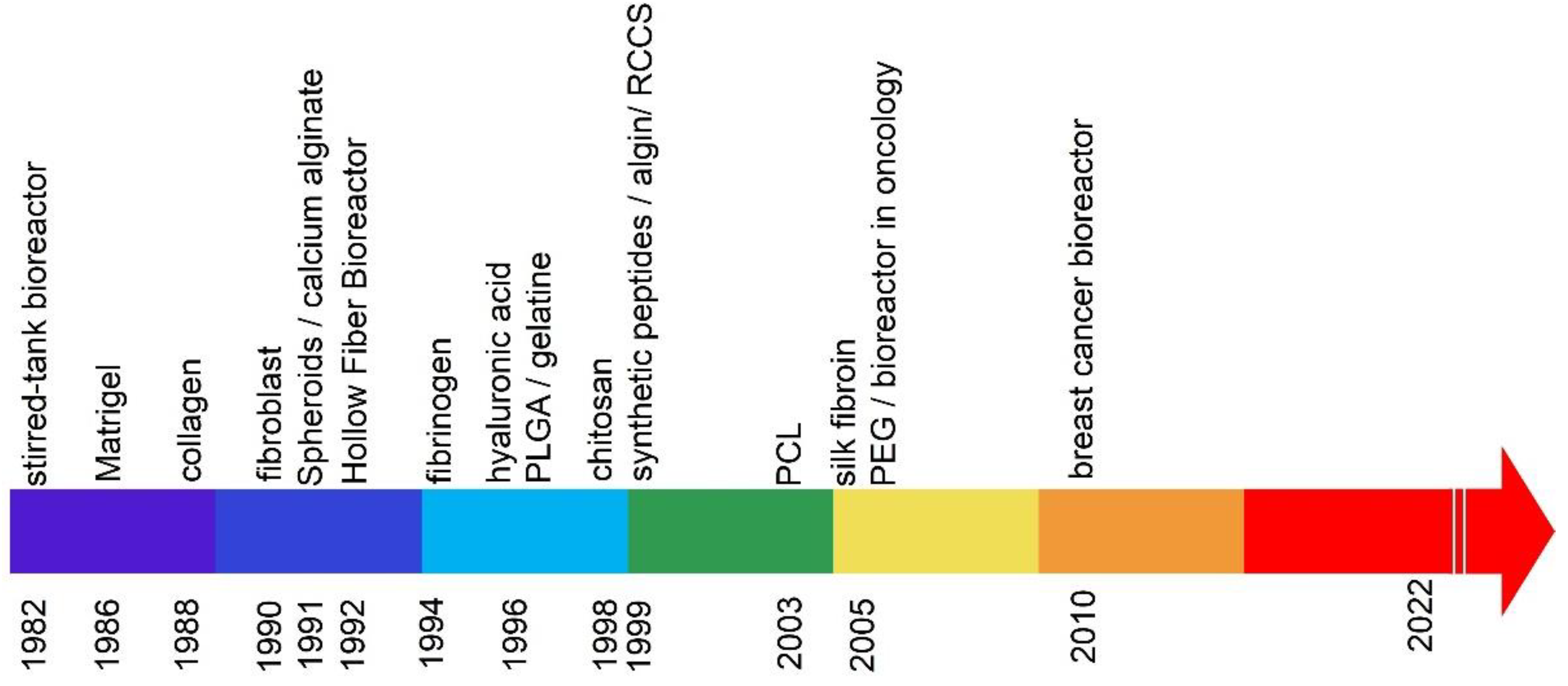
Figure 1. Cell culture methods: general division.
Ijms 23 10109 g001
Figure 2. Years of publications on hydrogel-based 3D matrices for cultivation.
Ijms 23 10109 g002
Table 1. Historical development of 3D breeding methods.
Cell Culture MethodNo.Type of Cell CultureReferences
3D cell culture1Spheroidal[16]
3D culture matrices based on a hydrogel
2Natural PolymersMatrigel [17]
3Collagen[18]
4Fibroblast[19]
5Calcium alginate[20]
6Fibrinogen[21]
7Hyaluronic acid[22]
8Gelatine[23]
9Chitosan[24]
10Algin[25]
11Silk fibroin[26]
12Synthetic PolymersPoly (lactic-co-glycolic acid) (PLGA) [27]
13Synthetic peptides[28]
14Scaffolding made of electro-spun poly (ε-caprolactone) (PCL)[29]
15Poly (ethylene glycol) (PEG),[30]
163D bioreactorsHollow fiber bioreactor[31]
17Stirred-tank[32]
18Rotary cell culture system (RCCS)[33]
Publisher’s Note: MDPI stays neutral with regard to jurisdictional claims in published maps and institutional affiliations.

