Review

The Pleiotropy of PAX5 Gene Products and Function

by
Parinaz Nasri Nasrabadi
1,2,
Danick Martin
1,2,
Ehsan Gharib
1,2 and
Gilles A. Robichaud
1,2,*
1
Département de Chimie et Biochimie, Université de Moncton, Moncton, NB E1A 3E9, Canada
2
Atlantic Cancer Research Institute, Université de Moncton, Moncton, NB E1C 8X3, Canada
*
Author to whom correspondence should be addressed.
Int. J. Mol. Sci. 2022, 23(17), 10095; https://doi.org/10.3390/ijms231710095
Submission received: 21 July 2022 / Revised: 23 August 2022 / Accepted: 26 August 2022 / Published: 3 September 2022
(This article belongs to the Special Issue PAX Genes in Health and Diseases)

Abstract

:
PAX5, a member of the Paired Box (PAX) transcription factor family, is an essential factor for B-lineage identity during lymphoid differentiation. Mechanistically, PAX5 controls gene expression profiles, which are pivotal to cellular processes such as viability, proliferation, and differentiation. Given its crucial function in B-cell development, PAX5 aberrant expression also correlates with hallmark cancer processes leading to hematological and other types of cancer lesions. Despite the well-established association of PAX5 in the development, maintenance, and progression of cancer disease, the use of PAX5 as a cancer biomarker or therapeutic target has yet to be implemented. This may be partly due to the assortment of PAX5 expressed products, which layers the complexity of their function and role in various regulatory networks and biological processes. In this review, we provide an overview of the reported data describing PAX5 products, their regulation, and function in cellular processes, cellular biology, and neoplasm.

1. Introduction

The Paired Box (PAX) gene family encodes nine transcription factors (PAX1–9), which regulate gene expression programs in tissue development [1]. Although PAX transcription factors share a highly similar paired-box DNA-binding domain, they are classified into four subgroups (I–IV) based on additional functional domains such as the octapeptide and the homeodomain, which are generally located in the protein’s internal and amino-terminal regions respectively [2,3]. Given their structural resemblance, PAX members from a particular subgroup account for similar activities and functions. For example, PAX genes in subgroups II (PAX2, PAX5, and PAX8) and III (PAX3 and PAX7) are commonly involved in processes including cell survival, motility, and tumor progression. Conversely, members from subgroup I (PAX1 and PAX9) and IV (PAX4 and PAX6) seem less involved in cancer processes [4]. The expression of PAX family gene products is also generally tissue specific. For instance, PAX2 expression has been described in kidney and optic nerve development [5], whereas PAX5 has mostly been associated with the development of the central nervous system, of B-lymphocytes, and spermatogenesis [6]. Furthermore, the expression patterns of subgroup II members are reported to be altered in various cancer tissues, which suggests a distinctive role for these PAX gene products in the regulation of specific malignancies [1]. Amongst these members, PAX5 has been extensively studied and characterized for its role in cancer pathogenesis.
This review will summarize the upstream and downstream pathways associated with the regulated expression of various PAX5 gene products in multiple tissues. Specifically, we will describe the most prevalent roles of PAX5 in vital biological processes (e.g., B-cell maturation) and neoplasia (e.g., leukemia and lymphoma) upon its aberrant expression. We will also review the mechanisms and impact of spatiotemporal regulated expression of PAX5 in both healthy and malignant cellular settings. Most importantly, we will provide a detailed overview of PAX5’s remarkable capacity to generate a range of gene products, which accounts for its pleiotropic function in an array of cell types and tissues (Figure 1).

2. The PAX5 Transcription Factor and B-Cell Development

2.1. Expression and Tissue Specificity

The human PAX5 gene locus is located on the 9p13 chromosomal region known to undergo a high degree of alterations leading to its implication in cancer development and progression [22]. Structurally, the PAX5 gene is characterized by two known distinct promoters, resulting in two alternative transcriptional initiation sites known as PAX5A and PAX5B [11]. Both transcripts share the same sequence encoded by exon 2 through exon 10. However, they have different sequences in their first exon (1A or 1B), which is dependently linked to their respective promoter regions (PAX5 1A versus PAX5 1B). Both PAX5A and PAX5B protein variants consist of a 52-kD protein known as the B-cell lineage-Specific Activator Protein (BSAP), which was initially identified as an essential regulator of early B-cell differentiation and commitment [6,23]. Despite their structural similarities, PAX5A and PAX5B gene products display differential expression signatures and tissue specificity [24,25]. For example, PAX5A expression is reported to be mostly restricted to the B-cell lineage, whereas PAX5B can also be found in the central nervous system and testis [6,11]. Functionally, although PAX5A and PAX5B have mutually been described to drive B-cell differentiation, other studies have demonstrated that PAX5 isoforms 1A and 1B regulate distinct target genes and functions in B-cell models [24,25,26].

2.2. Role of PAX5 in B-Cell Lineage Commitment and Maturation

Differentiation of common lymphoid progenitors (CLPs) into immunoglobulin (Ig) producing plasma cells is a complex spatiotemporal process where each developmental step of B-lymphocyte maturation requires controlled signaling through specific expression profiles of transcription factor associated with B-cell identity [27]. PAX5 has been well established as the central coordinator of B-cell commitment and maturation.
Initially, hematopoietic stem cells differentiate in CLPs in the bone marrow through the activity of Fms-Like Tyrosine kinase-3 [28,29]. During early B-cell differentiation, pre-B-cells proliferate under interleukin-7 activation, which is secreted from the stromal environment [30]. During this time, CLPs initiate V(D)J recombination of the immunoglobulin heavy-chain (IgH) and light-chain (Igk or Igl) gene loci to assemble the pre-B-cell receptor (BCR) complex (reviewed in [31]). Subsequent B-cell activation enables the induction of the remaining BCR components, which synergistically function to induce PAX5 expression and other B-cell phenotypic markers such as the Cluster Differentiation-19 (CD19) and the CD79a (MB-1 gene) transmembrane proteins involved in B-cell activation through the BCR complex [32,33,34,35]. PAX5 also functionally cooperates with other transcription factors including, PU.1, B-cell Linker, IKAROS Family Zinc Finger-1 (IKZF1), Early B-cell Factor (EBF), and Transcription Factor-3 (TCF3) to support PAX5 activity during early B-cell identity and maturation [36,37,38]. Further development of the BCR induces the expression and maturation of surface IgM and allows immature B-cells to infiltrate peripheral circulation for eventual antigen-dependent developmental stages of B-cell maturation [39]. Upon encountering antigens by way of the BCR, B-cells migrate to peripheral lymphoid organs for further Ig rearrangement to produce high-affinity antibody isotypes. Through this, PAX5 stimulates the Activation-Induced cytidine Deaminase (AID) gene, which is essential for somatic hypermutation and antibody class switching as part of B-cells’ capacity to diversify its Ig repertoire [40,41]. Mature and activated B-cells can then differentiate either into short-lived antibody-secreting plasma cells or germinal center memory B-cells for immunological memory, depending on the signaling and requirements of the extracellular environment.
In addition to the induction of pro-B lineage specific genes, PAX5 also concomitantly suppresses B-lineage inappropriate genes [35,42]. Accordingly, PAX5 inhibits the expression of Fms-Like Tyrosine kinase-3 (mediator of CLP multipotency), NOTCH receptor-1 (mediator of T-cell differentiation) in addition to plasma cell differentiation factors B-cell Induced Maturation Protein-1 (BLIMP-1) and X-Box Binding Protein-1 [43,44,45]. However, once all PAX5-mediated early development checkpoints are achieved, coordinated attenuation of PAX5 expression through BLIMP-1 is implemented to pursue B-cell maturation. Accordingly, many studies have established a bidirectional negative regulatory loop between PAX5 and BLIMP-1 where the activation of BLIMP-1 and concomitant suppression of PAX5 represent a critical step in plasma cell differentiation [43,45,46,47]. Although recent studies demonstrate that complete PAX5 repression is not essential for developing plasma cells and antibody secretion, it is required for optimal high-affinity IgG production and sustained accumulation of long-lived plasma cells [48].
Altogether, PAX5 not only mediates B-cell identity, it also concomitantly blocks the differentiation of other (non-B) hematopoietic specification. In fact, studies have shown that PAX5 is required for the progression of B-cell development beyond the pro-B-cell stage as the suppression of PAX5 during early B-cell development enables the reversal of B-lineage commitment fate [49,50]. Interestingly, these latter PAX5-silenced cells can initiate myeloid differentiation until PAX5 expression is restored [50]. Furthermore, the potential for B-cell retro-differentiation upon ectopic modulation of PAX5 expression appears unique to early developmental stages in uncommitted mature B-cells. Accordingly, experiments conducted by Proulx et al. (2010) show that restoration of PAX5 expression in terminally differentiated cancer B-cells (i.e., multiple myeloma) induces apoptosis of myeloma cells [51]. These findings highlight the impact of PAX5 and its involvement in various pathways leading to B-lineage commitment, maturation, and function. PAX5 expression is therefore stringently regulated during B-cell development. However, aberrant expression of PAX5 also consequently leads to pathological disorders, notably cancer.

3. PAX5 and Cancer

3.1. PAX5 Is a Key Driver of B-Cell Malignancies

Given its pivotal regulatory role in B-cell development, PAX5 also represents a potent oncogene in hematological cancers, particularly lymphoma and lymphocytic leukemia. These findings have been validated by numerous studies showing a direct link between PAX5 and the onset of B-cell cancer lesions using various B-cell lines and in murine models [50,52]. In fact, PAX5 expression is associated with the most common non-Hodgkin B-cell malignancies including: diffused large B-cell lymphoma (DLBCL) [53,54]; chronic lymphocytic leukemia [55]; and B-cell acute lymphoblastic leukemia (B-ALL) [55]. In most cases, deregulated PAX5 expression impedes the progression of B-cell differentiation leading to hallmark cancer features such as proliferation, apoptosis, and phenotype transitioning processes [56]. Genetic analyses demonstrate that aberrant PAX5 expression and function in most B-cell malignancies are caused by genetic instability of the PAX5 locus. For example, somatic mutations of the PAX5 enhancer region are prevalent in DLBCL, follicular lymphoma, Burkitt’s lymphoma, and mantle cell lymphoma [55,57,58,59]. PAX5 is also prone to recurrent chromosomal rearrangements, which represent the causal root for specific subsets of aggressive B-cell cancer lesions, notably B-ALL [60,61].
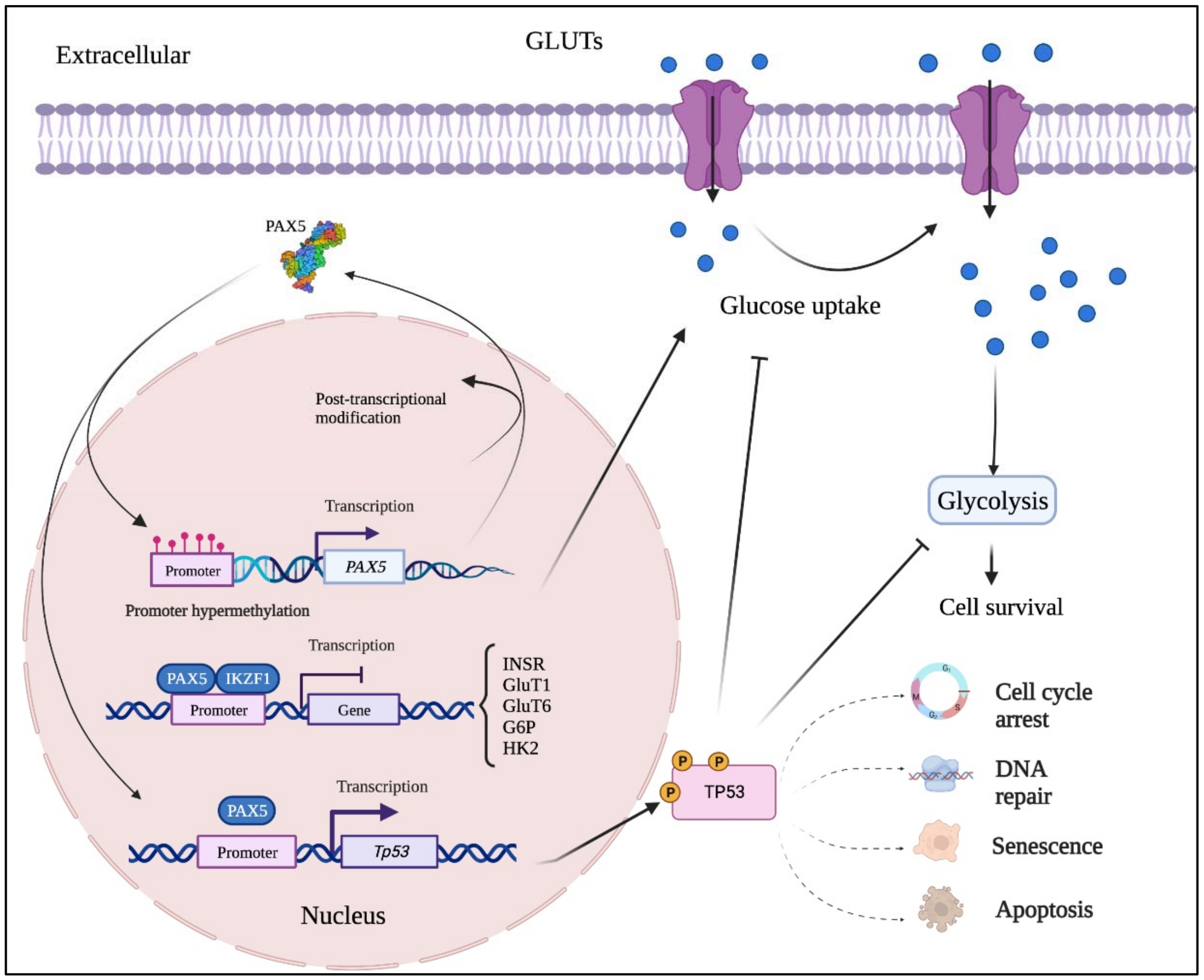
Acute lymphoblastic leukemia is the most common hematological (blood) cancer in children, where the B-cell subtype (B-ALL) is the prevalent form. B-ALL is characterized by recurrent genetic changes that suppress developmental stages beyond B-cell precursors, thus preserving the self-renewal phenotype of non-differentiated cells [7,62,63,64]. It is therefore not surprising that most B-ALL cases are associated to genetic alterations from genes governing the earliest stages of B-lymphoid specification (i.e., TCF3, EBF, IKZF1, and PAX5) [38,65,66]. Over 80% of gene lesions found in childhood B-ALL are associated with the PAX5 and IKZF1 genes alone [7,67]. A proposed mechanism recently described by Chan et al., (2017) suggests that PAX5 and IKZF1 mutations disrupt adequate regulation of downstream metabolic-related genes such as Insulin Receptor, Glucose Transporter-1 (GLUT1), Glucose Transporter-6, Glucose-6-Phosphate Dehydrogenase and Hexokinase-2, all of which encode proteins governing glucose uptake and utilization [68]. PAX5 and IKZF1 are commonly depicted as metabolic gatekeepers by conferring a chronic state of energy deprivation in hematopoietic stem cells, which limits the energy supply necessary for oncogenic transformation [68]. Aberrant glucose uptake and ATP overproduction are the main components fueling tumor development and oncogenic growth [69,70]. In addition, the overexpression of GLUT1, a major glucose transporter, contributes to the maintenance of cancer cell metabolism by increasing glycolysis and energy production [71]. Consequently, dominant negative mutants of PAX5 and IKZF1 found in the majority of B-ALL lesions alleviate glucose and energy restrictions, which foster malignant transformation [68]. These findings are also corroborated by Kurimoto et al. (2017) who observed significant increases in GLUT1 expression following PAX5 silencing in head and neck squamous cell carcinoma [72]. In addition, they suggest that the GLUT1-PAX5 regulatory axis can also be controlled by the Tumor suppressor Protein-53 (Tp53), which is upregulated by PAX5 via a positive feedback loop [73,74,75,76].
More than a third of all B-ALL cases arise solely from PAX5 genetic alterations, which include deletions, sequence mutations, and translocations to a range of fusion partners [7,77,78]. Concurrently, studies have demonstrated that PAX5 haploinsufficiency plays an essential role in lymphoblastic leukemia [8,62,79]. Accordingly, insufficient PAX5 expression due to somatic alterations in high-risk BCR-ABL1-positive (most frequent B-ALL subtype in adults) and Philadelphia-like B-ALL account for 50% of lymphoid blast crises [67,80]. In the case of germinal mutations, inheritance of altered PAX5 gene sequences predisposes children to a four-fold increased risk of being diagnosed with B-ALL when they have an affected sibling [81]. On the other hand, a growing number of chromosomal translocations in B-ALL regarding PAX5 are also reported to produce genetic fusions with different gene partners including: Forkhead Box P1 (3p13) [7,82]; Janus Kinase 2 (9p24) [83]; Autism Susceptibility Candidate 2 (7q11) [82]; Elastin (7q11) [8]; ETS Translocation Variant 6 (12p13) [7,60,82]; Promyelocytic Leukemia Protein (15q24) [61]; Zinc Finger Protein 521 (18q11) [7]; Chromosome 20 Open Reading Frame 112 (20q11) [82,84]; Ribosome Binding Protein 1 Pseudogene (7p12.1) [84]; Solute Carrier Organic Anion Transporter Family Member 1B3 (12p12) [84]; Additional Sex Combs Like 1 Transcriptional Regulator 1 (20q11.1) [84]; Kinesin Family Member 3B (20q11.21) [84]; Homeodomain Interacting Protein Kinase 1 (1p13) [85]; Pore Membrane Protein of 121 kDa (7q11) [85]; Dachslund Family Transcription Factor 1 (13q21) [85]; and, Bromodomain Containing 1 (22q13.33) [85]. Interestingly, these fusion proteins all contain the 5′end of the PAX5 coding region (corresponding to the DNA-binding domain) fused with a functional domain of the partner gene. Although these putative chimeric transcription factors retain DNA PAX5-specific motif binding, they are characterized by attenuated PAX5 transcriptional activities or loss of function [10,61,64,84,86]. Mechanistically, some studies have reported that PAX5 fusion proteins (e.g., PAX5-ETS Translocation Variant 6, PAX5-Forkhead Box P1, and PAX5-Elastin) function as dominant negative factors against their wild-type PAX5 counterpart [7,8,9,10,87]. Meanwhile, others show that PAX5 fusion proteins regulate an independent profile of target genes in B-cells [64,88,89]. Altogether, these studies highlight the importance of tightly orchestrated expression of PAX5 during early B-cell development where insufficient PAX5 activity leads to B-cell differentiation blockade and uncontrolled proliferation of immature B-cells [7,62,63,64].
In opposition to early-stage B-cell cancer lesions, which are normally characterized with PAX5 translocations conferring attenuated forms of PAX5 function, PAX5 genetic rearrangement in late B-cell developmental stages is mainly associated with increases in PAX5 expression and activities. For example, the PAX5 t(9;14) translocation is a genetic rearrangement involving the complete coding region of PAX5 (chromosome 9), which is relocated under the control of the potent Emµ promoter of the IgH gene (chromosome 14) [11,90]. The PAX5 t(9;14) recombination is by far the most studied and prevalent PAX5 genetic alteration in non-Hodgkin’s lymphoma subtypes such as lymphoplasmacytic lymphoma (LPL) and DLBCL [11,54,90]. Given that the repression of PAX5 expression is generally required for adequate B-cell terminal differentiation, its constitutive overexpression caused by the PAX5-IgH fusion blocks mature B-cell activation, accumulates inactivated mature B-cell populations, and lowers differentiated plasma cell numbers [11,54,90]. Similarly, studies have substantiated these findings by experimentally inserting a PAX5 minigene into the IgH locus (IgHP5ki), which provokes a t(9;14)(p13;q32) rearrangement of germline mutant knock-in mice [91]. Although the development of the IgHP5ki model was reminiscent of human t(9;14) B-cell lymphoma pathology, it harbors a germline rather than a somatic translocation mutation. Hence, forced expression of PAX5 in the hematopoietic lineage affected overall hematopoiesis in IgHP5ki mice, notably in T-cell development, which led to the development of T-lymphoblastic lymphomas [91]. Altogether, these studies not only demonstrate that t(9;14)-mediated overexpression PAX5 is an important cause of B-cell lymphoma; but also suggest a particularly harmful role of t(9;14) in the T-lymphoid system [91].

3.2. PAX5 in Non-Hematopoietic Cancers

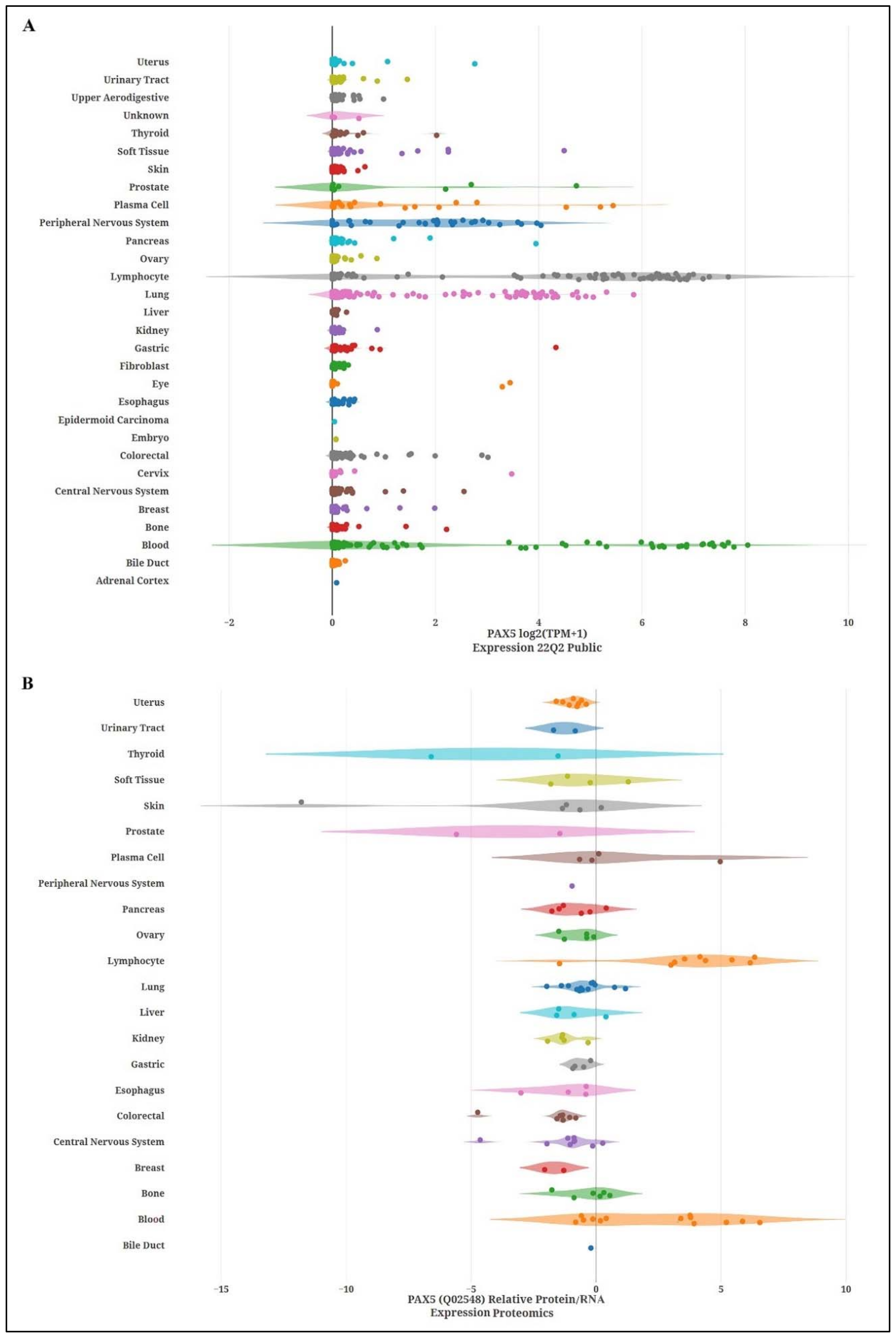
For nearly two decades, the PAX5 gene has also been revealed to be an imperceptible regulator of cellular processes in various non-hematological tissues and cancers. To obtain a global perspective of relative PAX5 expression profiles in non-hematological tissues, levels of PAX5 transcripts and proteins were recovered from data mining, processed, and plotted across a collection of tissue samples (Figure 2).
In contrast to B-cells, the functional role and outcome of aberrant PAX5 expression in non-hematological cancers are as diverse as the types of tissue that express PAX5. For example, Baumann et al. (2004) observed that elevated PAX5 expression levels correlate with a subset of malignant (N-type) neuroblastoma in comparison to their benign counterpart (S-type) [92]. This study also demonstrates that recombinant expression of PAX5 in benign neuroblastoma cell models resulted in more invasive cancer phenotypes [92]. Similarly, elevated PAX5 expression has been associated with pediatric brain tumors (medulloblastomas), where PAX5 expression positively correlates with cell proliferation and inversely with neuronal differentiation in desmoplastic medulloblastoma [93]. Another study by Kanteti et al. (2009) demonstrated that elevated levels of PAX5 expression in small-cell lung cancer induced the expression and activation of the c-MET proto-oncogene, also known as the Hepatocyte Growth Factor Receptor and a potent regulator of cancer cell motility and angiogenesis [94]. More recently, Dong et al. (2018) have shown that PAX5 potentiates cisplatin-based systematic chemotherapy resistance of muscle-invasive bladder cancers [95]. Mechanistically, this latter study demonstrates that the PAX5 transcription factor transactivates Prostaglandin-Endoperoxide Synthase-2 gene transcription, which promotes bladder cancer pathogenic features [95]. Other reports have also shown a role for PAX5 in oncogenic or pro-aggressive features in astrocytoma [96,97], lung cancer [94], cervical carcinoma [98], bladder cancer [95], oral carcinoma [99], neuroblastoma [92], and medulloblastoma [93]. In opposition, numerous studies functionally characterize PAX5 as a tumor suppressor in hepatocellular carcinoma [73,100], breast carcinoma [75,101,102,103], esophageal squamous cell carcinoma [72], retinoblastoma [104], gastric cancer [74,105], Merkel cell carcinoma [106], ovarian cancer [107], and head and neck squamous cell carcinoma [76]. Accordingly, a study by Kurimoto et al. (2017) shows that PAX5 overexpression inhibits cell proliferation and chemoresistance of esophageal squamous cells [72], whereas reporting from Liu et al. (2011) demonstrates that PAX5 blocks cell viability and colony formation of hepatocytes [73].
Despite PAX5 expression prevalence amongst different non-hematopoietic tissues, the upstream mechanisms responsible for its aberrant expression and often opposing impacts between tissue types are not entirely understood. However, many large-scale and functional genomic analyses commonly demonstrate that the tumor suppressive features of PAX5 are inhibited by promoter hypermethylation events in non-hematological cancers [73,76,100,105,107,108]. Accordingly, the methylation profile of the PAX5 promoter region has been proposed as a potential tool with clinical applicability for prognostics and diagnostics in the classification of non-hematological cancer subtypes [74,105,106,107]. Another possible explanation has been provided by reports describing PAX5 regulation through a direct relationship with Tp53 [73,74,75,76,96]. Yet, the regulation and outcome of PAX5/Tp53 interplay in non-hematological cancer are still conflicted and appear to be tissue type specific. For example, a study by Stuart et al. (1995) demonstrates that PAX5-mediated tumorigenesis of primary human diffuse astrocytoma is supported through the direct binding and inhibition of Tp53 promoter transactivation [96]. In contrast, when adopting the role of a tumor suppressor, PAX5 has been shown to transactivate Tp53 expression and activity in gastric cancer [74], breast cancer [75,103], and hepatocellular carcinoma [73]. A study by Guerrero-Preston et al., (2014) further substantiates the antitumor activity of the PAX5/Tp53 axis by demonstrating that PAX5 promoter hypermethylation in head and neck squamous cell carcinoma results in inadequate Tp53 activation [76]. This study also shows that PAX5 promoter methylation status is directly linked to Tp53 mutational profiles. These findings are significant given the high frequency of Tp53 mutation in cancer tissues [109] and its association to epigenetic control of PAX5 expression and function in non-hematological cancer cell fate.

3.3. PAX5 and Breast Cancer

To date, the functional role of PAX5 expression and function in non-hematopoietic cancers has largely been elucidated in breast cancer models. Studies demonstrate that PAX5 overexpression in breast cancer cells leads to a decrease in proliferation, colony formation, and migration through the induction of pro-epithelial features [75,101]. Specifically, PAX5 is shown to regulate hallmark phenotypic transitioning programs known as the epithelial to mesenchymal transition (EMT) during breast cancer cell metastasis and disease progression [75,101]. This phenotypic plasticity is widely accepted as a multistep biological program enabling breast cancer cells to successfully navigate through the various microenvironments confronted during the metastatic journey [110,111,112]. Generally, EMT initiates metastasis by fostering phenotypic change where mammary epithelial cells gain invasive properties (e.g., anoïkis resistance and migration) to infiltrate the circulatory system and distant organs. Thereafter, reversal of EMT or mesenchymal to epithelial transition (MET) would enable circulating metastatic cancer cells to reboot an epithelial program (e.g., adherence and intercellular tight junctions) to colonize metastatic tumor niches [111,112,113]. A study by Vidal et al. (2010) has demonstrated that PAX5 promotes pro-epithelial dominant features in breast cancer cells and thereby attenuates malignancy and disease progression in murine models [75]. In parallel, Benzina et al. (2017) have shown that PAX5 not only promotes breast cancer epithelial identity but also induces MET of aggressive breast cancer cells through direct transactivation of E-cadherin, a pivotal regulator of epithelialization [114]. In fact, E-cadherin is not only a predominant surface phenotypic marker of epithelial cells, but also a key regulator of anti-invasive properties and MET. Surface expression of E-cadherin results in the direct suppression of pro-mesenchymal gene expression (e.g., SNAIL [115], TWIST [116], SLUG [117], and Zinc Finger E-Box Binding Homeobox 1 [118]) deployed during EMT and disease progression [119,120,121].
Altogether, given its pro-epithelialization role in breast tumors, PAX5 suppresses invasive properties leading to better prognostic value with a lower risk of disease progression and relapse as long as cancer cells remain in their primary tumor sites (in situ) [102]. However, PAX5 expression during the shifts of phenotypic programs (EMT and MET) necessary for successful metastasis may also produce dichotomous outcomes for breast cancer disease. For instance, PAX5 may trigger MET in circulating cancer cells thus implementing epithelial features essential for the establishment of distant metastatic colonization [102]. Accordingly, a study by Ellsworth et al. (2009) demonstrates that relative PAX5 expression levels are 100-fold greater in metastasized breast cancer cells located in patient lymph nodes in comparison to levels found in their primary tumor counterparts [122]. These observations are largely reminiscent of the paradoxical role of PAX5 during the early and late stages of B-cell development. As discussed previously, disturbance of PAX5 spatial and temporal regulated expression ultimately leads to aberrant cellular processes and cancer. In the following sections, we will discuss the potential mechanisms of deregulated PAX5 expression in addition to the diversity of PAX5 products, which ultimately determine cellular function and cancer outcome.

4. PAX5 Expression and Regulation

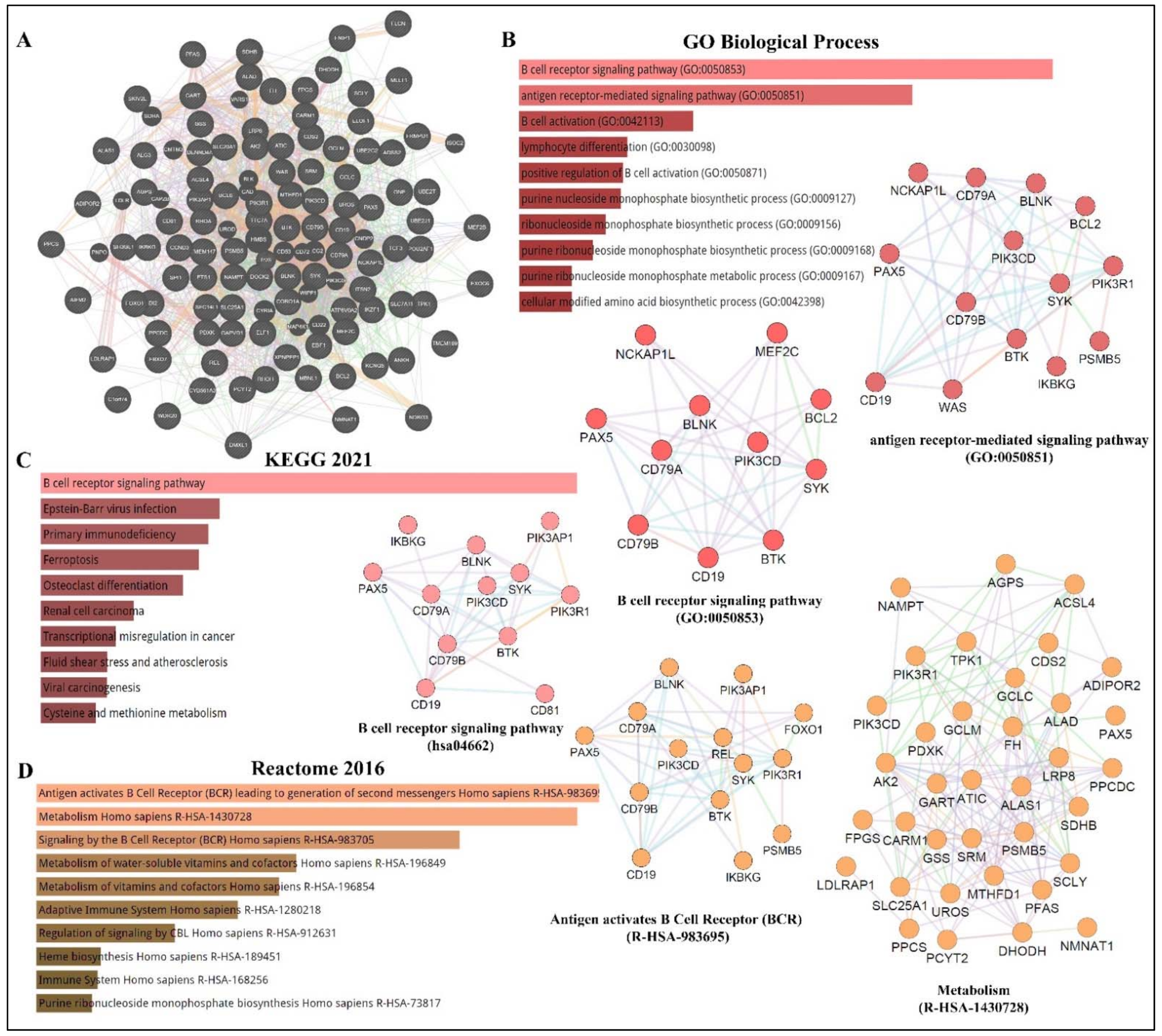
As depicted in Figure 3, PAX5 is widely associated with various cellular processes and pathologies (Figure 3). Given the essential role of PAX5-mediated transactivation of vital genes for cell biology, its deregulation will have consequences on basic cellular processes such as differentiation, viability, and proliferation (reviewed in [4,40,56]). Investigation of deregulated mechanisms leading to aberrant PAX5 expression and activity is therefore relevant and warranted to provide more insight into the overall comprehension of PAX5 mechanisms of action. Although the literature provides abundant research characterizing PAX5-mediated pathways and interactions, the upstream mechanisms regulating PAX5 expression are much less defined.

4.1. PAX5 Epigenetic Regulation

Many genomic studies have described the PAX5 locus as a genetic hot-spot susceptible to structural variation [57,58,59,123,124]. For example, PAX5 expression and function are altered by various genetic alterations, including somatic mutation, translocation, and duplication/polyploidy [10,11,62,78,83]. In addition to genetic mutation, which changes both the transcriptional levels and protein sequences, genes are also submitted to epigenetic deregulation, which impacts overall expression levels [125]. These epigenetic processes include methylation of 5′-cytosine-phosphate-guanine-3′ (CpG) islands, chromatin remodeling via histone modifications, and various RNA-mediated mechanisms, which involve regulatory non-coding RNAs [125,126]. A brief description of each regulatory mechanism and its impact on PAX5-mediated function is discussed below.
First, methylation of CpG islands to form 5′-methylcytosine (5mC) is a well-described mechanism to repress transcriptional expression of unwanted genes during fundamental cellular processes such as development and differentiation [127,128,129]. DNA methylation is catalyzed by a group of DNA methyltransferase (DNMT) enzyme members (e.g., DNMT1, DNMT3a, and DNMT3b) [130,131]. DNA methylation can also be reversed by demethylation, which is mediated by Ten-Eleven Translocation (TET) family dioxygenase enzymes, which include TET1, TET2, and TET3 [132]. In fact, B-lineage development is coordinated by the well-timed deployment of B-cell fate transcription factors, which are regulated by epigenetic events and post-transcriptional modifications [133,134]. For example, DNMT1, DNMT3a, and DNMT3b are required for the maturation of hematopoietic stem cells into CLPs, whereas DNMT1 is particularly essential for pre-B-cell differentiation to immature B-cell [128,135]. Subsequent studies have since demonstrated that TET function is required for developing B-cells to transit from the pro-B to pre-B developmental stage [136]. Mechanistically, the B-cell-specific MB-1 (CD79a) promoter is known to be hypermethylated during hematopoietic stem cells transition to CLPs and then progressively demethylated during the expression and assembling of the BCR components. These events upregulate PAX5 expression and concomitant target genes to achieve B-lineage identity [63,137,138,139]. On the other hand, attenuation of PAX5 expression during terminal B-cell differentiation is reported to partly mediated by methylation of PAX5 [140]. In support of these events, a study by Danbara et al., (2002) demonstrates that genomic demethylation using 5-aza-2′-deoxycytidine in myeloma cell lines results in the reconstitution of PAX5 expression and its transcriptional target genes (CD19 and MB-1) [140]. Although the regulation of the complex networks of epigenetic modifications governing B-cell differentiation is only partially understood, one aberrant mechanism leading to deregulated PAX5 methylation has been described for the inadequate function of AID [41,141]. The PAX5/AID pathway is essential for somatic hypermutation and antibody class switching during Ig production [142]. However, constitutive expression of AID has been associated with lymphomagenesis through its capacity to alter the sequence of non-Ig genes (i.e., PAX5) or through AID-mediated deamination of the PAX5 gene [141,143]. As a result, changes in PAX5 gene sequences redefine motif-specific regions marked for epigenetic modifications and subsequent expression control [41,141].
Given the importance of adequate methylation processes regulating PAX5-induced B-cell development, deregulated methylation results in the destabilization of B-cell homeostasis and cancer phenotypes [139,144]. This phenomenon has been further substantiated by the demonstration that PAX5 methylation status directly correlates with overall survival rates of cancer patients [72,74,105]. Furthermore, studies profiling methylation signatures in pediatric ALL patients have correlated PAX5 hypermethylation to the pathogenesis of B-ALL and T-ALL subtypes [145,146]. These findings have also prompted Nordlund et al. (2015) to propose that PAX5 methylation status combined with the mapping of PAX5 gene recombinations with other partner genes represent an effective diagnostic tool to classify heterogeneous and cytogenetically undefined ALL subtypes [147].
PAX5 aberrant methylation is not a tissue-specific phenomenon. In fact, PAX5 hypermethylation has been described in many non-hematological cancers, particularly where PAX5 is characterized as a tumor suppressor (e.g., hepatocellular carcinoma [100], ovarian carcinoma [107]; head and neck cancer [76], gastric cancer [105], lung and breast cancer malignancies [108,148]). Mechanistically, many of these latter studies demonstrate that silencing of PAX5 expression by hypermethylation leads to the inadequate transactivation of Tp53 expression, thus ensuing uncontrolled proliferation or decreased chemosensitivity to anticancer treatment regimens [72,73,149].
Gene expression profiles are also epigenetically regulated by multiple histone-modifying enzymes, which change chromatin structure to alter promoter region accessibility and recruit other modifications [150]. Histones, which assemble the nucleosomes, are prone to modifications, which include acetylation, methylation, ubiquitination, phosphorylation, and sumoylation [150]. The most common modifications consist of arginine methylation and/or lysine acetylation, where acetylation generally promotes gene expression whereas methylation elicits the opposite effects. Many histone modifying enzymes have been characterized including histone acetyltransferases (HATs), histone deacetylases (HDACs), histone demethylases, and various methyltransferases (e.g., Euchromatic Histone-Lysine N-Methyltransferase-2/EHMT2 and Lysine Methyltransferase-2A [128,144]). Like CpG island methylation, chromatin modifications represent an intrinsic part of B-cell activation and differentiation. For example, during early B-cell development, PAX5 secures B-cell commitment through activating B-cell specific genes. In addition, PAX5 concomitantly inhibits B-lineage inappropriate genes through the recruitment of HDACs to modify and silence promoter activation of these genes [139]. Studies show that the PAX5 locus is also continuously regulated by histone modifications throughout B-cell maturation. Specifically, the PAX5 promoter in pro-B-cells are modified by HDACs, whereas EHMT2 regulates mature B-cells located in germinal centers [128]. Another example is the EBF transcription factor, which is shown to be implicated in PAX5 and CD19 transactivation through the silencing of Lysine Methyltransferase-2A during early B-cell development [134,151,152]. Another example is the previously mentioned PAX5/BLIMP-1 axis during terminal B-cell differentiation into plasma cells. It is reported that BLIMP-1 suppresses PAX5 expression through the recruitment of histone demethylases and EHMT2 activities on the PAX5 promoter [153,154]. Furthermore, transcription factor Forkhead Box Protein-O1, which is essential for B-cell development beyond the pro-B-cell stage [155], is only activated upon histone methylation of TCF3, which only then can elicit histone modifications and silence PAX5 to enable the progression of B-cell development [156]. Another study conducted by Danbara et al., (2002) has specifically demonstrated that the upstream PAX5 promoter (exon 1A) is predominantly inactivated by DNA methylation, whereas the downstream promoter (exon 1B) is repressed by histone deacetylation during the final stages of B-cell terminal differentiation [140]. Comprehensively, deregulation of histone modifying events on PAX5 or its upstream regulators lead to aberrant PAX5 transcript levels and the development of diseases [144]. Accordingly, a recent study from Jin et al. (2021) has not only shown that PAX5 is hypermethylated in retinoblastoma tumors but also, the treatment of patients with cyclophosphamide (a common antineoplastic agent to treat retinoblastoma) increases PAX5 expression via gene demethylation and concomitant DNMT inhibition, which result in tumor regression [104,157].
To add complexity and appreciation for epigenetic mechanisms, different ATP-dependent chromatin remodeling complexes (CRC) capable of moving, ejecting, or restructuring nucleosomes (events often associated with DNA repair) have also been associated with PAX5 regulation and function [138,158]. For example, SWItch/Sucrose Non-Fermentable and the Nucleosome Remodeling Deacetylase CRCs are known to mediate PAX5-dependant induction or repression respectively of MB-1 (CD79a) gene expression during BCR assembly [138]. Therefore, the opposing functions of CRCs provide another layer of PAX5 function during B-cell development [138]. Another example is the histone modifying enzymes HATs, which can acetylate other cellular proteins (e.g., transcription factors) besides histones. A study by He et al., (2011) has found that histone acetyltransferase E1A binding protein p300 interacts with the C-terminal region of PAX5 to acetylate multiple lysine residues of the paired box DNA binding domain [19]. They also demonstrate that acetylation of the PAX5 transcription factor dramatically enhances the transactivation potential of its target genes [19]. This interaction was also investigated in B-cell lymphoma, where the Metastasis-Associated Protein-1 represents a substrate for acetylation upon its interaction with the HAT p300 [159]. This study found that Metastasis-Associated Protein-1 acetylation leads to the direct transactivation and overexpression of PAX5, a widespread phenomenon in human DLBCL [159].
The final contributing mechanism in epigenetic control is mediated by non-coding RNAs, which include small interfering RNAs, microRNAs (miRNAs), piwi-interacting RNAs, long non-coding RNAs, and circular RNAs (circRNAs) [160,161,162,163]. In comparison to DNA and histone modifications, only a paucity of studies has directly elucidated ncRNA-mediated mechanisms governing PAX5 expression and function. A recent study from Harquail et al., (2019) has used a bioinformatic approach to establish a causal link between differentially expressed miRNAs in cancer cells in relation to their putative targeting of PAX5-dependent cancer processes and identified miRs-484 and 210 as directly regulators for PAX5 expression and function [164]. Interestingly, miR-210 has been extensively studied as a potent oncogenic miRNA, which targets critical tumor suppressors such as E2F3 and Tp53 [165,166]. It is also well established that miR-210 is upregulated during hypoxia to induce EMT and tumor progression [167,168,169]. Given the prevalent role of PAX5 in epithelialization and EMT-MET processes in breast cancer cells [75,101], it has been suggested that miR-210 likely targets PAX5 during tumor neoplasm and hypoxia to produce a robust, comprehensive shift from epithelial to mesenchymal phenotypic features to evade hypoxic insult [164]. PAX5 has also been reported to be part of a regulatory feedback loop with miR-155 in cancer cells [170]. MiR-155 is known to play a vital role in the differentiation of memory B-cells where it targets PU.1 and AID necessary for B-cell commitment into plasma cell [171,172]. Despite the rapidly growing field of non-coding RNA function in biological processes, the elucidation of non-coding RNA-dependent control of PAX5 expression and function in B-cell development and disease is still under investigation. As our knowledge expands on the deregulation of miRNA profiles and its impact on biological processes, we notice that changes to the mRNA sequences targeted by miRNAs will also have significant consequences, including miRNA motif accessibility and disruption of translational control. Accordingly, the next section will discuss PAX5 post-transcriptional modifications and editing, which alter miRNA-specific targeting and impede the potential binding capacity of any motif-specific interacting partners of PAX5 products.

4.2. PAX5 Post-Transcriptional Regulation

Similar to most human gene transcripts, PAX5 mRNAs undergo alternative splicing processes, which translate into altered translational reading frames and often multiple protein isoforms [15,16,17,173]. To date, alternative splicing events of PAX5 transcripts in humans and other species result in translated products with deleted regions corresponding to single or multiple coding exons [15,17,173]. Specifically, studies have shown that alternative splicing of the 5′ or 3′ end of PAX5 mRNA leads to structural and functional alterations of the PAX5 transcription factor in the DNA binding (exons 2-3) and transactivation domains (exons 8-9) respectively [17,174,175]. A study performed by Robichaud et al., (2004) has characterized alternatively spliced PAX5 transcripts in CD19+ peripheral blood lymphocytes from healthy adult donors and found that B-cells simultaneously co-express multiple isoforms, including full-length mRNA (exons 1-10), in addition to transcripts lacking either exon 7 (∆7); exon 8 (∆8); exon 9 (∆9); exons 7-8 (∆7/8); or exons 7-8-9 (∆7/8/9) [17]. Interestingly, this study also demonstrates that each PAX5 protein variant elicits a unique transactivation potential upon downstream target genes [17]. Other studies have since reported additional C-terminal isoforms lacking exons 6-7-8-9 (∆6/7/8/9); exons 6-7-8 (∆6/7/8); exons 8-9 (∆8/9); and finally, a transcript containing a partial intronic sequence (intron 6) in healthy B-cells and lymphoma [175]. These findings underscore the complexity of potential dominant-negative effects and the outcome of downstream target gene expression due to a network of multiple PAX5 transcription factor variants. Despite various reports characterizing the expression of alternatively spliced PAX5 variants, the specific role of each isoform and their capacity to compete for putative PAX5 targets are still undefined. However, one study conducted by Sadakane et al., (2007) has correlated a specific expression profile comprising of the wild-type and the ∆8 PAX5 variants in over 90% of childhood acute lymphoblastic leukemia samples tested [176]. These findings suggest a possible role for individual PAX5 alternatively spliced isoforms in the regulation (or deregulation) of PAX5 function.
More recently, PAX5 transcripts have also been characterized to undergo 3′end shortening [18]. This type of transcriptional modification has significant repercussions on translational fate given that mRNA untranslated regions (UTRs), notably at the 3′end, harbor multiple binding sites for RNA binding proteins and other translational regulatory elements (e.g., miRNAs), which control transcript stability and translation efficiency [177,178]. A study by Beauregard et al., (2021) has recently reported that although 3′-editing of PAX5 transcripts is prevalent in healthy peripheral B-cells, shortening of the 3′UTR is directly linked to increased translation of PAX5 and correlates with leukemic disease progression [18]. Mechanistically, the study reveals that PAX5 3′UTR shortening is mainly due to sequence excision (up to 86%) by alternative splicing events. PAX5 mRNA shortening was also investigated in non-hematological cancers. Interestingly, conversely to 3′UTR splicing in B-cells, PAX5 3′UTR shortening in breast cancer cells is primarily manifested by alternative polyadenylation (APA) [18]. APA is another type of post-transcriptional modification where gene transcription is prompted to use alternative polyadenylation motifs (transcription termination signals), which alter the overall length of the mRNA sequences at their 3′ end. In fact, APA motifs are prevalent in more than half of all human transcripts, notably in oncogenes, to evade translational control at their 3′UTR, resulting in increased mRNA stability and translation [179,180,181]. To further elucidate the impact of PAX5 3′UTR shortening on miRNA targeting and regulation, a bioinformatic approach was used to identify predicted miRNAs targeting the excised 3′UTR in truncated PAX5 transcripts from cancer cells [18]. The study then experimentally validated that miR-181a, miR-217, and miR-1275 represent the most impactful tumor suppressors lost during PAX5 3′UTR shortening in cancer cell models [18]. Nevertheless, more studies are required to fully understand the impact of regulatory elements (e.g., miRNAs) and the accessibility of their corresponding binding sites deleted from truncated PAX5 transcripts in oncogenic processes and disease.
The previous sections describe multiple regulatory mechanisms, which lead to very diverse PAX5 transcripts, proteins, and functions. More recently, we and others have also characterized a new class of transcriptional PAX5 products, circular PAX5 RNAs (circPAX5) [12,13,14]. Circular RNAs (circRNAs) represent a relatively new category of non-coding RNAs characterized by a covalent phosphodiester bond between the 5′ and 3′ extremities of the transcript [182,183]. After being discovered in viruses in 1976, circular RNA was first observed in humans in 1991, when it was initially thought to be the product of improper post-translational editing [184]. Since then, circRNAs have been shown to be abundantly expressed and play essential roles in cell biology and disease [163,182]. Accordingly, circular RNAs can encode proteins through cap-independent translation pathways, regulate gene transcription, regulate gene translation, interact with proteins, and even mop up (sponge) small RNAs such as miRNAs [185,186,187]. Due to their essential role in cellular processes, circular RNA aberrant expression and function are consequently associated with diseases, including cancer [184,188,189]. In fact, a study by Gaffo et al. (2019) describes circPAX5 as one of the most differentially overexpressed products in pediatric B-ALL patients [12]. They also demonstrate that circPAX5 directly binds to miR-124-5p in B-cell precursors to promote B-ALL progression through the interference of the B-cell maturation process [12]. More recently, we have mapped multiple circPAX5 isoforms in B-cells including: circPAX5_2-3 (containing exons 2 and 3); circPAX5_2-4 (exons 2, 3 and 4); circPAX5_2-5 (exons 2, 3, 4 and 5); circPAX5_2-6 (exons 2, 3, 4, 5 and 6); circPAX5_2-7 (exons 2, 3, 4, 5, 6, and 7); circPAX5_2-8 (exons 2, 3, 4, 5, 6, 7, and 8); circPAX5_8 (exon 8); circPAX5_7-8 (exons 7 and 8); circPAX5_5-8 (exons 5, 6, 7, 8); and finally, circPAX5_2-6+intron 5 (exons 2, 3, 4, 5, partial intron 5, and exon 6) [13,14]. Furthermore, using TaqMan probes designed to target each unique circPAX5 junction region created by both extremities, we demonstrate that circPAX5_2-5 and circPAX5_2-6 are overexpressed in chronic lymphocytic leukemia patients in comparison to peripheral B-cells from healthy individuals. Mechanistically, we demonstrate that circPAX5 products interact with important microRNAs such as miR-146a and the miR-17-92 cluster. Previous reports demonstrate that miR-146a is a critical regulator of BLIMP-1 during B-cell differentiation [190], whereas microRNAs from the miR-17-92 cluster mediate the developmental transition of pro-B to pre-B-cells [191,192]. In addition, the miR-17-92 cluster is also associated with many oncogenic processes and phenotypes of hematopoietic cancers, notably in Burkitt lymphoma [193]. Altogether, these findings not only identify a new class of PAX5 products (i.e., circPAX5) but also provide new potential signaling avenues for PAX5-mediated function in B-cell development and disease.

4.3. Post-Translational Regulation of PAX5

Post-translational modifications and regulation of PAX5 function have not been extensively characterized. However, a few studies have reported specific PAX5-interacting regulators, which modify the PAX5 transcription factor to regulate its transactivation potential. As described earlier, PAX5 can be acetylated by HATs on multiple lysine residues, which enhances its transcriptional activation of downstream target genes [19]. Another example is how the PAX5 transcription factor can be regulated through phosphorylation events. Accordingly, studies show that PAX5 phosphorylation is responsible for the BLIMP-1/PAX5 regulatory axis during the critical stages of plasma cell differentiation [20,21]. Specifically, upon BCR engagement of pro-B cells, PAX5 is phosphorylated on serine and tyrosine residues by Extracellular Regulated Kinases-1/2 and Spleen Associated Tyrosine Kinase respectively, which revoke PAX5′s ability to repress BLIMP-1 expression, thus enabling the progression of plasma cell development. On the other hand, a study conducted by Kovac et al., (2000) has demonstrated that Importin alpha-1 interacts with the nuclear localization signal on PAX5 to confer its nuclear localization and import, leading to greater PAX5 transactivation of downstream target genes [194].

5. Discussion

It is well established that PAX5 products are important regulators of cell biology, notably in B-lineage commitment and maturation. It is also apparent that PAX5 is plagued not only by the high-profile genes it regulates but also, by its incredible vulnerability to genetic alterations leading to aberrant expression of PAX5 products. Given the reliance of crucial developmental program genes on PAX5 transactivity, perturbation of PAX5 expression and function at any level ultimately derails basic cellular processes, lending way to oncogenic manifestations. Moreover, given the requirements for coordinated and transitional PAX5 expression profiles during early (PAX5 activation) and late (PAX5 attenuation) phases of B-cell maturation, inadequate PAX5 activity leads to blockade of B-cell differentiation and uncontrolled proliferation of immature B-cells [7,62,63,64].
This review first describes how PAX5 liability is evidenced through its high susceptibility to genetic alterations, which either increase or decrease PAX5 expression and/or activity depending on the type of rearrangement. For example, impairment of PAX5 expression homeostasis can be allocated through multiple events, including unsuitable promoter regions (e.g., t(9;14)) [11,90], substitution of PAX5 protein domains (chimeric proteins) [8,10,60,88], and inadequate DNA motif marking for epigenetic control [140,145]. Although genomic instability has been extensively studied and correlated to aberrant PAX5 expression in hematological tissues, studies have also ruled out PAX5 genetic mutation as a causal link for the deregulation of PAX5 expression and function. In fact, PAX5 translocations are not well described in non-hematological cancer lesions. Regulated PAX5 expression and function are therefore highly dependent on post-transcriptional and translational mechanisms, including alternative splicing, use of alternative promoters (1A versus 1B), 3′UTR shortening, RNA circularization, and protein phosphorylation/acetylation, all of which play an essential role in PAX5 regulation (Figure 4).
PAX5 post-transcriptional and translational processes not only regulate PAX5 expression, but also contribute to PAX5 product diversity. The variety of PAX5-generated products likely confer multifaceted functions and influence on biological processes in various tissue types. The assortment of PAX5 products therefore complexifies our mechanistic elucidation of gene function and may account for the seemingly conflicting reports on PAX5 function in some cancer processes. Studies have also shown that multiple PAX5 products are expressed simultaneously within a cellular context where specific variants can elicit unique functions [25,26,176]. Therefore, functional genomics should not only consider the individual role of a particular PAX5 variant but rather PAX5 products altogether for a potential cellular outcome. For example, the evaluation of PAX5 function should consider expressed ratios, neutralizing actions, synergistic effects, or dominant negative events from different PAX5 products on overall PAX5-mediated gene expression programs and cellular processes. Additionally, technical scrutiny is advised during the evaluation and profiling of PAX5 expression as multiple PAX5-derived products have recurring homologous sequences with overlapping similarities. For example, the targeted PCR amplification of PAX5 exons 5-6 would reveal all expressed RNAs including: PAX5a (exon 1A); PAX5b (exon 1B); PAX5 full-length mRNA in addition to alternatively spliced isoforms (e.g., ∆7, ∆7/8, ∆7/8/9, etc.); truncated 3′UTR PAX5 mRNAs; and circular PAX5 RNA variants (e.g., circPAX5_2-3, circPAX5_2-5, circPAX5_2-6, etc.).
Clinically, the high specificity and sensitivity of the PAX5 transcription factor have made it a useful marker in identifying and distinguishing lymphomas and leukemias of B-cell origin. Indeed, some studies has proven PAX5 expression to be a more specific marker than CD79a for the diagnosis of B-ALL [195]. It is well established that PAX5 deletion is common in childhood and adult B-ALL, supporting its value in diagnosing or monitoring B-ALL [7,196]. PAX5 rearrangement is also a potent diagnostic marker where PAX5 t(9;14) is the most prevalent genetic alteration in lymphoplasmacytoid lymphoma and occasionally DLCL [11,90,197]. Other studies have also suggested the use of PAX5 recombination profiles can be used in a panel along with other transcription factor coding genes to predict disease outcomes and potential relapses following therapy [198]. Additionally, methylation of PAX5 has been characterized as a tumor-specific event in head and neck squamous cell carcinoma [72]. These findings have also been supported by others, which propose that PAX5 methylation status combined with mapping of PAX5 gene recombinations represent an effective diagnostic tool to classify undefined ALL subtypes [147]. On the other hand, PAX5 could also represent a potent therapeutic target for many hematological cancer lesions. However, these perspectives will only be achieved upon the careful investigation and functional elucidation of different PAX5 products.
Overall, the complexity of PAX5 products and signaling is growing at many levels. The expression profiles of various PAX5 products and their vast interacting networks ultimately determine the outcome of cell fate events and/or cancer processes. To this point, further elucidation is warranted to provide more insight into the overall comprehension of PAX5 function in cell biology and disease.

Author Contributions

Conceptualization, G.A.R.; Writing of original draft, equal contribution from P.N.N. and D.M.; Editing and review, P.N.N., D.M., E.G. and G.A.R.; Software and Data curation, E.G. and P.N.N.; Funding acquisition, G.A.R. All authors have read and agreed to the published version of the manuscript.

Funding

This work was supported by grants from: the New Brunswick (NB) Innovation Foundation; the NB Health Research Foundation (grant: OG-NBHRF/LLSC-2020); the Leukemia and Lymphoma Society of Canada (grant: OG2019); by the DUO research grant program from the Vitalité Health network of NB; the Canadian Cancer Society (grant: CHA-22); and the Natural Sciences and Engineering Research Council (grant: DDG-2020-00005). Student salary support was allocated by the MITACS Accelerate program (P.N.N.); MITACS Elevate program (E.G.); and the Beatrice Hunter Cancer Research Institute in partnership with GIVETOLIVE and the NBHRF (D.M.). We also acknowledge BioRender.com for the creation of our figures.

Institutional Review Board Statement

Healthy donor and clinical samples were obtained from the G.L.-Dumont University Hospital Centre (GLDHC, NB, Canada) biobank repository, which is also part of Exactis, a pan-Canadian repository for cancer biospecimens (https://www.exactis.ca, accessed on 1 May 2019). This study was conducted in accordance with the guidelines of the Declaration of Helsinki of 1975 (revised 2013, https://www.wma.net/what-we-do/medical-ethics/declaration-of-helsinki/, accessed on 1 May 2019). Institutional Review Board Statement and approval for studies involving human samples have been granted from both the GLDHC-Vitalité Health Network (project: CER7-3-17 Ver. 5) and the Université de Moncton (NB, Canada) (project: 1920-016) ethics evaluation committees.

Data Availability Statement

The illustrated transcriptional data from Figure 2 was obtained from DepMap (https://depmap.org/portal/depmap, accessed on 1 April 2022). Raw reads were aligned with the STAR algorithms (https://github.com/alexdobin/STAR, accessed on 1 April 2022). Data and comparison analysis of PAX5 protein levels were obtained from the Proteomics DB web tool (https://www.proteomicsdb.org, accessed on 4 April 2022). The illustrated transcriptional data from Figure 3 was obtained from the Cytoscape plugin GeneMANIA (https://genemania.org, accessed on 8 April 2022). Schematic illustrations of functional annotations and biological terms visualization were done using the Enrichr algorithms (https://maayanlab.cloud/Enrichr, accessed on 10 April 2022).

Conflicts of Interest

The authors declare no conflict of interest.

References

  1. Thompson, B.; Davidson, E.A.; Liu, W.; Nebert, D.W.; Bruford, E.A.; Zhao, H.; Dermitzakis, E.T.; Thompson, D.C.; Vasiliou, V. Overview of pax gene family: Analysis of human tissue-specific variant expression and involvement in human disease. Hum. Genet. 2021, 140, 381–400. [Google Scholar] [CrossRef]
  2. Wang, Q.; Fang, W.H.; Krupinski, J.; Kumar, S.; Slevin, M.; Kumar, P. Pax genes in embryogenesis and oncogenesis. J. Cell Mol. Med. 2008, 12, 2281–2294. [Google Scholar] [CrossRef] [PubMed]
  3. Underhill, D.A. Pax proteins and fables of their reconstruction. Crit. Rev. Eukaryot. Gene Expr. 2012, 22, 161–177. [Google Scholar] [CrossRef] [PubMed]
  4. Robson, E.J.; He, S.J.; Eccles, M.R. A panorama of pax genes in cancer and development. Nat. Rev. Cancer. 2006, 6, 52–62. [Google Scholar] [CrossRef] [PubMed]
  5. Underhill, D.A. Genetic and biochemical diversity in the pax gene family. Biochem. Cell Biol. 2000, 78, 629–638. [Google Scholar] [CrossRef] [PubMed]
  6. Adams, B.; Dorfler, P.; Aguzzi, A.; Kozmik, Z.; Urbanek, P.; Maurer-Fogy, I.; Busslinger, M. Pax-5 encodes the transcription factor bsap and is expressed in b lymphocytes, the developing cns, and adult testis. Genes Dev. 1992, 6, 1589–1607. [Google Scholar] [CrossRef]
  7. Mullighan, C.G.; Goorha, S.; Radtke, I.; Miller, C.B.; Coustan-Smith, E.; Dalton, J.D.; Girtman, K.; Mathew, S.; Ma, J.; Pounds, S.B.; et al. Genome-wide analysis of genetic alterations in acute lymphoblastic leukaemia. Nature 2007, 446, 758–764. [Google Scholar] [CrossRef]
  8. Bousquet, M.; Broccardo, C.; Quelen, C.; Meggetto, F.; Kuhlein, E.; Delsol, G.; Dastugue, N.; Brousset, P. A novel pax5-eln fusion protein identified in b-cell acute lymphoblastic leukemia acts as a dominant negative on wild-type pax5. Blood 2007, 109, 3417–3423. [Google Scholar] [CrossRef]
  9. Kurahashi, S.; Hayakawa, F.; Miyata, Y.; Yasuda, T.; Minami, Y.; Tsuzuki, S.; Abe, A.; Naoe, T. Pax5-pml acts as a dual dominant-negative form of both pax5 and pml. Oncogene 2011, 30, 1822–1830. [Google Scholar] [CrossRef]
  10. Kawamata, N.; Pennella, M.A.; Woo, J.L.; Berk, A.J.; Koeffler, H.P. Dominant-negative mechanism of leukemogenic pax5 fusions. Oncogene 2012, 31, 966–977. [Google Scholar] [CrossRef] [Green Version]
  11. Busslinger, M.; Klix, N.; Pfeffer, P.; Graninger, P.G.; Kozmik, Z. Deregulation of pax-5 by translocation of the emu enhancer of the igh locus adjacent to two alternative pax-5 promoters in a diffuse large-cell lymphoma. Proc. Natl. Acad. Sci. USA 1996, 93, 6129–6134. [Google Scholar] [CrossRef] [PubMed]
  12. Gaffo, E.; Boldrin, E.; Dal Molin, A.; Bresolin, S.; Bonizzato, A.; Trentin, L.; Frasson, C.; Debatin, K.-M.; Meyer, L.H.; te Kronnie, G.; et al. Circular rna differential expression in blood cell populations and exploration of circrna deregulation in pediatric acute lymphoblastic leukemia. Sci. Rep. 2019, 9, 14670. [Google Scholar] [CrossRef] [PubMed]
  13. Robichaud, G.A.; Hannay, B.; Martin, D.; Veilleux, V.; Finn, N. Characterization of new circular rna products from the pax-5 gene in b-cells. J. Immunol. 2020, 204, 223-14. [Google Scholar]
  14. Hannay, B.; Dumas, P.; Veilleux, V.; LeBlanc, N.; Robichaud, G.A. Discovery of novel non-coding products of the pax-5 gene and their clinical significance in lymphoid cancers. FASEB J. 2018, 32, 677-23. [Google Scholar] [CrossRef]
  15. Borson, N.D.; Lacy, M.Q.; Wettstein, P.J. Altered mrna expression of pax5 and blimp-1 in b cells in multiple myeloma. Blood 2002, 100, 4629–4639. [Google Scholar] [CrossRef]
  16. Zwollo, P.; Arrieta, H.; Ede, K.; Molinder, K.; Desiderio, S.; Pollock, R. The pax-5 gene is alternatively spliced during b-cell development. J. Biol. Chem. 1997, 272, 10160–10168. [Google Scholar] [CrossRef]
  17. Robichaud, G.A.; Nardini, M.; Laflamme, M.; Cuperlovic-Culf, M.; Ouellette, R.J. Human pax-5 c-terminal isoforms possess distinct transactivation properties and are differentially modulated in normal and malignant b cells. J. Biol. Chem. 2004, 279, 49956–49963. [Google Scholar] [CrossRef]
  18. Beauregard, A.-P.; Hannay, B.; Gharib, E.; Crapoulet, N.; Finn, N.; Guerrette, R.; Ouellet, A.; Robichaud, G.A. Pax-5 protein expression is regulated by transcriptional 3′utr editing. Cells 2021, 11, 76. [Google Scholar] [CrossRef]
  19. He, T.; Hong, S.Y.; Huang, L.; Xue, W.; Yu, Z.; Kwon, H.; Kirk, M.; Ding, S.-j.; Su, K.; Zhang, Z. Histone acetyltransferase p300 acetylates pax5 and strongly enhances pax5-mediated transcriptional activity. J. Biol. Chem. 2011, 286, 14137–14145. [Google Scholar] [CrossRef]
  20. Yasuda, T.; Hayakawa, F.; Kurahashi, S.; Sugimoto, K.; Minami, Y.; Tomita, A.; Naoe, T. B cell receptor-erk1/2 signal cancels pax5-dependent repression of blimp1 through pax5 phosphorylation: A mechanism of antigen-triggering plasma cell differentiation. J. Immunol. 2012, 188, 6127–6134. [Google Scholar] [CrossRef]
  21. Inagaki, Y.; Hayakawa, F.; Hirano, D.; Kojima, Y.; Morishita, T.; Yasuda, T.; Naoe, T.; Kiyoi, H. Pax5 tyrosine phosphorylation by syk co-operatively functions with its serine phosphorylation to cancel the pax5-dependent repression of blimp1: A mechanism for antigen-triggered plasma cell differentiation. Biochem. Biophys Res. Commun. 2016, 475, 176–181. [Google Scholar] [CrossRef] [PubMed]
  22. Andrieux, J.; Fert-Ferrer, S.; Copin, M.-C.; Huyghe, P.; Pocachard, P.; Lespinasse, J.; Bauters, F.; Laï, J.L.; Quesnel, B. Three new cases of non-hodgkin lymphoma with t (9; 14)(p13; q32). Cancer Genet. Cytogenet. 2003, 145, 65–69. [Google Scholar] [CrossRef]
  23. Barberis, A.; Widenhorn, K.; Vitelli, L.; Busslinger, M. A novel b-cell lineage-specific transcription factor present at early but not late stages of differentiation. Genes Dev. 1990, 4, 849–859. [Google Scholar] [CrossRef]
  24. Cresson, C.; Peron, S.; Jamrog, L.; Rouquie, N.; Prade, N.; Dubois, M.; Hebrard, S.; Lagarde, S.; Gerby, B.; Mancini, S.J.C.; et al. Pax5a and pax5b isoforms are both efficient to drive b cell differentiation. Oncotarget 2018, 9, 32841–32854. [Google Scholar] [CrossRef]
  25. Kikuchi, H.; Nakayama, M.; Kuribayashi, F.; Mimuro, H.; Imajoh-Ohmi, S.; Nishitoh, H.; Takami, Y.; Nakayama, T. Paired box gene 5 isoforms a and b have different functions in transcriptional regulation of b cell development-related genes in immature b cells. Microbiol. Immunol. 2015, 59, 426–431. [Google Scholar] [CrossRef] [PubMed]
  26. Robichaud, G.A.; Perreault, J.-P.; Ouellette, R.J. Development of an isoform-specific gene suppression system: The study of the human pax-5b transcriptional element. Nucleic Acids Res. 2008, 36, 4609–4620. [Google Scholar] [CrossRef]
  27. Horcher, M.; Souabni, A.; Busslinger, M. Pax5/bsap maintains the identity of b cells in late b lymphopoiesis. Immunity 2001, 14, 779–790. [Google Scholar] [CrossRef]
  28. Cobaleda, C.; Schebesta, A.; Delogu, A.; Busslinger, M. Pax5: The guardian of b cell identity and function. Nat. Immunol. 2007, 8, 463–470. [Google Scholar] [CrossRef]
  29. Zriwil, A.; Böiers, C.; Kristiansen, T.A.; Wittmann, L.; Yuan, J.; Nerlov, C.; Sitnicka, E.; Jacobsen, S.E.W. Direct role of flt 3 in regulation of early lymphoid progenitors. Br. J. Haematol. 2018, 183, 588–600. [Google Scholar] [CrossRef]
  30. Namen, A.E.; Lupton, S.; Hjerrild, K.; Wignall, J.; Mochizuki, D.Y.; Schmierer, A.; Mosley, B.; March, C.J.; Urdal, D.; Gillis, S. Stimulation of b-cell progenitors by cloned murine interleukin-7. Nature 1988, 333, 571–573. [Google Scholar] [CrossRef]
  31. Matsuuchi, L.; Gold, M.R. New views of bcr structure and organization. Curr. Opin. Immunol. 2001, 13, 270–277. [Google Scholar] [CrossRef]
  32. Kozmik, Z.; Wang, S.; Dorfler, P.; Adams, B.; Busslinger, M. The promoter of the cd19 gene is a target for the b-cell-specific transcription factor bsap. Mol. Cell. Biol. 1992, 12, 2662–2672. [Google Scholar] [PubMed]
  33. Maier, H.; Colbert, J.; Fitzsimmons, D.; Clark, D.R.; Hagman, J. Activation of the early b-cell-specific mb-1 (ig-alpha) gene by pax-5 is dependent on an unmethylated ets binding site. Mol. Cell. Biol. 2003, 23, 1946–1960. [Google Scholar] [CrossRef]
  34. Hirokawa, S.; Sato, H.; Kato, I.; Kudo, A. Ebf-regulating pax5 transcription is enhanced by stat5 in the early stage of b cells. Eur. J. Immunol. 2003, 33, 1824–1829. [Google Scholar] [CrossRef] [PubMed]
  35. Pridans, C.; Holmes, M.L.; Polli, M.; Wettenhall, J.M.; Dakic, A.; Corcoran, L.M.; Smyth, G.K.; Nutt, S.L. Identification of pax5 target genes in early b cell differentiation. J. Immunol. 2008, 180, 1719–1728. [Google Scholar] [CrossRef]
  36. Pang, S.H.M.; de Graaf, C.A.; Hilton, D.J.; Huntington, N.D.; Carotta, S.; Wu, L.; Nutt, S.L. Pu.1 is required for the developmental progression of multipotent progenitors to common lymphoid progenitors. Front. Immunol. 2018, 9, 1264. [Google Scholar] [CrossRef]
  37. Carotta, S.; Holmes, M.; Pridans, C.; Nutt, S.L. Pax5 maintains cellular identity by repressing gene expression throughout b cell differentiation. Cell Cycle 2006, 5, 2452–2456. [Google Scholar] [CrossRef]
  38. Schebesta, M.; Pfeffer, P.L.; Busslinger, M. Control of pre-bcr signaling by pax5-dependent activation of the blnk gene. Immunity 2002, 17, 473–485. [Google Scholar] [CrossRef]
  39. Reth, M.; Nielsen, P. Signaling circuits in early b-cell development. Adv. Immunol. 2014, 122, 129–175. [Google Scholar]
  40. Shahjahani, M.; Norozi, F.; Ahmadzadeh, A.; Shahrabi, S.; Tavakoli, F.; Asnafi, A.A.; Saki, N. The role of pax5 in leukemia: Diagnosis and prognosis significance. Med. Oncol. 2015, 32, 360. [Google Scholar] [CrossRef]
  41. Gonda, H.; Sugai, M.; Nambu, Y.; Katakai, T.; Agata, Y.; Mori, K.J.; Yokota, Y.; Shimizu, A. The balance between pax5 and id2 activities is the key to aid gene expression. J. Exp. Med. 2003, 198, 1427–1437. [Google Scholar] [CrossRef] [PubMed]
  42. Nutt, S.L.; Morrison, A.M.; Dorfler, P.; Rolink, A.; Busslinger, M. Identification of bsap (pax-5) target genes in early b-cell development by loss- and gain-of-function experiments. EMBO J. 1998, 17, 2319–2333. [Google Scholar] [CrossRef] [PubMed] [Green Version]
  43. Nera, K.-P.; Kohonen, P.; Narvi, E.; Peippo, A.; Mustonen, L.; Terho, P.; Koskela, K.; Buerstedde, J.-M.; Lassila, O. Loss of pax5 promotes plasma cell differentiation. Immunity 2006, 24, 283–293. [Google Scholar] [CrossRef]
  44. Souabni, A.; Cobaleda, C.; Schebesta, M.; Busslinger, M. Pax5 promotes b lymphopoiesis and blocks t cell development by repressing notch1. Immunity 2002, 17, 781–793. [Google Scholar] [CrossRef]
  45. Lin, K.-I.; Angelin-Duclos, C.; Kuo, T.C.; Calame, K. Blimp-1-dependent repression of pax-5 is required for differentiation of b cells to immunoglobulin m-secreting plasma cells. Mol. Cell. Biol. 2002, 22, 4771–4780. [Google Scholar] [CrossRef] [PubMed]
  46. Kallies, A.; Nutt, S.L. Terminal differentiation of lymphocytes depends on blimp-1. Curr. Opin. Immunol. 2007, 19, 156–162. [Google Scholar] [CrossRef]
  47. Delogu, A.; Schebesta, A.; Sun, Q.; Aschenbrenner, K.; Perlot, T.; Busslinger, M. Gene repression by pax5 in b cells is essential for blood cell homeostasis and is reversed in plasma cells. Immunity 2006, 24, 269–281. [Google Scholar] [CrossRef]
  48. Liu, G.J.; Jaritz, M.; Wohner, M.; Agerer, B.; Bergthaler, A.; Malin, S.G.; Busslinger, M. Repression of the b cell identity factor pax5 is not required for plasma cell development. J. Exp. Med. 2020, 217, e20200147. [Google Scholar] [CrossRef]
  49. Cobaleda, C.; Jochum, W.; Busslinger, M. Conversion of mature b cells into t cells by dedifferentiation to uncommitted progenitors. Nature 2007, 449, 473–477. [Google Scholar] [CrossRef]
  50. Nutt, S.L.; Heavey, B.; Rolink, A.G.; Busslinger, M. Commitment to the b-lymphoid lineage depends on the transcription factor pax5. Nature 1999, 401, 556–562. [Google Scholar] [CrossRef]
  51. Proulx, M.; Cayer, M.-P.; Drouin, M.; Laroche, A.; Jung, D. Overexpression of pax5 induces apoptosis in multiple myeloma cells. Int. J. Hematol. 2010, 92, 451–462. [Google Scholar] [CrossRef] [PubMed]
  52. Cozma, D.; Yu, D.; Hodawadekar, S.; Azvolinsky, A.; Grande, S.; Tobias, J.W.; Metzgar, M.H.; Paterson, J.; Erikson, J.; Marafioti, T.; et al. B cell activator pax5 promotes lymphomagenesis through stimulation of b cell receptor signaling. J. Clin. Investig. 2007, 117, 2602–2610. [Google Scholar] [CrossRef] [PubMed] [Green Version]
  53. Torlakovic, E.; Slipicevic, A.; Robinson, C.; DeCoteau, J.F.; Alfsen, G.C.; Vyberg, M.; Chibbar, R.; Flørenes, V.A. Pax-5 expression in nonhematopoietic tissues. Am. J. Clin. Pathol. 2006, 126, 798–804. [Google Scholar] [CrossRef] [PubMed]
  54. Poppe, B.; De Paepe, P.; Michaux, L.; Dastugue, N.; Bastard, C.; Herens, C.; Moreau, E.; Cavazzini, F.; Yigit, N.; Van Limbergen, H.; et al. Pax5/igh rearrangement is a recurrent finding in a subset of aggressive b-nhl with complex chromosomal rearrangements. Genes Chromosomes Cancer 2005, 44, 218–223. [Google Scholar] [CrossRef]
  55. Krenacs, L.; Himmelmann, A.; Quintanilla-Martinez, L.; Fest, T.; Riva, A.; Wellmann, A.; Bagdi, E.; Kehrl, J.; Jaffe, E.; Raffeld, M. Transcription factor b-cell-specific activator protein (bsap) is differentially expressed in b cells and in subsets of b-cell lymphomas. Blood 1998, 92, 1308–1316. [Google Scholar] [CrossRef] [PubMed]
  56. O’Brien, P.; Morin, P.; Ouellette, R.; Robichaud, G. The pax-5 gene: A pluripotent regulator of b-cell differentiation and cancer disease. Cancer Res. 2011, 71, 7345–7350. [Google Scholar] [CrossRef] [PubMed]
  57. Arthur, S.E.; Jiang, A.; Grande, B.M.; Alcaide, M.; Cojocaru, R.; Rushton, C.K.; Mottok, A.; Hilton, L.K.; Lat, P.K.; Zhao, E.Y.; et al. Genome-wide discovery of somatic regulatory variants in diffuse large b-cell lymphoma. Nat. Commun. 2018, 9, 4001. [Google Scholar] [CrossRef]
  58. Puente, X.S.; Bea, S.; Valdes-Mas, R.; Villamor, N.; Gutierrez-Abril, J.; Martin-Subero, J.I.; Munar, M.; Rubio-Perez, C.; Jares, P.; Aymerich, M.; et al. Non-coding recurrent mutations in chronic lymphocytic leukaemia. Nature 2015, 526, 519–524. [Google Scholar] [CrossRef]
  59. Grande, B.M.; Gerhard, D.S.; Jiang, A.; Griner, N.B.; Abramson, J.S.; Alexander, T.B.; Allen, H.; Ayers, L.W.; Bethony, J.M.; Bhatia, K.; et al. Genome-wide discovery of somatic coding and noncoding mutations in pediatric endemic and sporadic burkitt lymphoma. Blood 2019, 133, 1313–1324. [Google Scholar] [CrossRef]
  60. Cazzaniga, G.; Daniotti, M.; Tosi, S.; Giudici, G.; Aloisi, A.; Pogliani, E.; Kearney, L.; Biondi, A. The paired box domain gene pax5 is fused to etv6/tel in an acute lymphoblastic leukemia case. Cancer Res. 2001, 61, 4666–4670. [Google Scholar]
  61. Nebral, K.; Konig, M.; Harder, L.; Siebert, R.; Haas, O.A.; Strehl, S. Identification of pml as novel pax5 fusion partner in childhood acute lymphoblastic leukaemia. Br. J. Haematol. 2007, 139, 269–274. [Google Scholar] [CrossRef]
  62. Familiades, J.; Bousquet, M.; Lafage-Pochitaloff, M.; Béné, M.C.; Beldjord, K.; De Vos, J.; Dastugue, N.; Coyaud, E.; Struski, S.; Quelen, C.; et al. Pax5 mutations occur frequently in adult b-cell progenitor acute lymphoblastic leukemia and pax5 haploinsufficiency is associated with bcr-abl1 and tcf3-pbx1 fusion genes: A graall study. Leukemia 2009, 23, 1989–1998. [Google Scholar] [CrossRef] [PubMed]
  63. Medvedovic, J.; Ebert, A.; Tagoh, H.; Busslinger, M. Pax5: A master regulator of b cell development and leukemogenesis. Adv. Immunol. 2011, 111, 179–206. [Google Scholar]
  64. Jamrog, L.; Chemin, G.; Fregona, V.; Coster, L.; Pasquet, M.; Oudinet, C.; Rouquié, N.; Prade, N.; Lagarde, S.; Cresson, C.; et al. Pax5-eln oncoprotein promotes multistep b-cell acute lymphoblastic leukemia in mice. Proc. Natl. Acad. Sci. USA 2018, 115, 10357–10362. [Google Scholar] [CrossRef] [Green Version]
  65. Medina, K.L. Assembling a gene regulatory network for specification of the b cell fate. Dev. Cell 2004, 7, 607–617. [Google Scholar] [CrossRef]
  66. Okuyama, K.; Strid, T.; Kuruvilla, J.; Somasundaram, R.; Cristobal, S.; Smith, E.; Prasad, M.; Fioretos, T.; Lilljebjörn, H.; Soneji, S.; et al. Pax5 is part of a functional transcription factor network targeted in lymphoid leukemia. PLoS Genet. 2019, 15, e1008280. [Google Scholar] [CrossRef]
  67. Mullighan, C.G.; Miller, C.B.; Radtke, I.; Phillips, L.A.; Dalton, J.; Ma, J.; White, D.; Hughes, T.P.; Le Beau, M.M.; Pui, C.-H. Bcr–abl1 lymphoblastic leukaemia is characterized by the deletion of ikaros. Nature 2008, 453, 110–114. [Google Scholar] [CrossRef]
  68. Chan, L.N.; Chen, Z.; Braas, D.; Lee, J.-W.; Xiao, G.; Geng, H.; Cosgun, K.N.; Hurtz, C.; Shojaee, S.; Cazzaniga, V.; et al. Metabolic gatekeeper function of b-lymphoid transcription factors. Nature 2017, 542, 479–483. [Google Scholar] [CrossRef]
  69. Szablewski, L. Expression of glucose transporters in cancers. Biochim. Biophys. Acta (BBA)-Rev. Cancer 2013, 1835, 164–169. [Google Scholar] [CrossRef]
  70. Sadras, T.; Chan, L.N.; Xiao, G.; Muschen, M. Metabolic gatekeepers of pathological b cell activation. Annu. Rev. Pathol. 2021, 16, 323–349. [Google Scholar] [CrossRef]
  71. Li, S.-J.; Yang, X.-N.; Qian, H.-Y. Antitumor effects of wnt2b silencing in glut1 overexpressing cisplatin resistant head and neck squamous cell carcinoma. Am. J. Cancer Res. 2015, 5, 300. [Google Scholar] [PubMed]
  72. Kurimoto, K.; Hayashi, M.; Guerrero-Preston, R.; Koike, M.; Kanda, M.; Hirabayashi, S.; Tanabe, H.; Takano, N.; Iwata, N.; Niwa, Y.; et al. Pax5 gene as a novel methylation marker that predicts both clinical outcome and cisplatin sensitivity in esophageal squamous cell carcinoma. Epigenetics 2017, 12, 865–874. [Google Scholar] [CrossRef] [PubMed]
  73. Liu, W.; Li, X.; Chu, E.; Go, M.Y.; Xu, L.; Zhao, G.; Li, L.; Dai, N.; Si, J.; Tao, Q.; et al. Paired box gene 5 is a novel tumor suppressor in hepatocellular carcinoma through interaction with p53 signaling pathway. Hepatology 2011, 53, 843–853. [Google Scholar] [CrossRef] [PubMed]
  74. Li, X.; Cheung, K.F.; Ma, X.; Tian, L.; Zhao, J.; Go, M.Y.; Shen, B.; Cheng, A.S.; Ying, J.; Tao, Q.; et al. Epigenetic inactivation of paired box gene 5, a novel tumor suppressor gene, through direct upregulation of p53 is associated with prognosis in gastric cancer patients. Oncogene 2012, 31, 3419–3430. [Google Scholar] [CrossRef] [PubMed]
  75. Vidal, L.; Perry, J.; Vouyovitch, C.; Pandey, V.; Brunet-Dunand, S.; Mertani, H.; Liu, D.-X.; Lobie, P. Pax5alpha enhances the epithelial behavior of human mammary carcinoma cells. Mol. Cancer Res. MCR 2010, 8, 444–456. [Google Scholar] [CrossRef]
  76. Guerrero-Preston, R.; Michailidi, C.; Marchionni, L.; Pickering, C.R.; Frederick, M.J.; Myers, J.N.; Yegnasubramanian, S.; Hadar, T.; Noordhuis, M.G.; Zizkova, V.; et al. Key tumor suppressor genes inactivated by “greater promoter” methylation and somatic mutations in head and neck cancer. Epigenetics 2014, 9, 1031–1046. [Google Scholar] [CrossRef]
  77. Mullighan, C.G.; Downing, J.R. Genome-wide profiling of genetic alterations in acute lymphoblastic leukemia: Recent insights and future directions. Leukemia 2009, 23, 1209–1218. [Google Scholar] [CrossRef]
  78. Shah, S.; Schrader, K.A.; Waanders, E.; Timms, A.E.; Vijai, J.; Miething, C.; Wechsler, J.; Yang, J.; Hayes, J.; Klein, R.J.; et al. A recurrent germline pax5 mutation confers susceptibility to pre-b cell acute lymphoblastic leukemia. Nat. Genet. 2013, 45, 1226–1231. [Google Scholar] [CrossRef]
  79. Heltemes-Harris, L.; Willette, M.; Ramsey, L.; Qiu, Y.; Neeley, E.; Zhang, N.; Thomas, D.; Koeuth, T.; Baechler, E.; Kornblau, S.; et al. Ebf1 or pax5 haploinsufficiency synergizes with stat5 activation to initiate acute lymphoblastic leukemia. J. Exp. Med. 2011, 208, 1135–1149. [Google Scholar] [CrossRef]
  80. Roberts, K.G.; Morin, R.D.; Zhang, J.; Hirst, M.; Zhao, Y.; Su, X.; Chen, S.C.; Payne-Turner, D.; Churchman, M.L.; Harvey, R.C.; et al. Genetic alterations activating kinase and cytokine receptor signaling in high-risk acute lymphoblastic leukemia. Cancer Cell 2012, 22, 153–166. [Google Scholar] [CrossRef]
  81. Pui, C.-H.; Robison, L.L.; Look, A.T. Acute lymphoblastic leukaemia. Lancet 2008, 371, 1030–1043. [Google Scholar] [CrossRef]
  82. Kawamata, N.; Ogawa, S.; Zimmermann, M.; Niebuhr, B.; Stocking, C.; Sanada, M.; Hemminki, K.; Yamatomo, G.; Nannya, Y.; Koehler, R.; et al. Cloning of genes involved in chromosomal translocations by high-resolution single nucleotide polymorphism genomic microarray. Proc. Natl. Acad. Sci. USA 2008, 105, 11921–11926. [Google Scholar] [CrossRef] [PubMed]
  83. Coyaud, E.; Struski, S.; Prade, N.; Familiades, J.; Eichner, R.; Quelen, C.; Bousquet, M.; Mugneret, F.; Talmant, P.; Pages, M.P.; et al. Wide diversity of pax5 alterations in b-all: A groupe francophone de cytogenetique hematologique study. Blood 2010, 115, 3089–3097. [Google Scholar] [CrossRef] [PubMed]
  84. An, Q.; Wright, S.L.; Konn, Z.J.; Matheson, E.; Minto, L.; Moorman, A.V.; Parker, H.; Griffiths, M.; Ross, F.M.; Davies, T.; et al. Variable breakpoints target pax5 in patients with dicentric chromosomes: A model for the basis of unbalanced translocations in cancer. Proc. Natl. Acad. Sci. USA 2008, 105, 17050–17054. [Google Scholar] [CrossRef] [Green Version]
  85. Nebral, K.; Denk, D.; Attarbaschi, A.; König, M.; Mann, G.; Haas, O.A.; Strehl, S. Incidence and diversity of pax5 fusion genes in childhood acute lymphoblastic leukemia. Leukemia 2009, 23, 134–143. [Google Scholar] [CrossRef]
  86. Fazio, G.; Daniele, G.; Cazzaniga, V.; Impera, L.; Severgnini, M.; Iacobucci, I.; Galbiati, M.; Leszl, A.; Cifola, I.; De Bellis, G.; et al. Three novel fusion transcripts of the paired box 5 gene in b-cell precursor acute lymphoblastic leukemia. Haematologica 2015, 100, e14–e17. [Google Scholar] [CrossRef]
  87. Fazio, G.; Cazzaniga, V.; Palmi, C.; Galbiati, M.; Giordan, M.; te Kronnie, G.; Rolink, A.; Biondi, A.; Cazzaniga, G. Pax5/etv6 alters the gene expression profile of precursor b cells with opposite dominant effect on endogenous pax5. Leukemia 2013, 27, 992–995. [Google Scholar] [CrossRef]
  88. Smeenk, L.; Fischer, M.; Jurado, S.; Jaritz, M.; Azaryan, A.; Werner, B.; Roth, M.; Zuber, J.; Stanulla, M.; den Boer, M.L.; et al. Molecular role of the pax5-etv6 oncoprotein in promoting b-cell acute lymphoblastic leukemia. EMBO J. 2017, 36, 718–735. [Google Scholar] [CrossRef]
  89. Revilla, I.D.R.; Bilic, I.; Vilagos, B.; Tagoh, H.; Ebert, A.; Tamir, I.M.; Smeenk, L.; Trupke, J.; Sommer, A.; Jaritz, M.; et al. The b-cell identity factor pax5 regulates distinct transcriptional programmes in early and late b lymphopoiesis. EMBO J. 2012, 31, 3130–3146. [Google Scholar] [CrossRef]
  90. Iida, S.; Rao, P.H.; Nallasivam, P.; Hibshoosh, H.; Butler, M.; Louie, D.C.; Dyomin, V.; Ohno, H.; Chaganti, R.S.; Dalla-Favera, R. The t(9;14)(p13;q32) chromosomal translocation associated with lymphoplasmacytoid lymphoma involves the pax-5 gene. Blood 1996, 88, 4110–4117. [Google Scholar] [CrossRef]
  91. Souabni, A.; Jochum, W.; Busslinger, M. Oncogenic role of pax5 in the t-lymphoid lineage upon ectopic expression from the immunoglobulin heavy-chain locus. Blood 2007, 109, 281–289. [Google Scholar] [CrossRef] [PubMed]
  92. Baumann Kubetzko, F.; Di Paolo, C.; Maag, C.; Meier, R.; Schäfer, B.; Betts, D.; Stahel, R.; Himmelmann, A. The pax5 oncogene is expressed in n-type neuroblastoma cells and increases tumorigenicity of a s-type cell line. Carcinogenesis 2004, 25, 1839–1846. [Google Scholar] [CrossRef]
  93. Kozmik, Z.; Sure, U.; Rüedi, D.; Busslinger, M.; Aguzzi, A. Deregulated expression of pax5 in medulloblastoma. Proc. Natl. Acad. Sci. USA 1995, 92, 5709–5713. [Google Scholar] [CrossRef]
  94. Kanteti, R.; Nallasura, V.; Loganathan, S.; Tretiakova, M.; Kroll, T.; Krishnaswamy, S.; Faoro, L.; Cagle, P.; Husain, A.N.; Vokes, E.E.; et al. Pax5 is expressed in small-cell lung cancer and positively regulates c-met transcription. Lab. Investig. 2009, 89, 301–314. [Google Scholar] [CrossRef] [Green Version]
  95. Dong, B.W.; Zhang, W.B.; Qi, S.M.; Yan, C.Y.; Gao, J. Transactivation of ptgs2 by pax5 signaling potentiates cisplatin resistance in muscle-invasive bladder cancer cells. Biochem. Biophys. Res. Commun. 2018, 503, 2293–2300. [Google Scholar] [CrossRef]
  96. Stuart, E.T.; Haffner, R.; Oren, M.; Gruss, P. Loss of p53 function through pax-mediated transcriptional repression. EMBO J. 1995, 14, 5638–5645. [Google Scholar] [CrossRef] [PubMed]
  97. Stuart, E.T.; Kioussi, C.; Aguzzi, A.; Gruss, P. Pax5 expression correlates with increasing malignancy in human astrocytomas. Clin. Cancer Res. 1995, 1, 207–214. [Google Scholar]
  98. Yang, R.; Klimentova, J.; Gockel-Krzikalla, E.; Ly, R.; Gmelin, N.; Hotz-Wagenblatt, A.; Rehulkova, H.; Stulik, J.; Rosl, F.; Niebler, M. Combined transcriptome and proteome analysis of immortalized human keratinocytes expressing human papillomavirus 16 (hpv16) oncogenes reveals novel key factors and networks in hpv-induced carcinogenesis. mSphere 2019, 4, e00129-19. [Google Scholar] [CrossRef]
  99. Norhany, S.; Kouzu, Y.; Uzawa, K.; Hayama, M.; Higo, M.; Koike, H.; Kasamatu, A.; Tanzawa, H. Overexpression of pax5 in oral carcinogenesis. Oncol. Rep. 2006, 16, 1003–1008. [Google Scholar] [CrossRef]
  100. Mžik, M.; Chmelařová, M.; John, S.; Laco, J.; Slabý, O.; Kiss, I.; Bohovicová, L.; Palička, V.; Nekvindová, J. Aberrant methylation of tumour suppressor genes wt1, gata5 and pax5 in hepatocellular carcinoma. Clin. Chem. Lab. Med. (CCLM) 2016, 54, 1971–1980. [Google Scholar] [CrossRef]
  101. Benzina, S.; Beauregard, A.-P.; Guerrette, R.; Jean, S.; Faye, M.D.; Laflamme, M.; Maïcas, E.; Crapoulet, N.; Ouellette, R.J.; Robichaud, G.A. Pax-5 is a potent regulator of e-cadherin and breast cancer malignant processes. Oncotarget 2017, 8, 12052–12066. [Google Scholar] [PubMed]
  102. Crapoulet, N.; O’Brien, P.; Ouellette, R.; Robichaud, G. Coordinated expression of pax-5 and fak1 in metastasis. Anti-Cancer Agents Med. Chem. 2011, 11, 643–649. [Google Scholar] [CrossRef] [PubMed]
  103. Benzina, S.; Harquail, J.; Guerrette, R.; O’Brien, P.; Jean, S.; Crapoulet, N.; Robichaud, G.A. Breast cancer malignant processes are regulated by pax-5 through the disruption of fak signaling pathways. J. Cancer 2016, 7, 2035–2044. [Google Scholar] [CrossRef]
  104. Livide, G.; Epistolato, M.C.; Amenduni, M.; Disciglio, V.; Marozza, A.; Mencarelli, M.A.; Toti, P.; Lazzi, S.; Hadjistilianou, T.; De Francesco, S.; et al. Epigenetic and copy number variation analysis in retinoblastoma by ms-mlpa. Pathol. Oncol. Res. 2012, 18, 703–712. [Google Scholar]
  105. Deng, J.; Liang, H.; Zhang, R.; Dong, Q.; Hou, Y.; Yu, J.; Fan, D.; Hao, X. Applicability of the methylated cpg sites of paired box 5 (pax5) promoter for prediction the prognosis of gastric cancer. Oncotarget 2014, 5, 7420–7430. [Google Scholar]
  106. Kolhe, R.; Reid, M.D.; Lee, J.R.; Cohen, C.; Ramalingam, P. Immunohistochemical expression of pax5 and tdt by merkel cell carcinoma and pulmonary small cell carcinoma: A potential diagnostic pitfall but useful discriminatory marker. Int. J. Clin. Exp. Pathol. 2013, 6, 142–147. [Google Scholar]
  107. Chmelarova, M.; Krepinska, E.; Spacek, J.; Laco, J.; Nekvindova, J.; Palicka, V. Methylation analysis of tumour suppressor genes in ovarian cancer using ms-mlpa. Folia. Biol. 2012, 58, 246–250. [Google Scholar]
  108. Moelans, C.B.; Verschuur-Maes, A.H.; van Diest, P.J. Frequent promoter hypermethylation of brca2, cdh13, msh6, pax5, pax6 and wt1 in ductal carcinoma in situ and invasive breast cancer. J. Pathol. 2011, 225, 222–231. [Google Scholar]
  109. Rivlin, N.; Brosh, R.; Oren, M.; Rotter, V. Mutations in the p53 tumor suppressor gene: Important milestones at the various steps of tumorigenesis. Genes Cancer 2011, 2, 466–474. [Google Scholar] [CrossRef]
  110. Micalizzi, D.; Farabaugh, S.; Ford, H. Epithelial-mesenchymal transition in cancer: Parallels between normal development and tumor progression. J. Mammary Gland Biol. Neoplasia 2010, 15, 117–134. [Google Scholar]
  111. Chao, Y.L.; Shepard, C.R.; Wells, A. Breast carcinoma cells re-express e-cadherin during mesenchymal to epithelial reverting transition. Mol. Cancer 2010, 9, 179. [Google Scholar]
  112. Hugo, H.; Ackland, M.L.; Blick, T.; Lawrence, M.G.; Clements, J.A.; Williams, E.D.; Thompson, E.W. Epithelial--mesenchymal and mesenchymal--epithelial transitions in carcinoma progression. J. Cell Physiol. 2007, 213, 374–383. [Google Scholar]
  113. Chaffer, C.L.; Brennan, J.P.; Slavin, J.L.; Blick, T.; Thompson, E.W.; Williams, E.D. Mesenchymal-to-epithelial transition facilitates bladder cancer metastasis: Role of fibroblast growth factor receptor-2. Cancer Res. 2006, 66, 11271–11278. [Google Scholar] [PubMed] [Green Version]
  114. Benzina, S.; Beauregard, A.P.; Guerrette, R.; Jean, S.; Faye, M.D.; Laflamme, M.; Maïcas, E.; Crapoulet, N.; Ouellette, R.J.; Robichaud, G.A. Pax-5 is a potent transcriptional regulator of e-cadherin and breast cancer malignancy. Submitt. Mol. Cancer Res. MCR 2015, 8, 12052–12066. [Google Scholar]
  115. Cano, A.; Perez-Moreno, M.A.; Rodrigo, I.; Locascio, A.; Blanco, M.J.; del Barrio, M.G.; Portillo, F.; Nieto, M.A. The transcription factor snail controls epithelial-mesenchymal transitions by repressing e-cadherin expression. Nat. Cell Biol. 2000, 2, 76–83. [Google Scholar]
  116. Yang, J.; Mani, S.A.; Donaher, J.L.; Ramaswamy, S.; Itzykson, R.A.; Come, C.; Savagner, P.; Gitelman, I.; Richardson, A.; Weinberg, R.A. Twist, a master regulator of morphogenesis, plays an essential role in tumor metastasis. Cell 2004, 117, 927–939. [Google Scholar]
  117. Hajra, K.M.; Chen, D.Y.; Fearon, E.R. The slug zinc-finger protein represses e-cadherin in breast cancer. Cancer Res. 2002, 62, 1613–1618. [Google Scholar]
  118. Eger, A.; Aigner, K.; Sonderegger, S.; Dampier, B.; Oehler, S.; Schreiber, M.; Berx, G.; Cano, A.; Beug, H.; Foisner, R. Deltaef1 is a transcriptional repressor of e-cadherin and regulates epithelial plasticity in breast cancer cells. Oncogene 2005, 24, 2375–2385. [Google Scholar]
  119. Baranwal, S.; Alahari, S.K. Molecular mechanisms controlling e-cadherin expression in breast cancer. Biochem. Biophys Res. Commun. 2009, 384, 6–11. [Google Scholar]
  120. Wells, A.; Yates, C.; Shepard, C.R. E-cadherin as an indicator of mesenchymal to epithelial reverting transitions during the metastatic seeding of disseminated carcinomas. Clin. Exp. Metastasis 2008, 25, 621–628. [Google Scholar]
  121. Conacci-Sorrell, M.; Simcha, I.; Ben-Yedidia, T.; Blechman, J.; Savagner, P.; Ben-Ze’ev, A. Autoregulation of e-cadherin expression by cadherin-cadherin interactions: The roles of beta-catenin signaling, slug, and mapk. J. Cell Biol. 2003, 163, 847–857. [Google Scholar] [PubMed]
  122. Ellsworth, R.E.; Seebach, J.; Field, L.A.; Heckman, C.; Kane, J.; Hooke, J.A.; Love, B.; Shriver, C.D. A gene expression signature that defines breast cancer metastases. Clin. Exp. Metastasis 2009, 26, 205–213. [Google Scholar] [PubMed]
  123. Heinaniemi, M.; Vuorenmaa, T.; Teppo, S.; Kaikkonen, M.U.; Bouvy-Liivrand, M.; Mehtonen, J.; Niskanen, H.; Zachariadis, V.; Laukkanen, S.; Liuksiala, T.; et al. Transcription-coupled genetic instability marks acute lymphoblastic leukemia structural variation hotspots. eLife 2016, 5, e13087. [Google Scholar]
  124. Kasprzyk, M.E.; Sura, W.; Dzikiewicz-Krawczyk, A. Enhancing b-cell malignancies-on repurposing enhancer activity towards cancer. Cancers 2021, 13, 3270. [Google Scholar] [PubMed]
  125. Gibney, E.R.; Nolan, C.M. Epigenetics and gene expression. Heredity 2010, 105, 4–13. [Google Scholar]
  126. Wei, J.W.; Huang, K.; Yang, C.; Kang, C.S. Non-coding rnas as regulators in epigenetics (review). Oncol. Rep. 2017, 37, 3–9. [Google Scholar]
  127. Dunn, B.K.; Verma, M.; Umar, A. Epigenetics in cancer prevention: Early detection and risk assessment: Introduction. Ann. N. Y. Acad. Sci. 2003, 983, 1–4. [Google Scholar]
  128. Bao, Y.; Cao, X. Epigenetic control of b cell development and b-cell-related immune disorders. Clin. Rev. Allergy Immunol. 2016, 50, 301–312. [Google Scholar]
  129. Kurogi, T.; Inoue, H.; Guo, Y.; Nobukiyo, A.; Nohara, K.; Kanno, M. A methyl-deficient diet modifies early b cell development. Pathobiology 2012, 79, 209–218. [Google Scholar]
  130. Goll, M.G.; Bestor, T.H. Eukaryotic cytosine methyltransferases. Annu. Rev. Biochem. 2005, 74, 481–514. [Google Scholar]
  131. Jurkowska, R.Z.; Jurkowski, T.P.; Jeltsch, A. Structure and function of mammalian DNA methyltransferases. Chembiochem 2011, 12, 206–222. [Google Scholar] [PubMed]
  132. Mohr, F.; Dohner, K.; Buske, C.; Rawat, V.P. Tet genes: New players in DNA demethylation and important determinants for stemness. Exp. Hematol. 2011, 39, 272–281. [Google Scholar] [PubMed]
  133. Li, G.; Zan, H.; Xu, Z.; Casali, P. Epigenetics of the antibody response. Trends Immunol. 2013, 34, 460–470. [Google Scholar] [PubMed]
  134. Choukrallah, M.A.; Matthias, P. The interplay between chromatin and transcription factor networks during b cell development: Who pulls the trigger first? Front. Immunol. 2014, 5, 156. [Google Scholar]
  135. Cherry, S.R.; Beard, C.; Jaenisch, R.; Baltimore, D. V(d)j recombination is not activated by demethylation of the kappa locus. Proc. Natl. Acad. Sci. USA 2000, 97, 8467–8472. [Google Scholar]
  136. Lio, C.W.; Zhang, J.; Gonzalez-Avalos, E.; Hogan, P.G.; Chang, X.; Rao, A. Tet2 and tet3 cooperate with b-lineage transcription factors to regulate DNA modification and chromatin accessibility. eLife 2016, 5, e18290. [Google Scholar]
  137. Maier, H. Early b cell factor cooperates with runx1 and mediates epigenetic changes associated with mb-1 transcription. Nat. Immunol. 2004, 5, 1069–1077. [Google Scholar]
  138. Gao, H.; Lukin, K.; Ramírez, J.; Fields, S.; Lopez, D.; Hagman, J. Opposing effects of swi/snf and mi-2/nurd chromatin remodeling complexes on epigenetic reprogramming by ebf and pax5. Proc. Natl. Acad. Sci. USA 2009, 106, 11258–11263. [Google Scholar]
  139. McManus, S.; Ebert, A.; Salvagiotto, G.; Medvedovic, J.; Sun, Q.; Tamir, I.; Jaritz, M.; Tagoh, H.; Busslinger, M. The transcription factor pax5 regulates its target genes by recruiting chromatin-modifying proteins in committed b cells. EMBO J. 2011, 30, 2388–2404. [Google Scholar]
  140. Danbara, M.; Kameyama, K.; Higashihara, M.; Takagaki, Y. DNA methylation dominates transcriptional silencing of pax5 in terminally differentiated b cell lines. Mol. Immunol. 2002, 38, 1161–1166. [Google Scholar]
  141. Dominguez, P.M.; Shaknovich, R. Epigenetic function of activation-induced cytidine deaminase and its link to lymphomagenesis. Front. Immunol. 2014, 5, 642. [Google Scholar] [PubMed]
  142. Muramatsu, M.; Kinoshita, K.; Fagarasan, S.; Yamada, S.; Shinkai, Y.; Honjo, T. Class switch recombination and hypermutation require activation-induced cytidine deaminase (aid), a potential rna editing enzyme. Cell 2000, 102, 553–563. [Google Scholar] [CrossRef]
  143. Pasqualucci, L.; Neumeister, P.; Goossens, T.; Nanjangud, G.; Chaganti, R.S.; Kuppers, R.; Dalla-Favera, R. Hypermutation of multiple proto-oncogenes in b-cell diffuse large-cell lymphomas. Nature 2001, 412, 341–346. [Google Scholar] [CrossRef] [PubMed]
  144. Wu, H.; Deng, Y.; Feng, Y.; Long, D.; Ma, K.; Wang, X.; Zhao, M.; Lu, L.; Lu, Q. Epigenetic regulation in b-cell maturation and its dysregulation in autoimmunity. Cell. Mol. Immunol. 2018, 15, 676–684. [Google Scholar] [PubMed] [Green Version]
  145. Hütter, G.; Kaiser, M.; Neumann, M.; Mossner, M.; Nowak, D.; Baldus, C.D.; Gökbuget, N.; Hoelzer, D.; Thiel, E.; Hofmann, W.-K. Epigenetic regulation of pax5 expression in acute t-cell lymphoblastic leukemia. Leuk. Res. 2011, 35, 614–619. [Google Scholar] [CrossRef]
  146. Mullighan, C.G.; Downing, J.R. Global genomic characterization of acute lymphoblastic leukemia. Semin. Hematol. 2009, 46, 3–15. [Google Scholar] [PubMed]
  147. Nordlund, J.; Bäcklin, C.L.; Zachariadis, V.; Cavelier, L.; Dahlberg, J.; Öfverholm, I.; Barbany, G.; Nordgren, A.; Övernäs, E.; Abrahamsson, J.; et al. DNA methylation-based subtype prediction for pediatric acute lymphoblastic leukemia. Clin. Epigenet. 2015, 7, 11. [Google Scholar] [CrossRef]
  148. Palmisano, W.A.; Crume, K.P.; Grimes, M.J.; Winters, S.A.; Toyota, M.; Esteller, M.; Joste, N.; Baylin, S.B.; Belinsky, S.A. Aberrant promoter methylation of the transcription factor genes pax5 α and β in human cancers. Cancer Res. 2003, 63, 4620–4625. [Google Scholar] [PubMed]
  149. Zhang, W.; Yan, W.; Qian, N.; Han, Q.; Zhang, W.; Dai, G. Paired box 5 increases the chemosensitivity of esophageal squamous cell cancer cells by promoting p53 signaling activity. Chin. Med. J. 2022, 135, 606–618. [Google Scholar] [CrossRef]
  150. Rothbart, S.B.; Strahl, B.D. Interpreting the language of histone and DNA modifications. Biochim. Biophys Acta 2014, 1839, 627–643. [Google Scholar] [PubMed]
  151. Liu, G.J.; Cimmino, L.; Jude, J.G.; Hu, Y.; Witkowski, M.T.; McKenzie, M.D.; Kartal-Kaess, M.; Best, S.A.; Tuohey, L.; Liao, Y.; et al. Pax5 loss imposes a reversible differentiation block in b-progenitor acute lymphoblastic leukemia. Genes Dev. 2014, 28, 1337–1350. [Google Scholar] [CrossRef] [PubMed]
  152. Bullerwell, C.E.; Robichaud, P.P.; Deprez, P.M.L.; Joy, A.P.; Wajnberg, G.; D’Souza, D.; Chacko, S.; Fournier, S.; Crapoulet, N.; Barnett, D.A.; et al. Ebf1 drives hallmark b cell gene expression by enabling the interaction of pax5 with the mll h3k4 methyltransferase complex. Sci. Rep. 2021, 11, 1537. [Google Scholar] [CrossRef] [PubMed]
  153. Yu, J.; Angelin-Duclos, C.; Greenwood, J.; Liao, J.; Calame, K. Transcriptional repression by blimp-1 (prdi-bf1) involves recruitment of histone deacetylase. Mol. Cell. Biol. 2000, 20, 2592–2603. [Google Scholar] [CrossRef] [PubMed]
  154. Gyory, I.; Wu, J.; Fejer, G.; Seto, E.; Wright, K.L. Prdi-bf1 recruits the histone h3 methyltransferase g9a in transcriptional silencing. Nat. Immunol. 2004, 5, 299–308. [Google Scholar] [CrossRef] [PubMed]
  155. Dominguez-Sola, D.; Kung, J.; Holmes, A.B.; Wells, V.A.; Mo, T.; Basso, K.; Dalla-Favera, R. The foxo1 transcription factor instructs the germinal center dark zone program. Immunity 2015, 43, 1064–1074. [Google Scholar] [CrossRef]
  156. Lin, Y.C.; Jhunjhunwala, S.; Benner, C.; Heinz, S.; Welinder, E.; Mansson, R.; Sigvardsson, M.; Hagman, J.; Espinoza, C.A.; Dutkowski, J.; et al. A global network of transcription factors, involving e2a, ebf1 and foxo1, that orchestrates b cell fate. Nat. Immunol. 2010, 11, 635–643. [Google Scholar] [CrossRef]
  157. Jin, L.; Ma, X.; Lei, X.; Tong, J.a.; Wang, R. Cyclophosphamide inhibits pax5 methylation to regulate the growth of retinoblastoma via the notch1 pathway. Hum. Exp. Toxicol. 2021, 40, S497–S508. [Google Scholar] [CrossRef]
  158. Gangaraju, V.K.; Bartholomew, B. Mechanisms of atp dependent chromatin remodeling. Mutat. Res. 2007, 618, 3–17. [Google Scholar] [CrossRef]
  159. Balasenthil, S.; Gururaj, A.; Talukder, A.; Bagheri-Yarmand, R.; Arrington, T.; Haas, B.; Braisted, J.; Kim, I.; Lee, N.; Kumar, R. Identification of pax5 as a target of mta1 in b-cell lymphomas. Cancer Res. 2007, 67, 7132–7138. [Google Scholar] [CrossRef]
  160. Holoch, D.; Moazed, D. Rna-mediated epigenetic regulation of gene expression. Nat. Rev. Genet. 2015, 16, 71–84. [Google Scholar] [CrossRef]
  161. Butler, A.A.; Webb, W.M.; Lubin, F.D. Regulatory rnas and control of epigenetic mechanisms: Expectations for cognition and cognitive dysfunction. Epigenomics 2016, 8, 135–151. [Google Scholar] [CrossRef] [PubMed]
  162. Liu, H.; Lei, C.; He, Q.; Pan, Z.; Xiao, D.; Tao, Y. Nuclear functions of mammalian micrornas in gene regulation, immunity and cancer. Mol. Cancer 2018, 17, 64. [Google Scholar] [CrossRef] [PubMed]
  163. Holdt, L.M.; Kohlmaier, A.; Teupser, D. Molecular roles and function of circular rnas in eukaryotic cells. Cell. Mol. Life Sci. 2018, 75, 1071–1098. [Google Scholar] [CrossRef] [PubMed]
  164. Harquail, J.; LeBlanc, N.; Ouellette, R.J.; Robichaud, G.A. Mirnas 484 and 210 regulate pax-5 expression and function in breast cancer cells. Carcinogenesis 2019, 40, 1010–1020. [Google Scholar] [CrossRef] [PubMed]
  165. Rothe, F.; Ignatiadis, M.; Chaboteaux, C.; Haibe-Kains, B.; Kheddoumi, N.; Majjaj, S.; Badran, B.; Fayyad-Kazan, H.; Desmedt, C.; Harris, A.L.; et al. Global microrna expression profiling identifies mir-210 associated with tumor proliferation, invasion and poor clinical outcome in breast cancer. PLoS ONE 2011, 6, e20980. [Google Scholar] [CrossRef]
  166. Huang, X.; Ding, L.; Bennewith, K.L.; Tong, R.T.; Welford, S.M.; Ang, K.K.; Story, M.; Le, Q.T.; Giaccia, A.J. Hypoxia-inducible mir-210 regulates normoxic gene expression involved in tumor initiation. Mol. Cell 2009, 35, 856–867. [Google Scholar]
  167. Qin, Q.; Furong, W.; Baosheng, L. Multiple functions of hypoxia-regulated mir-210 in cancer. J. Exp. Clin. Cancer Res. 2014, 33, 50. [Google Scholar]
  168. Devlin, C.; Greco, S.; Martelli, F.; Ivan, M. Mir-210: More than a silent player in hypoxia. IUBMB Life 2011, 63, 94–100. [Google Scholar] [CrossRef]
  169. Huang, X.; Le, Q.T.; Giaccia, A.J. Mir-210--micromanager of the hypoxia pathway. Trends Mol. Med. 2010, 16, 230–237. [Google Scholar] [CrossRef]
  170. Harquail, J.; LeBlanc, N.; Landry, C.; Crapoulet, N.; Robichaud, G.A. Pax-5 inhibits nf-kappab activity in breast cancer cells through ikkepsilon and mirna-155 effectors. J. Mammary Gland. Biol. Neoplasia 2018, 23, 177–187. [Google Scholar] [CrossRef]
  171. Lu, D.; Nakagawa, R.; Lazzaro, S.; Staudacher, P.; Abreu-Goodger, C.; Henley, T.; Boiani, S.; Leyland, R.; Galloway, A.; Andrews, S.; et al. The mir-155–pu.1 axis acts on pax5 to enable efficient terminal b cell differentiation. J. Exp. Med. 2014, 211, 2183–2198. [Google Scholar] [PubMed]
  172. Calame, K. Microrna-155 function in b cells. Immunity 2007, 27, 825–827. [Google Scholar] [CrossRef] [PubMed]
  173. MacMurray, E.; Barr, M.; Bruce, A.; Epp, L.; Zwollo, P. Alternative splicing of the trout pax5 gene and identification of novel b cell populations using pax5 signatures. Dev. Comp. Immunol. 2013, 41, 270–281. [Google Scholar]
  174. Anspach, J.; Poulsen, G.; Kaattari, I.; Pollock, R.; Zwollo, P. Reduction in DNA binding activity of the transcription factor pax-5a in b lymphocytes of aged mice. J. Immunol. 2001, 166, 2617–2626. [Google Scholar] [PubMed]
  175. Arseneau, J.R.; Laflamme, M.; Lewis, S.M.; Maicas, E.; Ouellette, R.J. Multiple isoforms of pax5 are expressed in both lymphomas and normal b-cells. Br. J. Haematol. 2009, 147, 328–338. [Google Scholar] [PubMed]
  176. Sadakane, Y.; Zaitsu, M.; Nishi, M.; Sugita, K.; Mizutani, S.; Matsuzaki, A.; Sueoka, E.; Hamasaki, Y.; Ishii, E. Expression and production of aberrant pax5 with deletion of exon 8 in b-lineage acute lymphoblastic leukaemia of children. Br. J. Haematol. 2007, 136, 297–300. [Google Scholar] [CrossRef] [PubMed]
  177. Andreassi, C.; Riccio, A. To localize or not to localize: Mrna fate is in 3’utr ends. Trends Cell Biol. 2009, 19, 465–474. [Google Scholar] [PubMed]
  178. Sachs, A. The role of poly(a) in the translation and stability of mrna. Curr. Opin. Cell Biol. 1990, 2, 1092–1098. [Google Scholar]
  179. Tian, B.; Hu, J.; Zhang, H.; Lutz, C. A large-scale analysis of mrna polyadenylation of human and mouse genes. Nucleic Acids Res. 2005, 33, 201–212. [Google Scholar] [CrossRef]
  180. Sandberg, R.; Neilson, J.R.; Sarma, A.; Sharp, P.A.; Burge, C.B. Proliferating cells express mrnas with shortened 3’ untranslated regions and fewer microrna target sites. Science 2008, 320, 1643–1647. [Google Scholar] [CrossRef]
  181. Mayr, C.; Bartel, D. Widespread shortening of 3′utrs by alternative cleavage and polyadenylation activates oncogenes in cancer cells. Cell 2009, 138, 673–684. [Google Scholar] [PubMed]
  182. Salzman, J.; Gawad, C.; Wang, P.L.; Lacayo, N.; Brown, P.O. Circular rnas are the predominant transcript isoform from hundreds of human genes in diverse cell types. PLoS ONE 2012, 7, e30733. [Google Scholar] [CrossRef] [PubMed]
  183. Lu, M. Circular rna: Functions, applications and prospects. ExRNA 2020, 2, 1. [Google Scholar]
  184. Santer, L.; Bär, C.; Thum, T. Circular rnas: A novel class of functional rna molecules with a therapeutic perspective. Mol. Ther. 2019, 27, 1350–1363. [Google Scholar] [PubMed]
  185. Panda, A.C. Circular rnas act as mirna sponges. Circ. RNAs 2018, 1087, 67–79. [Google Scholar]
  186. Prats, A.-C.; David, F.; Diallo, L.; Roussel, E.; Tatin, F.; Garmy-Susini, B.; Lacazette, E. Circular rna, the key for translation. Int. J. Mol. Sci. 2020, 21, 8591. [Google Scholar]
  187. Yang, Y.; Fan, X.; Mao, M.; Song, X.; Wu, P.; Zhang, Y.; Jin, Y.; Yang, Y.; Chen, L.-L.; Wang, Y. Extensive translation of circular rnas driven by n6-methyladenosine. Cell Res. 2017, 27, 626–641. [Google Scholar]
  188. Nisar, S.; Bhat, A.A.; Singh, M.; Karedath, T.; Rizwan, A.; Hashem, S.; Bagga, P.; Reddy, R.; Jamal, F.; Uddin, S.; et al. Insights into the role of circrnas: Biogenesis, characterization, functional, and clinical impact in human malignancies. Front. Cell Dev. Biol. 2021, 9, 617281. [Google Scholar]
  189. Fontemaggi, G.; Turco, C.; Esposito, G.; Di Agostino, S. New molecular mechanisms and clinical impact of circrnas in human cancer. Cancers 2021, 13, 3154. [Google Scholar]
  190. King, J.K.; Ung, N.M.; Paing, M.H.; Contreras, J.R.; Alberti, M.O.; Fernando, T.R.; Zhang, K.; Pellegrini, M.; Rao, D.S. Regulation of marginal zone b-cell differentiation by microrna-146a. Front. Immunol. 2016, 7, 670. [Google Scholar]
  191. Lai, M.; Gonzalez-Martin, A.; Cooper, A.B.; Oda, H.; Jin, H.Y.; Shepherd, J.; He, L.; Zhu, J.; Nemazee, D.; Xiao, C. Regulation of b-cell development and tolerance by different members of the mir-17 approximately 92 family micrornas. Nat. Commun. 2016, 7, 12207. [Google Scholar]
  192. Psathas, J.N.; Doonan, P.J.; Raman, P.; Freedman, B.D.; Minn, A.J.; Thomas-Tikhonenko, A. The myc-mir-17-92 axis amplifies b-cell receptor signaling via inhibition of itim proteins: A novel lymphomagenic feed-forward loop. Blood 2013, 122, 4220–4229. [Google Scholar] [PubMed]
  193. Dal Bo, M.; Bomben, R.; Hernández, L.; Gattei, V. The myc/mir-17-92 axis in lymphoproliferative disorders: A common pathway with therapeutic potential. Oncotarget 2015, 6, 19381–19392. [Google Scholar] [PubMed]
  194. Kovac, C.R.; Emelyanov, A.; Singh, M.; Ashouian, N.; Birshtein, B.K. Bsap (pax5)-importin alpha 1 (rch1) interaction identifies a nuclear localization sequence. J. Biol Chem. 2000, 275, 16752–16757. [Google Scholar] [PubMed]
  195. Tiacci, E.; Pileri, S.; Orleth, A.; Pacini, R.; Tabarrini, A.; Frenguelli, F.; Liso, A.; Diverio, D.; Lo-Coco, F.; Falini, B. Pax5 expression in acute leukemias: Higher b-lineage specificity than cd79a and selective association with t(8;21)-acute myelogenous leukemia. Cancer Res. 2004, 64, 7399–7404. [Google Scholar] [CrossRef] [PubMed]
  196. Kim, M.; Choi, J.E.; She, C.J.; Hwang, S.M.; Shin, H.Y.; Ahn, H.S.; Yoon, S.S.; Kim, B.K.; Park, M.H.; Lee, D.S. Pax5 deletion is common and concurrently occurs with cdkn2a deletion in b-lineage acute lymphoblastic leukemia. Blood Cells Mol. Dis. 2011, 47, 62–66. [Google Scholar] [PubMed]
  197. Erickson, P.; Gao, J.; Chang, K.S.; Look, T.; Whisenant, E.; Raimondi, S.; Lasher, R.; Trujillo, J.; Rowley, J.; Drabkin, H. Identification of breakpoints in t(8;21) acute myelogenous leukemia and isolation of a fusion transcript, aml1/eto, with similarity to drosophila segmentation gene, runt. Blood 1992, 80, 1825–1831. [Google Scholar]
  198. Russell, L.J.; Akasaka, T.; Majid, A.; Sugimoto, K.J.; Loraine Karran, E.; Nagel, I.; Harder, L.; Claviez, A.; Gesk, S.; Moorman, A.V.; et al. T(6;14)(p22;q32): A new recurrent igh@ translocation involving id4 in b-cell precursor acute lymphoblastic leukemia (bcp-all). Blood 2008, 111, 387–391. [Google Scholar]
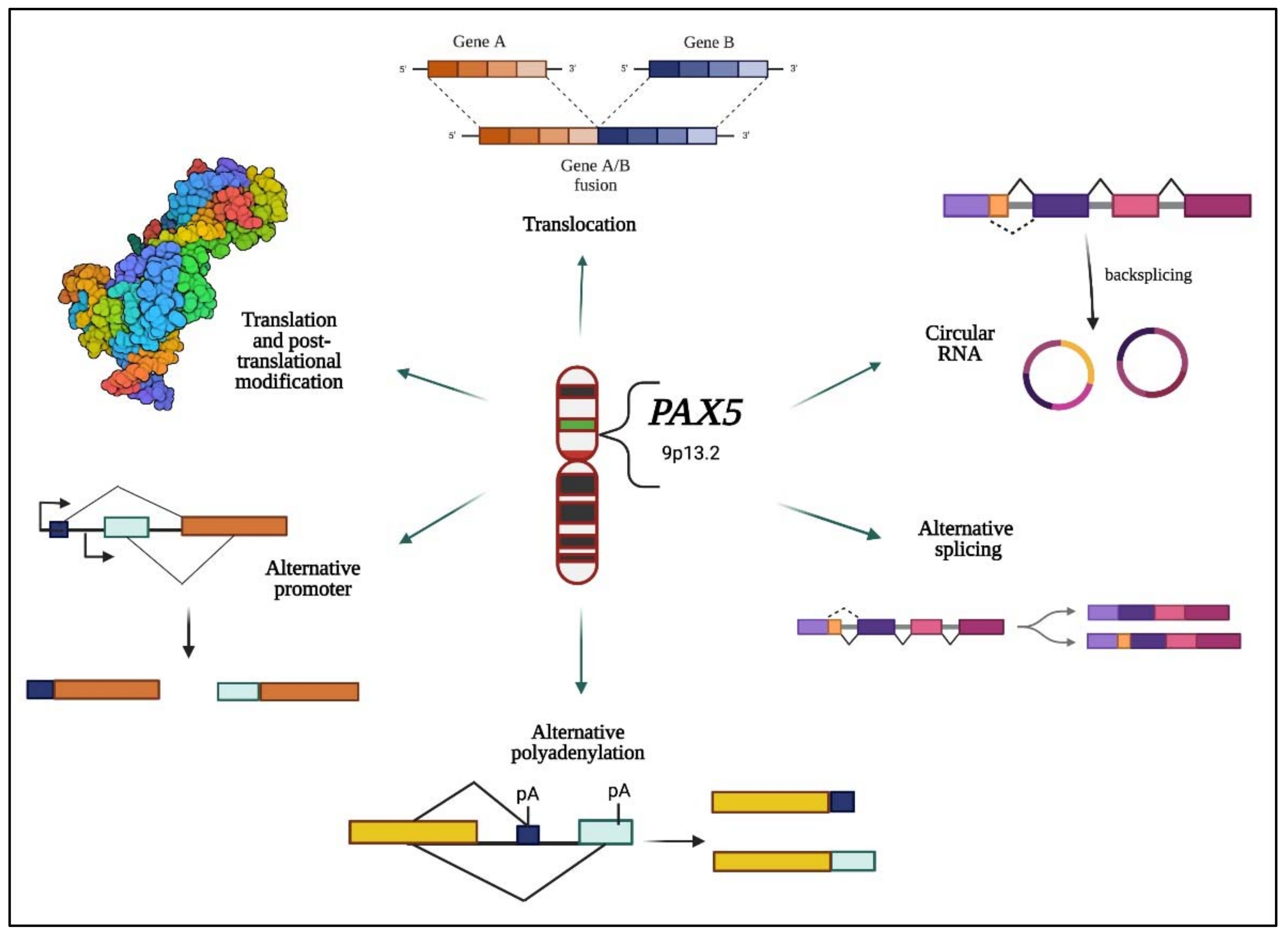
Figure 1. Hallmarks of PAX5 gene alterations and expressed products. This schematic illustration represents the reported modifications of the PAX5 gene sequence and expressed products. Multiple studies indicate that the human PAX5 gene undergoes various alterations that affect its spatiotemporal expression to function as an oncogene or tumor suppressor. Specifically (clockwise from the top), the PAX5 locus can undergo: genetic translocations resulting in chimeric fusion proteins and alternative promoter regulation [7,8,9,10,11]; transcriptional back-splicing resulting in circular PAX5 RNAs [12,13,14]; transcriptional alternative splicing of coding exons [15,16,17]; alternative use of poly-adenylation (pA) termination signals [18]; use of alternative promoter regions and transcription initiation sites [11]; and post-translational modifications, which impact its transactivation potential [19,20,21].
Ijms 23 10095 g001
Figure 2. PAX5 expression profiles in human tissue collections. (A) Relative PAX5 transcription levels from deep sequencing data in over 560 cancer cell lines depict a significant level of PAX5 RNA expression in human malignancies. The data was plotted using DepMap (https://depmap.org/portal/depmap, accessed on 1 April 2022). Raw reads were aligned with the STAR algorithms (https://github.com/alexdobin/STAR, accessed on 1 April 2022). Relative transcript quantification was performed with the RSEM and subsequently TPM-normalized. Final data were plotted as log2(TPM+1). (B) Comparison analysis of PAX5 protein/RNA levels in different cancer types are plotted and show a significant translation of PAX5 RNA in blood, bone, and human lymphocytes. In contrast, despite the high RNA expression, PAX5 protein level remains low in most examined solid cancer tumors, suggesting that these cells use alternative regulatory mechanism affected by PAX5 RNA products. The data was analyzed using the Proteomics DB web tool (https://www.proteomicsdb.org, accessed on 4 April 2022). Values are shown as median value ± interquartile range in log2-transformed iBAQ intensities.
Ijms 23 10095 g002
Figure 3. PAX5 interaction networks and related biological pathways. (A) PAX5 gene interaction networks have been mapped using the Cytoscape plugin GeneMANIA (https://genemania.org, accessed on 8 April 2022). Schematic illustrations of functional annotations and biological terms visualization are represented by: (B) PAX5 gene ontology (GO) in terms of functional orthologs and their relative implication in each predicted biological processes; (C) PAX5 pathway analysis using the Kyoto Encyclopedia of Genes and Genomes (KEGG) database, which provides an integrated evaluation of genomic, chemical, and biochemical functions; and (D) relative functional association to biological reactomes based on PAX5-related network genes. Annotations were done using the Enrichr algorithms (https://maayanlab.cloud/Enrichr, accessed on 10 April 2022). Significance was considered if p < 0.05.
Ijms 23 10095 g003
Figure 4. Mutation-independent mechanisms leading to aberrant PAX5 signaling and cell processes. Aside from PAX5 gene sequence alterations, deregulated PAX5 expression can also result from epigenetic events and post-transcription modifications. First, PAX5 gene promoter hypermethylation has been described in many cancers, notably when PAX5 behaves as a tumor suppressor. Post-transcriptional modifications (e.g., coding exon alternative splicing, 3′UTR shortening, and RNA circularization) also contribute to overall PAX5 expression and function. The net production of functional PAX5 transcription factors can thereafter collaborate with IKZF1 to regulate downstream metabolic genes to limit glucose uptake and energy supply required for oncogenic transformation. Adequate PAX5 function is also required to regulate Tp53 expression and avoid uncontrolled cancer phenotypes. Tp53 is also intimately linked to metabolic disfunction leading to cancer processes.
Ijms 23 10095 g004
Publisher’s Note: MDPI stays neutral with regard to jurisdictional claims in published maps and institutional affiliations.