Article

Rapid Synergistic Biofilm Production of Pseudomonas and Candida on the Pulmonary Cell Surface and in Mice, a Possible Cause of Chronic Mixed Organismal Lung Lesions

by
Pornpimol Phuengmaung
1,2,
Jiradej Mekjaroen
3,
Wilasinee Saisorn
1,2,
Tanittha Chatsuwan
1,
Poorichaya Somparn
3,* and
Asada Leelahavanichkul
1,2,*,†
1
Department of Microbiology, Faculty of Medicine, Chulalongkorn University, Bangkok 10330, Thailand
2
Center of Excellence on Translational Research in Inflammation and Immunology (CETRII), Department of Microbiology, Faculty of Medicine, Chulalongkorn University, Bangkok 10330, Thailand
3
Center of Excellence in Systems Biology, Research Affairs, Faculty of Medicine, Chulalongkorn University, Bangkok 10330, Thailand
*
Authors to whom correspondence should be addressed.
Current address: Immunology Unit, Department of Microbiology, Chulalongkorn University, Bangkok 10330, Thailand.
Int. J. Mol. Sci. 2022, 23(16), 9202; https://doi.org/10.3390/ijms23169202
Submission received: 20 July 2022 / Revised: 12 August 2022 / Accepted: 15 August 2022 / Published: 16 August 2022

Abstract

:
Due to the possible co-presence of Pseudomonas aeruginosa and Candida albicans (the most common nosocomial pathogens) in lungs, rapid interkingdom biofilm production is possible. As such, PA+CA produced more dominant biofilms on the pulmonary epithelial surface (NCI-H292) (confocal fluorescent extracellular matrix staining) with dominant psl upregulation, as demonstrated by polymerase chain reaction (PCR), after 8 h of experiments than PA alone. With a proteomic analysis, rhamnosyltransferase RhlB protein (Psl-associated quorum-sensing protein) was found to be among the high-abundance proteins in PA+CA than in PA biofilms, supporting psl-mediated biofilms in PA+CA on the cell surface. Additionally, PA+CA increased supernatant cytokines (IL-8 and IL-13, but not TNF-α, IL-6, and IL-10) with a similar upregulation of TLR-4, TLR-5, and TLR-9 (by PCR) compared with PA-stimulated cells. The intratracheal administration of PA+CA induced a greater severity of sepsis (serum creatinine, alanine transaminase, serum cytokines, and histology score) and prominent biofilms (fluorescent staining) with psl upregulation (PCR). In comparison with PA+CA biofilms on glass slides, PA+CA biofilms on biotic surfaces were more prominent (fluorescent staining). In conclusion, PA+CA induced Psl-predominant biofilms on the pulmonary cell surface and in mice with acute pneumonia, and these biofilms were more prominent than those induced by PA alone, highlighting the impact of Candida on rapid interkingdom biofilm production.

1. Introduction

Biofilms are microbial communities that are surrounded by heterogeneously complex extracellular polymeric substances (EPSs) resulting, in part, from the harsh micro-environment, caused by host immunity, limited nutrient availability, and competition from other microbes [1]. Quorum-sensing is one of the most potent molecules for the initiation of cell-to-cell communication within the microbial communities of microorganisms, followed by the synthesis of the functional molecules that have an impact on the different phenotypes of biofilms, such as the ability to attach to abiotic and biotic surfaces [2,3,4]. Because biofilm-residing microorganisms are protected from host immune responses and antibiotics, infections with biofilms are usually difficult to treat [5].
A mixed infection by different bacteria (polymicrobial infection) or organisms (interspecies infection) is well-known, as Pseudomonas aeruginosa and Staphylococcus aureus are the two of the most prevalent bacterial species in the lungs of cystic fibrosis [6]. Likewise, 20% of systemic Candida albicans infections are polymicrobial, with Staphylococcus epidermidis and Staphylococcus aureus being the first and third most common co-isolated organisms, respectively, with a strong interspecies biofilm formation [7,8]. Moreover, an enhanced severity of infection with the co-presence of C. albicans and Klebsiella pneumoniae has been reported [9]. However, Pseudomonas aeruginosa and Candida albicans are two of the most common opportunistic pathogens that usually colonize numerous sites of the human body, resulting in several diseases, including burn wounds, contaminated catheters, and infections in several organs (the genitourinary tract, gut, and lung). Interestingly, the co-presence of Pseudomonas and Candida induces prominent interkingdom biofilms [10,11,12,13] with more severe infections [14,15,16] as indicated in lungs with cystic fibrosis and ventilation-associated pneumonia (VAP) [17,18,19,20,21], which is partly due to the potent Pseudomonas quorum-sensing proteins (growth promotion) and elastases (disruption in the tight junctions of host cells) [22,23]. Additionally, some molecules from Candida spp. also facilitate the biofilm productivity of Pseudomonas, as the addition of Candida spp. into preformed Pseudomonas biofilms increases biofilm production more than does the initial simultaneous incubation of Pseudomonas with Candida [13].
Because the presence of C. albicans in the human pulmonary system is frequently reported [24] and because of the high prevalence of nosocomial pneumonia from Pseudomonas spp. [25], the co-existence of Pseudomonas and Candida in pulmonary lesions and their synergy in biofilm production is possible. However, most of the previous publications on the synergy of Pseudomonas–Candida biofilms are conducted using abiotic surfaces (glass slides, plastics, and catheters) [13]. While Pseudomonas biofilms on abiotic surfaces consist of an alginate-predominant matrix, biofilms on the bronchial airway are composed of proteins mainly from the polysaccharide synthesis locus (Psl) gene [26,27], implying that biofilm properties depend on surface characteristics. Thus, studies of biofilms on biotic surfaces might better resemble patients’ conditions.
Moreover, in vivo biofilm formation is mostly demonstrated in chronic infections, especially Pseudomonas and Candida, despite the rapid formation of biofilms (within 1–2 days) after in vitro incubation and the common co-presence of both organisms [13]. Hence, the co-presentation of both organisms on the biotic surface of the pulmonary system for a short period might cause chronic interkingdom lesions, and the understanding of this pathophysiology is beneficial for the treatment of Pseudomonas–Candida infections in several diseases (cystic fibrosis, VAP, and other nosocomial pneumonias). Therefore, we tested the influence of the co-presence of Pseudomonas–Candida on a human pulmonary mucoepidermoid carcinoma cell line (NCI-H292) and on glass slides, together with a mouse model of aspiration pneumonia.

2. Results

2.1. Prominent Mucoid Biofilms from Pseudomonas plus Candida on Human Pulmonary Cells

Due to the lack of data on interkingdom Pseudomonas–Candida biofilms on pulmonary cell surfaces, biofilms from mixed organisms and Pseudomonas alone were stained for extracellular matrix, bacterial DNA, and fungi (Figure 1A,B). At 24 h after inoculation, there were more prominent Pseudomonas–Candida biofilms (PA+CA) with a higher abundance of bacteria and fungi than Pseudomonas alone (PA) biofilms (Figure 1A,B). Additionally, PA+CA and PA alone reduced the number of pulmonary cells as indicated by an MTT assay (this was more prominent in the PA+CA group than in the PA alone group) (Supplement Figure S1A) due to Pseudomonas cytotoxicity [6]. There was an increase in the intensity of yellow fluorescence (Figure 1A, middle), which might represent elevated cell tight junctions and bacterial DNA abundance; however, bacterial abundance, determined by culturing, was higher in the PA+CA biofilms than in the PA alone biofilms (Supplement Figure S1B). Although the enhanced blue color in the PA+CA biofilms than that in the PA alone biofilms (Figure 1A right) might be due to an increase in both pulmonary and yeast cells, the fungal abundance in the PA+CA biofilms was higher than that in the PA alone biofilms (Supplement Figure S1C). The cell injury, as determined by the loss of cell tight junctions (ZO-1), was higher in the PA alone biofilms than in the PA+CA biofilms, as the linear yellow staining color in the PA+CA biofilms was more prominent than that in the PA alone biofilms (Figure 1B), supported by the higher number of dead cells observed in the PA alone biofilms than in the PA+CA biofilms using the MTT assay (CA alone did not induce cell death) (Supplement Figure S1A). Notably, the incubation of Candida alone did not induce the formation of biofilms on the pulmonary cells (Supplementary Figure S2). It is possible that the better conditions of the biotic surface in the PA+CA group than those of the unhealthy surface in the PA alone group were partly responsible for the more prominent biofilms in the PA+CA condition.
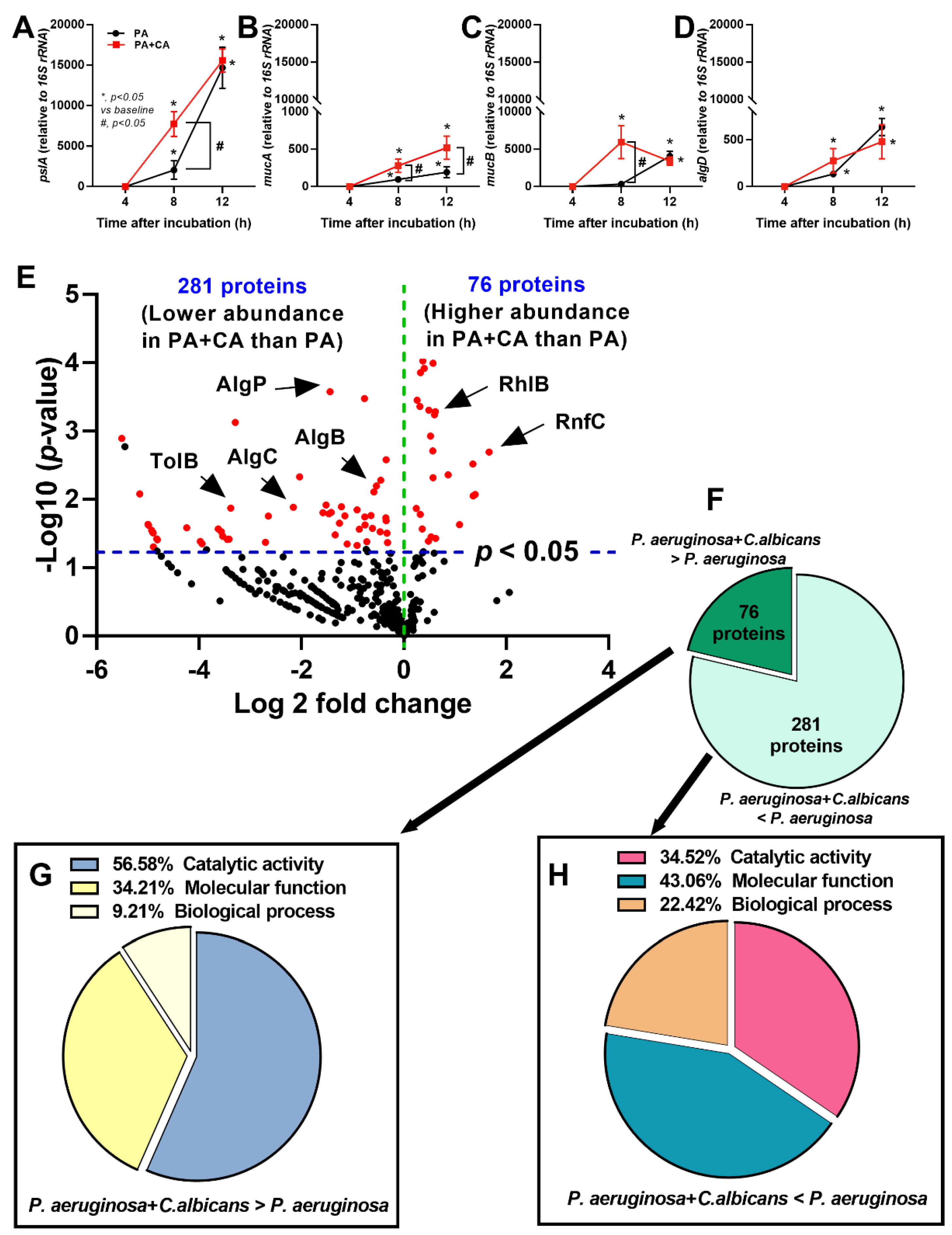
At 8 h post-incubation, PA+CA induced a lower expression of pslA (a gene required for Psl production) with higher mucA and mucB (negative regulators of alginate) but similar algD (a gene required for alginate production) than PA alone (Figure 2A–D). After 24 h of incubation, the expression of mucA, but not other genes, was higher in the PA+CA group than in the PA alone group (Figure 2A–D). These data suggest a possible difference between the biofilm properties of PA+CA and PA alone through the more prominent psl-mediated biofilm components, with a more dominant alginate negative regulator (mucA) in PA+CA biofilms than in PA alone biofilms on the lung epithelial cell surface [28,29].
Additionally, the differences between both groups were demonstrated by a proteomic analysis based on the fold change of the proteins in the PA+CA biofilms compared with that of the proteins in the PA alone biofilms, as indicated by a volcano plot (Figure 2E), and only 76 proteins were demonstrated to be higher in the PA+CA biofilms than in the PA alone biofilms (Figure 2F) using the protein biological process based on the PANTHER classification (http://www.pantherdb.org; accessed on 15 June 2022). These proteins included the proteins for rhamnosyltransferase RhlB (Psl-associated quorum-sensing) and iron-translocating oxidoreductase complex subunit C (RnfC) (Figure 2G). Meanwhile, there were 281 proteins that were expressed in the PA+CA group at a lower level than in the PA alone group, including the proteins for alginate-mediated biofilms, transcriptional regulatory protein (AlgP), alginate biosynthesis transcriptional regulatory protein (AlgB), phosphomannomutase/phosphoglucomutase (AlgC), and Pseudomonas pathogenesis (Tol–Pal system protein; TolB) (Figure 2H). Notably, the initial abundance of bacteria in the PA+CA group was half of that in the PA alone group (organismal abundance between PA+CA and PA alone groups was controlled), and most of the bacterial proteins in the PA biofilms were higher than those in the PA+CA biofilms. The higher proteins in the PA+CA group than those in the PA alone group are the interesting Candida-inducing Pseudomonas proteins.

2.2. The Pulmonary Responses against Interkingdom Pseudomonas–Candida Biofilms

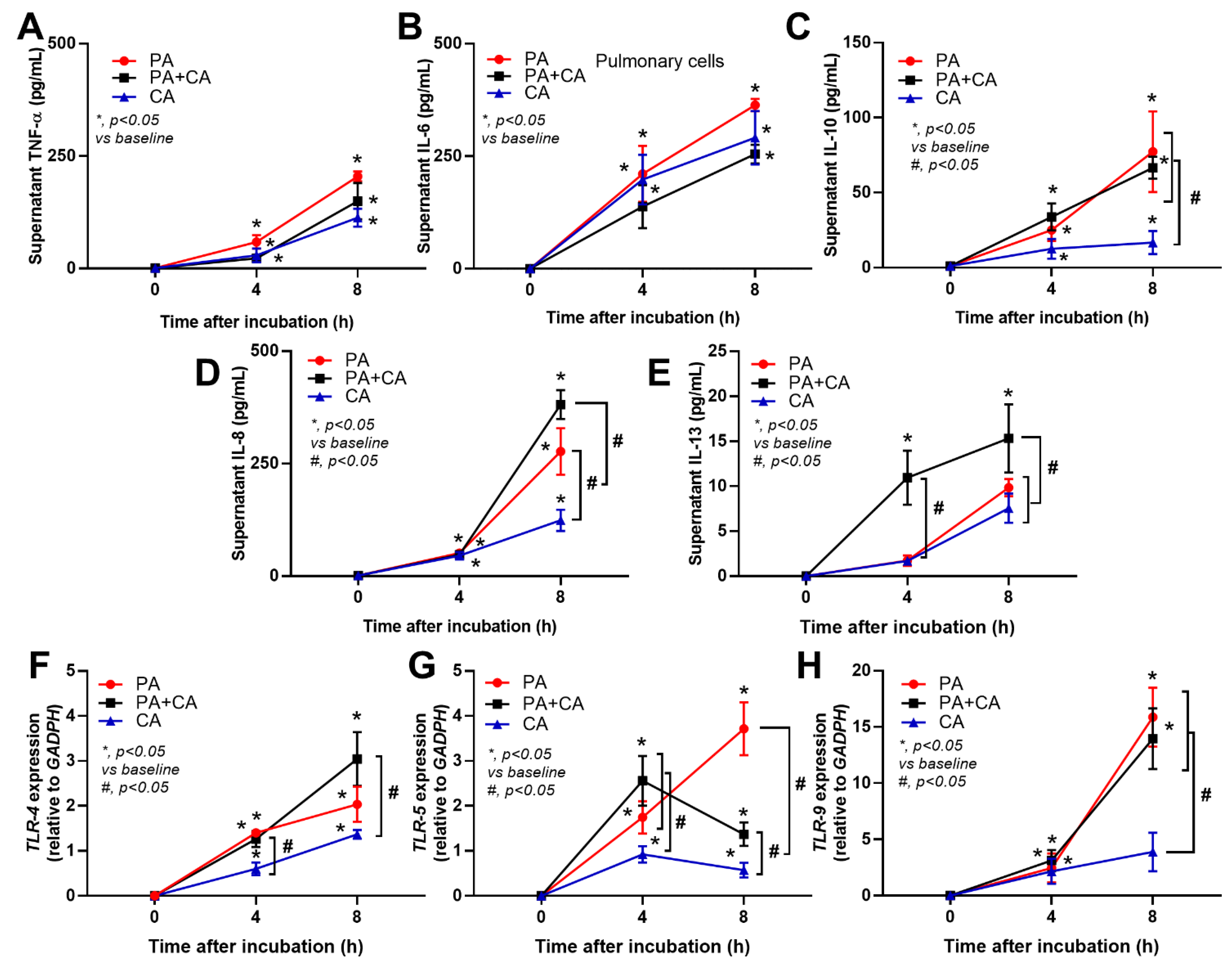
Because (i) the better pulmonary cell tight junction in the PA+CA group than that in the PA alone group (Figure 1B, the linear yellow color) might be due to the higher psl-mediated Pseudomonas exopolysaccharides (Figure 2A–D) (possibly induced by fungal ethanol [30]) and because (ii) pulmonary impacts from psl-mediated lung responses [26,31] with Pseudomonas–Candida competition is possible [32], the cell responses against PA+CA and PA alone might be different. Indeed, PA+CA biofilms induced higher proinflammatory responses in pulmonary epithelial cells, mostly at 8 h post-stimulation as indicated by supernatant IL-8 and IL-13, but not TNF-α, IL-6, or IL-10 than PA alone biofilms (Figure 3A–E). Notably, IL-8 and IL-13 are cytokines for chemotaxis and pulmonary mucosal inducers (inflammation, mucus hyperproduction, goblet cell hyperplasia, and subepithelial airway fibrosis), respectively [33,34]. Meanwhile, PA biofilms increased supernatant levels of IL-8 and IL-10, but did not increase the levels of other cytokines, when compared with CA biofilms (CA induced the lowest cytokine levels among these triggers) (Figure 3A–E). In parallel, similar Toll-like receptor 4 (TLR-4) upregulation was found in pulmonary epithelial cells after PA alone and PA+CA (higher than CA activation) (Figure 3F), implying a more potent TLR-4 inducer of LPS from P. aeruginosa than the stimulation from Candida antigens [35]. However, the upregulation of TLR-5 was the most prominent in PA+CA activation (at 8 h after incubation) than in PA or CA alone (Figure 3G), supporting the more potent TLR-5 activation by Pseudomonas flagella than Candida antigens [36]. Furthermore, TLR-9 upregulation was similar between PA+CA and PA alone groups, and this was higher than in the CA group (Figure 3H), possibly due to the higher cytosine–guanine dinucleotide (CpG) DNA in bacteria than in fungi [37]. Hence, the co-presence of Pseudomonas with Candida induced the most potent hyper-inflammation, as indicated by cytokines (IL-8 and IL-13), partly though the activation of TLR-4, TLR-5, and TLR-9.

2.3. Biofilm Formation in the Lungs of Acute Pneumonia, a Possible Initiator of Chronic Lesions

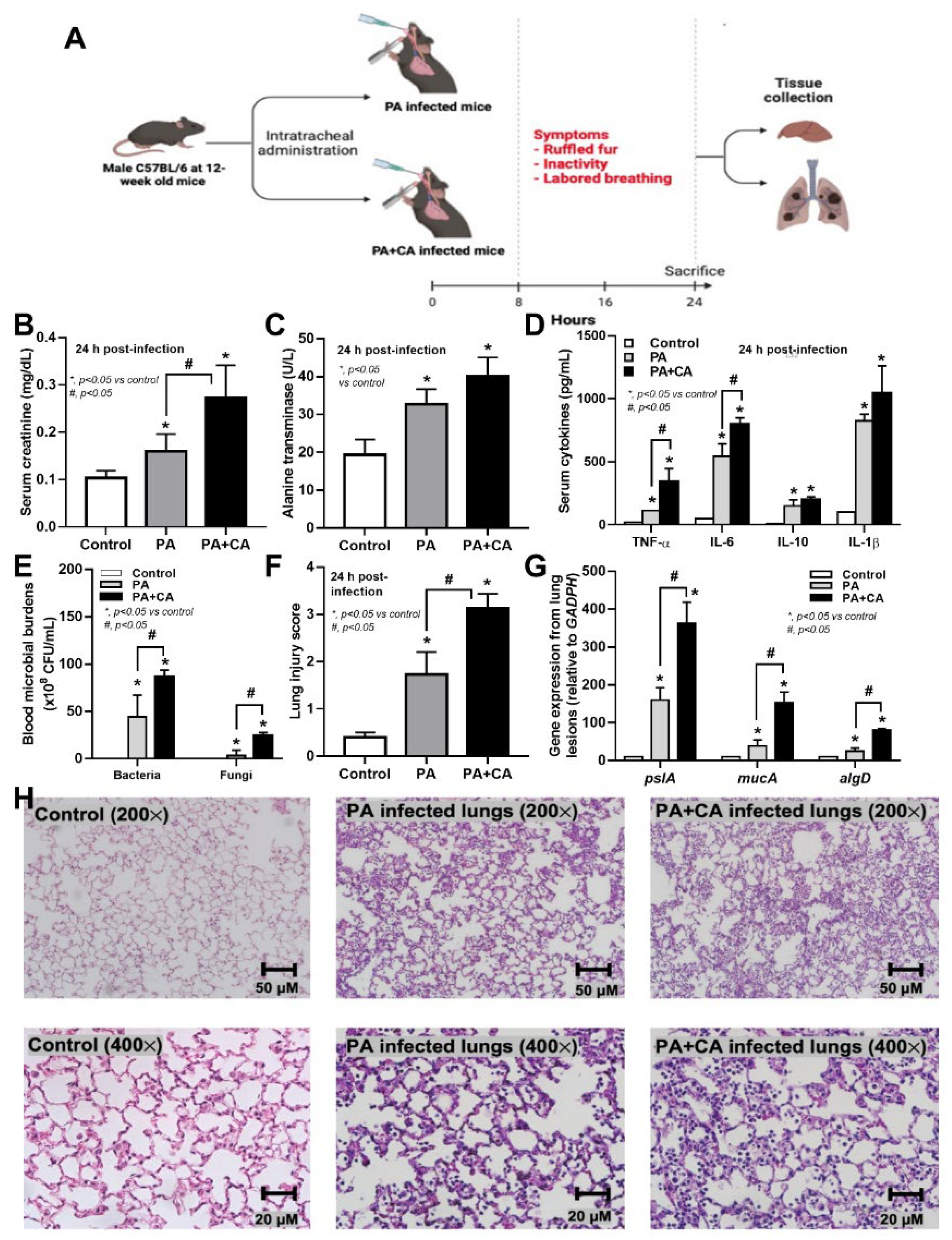
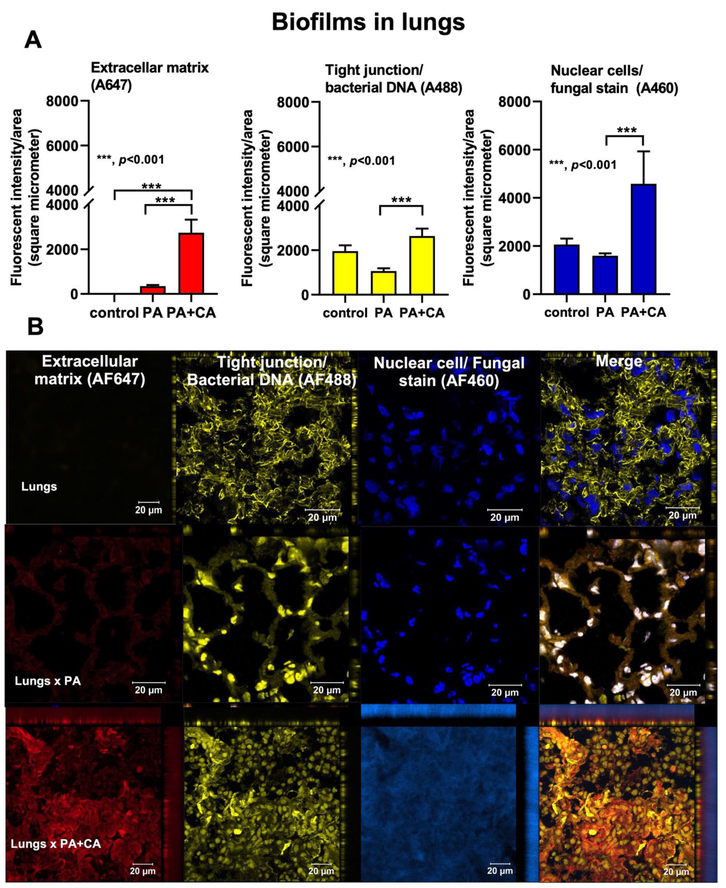
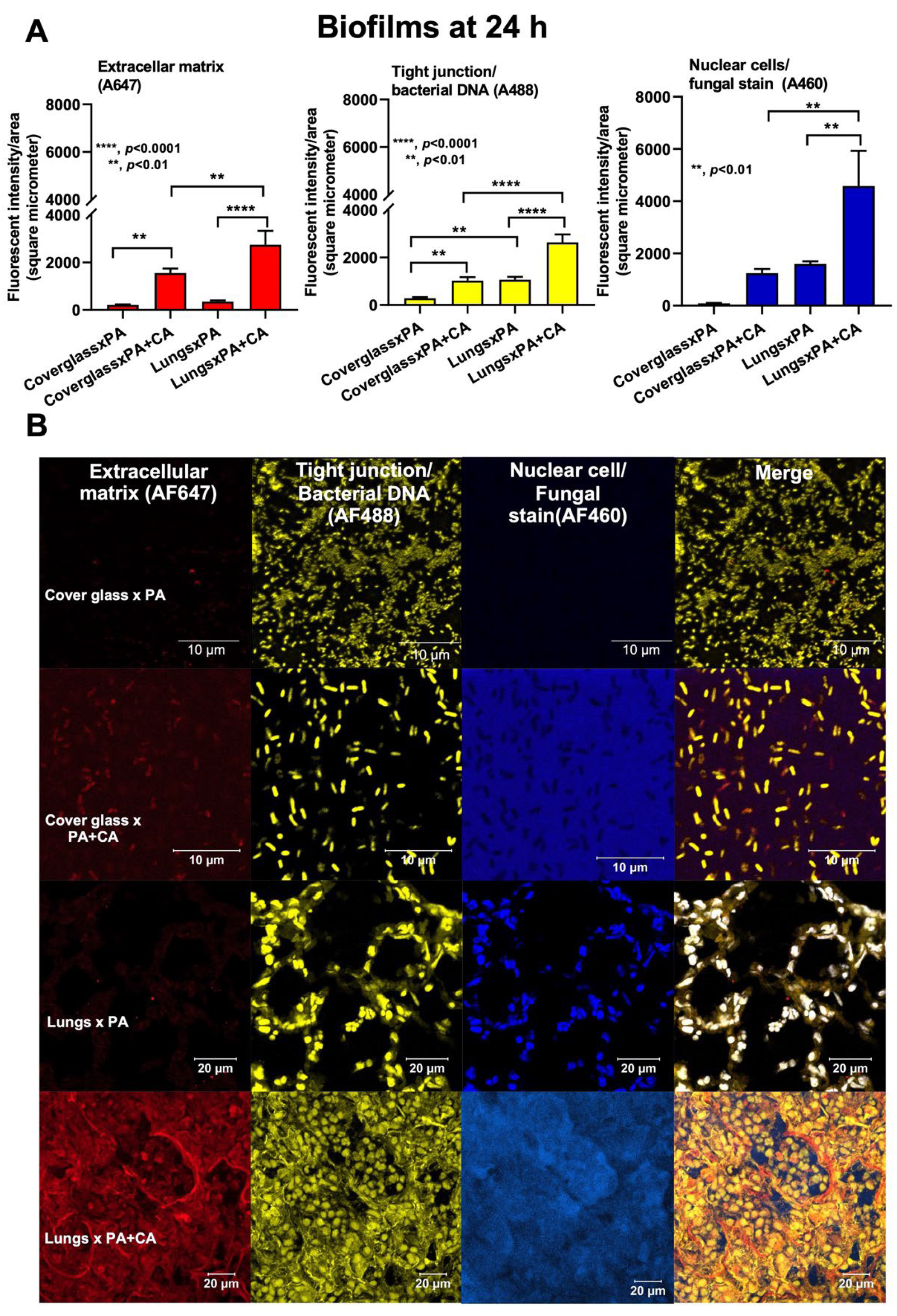
Because of the initial epithelial adhesion property of psl-mediated biofilms in mucoid Pseudomonas being more potent than that of alginate-predominant biofilms [26] and because of Candida-induced psl upregulation (Figure 2A), we hypothesized that Pseudomonas with Candida could rapidly produce biofilms and tested it in vivo (Figure 4A). At 24 h post-infection, sepsis severity was more prominent in the PA+CA group than in the PA alone group, as indicated by acute kidney injury (serum creatinine), liver damage (alanine transaminase), serum cytokines (TNF-α and IL-6, but not IL-10 or IL-1β), bacteremia, lung injury score, and the expression of biofilm-associated genes (pslA, mucA, and algD) (Figure 4B–H). There was no fungemia in the PA+CA group (data not shown), and the more prominent bacteremia in the PA+CA group than that in the PA alone group might be due to the higher bacterial abundance in the PA+CA group than that in the PA alone group on the pulmonary surface (Supplement Figure S1B). The psl upregulation in the PA+CA group supports the enhanced Pseudomonas biofilms by Candida on the pulmonary cell line at 24 post-incubation in vitro (Figure 1A,B). Despite the limited data on biofilms in acute pneumonia, the fluorescent staining of mouse lung tissue demonstrated the dominant extracellular matrix along with higher bacterial and fungal DNA in the PA+CA group when compared with those in the PA alone group (Figure 5A,B). There was a higher abundance of bacterial DNA in the PA+CA group at 24 h of the pneumonia model (Figure 5A,B), although the initial administration of fewer bacterial abundances in the PA+CA group than in the PA alone group was used as a control for the equivalent microbial abundance between groups (see Materials and Methods Section). Notably, the possible protective pulmonary tight junction of the PA+CA group on the cell line (Figure 1B, yellow) was not obvious in the mice (Figure 5B, yellow), suggesting a difference between the in vitro and in vivo conditions. Additionally, the biofilm abundance of the PA+CA group on the mouse lung tissue as indicated by extracellular matrix staining (red) (Figure 5A,B) was similar to the in vitro biofilms on the cell line (Figure 1A,B), which were clearly more prominent than the biofilms on the abiotic surface (glass slide) (Figure 6A,B). Indeed, the fluorescent intensities of the extracellular matrix and the microbial DNA (bacteria and fungi) of the PA+CA biofilms on the biotic surfaces were higher than those on the abiotic glass slides (Figure 6A,B). Meanwhile, the abundance of the PA alone biofilms on abiotic glass slides was not different to that on biotic surfaces (Figure 6A,B). These data indicate the possible impact of biotic surface on biofilm formation and rapid biofilm formation, especially on bacterial–fungal interkingdom biofilms. The abundance of Candida in the respiratory system of patients with pneumonia might be associated with the risk of biofilm formation and chronic infection. More studies on these topics are interesting

3. Discussion

Although the physical and molecular interactions between P. aeruginosa and C. albicans have been extensively studied [38,39], the data on Pseudomonas–Candida interkingdom biofilms are still limited. The co-existence of P. aeruginosa and C. albicans on catheters enhances biofilm thickness via the alginate-related extracellular matrix [13], whereas the presence of Pseudomonas–Candida colonization on airway epithelial cells might be different. Indeed, Pseudomonas produces some molecules (such as phenazines) that induce ethanol from Candida, and ethanol facilitates the Pseudomonas extracellular matrix through the psl operon as a positive feedback loop [13], perhaps in order to adapt to the micro-environment. Because (i) the prominent biofilm production of PA+CA over PA alone might be due to the different biofilm properties and because of (ii) the importance of several genes for Pseudomonas biofilm production, especially for biofilms from the polysaccharide synthesis locus (Psl) versus the alginate-mediated pathway [22,25], the expression of some genes was explored. Pseudomonas biofilm-producing genes (alginate, psl, and pel) are important Pseudomonas virulent factors [40]. As such, alginate is a negatively charged acetylated polymer containing nonrepetitive β-1,4-linked l-guluronic and d-mannuronic acids used for inhibiting phagocytosis and enhancing reactive oxygen species in host cells [41,42,43,44]. Additionally, Psl is a neutral-branched pentasaccharide with a ratio of 1:1:3 of d-glucose, d-rhamnose, and d-mannose, and it mediates attachment to lung epithelial cells, promoting a pro-inflammatory response and host cell damage [31,45]. Meanwhile, Pel is a positively charged polysaccharide containing acetylated 1→4 glycosidic branches of N-acetylgalactosamine and N-acetylglucosamine, which stabilize the biofilm structure [46,47]. In parallel, Candida biofilms, polysaccharide-rich extracellular matrices [48], especially in the hyphae form, can be facilitated by yeast-to-hyphae accelerators of Pseudomonas (quorum-sensing molecules) together with hyphae-to-yeast inhibitors (such as farnesol) from the Candida themselves [49,50]. Different from Pseudomonas–Candida alginate-dominant biofilms on catheters [13], the biotic surfaces upregulated pslA that was more profound than the Pseudomonas alone biofilms, implying an important role of the lung cell surface on interkingdom biofilms. In the proteomic analysis, Pseudomonas–Candida proteins demonstrated the prominent proteins for iron-translocating oxidoreductase complex subunit C (RnfC) and rhamnosyltransferase RhlB (Psl-related quorum-sensing protein) with lower alginate-associated proteins (AlgP, AlgB, and AlgC) when compared to Pseudomonas alone, suggesting that the presence of Candida possibly led to the competition for iron resources (a source of nutrition) and that it induced Psl-dominated biofilms, which effectively attach to epithelial cells with lower cell responses than alginate-based biofilms [51].
Because the epithelial barrier integrity [32,52] is important for the first defense against pathogens [53,54] and because the loss of ZO-1 tight junction molecules in human lungs with P. aeruginosa infection has also been previously demonstrated [55,56], studies of biofilms on epithelial cell surfaces might better resemble the clinical situation. Here, there were increased biofilms in Pseudomonas–Candida compared to Pseudomonas alone on both the cell line and lung tissue, with an increased cell abundance of both bacteria and fungi, which was more prominent than the abundance on the glass slides, implying that the biotic surface has an impact on microbial growth and biofilm production. Despite Pseudomonas–Candida seeming to protect the pulmonary tight junction better than Pseudomonas alone (possibly for a better biofilm structure), the increased bacterial abundance of the Pseudomonas–Candida group might have resulted in higher bacteremia and more severe sepsis in our mouse model. The prominent psl upregulation, with the reduced expression of alginate-associated genes, was indicated by the high mucA (an alginate regulator) and low algD (an alginate promotor) of Pseudomonas–Candida compared to Pseudomonas alone, and this was similarly demonstrated both on pulmonary cells and in mouse lungs, despite the possible better nourishment and host–organism ratio in mice. Regarding the host responses against biofilms, Pseudomonas–Candida biofilms induced more severe responses than Pseudomonas alone biofilms through several well-known signals, including TLR-4, TLR-5, and TLR-9 from the activation by LPS, flagella, and microbial DNA [57,58,59]. Interestingly, microbial dissemination was demonstrated only from bacteremia but not fungemia, perhaps the yeast cells were too large to pass through the damaged pulmonary barrier, and the more severe sepsis in the Pseudomonas–Candida group might be mainly due to the higher bacterial abundance in the blood than in the mice with Pseudomonas alone. Despite the similar bacterial abundance demonstrated between Pseudomonas–Candida biofilms and Pseudomonas alone biofilms on the abiotic surface (glass slide) in our previous report [13], the microbial abundance (both bacteria and fungi) of Pseudomonas–Candida biofilms was higher than that of Pseudomonas alone biofilms, highlighting that host cells have an impact on microbial growth (in a similar way to microbial contamination on the cell culture) [60].
Interestingly, the treatment of bacterial–Candida interspecies biofilms is challenging, as the effect of antibacterials alone seems to be decreased in dual-species biofilms compared with bacteria alone biofilms [61]. Meanwhile, in Pseudomonas–Candida biofilms, a single treatment with an anti-fungal drug (fluconazole) alone synergistically with Pseudomonas on the biofilms more effectively reduces Candida burdens, partly through iron sequestration by P. aeruginosa [11]. These data indicate that anti-fungals might be beneficial in the treatment of bacterial–Candida interspecies biofilms. Moreover, quorum-quenching (quorum-sensing blockage) and anti-virulence compounds (inhibitors against the virulent factors that develop from dual biofilms) are new, interesting strategic treatments of inter-species biofilms [30]. More studies on this topic are required.
In our acute pneumonia model, the biofilms were surprisingly rapidly produced, which, in parts, was associated with the more severe antibiotic resistance and microbial persistence in the respiratory tract with the co-infection of Pseudomonas–Candida [62,63]. In this regard, the studies on biofilms, which currently are mostly only being conducted in some time-consuming chronic infection models, could be conducted in acute infection models (especially for the early phase of biofilms) and may rapidly expand the knowledge on the topic. However, chronic infection models are still necessary for the evaluation of the progression from the acute phase to the chronic phase of biofilms. For clinical translation, we propose that Candida abundance in the pulmonary tract of patients with Pseudomonas pneumonia might be associated with rapid biofilm production, treatment difficulties, and progression into a chronic form of infection. A further exploration of Candida abundance in lungs and pneumonia is warranted.

4. Materials and Methods

4.1. Microbial Preparation and Biofilms on Human Cells

P. aeruginosa and C. albicans were isolated from the endotracheal specimens of patients from the King Chulalongkorn Memorial Hospital, Bangkok, Thailand, following the protocol (MDCU-IBC001/2022) approved by the ethical institutional review board, Faculty of Medicine, Chulalongkorn University, according to the Declaration of Helsinki, with written informed consents. Human mucoepidermoid carcinoma (NCI-H292) cells were grown and maintained in Dulbecco’s modified Eagle medium (DMEM; Life Technologies, Carlsbad, CA, USA), supplemented with 10% heat-inactivated fetal bovine serum (FBS; Life Technologies) and 1% penicillin/streptomycin in a humidified 5% CO2 incubator at 37 °C. Both P. aeruginosa and C. albicans were grown in tryptic soy broth (TSB; Difco™, Becton, NJ, USA) to induce the early stationary phase, which is appropriate for biofilm formation, and they were washed and resuspended in fresh prewarmed completed DMEM containing 10% FBS to adjust the turbidity of 1 × 10⁶ CFU/mL. The suspensions (P. aeruginosa plus C. albicans or each organism alone) were added to 12-well polystyrene plates with 1 × 10⁶ confluent NCI-H292 cells at the multiplicity of infection (MOI) between organisms per cells at 1:100 on a biotic surface or on the polystyrene well with a round glass coverslip in the bottom (abiotic surface) in order to evaluate biofilm formation. To control the number of organisms in each condition, 1 × 10⁶ CFU/mL of the preparation of P. aeruginosa alone (PA) or C. albicans alone (CA) was used, while 0.5 × 10⁶ CFU/mL of Pseudomonas and Candida preparations was combined to use for the preparations of Pseudomonas plus Candida (PA+CA).
Biofilms were grown at 37 °C with 5% CO2, and the culture medium was changed every 12 h. The viability of NCI-H292 in these biofilms was measured using an MTT (3-[4,5-dimethylthiazol-2-yl]-2,5 diphenyltetrazolium bromide) assay (Invitrogen, Waltham, MA, USA) according to the manufacturer’s procedures. Briefly, the 24 h activated cells were incubated with 0.5 mg/mL of MTT solution for 2 h at 37 °C in the dark before the removal of MTT, diluted with dimethyl sulfoxide (DMSO) (Thermo Fisher Scientific, Waltham, MA, USA), and measured using a Varioskan Flash microplate reader at an absorbance of optical density of 570 nm. Supernatants were quantified for cytokine measurement using an enzyme-linked immunosorbent assay (ELISA; Invitrogen). The ability of biofilm production on in vitro (biotic and abiotic surfaces) and in vivo the pulmonary lesions prepared in Tissue-Tek O.C.T Compound (Sakura Finetek, Torrace, CA, USA) (details later) was evaluated by employing confocal microscopy using ZEISS LSM 800 (Carl Zeiss, Jena, Germany) with the following antibodies: (i) extracellular matrix using Alexa FluorTM647-tagged Concanavalin A (Invitrogen) (red color of AF647), (ii) bacterial DNA by SYTO9 color (Invitrogen) (yellow color of AF488), (iii) pulmonary cell borders through tight junction zonula occluden-1 (ZO-1) (Thermo Fisher Scientific) with the goat anti-rabbit secondary IgG (Cell Signaling Technology, Danvers, MA, USA) (yellow color of AF488), (iv) Calcofluor white of fungal stain (Sigma-Aldrich, St. Louis, MI, USA) (blue color of AF460), and (v) nuclei of pulmonary cells by DAPI (4′,6-diamidino-2-phenylindole) color (Cell Signaling Technology) (blue color of AF460). Notably, AF488 yellow was used for both pulmonary tight junctions and bacterial DNA, while AF460 blue was stained for the nuclei of both human cells and fungi due to technical limitations. However, the different characteristics (shapes and sizes) between tight junctions and bacterial DNA, and the dissimilar nucleus sizes between human cells and fungi were used for differentiation.

4.2. Quantitative Real-Time Polymerase Chain Reaction

Several molecules associated with P. aeruginosa biofilms with and without C. albicans on NCI-H292 cells (inflammation and biofilm formation) were evaluated using quantitative real-time polymerase chain reaction (qRT-PCR). Total RNA was extracted by TRIzol reagent (Thermo Fisher Scientific), converted by reverse transcriptase (RevertAid First Stand cDNA), and conducted qRT-PCR using Applied Biosystems QuantStudio 6 Flex Real-Time PCR System (Applied Biosystems, Warrington, UK) with SYBR® Green PCR master mix. The housekeeping gene 16S rRNA was used to normalize the transcriptional levels of target genes from P. aeruginosa, and GADPH was used to normalize the gene expression in NCI-H292 cells, with the comparative cycle threshold against the expression. The list of primers for PCR is presented in Table 1.

4.3. Proteomic Analysis

Due to the biofilm production of C. albicans being less than that of P. aeruginosa [9], the pulmonary cells (NCI-H292) with biofilms from P. aeruginosa plus C. albicans or P. aeruginosa alone were used for a proteomic analysis. Notably, the initial abundance of P. aeruginosa in the Pseudomonas alone (PA) group was approximately 2-fold higher than that in the Pseudomonas plus Candida (PA+CA) group due to the protocol to ensure a similar total organismal abundance between both conditions (PA vs. PA+CA). Then, acetone precipitation of proteins was performed, and the pellet was resuspended with 8 M urea in 50 mM Tris-HCl (pH 8) to estimate protein concentration using a BCA protein assay (Thermo Fisher Scientific). Subsequently, the dissolved proteins were incubated with dithiothreitol (DTT), alkylated by iodoacetamide (IAA), digested with trypsin, inhibited the digestion by trifluoroacetic acid (TFA), and lyophilized by SpeedVac centrifugation (Thermo Fisher Scientific). After that, the peptides were analyzed using an EASY-nLC1000 system coupled to a Q-Exactive Orbitrap Plus mass spectrometer (Thermo Fisher Scientific) equipped with a nano-electrospray ion source (Thermo Fisher Scientific) at a flow rate of 300 nL/min, followed by a linear gradient from 5% to 40% acetonitrile in 0.1% formic acid for 60 min, with 40–95% acetonitrile in 0.1% formic acid for 30 min. Fractioned peptide samples were carried out with a liquid chromatography-mass spectrometer (LC-MS) using a 10 data-dependent acquisition method with an MS scan range of 350–1400 m/z accumulated at a resolution of 70,000 full widths at half maximum (FWHM) followed by a resolution of 17,500 FWHM. The normalized collision energy of higher energy collisional dissociation (HCD) was controlled at 32%. Precursor ions with unassigned charge states of +1 or those greater than +8 were excluded, and the dynamic exclusion time was set to 30 sec. Data acquisition and interpretation were monitored using Proteome DiscovererTM software 2.1 (Thermo Fisher Scientific). Peptide spectra were evaluated using the SEQUEST-HT search engine against P. aeruginosa (strain ATCC 15692/DSM 22644/CIP 104116/JCM 14847/LMG 12228/1C/PRS 101/PAO1) Swiss-Prot Database (1435 proteins, March 2022). The following parameters were set for the search: (i) digestion enzyme: trypsin; (ii) maximum allowance for missed cleavages: 2; (iii) maximum of modifications: 4; (iv) fixed modifications: carbamidomethylation of cysteine (+57.02146 Da), as well as light and medium dimethylation of N termini and lysine (+28.031300 and +32.056407 Da); and (v) variable modifications: oxidation of methionine (+15.99491 Da). A biological process analysis of the host proteins was performed using the PANTHER (http://www.pantherdb.org/; accessed on 15 June 2022) program. Mass spectrometry proteomics data were determined and submitted to the ProteomeXchange Consortium via PRIDE (http://www.proteomexchange.org, accessed on 15 June 2022) with the data set identifier PXD034528.

4.4. Animals and Animal Model

Male, 12-week-old C57BL/6 mice, purchased from Nomura Siam International (Pathumwan, Bangkok, Thailand), were intratracheally administered with 0.2 mL of P. aeruginosa (1 × 10⁸ CFU/mL) or P. aeruginosa (0.5 × 10⁸ CFU/mL) with C. albicans (0.5 × 10⁸ CFU/mL) before sacrificing by cardiac puncture under isoflurane anesthesia with sample collections at 24 h post-administration. For a histological analysis, paraffined-embedded lungs were fixed with 10% neutral-buffered formalin before the processes with Hematoxylin and Eosin (H&E) staining, and the lung injury was evaluated at 200× magnification in 10 randomly selected fields for each animal. The lung injury score was determined based on desquamation, dystelectasis/atelectasis, congestion, interstitial thickness, infiltration, and bronchial exudate with a modified lung injury score [24] as follows: 0, no injury; 1, minimal/discrete; 2, mild; 3, moderate; and 4, severe. For bacteremia and fungemia, 10 μL of mouse blood was spread onto tryptic soy agar (TSA; Difco™, Becton, NJ, USA) and Sabouraud dextrose agar (SDA; Oxoid, Cambridge, UK) and incubated at 37 °C for 24 h before colony enumeration.

4.5. Statistical Analysis

Data are presented as mean ± standard error (SE). The differences between groups were examined using a one-way analysis of variance (ANOVA) with Tukey’s analysis or Student’s t-test for comparisons of multiple groups or two groups, respectively. Log-rank test was used for a survival analysis. The time-point data were analyzed using a repeat measured ANOVA. SPSS 11.5 software (SPSS, Chicago, IL, USA) and GraphPad Prism version 8.0 were used for all statistical analyses. A p-value of <0.05 was considered statistically significant.

5. Conclusions

In conclusion, in this study, the Pseudomonas–Candida biofilms on biotic surfaces were more psl predominant than the mixed-organism biofilms on abiotic surfaces. Moreover, the Pseudomonas–Candida biofilms were more profound than the monomicrobial Pseudomonas biofilms, highlighting that the impact of Candida and the attachment to host cells enhance Pseudomonas biofilm production.

Supplementary Materials

The following supporting information can be downloaded at: https://www.mdpi.com/article/10.3390/ijms23169202/s1.

Author Contributions

Conceptualization, P.P. and A.L.; methodology, P.P., J.M., and W.S.; software, P.P., J.M., and A.L.; validation, P.P., J.M., and A.L.; formal analysis, P.P., J.M., and A.L.; investigation, P.P., J.M., and A.L.; resources, P.P., W.S., J.M., and A.L.; data curation, P.P. and A.L.; writing—original draft preparation, P.P., J.M., T.C. and A.L.; writing—review and editing, P.P., P.S., and A.L.; supervision, P.S. and A.L.; funding, A.L. All authors have read and agreed to the published version of the manuscript.

Funding

This research was supported by Chulalongkorn University through the Fundamental Fund 65 [CUFRB65_hea[33]_040_30_21]; the National Research Council of Thailand (grant number NRCT-N41A640076); and (811/2563) with NSRF via the Program Management Unit for Human Resources & Institutional Development, Research, and Innovation (B16F640175 and B05F640144). P.P. was supported by the Second Century Fund (C2F) for Postdoctoral Fellowship, Chulalongkorn University.

Institutional Review Board Statement

The protocol (STT012/2564) was approved by the Institutional Animal Care and Use Committee of the Faculty of Medicine, Chulalongkorn University, Bangkok, Thailand, in accordance with the National Institutes of Health (NIH), USA.

Informed Consent Statement

Informed consent was obtained from all subjects involved in the study.

Data Availability Statement

The data presented in this study are available on request from the corresponding author.

Conflicts of Interest

The authors declare that there are no conflicts of interest.

References

  1. Thi, M.T.T.; Wibowo, D.; Rehm, B.H.A. Pseudomonas aeruginosa Biofilms. Int. J. Mol. Sci. 2020, 21, 8671. [Google Scholar] [CrossRef] [PubMed]
  2. Romling, U.; Balsalobre, C. Biofilm infections, their resilience to therapy and innovative treatment strategies. J. Intern. Med. 2012, 272, 541–561. [Google Scholar] [CrossRef] [PubMed]
  3. Kostylev, M.; Kim, D.Y.; Smalley, N.E.; Salukhe, I.; Greenberg, E.P.; Dandekar, A.A. Evolution of the Pseudomonas aeruginosa quorum-sensing hierarchy. Proc. Natl. Acad. Sci. USA 2019, 116, 7027–7032. [Google Scholar] [CrossRef]
  4. Hossain, M.A.; Sattenapally, N.; Parikh, H.I.; Li, W.; Rumbaugh, K.P.; German, N.A. Design, synthesis, and evaluation of compounds capable of reducing Pseudomonas aeruginosa virulence. Eur. J. Med. Chem. 2020, 185, 111800. [Google Scholar] [CrossRef] [PubMed]
  5. Otto, M. Staphylococcus epidermidis pathogenesis. Methods Mol. Biol. 2014, 1106, 17–31. [Google Scholar] [CrossRef] [PubMed]
  6. Camus, L.; Briaud, P.; Vandenesch, F.; Moreau, K. How Bacterial Adaptation to Cystic Fibrosis Environment Shapes Interactions Between Pseudomonas aeruginosa and Staphylococcus aureus. Front. Microbiol. 2021, 12, 617784. [Google Scholar] [CrossRef]
  7. Carolus, H.; Van Dyck, K.; Van Dijck, P. Candida albicans and Staphylococcus Species: A Threatening Twosome. Front. Microbiol. 2019, 10, 2162. [Google Scholar] [CrossRef]
  8. Phuengmaung, P.; Panpetch, W.; Singkham-In, U.; Chatsuwan, T.; Chirathaworn, C.; Leelahavanichkul, A. Presence of Candida tropicalis on Staphylococcus epidermidis biofilms facilitated biofilm production and Candida dissemination: An impact of fungi on bacterial biofilms. Front. Cell Infect. Microbiol. 2021, 11, 763239. [Google Scholar] [CrossRef]
  9. Panpetch, W.; Phuengmaung, P.; Hiengrach, P.; Issara-Amphorn, J.; Cheibchalard, T.; Somboonna, N.; Tumwasorn, S.; Leelahavanichkul, A. Candida worsens Klebsiella pneumoniae induced-sepsis in a mouse model with low dose dextran sulfate solution through gut dysbiosis and enhanced inflammation. Int. J. Mol. Sci. 2022, 23, 7050. [Google Scholar] [CrossRef]
  10. Fourie, R.; Pohl, C.H. Beyond Antagonism: The Interaction Between Candida Species and Pseudomonas aeruginosa. J. Fungi 2019, 5, 34. [Google Scholar] [CrossRef]
  11. Hattab, S.; Dagher, A.M.; Wheeler, R.T. Pseudomonas synergizes with fluconazole against Candida during treatment of polymicrobial Infection. Infect. Immun. 2022, 90, e0062621. [Google Scholar] [CrossRef] [PubMed]
  12. El-Azizi, M.A.; Starks, S.E.; Khardori, N. Interactions of Candida albicans with other Candida spp. and bacteria in the biofilms. J. Appl. Microbiol. 2004, 96, 1067–1073. [Google Scholar] [CrossRef] [PubMed]
  13. Phuengmaung, P.; Somparn, P.; Panpetch, W.; Singkham-In, U.; Wannigama, D.L.; Chatsuwan, T.; Leelahavanichkul, A. Coexistence of Pseudomonas aeruginosa with Candida albicans enhances biofilm thickness through alginate-related extracellular matrix but is attenuated by N-acetyl-l-cysteine. Front. Cell. Infect. Microbiol. 2020, 10, 594336. [Google Scholar] [CrossRef] [PubMed]
  14. Bergeron, A.C.; Seman, B.G.; Hammond, J.H.; Archambault, L.S.; Hogan, D.A.; Wheeler, R.T. Candida albicans and Pseudomonas aeruginosa interact to enhance virulence of mucosal infection in transparent zebrafish. Infect. Immun. 2017, 85, e00475-17. [Google Scholar] [CrossRef]
  15. Gileles-Hillel, A.; Shoseyov, D.; Polacheck, I.; Korem, M.; Kerem, E.; Cohen-Cymberknoh, M. Association of chronic Candida albicans respiratory infection with a more severe lung disease in patients with cystic fibrosis. Pediatr. Pulmonol. 2015, 50, 1082–1089. [Google Scholar] [CrossRef]
  16. Leclair, L.W.; Hogan, D.A. Mixed bacterial-fungal infections in the CF respiratory tract. Med. Mycol. 2010, 48 (Suppl. S1), S125–S132. [Google Scholar] [CrossRef]
  17. Garau, J.; Gomez, L. Pseudomonas aeruginosa pneumonia. Curr. Opin. Infect. Dis. 2003, 16, 135–143. [Google Scholar] [CrossRef]
  18. Roux, D.; Gaudry, S.; Khoy-Ear, L.; Aloulou, M.; Phillips-Houlbracq, M.; Bex, J.; Skurnik, D.; Denamur, E.; Monteiro, R.C.; Dreyfuss, D.; et al. Airway fungal colonization compromises the immune system allowing bacterial pneumonia to prevail. Crit. Care Med. 2013, 41, e191–e199. [Google Scholar] [CrossRef]
  19. Dhamgaye, S.; Qu, Y.; Peleg, A.Y. Polymicrobial infections involving clinically relevant Gram-negative bacteria and fungi. Cell. Microbiol. 2016, 18, 1716–1722. [Google Scholar] [CrossRef]
  20. Kong, E.F.; Kucharikova, S.; Van Dijck, P.; Peters, B.M.; Shirtliff, M.E.; Jabra-Rizk, M.A. Clinical implications of oral candidiasis: Host tissue damage and disseminated bacterial disease. Infect. Immun. 2015, 83, 604–613. [Google Scholar] [CrossRef]
  21. Kruger, W.; Vielreicher, S.; Kapitan, M.; Jacobsen, I.D.; Niemiec, M.J. Fungal-bacterial interactions in health and disease. Pathogens 2019, 8, 70. [Google Scholar] [CrossRef] [PubMed]
  22. Wittekindt, O.H. Tight junctions in pulmonary epithelia during lung inflammation. Pflug. Arch. 2017, 469, 135–147. [Google Scholar] [CrossRef] [PubMed]
  23. Alonso, B.; Fernandez-Barat, L.; Di Domenico, E.G.; Marin, M.; Cercenado, E.; Merino, I.; de Pablos, M.; Munoz, P.; Guembe, M. Characterization of the virulence of Pseudomonas aeruginosa strains causing ventilator-associated pneumonia. BMC Infect. Dis. 2020, 20, 909. [Google Scholar] [CrossRef]
  24. Pendleton, K.M.; Huffnagle, G.B.; Dickson, R.P. The significance of Candida in the human respiratory tract: Our evolving understanding. Pathog. Dis. 2017, 75, ftx029. [Google Scholar] [CrossRef] [PubMed]
  25. Ding, C.; Yang, Z.; Wang, J.; Liu, X.; Cao, Y.; Pan, Y.; Han, L.; Zhan, S. Prevalence of Pseudomonas aeruginosa and antimicrobial-resistant Pseudomonas aeruginosa in patients with pneumonia in mainland China: A systematic review and meta-analysis. Int. J. Infect. Dis. 2016, 49, 119–128. [Google Scholar] [CrossRef] [PubMed]
  26. Jones, C.J.; Wozniak, D.J. Psl Produced by mucoid Pseudomonas aeruginosa contributes to the establishment of biofilms and immune evasion. mBio 2017, 8, e00864-17. [Google Scholar] [CrossRef] [PubMed]
  27. Stoner, S.N.; Baty, J.J.; Scoffield, J.A. Pseudomonas aeruginosa polysaccharide Psl supports airway microbial community development. ISME J. 2022, 16, 1730–1739. [Google Scholar] [CrossRef]
  28. Vaschetto, R.; Kuiper, J.W.; Chiang, S.R.; Haitsma, J.J.; Juco, J.W.; Uhlig, S.; Plotz, F.B.; Della Corte, F.; Zhang, H.; Slutsky, A.S. Inhibition of poly(adenosine diphosphate-ribose) polymerase attenuates ventilator-induced lung injury. Anesthesiology 2008, 108, 261–268. [Google Scholar] [CrossRef]
  29. Overhage, J.; Schemionek, M.; Webb, J.S.; Rehm, B.H. Expression of the psl operon in Pseudomonas aeruginosa PAO1 biofilms: PslA performs an essential function in biofilm formation. Appl. Environ. Microbiol. 2005, 71, 4407–4413. [Google Scholar] [CrossRef]
  30. Grainha, T.; Jorge, P.; Alves, D.; Lopes, S.P.; Pereira, M.O. Unraveling Pseudomonas aeruginosa and Candida albicans communication in coinfection scenarios: Insights through network analysis. Front. Cell. Infect. Microbiol. 2020, 10, 550505. [Google Scholar] [CrossRef]
  31. Byrd, M.S.; Pang, B.; Mishra, M.; Swords, W.E.; Wozniak, D.J. The Pseudomonas aeruginosa exopolysaccharide Psl facilitates surface adherence and NF-κB activation in A549 cells. mBio 2010, 1, e00140-10. [Google Scholar] [CrossRef] [PubMed]
  32. Maurice, N.M.; Bedi, B.; Sadikot, R.T. Pseudomonas aeruginosa biofilms: Host response and clinical implications in lung infections. Am. J. Respir. Cell Mol. Biol. 2018, 58, 428–439. [Google Scholar] [CrossRef] [PubMed]
  33. Cesta, M.C.; Zippoli, M.; Marsiglia, C.; Gavioli, E.M.; Mantelli, F.; Allegretti, M.; Balk, R.A. The role of interleukin-8 in lung inflammation and injury: Implications for the management of COVID-19 and hyperinflammatory acute respiratory distress syndrome. Front. Pharmacol. 2021, 12, 808797. [Google Scholar] [CrossRef]
  34. Nie, Y.; Hu, Y.; Yu, K.; Zhang, D.; Shi, Y.; Li, Y.; Sun, L.; Qian, F. Akt1 regulates pulmonary fibrosis via modulating IL-13 expression in macrophages. Innate Immun. 2019, 25, 451–461. [Google Scholar] [CrossRef]
  35. Gasparoto, T.H.; Tessarolli, V.; Garlet, T.P.; Torres, S.A.; Garlet, G.P.; da Silva, J.S.; Campanelli, A.P. Absence of functional TLR4 impairs response of macrophages after Candida albicans infection. Med. Mycol. 2010, 48, 1009–1017. [Google Scholar] [CrossRef] [PubMed]
  36. Gao, N.; Kumar, A.; Guo, H.; Wu, X.; Wheater, M.; Yu, F.S. Topical flagellin-mediated innate defense against Candida albicans keratitis. Investig. Ophthalmol. Vis. Sci. 2011, 52, 3074–3082. [Google Scholar] [CrossRef] [PubMed]
  37. Ramirez-Ortiz, Z.G.; Specht, C.A.; Wang, J.P.; Lee, C.K.; Bartholomeu, D.C.; Gazzinelli, R.T.; Levitz, S.M. Toll-like receptor 9-dependent immune activation by unmethylated CpG motifs in Aspergillus fumigatus DNA. Infect. Immun. 2008, 76, 2123–2129. [Google Scholar] [CrossRef]
  38. Mear, J.B.; Kipnis, E.; Faure, E.; Dessein, R.; Schurtz, G.; Faure, K.; Guery, B. Candida albicans and Pseudomonas aeruginosa interactions: More than an opportunistic criminal association? Med. Mal. Infect. 2013, 43, 146–151. [Google Scholar] [CrossRef]
  39. Bisht, K.; Baishya, J.; Wakeman, C.A. Pseudomonas aeruginosa polymicrobial interactions during lung infection. Curr. Opin. Microbiol. 2020, 53, 1–8. [Google Scholar] [CrossRef]
  40. Franklin, M.J.; Nivens, D.E.; Weadge, J.T.; Howell, P.L. Biosynthesis of the Pseudomonas aeruginosa extracellular polysaccharides, alginate, Pel, and Psl. Front. Microbiol. 2011, 2, 167. [Google Scholar] [CrossRef]
  41. Evans, L.R.; Linker, A. Production and characterization of the slime polysaccharide of Pseudomonas aeruginosa. J. Bacteriol. 1973, 116, 915–924. [Google Scholar] [CrossRef] [PubMed]
  42. Simpson, J.A.; Smith, S.E.; Dean, R.T. Alginate inhibition of the uptake of Pseudomonas aeruginosa by macrophages. Microbiology 1988, 134, 29–36. [Google Scholar] [CrossRef] [PubMed]
  43. Wozniak, D.J.; Wyckoff, T.J.; Starkey, M.; Keyser, R.; Azadi, P.; O’Toole, G.A.; Parsek, M.R. Alginate is not a significant component of the extracellular polysaccharide matrix of PA14 and PAO1 Pseudomonas aeruginosa biofilms. Proc. Natl. Acad. Sci. USA 2003, 100, 7907–7912. [Google Scholar] [CrossRef] [PubMed]
  44. Stapper, A.P.; Narasimhan, G.; Ohman, D.E.; Barakat, J.; Hentzer, M.; Molin, S.; Kharazmi, A.; Hoiby, N.; Mathee, K. Alginate production affects Pseudomonas aeruginosa biofilm development and architecture, but is not essential for biofilm formation. J. Med. Microbiol. 2004, 53, 679–690. [Google Scholar] [CrossRef]
  45. Ma, L.; Lu, H.; Sprinkle, A.; Parsek, M.R.; Wozniak, D.J. Pseudomonas aeruginosa Psl is a galactose- and mannose-rich exopolysaccharide. J. Bacteriol. 2007, 189, 8353–8356. [Google Scholar] [CrossRef]
  46. Colvin, K.M.; Gordon, V.D.; Murakami, K.; Borlee, B.R.; Wozniak, D.J.; Wong, G.C.; Parsek, M.R. The pel polysaccharide can serve a structural and protective role in the biofilm matrix of Pseudomonas aeruginosa. PLoS Pathog. 2011, 7, e1001264. [Google Scholar] [CrossRef]
  47. Jennings, L.K.; Storek, K.M.; Ledvina, H.E.; Coulon, C.; Marmont, L.S.; Sadovskaya, I.; Secor, P.R.; Tseng, B.S.; Scian, M.; Filloux, A.; et al. Pel is a cationic exopolysaccharide that cross-links extracellular DNA in the Pseudomonas aeruginosa biofilm matrix. Proc. Natl. Acad. Sci. USA 2015, 112, 11353–11358. [Google Scholar] [CrossRef]
  48. Chandra, J.; Mukherjee, P.K. Candida Biofilms: Development, architecture, and resistance. Microbiol. Spectr. 2015, 3, MB-0020-2015. [Google Scholar] [CrossRef]
  49. Padder, S.A.; Prasad, R.; Shah, A.H. Quorum sensing: A less known mode of communication among fungi. Microbiol. Res. 2018, 210, 51–58. [Google Scholar] [CrossRef]
  50. Sebaa, S.; Boucherit-Otmani, Z.; Courtois, P. Effects of tyrosol and farnesol on Candida albicans biofilm. Mol. Med. Rep. 2019, 19, 3201–3209. [Google Scholar] [CrossRef]
  51. Smith, E.E.; Buckley, D.G.; Wu, Z.; Saenphimmachak, C.; Hoffman, L.R.; D’Argenio, D.A.; Miller, S.I.; Ramsey, B.W.; Speert, D.P.; Moskowitz, S.M.; et al. Genetic adaptation by Pseudomonas aeruginosa to the airways of cystic fibrosis patients. Proc. Natl. Acad. Sci. USA 2006, 103, 8487–8492. [Google Scholar] [CrossRef] [PubMed]
  52. Ruffin, M.; Brochiero, E. Repair process impairment by Pseudomonas aeruginosa in epithelial tissues: Major features and potential therapeutic avenues. Front. Cell Infect. Microbiol. 2019, 9, 182. [Google Scholar] [CrossRef] [PubMed]
  53. Saatian, B.; Rezaee, F.; Desando, S.; Emo, J.; Chapman, T.; Knowlden, S.; Georas, S.N. Interleukin-4 and interleukin-13 cause barrier dysfunction in human airway epithelial cells. Tissue Barriers 2013, 1, e24333. [Google Scholar] [CrossRef] [PubMed]
  54. Davis, J.D.; Wypych, T.P. Cellular and functional heterogeneity of the airway epithelium. Mucosal Immunol. 2021, 14, 978–990. [Google Scholar] [CrossRef] [PubMed]
  55. Clark, C.A.; Thomas, L.K.; Azghani, A.O. Inhibition of protein kinase C attenuates Pseudomonas aeruginosa elastase-induced epithelial barrier disruption. Am. J. Respir. Cell Mol. Biol. 2011, 45, 1263–1271. [Google Scholar] [CrossRef] [PubMed]
  56. Lee, T.J.; Choi, Y.H.; Song, K.S. The PDZ motif peptide of ZO-1 attenuates Pseudomonas aeruginosa LPS-induced airway inflammation. Sci. Rep. 2020, 10, 19644. [Google Scholar] [CrossRef]
  57. Moser, C.; Pedersen, H.T.; Lerche, C.J.; Kolpen, M.; Line, L.; Thomsen, K.; Hoiby, N.; Jensen, P.O. Biofilms and host response—Helpful or harmful. APMIS 2017, 125, 320–338. [Google Scholar] [CrossRef]
  58. Nakamoto, K.; Watanabe, M.; Sada, M.; Inui, T.; Nakamura, M.; Honda, K.; Wada, H.; Ishii, H.; Takizawa, H. Pseudomonas aeruginosa-derived flagellin stimulates IL-6 and IL-8 production in human bronchial epithelial cells: A potential mechanism for progression and exacerbation of COPD. Exp. Lung Res. 2019, 45, 255–266. [Google Scholar] [CrossRef]
  59. Xia, P.; Wu, Y.; Lian, S.; Yan, L.; Meng, X.; Duan, Q.; Zhu, G. Research progress on Toll-like receptor signal transduction and its roles in antimicrobial immune responses. Appl. Microbiol. Biotechnol. 2021, 105, 5341–5355. [Google Scholar] [CrossRef]
  60. Cobo, F.; Cortes, J.L.; Cabrera, C.; Nieto, A.; Concha, A. Microbiological contamination in stem cell cultures. Cell Biol. Int. 2007, 31, 991–995. [Google Scholar] [CrossRef]
  61. Roszak, M.; Dotegowska, B.; Cecerska-Heryc, E.; Serwin, N.; Jabtonska, J.; Grygorcewicz, B. Bacteriophage-Ciprofloxacin combination effectiveness depends on Staphylococcus aureus-Candida albicans dual-species communities’ growth model. Microb. Drug Resist. 2022, 28, 613–622. [Google Scholar] [CrossRef] [PubMed]
  62. Ader, F.; Jawhara, S.; Nseir, S.; Kipnis, E.; Faure, K.; Vuotto, F.; Chemani, C.; Sendid, B.; Poulain, D.; Guery, B. Short term Candida albicans colonization reduces Pseudomonas aeruginosa-related lung injury and bacterial burden in a murine model. Crit. Care. 2011, 15, R150. [Google Scholar] [CrossRef] [PubMed]
  63. Morales, D.K.; Hogan, D.A. Candida albicans interactions with bacteria in the context of human health and disease. PLoS Pathog. 2010, 6, e1000886. [Google Scholar] [CrossRef] [PubMed]
Figure 1. The characteristics of biofilms on pulmonary epithelial cells (NCI-H292) from Pseudomonas alone (PA), Pseudomonas with Candida (PA+CA), and control after staining for extracellular matrix (AF467, red), cell tight junction, bacterial DNA (AF488, yellow), and nuclei of host cells and fungi (AF460, blue) as indicated by fluorescent intensities (A), and the representative pictures (B) (independent experiments were performed in triplicate). Despite being the same color, the linear feature of the tight junction and the smaller size of the fungal nuclei compared to the host are used for differentiation (see text).
Ijms 23 09202 g001
Figure 2. The characteristics of gene-associated biofilms using polymerase chain reaction (PCR) on pulmonary epithelial cells (NCI-H292) from Pseudomonas alone (PA) and Pseudomonas with Candida (PA+CA) are demonstrated (AD) (independent experiments were performed in triplicate). A proteomic analysis of protein from PA+CA in relative to the protein from PA group is indicated by a volcano plot (E), and proteins with a higher abundance in PA+CA (P. aeruginosa + C. albicans > P. aeruginosa) or a lower abundance (P. aeruginosa + C. albicans < P. aeruginosa) with the functional characteristics of proteins from each group (FH) are demonstrated (five samples per group for the proteomic analysis). AlgB, AlgC, and AlgP, proteins used for alginate-based biofilms; TolB, a part of the Tol–Pal system protein on bacterial cell membrane; rhamnosyltransferase RhlB, Psl-associated quorum-sensing protein); and RnfC, iron-translocating oxidoreductase complex subunit C.
Ijms 23 09202 g002
Figure 3. The responses of the pulmonary epithelial cells (NCI-H292) after activation by Pseudomonas alone (PA), Candida alone (CA), and Pseudomonas with Candida (PA+CA), as indicated by supernatant cytokines (TNF-α, IL-6, IL-10, IL-8, and IL-13) (AE), along with the gene expression of TLR-4, TLR-5, and TLR-9 using polymerase chain reaction (PCR) (FH) (independent experiments were performed in triplicate).
Ijms 23 09202 g003
Figure 4. The experimental schema (A) was created using BioRender (http://app.biorender.com/) on 25 June 2022. The characteristics of sepsis in mice with pneumonia after administration of Pseudomonas alone (PA), Pseudomonas with Candida (PA+CA), or control, as indicated by serum creatinine (B), liver enzyme (C), serum cytokines (TNF-α, IL-6, IL-10, and IL-1β) (D), bacteremia burdens (E), lung injury score (F), and the expression of biofilm-associated genes (pslA, mucA, and algD) (G), and the representative pictures of lung lesions (H) (n = 6–7 per group).
Ijms 23 09202 g004
Figure 5. The characteristics of biofilms on mouse lung tissue after the administration of Pseudomonas alone (PA), Pseudomonas with Candida (PA+CA), or control after staining for extracellular matrix (AF467, red), cell tight junction, bacterial DNA (AF488, yellow), and nuclei of host cells and fungi (AF460, blue) as indicated by fluorescent intensities (A) and the representative pictures (B) (n = 6–7 per group). Despite being the same color, the linear feature of the tight junction and the smaller size of fungal nuclei compared to the host are used for differentiation (see text).
Ijms 23 09202 g005
Figure 6. The characteristics of biofilms on abiotic glass slides and on biotic mouse lung tissue from Pseudomonas alone (PA), Pseudomonas with Candida (PA+CA), and control after staining for extracellular matrix (AF467, red), cell tight junction, bacterial DNA (AF488, yellow), and nuclei of host cells and fungi (AF460, blue), as indicated by fluorescent intensities (A), and the representative pictures (B) (independent experiments were performed in triplicate for biofilms on glass slides, and data of the in vivo biofilms were derived from n = 6–7 per group). Despite being the same color, the linear feature of the tight junction and the smaller size of fungal nuclei compared to the host are used for differentiation (see text).
Ijms 23 09202 g006
Table 1. Quantitative real-time PCR primers.
Target GeneOligonucleotide Sequence (5-3)
Anti-sigma factor MucA (mucA)F-GCGGATGAACTCGAGTTG
R-CACTGACGGCGGATTGTT
Negative regulator for alginate biosynthesis MucB (mucB)F-CTCCCTGTTGCTTTTGCTTG
R-GATCTCATGGGTGGAGAAGC
GDP-mannose-6-dehydrogenase AlgD (algD)F-AGGGCAACTGGACGGCTATC
R-TGTGGTCGGCAATGAAGAAGA
Exopolysaccharide Psl (pslA)F-ATGAACGCTCTGTTCGATTGTCCAC
R-TCAAGCACTTGCACAGCAGACCT
16S rRNAF-ACGCAACTGACGAGTGTGAC
R-GATCGCGACACCGAACTAAT
Toll-like receptor 4 (TLR4)F-CACAGACTTGCGGGTTCTAC
R-AGGACCGACACACCAATGATG
Toll-like receptor 5 (TLR5)F-ATTGCGTGTACCCTGACTCG
R-TTGAACACCAGTCTCTGGGC
Toll-like receptor 9 (TLR9)F-GTGACAGATCCAAGGTGAAGT
R-CTTCCTCTACAAATGCATCACT
Interleukin-8 (IL-8)F-TAGCAAAATTGAGGCCAAGG
R-GGACTTGTGGATCCTGGCTA
Interleukin-13 (IL-13)F-GCAATGGCAGCATGGTATGG
R-AAGGAATTTTACCCCTCCCTAACC
Glyceroldehyde-3-phosphate dehydrogenase (GADPH)F-GTGAAGGTCGGTGTCAACGGATTT
R-CACAGTCTTCTGAGTGGCAGTGAT
Publisher’s Note: MDPI stays neutral with regard to jurisdictional claims in published maps and institutional affiliations.