Review

Pathophysiology of Sepsis and Genesis of Septic Shock: The Critical Role of Mesenchymal Stem Cells (MSCs)

1
Unité de Recherche en Pharmaco-Immunologie (UR-EPI), Université et CHU de La Réunion, 97400 Saint-Denis, France
2
Service de Médecine d’Urgences-SAMU-SMUR, CHU de La Réunion, 97400 Saint-Denis, France
3
Laboratoire d’Immunologie Clinique et Expérimentale Océan Indien LICE-OI, Université de La Réunion, 97400 Saint-Denis, France
4
Service de Médecine Interne, CHU de La Réunion, 97400 Saint-Denis, France
5
Service d’Anatomo-Pathologie, CHU de la Réunion, 97400 Saint Pierre, France
6
Service de Génétique, CHU de La Réunion, 97400 Saint-Denis, France
*
Author to whom correspondence should be addressed.
Int. J. Mol. Sci. 2022, 23(16), 9274; https://doi.org/10.3390/ijms23169274
Submission received: 31 July 2022 / Revised: 14 August 2022 / Accepted: 15 August 2022 / Published: 17 August 2022

Abstract

:
The treatment of sepsis and septic shock remains a major public health issue due to the associated morbidity and mortality. Despite an improvement in the understanding of the physiological and pathological mechanisms underlying its genesis and a growing number of studies exploring an even higher range of targeted therapies, no significant clinical progress has emerged in the past decade. In this context, mesenchymal stem cells (MSCs) appear more and more as an attractive approach for cell therapy both in experimental and clinical models. Pre-clinical data suggest a cornerstone role of these cells and their secretome in the control of the host immune response. Host-derived factors released from infected cells (i.e., alarmins, HMGB1, ATP, DNA) as well as pathogen-associated molecular patterns (e.g., LPS, peptidoglycans) can activate MSCs located in the parenchyma and around vessels to upregulate the expression of cytokines/chemokines and growth factors that influence, respectively, immune cell recruitment and stem cell mobilization. However, the way in which MSCs exert their beneficial effects in terms of survival and control of inflammation in septic states remains unclear. This review presents the interactions identified between MSCs and mediators of immunity and tissue repair in sepsis. We also propose paradigms related to the plausible roles of MSCs in the process of sepsis and septic shock. Finally, we offer a presentation of experimental and clinical studies and open the way to innovative avenues of research involving MSCs from a prognostic, diagnostic, and therapeutic point of view in sepsis.

1. Concept of Sepsis and Genesis of Septic Shock
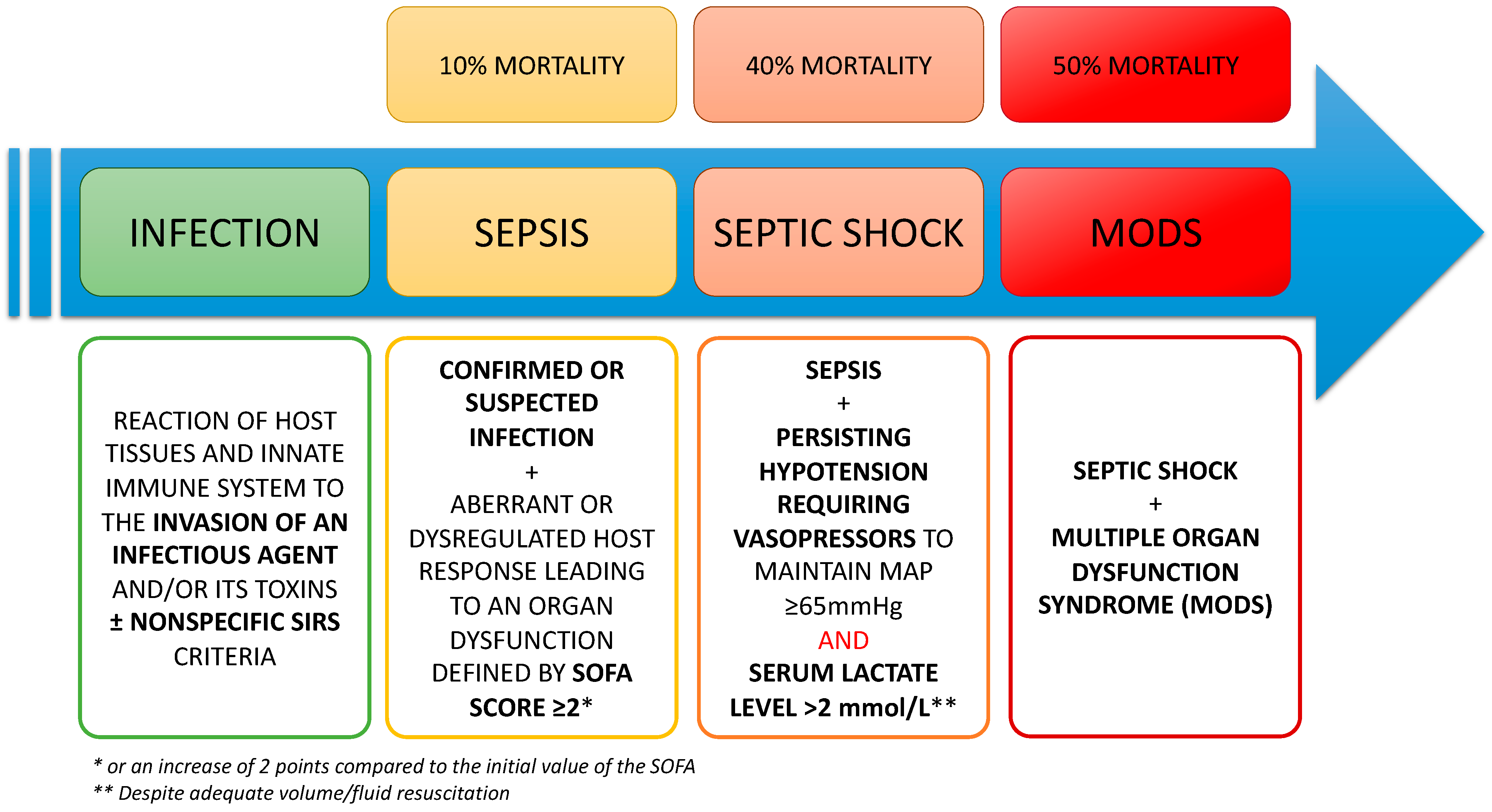
1.1. Definitions

Sepsis and septic shock represent the consecrated terms to designate the systemic response, in terms of severity, generated by the innate immune system (IIS) in the presence of a pathogen. This aggression, in connection with the infection, is responsible for a sequence of molecular and cellular interactions that triggers an inflammatory response. This host response may be dysregulated, inducing an immune system dysfunction. This dysregulated host response associated with at least one organ dysfunction, identified as an acute change ≥ 2 in Sepsis-Related Organ Failure Assessment score (SOFA score) currently defines the “sepsis” according to SEPSIS-3 criteria [1]. In most cases, this inflammation is controlled and balanced by the host immune system and the disease is only represented by a reaction called “infection” often accompanied by a systemic inflammatory response syndrome formerly named “SIRS” [2]. On the other side, if the triggering is too massive (pathogen’s virulence) or associated with an old age, underlying comorbidities, concurrent injuries, medications or genetic predispositions, the inflammatory phase is racing to a more severe form: the “sepsis” which can often lead to the “septic shock”. Sepsis and, a fortiori, septic shock are responsible for the onset of acute organ dysfunctions or even a multiple organ dysfunction syndrome (MODS) requiring the management of patients in intensive care units (ICU). MODS associated with sepsis represents one of the main causes of morbidity and mortality worldwide in ICU with more than 50% of in-hospital deaths linked to sepsis [1,3]. It is commonly accepted that, when accompanied by MODS, septic shock reflects the exacerbated response to the presence of the pathogen and frequently leads to immune, metabolic, and hematological dysfunctions [4,5]. The different definitions related to sepsis and their continuum from infection to the picture of multiple organ failure are presented in Figure 1.

1.2. Physiopathology and Treatments of the Septic Shock

1.2.1. Cell-Mediated Innate and Adaptative Immune Responses

The triggering event of a septic state is represented by the colonization of an organ, compartment or fluid of the organism by a pathogen inducing a local inflammatory reaction. Resident macrophages and dendritic cells, known as professional antigen presenting cells (APCs), are the first line of defense [6]. These cells are able to recognize pathogen-associated molecular patterns (PAMPs) using specific pathogen recognition receptors (PRR), including Toll-like receptors (TLRs). The first elements involved in the genesis of this IIS reaction are the PAMPs but also the host-derived debris released by damaged cells and known as damage (or danger)-associated molecular patterns (DAMPs). These specialized components interact with PRRs as TLRs, C-type lectin receptors (CTLRs), NOD-like receptors (NLRs), and RIG-I-like receptors (RLRs) [7]. DAMPs such as defensins, cathelicidin (LL-37), eosinophil-derived neurotoxin, and High Mobility Group Box-1 (HMGB-1) are also able to activate these PRRs and are called alarmins [8]. Activation of PPRs results in the engagement of complex metabolic cascades responsible for the formation of the inflammasome [9]. The inflammasome is, in turn, responsible for the synthesis and release of pro-inflammatory cytokines among which we can cite, interleukin-1-beta (IL-1ß), tumor necrosis factor-alpha (TNF-α), interleukin-6 (IL-6), interleukin-18 (IL-18), and HMGB-1 [10] sometimes carrying out the cytokine released syndrome or “cytokine storm” during an exacerbated activity of the inflammasome. The activation of the PRRs and the cytokine release are also responsible for a cascade of reactions leading, in immune cells, to a nuclear translocation of the nuclear factor kappa B (NFκB) [11] and induction of the transcription of targets genes involved in the synthesis of other pro-inflammatory cytokines including IL-1ß, IL-6, and chemokines such as IL-8 (CXCL8) [12]. This inflammatory microenvironment is essential to provoke the release of molecules such as complement proteins involved in direct lysis of the pathogen and to promote the recruitment of effector cells of the innate and then the adaptive immune system (AIS). In turn, these effector cells recruited at the site of the attack, will participate in the management and destruction of the pathogen.
The adaptive immune system (AIS) response is time-lagged relative to IIS. It consists of presenting the pathogen incriminated by the APC to the naive T lymphocytes (LyT) of the nearby secondary lymphoid organs (SLO). These LyT will, for the most part, migrate to the site of infection to participate in cell-mediated immunity. A portion of these T lymphocytes remains in SLO to elicit humoral mediated immunity by activating B lymphocytes (LyB) specific for the presented antigenic motifs [13]. There are currently several types of adaptive immune responses depending on signaling and differentiation pathways [14]. Among these different ways we can cite the T helper 17 (Th17) pathway, a pathway defined by differentiation into T helper 1 (Th1) lymphocytes promoting cell-mediated immunity and a T helper 2 (Th2) pathway for humoral-mediated immunity. This differentiation is determined by the cytokine pattern secreted in response to infection. The Th1 response is induced by interferon-gamma (IFN-γ) secreted by Th1 cells and by IL-12 produced by APCs. It is accompanied by the activation of cytotoxic CD8 + lymphocytes and monocytes. The Th2 response is triggered by the secretion of IL-4 and by the expression of CD40 ligand on the surface of macrophages and LyT and allows the activation of LyB secreting immunoglobulins directed against the constituents of microorganisms and/or their toxins [15].

1.2.2. Peripheral Neuro-Immune Control of the Inflammatory Process in Sepsis

All the initial inflammatory reactions normally resolve once the pathogen has been controlled by the organism, giving way to anti-inflammatory processes to form the basis for tissue repair. Patients with a form of infection with a so-called “adapted” immune response are characterized by the appearance of anti-inflammatory factors allowing to counterbalance the inflammatory response after the initial phase which, when not controlled, leads to a deleterious chronic inflammatory syndrome. The early neuroinflammatory modulation results notably in an activation of the autonomic nervous system (ANS) with a reflex loop involving the descending parasympathetic (vagus nerve) and sympathetic (orthosympathetic system by ganglia nodosis) pathways [16,17,18]. Tracey et al. thus showed that activation of the vagus nerve by signals originating from the IIS in response to the presence of DAMPs enables neuromodulation of the immune response [18]. This reflex loop carries out a relay at the level of the brainstem and leads to the activation of acetylcholine (Ach) expressing LyT cells (ChAT+ cells), representing a particular lymphocyte population expressing the choline acetyltransferase (ChAT) enzyme and playing a role in inhibiting TNF-α levels [19,20], regulating blood pressure [21] as well as circulating cytokine levels in mouse [19]. Activation of the ANS stimulates the secretion of Ach and norepinephrine in the spleen, resulting in an activation of the population of ChAT+ and, among other effects, an inhibition of the macrophages producing pro-inflammatory cytokines via an Ach-dependent alpha7-nicotinic acetylcholine receptor activation [19]. This biofeedback allows curbing the inflammatory response and prevents a “runaway” of the immune response. Several studies have highlighted a defect in the regulatory processes of the immune response in severe sepsis patients [22] and to PAMPs and DAMPs [23].
There are also important interactions between the ANS, notably the sympathetic system and innate or adaptive immunity through specific receptors [24]. Adrenergic receptors thus constitute receptors belonging to the ANS, classified into different categories and having a role in modulating the immune response. These receptors are mainly located in the primary and secondary lymphoid organs and are responsible for down-regulation of the immune response [25]. Among IIS cells, neutrophils (polymorphonuclear neutrophils, PNN) strongly express some adrenergic type receptors, particularly beta-2 receptors [26]. Their activation is responsible for a decrease in the production of reactive oxygen species (ROS), the migration of PNNs, and the formation of neutrophil extracellular traps (NETs) involved in bactericidal activities [26,27].

1.2.3. The Balance between Pro-Inflammatory and Anti-Inflammatory Processes

The infectious process is actually described as a balanced state associating concomitant inflammatory (e.g., IL1β, TNF-α) and anti-inflammatory responses (mediated chiefly by IL-10 and TGF-beta2). Sepsis and septic shock states can be interpreted as an imbalance between those two parts of the immune and inflammatory manifestations in favor of an unsuitable reaction of the IIS and AIS. This inappropriate immune response can persist for several hours or days after the initial infectious trigger and can be responsible, at least in part, for damage to various organs. The initial response observed during sepsis is similar to that observed during systemic inflammation of other origins and processes related to ischemia-reperfusion [28]. This immune response dysfunction will have various consequences on the organ functions at the initial site of the infection but also systemically. The genesis of septic shock is accompanied by attacks of multiple systems (neurons, endothelium among others) leading to acute kidney injury (AKI), acute liver injury, myocardial dysfunction, acute lung injury (ALI), and/or acute respiratory distress syndrome (ARDS). Added together and exceeding the host’s adaptive capacities, these dysfunctions lead to the development of multiple system organ failure.

1.2.4. Endothelial Dysfunction and Hemostasis Troubles

The circulating cytokines will activate the endothelial cells which represent one of the first targets to endothelial and cardiovascular dysfunctions [29]. Cardiovascular system failure affects more than 50% of patients with sepsis within 24 h of admission to ICU [30] and is found in this same proportion post-mortem [31]. The vascular endothelium plays a major role in the homeostasis of many richly vascularized tissues such as the cardiovascular, respiratory, cerebral, and renal systems. While septic shock is generally accompanied by an array of MODS, endothelial dysfunctions appear to be the major factor associated with a poor prognosis of patients [32,33]. The endothelium participates in maintaining the internal environment’s homeostasis and regulating the function of the various cardiovascular, pulmonary, renal, and cerebral systems [34,35]. There is a decrease or even a loss of this regulation with the loss of the endothelial barrier integrity [36]. Endothelial dysfunction is a well-described entity now occurring in critical illness. The consequences of this endothelial dysfunction are multiple: loss of regulation of regional microcirculations, ischemia/reperfusion and oxidative stress mechanisms, hemostasis disorders and formation of microthrombi and tissular hypoxia [36]. These disorders result in a deregulation of the balance between cellular energy needs (respiratory cycle) and oxygen and nutrient inputs as well as activation of pro-apoptotic mechanisms [37]. The mechanisms associated with sepsis leading to cell death are still the subject of many studies but the physiopathology of sepsis-associated cell death seems to be multifactorial and linked to both mitochondrial dysfunction (loss of membrane permeability), transcription and oxidative stress with increased cellular ROS production [38,39].
The uncontrolled systemic inflammatory cascade is also responsible for an overactivation of coagulation that can progress to disseminated intravascular coagulation (DIC) with microthrombi formation within the organs, aggravating hypoperfusion lesions. These coagulation disorders result from the activation of pro-coagulant molecules such as thrombin and tissue factor by activated endothelial and monocyte cells and the inactivation of molecules responsible for the fibrinolysis (protein C and endothelial pathways of anticoagulation) [40].

1.2.5. Central Nervous System Involvement in Sepsis

The central nervous system (CNS) is also a major actor in septic states genesis, affected by the aberrant immune response to infection through activation of the ANS (as aforementioned), the hypothalamic pituitary axis, and the release of hormones (corticoids), cytokines and neurotransmitters [41]. The CNS is involved both in detecting the pathogen, cytokines, hormones, and other molecules secreted by immune effectors [42]. The CNS is a part of a general neuroimmune reflex loop and exerts, with the help of the vagus nerve, a negative feedback on the inflammasome by reducing the circulating levels of TNF-α [16] and by activating ChAT+ cells. Interestingly, ChAT+ cells are also known to be derived, at least in vitro, from human umbilical cord mesenchymal stem cells (hUC-MSCs) [43].
This reflex loop also involves the brainstem and in particular the nucleus of the solitary tract, which acts as a nucleus serving as a relay in the higher centers between the afferents and the efferents of the ANS. During septic states, the secretion of proinflammatory cytokines is thus the cause but also the consequence of cerebral aggression and septic encephalopathy by altering the blood-brain barrier (BBB) and activating glial cells [44,45].

1.2.6. Sepsis and Immunoparalysis

Sepsis is characterized by a phenomenon called “immunoparalysis” which can be described as an impairment of innate and adaptive immunity responsible for a long-term susceptibility to infections including viral reactivations [46,47]. This affection is accompanied by a decreased survival in septic patients [47]. Septic states particularly impact LyT populations, both from a quantitative and qualitative point of view. Within the LyT population, a distinction is made in particular between CD8+ (CD8+) defined by a CD56, CD57, CD8+ phenotype, natural killer (NK) and natural killer-“type T” cells (NKT) respectively possessing CD56+ or CD57+ phenotypes [48]. CD8+ requiring activation by CD4+ T cells and APC, will convey cytotoxic functions attributed to perforin and granzymes directed against infected cells. NK and NKT are cells of innate immunity possessing intrinsic cytotoxicity functions [49]. Among T cells repertory, a decrease in the pool of naive CD8+ cells (i.e., antigen-inexperienced T cells) significantly in patients with septic shock as well as an alteration of their cytotoxicity functions has been reported [50]. Furthermore, expression of inhibitory receptors (2B4 and PD-1 receptors) has been reported on the surface of naive post-sepsis T cells, which may increase susceptibility to infection and morbidity and mortality in the host [51,52].

1.3. Therapeutics for the Treatment of Septic Shock

Even today, the treatment of septic shock remains the subject of many debates and studies. The current validated treatments, reaffirmed by the Surviving Sepsis Campaign [53], can be divided into two treatment groups. Early treatment (<6 h) consists of the administration of empiric antibiotic therapy and treatment necessary to optimize the flow rates of the macrocirculation and the microcirculation and therefore, to manage the imbalance between inputs and metabolic needs. Maintenance and control, in the first 24 h, of these various biological and clinical parameters improve survival. While the implementation of these recommendations was gradually accompanied by an improvement in patient outcomes through standardization of practices, they were ineffective in obtaining an additional reduction in the overall mortality of these patients. Consequently, many teams are working to evaluate new therapies for sepsis, such as TNF-α antagonist [54], recombinant human activated protein C (APC) [55], intravenous immunoglobulin G therapy [56], TLR4 antagonist [57], IL-1 receptor antagonist [58], and talactoferrin [59].
Neither of these new therapies nor the early goal-directed therapy has shown significant efficacy in the treatment of sepsis in terms of morbidity and mortality [60]. Among the therapies cited, only subgroup analyses were able to show efficacy in certain categories of patients with particularly high mortality [61], leading to the following question: would the development of novel immunoregulatory avenues for instance using mesenchymal stem cells (MSCs) be of clinical benefit in sepsis and septic shock?
The sepsis and a fortiori the septic shock remain pathologies encountered in emergency and intensive care medicine with significant morbidity and mortality [62]. The therapies offered to patients remain ineffective due to the complexity of the pathophysiology of this affection involving, among others, the role of the effectors of innate and adaptive immunity. MSCs, by virtue of their particularly immunomodulatory characteristics (as described below), can represent both major players in the intrinsic control of septic shock and a source for the establishment of new therapeutic alternatives to what currently remains an issue in critical care medicine of the 21st century. The main objectives of this review are to identify the roles played by MSCs during septic states in the detection of the infection to instruct immunity, to understand their implications in the process of uncontrolled sepsis leading to vasoplegic and cardiogenic shock states and their prophylactic and curative therapeutic applications.

2. Origins and Roles of Mesenchymal Stem/Stromal Cells

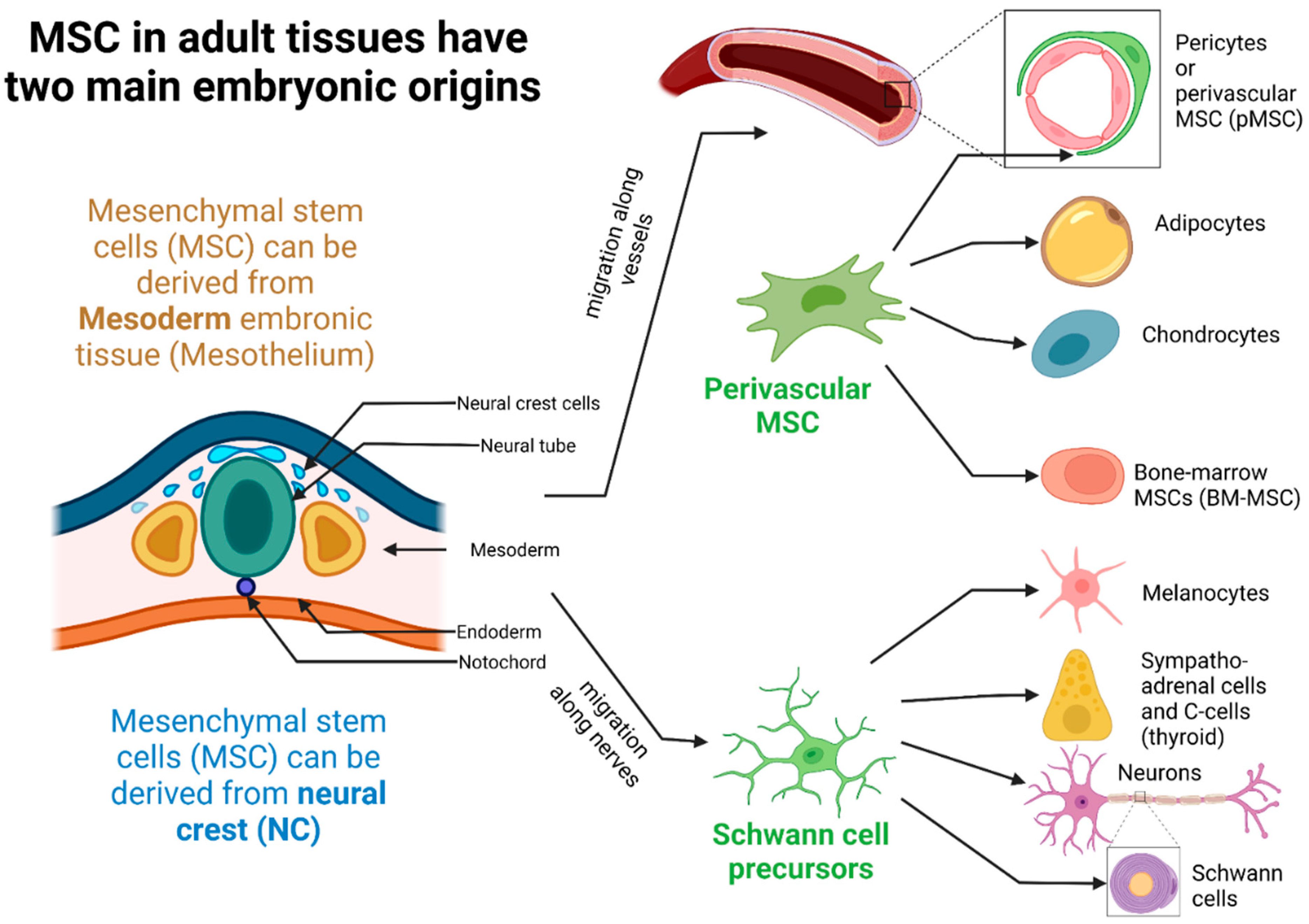
2.1. Ontogeny, Tissue Localization, and Different Populations of MSCs in Tissues

MSC were first described by Friedenstein and co-workers in the 1990s as spindle-shaped cells isolated from the bone marrow (BM) in an attempt to identify multipotent stromal precursor cells. Those cells were first named colony-forming unit fibroblasts (CFU-Fs) due to their presupposed origin from the stromal compartment of the BM in connection with their capacity of adherence to tissue culture vessels and the fibroblast-like appearance of the common progeny [63]. Stromal cells derived from CFU-Fs have been identified as feeder cells for the culture of hematopoietic stem cells (HSCs) and have the ability to differentiate into several subsets of cells both in vitro and in vivo: adipocytes, chondrocytes, and osteocytes among others [63]. The potential implications of these main discoveries were initially evaluated only in the light of experimental hematology and the entire role and physiology of the MSC remained, almost today and despite an important research, unappreciated [64,65].
MSCs are principally derived from mesodermal cells (MC) [66,67]. They can also be defined as multipotent mesenchymal stromal cells encompassing a heterogenous population of cells that have the ability to proliferate in vitro as plastic-adherent cells, forming colonies, with fibroblast-like characteristics, self-renewing potential, and to differentiate in several types of mature cells from different embryologic origins: ectodermal cells (epithelial cells and neurons), endodermal cells (gut and lung epithelial cells, muscle cells), and mesodermal cells as connective stromal cells, bone, cartilage, and fat cells [68]. If MSCs were first identified and extracted from BM, they have been isolated from every type of connective tissue [69]. It is important to note that the MSCs are localized at the perivascular level in the different organs playing the role of an interface between the inner and outer environments, thus functioning as a possible gatekeeper against possible infection and yet being able to alert professional immune cells to trigger the IIR.
Although MSCs are considered of MC origin, MSCs can also derive from the neural crest (NC) ectodermal embryonic tissue [70,71]. The NC gives rise to numerous progenitors: NC-MSCs but also melanocytes, Schwann cells, thyroid C cells, and adrenergic cells of the adrenal medulla [72]. NC-MSCs have the particularity of being able to differentiate into many cell types, among which we find endoneural fibroblasts, osteoblasts, and chondrocytes [73,74]. All NC-derived progenitors (NC-Pg) have nurturing functions and aid in the education and maturation of other cell subpopulations. Some NC-Pg (e.g., melanocytes) thus have an essential nourishing function with respect to neurons and keratinocytes. NC-MSCs have educational and maturation support functions for the cell populations contained within organs as varied as the lung, stomach, skin, bone marrow, or even the immune system [71].
Perivascular MSCs (pMSCs) result from an embryologic process of migration to the wall of blood vessels around endothelial cells and next to peripheral nerves [69,75,76,77]. pMSCs are able to differentiate into collagenhigh-producing myofibroblasts contributing to kidney or liver fibrosis and express canonical glial markers as glial fibrillary acidic protein (GFAP) and myelin P zero protein (P0), surface markers of astrocytes and Schwann cells respectively. This particular population, expressing glial markers and derived from NC was consecrated as glial MSC (gMSC) [78]. pMSCs are found around the vessels of different organs. The pMSCs located within the liver, as recently emphasized by Mederacke et al., are better known as hepatic stellate cells (HepSC) [79,80].
The ontogeny of MSC deriving from either the ectoderm (neural crest, NC) or the mesoderm is shown in Figure 2.

2.2. MSCs Characterization and Surface Markers Expression

As aforementioned, an MSC is characterized by its origin, its multipotent stem cell capacity, and its ability to differentiate notably into lipo- or myo-fibroblasts. MSCs might also be identified by the summation of expressed selective surface markers such as CD29 (integrin β1), CD44 (hyaluronic acid receptor), CD71 (transferrin receptor), CD73 (ecto-5′-nucleotidase), CD90 (GPI-anchored THY1), CD105 (TGF-ß R or endoglin), CD140 (PDGFR), CD146 (MUC18/melanoma cell adhesion molecule), CD271 (low-affinity nerve growth factor receptor and also known as p75 neurotrophin receptor), CD248 (endosialin, tumor endothelial marker 1), nestin (type IV intermediate filament of the NC), NG2 (Chondroitin sulfate), GFAP (glial fibrillary acidic protein of astrocytes), desmin (type III intermediate filament), and ganglioside GD2 [66,78]. The relative amount of each of these surface molecules varies with the MSC tissue localization and state of activation. MSCs are also characterized by the absence of several markers including those of the hematopoietic lineages (CD14, CD34, CD45, CD19), co-stimulatory molecules (CD80, CD86, and CD40), and other immune surface markers (CD11b, CD79alpha, and HLA-DR) [81]. These surface markers have been characterized on cell populations cultured in vitro and it is commonly accepted that several markers may be lost compared to the MSCs observed in vivo.

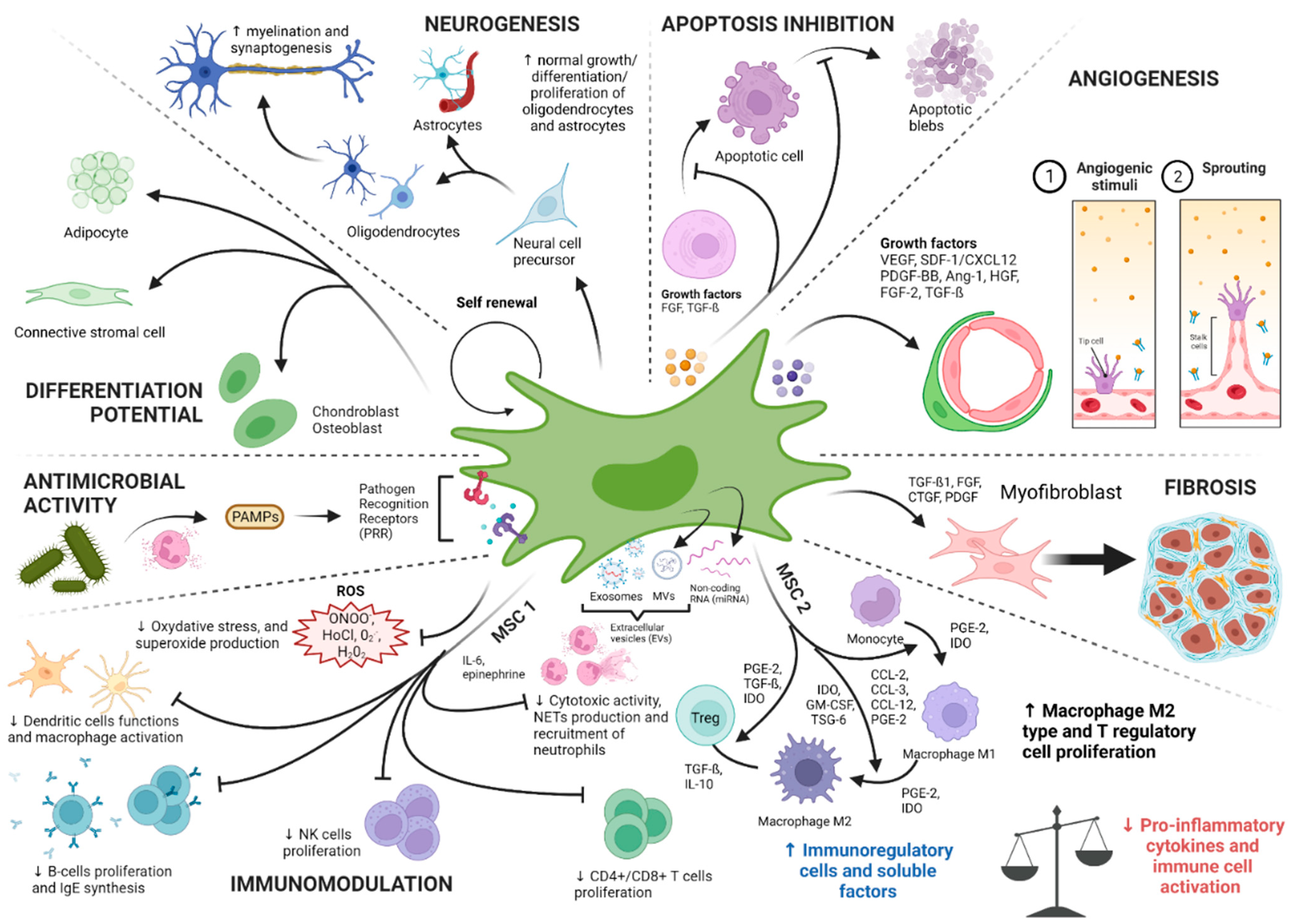
2.3. Physiological Roles of MSCs: A Niche for Stem Cells and Tissue Gatekeepers Alerted in Response to Tissue Injuries

MSCs are stromal cells that can be identified in adult tissues such as BM, lung [82], heart [83], synovial membrane, periosteum, skeletal muscles, dental pulp [84], adipose tissue [85], bones, and around the vessels. It is also possible to find them in large numbers in many fetal tissues such as amniotic fluid and membrane [86], placenta [87], umbilical cord blood, and Wharton’s Jelly where they may exert important immunoregulatory functions [88,89].
The main role of MSCs has long been a subject of ongoing debates on the ground that they are ultimately and extremely pleiotropic and totipotent [90]. Their pleiotropism would suggest a potential for actions out of proportion compared to other cell types, including professional immune cells. The ability of MSCs to migrate to the site of injury, to remove the intruders and to engage stem cell recruitment makes them “master orchestrator” of tissue repair in response to diverse injuries. MSCs, notably those from the NC, act as a niche for stem cells in the BM [91,92]. They interact with other cell types present in different organs with the help of growth factors. These growth factors produced by MSC are epidermal growth factor (EGF), fibroblast growth factor (FGF), transforming growth factor (TGF-beta), vascular endothelial growth factor (VEGF), hepatocyte growth factor (HGF), insulin-like growth factor (IGF), and angiopoietin-1 [93,94].
In response to tissue injuries associated with infectious diseases, MSC expressing PRRs responds to DAMPS or “alarmins” [95,96] and PAMPs represented by constituents of pathogens (viruses, bacteria, parasites). Pro-inflammatory molecules and hypoxia which are observed during acute critical attacks such as sepsis, are vectors of upregulated expression of PRRs such as TLRs [97]. These PRRs localized at the MSC surface are capable of binding both PAMPs [98,99,100] and DAMPS (HMGB1, HSP, and DNA proteins), degradation products of host cells or pathogens. Fine regulation of the response to the presence of DAMPs/PAMPs is crucial to avoid a “runaway” phenomenon of inflammation and a risk of an inappropriate autoimmune reaction [101].
MSC can also regulate the recruitment of innate and adaptive immune cells. Indeed, they can produce chemoattractant molecules (e.g., C3a, ATP, lysophosphatidic acid LPA1) and chemokines (e.g., CCL2/MCP1, CCL5/RANTES) to recruit professional immune cells at the site of the injury. This will lead to the activation of innate immune cells (PNN and monocytes) which play a critical role in local inflammation by expression of diverse inflammatory factors such the migration inhibitory factor (MIF) [81]. These recruited immune cells will greatly contribute to the phagocytosis of the infectious agents as well as the host-derived cell debris, both fueling the local and systemic inflammatory responses.
The identified roles of MSCs in controlling effectors of innate and adaptive immunities represent one of the cornerstones of its implication in the pathophysiology of septic states as explained below.
An outline of the different canonical and physiological roles of the MSCs is proposed in the Figure 3.

3. Study of the Role of the MSCs in Sepsis/Septic Shock: What Do We Know?

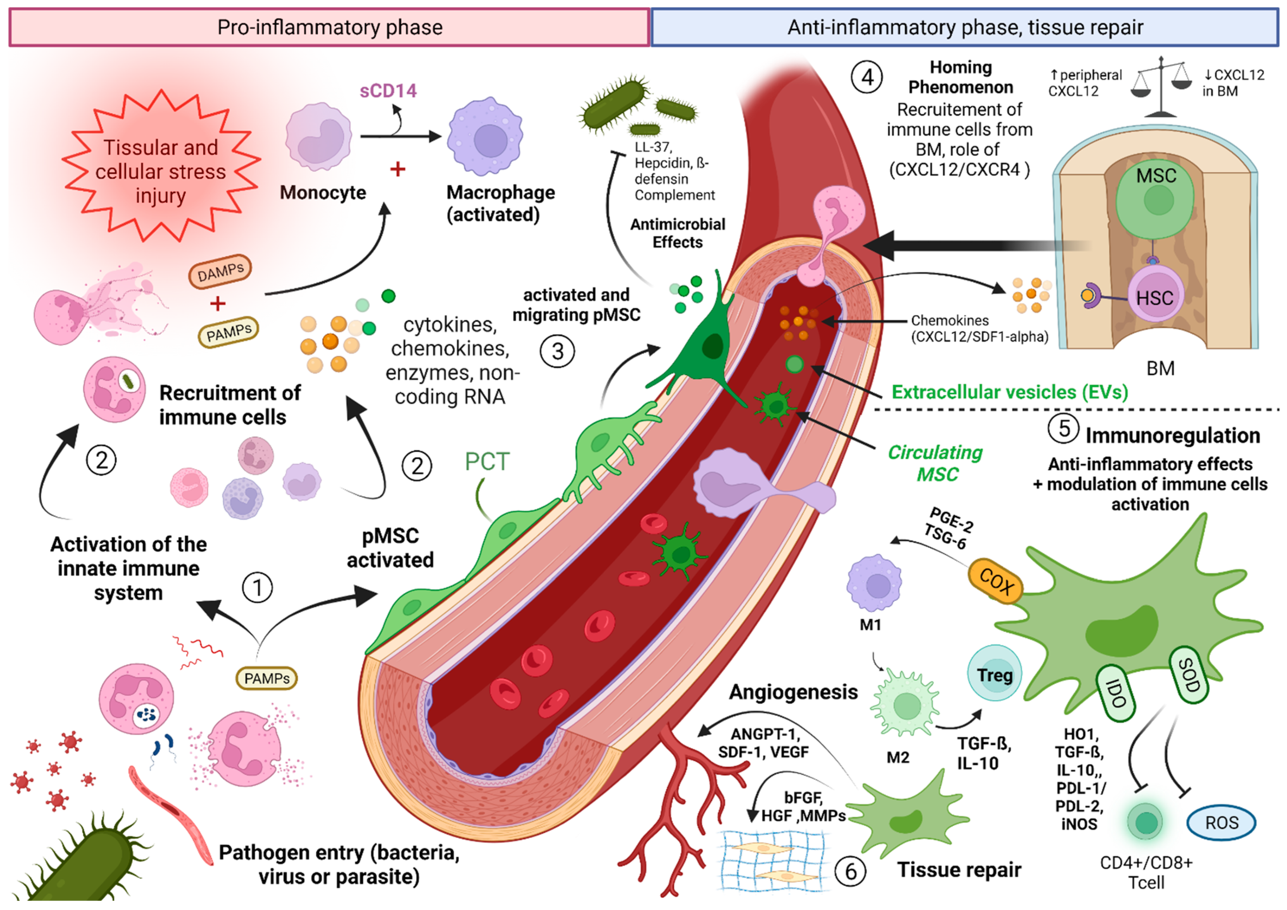
3.1. Contribution of Knowledge of the Role of MSCs in the Comprehension of the Septic Shock: Interactions between MSCs and the Immune System

It is increasingly recognized that MSCs represent cornerstones of the immune system and possess the capacities to modulate the immune response. Although this immunomodulatory potential of MSCs is the subject of numerous studies, the interactions of MSCs with target cells and their role in sepsis are still poorly understood. This key role is provided through two of their main characteristics: (1) Their ability to express soluble proteins and surface markers of innate immunity allowing the detection, management, and destruction of pathogens; and (2) their immunomodulating capacity in inflammatory conditions avoiding overexpression and overactivation of innate immune effectors. MSCs therefore express numerous markers specific of innate immune cells which can be divided into three categories: (1) Pathogen recognition receptors (PRR); (2) pro-inflammatory cytokines and chemokines; (3) immune effectors playing a direct anti-microbial or toxic role on pathogens.
Several authors have noticed that MSCs have the ability to regulate innate and adaptive immune responses both in vitro and in vivo [81,93,102]. MSCs are able to activate several pathways involved in maintaining the balance between proinflammatory and anti-inflammatory aspects of the immune system. More precisely, several authors noticed the capacities of MSCs to regulate the inflammatory response by activating different axes. This modulation of the IR can occur through two distinct mechanisms: (1) The secretion of soluble factors having a paracrine action such as IFN gamma, nitric oxide (NO), indoleamine 2,3-dioxygenase (IDO), prostaglandin E2 (PGE2), TGF-ß, and IL-10; (2) the direct cell–cell contact between some specific membrane receptors of MSCs and those located on the surface of target cells.
The modulation of immune responses by MSCs thus occurs through the activation of numerous mediators:
-
IFN-γ, alone or combined with other pro-inflammatory cytokines such as TNF, IL-1α, or IL-1β are able to induce the secretion of chemokines responsible for the activation of iNOS and the attraction of T cells. MSCs are also able to control the proliferation and activation of macrophages, natural killer T (NKT) cells, and γδ T cells [103]. MSCs, after stimulation with the inflammatory cytokines IFN-γ, TNF-α, or IL-1, can express inducible (i) NOS, releasing NO. High NO concentrations can, in turn, inhibit the transcription of (STAT)-5 phosphorylation in T cells and decrease the apoptosis of immune cells, participating in immunomodulation [104].
-
Indoleamine 2,3-dioxygenase, also expressed by MSCs stimulated by IFN-γ, inhibits lymphocyte proliferation by depleting tryptophan in the microenvironment. IDO-secreting MSCs are also potent inhibitors of Th1 cells and NK activity with the help of PGE2.
-
Role of cyclooxygenase-2 (COX2) and prostaglandins expressed by MSCs: In the context of a severe infection associated with high levels of LPS and/or host-derived factors (e.g., TNF-α) or even in hypoxic conditions, MSCs will engage the stimulation of the NFκB pathway. Activation of NF-κB signaling can upregulate the expression of COX2 and the COX2-dependent increase of PGE2 synthesis. PGE2 in turn will bind to G-protein-coupled receptors EP2 and EP4 on macrophages to increase the expression of the canonical anti-inflammatory cytokine IL-10 chiefly involved in the control of an overt inflammatory response [96,97].
-
Other soluble factors released by MSCs, such as IL-6, have the ability to slow down oxidative stress, apoptosis of neutrophils, and the differentiation of bone marrow progenitors into APC [105,106].
-
HLA-G5 is secreted by MSCs stimulated by IL-10 and following contact between MSCs and activated T cells. HLA-G5 has an anti-proliferative action on T cells, NK cells, and cytotoxic T lymphocytes.
From a cellular point of view, MSCs are known to control proliferation, IFN-γ production, and cytotoxicity of both CD4+ and CD8+ T cells [105]. Regulatory T cells (Tregs) are a subpopulation of CD4+ T lymphocytes originating from the thymus (tTregs) or from the periphery (pTregs) [107,108]. These immune cells are involved in the tolerance and homeostasis of the immune system. During septic states, these Tregs act as modulators of the immune system allowing control of the inflammatory response. Most of the experimental studies focusing on Tregs during sepsis observed an up-regulation of this population which can explain a long-term immunosuppression [109,110]. Conversely, Carvelli et al. found a decrease of these lymphocytes during septic shock [111]. If the proportion of Tregs remains to be better determined during sepsis, it appears that these cells play a key role in restoring the immune balance after the early inflammatory phase. MSCs are reported to favorably alter the Th1/Th2 balance toward Th2 cells [112] and are able to convert conventional T cells into Tregs [81].
It is well described that in the event of critical tissue damage in vivo, MSCs are attracted to various target sites (ischemic or injured sites) by a phenomenon of “homing” [113] mediated by the pathways of SDF1-α/CXCR4 or CD44 present on their surface and which can interact with hyaluronic acid (molecule exposed in the event of an acute lesion with attack of the connective tissue) [106,114]. MSCs possess the ability, in response to pro-inflammatory cytokines secreted upon contact with a pathogen, to produce chemokines and intercellular adhesion or vascular adhesion molecules such as (ICAM)-1 or (VCAM)-1 respectively [115]. The activation of these chemokines and cellular adhesion factors in return promotes the migration of immune cells.
Ischemia-reperfusion represents a mechanism that occurs in sepsis and septic shock conditions. It has been suggested that IL-10-overexpressing BM-MSCs could prevent lung IR-injuries in rats with a decrease of CD4(+) and CD8(+) T cells in the lung, improving blood oxygenation in the treated group [116].
Experimentally, the dynamic in vivo distribution of MSCs has been monitored in animal models. When MSCs are administered intravenously, several phases are distinguished, characterized by different MSC distributions. MSCs first migrate to the pulmonary and hepatic capillary bed [117]. They seem to reside at this level for a period extending over 96 h, before becoming undetectable within the sites considered [118]. Although this homing phenomenon to the lung and the liver remains unclear [119,120], these observations are at the origin of the theory according to which MSCs may act at a distance on their targets using mediators by two distinct pathways: (1) a paracrine pathway mediated by a direct secretion in the environment and (2) a pathway mediated by the secretion of extracellular vesicles (EVs). Whether this paracrine pathway is mediated by EVs or not, the two pathways have in common the fact that they can exert a remote action on the immune effector cells of immunity and do not require mandatory proximity of the MSCs to mediate their immunoregulatory effects.
EVs encompass several types of particles made up of membrane material formed from a parent cell. The internal content is thus protected during its transit to the target site by a lipidic bilayer which differentiates from the mother cell membrane by an enrichment in specific lipids (cholesterol, glycosphingolipids, and phosphatidylserine) allowing increased longevity within the compartments of the organism, especially the vascular environment [121,122] and direct physiological actions [123]. They can be classified according to their size and content. A distinction is thus made between apoptotic bodies derived from cells in apoptosis (1000 to 5000 nm), microvesicles (MVs) formed directly from the plasma membrane (100–1000 nm) and exosomes (Exos) formed from the endosomal system (40–150 nm). Usually, it is easy to distinguish apoptotic bodies from Exos and MVs due to the possible separation by ultracentrifugation. The EVs thus commonly designate the group of vesicles comprising the MVs and the Exos obtained after extraction. The content of the EVs, once extracted, can be analyzed. This heterogeneous EV’s material and content includes nucleic acids of which coding and non-coding RNA (mRNAs, miRNAs), lipids and proteins [124,125,126]. Among the non-coding RNAs, we can distinguish the following microRNAs (miRNAs) in MSC: miR-221, miR-23b, miR-125b, miR-451, miR-31, miR-24, miR-214, miR-122, miR-16, miR-150, and miR133b [127,128,129]. These miRNAs are involved in vitro in the mechanisms of tumor genesis, apoptosis, angiogenesis, and modulation of immunity [127,128,129]. More than 5000 different proteins derived from MSC EVs and possessing roles in self-renewal, differentiation, homing and signal transduction have so far been identified [130]. These EVs and their content may be essential players in the immunomodulatory and repair roles of MSCs. Several studies have thus been able to demonstrate the presence of cytokines such as IL-10, IL-6, IL-37, lipocalin-2, TGF-β, programmed death ligand-1 (PD-L1), and galectin-1 within these EVs [130,131,132]. The above findings are supported by studies that have observed an increase in the number of EVs released by MSCs in physiological inflammatory and stressful situations, such as sepsis [131,133,134]. Beyond their use in therapeutic trials during sepsis, the Exos derived from MSCs seem to be involved in their immunomodulatory potential during pro-inflammatory conditions and acute injuries [135]. Exos are capable of presenting antigenic motifs directly to specialized immune cells and activate CD8+ T lymphocytes and NK cells [136]. They are also carriers of bioactive molecules and indirect antigen presenters through modulation of the response of subpopulations of immune cells [137]. MSC-Exos finally have immunosuppressive capacities and control the activation and proliferation of a large panel of innate or adaptive immune cells such as B lymphocyte cells (LyB), NKs, LyT or LyT CD3+ and LyT CD4+ populations while preserving the activity of Tregs [138,139]. They also have, like the MSCs themselves, the capacity to down-regulate the production of pro-inflammatory cytokines such as TNF-α [140] and to up-regulate the production of anti-inflammatory cytokines (IL-6, IL-10, and TGF-β) [141]. MSC-Exos are therefore able to orient the individual’s immunity toward immune tolerance with a preferential M2 type polarization [142,143,144].
The different interactions between MSCs and immunity effector cells in acute tissue injury conditions and the hypothesis of interactions with immune and pro-inflammatory effectors in septic conditions are summarized in Figure 4.

3.2. MSCs Interacting with PNN, and the Key Role of the SDF-1/CXCR4/CXCR7 Pathway

Sepsis involves a very early innate immune response, in particular throughout the systemic inflammatory response to the aggressor. One of the key cells of this IIR is represented by PNN. The PNN participates in the control of the pathogen by its antimicrobial action (i.e., Release of ROS such as H2O2) in the early phase of the infection. The PNN is thus one of the first specialized cells recruited to the site of infection [145]. Its recruitment and early migration from the circulation to the site of inflammation make it a good prognostic marker predicting mortality [146]. A dysregulation of PNN functions is the cause of increased morbidity and mortality [147].
The PNN has a short lifespan and quickly moves toward cell death once its mission has been carried out using a programmed cell death mechanism called “pyroptosis” dependent on the caspase 1, 4, 5, and 11 pathways in particular [148]. PNN is continuously produced in the BM from progenitors of the granulocyte lineage and the “mature” forms are released into the systemic circulation just after the pathogen aggression [149]. During sepsis, an increase in the lifespan of these PNNs [147] as well as in the recruitment of immature forms were observed and is associated with increased mortality [150,151].
The SDF-1/chemokine (C-X-C motif) ligand 12 (CXCL12) pathway is a major pathway involved in the mechanisms of IIS. It is at the origin of the activation of multiple cellular pathways responsible for the recruitment and migration of PNNs from the production site to the site of aggression or “homing” phenomenon [152]. SDF-1 has two types of receptors: CXCR4 and CXCR7, both expressed on the surface of hematopoietic cells and in particular PNNs [153,154,155]. The SDF-1/CXCR4 pathway participates in the maintenance of the hematopoietic niche of PNNs and their release from the BM into the circulation during inflammatory and septic states [156]. Several authors have demonstrated a down-regulation of this pathway during septic states at the level of the BM contrasting with an up-regulation at the level of peripheral tissues with the consequence of chemotaxis of the PNNs toward the production sites in response to the presence of the pathogen [157,158].
Several studies have demonstrated an increase in the tissue expression of SDF-1 and its CXCR4 and CXCR7 receptors in the peritoneum, lungs, and liver in mouse models of peritonitis [159], and as confirmed in vitro [160,161]. SDF-1 and CXCR4 have also been the subject of studies showing their potential as early markers of sepsis [162]. If the involvement of the SDF-1/CXCR4/CXCR7 pathway in inflammatory and septic states is beyond doubt, its exact role in the regulation of the inflammatory balance is however unclear and is the subject of contradictory observations. Thus, the teams of Delano et al. and Guan et al. observed in experimental in vivo models that the administration of SDF-1/CXCL12 analogues increased the survival of mice in septic shock, whereas its depletion was responsible for increased mortality in individuals [158,163]. Conversely, Ramonell et al. showed that inhibition of CXCR4 reduced the mortality associated with sepsis in an experimental model of multimicrobial sepsis [164]. Gosh et al. clarified this conundrum by showing that inhibition of CXCR4 reduced cell migration by regulating the modulation of the cytoskeleton [165]. Other authors have demonstrated that inhibition of the CXCR4 pathway using pharmacological antagonists allowed protection of the lung and participated in the maintenance of tissue homeostasis during acute and chronic pulmonary inflammatory processes by reducing the CXCR4+ PNN infiltrate.
More recently the team of Kwon et al. have shown that the SDF-1/CXCL12 pathway activation tightly involves MSCs and results not only in the recruitment of PNNs from the BM, but also in increased phagocytotic activities of mature and immature PNNs [166]. The PNN plays a central role in circumspection of infection. If the infection persists, mature PNNs will no longer be sufficient and the SDF-1/CXCL12 pathway activation then participates in the recruitment of immature PNNs [158]. Although the bactericidal and phagocytotic capacities of these PNNs are less pronounced [167], they participate in the fight against the invading pathogen. These phagocytic capacities are significantly diminished when this pathway is silenced [166].
Finally, the activation of this pathway is at the origin of the synthesis of pro-inflammatory cytokines and is involved in the integrity of the endothelial cell barrier through the MAP-kinase (MAPKs) pathway and the transcription of NFκB p65 [168]. More recently, Ngamsri et al. have demonstrated the involvement of the SDF-1/CXCR4/CXCR7 pathway in cell-tissue interactions via tight junctions in experimental models of peritonitis and associated sepsis. Thus, blocking this pathway makes it possible to restore the integrity of the endothelial barrier, to reduce tissue edema and its consequences by a mechanism dependent on the A2B adenosine receptor [159].

3.3. Circulating MSCs and Sepsis: Toward a Novel Entity?

It is currently well established, and this for about ten years, that MSCs can be observed in vivo around vessels (arterioles and arterial and venous capillaries). These MSCs, conveniently called pericytes, maintain contact with the basement membrane and are separated from the vessel lumen by endothelial cells.
The ability of MSCs and in particular of pMSCs to evade and gain access to the blood circulation (see Figure 4, ③) in vivo remains ill-characterized. Several works denoted the impossibility of detecting MSC in the circulation [169]. In the same way, authors have tried to obtain, without success, cultures of MSCs from blood puncture from the portal vein, a technique allowing to avoid peripheral tissue contamination [66]. Kuznetsov et al. have successfully isolated, in minute quantities, circulating MSCs from four different mammalian species, capable of donating osteogenic cells and adipocytes [170]. Other studies were able to individualize MSCs in the circulating blood under stimulation conditions by G-CSF or VEGF [171,172,173]. Likewise, searching for circulating MSCs in injured mice have allowed the isolation of larger quantities of these cells, associated with increased circulating levels of G-CSF and VEGF. Given the literature, two facts are interesting to note: first, obtaining circulating MSCs seems easier under stimulatory conditions and, second, MSCs from the blood of individuals subjected to these conditions, appear to have a greater differentiation potential in culture than those obtained from peripheral blood from healthy individuals. The capacity for contraction and mobilization of pMSCs deeply in the vascular bed seems to be related to several cellular pathways independent of the iNOS pathway in mouse models of sepsis [174]. Of critical note, circulating MSCs have recently been found in patients suffering from rheumatoid arthritis (RA) or from cancer [175,176]. Blood transcriptional profiling experiments indicated that, in RA, B-cell autoimmune activation was followed by expansion of circulating podoplanin + MSCs (but negatives for myeloid CD45 and endothelial CD31 markers) corresponding to pre-inflammatory mesenchymal, or PRIME, cells in the blood of patients [175]. This finding has exciting and important implications from a biomarker point of view and also from a pathological standpoint given that these circulating MSCs may recapitulate secondary inflammatory-autoimmune sites, distant from the primary sites, a phenomenon which is well-known in cancer metastasis particularly as a side effect of irradiation [176]. Of note, a recent study using FACS analysis revealed increased numbers of circulating MSC (CD29+ CD73+) in the blood of patients with ARDS [177]. High levels of PCT and basal blood levels of VEGF and angiopoietin two were observed in these patients, arguing that the latter two molecules should not be considered as the mobilizing factors.
These different works lead us to formulate the hypothesis that pMSCs must play a dual role after recirculation: (1) an immunomodulatory role in the blood stage after leaving their perivascular niche in response to the systemic injuries. The migrating phenotypes may be associated to high levels of PAMPs. (2) A trophic role while responding to host cell debris (DAMPs) to help to repopulate the damaged tissue with stem cells. The immunomodulation phase in blood would concern the pMSCs co-responding to specific pro-inflammatory cytokines and chemokines produced by circulating and activated immune cells. This migration should involve specific set of adhesion-ligand molecules uniquely expressed at the basal membranes of the endothelium. When returning to tissue bed and in essence recapitulating their embryonic behavior (NC-like), they are likely to affect the perivascular niche with two possible scenarios: 1. They will recruit stem cells (including those from the BM) but also, 2. favor immune cell recruitment. In severe inflammatory conditions, the scenario 2 may contribute to autoimmune flare as recently described for the PRIME cells in RA patients. The unanticipated role of B cells and derived factors in this process needs to be explored further.

3.4. Diagnostic and Therapeutic Consequences

If these different hypothesis prove to be correct, MSCs could play a major role in the genesis of septic shock. Thus, septic shock could be the consequence of several isolated or intricate phenomena: (1) a dysfunction of the pMSCs allegedly involved in immunoregulatory activities but which may reveal a more aggressive phenotype while being harassed by the myriad of inflammatory factors. In light of their past education program while being derived from the NC, MSCs have a unique relationship with endothelial cells and peripheral nerves. For unclear reasons, when they are dissociated from this cell–cell communication settings, they are uncontrolled and behave abnormally. This original paradigm makes us argue that returning to homeostasis should aim to restore the crosstalks between pMSCs and endothelial cells as well as pMSCs and nerves. pMSCs express several receptors for neurotransmitters and provide opening new pharmacological therapeutic avenues. From a therapeutic point of view, the identification of different cellular profiles of pericytes, pMSCs, and of circulating activated MSCs would improve our physiological knowledge and could pave the way for new molecules targeting these cellular pathways in the treatment of septic shock.
From a diagnostic standpoint, the critical involvement of MSCs in the processes leading from sepsis to septic shock could result in very early changes in MSCs circulating in the blood. It would be interesting to perform kinetics of the circulating levels of these MSCs during sepsis in order to identify the unique disease state patterns. The use of RNA sequencing as performed for the PRIME studies will help to associate the presence of these circulating MSCs with a unique “mesenchymal” RNA signature in blood samples. This type of analysis will benefit from the isolation of exosomes-derived from MSCs prior to the molecular analyses.

4. Perspectives for the Study of MSCs in Sepsis Pathophysiology

4.1. Use of MSCs as In Vivo Immunomodulators

The natural history of sepsis and the possible rapid onset of septic shock make it difficult to resort to an administration of autologous MSCs conditioned in vitro, because of a limited time for clonal expansion. The possibilities offered by the transfusion of allogeneic MSCs, from healthy subjects, are thus the basis of more therapeutic trials. Allogeneic MSCs also have the advantage of being poorly immunogenic in vitro [178], facilitating their tolerance by the host, both in preclinical [179] and clinical studies [96]. Nevertheless, the less immunogenic nature of allogeneic MSCs [180,181] raises the question of their use in the context of sepsis, in particular, due to major inter-individual phenotypic variations which may interfere with their immunomodulatory capacities depending on the inter-cellular contacts that they may encounter while being in the blood.

4.1.1. Preclinical Data and Control of Inflammatory Processes in Experimental Studies

For the past ten years, many authors have shown interest in the therapeutic contribution of MSCs to treat critical illness or organ failures in the context of inflammatory or septic aggression. Numerous preclinical data were able to show that MSCs can have a protective action in murine models of sepsis, modify the potential evolution of sepsis toward a septic shock [182], and reduce its consequences through their immunomodulatory, anti-bacterial, and anti-inflammatory properties as well as their restorative potential after elimination of the initial threat. The time window for the injection of the MSCs is important and aims, according to our hypothesis, to have low levels of circulating PAMPs before the injection. It remains to be tested whether antibiotics may directly affect MSCs immunoregulatory activities.
The systemic administration of allogeneic MSCs in a mouse model of acute renal failure has made it possible to improve the recovery of renal function in association with an inhibitory effect on the production of pro-inflammatory cytokines (Il-1β, TNF-α, and IFN-γ) and an anti-apoptotic effect on kidney cells [183]. A study involving the injection of MSCs in a model of pulmonary fibrosis showed an effect on the secretion of the IL-1 receptor antagonist (IL-1RA), inhibiting Il-1α-producing T cells and TNF-α producing macrophages [184]. Parekkadan et al. have also observed the anti-inflammatory and immunomodulatory roles of conditioned MSCs in an experimental model of fulminant hepatic failure, in connection with a reduction in leukocyte proliferation and infiltrate and apoptosis of hepatic cells [185]. MSCs also have a protective effect against cerebral deleterious consequences of sepsis by controlling neuroinflammation. Several studies have thus been able to show the protective role of MSCs in mouse models of autoimmune encephalitis by inducing immune tolerance by the targeted inhibition of myelin-specific T cells [102] and by inhibiting the expression of anti-myelin autoantibodies by T cells, preserving axonal capital [186]. While many authors have found an antagonist effect of the administration of MSCs on the secreted pro-inflammatory factors [187,188], other studies had different results, showing no impact on the inflammasome, especially in porcine models [189]. Despite sometimes discordant results, meta-analysis are available on the subject, highlighting an overall downward trend in the mortality of individuals with septic conditions, after administration of MSCs [190].
These data and a selection of studies providing preclinical data on the administration of MSCs during sepsis are summarized in Table 1.

4.1.2. Infusion of Derived-MSCs and Clinical Studies of Sepsis Models

Many works have focused on testing the infusion of MSCs during inflammatory conditions complicating septic states, with acute respiratory distress syndrome (ARDS) in the foreground. In contrast, we only find a few clinical trials including MSCs in septic states strictly speaking in the literature. Most are phase I to II trials focusing on the safety of injecting MSCs in sepsis and septic shock using allogeneic MSCs derived from adipose tissue, BM or UC harvest (UC-MSC). A first phase I study dating from 2017 looked at the safety of injection of MSCs in septic shock. A “phase I/II” clinical trial (NCT01849237) has been conducted in neutropenic patients (<1000 PNN/mm3) with an administration of doses of 1 to 2 million MSC/kg/day started within 10 h of the onset of the septic shock. This trial, which is currently underway, is looking at D28 mortality, the reversibility of septic shock, the extent of organ dysfunction and the impact of MSCs on the biological parameters of inflammation [191]. Another phase I study has compared MSC infusion in increasing doses (1 × 10⁶, 2 × 10⁶ and 3 × 10⁶ cells/kg) in 15 patients with severe sepsis [192]. Finally, a phase I trial (NCT02328612) analyzes the impact of infusion of MSCs derived from adipose tissue on the systemic inflammatory response in healthy patients aged 18 to 35 years in a model of inflammation by injection of lipopolysaccharide (LPS) and attempts to determine the optimal dose of MSCs to be administered for the control of inflammation (four groups: placebo, groups of 0.25 × 10⁶ cells/kg, 1 × 10⁶ cells/kg, and 4 × 10⁶ cells/kg) [193].
The preclinical data also led to the realization of clinical trials which confirm the decrease of pro-inflammatory states complicating septic states and the associated organ failure. The START trial (STem cells for ARDS Treatment, NCT01775774) was a multi-center, randomized, controlled trial that tested a single dose of intravenous BM-MSCs-type MSCs in the treatment of moderate to severe ARDS [194]. The lung injury score (LIS) and sepsis-related organ failure assessment score (SOFA score) were lower at H72 in the group receiving high doses of MSCs although these differences were not significant (p = 0.87 and p = 0.76 respectively). The authors observed very good tolerance in the different groups without any serious adverse events related to the treatment being able to be highlighted.
More recently, two studies have focused on therapeutic infusion of MSCs during ARDS complicating SARS-CoV-2 infection. The first study was conducted on a 65 years unique woman, treated by allogenic human UC-MSCs at three different time-points (5 × 10⁷ cells each time) with a remission of the inflammation symptoms showed by laboratory indexes and CT images attributed to the injection but occurring on the 16th day after the diagnosis, which may correspond to the natural course of the disease [195]. A second study tested the injection of MSCs in 7 patients with an improvement of inflammation parameters but with very heterogeneous severity groups, limited follow-up, and without a control group [196].
Most of the clinical studies conducted using MSCs have many limitations: they relate to small numbers, show large disparities in the design adopted and in the associated biomarker analyses, and probably lack standardization in the selection of types of sepsis. In addition, many clinical studies concern states of systemic inflammation such as ARDS or ALI and few have been carried out in septic patients per se. Finally, the vast majority of these studies are phase I clinical trials. The various clinical trials concerning the use of MSCs in the context of inflammatory and septic states are summarized in Table 2.
While most of these trials confirm the safety of administration of allogeneic MSCs, there are very few results available from phase II trials. A meta-analysis by Can et al. published in 2017 grouping together more than 90 phase I clinical trials investigating the use of MSCs derived from umbilical cord harvesting also reported excellent tolerance of these IV infusion with minimal adverse effects marked by pain at the injection site and rapidly resolving flu-like symptoms [201]. The various clinical studies published to date on the use of allogeneic MSCs for the treatment of inflammatory and septic conditions are summarized in Table 2 and classified according to their type (phase I or II trial) and the pathophysiological characteristics of the populations studied.
Among the studies of interest concerning sepsis not yet published, we can cite the phase I CISS trial carried out in open label using allogeneic BM-MSCs (NCT02421484) in three dose cohorts with three participants per cohort who received allogeneic BM-MSCs at doses of 0.3 × 10⁶ cells/kg, 1 × 10⁶ cells/kg, and 3 × 10⁶ cells/kg from the lower dose to the higher dose. Another phase Ib/IIa, randomized, double-blind, placebo-controlled study (SEPCELL N°2015-002994-39) looked at the injection safety of AD-MSCs (injections on D1 then D3 of admission) for the treatment of several bacterial pneumonia acquired in ICU-patients [204].

4.1.3. Cell-Free Based Therapies: MSCs Extracellular Vesicles and Paracrine Factors Incriminated

As aforementioned, EVs group together the endosomes and MVs of MSCs which contain proteins, mitochondrial material (a major DAMP), and a myriad of miRNAs. Numerous pre-clinical studies have demonstrated the beneficial role of the administration of MSCs exosomes in tissue repair processes (cartilage, skeletal muscles), angiogenesis, modulation of the immune response and acute or chronic inflammatory states [205,206,207,208,209]. In addition to these pleitropic effects linked to the “cargo” function of the EVs of MSCs, allowing the transport of mediators with a direct or indirect role (inter-cellular communication) on the target tissues, EVs own a preponderant immunomodulatory action, at the image of the cells that secrete them. Exosomes thus modulate the activity of many effectors of the immune response such as LyT, macrophages (M1 and M2), NK cells, and Treg as well as microglia and dendritic cells [210,211,212,213,214]. These pre-clinical studies gradually led to the use of EVs in clinical studies.
The administration of EVs has also been tested as a therapy. Many clinical trials dealing with tissue repair, the control of autoimmune diseases, cancers, as well as cardiovascular diseases are available in the literature and their main results have been well summarized in the review by Lee et al. published in 2021 (revue Lee J Clin Med). However, few clinical trials have looked at the potential benefit of the administration of MSC-derived EVs during systemic inflammatory states and septic states. A clinical trial (NCT04356300), the results of which have not yet been communicated, used exosomes derived from UC-MSCs in the treatment of MODS after cardiovascular surgery. MVs obtained from the ultracentrifugation of cultures of human BM-MSCs were thus compared to the injection of MSCs in the control of pulmonary sepsis due to Escherichia coli in mice. Unfortunately, no statistical difference concerning survival and bacterial clearance was noticed between the two groups [215]. A prior infusion of the culture media of MSCs with an anti-CD44 antibody impaired the survival of mice. This result is not surprising given that the EVs obtained from BM-MSCs express canonical markers (CD90, CD44 and CD73) and are negative for CD34 and CD45 (hematopoietic markers) [216]. Thus, the use of anti-CD44 could interfere with the extracellular transport of MVs. Another mean of overcoming the drawbacks associated with the administration of MSCs (potentially carrying viral materials and/or progenies) is to administer soluble factors such as TSG-6, FGF-7, KGF, PGE-2 and other components such as non-coding RNAs (lncRNAs, circRNAs, and miRNAs). The available pre-clinical and clinical data regarding the use of MSC EVs has, however, recently been expanded considering the COVID-19 pandemic. Many phase 1 or 2 clinical trials using MSCs-derived EVs from sources as varied as bone marrow, adipose tissue, umbilical cord, are being recruited. Completed clinical studies, having used EVs administered by intravenous or inhaled route, seem to show a safety of their administration. A study published in 2020 already suggested a significant association between the administration of Exos and the improvement of the PaO2/FiO2 ratio (p < 0.001), a decrease in the absolute number of neutrophils [mean reduction 32% (p < 0.001)], an increase in CD3+, CD4+, and CD8+ LyT by 46% (p < 0.05), 45% (p < 0.05), and 46% (p < 0.001), respectively. They also found a reduction in CRP, ferritin, and D-dimer levels of 77% (p < 0.001), 43% (p < 0.001), and 42% (p < 0.05) [217]. The main trials using MSCs-derived EVs in COVID-19 have been described in a review recently published by Krishnan et al. [218].

4.2. Biomarkers of Disease States in Sepsis Including Those Related to Immune Cells and MSC Behaviors

The ability to diagnose sepsis but above all to predict the progression to septic shock remains a medical grail. To this end, many authors have attempted to demonstrate early biomarkers of these states of acute aggression. In a review, Pierrakos et al. studied 34 biological markers of sepsis including 16 for its early diagnosis. No molecule was found to have sufficient sensitivity (Se) and specificity (Sp) for routine use [219]. Thus, the study of pro-inflammatory cytokines (TNF-α, IL-6, and IL-1) has proved to be deceiving in this indication with low Se and Sp [220]. The CRP, which reflects the acute phase of inflammation, increases too late compared to the onset of sepsis (4–6 h delay) and has very poor Sp [221,222].
Other biological markers of the “ligand-receptor pair” type intervening early in the processes of adaptive immunity have also been suggested as good candidates. TREM-1 and TREM-2 (triggering receptors expressed on myeloid cells) are transmembrane glycoproteins of the immunoglobulin superfamily (Ig-SF) and lectins acting as receptors “inhibitors” of the IIR. They are constantly engaged by the presence of ubiquitous endogenous molecules (sialic acid, MHC-I, CD200) and activate tyrosine phosphatases which restrain stimuli in favor of this immune response. TREM-1 is found on the surface of PNNs, macrophages, and monocytes. The activation of TLRs during sepsis induces an overexpression of pro-inflammatory cytokines (TNF-α and IL-1α), of the membrane form of TREM-1 as well as the secretion of a soluble form of TREM-1 (sTREM- 1). This role of amplifier of the inflammatory response has moreover been confirmed in vivo in a murine model of septic shock, where the blocking of TREM-1 reduced the overall mortality. The determination of the sTREM-1 seems to be more interesting with an AUC of 0.91 (95% CI 0.88–0.93) in a meta-analysis involving 980 patients [223]. The measurement of the expression of CD64 (Fc gamma receptor), a surface marker of PNN highly induced by sepsis is also found to be efficient with an AUC of 0.94 (95% CI 0.92–0.97; p < 0.001) for the discrimination of septic patients [224]. The early determination of CD64 combined with the SOFA score also appears to have good area under curve (AUC) for the early diagnosis of sepsis in emergency medicine [225]. However, these results concerning the last two markers require confirmation by interventional studies.
The presepsin or soluble CD14 subtype (SCD14-ST) is a soluble form isotype of the GPI-anchored molecule of macrophages and monocytes serving as a co-receptor for the LPS-lipopolysaccharide binding protein (LPS-LPB) and TLR4; it is currently recognized as a marker for sepsis [226,227]. suPAR is a soluble form of uPAR which is a urokinase-type membrane receptor plasminogen activator receptor. Its concentration is correlated with the activity of the immune system and is also stable under physiological conditions [228,229]. Presepsin and suPAR are also of interest for the progressive prognosis of sepsis but also have many limitations [230,231].
Other works have studied the dosage of endothelial progenitors cells (EPCs), derived from the BM, as early markers of sepsis and its severity, finding circulating levels of EPCs (cEPCs) statistically higher in septic patients than in healthy subjects (45 +/− 4.5% vs. 12 +/− 3.6%, p < 0.001) [232]. These levels of cEPCs were higher in surviving patients than in nonsurvivors, defined as death within 28 days after onset of sepsis (p < 0.0001) [233].
Procalcitonin or PCT, a precursor peptide of calcitonin, is secreted ubiquitously in response to microbial infection consecutively to an up-regulation of the CALC-I gene in response to stimulation by pro-inflammatory cytokines. Thyroid cells (derived from the NC embryonic tissue) have high expression levels of PCT. Its kinetics are more interesting than CRP because its serum concentrations, <0.1 μg/mL in healthy subjects, increase as early as 4 h after the onset of sepsis with an earlier peak [222,234]. Its usefulness in the discrimination of sepsis, proven, is undoubtedly more linked to its good negative predictive value (NPV) and its sequential dosage in intensive care units for the diagnosis of secondary infections. It is also increasingly used as a marker for the follow-up of the effectiveness of antibiotic therapy and its early de-escalation [222]. In a meta-analysis of 30 studies and 3244 patients, PCT had a mean sensitivity of 0.77 (95% CI 0.72–0.81) and specificity of 0.79 (95% CI 0.74–0.84) and the area under the receiver operating curve (ROC) was 0.85 (95% CI 0.81–0.88) [235]. Cultured adipocytes have been shown to secrete PCT in response to the addition of IL-1β [236]. The levels of PCT secreted seem to depend on the type of bacteria found to cause sepsis (more important in Gram-negative infections, especially in obligate anaerobes) [237]. This observed difference in concentrations is explained by the different levels of stimulation linked to the polymorphism of PAMPs and DAMPs, responsible for different cytokine profiles depending on the type of aggression [238]. The addition of IFN-gamma in adipocytes culture, a molecule secreted in particular by helper T lymphocytes during viral infections, has the effect of blocking the synthesis of PCT [236]. IFN-gamma therefore has a negative biofeedback effect on the induction of PCT synthesis, which also explains the lower levels of PCT observed during invasive fungal infections which are also accompanied by IL-17 secretion with an effect similar to IFN-gamma [239]. While the mechanisms that lead to the secretion of PCT seem well understood, a crucial question remains: which cell types are responsible for this secretion? While it is well established that this is ubiquitous in vivo and that cell populations of cultured adipocytes can synthesize it, it is important to note that no work provides a clear answer on the subject.
However, some data allow us to define avenues concerning the sources of PCT in vivo. It has thus been established that human adipose tissue is a major player in inflammation and is involved in the elevation of circulating levels of PCT levels during sepsis [240]. The lipolytic effect of the calcitonin-gene (CT-gene) related peptide and adrenomedullin are moreover involved in the metabolic processes encountered during these physiological conditions. In a 2005 study investigating the expression of calcitonin-derived peptides by quantification of their respective transcripts in RT-PCR, the authors noted a significant production of PCT from AT-MSC obtained by adipose tissue biopsies of patients subjected to IL-1β and stimulation with LPS but not from undifferentiated MSCs from the same donors [236]. PCT may therefore well be secreted by subpopulations of specialized MSCs (particularly the lipofibroblast-like MSC subset, see Figure 2), and in other tissues where MSCs are present. It is interesting to note that some authors have been able to identify the presence of PCT within specific extracellular vesicles enriched with GM1 ganglioside isolated from the plasma of pregnant patients presenting pre-eclampsia, a pro-inflammatory placental disease characterized by a dysimmunity [241]. However, this type of EVs is isolated by its affinity to the B chain of cholera toxin (CTB). These CTB-EVs are the only true exosomes derived from endosomes secreted by MSCs and which may suggest a major role of these activated MSCs in the increased levels of circulating PCT during sepsis [242].

4.3. Interest of the Study of miRNA in Septic States

The septic syndrome is associated with a profound alteration of the genome and its expression within the cells of the various tissues of the host [243]. Among the possible actors involved in these changes in the genome, we can distinguish the class of non-coding RNAs. This class of ribonucleotides is recognized by a growing number of studies as being involved in many biological processes including innate immunity, apoptosis, and the regulation of mitochondrial function [244,245,246]. miRNAs are a class of RNA molecules of 21 to 25 nucleotides, acting as post-transcriptional regulators of gene in cells of healthy or damaged tissues [247]. miRNAs are known to play a major role in the regulation of pathologies linked to the immune system due to their capacity to modulate the expression of genes responsible for the secretion of pro-inflammatory cytokines such as TNF-α and interleukin IL-1β but also proteins linked to major intracellular communication pathways (MAP kinase pathway) [245,248,249,250]. The studies currently available concerning the detection and the assay of the non-coding RNA during the septic states relate primarily to their role in the immune processes and their potential as biomarkers. A meta-analysis studied the involvement of miRNAs in sepsis and found great heterogeneity in the themes studied and their interest as diagnostic or prognostic markers [251]. For the time being, the use of regulatory RNAs as biomarkers, although mentioned for more than ten years, concerns only a few works and limited set of miRNAs. These studies found a wide variety of miRNAs that can differentiate between inflammatory and septic states, but none has been validated. The prediction of morbidity and mortality and the risk of complications (organ dysfunctions) associated with septic patients by the detection of specific miRNAs is still in its early stages, even if miR-574-5p and miR-155 seem interesting for this purpose.

5. Conclusions

Despite many advances observed in intensive medicine since the 20th century, septic pathology and septic shock remain burdened with significant morbidity and mortality. The recent decision-making algorithms and recommendations issued by the sepsis-surviving campaign are struggling to obtain clear results in terms of survival for sepsis patient. The study of MSCs, which began about fifty years ago in the field of cell therapy, opens up new avenues in the understanding and control of this clinical entity. Although their therapeutic use in intensive care medicine, related to their immunomodulatory capacities, is at the center of ongoing interests, hence studies on their diverse interactions with the different effectors of immunity in vitro and during septic states must be continued. The use of cellular material derived from these cells such as MSC-derived extracellular vesicles for therapeutic purposes, or the study of miRNAs in vivo are thus good examples of therapeutic alternatives to the infusion of MSCs during sepsis. Likewise, the exploitation of their role in sepsis could lead to discoveries opening the way to diagnostic and prognostic models of septic shock. While much progress must be made before the routine use of these stem cells can be considered, further investigations on their overall physio pathological roles in diverse acute and chronic disease settings will be of great value.

Author Contributions

Conceptualization, M.D. and P.G.; methodology, P.G.; software, M.D.; validation, P.G., M.D., Y.B. and D.V.; formal analysis, M.D.; investigation, M.D.; resources, M.D.; data curation, M.D.; writing—original draft preparation, M.D.; writing—review and editing, M.D, P.G., D.V., F.A.-P., L.R. and B.D.; visualization, M.D.; supervision, P.G.; project administration, P.G.; funding acquisition, M.D. All authors have read and agreed to the published version of the manuscript.

Funding

The work carried out by members of the EPI team is supported by fundings from University of La Réunion and European funding. EPI’s team received funding from the Regional Council of La Réunion, French State and Europe (CPER-FEDER, GURDTI 2017-1198-0002583 VIROPAM) and (CPER-FEDER 20192211-0022768, EPIGEN).

Institutional Review Board Statement

Not applicable.

Informed Consent Statement

Not applicable.

Data Availability Statement

Not applicable.

Acknowledgments

The authors thank all members of the EPI team. All authors have consented to the acknowledgement Figures were created with Biorender.com.

Conflicts of Interest

The authors declare no conflict of interest.

Abbreviations

AT-MSCAdipose Tissue-Derived Mesenchymal Stem Cell
AISAdaptive Immune System
AKIAcute Kidney Injury
ALIAcute Lung Injury
ANSAutonomic Nervous System
APCAntigen Presenting Cell
ARDSAcute Respiratory Distress Syndrome
BMBone Marrow
BM-MSCBone Marrow-Derived Mesenchymal Stem Cell
ChATCholine Acetyl Transferase
ChAT+ cellsAcetylcholine expressing T cells
CFU-FsColony Forming Unit-Fibroblasts
CTLRC-Type Lectin Receptor
DAMPDamage Associated Molecular Pattern
DICDisseminated Intravascular Coagulation
EPCEndothelial Progenitor Cell
EVsExtracellular Vesicles
gMSCglial Mesenchymal Stem Cell
HepSCHepatic Stellate Cell
HSCHematopoietic Stem Cell
ICUIntensive Care Unit
IDOInDoleamine 2,3-Dioxygenase
IISInnate Immune System
iNOSinducible NO Synthase
IRImmune Response
IIRInnate Immune Response
LyBB Lymphocyte
LTCD4+Lymphocyte CD4+ T Cell
LTCD8+Lymphocyte CD8+ T Cell
LyTLymphocyte T
MB-MSCMenstrual Blood-Derived Mesenchymal Stem Cell
MCMesodermal Cell
mRNAmessenger ARN
miRNAmicroRNA
MODSMultiple Organ Dysfunction Syndrome
MSCMesenchymal Stem Cell
NC-MSCNeural Crest-Derived Mesenchymal Stem Cell
NC-PgNeural Crest-Derived Progenitors
NKNatural Killer Cell
NKTNatural Killer “type T” cell
NLRNOD-Like Receptor
NPVNegative Predictive Value
NF-kBNuclear Factor kappa B
NONitric Oxide
PAMPPathogen-Associated Molecular Pattern
pMSCperivascular Mesenchymal Stem Cell
PRRPathogen Recognition Receptor
RLRRIG-1-Like Receptor
ROSReactive Oxygen Species
SLOSecondary Lymphoid Organs
SOFASepsis-related Organ Failure Assessment
TH1T Helper 1
TH2T Helper 2
TLRToll Like Receptor
Treg cellsregulatory T Cells
UC-MSCUmbilical Cord-Derived Mesenchymal Stem Cell

References

  1. Shankar-Hari, M.; Phillips, G.S.; Levy, M.L.; Seymour, C.W.; Liu, V.X.; Deutschman, C.S.; Angus, D.C.; Rubenfeld, G.D.; Singer, M.; for the Sepsis Definitions Task Force. Developing a New Definition and Assessing New Clinical Criteria for Septic Shock: For the Third International Consensus Definitions for Sepsis and Septic Shock (Sepsis-3). JAMA 2016, 315, 775. [Google Scholar] [CrossRef] [PubMed]
  2. Muckart, D.J.; Bhagwanjee, S. American College of Chest Physicians/Society of Critical Care Medicine Consensus Conference: Definitions for Sepsis and Organ Failure and Guidelines for the Use of Innovative Therapies in Sepsis. Crit. Care Med. 1992, 20, 864–874. [Google Scholar]
  3. Paoli, C.J.; Reynolds, M.A.; Sinha, M.; Gitlin, M.; Crouser, E. Epidemiology and Costs of Sepsis in the United States—An Analysis Based on Timing of Diagnosis and Severity Level. Crit. Care Med. 2018, 46, 1889–1897. [Google Scholar] [CrossRef]
  4. Kaneki, M. Metabolic Inflammatory Complex in Sepsis: Septic Cachexia as a Novel Potential Therapeutic Target. Shock 2017, 48, 600–609. [Google Scholar] [CrossRef] [PubMed]
  5. Rossaint, J.; Zarbock, A. Pathogenesis of Multiple Organ Failure in Sepsis. Crit. Rev. Immunol. 2015, 35, 277–291. [Google Scholar] [CrossRef] [PubMed]
  6. Efron, P.; Moldawer, L.L. Sepsis and the Dendritic Cell. Shock 2003, 20, 386–401. [Google Scholar] [CrossRef] [PubMed]
  7. Takeuchi, O.; Akira, S. Pattern Recognition Receptors and Inflammation. Cell 2010, 140, 805–820. [Google Scholar] [CrossRef]
  8. Oppenheim, J.J.; Yang, D. Alarmins: Chemotactic Activators of Immune Responses. Curr. Opin. Immunol. 2005, 17, 359–365. [Google Scholar] [CrossRef]
  9. Martinon, F.; Burns, K.; Tschopp, J. The Inflammasome: A Molecular Platform Triggering Activation of Inflammatory Caspases and Processing of ProIL-Beta. Mol. Cell 2002, 10, 417–426. [Google Scholar] [CrossRef]
  10. Huang, C.; Wang, Y.; Li, X.; Ren, L.; Zhao, J.; Hu, Y.; Zhang, L.; Fan, G.; Xu, J.; Gu, X.; et al. Clinical Features of Patients Infected with 2019 Novel Coronavirus in Wuhan, China. Lancet 2020, 395, 497–506. [Google Scholar] [CrossRef]
  11. Brown, M.A.; Jones, W.K. NF-KappaB Action in Sepsis: The Innate Immune System and the Heart. Front. Biosci. J. Virtual Libr. 2004, 9, 1201–1217. [Google Scholar] [CrossRef] [PubMed]
  12. van der Poll, T.; van de Veerdonk, F.L.; Scicluna, B.P.; Netea, M.G. The Immunopathology of Sepsis and Potential Therapeutic Targets. Nat. Rev. Immunol. 2017, 17, 407–420. [Google Scholar] [CrossRef] [PubMed]
  13. Oberholzer, A.; Oberholzer, C.; Moldawer, L.L. Sepsis Syndromes: Understanding the Role of Innate and Acquired Immunity. Shock 2001, 16, 83–96. [Google Scholar] [CrossRef] [PubMed]
  14. Brady, J.; Horie, S.; Laffey, J.G. Role of the Adaptive Immune Response in Sepsis. Intensive Care Med. Exp. 2020, 8, 20. [Google Scholar] [CrossRef] [PubMed]
  15. MacConmara, M.; Lederer, J.A. B Cells. Crit. Care Med. 2005, 33, S514–S516. [Google Scholar] [CrossRef] [PubMed]
  16. Bonaz, B.; Sinniger, V.; Pellissier, S. The Vagus Nerve in the Neuro-Immune Axis: Implications in the Pathology of the Gastrointestinal Tract. Front. Immunol. 2017, 8, 1452. [Google Scholar] [CrossRef]
  17. Huston, J.M.; Gallowitsch-Puerta, M.; Ochani, M.; Ochani, K.; Rosas-Ballina, M.; Ashok, M.; Pavlov, V.; Czura, C.J.; Ulloa, L.; Goldstein, R.S.; et al. Vagus Nerve Stimulation Improves Survival in Experimental Sepsis.: 118-T. Crit. Care Med. 2005, 33, A137. [Google Scholar] [CrossRef]
  18. Tracey, K.J. The Inflammatory Reflex. Nature 2002, 420, 853–859. [Google Scholar] [CrossRef]
  19. Rosas-Ballina, M.; Olofsson, P.S.; Ochani, M.; Valdés-Ferrer, S.I.; Levine, Y.A.; Reardon, C.; Tusche, M.W.; Pavlov, V.A.; Andersson, U.; Chavan, S.; et al. Acetylcholine-Synthesizing T Cells Relay Neural Signals in a Vagus Nerve Circuit. Science 2011, 334, 98–101. [Google Scholar] [CrossRef]
  20. Reardon, C.; Duncan, G.S.; Brüstle, A.; Brenner, D.; Tusche, M.W.; Olofsson, P.S.; Olofsson, P.; Rosas-Ballina, M.; Tracey, K.J.; Mak, T.W. Lymphocyte-Derived ACh Regulates Local Innate but Not Adaptive Immunity. Proc. Natl. Acad. Sci. USA 2013, 110, 1410–1415. [Google Scholar] [CrossRef]
  21. Olofsson, P.S.; Steinberg, B.E.; Sobbi, R.; Cox, M.A.; Ahmed, M.N.; Oswald, M.; Szekeres, F.; Hanes, W.M.; Introini, A.; Liu, S.F.; et al. Blood Pressure Regulation by CD4+ Lymphocytes Expressing Choline Acetyltransferase. Nat. Biotechnol. 2016, 34, 1066–1071. [Google Scholar] [CrossRef] [PubMed]
  22. Boomer, J.S.; To, K.; Chang, K.C.; Takasu, O.; Osborne, D.F.; Walton, A.H.; Bricker, T.L.; Jarman, S.D.; Kreisel, D.; Krupnick, A.S.; et al. Immunosuppression in Patients Who Die of Sepsis and Multiple Organ Failure. JAMA 2011, 306, 2594–2605. [Google Scholar] [CrossRef] [PubMed]
  23. Monneret, G.; Venet, F.; Pachot, A.; Lepape, A. Monitoring Immune Dysfunctions in the Septic Patient: A New Skin for the Old Ceremony. Mol. Med. Camb. Mass 2008, 14, 64–78. [Google Scholar] [CrossRef] [PubMed]
  24. Elenkov, I.J.; Wilder, R.L.; Chrousos, G.P.; Vizi, E.S. The Sympathetic Nerve—An Integrative Interface between Two Supersystems: The Brain and the Immune System. Pharmacol. Rev. 2000, 52, 595–638. [Google Scholar] [PubMed]
  25. Sanders, V.M.; Straub, R.H. Norepinephrine, the Beta-Adrenergic Receptor, and Immunity. Brain. Behav. Immun. 2002, 16, 290–332. [Google Scholar] [CrossRef]
  26. Nicholls, A.J.; Wen, S.W.; Hall, P.; Hickey, M.J.; Wong, C.H.Y. Activation of the Sympathetic Nervous System Modulates Neutrophil Function. J. Leukoc. Biol. 2018, 103, 295–309. [Google Scholar] [CrossRef]
  27. Scanzano, A.; Schembri, L.; Rasini, E.; Luini, A.; Dallatorre, J.; Legnaro, M.; Bombelli, R.; Congiu, T.; Cosentino, M.; Marino, F. Adrenergic Modulation of Migration, CD11b and CD18 Expression, ROS and Interleukin-8 Production by Human Polymorphonuclear Leukocytes. Inflamm. Res. 2015, 64, 127–135. [Google Scholar] [CrossRef]
  28. Andersson, U.; Tracey, K.J. HMGB1 Is a Therapeutic Target for Sterile Inflammation and Infection. Annu. Rev. Immunol. 2011, 29, 139–162. [Google Scholar] [CrossRef]
  29. Ait-Oufella, H.; Maury, E.; Lehoux, S.; Guidet, B.; Offenstadt, G. The Endothelium: Physiological Functions and Role in Microcirculatory Failure during Severe Sepsis. Intensive Care Med. 2010, 36, 1286–1298. [Google Scholar] [CrossRef]
  30. Brun-Buisson, C.; Meshaka, P.; Pinton, P.; Vallet, B.; EPISEPSIS Study Group. EPISEPSIS: A Reappraisal of the Epidemiology and Outcome of Severe Sepsis in French Intensive Care Units. Intensive Care Med. 2004, 30, 580–588. [Google Scholar] [CrossRef]
  31. Torgersen, C.; Moser, P.; Luckner, G.; Mayr, V.; Jochberger, S.; Hasibeder, W.R.; Dünser, M.W. Macroscopic Postmortem Findings in 235 Surgical Intensive Care Patients with Sepsis. Anesth. Analg. 2009, 108, 1841–1847. [Google Scholar] [CrossRef] [PubMed]
  32. Wright, J.K.; Hayford, K.; Tran, V.; Al Kibria, G.M.; Baqui, A.; Manajjir, A.; Mahmud, A.; Begum, N.; Siddiquee, M.; Kain, K.C.; et al. Biomarkers of Endothelial Dysfunction Predict Sepsis Mortality in Young Infants: A Matched Case-Control Study. BMC Pediatr. 2018, 18, 118. [Google Scholar] [CrossRef] [PubMed]
  33. Iba, T.; Connors, J.M.; Nagaoka, I.; Levy, J.H. Recent Advances in the Research and Management of Sepsis-Associated DIC. Int. J. Hematol. 2021, 113, 24–33. [Google Scholar] [CrossRef]
  34. Coletta, C.; Módis, K.; Oláh, G.; Brunyánszki, A.; Herzig, D.S.; Sherwood, E.R.; Ungvári, Z.; Szabo, C. Endothelial Dysfunction Is a Potential Contributor to Multiple Organ Failure and Mortality in Aged Mice Subjected to Septic Shock: Preclinical Studies in a Murine Model of Cecal Ligation and Puncture. Crit. Care 2014, 18, 511. [Google Scholar] [CrossRef] [PubMed]
  35. Roquer, J.; Segura, T.; Serena, J.; Castillo, J. Endothelial Dysfunction, Vascular Disease and Stroke: The ARTICO Study. Cerebrovasc. Dis. 2009, 27, 25–37. [Google Scholar] [CrossRef]
  36. Boisramé-Helms, J.; Kremer, H.; Schini-Kerth, V.; Meziani, F. Endothelial Dysfunction in Sepsis. Curr. Vasc. Pharmacol. 2013, 11, 150–160. [Google Scholar]
  37. Ince, C.; Mayeux, P.R.; Nguyen, T.; Gomez, H.; Kellum, J.A.; Ospina-Tascón, G.A.; Hernandez, G.; Murray, P.; De Backer, D. The Endothelium in Sepsis. Shock 2016, 45, 259–270. [Google Scholar] [CrossRef]
  38. Berger, M.M.; Grocott, M.P.W. Facing Acute Hypoxia: From the Mountains to Critical Care Medicine. Br. J. Anaesth. 2017, 118, 283–286. [Google Scholar] [CrossRef]
  39. Kiers, H.D.; Scheffer, G.-J.; van der Hoeven, J.G.; Eltzschig, H.K.; Pickkers, P.; Kox, M. Immunologic Consequences of Hypoxia during Critical Illness. Anesthesiology 2016, 125, 237–249. [Google Scholar] [CrossRef]
  40. Levi, M.; van der Poll, T. Coagulation and Sepsis. Thromb. Res. 2017, 149, 38–44. [Google Scholar] [CrossRef]
  41. Schiller, M.; Ben-Shaanan, T.L.; Rolls, A. Neuronal Regulation of Immunity: Why, How and Where? Nat. Rev. Immunol. 2021, 21, 20–36. [Google Scholar] [CrossRef] [PubMed]
  42. Pavlov, V.A.; Chavan, S.S.; Tracey, K.J. Molecular and Functional Neuroscience in Immunity. Annu. Rev. Immunol. 2018, 36, 783–812. [Google Scholar] [CrossRef] [PubMed]
  43. Zhang, L.; Tan, X.; Dong, C.; Zou, L.; Zhao, H.; Zhang, X.; Tian, M.; Jin, G. In Vitro Differentiation of Human Umbilical Cord Mesenchymal Stem Cells (hUCMSCs), Derived from Wharton’s Jelly, into Choline Acetyltransferase (ChAT)-Positive Cells. Int. J. Dev. Neurosci. 2012, 30, 471–477. [Google Scholar] [CrossRef]
  44. Gao, Q.; Hernandes, M.S. Sepsis-Associated Encephalopathy and Blood-Brain Barrier Dysfunction. Inflammation 2021, 44, 2143–2150. [Google Scholar] [CrossRef] [PubMed]
  45. Danielski, L.G.; Giustina, A.D.; Badawy, M.; Barichello, T.; Quevedo, J.; Dal-Pizzol, F.; Petronilho, F. Brain Barrier Breakdown as a Cause and Consequence of Neuroinflammation in Sepsis. Mol. Neurobiol. 2018, 55, 1045–1053. [Google Scholar] [CrossRef]
  46. Walton, A.H.; Muenzer, J.T.; Rasche, D.; Boomer, J.S.; Sato, B.; Brownstein, B.H.; Pachot, A.; Brooks, T.L.; Deych, E.; Shannon, W.D.; et al. Reactivation of Multiple Viruses in Patients with Sepsis. PLoS ONE 2014, 9, e98819. [Google Scholar] [CrossRef]
  47. Gaieski, D.F.; Edwards, J.M.; Kallan, M.J.; Carr, B.G. Benchmarking the Incidence and Mortality of Severe Sepsis in the United States. Crit. Care Med. 2013, 41, 1167–1174. [Google Scholar] [CrossRef]
  48. Ohkawa, T.; Seki, S.; Dobashi, H.; Koike, Y.; Habu, Y.; Ami, K.; Hiraide, H.; Sekine, I. Systematic Characterization of Human CD8+ T Cells with Natural Killer Cell Markers in Comparison with Natural Killer Cells and Normal CD8+ T Cells. Immunology 2001, 103, 281–290. [Google Scholar] [CrossRef]
  49. Etogo, A.O.; Nunez, J.; Lin, C.Y.; Toliver-Kinsky, T.E.; Sherwood, E.R. NK but Not CD1-Restricted NKT Cells Facilitate Systemic Inflammation during Polymicrobial Intra-Abdominal Sepsis. J. Immunol. 2008, 180, 6334–6345. [Google Scholar] [CrossRef]
  50. Condotta, S.A.; Rai, D.; James, B.R.; Griffith, T.S.; Badovinac, V.P. Sustained and Incomplete Recovery of Naive CD8+ T Cell Precursors after Sepsis Contributes to Impaired CD8+ T Cell Responses to Infection. J. Immunol. 2013, 190, 1991–2000. [Google Scholar] [CrossRef]
  51. Chen, C.-W.; Mittal, R.; Klingensmith, N.J.; Burd, E.M.; Terhorst, C.; Martin, G.S.; Coopersmith, C.M.; Ford, M.L. Cutting Edge: 2B4-Mediated Coinhibition of CD4+ T Cells Underlies Mortality in Experimental Sepsis. J. Immunol. 2017, 199, 1961–1966. [Google Scholar] [CrossRef] [PubMed]
  52. Spec, A.; Shindo, Y.; Burnham, C.-A.D.; Wilson, S.; Ablordeppey, E.A.; Beiter, E.R.; Chang, K.; Drewry, A.M.; Hotchkiss, R.S. T Cells from Patients with Candida Sepsis Display a Suppressive Immunophenotype. Crit. Care 2016, 20, 15. [Google Scholar] [CrossRef] [PubMed]
  53. Dellinger, R.P.; Levy, M.M.; Rhodes, A.; Annane, D.; Gerlach, H.; Opal, S.M.; Sevransky, J.E.; Sprung, C.L.; Douglas, I.S.; Jaeschke, R.; et al. Surviving Sepsis Campaign: International Guidelines for Management of Severe Sepsis and Septic Shock, 2012. Intensive Care Med. 2013, 39, 165–228. [Google Scholar] [CrossRef] [PubMed]
  54. Abraham, E.; Laterre, P.F.; Garbino, J.; Pingleton, S.; Butler, T.; Dugernier, T.; Margolis, B.; Kudsk, K.; Zimmerli, W.; Anderson, P.; et al. Lenercept (P55 Tumor Necrosis Factor Receptor Fusion Protein) in Severe Sepsis and Early Septic Shock: A Randomized, Double-Blind, Placebo-Controlled, Multicenter Phase III Trial with 1342 Patients. Crit. Care Med. 2001, 29, 503–510. [Google Scholar] [CrossRef] [PubMed]
  55. Bernard, G.R.; Vincent, J.L.; Laterre, P.F.; LaRosa, S.P.; Dhainaut, J.F.; Lopez-Rodriguez, A.; Steingrub, J.S.; Garber, G.E.; Helterbrand, J.D.; Ely, E.W.; et al. Efficacy and Safety of Recombinant Human Activated Protein C for Severe Sepsis. N. Engl. J. Med. 2001, 344, 699–709. [Google Scholar] [CrossRef]
  56. Werdan, K.; Pilz, G.; Bujdoso, O.; Fraunberger, P.; Neeser, G.; Schmieder, R.E.; Viell, B.; Marget, W.; Seewald, M.; Walger, P.; et al. Score-Based Immunoglobulin G Therapy of Patients with Sepsis: The SBITS Study. Crit. Care Med. 2007, 35, 2693–2701. [Google Scholar]
  57. Rice, T.W.; Wheeler, A.P.; Bernard, G.R.; Vincent, J.-L.; Angus, D.C.; Aikawa, N.; Demeyer, I.; Sainati, S.; Amlot, N.; Cao, C.; et al. A Randomized, Double-Blind, Placebo-Controlled Trial of TAK-242 for the Treatment of Severe Sepsis. Crit. Care Med. 2010, 38, 1685–1694. [Google Scholar] [CrossRef]
  58. Opal, S.M.; Fisher, C.J.; Dhainaut, J.F.; Vincent, J.L.; Brase, R.; Lowry, S.F.; Sadoff, J.C.; Slotman, G.J.; Levy, H.; Balk, R.A.; et al. Confirmatory Interleukin-1 Receptor Antagonist Trial in Severe Sepsis: A Phase III, Randomized, Double-Blind, Placebo-Controlled, Multicenter Trial. The Interleukin-1 Receptor Antagonist Sepsis Investigator Group. Crit. Care Med. 1997, 25, 1115–1124. [Google Scholar] [CrossRef]
  59. Guntupalli, K.; Dean, N.; Morris, P.E.; Bandi, V.; Margolis, B.; Rivers, E.; Levy, M.; Lodato, R.F.; Ismail, P.M.; Reese, A.; et al. A Phase 2 Randomized, Double-Blind, Placebo-Controlled Study of the Safety and Efficacy of Talactoferrin in Patients with Severe Sepsis. Crit. Care Med. 2013, 41, 706–716. [Google Scholar] [CrossRef]
  60. Angus, D.C.; Barnato, A.E.; Bell, D.; Bellomo, R.; Chong, C.-R.; Coats, T.J.; Davies, A.; Delaney, A.; Harrison, D.A.; Holdgate, A.; et al. A Systematic Review and Meta-Analysis of Early Goal-Directed Therapy for Septic Shock: The ARISE, ProCESS and ProMISe Investigators. Intensive Care Med. 2015, 41, 1549–1560. [Google Scholar] [CrossRef]
  61. Gupta, R.G.; Hartigan, S.M.; Kashiouris, M.G.; Sessler, C.N.; Bearman, G.M.L. Early Goal-Directed Resuscitation of Patients with Septic Shock: Current Evidence and Future Directions. Crit. Care 2015, 19, 286. [Google Scholar] [CrossRef] [PubMed]
  62. Vincent, J.-L.; Jones, G.; David, S.; Olariu, E.; Cadwell, K.K. Frequency and Mortality of Septic Shock in Europe and North America: A Systematic Review and Meta-Analysis. Crit. Care 2019, 23, 196. [Google Scholar] [CrossRef] [PubMed]
  63. Friedenstein, A.J.; Chailakhyan, R.K.; Gerasimov, U.V. Bone Marrow Osteogenic Stem Cells: In Vitro Cultivation and Transplantation in Diffusion Chambers. Cell Tissue Kinet. 1987, 20, 263–272. [Google Scholar] [CrossRef] [PubMed]
  64. Bianco, P.; Robey, P.G.; Simmons, P.J. Mesenchymal Stem Cells: Revisiting History, Concepts, and Assays. Cell Stem Cell 2008, 2, 313–319. [Google Scholar] [CrossRef] [PubMed]
  65. Caplan, A.I. New MSC: MSCs as Pericytes Are Sentinels and Gatekeepers. J. Orthop. Res. 2017, 35, 1151–1159. [Google Scholar] [CrossRef]
  66. da Silva Meirelles, L.; Caplan, A.I.; Nardi, N.B. In Search of the in Vivo Identity of Mesenchymal Stem Cells. Stem Cells 2008, 26, 2287–2299. [Google Scholar] [CrossRef]
  67. Crisan, M.; Yap, S.; Casteilla, L.; Chen, C.-W.; Corselli, M.; Park, T.S.; Andriolo, G.; Sun, B.; Zheng, B.; Zhang, L.; et al. A Perivascular Origin for Mesenchymal Stem Cells in Multiple Human Organs. Cell Stem Cell 2008, 3, 301–313. [Google Scholar] [CrossRef]
  68. Horwitz, E.M.; Le Blanc, K.; Dominici, M.; Mueller, I.; Slaper-Cortenbach, I.; Marini, F.C.; Deans, R.J.; Krause, D.S.; Keating, A.; International Society for Cellular Therapy. Clarification of the Nomenclature for MSC: The International Society for Cellular Therapy Position Statement. Cytotherapy 2005, 7, 393–395. [Google Scholar] [CrossRef]
  69. da Silva Meirelles, L.; Chagastelles, P.C.; Nardi, N.B. Mesenchymal Stem Cells Reside in Virtually All Post-Natal Organs and Tissues. J. Cell Sci. 2006, 119, 2204–2213. [Google Scholar] [CrossRef]
  70. Takashima, Y.; Era, T.; Nakao, K.; Kondo, S.; Kasuga, M.; Smith, A.G.; Nishikawa, S.-I. Neuroepithelial Cells Supply an Initial Transient Wave of MSC Differentiation. Cell 2007, 129, 1377–1388. [Google Scholar] [CrossRef]
  71. Le Douarin, N.M.; Creuzet, S.; Couly, G.; Dupin, E. Neural Crest Cell Plasticity and Its Limits. Development 2004, 131, 4637–4650. [Google Scholar] [CrossRef] [PubMed]
  72. Furlan, A.; Adameyko, I. Schwann Cell Precursor: A Neural Crest Cell in Disguise? Dev. Biol. 2018, 444, S25–S35. [Google Scholar] [CrossRef] [PubMed]
  73. Dupin, E.; Sommer, L. Neural Crest Progenitors and Stem Cells: From Early Development to Adulthood. Dev. Biol. 2012, 366, 83–95. [Google Scholar] [CrossRef] [PubMed]
  74. Pittenger, M.F.; Mackay, A.M.; Beck, S.C.; Jaiswal, R.K.; Douglas, R.; Mosca, J.D.; Moorman, M.A.; Simonetti, D.W.; Craig, S.; Marshak, D.R. Multilineage Potential of Adult Human Mesenchymal Stem Cells. Science 1999, 284, 143–147. [Google Scholar] [CrossRef]
  75. Hirschi, K.K.; D’Amore, P.A. Pericytes in the Microvasculature. Cardiovasc. Res. 1996, 32, 687–698. [Google Scholar] [CrossRef]
  76. Traktuev, D.O.; Merfeld-Clauss, S.; Li, J.; Kolonin, M.; Arap, W.; Pasqualini, R.; Johnstone, B.H.; March, K.L. A Population of Multipotent CD34-Positive Adipose Stromal Cells Share Pericyte and Mesenchymal Surface Markers, Reside in a Periendothelial Location, and Stabilize Endothelial Networks. Circ. Res. 2008, 102, 77–85. [Google Scholar] [CrossRef]
  77. Sacchetti, B.; Funari, A.; Michienzi, S.; Di Cesare, S.; Piersanti, S.; Saggio, I.; Tagliafico, E.; Ferrari, S.; Robey, P.G.; Riminucci, M.; et al. Self-Renewing Osteoprogenitors in Bone Marrow Sinusoids Can Organize a Hematopoietic Microenvironment. Cell 2007, 131, 324–336. [Google Scholar] [CrossRef]
  78. Bedoui, Y.; Lebeau, G.; Guillot, X.; Dargai, F.; Guiraud, P.; Neal, J.W.; Ralandison, S.; Gasque, P. Emerging Roles of Perivascular Mesenchymal Stem Cells in Synovial Joint Inflammation. J. Neuroimmune Pharmacol. 2020, 15, 838–851. [Google Scholar] [CrossRef]
  79. Mederacke, I.; Hsu, C.C.; Troeger, J.S.; Huebener, P.; Mu, X.; Dapito, D.H.; Pradere, J.-P.; Schwabe, R.F. Fate Tracing Reveals Hepatic Stellate Cells as Dominant Contributors to Liver Fibrosis Independent of Its Aetiology. Nat. Commun. 2013, 4, 2823. [Google Scholar] [CrossRef]
  80. Kordes, C.; Sawitza, I.; Götze, S.; Herebian, D.; Häussinger, D. Hepatic Stellate Cells Contribute to Progenitor Cells and Liver Regeneration. J. Clin. Investig. 2014, 124, 5503–5515. [Google Scholar] [CrossRef]
  81. Le Blanc, K.; Mougiakakos, D. Multipotent Mesenchymal Stromal Cells and the Innate Immune System. Nat. Rev. Immunol. 2012, 12, 383–396. [Google Scholar] [CrossRef] [PubMed]
  82. Griffiths, M.J.D.; Bonnet, D.; Janes, S.M. Stem Cells of the Alveolar Epithelium. Lancet 2005, 366, 249–260. [Google Scholar] [CrossRef]
  83. Beltrami, A.P.; Barlucchi, L.; Torella, D.; Baker, M.; Limana, F.; Chimenti, S.; Kasahara, H.; Rota, M.; Musso, E.; Urbanek, K.; et al. Adult Cardiac Stem Cells Are Multipotent and Support Myocardial Regeneration. Cell 2003, 114, 763–776. [Google Scholar] [CrossRef]
  84. Chen, Y.; Shao, J.-Z.; Xiang, L.-X.; Dong, X.-J.; Zhang, G.-R. Mesenchymal Stem Cells: A Promising Candidate in Regenerative Medicine. Int. J. Biochem. Cell Biol. 2008, 40, 815–820. [Google Scholar] [CrossRef] [PubMed]
  85. Fraser, J.K.; Wulur, I.; Alfonso, Z.; Hedrick, M.H. Fat Tissue: An Underappreciated Source of Stem Cells for Biotechnology. Trends Biotechnol. 2006, 24, 150–154. [Google Scholar] [CrossRef]
  86. Wolbank, S.; van Griensven, M.; Grillari-Voglauer, R.; Peterbauer-Scherb, A. Alternative Sources of Adult Stem Cells: Human Amniotic Membrane. Adv. Biochem. Eng. Biotechnol. 2010, 123, 1–27. [Google Scholar] [CrossRef]
  87. Mushahary, D.; Spittler, A.; Kasper, C.; Weber, V.; Charwat, V. Isolation, Cultivation, and Characterization of Human Mesenchymal Stem Cells. Cytom. Part J. Int. Soc. Anal. Cytol. 2018, 93, 19–31. [Google Scholar] [CrossRef]
  88. Bieback, K.; Kern, S.; Klüter, H.; Eichler, H. Critical Parameters for the Isolation of Mesenchymal Stem Cells from Umbilical Cord Blood. Stem Cells 2004, 22, 625–634. [Google Scholar] [CrossRef]
  89. Ding, D.-C.; Chang, Y.-H.; Shyu, W.-C.; Lin, S.-Z. Human Umbilical Cord Mesenchymal Stem Cells: A New Era for Stem Cell Therapy. Cell Transplant. 2015, 24, 339–347. [Google Scholar] [CrossRef]
  90. Caplan, A. Mesenchymal Stem-Cells. J. Orthop. Res. 1991, 9, 641–650. [Google Scholar] [CrossRef]
  91. Mendez-Ferrer, S.; Michurina, T.V.; Ferraro, F.; Mazloom, A.R.; MacArthur, B.D.; Lira, S.A.; Scadden, D.T.; Ma’ayan, A.; Enikolopov, G.N.; Frenette, P.S. Mesenchymal and Haematopoietic Stem Cells Form a Unique Bone Marrow Niche. Nature 2010, 466, 829–834. [Google Scholar] [CrossRef] [PubMed]
  92. Isern, J.; García-García, A.; Martín, A.M.; Arranz, L.; Martín-Pérez, D.; Torroja, C.; Sánchez-Cabo, F.; Méndez-Ferrer, S. The Neural Crest Is a Source of Mesenchymal Stem Cells with Specialized Hematopoietic Stem Cell Niche Function. eLife 2014, 3, e03696. [Google Scholar] [CrossRef] [PubMed]
  93. Shi, Y.; Su, J.; Roberts, A.I.; Shou, P.; Rabson, A.B.; Ren, G. How Mesenchymal Stem Cells Interact with Tissue Immune Responses. Trends Immunol. 2012, 33, 136–143. [Google Scholar] [CrossRef] [PubMed]
  94. Kortesidis, A.; Zannettino, A.; Isenmann, S.; Shi, S.T.; Lapidot, T.; Gronthos, S. Stromal-Derived Factor-1 Promotes the Growth, Survival, and Development of Human Bone Marrow Stromal Stem Cells. Blood 2005, 105, 3793–3801. [Google Scholar] [CrossRef] [PubMed]
  95. Dominici, M.; Le Blanc, K.; Mueller, I.; Slaper-Cortenbach, I.; Marini, F.; Krause, D.; Deans, R.; Keating, A.; Prockop, D.; Horwitz, E. Minimal Criteria for Defining Multipotent Mesenchymal Stromal Cells. The International Society for Cellular Therapy Position Statement. Cytotherapy 2006, 8, 315–317. [Google Scholar] [CrossRef]
  96. Le Blanc, K.; Frassoni, F.; Ball, L.; Locatelli, F.; Roelofs, H.; Lewis, I.; Lanino, E.; Sundberg, B.; Bernardo, M.E.; Remberger, M.; et al. Mesenchymal Stem Cells for Treatment of Steroid-Resistant, Severe, Acute Graft-versus-Host Disease: A Phase II Study. Lancet 2008, 371, 1579–1586. [Google Scholar] [CrossRef]
  97. Prockop, D.J.; Oh, J.Y. Mesenchymal Stem/Stromal Cells (MSCs): Role as Guardians of Inflammation. Mol. Ther. 2012, 20, 14–20. [Google Scholar] [CrossRef]
  98. Gasque, P.; Jaffar-Bandjee, M.C. The Immunology and Inflammatory Responses of Human Melanocytes in Infectious Diseases. J. Infect. 2015, 71, 413–421. [Google Scholar] [CrossRef]
  99. Saint-Pastou Terrier, C.; Gasque, P. Bone Responses in Health and Infectious Diseases: A Focus on Osteoblasts. J. Infect. 2017, 75, 281–292. [Google Scholar] [CrossRef]
  100. Neal, J.W.; Gasque, P. The Role of Primary Infection of Schwann Cells in the Aetiology of Infective Inflammatory Neuropathies. J. Infect. 2016, 73, 402–418. [Google Scholar] [CrossRef]
  101. Vogel, S.; Börger, V.; Peters, C.; Förster, M.; Liebfried, P.; Metzger, K.; Meisel, R.; Däubener, W.; Trapp, T.; Fischer, J.C.; et al. Necrotic Cell-Derived High Mobility Group Box 1 Attracts Antigen-Presenting Cells but Inhibits Hepatocyte Growth Factor-Mediated Tropism of Mesenchymal Stem Cells for Apoptotic Cell Death. Cell Death Differ. 2015, 22, 1219–1230. [Google Scholar] [CrossRef] [PubMed]
  102. Zappia, E.; Casazza, S.; Pedemonte, E.; Benvenuto, F.; Bonanni, I.; Gerdoni, E.; Giunti, D.; Ceravolo, A.; Cazzanti, F.; Frassoni, F.; et al. Mesenchymal Stem Cells Ameliorate Experimental Autoimmune Encephalomyelitis Inducing T-Cell Anergy. Blood 2005, 106, 1755–1761. [Google Scholar] [CrossRef] [PubMed]
  103. Prigione, I.; Benvenuto, F.; Bocca, P.; Battistini, L.; Uccelli, A.; Pistoia, V. Reciprocal Interactions between Human Mesenchymal Stem Cells and γδ T Cells Or Invariant Natural Killer T Cells. Stem Cells 2009, 27, 693–702. [Google Scholar] [CrossRef]
  104. Sato, K.; Ozaki, K.; Oh, I.; Meguro, A.; Hatanaka, K.; Nagai, T.; Muroi, K.; Ozawa, K. Nitric Oxide Plays a Critical Role in Suppression of T-Cell Proliferation by Mesenchymal Stem Cells. Blood 2007, 109, 228–234. [Google Scholar] [CrossRef] [PubMed]
  105. Bouffi, C.; Bony, C.; Courties, G.; Jorgensen, C.; Noël, D. IL-6-Dependent PGE2 Secretion by Mesenchymal Stem Cells Inhibits Local Inflammation in Experimental Arthritis. PLoS ONE 2010, 5, e14247. [Google Scholar] [CrossRef]
  106. Honczarenko, M.; Le, Y.; Swierkowski, M.; Ghiran, I.; Glodek, A.M.; Silberstein, L.E. Human Bone Marrow Stromal Cells Express a Distinct Set of Biologically Functional Chemokine Receptors. Stem Cells 2006, 24, 1030–1041. [Google Scholar] [CrossRef]
  107. Yang, J.; Zou, M.; Pezoldt, J.; Zhou, X.; Huehn, J. Thymus-Derived Foxp3+ Regulatory T Cells Upregulate RORγt Expression under Inflammatory Conditions. J. Mol. Med. 2018, 96, 1387–1394. [Google Scholar] [CrossRef]
  108. Sakaguchi, S.; Ono, M.; Setoguchi, R.; Yagi, H.; Hori, S.; Fehervari, Z.; Shimizu, J.; Takahashi, T.; Nomura, T. Foxp3+ CD25+ CD4+ Natural Regulatory T Cells in Dominant Self-Tolerance and Autoimmune Disease. Immunol. Rev. 2006, 212, 8–27. [Google Scholar] [CrossRef]
  109. Yoon, S.-J.; Kim, S.-J.; Lee, S.-M. Overexpression of HO-1 Contributes to Sepsis-Induced Immunosuppression by Modulating the Th1/Th2 Balance and Regulatory T-Cell Function. J. Infect. Dis. 2017, 215, 1608–1618. [Google Scholar] [CrossRef]
  110. Jeremias, I.C.; Victorino, V.J.; Barbeiro, H.V.; Kubo, S.A.; Prado, C.M.; Lima, T.M.; Soriano, F.G. The Role of Acetylcholine in the Inflammatory Response in Animals Surviving Sepsis Induced by Cecal Ligation and Puncture. Mol. Neurobiol. 2016, 53, 6635–6643. [Google Scholar] [CrossRef]
  111. Carvelli, J.; Piperoglou, C.; Bourenne, J.; Farnarier, C.; Banzet, N.; Demerlé, C.; Gainnier, M.; Vély, F. Imbalance of Circulating Innate Lymphoid Cell Subpopulations in Patients with Septic Shock. Front. Immunol. 2019, 10, 2179. [Google Scholar] [CrossRef] [PubMed]
  112. Duffy, M.M.; Ritter, T.; Ceredig, R.; Griffin, M.D. Mesenchymal Stem Cell Effects on T-Cell Effector Pathways. Stem Cell Res. Ther. 2011, 2, 34. [Google Scholar] [CrossRef]
  113. Karp, J.M.; Teo, G.S.L. Mesenchymal Stem Cell Homing: The Devil Is in the Details. Cell Stem Cell 2009, 4, 206–216. [Google Scholar] [CrossRef] [PubMed]
  114. Zhu, H.; Mitsuhashi, N.; Klein, A.; Barsky, L.W.; Weinberg, K.; Barr, M.L.; Demetriou, A.; Wu, G.D. The Role of the Hyaluronan Receptor CD44 in Mesenchymal Stem Cell Migration in the Extracellular Matrix. Stem Cells 2006, 24, 928–935. [Google Scholar] [CrossRef]
  115. Ren, G.; Zhao, X.; Zhang, L.; Zhang, J.; L’Huillier, A.; Ling, W.; Roberts, A.I.; Le, A.D.; Shi, S.; Shao, C.; et al. Inflammatory Cytokine-Induced Intercellular Adhesion Molecule-1 and Vascular Cell Adhesion Molecule-1 in Mesenchymal Stem Cells Are Critical for Immunosuppression. J. Immunol. 2010, 184, 2321–2328. [Google Scholar] [CrossRef] [PubMed]
  116. Manning, E.; Pham, S.; Li, S.; Vazquez-Padron, R.I.; Mathew, J.; Ruiz, P.; Salgar, S.K. Interleukin-10 Delivery via Mesenchymal Stem Cells: A Novel Gene Therapy Approach to Prevent Lung Ischemia-Reperfusion Injury. Hum. Gene Ther. 2010, 21, 713–727. [Google Scholar] [CrossRef]
  117. Gao, J.; Dennis, J.E.; Muzic, R.F.; Lundberg, M.; Caplan, A.I. The Dynamic in Vivo Distribution of Bone Marrow-Derived Mesenchymal Stem Cells after Infusion. Cells Tissues Organs 2001, 169, 12–20. [Google Scholar] [CrossRef] [PubMed]
  118. de Witte, S.F.H.; Luk, F.; Sierra Parraga, J.M.; Gargesha, M.; Merino, A.; Korevaar, S.S.; Shankar, A.S.; O’Flynn, L.; Elliman, S.J.; Roy, D.; et al. Immunomodulation By Therapeutic Mesenchymal Stromal Cells (MSC) Is Triggered Through Phagocytosis of MSC by Monocytic Cells. Stem Cells 2018, 36, 602–615. [Google Scholar] [CrossRef]
  119. Weiss, D.J.; Bates, J.H.T.; Gilbert, T.; Liles, W.C.; Lutzko, C.; Rajagopal, J.; Prockop, D. Stem Cells and Cell Therapies in Lung Biology and Diseases: Conference Report. Ann. Am. Thorac. Soc. 2013, 10, S25–S44. [Google Scholar] [CrossRef]
  120. Matthay, M.A.; Anversa, P.; Bhattacharya, J.; Burnett, B.K.; Chapman, H.A.; Hare, J.M.; Hei, D.J.; Hoffman, A.M.; Kourembanas, S.; McKenna, D.H.; et al. Cell Therapy for Lung Diseases. Report from an NIH-NHLBI Workshop, November 13–14, 2012. Am. J. Respir. Crit. Care Med. 2013, 188, 370–375. [Google Scholar] [CrossRef]
  121. Haraszti, R.A.; Didiot, M.-C.; Sapp, E.; Leszyk, J.; Shaffer, S.A.; Rockwell, H.E.; Gao, F.; Narain, N.R.; DiFiglia, M.; Kiebish, M.A.; et al. High-Resolution Proteomic and Lipidomic Analysis of Exosomes and Microvesicles from Different Cell Sources. J. Extracell. Vesicles 2016, 5, 32570. [Google Scholar] [CrossRef] [PubMed]
  122. Simbari, F.; McCaskill, J.; Coakley, G.; Millar, M.; Maizels, R.M.; Fabriás, G.; Casas, J.; Buck, A.H. Plasmalogen Enrichment in Exosomes Secreted by a Nematode Parasite versus Those Derived from Its Mouse Host: Implications for Exosome Stability and Biology. J. Extracell. Vesicles 2016, 5, 30741. [Google Scholar] [CrossRef] [PubMed]
  123. Xiang, C.; Yang, K.; Liang, Z.; Wan, Y.; Cheng, Y.; Ma, D.; Zhang, H.; Hou, W.; Fu, P. Sphingosine-1-Phosphate Mediates the Therapeutic Effects of Bone Marrow Mesenchymal Stem Cell-Derived Microvesicles on Articular Cartilage Defect. Transl. Res. J. Lab. Clin. Med. 2018, 193, 42–53. [Google Scholar] [CrossRef] [PubMed]
  124. Wang, X.-Q.; Zhu, X.-J.; Zou, P. Research progress of mesenchymal stem cell-derived microvesicle. Zhongguo Shi Yan Xue Ye Xue Za Zhi 2013, 21, 227–230. [Google Scholar] [CrossRef]
  125. Wang, L.; Gu, Z.; Zhao, X.; Yang, N.; Wang, F.; Deng, A.; Zhao, S.; Luo, L.; Wei, H.; Guan, L.; et al. Extracellular Vesicles Released from Human Umbilical Cord-Derived Mesenchymal Stromal Cells Prevent Life-Threatening Acute Graft-Versus-Host Disease in a Mouse Model of Allogeneic Hematopoietic Stem Cell Transplantation. Stem Cells Dev. 2016, 25, 1874–1883. [Google Scholar] [CrossRef] [PubMed]
  126. Zheng, G.; Huang, R.; Qiu, G.; Ge, M.; Wang, J.; Shu, Q.; Xu, J. Mesenchymal Stromal Cell-Derived Extracellular Vesicles: Regenerative and Immunomodulatory Effects and Potential Applications in Sepsis. Cell Tissue Res. 2018, 374, 1–15. [Google Scholar] [CrossRef]
  127. Fotuhi, S.N.; Khalaj-Kondori, M.; Hoseinpour Feizi, M.A.; Talebi, M. Long Non-Coding RNA BACE1-AS May Serve as an Alzheimer’s Disease Blood-Based Biomarker. J. Mol. Neurosci. 2019, 69, 351–359. [Google Scholar] [CrossRef]
  128. Pan, C.; Stevic, I.; Müller, V.; Ni, Q.; Oliveira-Ferrer, L.; Pantel, K.; Schwarzenbach, H. Exosomal MicroRNAs as Tumor Markers in Epithelial Ovarian Cancer. Mol. Oncol. 2018, 12, 1935–1948. [Google Scholar] [CrossRef]
  129. Shi, L.; Zhang, Z.; Yu, A.M.; Wang, W.; Wei, Z.; Akhter, E.; Maurer, K.; Costa Reis, P.; Song, L.; Petri, M.; et al. The SLE Transcriptome Exhibits Evidence of Chronic Endotoxin Exposure and Has Widespread Dysregulation of Non-Coding and Coding RNAs. PLoS ONE 2014, 9, e93846. [Google Scholar] [CrossRef]
  130. Qiu, G.; Zheng, G.; Ge, M.; Wang, J.; Huang, R.; Shu, Q.; Xu, J. Functional Proteins of Mesenchymal Stem Cell-Derived Extracellular Vesicles. Stem Cell Res. Ther. 2019, 10, 359. [Google Scholar] [CrossRef]
  131. Mokarizadeh, A.; Delirezh, N.; Morshedi, A.; Mosayebi, G.; Farshid, A.-A.; Mardani, K. Microvesicles Derived from Mesenchymal Stem Cells: Potent Organelles for Induction of Tolerogenic Signaling. Immunol. Lett. 2012, 147, 47–54. [Google Scholar] [CrossRef] [PubMed]
  132. Alcayaga-Miranda, F.; Cuenca, J.; Khoury, M. Antimicrobial Activity of Mesenchymal Stem Cells: Current Status and New Perspectives of Antimicrobial Peptide-Based Therapies. Front. Immunol. 2017, 8, 339. [Google Scholar] [CrossRef] [PubMed]
  133. Tan, X.; Gong, Y.-Z.; Wu, P.; Liao, D.-F.; Zheng, X.-L. Mesenchymal Stem Cell-Derived Microparticles: A Promising Therapeutic Strategy. Int. J. Mol. Sci. 2014, 15, 14348–14363. [Google Scholar] [CrossRef]
  134. Xunian, Z.; Kalluri, R. Biology and Therapeutic Potential of Mesenchymal Stem Cell-Derived Exosomes. Cancer Sci. 2020, 111, 3100–3110. [Google Scholar] [CrossRef] [PubMed]
  135. Testa, J.S.; Apcher, G.S.; Comber, J.D.; Eisenlohr, L.C. Exosome-Driven Antigen Transfer for MHC Class II Presentation Facilitated by the Receptor Binding Activity of Influenza Hemagglutinin. J. Immunol. 2010, 185, 6608–6616. [Google Scholar] [CrossRef] [PubMed]
  136. Besse, B.; Charrier, M.; Lapierre, V.; Dansin, E.; Lantz, O.; Planchard, D.; Le Chevalier, T.; Livartoski, A.; Barlesi, F.; Laplanche, A.; et al. Dendritic Cell-Derived Exosomes as Maintenance Immunotherapy after First Line Chemotherapy in NSCLC. Oncoimmunology 2016, 5, e1071008. [Google Scholar] [CrossRef]
  137. Théry, C.; Ostrowski, M.; Segura, E. Membrane Vesicles as Conveyors of Immune Responses. Nat. Rev. Immunol. 2009, 9, 581–593. [Google Scholar] [CrossRef]
  138. Fujii, S.; Miura, Y.; Fujishiro, A.; Shindo, T.; Shimazu, Y.; Hirai, H.; Tahara, H.; Takaori-Kondo, A.; Ichinohe, T.; Maekawa, T. Graft-Versus-Host Disease Amelioration by Human Bone Marrow Mesenchymal Stromal/Stem Cell-Derived Extracellular Vesicles Is Associated with Peripheral Preservation of Naive T Cell Populations. Stem Cells 2018, 36, 434–445. [Google Scholar] [CrossRef]
  139. Di Trapani, M.; Bassi, G.; Midolo, M.; Gatti, A.; Kamga, P.T.; Cassaro, A.; Carusone, R.; Adamo, A.; Krampera, M. Differential and Transferable Modulatory Effects of Mesenchymal Stromal Cell-Derived Extracellular Vesicles on T, B and NK Cell Functions. Sci. Rep. 2016, 6, 24120. [Google Scholar] [CrossRef]
  140. Liu, J.; Kuwabara, A.; Kamio, Y.; Hu, S.; Park, J.; Hashimoto, T.; Lee, J.-W. Human Mesenchymal Stem Cell-Derived Microvesicles Prevent the Rupture of Intracranial Aneurysm in Part by Suppression of Mast Cell Activation via a PGE2-Dependent Mechanism. Stem Cells 2016, 34, 2943–2955. [Google Scholar] [CrossRef]
  141. Favaro, E.; Carpanetto, A.; Caorsi, C.; Giovarelli, M.; Angelini, C.; Cavallo-Perin, P.; Tetta, C.; Camussi, G.; Zanone, M.M. Human Mesenchymal Stem Cells and Derived Extracellular Vesicles Induce Regulatory Dendritic Cells in Type 1 Diabetic Patients. Diabetologia 2016, 59, 325–333. [Google Scholar] [CrossRef] [PubMed]
  142. Li, J.; Xue, H.; Li, T.; Chu, X.; Xin, D.; Xiong, Y.; Qiu, W.; Gao, X.; Qian, M.; Xu, J.; et al. Exosomes Derived from Mesenchymal Stem Cells Attenuate the Progression of Atherosclerosis in ApoE-/- Mice via MiR-Let7 Mediated Infiltration and Polarization of M2 Macrophage. Biochem. Biophys. Res. Commun. 2019, 510, 565–572. [Google Scholar] [CrossRef] [PubMed]
  143. Shen, B.; Liu, J.; Zhang, F.; Wang, Y.; Qin, Y.; Zhou, Z.; Qiu, J.; Fan, Y. CCR2 Positive Exosome Released by Mesenchymal Stem Cells Suppresses Macrophage Functions and Alleviates Ischemia/Reperfusion-Induced Renal Injury. Stem Cells Int. 2016, 2016, 1240301. [Google Scholar] [CrossRef] [PubMed]
  144. Zhang, B.; Yin, Y.; Lai, R.C.; Tan, S.S.; Choo, A.B.H.; Lim, S.K. Mesenchymal Stem Cells Secrete Immunologically Active Exosomes. Stem Cells Dev. 2014, 23, 1233–1244. [Google Scholar] [CrossRef] [PubMed]
  145. Kolaczkowska, E.; Kubes, P. Neutrophil Recruitment and Function in Health and Inflammation. Nat. Rev. Immunol. 2013, 13, 159–175. [Google Scholar] [CrossRef]
  146. Ahn, C.; Kim, W.; Lim, T.H.; Cho, Y.; Choi, K.-S.; Jang, B.-H. The Delta Neutrophil Index (DNI) as a Prognostic Marker for Mortality in Adults with Sepsis: A Systematic Review and Meta-Analysis. Sci. Rep. 2018, 8, 6621. [Google Scholar] [CrossRef]
  147. Shen, X.-F.; Cao, K.; Jiang, J.-P.; Guan, W.-X.; Du, J.-F. Neutrophil Dysregulation during Sepsis: An Overview and Update. J. Cell. Mol. Med. 2017, 21, 1687–1697. [Google Scholar] [CrossRef]
  148. Liu, L.; Sun, B. Neutrophil Pyroptosis: New Perspectives on Sepsis. Cell. Mol. Life Sci. CMLS 2019, 76, 2031–2042. [Google Scholar] [CrossRef]
  149. Borregaard, N. Neutrophils, from Marrow to Microbes. Immunity 2010, 33, 657–670. [Google Scholar] [CrossRef]
  150. Demaret, J.; Venet, F.; Friggeri, A.; Cazalis, M.-A.; Plassais, J.; Jallades, L.; Malcus, C.; Poitevin-Later, F.; Textoris, J.; Lepape, A.; et al. Marked Alterations of Neutrophil Functions during Sepsis-Induced Immunosuppression. J. Leukoc. Biol. 2015, 98, 1081–1090. [Google Scholar] [CrossRef]
  151. Manz, M.G.; Boettcher, S. Emergency Granulopoiesis. Nat. Rev. Immunol. 2014, 14, 302–314. [Google Scholar] [CrossRef] [PubMed]
  152. Ratajczak, M.Z.; Zuba-Surma, E.; Kucia, M.; Reca, R.; Wojakowski, W.; Ratajczak, J. The Pleiotropic Effects of the SDF-1-CXCR4 Axis in Organogenesis, Regeneration and Tumorigenesis. Leukemia 2006, 20, 1915–1924. [Google Scholar] [CrossRef] [PubMed]
  153. Chatterjee, M.; von Ungern-Sternberg, S.N.I.; Seizer, P.; Schlegel, F.; Büttcher, M.; Sindhu, N.A.; Müller, S.; Mack, A.; Gawaz, M. Platelet-Derived CXCL12 Regulates Monocyte Function, Survival, Differentiation into Macrophages and Foam Cells through Differential Involvement of CXCR4-CXCR7. Cell Death Dis. 2015, 6, e1989. [Google Scholar] [CrossRef] [PubMed]
  154. Suratt, B.T.; Petty, J.M.; Young, S.K.; Malcolm, K.C.; Lieber, J.G.; Nick, J.A.; Gonzalo, J.-A.; Henson, P.M.; Worthen, G.S. Role of the CXCR4/SDF-1 Chemokine Axis in Circulating Neutrophil Homeostasis. Blood 2004, 104, 565–571. [Google Scholar] [CrossRef] [PubMed]
  155. Eash, K.J.; Greenbaum, A.M.; Gopalan, P.K.; Link, D.C. CXCR2 and CXCR4 Antagonistically Regulate Neutrophil Trafficking from Murine Bone Marrow. J. Clin. Investig. 2010, 120, 2423–2431. [Google Scholar] [CrossRef] [PubMed]
  156. Devi, S.; Wang, Y.; Chew, W.K.; Lima, R.; A-González, N.; Mattar, C.N.Z.; Chong, S.Z.; Schlitzer, A.; Bakocevic, N.; Chew, S.; et al. Neutrophil Mobilization via Plerixafor-Mediated CXCR4 Inhibition Arises from Lung Demargination and Blockade of Neutrophil Homing to the Bone Marrow. J. Exp. Med. 2013, 210, 2321–2336. [Google Scholar] [CrossRef]
  157. Strydom, N.; Rankin, S.M. Regulation of Circulating Neutrophil Numbers under Homeostasis and in Disease. J. Innate Immun. 2013, 5, 304–314. [Google Scholar] [CrossRef]
  158. Delano, M.J.; Kelly-Scumpia, K.M.; Thayer, T.C.; Winfield, R.D.; Scumpia, P.O.; Cuenca, A.G.; Harrington, P.B.; O’Malley, K.A.; Warner, E.; Gabrilovich, S.; et al. Neutrophil Mobilization from the Bone Marrow during Polymicrobial Sepsis Is Dependent on CXCL12 Signaling. J. Immunol. 2011, 187, 911–918. [Google Scholar] [CrossRef]
  159. Ngamsri, K.-C.; Jans, C.; Putri, R.A.; Schindler, K.; Gamper-Tsigaras, J.; Eggstein, C.; Köhler, D.; Konrad, F.M. Inhibition of CXCR4 and CXCR7 Is Protective in Acute Peritoneal Inflammation. Front. Immunol. 2020, 11, 407. [Google Scholar] [CrossRef]
  160. Ngamsri, K.-C.; Müller, A.; Bösmüller, H.; Gamper-Tsigaras, J.; Reutershan, J.; Konrad, F.M. The Pivotal Role of CXCR7 in Stabilization of the Pulmonary Epithelial Barrier in Acute Pulmonary Inflammation. J. Immunol. 2017, 198, 2403–2413. [Google Scholar] [CrossRef]
  161. Feng, Y.; Guo, H.; Yuan, F.; Shen, M. Lipopolysaccharide Promotes Choroidal Neovascularization by Up-Regulation of CXCR4 and CXCR7 Expression in Choroid Endothelial Cell. PLoS ONE 2015, 10, e0136175. [Google Scholar] [CrossRef] [PubMed]
  162. Badr, H.S.; El-Gendy, F.M.; Helwa, M.A. Serum Stromal-Derived-Factor-1 (CXCL12) and Its Alpha Chemokine Receptor (CXCR4) as Biomarkers in Neonatal Sepsis. J. Matern.-Fetal Neonatal Med. 2018, 31, 2209–2215. [Google Scholar] [CrossRef] [PubMed]
  163. Guan, S.; Guo, C.; Zingarelli, B.; Wang, L.; Halushka, P.V.; Cook, J.A.; Fan, H. Combined Treatment with a CXCL12 Analogue and Antibiotics Improves Survival and Neutrophil Recruitment and Function in Murine Sepsis. Immunology 2014, 144, 405–411. [Google Scholar] [CrossRef] [PubMed]
  164. Ramonell, K.M.; Zhang, W.; Hadley, A.; Chen, C.-W.; Fay, K.T.; Lyons, J.D.; Klingensmith, N.J.; McConnell, K.W.; Coopersmith, C.M.; Ford, M.L. CXCR4 Blockade Decreases CD4+ T Cell Exhaustion and Improves Survival in a Murine Model of Polymicrobial Sepsis. PLoS ONE 2017, 12, e0188882. [Google Scholar] [CrossRef] [PubMed]
  165. Ghosh, M.C.; Makena, P.S.; Gorantla, V.; Sinclair, S.E.; Waters, C.M. CXCR4 Regulates Migration of Lung Alveolar Epithelial Cells through Activation of Rac1 and Matrix Metalloproteinase-2. Am. J. Physiol. Lung Cell. Mol. Physiol. 2012, 302, L846–L856. [Google Scholar] [CrossRef]
  166. Kwon, M.-Y.; Ghanta, S.; Ng, J.; Tsoyi, K.; Lederer, J.A.; Bronson, R.T.; El-Chemaly, S.; Chung, S.W.; Liu, X.; Perrella, M.A. Expression of Stromal Cell-Derived Factor-1 by Mesenchymal Stromal Cells Impacts Neutrophil Function During Sepsis. Crit. Care Med. 2020, 48, e409–e417. [Google Scholar] [CrossRef]
  167. Drifte, G.; Dunn-Siegrist, I.; Tissières, P.; Pugin, J. Innate Immune Functions of Immature Neutrophils in Patients with Sepsis and Severe Systemic Inflammatory Response Syndrome. Crit. Care Med. 2013, 41, 820–832. [Google Scholar] [CrossRef]
  168. Yano, T.; Torisawa, T.; Oiwa, K.; Tsukita, S. AMPK-Dependent Phosphorylation of Cingulin Reversibly Regulates Its Binding to Actin Filaments and Microtubules. Sci. Rep. 2018, 8, 15550. [Google Scholar] [CrossRef]
  169. He, Q.; Wan, C.; Li, G. Concise Review: Multipotent Mesenchymal Stromal Cells in Blood. Stem Cells 2007, 25, 69–77. [Google Scholar] [CrossRef]
  170. Kuznetsov, S.A.; Mankani, M.H.; Gronthos, S.; Satomura, K.; Bianco, P.; Robey, P.G. Circulating Skeletal Stem Cells. J. Cell Biol. 2001, 153, 1133–1140. [Google Scholar] [CrossRef]
  171. Tondreau, T.; Meuleman, N.; Delforge, A.; Dejeneffe, M.; Leroy, R.; Massy, M.; Mortier, C.; Bron, D.; Lagneaux, L. Mesenchymal Stem Cells Derived from CD133-Positive Cells in Mobilized Peripheral Blood and Cord Blood: Proliferation, Oct4 Expression, and Plasticity. Stem Cells 2005, 23, 1105–1112. [Google Scholar] [CrossRef] [PubMed]
  172. Rochefort, G.Y.; Delorme, B.; Lopez, A.; Hérault, O.; Bonnet, P.; Charbord, P.; Eder, V.; Domenech, J. Multipotential Mesenchymal Stem Cells Are Mobilized into Peripheral Blood by Hypoxia. Stem Cells 2006, 24, 2202–2208. [Google Scholar] [CrossRef]
  173. Wang, C.-H.; Cherng, W.-J.; Yang, N.-I.; Kuo, L.-T.; Hsu, C.-M.; Yeh, H.-I.; Lan, Y.-J.; Yeh, C.-H.; Stanford, W.L. Late-Outgrowth Endothelial Cells Attenuate Intimal Hyperplasia Contributed by Mesenchymal Stem Cells after Vascular Injury. Arterioscler. Thromb. Vasc. Biol. 2008, 28, 54–60. [Google Scholar] [CrossRef] [PubMed]
  174. Speyer, C.L.; Steffes, C.P.; Tyburski, J.G.; Ram, J.L. Lipopolysaccharide Induces Relaxation in Lung Pericytes by an INOS-Independent Mechanism. Am. J. Physiol. Lung Cell. Mol. Physiol. 2000, 278, L880–L887. [Google Scholar] [CrossRef] [PubMed]
  175. Orange, D.E.; Yao, V.; Sawicka, K.; Fak, J.; Frank, M.O.; Parveen, S.; Blachere, N.E.; Hale, C.; Zhang, F.; Raychaudhuri, S.; et al. RNA Identification of PRIME Cells Predicting Rheumatoid Arthritis Flares. N. Engl. J. Med. 2020, 383, 218–228. [Google Scholar] [CrossRef]
  176. Klopp, A.H.; Spaeth, E.L.; Dembinski, J.L.; Woodward, W.A.; Munshi, A.; Meyn, R.E.; Cox, J.D.; Andreeff, M.; Marini, F.C. Tumor Irradiation Increases the Recruitment of Circulating Mesenchymal Stem Cells into the Tumor Microenvironment. Cancer Res. 2007, 67, 11687–11695. [Google Scholar] [CrossRef]
  177. Patry, C.; Doniga, T.; Lenz, F.; Viergutz, T.; Weiss, C.; Toenshoff, B.; Kalenka, A.; Yard, B.; Krebs, J.; Schaible, T.; et al. Increased Mobilization of Mesenchymal Stem Cells in Patients with Acute Respiratory Distress Syndrome Undergoing Extracorporeal Membrane Oxygenation. PLoS ONE 2020, 15, e0227460. [Google Scholar] [CrossRef]
  178. Tse, W.T.; Pendleton, J.D.; Beyer, W.M.; Egalka, M.C.; Guinan, E.C. Suppression of Allogeneic T-Cell Proliferation by Human Marrow Stromal Cells: Implications in Transplantation. Transplantation 2003, 75, 389–397. [Google Scholar] [CrossRef]
  179. Bartholomew, A.; Sturgeon, C.; Siatskas, M.; Ferrer, K.; McIntosh, K.; Patil, S.; Hardy, W.; Devine, S.; Ucker, D.; Deans, R.; et al. Mesenchymal Stem Cells Suppress Lymphocyte Proliferation In Vitro and Prolong Skin Graft Survival In Vivo. Exp. Hematol. 2002, 30, 42–48. [Google Scholar] [CrossRef]
  180. Eliopoulos, N.; Stagg, J.; Lejeune, L.; Pommey, S.; Galipeau, J. Allogeneic Marrow Stromal Cells Are Immune Rejected by MHC Class I- and Class II-Mismatched Recipient Mice. Blood 2005, 106, 4057–4065. [Google Scholar] [CrossRef]
  181. Nauta, A.J.; Westerhuis, G.; Kruisselbrink, A.B.; Lurvink, E.G.A.; Willemze, R.; Fibbe, W.E. Donor-Derived Mesenchymal Stem Cells Are Immunogenic in an Allogeneic Host and Stimulate Donor Graft Rejection in a Nonmyeloablative Setting. Blood 2006, 108, 2114–2120. [Google Scholar] [CrossRef] [PubMed]
  182. Laroye, C.; Gibot, S.; Reppel, L.; Bensoussan, D. Concise Review: Mesenchymal Stromal/Stem Cells: A New Treatment for Sepsis and Septic Shock? Stem Cells 2017, 35, 2331–2339. [Google Scholar] [CrossRef] [PubMed]
  183. Tögel, F.; Hu, Z.; Weiss, K.; Isaac, J.; Lange, C.; Westenfelder, C. Administered Mesenchymal Stem Cells Protect against Ischemic Acute Renal Failure through Differentiation-Independent Mechanisms. Am. J. Physiol. Renal Physiol. 2005, 289, F31–F42. [Google Scholar] [CrossRef] [PubMed]
  184. Ortiz, L.A.; Dutreil, M.; Fattman, C.; Pandey, A.C.; Torres, G.; Go, K.; Phinney, D.G. Interleukin 1 Receptor Antagonist Mediates the Antiinflammatory and Antifibrotic Effect of Mesenchymal Stem Cells during Lung Injury. Proc. Natl. Acad. Sci. USA 2007, 104, 11002–11007. [Google Scholar] [CrossRef]
  185. Parekkadan, B.; van Poll, D.; Suganuma, K.; Carter, E.A.; Berthiaume, F.; Tilles, A.W.; Yarmush, M.L. Mesenchymal Stem Cell-Derived Molecules Reverse Fulminant Hepatic Failure. PLoS ONE 2007, 2, e941. [Google Scholar] [CrossRef]
  186. Gerdoni, E.; Gallo, B.; Casazza, S.; Musio, S.; Bonanni, I.; Pedemonte, E.; Mantegazza, R.; Frassoni, F.; Mancardi, G.; Pedotti, R.; et al. Mesenchymal Stem Cells Effectively Modulate Pathogenic Immune Response in Experimental Autoimmune Encephalomyelitis. Ann. Neurol. 2007, 61, 219–227. [Google Scholar] [CrossRef] [PubMed]
  187. Laroye, C.; Lemarié, J.; Boufenzer, A.; Labroca, P.; Cunat, L.; Alauzet, C.; Groubatch, F.; Cailac, C.; Jolly, L.; Bensoussan, D.; et al. Clinical-Grade Mesenchymal Stem Cells Derived from Umbilical Cord Improve Septic Shock in Pigs. Intensive Care Med. Exp. 2018, 6, 24. [Google Scholar] [CrossRef]
  188. Laroye, C.; Boufenzer, A.; Jolly, L.; Cunat, L.; Alauzet, C.; Merlin, J.-L.; Yguel, C.; Bensoussan, D.; Reppel, L.; Gibot, S. Bone Marrow vs Wharton’s Jelly Mesenchymal Stem Cells in Experimental Sepsis: A Comparative Study. Stem Cell Res. Ther. 2019, 10, 192. [Google Scholar] [CrossRef]
  189. Horak, J.; Nalos, L.; Martinkova, V.; Tegl, V.; Vistejnova, L.; Kuncova, J.; Kohoutova, M.; Jarkovska, D.; Dolejsova, M.; Benes, J.; et al. Evaluation of Mesenchymal Stem Cell Therapy for Sepsis: A Randomized Controlled Porcine Study. Front. Immunol. 2020, 11, 126. [Google Scholar] [CrossRef]
  190. Sun, X.-Y.; Ding, X.-F.; Liang, H.-Y.; Zhang, X.-J.; Liu, S.-H.; Han, B.; Duan, X.-G.; Sun, T.-W. Efficacy of Mesenchymal Stem Cell Therapy for Sepsis: A Meta-Analysis of Preclinical Studies. Stem Cell Res. Ther. 2020, 11, 214. [Google Scholar] [CrossRef]
  191. Gennadiy, G.; Polina, M.; Elena, P.; Larisa, K.; Vera, T.; Eduard, G.; Valeriy, S. The Results of the Single Center Pilot Randomized Russian Clinical Trial of Mesenchymal Stromal Cells in Severe Neutropenic Patients with Septic Shock (RUMCESS). Int. J. Blood Res. Disord. 2018, 5, 33. [Google Scholar] [CrossRef]
  192. He, X.; Ai, S.; Guo, W.; Yang, Y.; Wang, Z.; Jiang, D.; Xu, X. Umbilical Cord-Derived Mesenchymal Stem (Stromal) Cells for Treatment of Severe Sepsis: Aphase 1 Clinical Trial. Transl. Res. J. Lab. Clin. Med. 2018, 199, 52–61. [Google Scholar] [CrossRef] [PubMed]
  193. Perlee, D.; van Vught, L.A.; Scicluna, B.P.; Maag, A.; Lutter, R.; Kemper, E.M.; van’t Veer, C.; Punchard, M.A.; González, J.; Richard, M.P.; et al. Intravenous Infusion of Human Adipose Mesenchymal Stem Cells Modifies the Host Response to Lipopolysaccharide in Humans: A Randomized, Single-Blind, Parallel Group, Placebo Controlled Trial. Stem Cells 2018, 36, 1778–1788. [Google Scholar] [CrossRef] [PubMed]
  194. Wilson, J.G.; Liu, K.D.; Zhuo, H.; Caballero, L.; McMillan, M.; Fang, X.; Cosgrove, K.; Vojnik, R.; Calfee, C.S.; Lee, J.-W.; et al. Mesenchymal Stem (Stromal) Cells for Treatment of ARDS: A Phase 1 Clinical Trial. Lancet Respir. Med. 2015, 3, 24–32. [Google Scholar] [CrossRef]
  195. Liang, B.; Chen, J.; Li, T.; Wu, H.; Yang, W.; Li, Y.; Li, J.; Yu, C.; Nie, F.; Ma, Z.; et al. Clinical Remission of a Critically Ill COVID-19 Patient Treated by Human Umbilical Cord Mesenchymal Stem Cells: A Case Report. Medicine 2020, 99, e21429. [Google Scholar] [CrossRef]
  196. Leng, Z.; Zhu, R.; Hou, W.; Feng, Y.; Yang, Y.; Han, Q.; Shan, G.; Meng, F.; Du, D.; Wang, S.; et al. Transplantation of ACE2- Mesenchymal Stem Cells Improves the Outcome of Patients with COVID-19 Pneumonia. Aging Dis. 2020, 11, 216–228. [Google Scholar] [CrossRef]
  197. Zheng, G.; Huang, L.; Tong, H.; Shu, Q.; Hu, Y.; Ge, M.; Deng, K.; Zhang, L.; Zou, B.; Cheng, B.; et al. Treatment of Acute Respiratory Distress Syndrome with Allogeneic Adipose-Derived Mesenchymal Stem Cells: A Randomized, Placebo-Controlled Pilot Study. Respir. Res. 2014, 15, 39. [Google Scholar] [CrossRef]
  198. Yip, H.-K.; Fang, W.-F.; Li, Y.-C.; Lee, F.-Y.; Lee, C.-H.; Pei, S.-N.; Ma, M.-C.; Chen, K.-H.; Sung, P.-H.; Lee, M.S. Human Umbilical Cord-Derived Mesenchymal Stem Cells for Acute Respiratory Distress Syndrome. Crit. Care Med. 2020, 48, e391–e399. [Google Scholar] [CrossRef]
  199. Chen, J.; Hu, C.; Chen, L.; Tang, L.; Zhu, Y.; Xu, X.; Chen, L.; Gao, H.; Lu, X.; Yu, L.; et al. Clinical Study of Mesenchymal Stem Cell Treatment for Acute Respiratory Distress Syndrome Induced by Epidemic Influenza A (H7N9) Infection: A Hint for COVID-19 Treatment. Engineering 2020, 6, 1153–1161. [Google Scholar] [CrossRef]
  200. Matthay, M.A.; Calfee, C.S.; Zhuo, H.; Thompson, B.T.; Wilson, J.G.; Levitt, J.E.; Rogers, A.J.; Gotts, J.E.; Wiener-Kronish, J.P.; Bajwa, E.K.; et al. Treatment with Allogeneic Mesenchymal Stromal Cells for Moderate to Severe Acute Respiratory Distress Syndrome (START Study): A Randomised Phase 2a Safety Trial. Lancet Respir. Med. 2019, 7, 154–162. [Google Scholar] [CrossRef]
  201. Can, A.; Celikkan, F.T.; Cinar, O. Umbilical Cord Mesenchymal Stromal Cell Transplantations: A Systemic Analysis of Clinical Trials. Cytotherapy 2017, 19, 1351–1382. [Google Scholar] [CrossRef] [PubMed]
  202. McIntyre, L.A.; Stewart, D.J.; Mei, S.H.J.; Courtman, D.; Watpool, I.; Granton, J.; Marshall, J.; Dos Santos, C.; Walley, K.R.; Winston, B.W.; et al. Cellular Immunotherapy for Septic Shock. A Phase I Clinical Trial. Am. J. Respir. Crit. Care Med. 2018, 197, 337–347. [Google Scholar] [CrossRef] [PubMed]
  203. Schlosser, K.; Wang, J.-P.; Dos Santos, C.; Walley, K.R.; Marshall, J.; Fergusson, D.A.; Winston, B.W.; Granton, J.; Watpool, I.; Stewart, D.J.; et al. Effects of Mesenchymal Stem Cell Treatment on Systemic Cytokine Levels in a Phase 1 Dose Escalation Safety Trial of Septic Shock Patients. Crit. Care Med. 2019, 47, 918–925. [Google Scholar] [CrossRef] [PubMed]
  204. Laterre, P.-F.; Sánchez-García, M.; van der Poll, T.; de la Rosa, O.; Cadogan, K.-A.; Lombardo, E.; François, B. A Phase Ib/IIa, Randomised, Double-Blind, Multicentre Trial to Assess the Safety and Efficacy of Expanded Cx611 Allogeneic Adipose-Derived Stem Cells (EASCs) for the Treatment of Patients with Community-Acquired Bacterial Pneumonia Admitted to the Intensive Care Unit. BMC Pulm. Med. 2020, 20, 309. [Google Scholar] [CrossRef]
  205. Zhang, S.; Chuah, S.J.; Lai, R.C.; Hui, J.H.P.; Lim, S.K.; Toh, W.S. MSC Exosomes Mediate Cartilage Repair by Enhancing Proliferation, Attenuating Apoptosis and Modulating Immune Reactivity. Biomaterials 2018, 156, 16–27. [Google Scholar] [CrossRef]
  206. Nakamura, Y.; Miyaki, S.; Ishitobi, H.; Matsuyama, S.; Nakasa, T.; Kamei, N.; Akimoto, T.; Higashi, Y.; Ochi, M. Mesenchymal-Stem-Cell-Derived Exosomes Accelerate Skeletal Muscle Regeneration. FEBS Lett. 2015, 589, 1257–1265. [Google Scholar] [CrossRef]
  207. Anderson, J.D.; Johansson, H.J.; Graham, C.S.; Vesterlund, M.; Pham, M.T.; Bramlett, C.S.; Montgomery, E.N.; Mellema, M.S.; Bardini, R.L.; Contreras, Z.; et al. Comprehensive Proteomic Analysis of Mesenchymal Stem Cell Exosomes Reveals Modulation of Angiogenesis via Nuclear Factor-KappaB Signaling. Stem Cells 2016, 34, 601–613. [Google Scholar] [CrossRef] [PubMed]
  208. Gonzalez-King, H.; García, N.A.; Ontoria-Oviedo, I.; Ciria, M.; Montero, J.A.; Sepúlveda, P. Hypoxia Inducible Factor-1α Potentiates Jagged 1-Mediated Angiogenesis by Mesenchymal Stem Cell-Derived Exosomes. Stem Cells 2017, 35, 1747–1759. [Google Scholar] [CrossRef] [PubMed]
  209. Zhao, J.; Li, X.; Hu, J.; Chen, F.; Qiao, S.; Sun, X.; Gao, L.; Xie, J.; Xu, B. Mesenchymal Stromal Cell-Derived Exosomes Attenuate Myocardial Ischaemia-Reperfusion Injury through MiR-182-Regulated Macrophage Polarization. Cardiovasc. Res. 2019, 115, 1205–1216. [Google Scholar] [CrossRef]
  210. Willis, G.R.; Fernandez-Gonzalez, A.; Anastas, J.; Vitali, S.H.; Liu, X.; Ericsson, M.; Kwong, A.; Mitsialis, S.A.; Kourembanas, S. Mesenchymal Stromal Cell Exosomes Ameliorate Experimental Bronchopulmonary Dysplasia and Restore Lung Function through Macrophage Immunomodulation. Am. J. Respir. Crit. Care Med. 2018, 197, 104–116. [Google Scholar] [CrossRef]
  211. He, X.; Dong, Z.; Cao, Y.; Wang, H.; Liu, S.; Liao, L.; Jin, Y.; Yuan, L.; Li, B. MSC-Derived Exosome Promotes M2 Polarization and Enhances Cutaneous Wound Healing. Stem Cells Int. 2019, 2019, 7132708. [Google Scholar] [CrossRef] [PubMed]
  212. Sotiropoulou, P.A.; Perez, S.A.; Gritzapis, A.D.; Baxevanis, C.N.; Papamichail, M. Interactions between Human Mesenchymal Stem Cells and Natural Killer Cells. Stem Cells 2006, 24, 74–85. [Google Scholar] [CrossRef] [PubMed]
  213. Riazifar, M.; Mohammadi, M.R.; Pone, E.J.; Yeri, A.; Lässer, C.; Segaliny, A.I.; McIntyre, L.L.; Shelke, G.V.; Hutchins, E.; Hamamoto, A.; et al. Stem Cell-Derived Exosomes as Nanotherapeutics for Autoimmune and Neurodegenerative Disorders. ACS Nano 2019, 13, 6670–6688. [Google Scholar] [CrossRef] [PubMed]
  214. Shahir, M.; Mahmoud Hashemi, S.; Asadirad, A.; Varahram, M.; Kazempour-Dizaji, M.; Folkerts, G.; Garssen, J.; Adcock, I.; Mortaz, E. Effect of Mesenchymal Stem Cell-derived Exosomes on the Induction of Mouse Tolerogenic Dendritic Cells. J. Cell. Physiol. 2020, 235, 7043–7055. [Google Scholar] [CrossRef] [PubMed]
  215. Monsel, A.; Zhu, Y.; Gennai, S.; Hao, Q.; Hu, S.; Rouby, J.-J.; Rosenzwajg, M.; Matthay, M.A.; Lee, J.W. Therapeutic Effects of Human Mesenchymal Stem Cell-Derived Microvesicles in Severe Pneumonia in Mice. Am. J. Respir. Crit. Care Med. 2015, 192, 324–336. [Google Scholar] [CrossRef]
  216. Ramos, T.L.; Sánchez-Abarca, L.I.; Muntión, S.; Preciado, S.; Puig, N.; López-Ruano, G.; Hernández-Hernández, Á.; Redondo, A.; Ortega, R.; Rodríguez, C.; et al. MSC Surface Markers (CD44, CD73, and CD90) Can Identify Human MSC-Derived Extracellular Vesicles by Conventional Flow Cytometry. Cell Commun. Signal. CCS 2016, 14, 2. [Google Scholar] [CrossRef]
  217. Sengupta, V.; Sengupta, S.; Lazo, A.; Woods, P.; Nolan, A.; Bremer, N. Exosomes Derived from Bone Marrow Mesenchymal Stem Cells as Treatment for Severe COVID-19. Stem Cells Dev. 2020, 29, 747–754. [Google Scholar] [CrossRef]
  218. Krishnan, A.; Muthusamy, S.; Fernandez, F.B.; Kasoju, N. Mesenchymal Stem Cell-Derived Extracellular Vesicles in the Management of COVID19-Associated Lung Injury: A Review on Publications, Clinical Trials and Patent Landscape. Tissue Eng. Regen. Med. 2022, 19, 659–673. [Google Scholar] [CrossRef]
  219. Pierrakos, C.; Vincent, J.-L. Sepsis Biomarkers: A Review. Crit. Care 2010, 14, R15. [Google Scholar] [CrossRef]
  220. Carrigan, S.D.; Scott, G.; Tabrizian, M. Toward Resolving the Challenges of Sepsis Diagnosis. Clin. Chem. 2004, 50, 1301–1314. [Google Scholar] [CrossRef]
  221. Póvoa, P. C-Reactive Protein: A Valuable Marker of Sepsis. Intensive Care Med. 2002, 28, 235–243. [Google Scholar] [CrossRef] [PubMed]
  222. Kibe, S.; Adams, K.; Barlow, G. Diagnostic and Prognostic Biomarkers of Sepsis in Critical Care. J. Antimicrob. Chemother. 2011, 66 (Suppl. S2), ii33–ii40. [Google Scholar] [CrossRef] [PubMed]
  223. Jing, J.; Huang, T.; Cui, W.; Shen, H. Diagnostic Value of the Soluble Triggering Receptor Expressed on Myeloid Cells-1 in Bacterial Infection: A Meta-Analysis. Intensive Care Med. 2009, 35, 587–595. [Google Scholar] [CrossRef] [PubMed]
  224. Hoffmann, J.J.M.L. Neutrophil CD64: A Diagnostic Marker for Infection and Sepsis. Clin. Chem. Lab. Med. 2009, 47, 903–916. [Google Scholar] [CrossRef]
  225. Yin, W.-P.; Li, J.-B.; Zheng, X.-F.; An, L.; Shao, H.; Li, C.-S. Effect of Neutrophil CD64 for Diagnosing Sepsis in Emergency Department. World J. Emerg. Med. 2020, 11, 79–86. [Google Scholar] [CrossRef]
  226. Pizzolato, E.; Ulla, M.; Galluzzo, C.; Lucchiari, M.; Manetta, T.; Lupia, E.; Mengozzi, G.; Battista, S. Role of Presepsin for the Evaluation of Sepsis in the Emergency Department. Clin. Chem. Lab. Med. 2014, 52, 1395–1400. [Google Scholar] [CrossRef]
  227. Ulla, M.; Pizzolato, E.; Lucchiari, M.; Loiacono, M.; Soardo, F.; Forno, D.; Morello, F.; Lupia, E.; Moiraghi, C.; Mengozzi, G.; et al. Diagnostic and Prognostic Value of Presepsin in the Management of Sepsis in the Emergency Department: A Multicenter Prospective Study. Crit. Care 2013, 17, R168. [Google Scholar] [CrossRef]
  228. Samraj, R.S.; Zingarelli, B.; Wong, H.R. Role of Biomarkers in Sepsis Care. Shock 2013, 40, 358–365. [Google Scholar] [CrossRef]
  229. Thunø, M.; Macho, B.; Eugen-Olsen, J. SuPAR: The Molecular Crystal Ball. Dis. Markers 2009, 27, 157–172. [Google Scholar] [CrossRef]
  230. Aliu-Bejta, A.; Atelj, A.; Kurshumliu, M.; Dreshaj, S.; Baršić, B. Presepsin Values as Markers of Severity of Sepsis. Int. J. Infect. Dis. IJID 2020, 95, 1–7. [Google Scholar] [CrossRef]
  231. Larsen, F.F.; Petersen, J.A. Novel Biomarkers for Sepsis: A Narrative Review. Eur. J. Intern. Med. 2017, 45, 46–50. [Google Scholar] [CrossRef] [PubMed]
  232. Becchi, C.; Pillozzi, S.; Fabbri, L.P.; Al Malyan, M.; Cacciapuoti, C.; Della Bella, C.; Nucera, M.; Masselli, M.; Boncinelli, S.; Arcangeli, A.; et al. The Increase of Endothelial Progenitor Cells in the Peripheral Blood: A New Parameter for Detecting Onset and Severity of Sepsis. Int. J. Immunopathol. Pharmacol. 2008, 21, 697–705. [Google Scholar] [CrossRef] [PubMed]
  233. Rafat, N.; Hanusch, C.; Brinkkoetter, P.T.; Schulte, J.; Brade, J.; Zijlstra, J.G.; van der Woude, F.J.; van Ackern, K.; Yard, B.A.; Beck, G.C. Increased Circulating Endothelial Progenitor Cells in Septic Patients: Correlation with Survival. Crit. Care Med. 2007, 35, 1677–1684. [Google Scholar] [CrossRef] [PubMed]
  234. Meisner, M.; Schmidt, J.; Hüttner, H.; Tschaikowsky, K. The Natural Elimination Rate of Procalcitonin in Patients with Normal and Impaired Renal Function. Intensive Care Med. 2000, 26 (Suppl. S2), S212–S216. [Google Scholar] [CrossRef]
  235. Wacker, C.; Prkno, A.; Brunkhorst, F.M.; Schlattmann, P. Procalcitonin as a Diagnostic Marker for Sepsis: A Systematic Review and Meta-Analysis. Lancet Infect. Dis. 2013, 13, 426–435. [Google Scholar] [CrossRef]
  236. Linscheid, P.; Seboek, D.; Nylen, E.S.; Langer, I.; Schlatter, M.; Becker, K.L.; Keller, U.; Müller, B. In Vitro and in Vivo Calcitonin I Gene Expression in Parenchymal Cells: A Novel Product of Human Adipose Tissue. Endocrinology 2003, 144, 5578–5584. [Google Scholar] [CrossRef]
  237. Brodská, H.; Malíčková, K.; Adámková, V.; Benáková, H.; Šťastná, M.M.; Zima, T. Significantly Higher Procalcitonin Levels Could Differentiate Gram-Negative Sepsis from Gram-Positive and Fungal Sepsis. Clin. Exp. Med. 2013, 13, 165–170. [Google Scholar] [CrossRef]
  238. Becker, K.L.; Snider, R.; Nylen, E.S. Procalcitonin in Sepsis and Systemic Inflammation: A Harmful Biomarker and a Therapeutic Target. Br. J. Pharmacol. 2010, 159, 253–264. [Google Scholar] [CrossRef]
  239. Delsing, C.E.; Gresnigt, M.S.; Leentjens, J.; Preijers, F.; Frager, F.A.; Kox, M.; Monneret, G.; Venet, F.; Bleeker-Rovers, C.P.; van de Veerdonk, F.L.; et al. Interferon-Gamma as Adjunctive Immunotherapy for Invasive Fungal Infections: A Case Series. BMC Infect. Dis. 2014, 14, 166. [Google Scholar] [CrossRef]
  240. Linscheid, P.; Seboek, D.; Zulewski, H.; Keller, U.; Müller, B. Autocrine/Paracrine Role of Inflammation-Mediated Calcitonin Gene-Related Peptide and Adrenomedullin Expression in Human Adipose Tissue. Endocrinology 2005, 146, 2699–2708. [Google Scholar] [CrossRef]
  241. Tan, K.H.; Tan, S.S.; Sze, S.K.; Lee, W.K.R.; Ng, M.J.; Lim, S.K. Plasma Biomarker Discovery in Preeclampsia Using a Novel Differential Isolation Technology for Circulating Extracellular Vesicles. Am. J. Obstet. Gynecol. 2014, 211, 380.e1–380.e13. [Google Scholar] [CrossRef] [PubMed]
  242. Lai, R.C.; Lim, S.K. Membrane Lipids Define Small Extracellular Vesicle Subtypes Secreted by Mesenchymal Stromal Cells. J. Lipid Res. 2019, 60, 318–322. [Google Scholar] [CrossRef] [PubMed]
  243. Singer, M.; Deutschman, C.S.; Seymour, C.W.; Shankar-Hari, M.; Annane, D.; Bauer, M.; Bellomo, R.; Bernard, G.R.; Chiche, J.-D.; Coopersmith, C.M.; et al. The Third International Consensus Definitions for Sepsis and Septic Shock (Sepsis-3). JAMA 2016, 315, 801–810. [Google Scholar] [CrossRef] [PubMed]
  244. Morris, K.V.; Mattick, J.S. The Rise of Regulatory RNA. Nat. Rev. Genet. 2014, 15, 423–437. [Google Scholar] [CrossRef]
  245. Brudecki, L.; Ferguson, D.A.; McCall, C.E.; El Gazzar, M. Mitogen-Activated Protein Kinase Phosphatase 1 Disrupts Proinflammatory Protein Synthesis in Endotoxin-Adapted Monocytes. Clin. Vaccine Immunol. CVI 2013, 20, 1396–1404. [Google Scholar] [CrossRef]
  246. Precone, V.; Stornaiuolo, G.; Amato, A.; Brancaccio, G.; Nardiello, S.; Gaeta, G.B. Different Changes in Mitochondrial Apoptotic Pathway in Lymphocytes and Granulocytes in Cirrhotic Patients with Sepsis. Liver Int. 2013, 33, 834–842. [Google Scholar] [CrossRef]
  247. Ha, M.; Kim, V.N. Regulation of MicroRNA Biogenesis. Nat. Rev. Mol. Cell Biol. 2014, 15, 509–524. [Google Scholar] [CrossRef]
  248. Tili, E.; Michaille, J.-J.; Cimino, A.; Costinean, S.; Dumitru, C.D.; Adair, B.; Fabbri, M.; Alder, H.; Liu, C.G.; Calin, G.A.; et al. Modulation of MiR-155 and MiR-125b Levels Following Lipopolysaccharide/TNF-Alpha Stimulation and Their Possible Roles in Regulating the Response to Endotoxin Shock. J. Immunol. 2007, 179, 5082–5089. [Google Scholar] [CrossRef]
  249. Ho, P.-C.; Chang, K.-C.; Chuang, Y.-S.; Wei, L.-N. Cholesterol Regulation of Receptor-Interacting Protein 140 via MicroRNA-33 in Inflammatory Cytokine Production. FASEB J. 2011, 25, 1758–1766. [Google Scholar] [CrossRef]
  250. How, C.-K.; Hou, S.-K.; Shih, H.-C.; Huang, M.-S.; Chiou, S.-H.; Lee, C.-H.; Juan, C.-C. Expression Profile of MicroRNAs in Gram-Negative Bacterial Sepsis. Shock 2015, 43, 121–127. [Google Scholar] [CrossRef]
  251. Ho, J.; Chan, H.; Wong, S.H.; Wang, M.H.T.; Yu, J.; Xiao, Z.; Liu, X.; Choi, G.; Leung, C.C.H.; Wong, W.T.; et al. The Involvement of Regulatory Non-Coding RNAs in Sepsis: A Systematic Review. Crit. Care 2016, 20, 383. [Google Scholar] [CrossRef] [PubMed]
Figure 1. Diagram representing the continuum between infection by a pathogen and the occurrence of sepsis complicated or not with a state of shock or even a picture of multi-organ failure and their respective definitions in accordance with the recommendations of the task force and from the survival sepsis campaign. MAP: mean arterial pressure; SOFA score: Sepsis-related Organ Failure Assessment Score.
Ijms 23 09274 g001
Figure 2. MSC originate essentially from the mesoderm or the ectoderm (neural crest-NC) embryonic tissues. Differentiated MSC notably of the NC are known to contribute to the peripheral nervous system (and the myelin-forming Schwann cells). They also contribute to important regulatory activities in response to environmental stress and for example to protect the skin from the toxic UV irradiation (role of melanocytes producing melanin pigment to protect keratinocytes). A pool of MSC from either the mesoderm or NC will migrate along blood vessels and will remain associated to endothelial cells later in life. These perivascular MSC form for instance the so-called bone-marrow stem cell niche but they are also the main gatekeepers in all major organs in adults. MSC (at least in culture) are known to differentiate into adipocytes, osteoblasts, or chondrocytes. This differentiation potential has been linked to different pathological settings whereby MSC may be involved either in tissue fibrosis (MSC differentiating into collagen-high producing myofibroblasts), in vessel calcification (osteoblast-like cells) or in fat-high producer adipocyte-like cells involved in atherosclerosis. MSC contribute to the tumor microenvironment while differentiating into cancer-associated fibroblasts (CAF).
Ijms 23 09274 g002
Figure 3. Representation of the different physiological roles attributed to MSCs according to their locations within different organs and their interactions with specific tissue-resident cell subpopulations. There are six major physiological roles associated with the MSC: (1) The capacity for neurogenesis represented by the potential for regeneration of myelin and synapses (pruning) and for the genesis of different neuronal and glial cell types; (2) control of apoptosis mediated by soluble mediators; (3) angiogenesis mediated by the secretion of numerous growth factors (e.g., VEGF, Angiopoietin) allowing the construction of nee-vessels and the repair of vessels damaged during tissue attacks; (4) anti-microbial activity by secretion of specialized proteins exerting a direct toxicity on the pathogens such as hepcidin, ß-defensin-2, and LL-37 (cathelicidin hCAP18); (5) the capacity immunomodulation and regulation of the various cellular actors of the immunity by modulating their activation, their proliferation/growth or their differentiation either by direct contact cell- cell either using soluble factors (cytokines, chemokines and non-coding RNAs) exported into the extracellular medium using EVs; and (6) the self-renewal potential of MSCs and their multipotent stem cell character which can lead to the formation of several cell types depending on the conditions of the medium in vivo and in vitro. Ac, astrocyte cells; Exos, exosomes; M1 and M2, macrophages type 1 and 2; MVs, microvesicles; Nc, neuronal cells; NK, natural killer cells; Oc, oligodendrocytes; PRR, pathogen recognition receptor; ROS, reactive oxygen species; TGFß, transforming growth factor ß; TLR, Toll-like receptor; Treg, regulatory T cell.
Ijms 23 09274 g003
Figure 4. Illustration of the crosstalks between MSCs and cells of the innate and acquired immune systems during the development and the resolution of septic shock in the host. ① The inflammatory response and activation of the innate immune system at the site of the initial injury allow differentiation and activation of resident immune cells (e.g., macrophages) as well as perivascular MSCs (pMSC), both expressing a myriad of pattern recognition receptors for pathogens (PAMPs) highly conserved motifs (e.g., LPS or nucleic acids for viruses). ② Both cell types will be activated and release factors such as chemokines, cytokines, and growth factors to attract and activate blood-derived innate and adaptative immune cells. Interestingly, pMSC are known to produce the pro-calcitonin (PCT) hormone which is an early marker of the infection (bacterial >> virus) long before the liver acute phase response exemplified by the rise in C reactive protein (CRP) levels. ③ Another biomarker of sepsis, the so-called presepin molecule is the soluble form of the GPI-anchored CD14, coreceptor for LPS and known to be associated with TLR4. The appearance of sCD14 may result from the acute differentiation of circulating monocytes CD14high/CD16low into CD14low/CD16high (hence releasing CD14) tissue infiltrating cells. Immune cells such as neutrophils and activated MSC can release several bactericidal proteins such as LL37 as well as proteins of the complement system. The latter will contribute to pathogen opsonization, a process in Greek which means “to make the target more appetizing” and that also leads to the formation of the lytic membrane attack complex (MAC, C5b9). MSC but not pathogens will be protected from complement attack on the ground that they express high levels of GPI-anchored regulators (CD55/DAF and CD59/Protectin). ④ pMSC notably derived from the neural crest (associated to vessels and nerves in the bone marrow (BM)) play a critical role in maintaining the hematopoietic stem cells (HSC) in an immuno-privileged niche. For this purpose, MSC of the BM express high levels of the stromal-derived factor 1 (SDF1a/CXCL12) retaining HSC expressing the chemokine receptor CXCR4 (see text). Higher concentrations of CXCL12 produced by pMSC at the site of injury in sepsis will lead to a chemokine gradient in favor of HSC migration in inflamed peripheral. ⑤ pMSC as well as a little-known blood circulating MSC pool (activated in response to PAMPs, DAMPs and immune cytokines (e.g., IFN-gamma produced by T and NK cells)) will be endowed with important immunoregulatory functions (cell-cell contact mechanisms or through the release of exosomes containing regulatory miR and anti-inflammatory cytokines). ⑥ With the ultimate aim to repair the injured tissue, pMSC are well known to release growth factors to drive angiogenesis (VEGF) and/or fibrosis (TGF-β1). Fibrosis is a natural response of tissue healing and associated with the production of extracellular cellular matrix (ECM) proteins (together with matrix metallo-proteases, MMPs) such as collagens. Immune cells and notably polarized M2 anti-inflammatory macrophages are also capable of releasing these growth factors to further contribute to the return of tissue homeostasis.
Ijms 23 09274 g004
Table 1. A non-exhaustive list of cell therapy MSC-based studies exploring the effectiveness of genetically modified-MSCs infusion in experimental animal models of sepsis or septic shock.
IndicationsMSC TypeAnimal ModelResultsReferences
ARDSBM-MSCsMurine modelMSCs-mediated inhibition of TNF-alpha, IL-1alpha, and IL1RN mRNA in lung, IL1RN protein in bronchoalveolar lavage (BAL) fluid, and trafficking of lymphocytes and neutrophils into the lung.Ortiz et al. [184]
Ischemia/reperfusion
Acute Renal Failure
BM-MSCsMurine modelBeneficial effects of MSCs characterized by a reduction of the expression of proinflammatory cytokines and an up-regulation of anti-inflammatory cytokines primarily mediated via complex paracrine actions and not by their differentiation into target cells.Tögel et al. [183]
Fulminant Hepatic Failure (FHF)BM-MSCsMurine modelMSCs can provide a significant survival benefit in rats undergoing FHF. The authors observed a cell mass-dependent reduction in mortality that was abolished at high cell numbers indicating a therapeutic window. Histopathological analysis of liver tissue after MSC treatment showed dramatic reduction of panlobular leukocytic infiltrates, hepatocellular death, and bile duct duplication.Parekkadan et al. [185]
Experimental Autoimmune Encephalomyelitis (EAE)BM-MSCsMurine modelImmunoregulatory properties of MSCs interfere with the autoimmune attack during EAE inducing an in vivo state of T-cell unresponsiveness occurring within secondary lymphoid organs.Zappia et al. [102]
Autoimmune EncephalomyelitisUC-MSCsMurine modelMSC-treated mice showed a significantly milder disease and fewer relapses compared to control mice related to a lower number of inflammatory infiltrates, a reduced demyelination and axonal loss.
In vivo, PLP-specific T-cell response and antibody titers were significantly lower in MSC-treated mice.
Gerdoni et al. [186]
Sepsis and septic shockAT-MSCsMurine modelMSCs-immunomodulatory capacities decrease tissue inflammation by regulating cytokine homeostasis and decreasing the traffic of immune cells into organs. They own antibacterial capacities mediated by direct action on the bacterial load through secreting antibacterial peptides and by indirect action through increasing the phagocytic activity of macrophages and neutrophils. MSC infusion reduced organ failure and mortality associated with sepsis and septic shock.Laroye et al. [182]
Septic shockBM-MSCsPorcine modelUC-MSCs infusion reduced peritonitis-associated hypotension, hyperlactatemia, and multiple organ failure. Cardiovascular failure was attenuated, as attested by a better mean arterial pressure and reduced lactatemia, despite lower norepinephrine requirements. UC-MSCs improved survival (60% survival vs. 0% at 24 h).Laroye et al. [187]
SepsisBM-MSCs and WJ-MSCsPorcine modelMSCs regulated leukocytes trafficking and reduced organ dysfunction. WJ-MSCs improved bacterial clearance and survival.Laroye et al. [188]
SepsisBM-MSCsPorcine modelBM-MSCs IV administration was well-tolerated. MSCs were not capable of reversing sepsis-induced disturbances in multiple biological, organ, and cellular systems.Horak et al. [189]
SepsisVarious types of MSCsVarious animal modelsThere was a statistically significant association between MSC therapy and lower mortality in sepsis animal models, supporting the potential therapeutic effect of MSC treatment in future clinical trials.Sun et al. [190]
Abbreviations. MSCs: mesenchymal stem cells; ARDS: acute respiratory distress syndrome; AT-MSCs: adipose tissue-derived MSCs; BM-MSCs: bone marrow-derived MSCs; UC-MSCs: umbilical cord-derived MSCs; MB-MSCs: menstrual blood derived MSCs; LPS: lipopolysaccharide; IV: intra venous; EAE: experimental autoimmune encephalitis; WJ-MSCs: Wharton’s Jelly mesenchymal stem cells.
Table 2. Clinical trials about the use of allogeneic MSCs in inflammatory and septic states classified according to indications and trial phases.
IndicationsStudy phase or typeMSC typePatients, nDoseResultsReferences
ARDSPhase I trialAT-MSCsControl: 6
Experimental: 6
1 × 10⁶ cells/kgSafety and feasibility of an AT-MSCs single infusion in treatment of ARDSZheng et al. [197]
ARDSPhase I trialBM-MSCs91, 5 and 10 × 106 cells/kgA single infusion of allogeneic BM-MSCs is well tolerated in patients with moderate to severe ARDSWilson et al. [194]
ARDSPhase I trialUC-MSCs91, 5 and 10 × 106 cells/kgSafety of a single infusion of UC-MSCsYip et al. [198]
H7N9-ARDSPhase I trialMB-MSCsControl: 44
Experimental: 17
1 × 10⁶ cells/kg in 3 or 4 injectionsNo harmful effects observedChen et al. [199]
SARS-CoV-2 ARDSPhase I trialUC-MSCs1 (Case report)50 × 106 cells/kg × 3 injectionsGood tolerance of allogenic UC-MSCsLiang et al. [195]
SARS-CoV-2 ARDSCase reportUC-MSCsControl: 3
Experimental: 7
1 × 10⁶ cells/kgNo adverse effects observedLeng et al. [196]
ARDSPhase IBM-MSCsControl: 20
Experimental: 40
10 × 106 cells/kgSafety of MSCs infusionMatthay et al. [200]
Various critical illness conditionsMeta-analysisUC-MSCs93 peer-reviewed full articles or abstractsvariousNo long-term adverse effects, tumor formation or cell rejection foundedCan et al. [201]
Septic shockPhase IBM-MSCsControl: 21
Experimental: 9
0.5, 1 and 3 × 10⁶ cells/kgInfusion of freshly cultured allogenic BM-MSCs up to 3 × 10⁶ cells/kg seems safeMcintyre et al. [202]
Severe sepsisPhase IUC-MSCsControl: 15
Historical case-matched: 15
1, 2 and 3 × 10⁶ cells/kgNo infusion-associated serious events or treatment-related adverse eventsHe et al. [192]
LPS-mediated sepsis (LPS at 2 ng/kg) 1 h after MSC infusion)Phase IAT-MSCs32 (healthy subjects)0.25, 1 and 4 × 10⁶ cells/kgIV infusion of AT-MSCs at a dose of 4.106 cells/kg is well tolerated and associated with various procoagulant, pro and anti-inflammatory effectsPerlee et al. [193]
Septic shockPhase IBM-MSCsControl: 21
Historical case-matched group: 9
0.3, 1 and 3 × 10⁶ cells/kgSafe response characterized by the absence of elevation of plasma-cytokine levelsSchlosser et al. [203]
ARDS
(the START study)
Phase IIaBM-MSCsControl: 20
Experimental: 40
10 × 106 cells/kgSignificant decrease of Angiopoietin-2, a marker of endothelial dysfunction.
No survival improvement
Matthay et al. [200]
Septic shock in severe neutropenic patients
(the RUMCESS study)
Phase IIBM-MSCsControl: 15
Experimental: 15
1 × 106 cells/kgGood tolerance and safety of MSCs infusion in neutropenic patients.
A faster hemodynamic stabilization, vasopressor with- drawal, attenuation of respiratory failure and shortening of the neutropenia duration period
Gennadiy et al. [191]
Abbreviations. MSCs: mesenchymal stem cells; ARDS: acute respiratory distress syndrome; AT-MSCs: adipose tissue-derived MSCs; BM-MSCs: bone marrow-derived MSCs; UC-MSCs: umbilical cord-derived MSCs; MB-MSCs: menstrual blood-derived MSCs; LPS: lipopolysaccharide; IV: intra venous.
Publisher’s Note: MDPI stays neutral with regard to jurisdictional claims in published maps and institutional affiliations.

