Article

Cadmium Toxicity Is Regulated by Peroxisome Proliferator-Activated Receptor δ in Human Proximal Tubular Cells

Laboratory of Pharmaceutical Health Sciences, School of Pharmacy, Aichi Gakuin University, 1-100 Kusumoto-cho, Chikusa-ku, Nagoya 464-8650, Japan
*
Author to whom correspondence should be addressed.
Int. J. Mol. Sci. 2022, 23(15), 8652; https://doi.org/10.3390/ijms23158652
Submission received: 23 June 2022 / Revised: 26 July 2022 / Accepted: 2 August 2022 / Published: 3 August 2022
(This article belongs to the Special Issue Mechanisms of Heavy Metal Toxicity)

Abstract

:
Cadmium (Cd) is a toxic heavy metal that is widely present in the environment. Renal proximal tubule disorder is the main symptom of Cd chronic poisoning. Our previous study demonstrated that Cd inhibits the total activities of peroxisome proliferator-activated receptor (PPAR) transcription factors in human and rat proximal tubular cells. In this study, we investigated the involvement of PPAR in Cd renal toxicity using the HK-2 human proximal tubular cell line. Among PPAR isoform genes, only PPARD knockdown significantly showed resistance to Cd toxicity in HK-2 cells. The transcriptional activity of PPARδ was decreased not only by PPARD knockdown but also by Cd treatment. DNA microarray analysis showed that PPARD knockdown changed the expression of apoptosis-related genes in HK-2 cells. PPARD knockdown decreased apoptosis signals and caspase-3 activity induced by Cd treatment. PPARD knockdown did not affect the intracellular Cd level after Cd treatment. These results suggest that PPARδ plays a critical role in the modification of susceptibility to Cd renal toxicity and that the apoptosis pathway may be involved in PPARδ-related Cd toxicity.

1. Introduction

Cadmium (Cd) is an environmentally toxic metal that adversely affects various tissues such as kidney, liver, and lung [1,2]. Because Cd has a long biological half-life (15–30 years), it accumulates in the kidney and liver upon chronic exposure through dietary intake of contaminated rice, vegetables, fish, and shellfish [1,2]. Several decades ago, many areas in Japan were affected by Cd contamination caused by mining, and long-term Cd ingestion causes renal toxicity [3]. Moreover, it is well known that itai-itai disease was caused by Cd concentration in Toyama, Japan [3]. Proximal tubular cells are the primary target of Cd-induced renal toxicity. Cd causes cell death through necrosis, apoptosis, autophagy, disruption of cell–cell adhesions, and production of reactive oxygen species in various tissues, including mouse kidney, and cultured cells [4,5,6]. Among the toxic pathways, cell death of proximal tubular cells of the kidney via the apoptotic pathway is one of the major events of Cd-induced nephrotoxicity.
Our recent studies show that changes in transcriptional activity may be involved in Cd renal toxicity [7,8,9]. Cd decreases the activity of transcription factor MEF2A in HK-2 human proximal tubular cells [9]. MEF2A regulates the expression of GLUT4. Cd decreases the cellular levels of GLUT4 and the transportation of glucose into cells. The decreased glucose level affects ATP production, which causes cytotoxicity in Cd-treated HK-2 cells [9].
Our previous studies demonstrated that Cd inhibits the activities of peroxisome proliferator-activated receptor (PPAR) transcription factors in human and rat proximal tubular cells [8,10]. PPARs belong to the nuclear receptor superfamily of transcription factors and have three isoforms (PPARα, PPARδ, and PPARγ) in humans. PPARs form heterodimers with the retinoid X receptor (RXR) and bind to specific DNA elements [11,12,13]. PPAR affects lipid metabolism, regulation of glucose homeostasis, cell proliferation, differentiation, and apoptosis. PPARα is abundant in tissues with active fatty acid conversion, such as the liver, heart, and digestive tract, and has functions such as fatty acid metabolism and anti-inflammatory actions [14]. PPARγ is mainly expressed in adipose tissue and the immune system, which functions in adipocyte differentiation and insulin regulation [14]. PPARδ is present in various tissues, such as the small intestine, colon, liver, and even cancer tissues; moreover, it regulates metabolism, inflammation, and cell proliferation [14,15,16,17].
As described above, PPARs play diverse and important physiological roles. Furthermore, we have demonstrated that Cd changed the transcriptional activity of PPAR. However, the roles of PPAR in the Cd renal toxicity has not been elucidated. Therefore, this study investigated the effect of PPAR knockdown on Cd toxicity and the role of PPAR in Cd-induced apoptosis of HK-2 cells.

2. Results

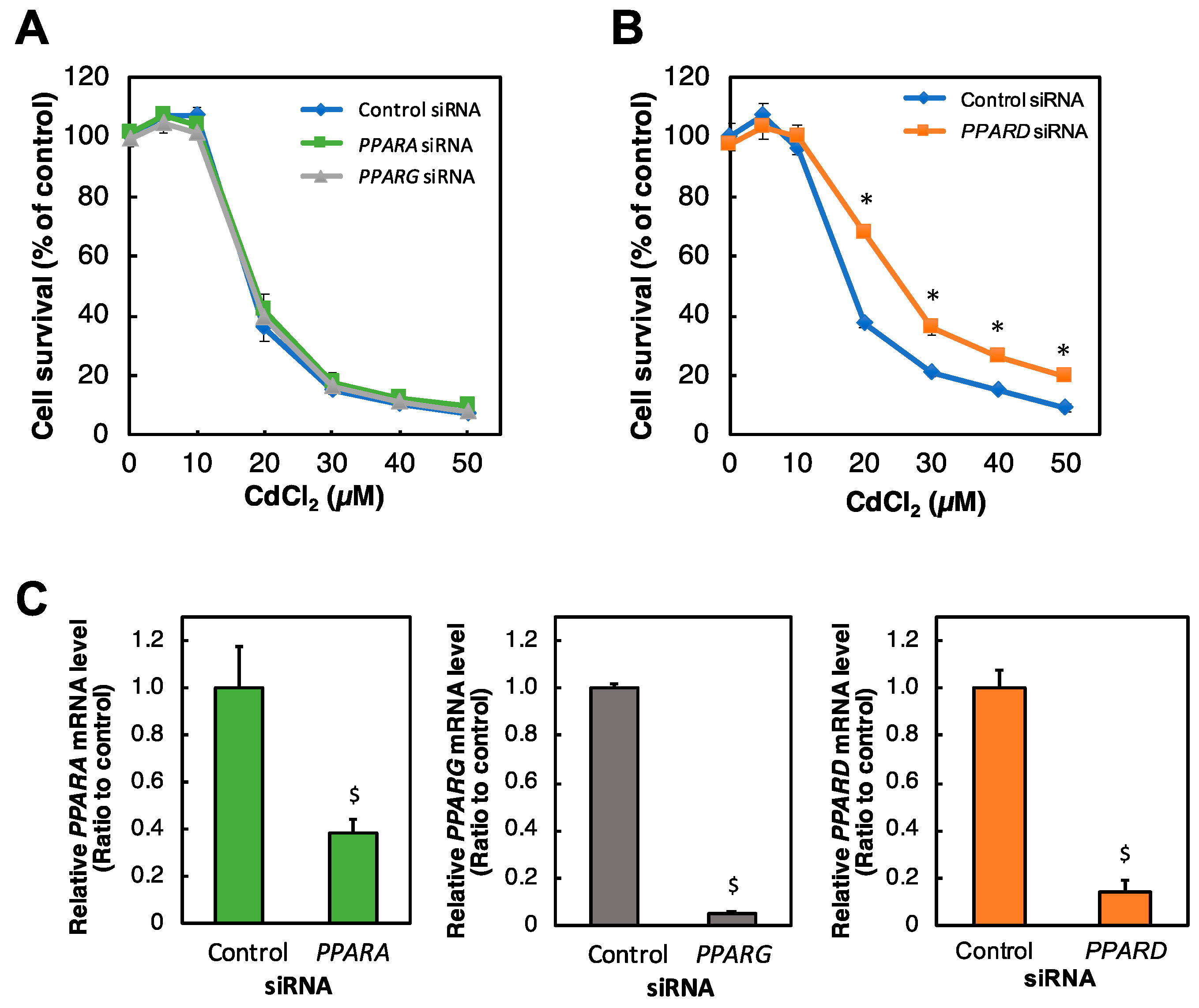
2.1. Identification of PPAR Affecting Cd Toxicity in HK-2 Cells

To investigate the involvement of PPAR in Cd toxicity in HK-2 cells, we examined the effect of knockdown of each PPAR gene on Cd toxicity. In human cells, PPARs consist of PPARα (PPARA), PPARδ (PPARD), and PPARγ (PPARG). Among the isoforms, only PPARD knockdown significantly conferred resistance to Cd toxicity (Figure 1A,B). Additionally, each siRNA against PPAR genes significantly reduced their mRNA levels in HK-2 cells (Figure 1C).

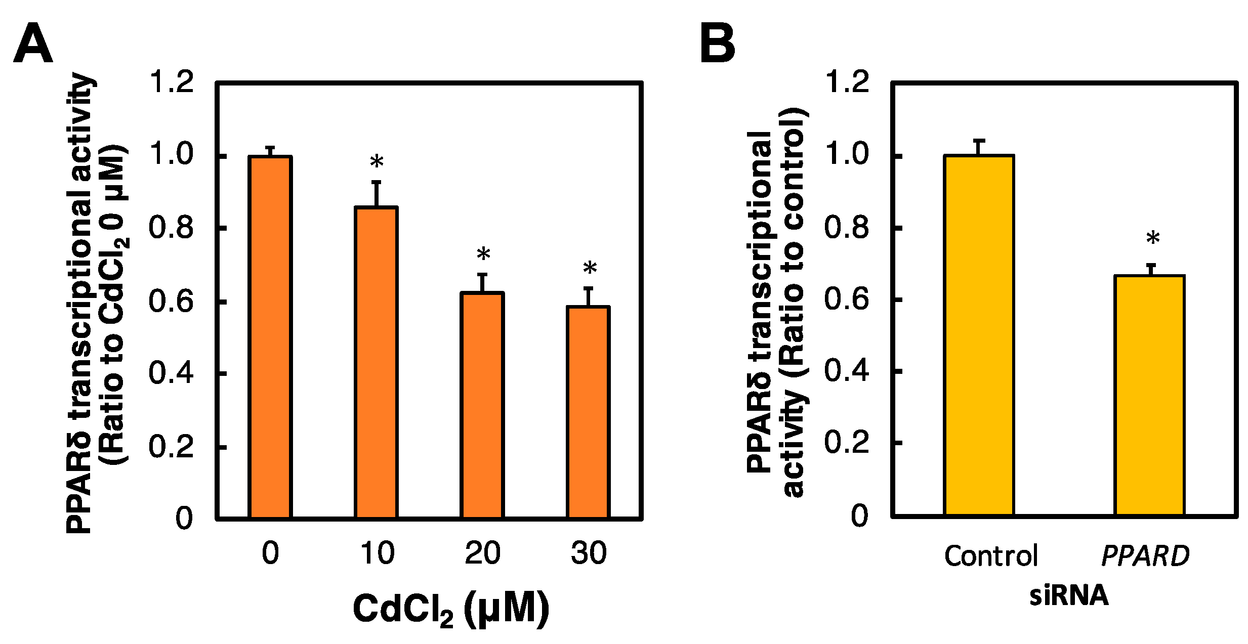
2.2. Effects of Cd and PPARD Knockdown on the PPARδ Transcriptional Activity in HK-2 Cells

To investigate the transcriptional activity of PPARδ by exposure to Cd and PPARD knockdown, we examined PPARδ transcriptional activity in Cd-treated or PPARD knockdown HK-2 cells. Cd treatment for 6 h significantly and dose-dependently decreased PPARδ transcriptional activity in HK-2 cells (Figure 2A). PPARδ transcriptional activity was significantly decreased after PPARD knockdown in HK-2 cells (Figure 2B).

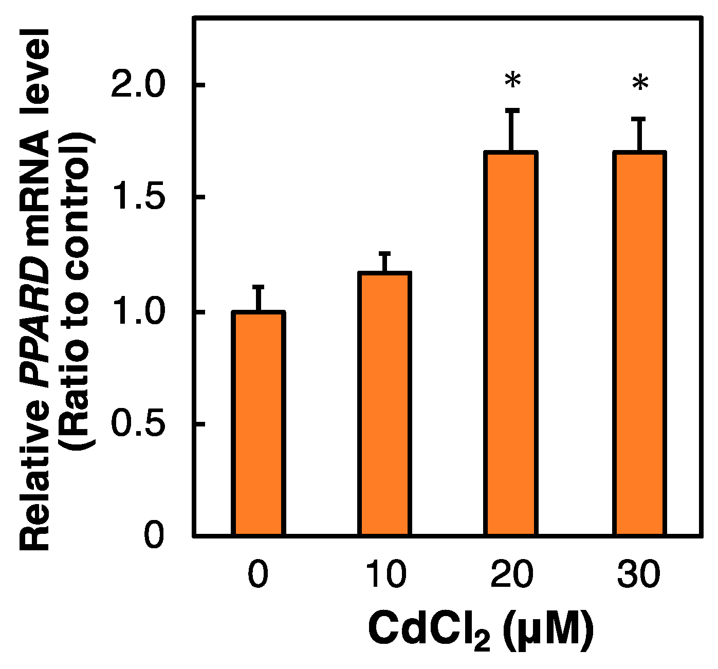
2.3. Effect of Cd on the PPARD mRNA Level in HK-2 Cells

To clarify the effect of Cd treatment on PPARD expression in HK-2 cells, the PPARD mRNA level was examined after HK-2 cells were treated with Cd. Cd treatment for 6 h significantly and dose-dependently increased the PPARD mRNA level in HK-2 cells (Figure 3). This result implies that the gene expression of PPARD may not contribute to the Cd-inhibited transcriptional activity of PPARδ in HK-2 cells.

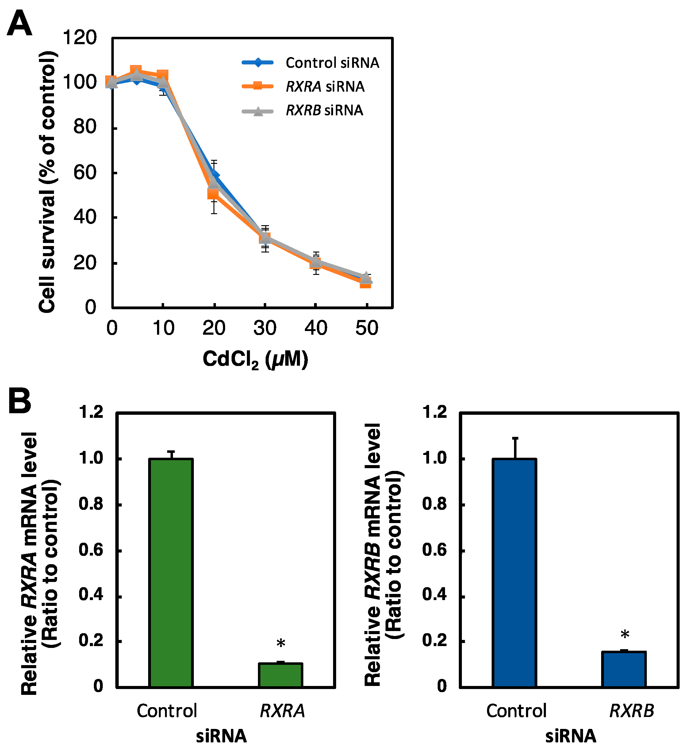
2.4. Effect of RXR Knockdown on the Viability of HK-2 Cells Treated with Cd

PPAR forms a dimer with RXR, and therefore resistance to Cd toxicity by PPARD knockdown may be associated with RXR activity. To investigate the involvement of RXR in the resistance Cd toxicity by PPARD knockdown in HK-2 cells, we examined the effect of RXR knockdown on Cd toxicity. RXR also consists of three isoforms [11,12,13]. Cd cytotoxicity in RXRA and RXRB knockdown cells was similar to that in control cells (Figure 4A). We also confirmed that RXRA and RXRB siRNA treatments significantly reduced the levels of RXRA and RXRB mRNAs in HK-2 cells (Figure 4B). Therefore, RXR may be independent of PPARD knockdown-decreased sensitivity to Cd toxicity in HK-2 cells.

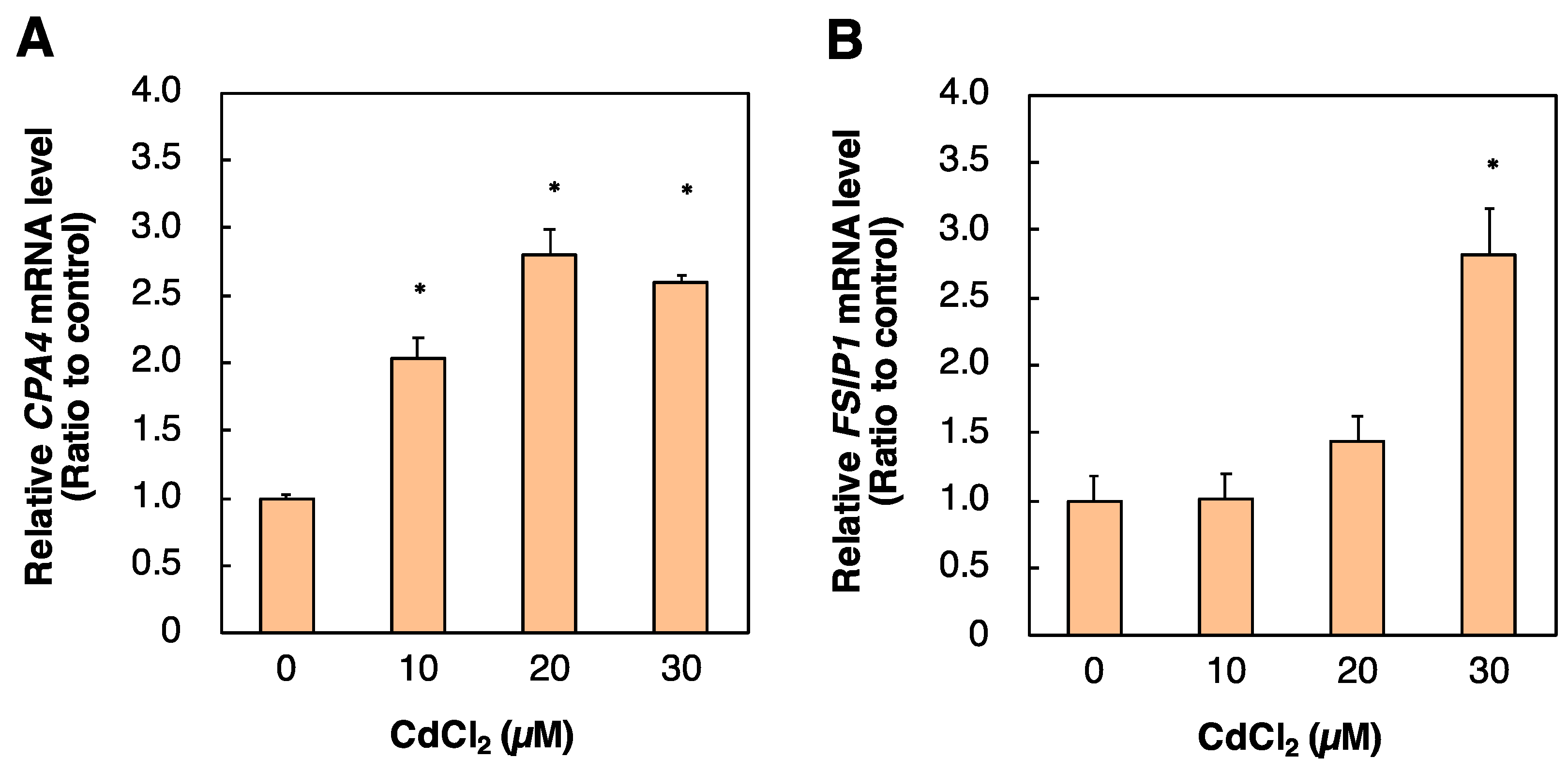
2.5. Identification of Genes Regulated by PPARD Knockdown in HK-2 Cells

PPARδ regulates gene expression by transcriptional activity, and therefore PPARδ-related Cd toxicity may affect downstream factor(s) of PPARδ. To identify genes regulated by PPARδ, we performed DNA microarray analysis of HK-2 cells transfected with PPARD siRNA. PPARD knockdown increased the expression of 53 genes by more than three-fold (Table 1). Among the genes, RYR2, ITPK1, PALD1, ZNF488, TFF2, IL9R, PANX2, CPA4, CCL19, FSIP1, and MLXIPL are involved in apoptosis [18,19,20,21,22,23,24,25,26,27,28]. PPARD knockdown decreased the expression of 39 genes by less than or equal to 0.5-fold (Table 2). Among these genes, LPAR3, GAL3ST1, PTPN11, and RORA are involved in apoptosis [29,30,31,32]. We examined the effect of Cd on the expression of 15 of the above genes (Figure 5, data not shown). Cd increased the mRNA levels of CPA4 and FSIP1 whose expression was induced by PPARD knockdown (Figure 5). Increases in cellular levels of CPA4 and FSIP1 negatively act on apoptosis signals [20,23]. These results indicated that apoptosis was associated with resistance to Cd toxicity by PPARD knockdown.

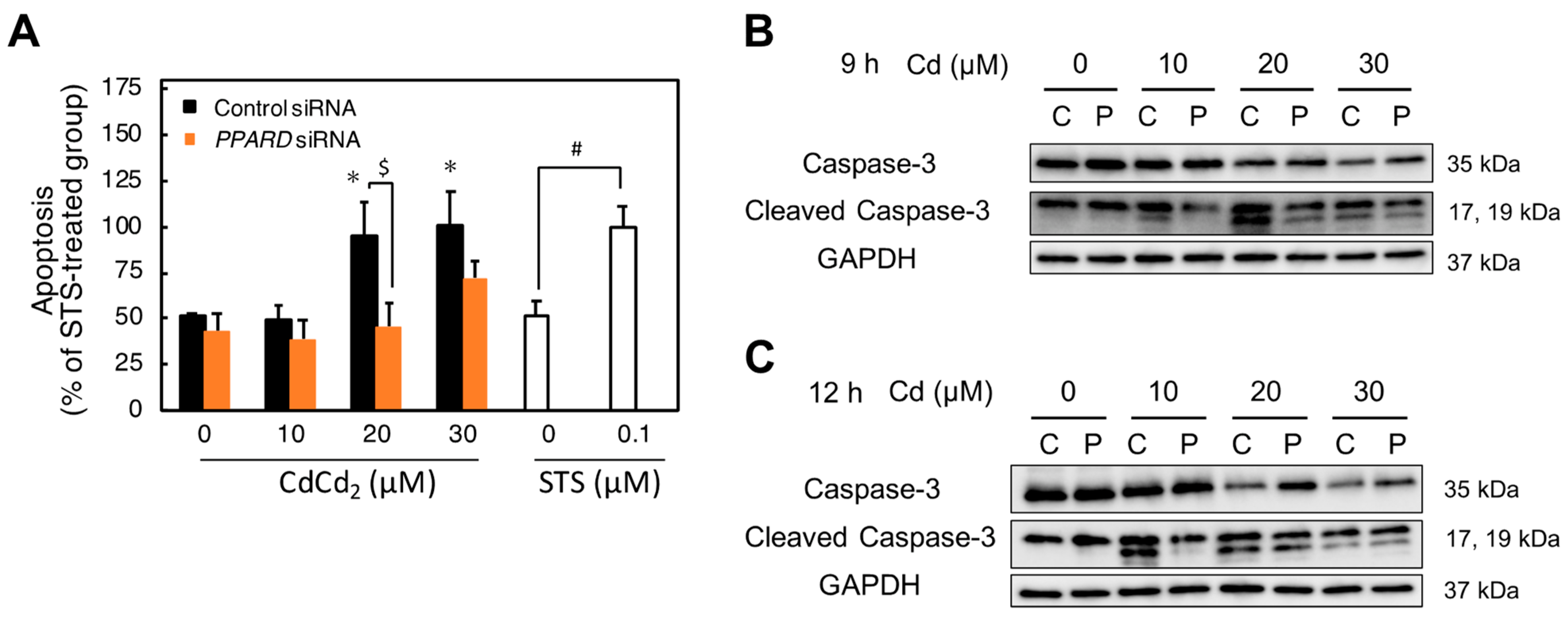
2.6. Involvement of PPARD Knockdown in Cd-Induced Apoptosis

Previous studies have demonstrated that Cd induces apoptosis in HK-2 cells [4,7,8,33]. Furthermore, some downstream factors of PPARδ are involved in the apoptosis pathway [34,35]. Therefore, we examined whether the resistance to Cd toxicity by PPARD knockdown was involved in apoptosis. To compare apoptosis levels, staurosporine (STS) was used as an apoptosis inducer. The apoptosis was significantly induced in control cells by 20 and 30 µM Cd treatment for 12 h. Additionally, Cd-induced apoptosis was significantly inhibited by PPARD knockdown. The apoptosis induced by 20 and 30 µM Cd treatment was similar to that induced by 0.1 µM STS treatment for 12 h (Figure 6A). Induction of apoptosis by Cd is mediated through caspase-3 activation in Cd renal toxicity [4]. Therefore, we investigated whether inhibition of Cd-induced apoptosis by PPARD knockdown was involved in caspase-3 activation. Cd treatment (10–30 µM) for 9 h increased the level of cleaved caspase-3, whereas PPARD knockdown decreased cleaved caspase-3 increased by Cd (Figure 6B). The treatment with 10 µM Cd for 12 h markedly increased the level of cleaved caspase-3 and PPARD knockdown decreased the increased one by Cd treatment (Figure 6C). However, the increased level of cleaved caspase-3 by 20 and 30 µM Cd for 12 h was not affected by PPARD knockdown. These results indicated that PPARD knockdown partly protected HK-2 cells from the Cd-induced apoptosis through a decrease in the cleaved caspase-3 level.

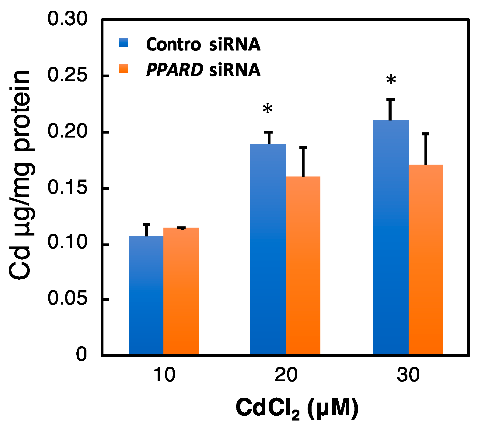
2.7. Effect of PPARD Knockdown on the Intracellular Cd Concentration

PPARD knockdown may affect the Cd accumulation in HK-2 cells. Therefore, we investigated the effect of PPARD knockdown on the intracellular Cd concentration after Cd treatment. The intracellular Cd level was increased dose-dependently on the treatment concentration (Figure 7). However, PPARD knockdown did not affect the intracellular Cd level (Figure 7).

3. Discussion

This study strongly suggests that the transcription factor PPARδ modifies susceptibility to Cd toxicity. Additionally, PPARδ transcriptional activity is inhibited in response to Cd stimulation in HK-2 cells. Furthermore, the decrease in PPARδ activity inhibits the apoptosis pathway induced by Cd in HK-2 cells and PPARD knockdown is resistant to Cd toxicity in HK-2 cells. The inhibitory effect of Cd on the transcriptional activity of PPARδ may be a biological reaction to protect against Cd toxicity. Moreover, PPARD knockdown did not change the concentration of Cd in cells, suggesting that PPARD knockdown is not involved in Cd uptake and excretion in HK-2 cells.
Cd increased the level of PPARD mRNA. However, transcriptional activity of PPARδ was suppressed by Cd. Therefore, the induction of gene expression of PPARD is unlikely to be associated with the suppression of PPARδ transcriptional activity. Several research groups have reported on the regulation of PPARδ activity. In the skeletal muscle, it has been reported that the activity of PPARδ is regulated by AMPK, CRYs, and PGC [36,37]. AMPK promotes PPARδ-dependent transcription. However, AMPK does not increase the PPARδ phosphorylation. AMPK may be present in a transcriptional complex with PPARδ [37]. CRY1 and CRY2 can selectively repress the transcriptional activity of PPARδ [36]. In addition, NCOA6 deficiency suppresses the activity of PPARδ in human and mouse hearts [38,39]. Recent studies demonstrated that Cd changes the AMPK-related pathway and disrupts the expressions of CRY1 [40,41]. These findings suggest that Cd may affect the transcriptional activity of PPARδ via the various mechanisms including the interaction with the cofactors and PPARδ.
Individual differences have been observed in the onset of chronic renal toxicity caused by Cd. Previous studies reported that there are gene polymorphisms in PPARD [42,43]. Our study demonstrated that PPARδ is a modification factor in Cd renal toxicity because PPARD knockdown is resistant to Cd renal toxicity. The presence of gene polymorphisms in PPARD means that there is a population with low PPARδ levels, which may be a population less sensitive to Cd renal toxicity. Moreover, changes in PPARδ may be one of the factors that cause individual differences in the development of Cd renal toxicity. Therefore, these findings suggest PPARδ plays an important role as a modification factor against Cd renal toxicity.
Cd decreases the activities of transcription factors YY1 and FOXF1. As a result, gene expression of the UBE2D family—downstream factors of YY1 and FOXF1—decreases in HK-2 cells [7]. Suppression of UBE2D family expression causes accumulation of apoptosis-inducing factor p53 in the cells, which induces apoptosis [7,33]. Cd decreases transcriptional activity of ARNT in HK-2 cells [8]. The cellular level BIRC3, a downstream factor of ARNT, was decreased by Cd treatment. BIRC3 is an inhibitor of apoptosis. Cd-decreased BIRC3 also induces apoptosis in the HK-2 cells [8]. PPARγ is involved in Cd-induced apoptosis and oxidative stress in renal epithelial cells and hepatocytes [44,45]. In the rat heart cells, DHA (docosahexaenoic acid), which acts as a ligand for PPARδ, has been reported to promote apoptotic cell death, increase caspase-3 activity, and reduce Akt phosphorylation via PPARδ [46]. Furthermore, it has been reported that intracellular prostacyclin promotes apoptosis by activating PPARδ in the human kidney cells [47]. On the other hand, it has also been reported that in mouse brain, PPARδ activation suppresses caspase-3 activation through miR-15a and its downstream Bcl-2 and protects cerebrovascular vessels by reducing apoptosis [48]. We demonstrated that PPARδ is a crucial factor that influences Cd-induced apoptosis in human proximal tubular cells. These findings indicate that Cd triggers renal toxicity via an apoptosis pathway with associations between various related factors. Depending on the various physiological circumstances, there may be differences in the contribution of each apoptosis-related factor to Cd-induced renal toxicity.

4. Materials and Methods

4.1. Cell Culture and Treatment

HK-2 cells purchased from the American Type Culture Collection (Manassas, MA, USA) were cultured in Dulbecco’s modified Eagle’s medium/Ham’s F-12 nutrient mixture (Sigma-Aldrich, St. Louis, MO, USA) containing 10% fetal bovine serum (Gibco, Grand Island, NY, USA), 25 U/mL penicillin (DS Pharm, Osaka, Japan), 25 μg/mL streptomycin (DS Pharm), 1% insulin-transferrin-selenium X (Gibco), 10 ng/mL epidermal growth factor (Sigma-Aldrich), and 5 ng/mL hydrocortisone at 37 °C in a humidified incubator with 5% CO2. The cells were cultured in test plates at a density of 250 cells/mm2 for 48 h. After discarding the culture medium, the cells were treated with Cd (CdCl2; 98.0%; Fujifilm Wako Pure Chemical Co., Tokyo, Japan) in serum-free culture medium.

4.2. Small Interfering RNA (siRNA) Transfection

Silencer Select Predesigned siRNAs against human PPAR and RXR mRNAs were purchased from Ambion (Grand Island, NY, USA). Control siRNA (Silencer Select Negative Control #1 siRNA) was also purchased from Ambion. siRNA transfection was performed with Lipofectamine RNAiMAX (Invitrogen, Grand Island, NY, USA). After the siRNA mixture was incubated for 15 min with Lipofectamine RNAiMAX and Opti-MEM I Reduced Serum Medium (Opti-MEM; Gibco), HK-2 cells were transfected with the siRNA mixture (1 nM siRNA per sequence, 0.2% Lipofectamine RNAiMAX, and 10% Opti-MEM) for 48 h.

4.3. Cell Survival Rate

HK-2 cells were treated with the siRNA mixture in 96-well plates for 48 h. After treatment, Alamar blue (10%; Invitrogen) was added and the cells were incubated for 4 h at 37 °C. Fluorescence was measured at an excitation wavelength of 540 nm and emission wavelength of 595 nm with a SpectraMax® iD3 microplate reader (Molecular Devices, San Jose, CA, USA).

4.4. RNA Extraction

HK-2 cells were treated with the siRNA mixture and Cd in 6-well plates. Cd-treated HK-2 cells were washed twice with ice-cold phosphate-buffered saline (PBS(−); Nissui, Tokyo, Japan). Total RNA was extracted with a PureLinkTM RNA Mini Kit (Ambion) in accordance with the manufacturer’s instructions. RNA quantity and purity were measured using a BioSpec-nano spectrophotometer (Shimadzu, Kyoto, Japan).

4.5. DNA Microarray Analysis

DNA microarray analysis was performed by Hokkaido System Science Co., Ltd. (Sapporo, Japan). Complementary RNA (cRNA) was synthesized from 50 ng total RNA using a Low Input Quick Amp Labeling Kit (Agilent Technologies, Santa Clara, CA, USA). HK-2 cells were treated with the siRNA mixture in 6-well plates for 48 h. The total RNAs were pooled from the independent three samples. Double-stranded cDNA from control siRNA- or PPARD siRNA-treated cells was transcribed in the presence of cyanine (Cy) 3-CTP or Cy5-CTP, respectively. These two sets of labeled cRNAs (300 ng each) were mixed and hybridized to a SurePrint G3 Human 8 × 60 K ver. 3.0 (Agilent Technologies) by a Gene Expression Hybridization Kit (Agilent Technologies) for 17 h at 65 °C. Fluorescent images were recorded with the Agilent Microarray Scanner (G2600D). Digitized image data were processed with Agilent Feature Extraction ver. 12.0.3.1. Information on each gene was obtained from the National Center for Biotechnology Information database.

4.6. Real-Time Reverse Transcription (RT)-PCR

To generate cDNA, total RNA was subjected to a PrimeScript RT Reagent Kit (Perfect Real Time) (Takara Bio, Shiga, Japan). Real-time PCR was performed with SYBR® Premix Ex TaqTM II (Perfect Real Time) (Takara Bio) on the Thermal Cycler Dice Real Time System (Takara Bio). The thermal cycling conditions were 10 s at 95 °C followed by 40 cycles of 5 s at 95 °C and 30 s at 60 °C. Gene expression was normalized to GAPDH mRNA levels. The oligonucleotide sequences of the primers used were as follows: sense, 5′-GAAACAGGCCTTCTCAGTGC-3′ and antisense, 5′-TTGCTGGGTCGTCTTTTTCT-3′ for the human PPARD gene; sense, 5′-ACGATTCGACTCAAGCTGGT-3′ and antisense, 5′-GTTGTGTGACATCCCGACAG-3′ for the human PPARA gene; sense, 5′-TTCAGAAATGCCTTGCAGTG-3′ and antisense, 5′-CCAACAGCTTCTCCTTCTCG-3′ for the human PPARG gene; sense, 5′-CAAGGACTGCCTGATTGACA-3′ and antisense, 5′-CTGGTCGACTCCACCTCATT-3′ for the human RXRA gene; sense, 5′-CCTGAGGGCAATCATTCTGT-3′, and antisense, 5′-CCTGCTGCTCAGGGTACTTC-3′ for the human RXRB gene; sense, 5′-GCACCGTCAAGGCTGAGAAC-3′ and antisense, 5′-TGGTGAAGACGCCAGTGGA-3′ for the human GAPDH gene.

4.7. Western Blot Analysis

HK-2 cells were treated with the siRNA mixture and Cd in 6 cm dishes. After treatment, the cells were washed twice with ice-cold PBS(−) and harvested in RIPA buffer (25 mM Tris, pH 7.6, 150 mM NaCl, 1% NP-40, 1% sodium deoxycholate, and 0.1% sodium dodecyl sulfate (SDS); Thermo Fisher Scientific, Waltham, MA, USA). Protein concentrations were measured by PierceTM BCA Protein Assay Kit (Thermo Fisher Scientific). Protein samples were separated on an SDS-polyacrylamide gel and transferred to a polyvinylidene difluoride membrane. The membrane was probed with primary antibodies and then with a horseradish peroxidase-conjugated secondary antibody (1:10,000 dilution; GE Healthcare, Little Chalfont, UK). The proteins were detected by enhanced chemiluminescence using Chemi-Lumi One Super (Nacalai Tesque, Kyoto, Japan). Chemiluminescence images were acquired with the ChemiDocTM imaging system (BIO-RAD, Hercules, CA, USA). The primary antibodies were anti-GAPDH (1:1000 dilution) from American Research Products (Waltham, MA, USA), and anti-Caspase-3 (1:1000 dilution) and anti-Cleaved Caspase-3 (diluted 1:1000) from Cell Signaling Technology (Danvers, MA, USA).

4.8. Nuclear Protein Extraction

Nuclei were extracted with a Nuclear Extraction Kit (ab113474; abcam, Tokyo, Japan). HK-2 cells were treated with Cd or the siRNA mixture in 10 cm dishes. The treated cells were pooled from the independent two samples. After treatment, HK-2 cells were washed twice with ice-cold PBS(−) and harvested in PBS(−). The cells were shaken at 200× g for 10 min at 4 °C in Pre-extraction Buffer that included a Protease Inhibitor Cocktail and dithiothreitol (DTT). The cytoplasmic fraction was collected by centrifugation at 14,000× g for 3 min at 4 °C. The nuclear pellet was resuspended in Extraction Buffer, which included the Protease Inhibitor Cocktail and DTT, and incubated at 4 °C for 1 h with agitation every 15 min. The mixture was centrifuged at 16,000× g for 10 min at 4 °C, and the supernatant was collected. Protein concentrations were measured by PierceTM BCA Protein Assay Kit.

4.9. PPARδ Transcriptional Activity Assay

PPARδ transcriptional activity was determined by a PPAR delta Transcriptional Factor Assay Kit (ab133106; abcam). After treatment, nuclei were extracted from HK-2 cells. Complete Transcription Factor Binding Assay Buffer (CTFB) was prepared by adding Transcription Factor Binding Assay Buffer, Reagent A, and DTT. Nuclei and CTFB were added to 96-well plates. After covering the 96-well plates with the included cover, the plates were incubated overnight at 4 °C without agitation. The 96-well plates were washed five times with Wash Buffer. Transcription Factor PPAR delta Primary Antibody was prepared by adding Transcription Factor Antibody Binding Buffer (ABB) and PPAR delta Primary Antibody. After completely removing the Wash Buffer, Transcription Factor PPAR delta Primary Antibody was added to the 96-well plates. After covering the 96-well plates with the included cover, the plates were incubated for 1 h at room temperature without agitation. The 96-well plates were then washed five times with Wash Buffer. Transcription Factor Goat Anti-Rabbit HRP conjugate was prepared by adding ABB and Goat Anti-Rabbit HRP conjugate. After completely removing the Wash Buffer, Transcription Factor Goat Anti-Rabbit HRP conjugate was added to the 96-well plates. After covering the 96-well plates with the included cover, the plates were incubated for 1 h at room temperature without agitation. The 96-well plates were washed five times with Wash Buffer. After completely removing the Wash Buffer, Transcription Factor Developing Solution was added to the 96-well plates. The 96-well plates were incubated for 45 min at room temperature with gentle agitation while protected from light. Absorbance at 450 nm was read within 5 min after adding Stop Solution to the 96-well plate with an iMarkTM Microplate Reader (BIO-RAD).

4.10. Apoptosis Assay

Apoptosis was examined by a Cell Death Detection ELIZAPLUS Kit (Roche, Basel, Switzerland). HK-2 cells were treated with the siRNA mixture and Cd in 96-well plates. After treatment, the HK-2 cells were washed twice with ice-cold PBS(−). Lysis Buffer was added to the cells, followed by incubation for 30 min at room temperature to lyse the cells. The cell lysates were repeatedly pipetted and then transferred to 96-well plates. Incubation buffer containing Anti-Histone Biotin and Anti-DNA POD was added to the 96-well plates. After covering with a close contact cover, the mixture was incubated at room temperature for 2 h while shaking at 300 rpm. The 96-well plate was washed three times with Incubation Buffer and then the Incubation Buffer was completely removed. ABTS solution, in which ABTS tablets were dissolved in the Substrate Buffer, was placed in a 96-well plate and incubated for 20 min with shaking at 250 rpm. After adding the ABTS stop solution, the absorbance was measured at 405 nm and 490 nm with the SpectraMax® iD3 microplate reader. Staurosporine (STS) was used for the positive control treatment [49]. The degree of Cd-induced apoptosis was normalized to apoptosis induced by 0.1 µM STS treatment (12 h).

4.11. Determination of Cd Content

After siRNA or Cd treatment using 6-well plates, cells were washed twice with ice-cold PBS(−) and then three times with PBS(−) containing 2 nM ethylene glycol tetraacetic acid (Nacalai Tesque). The cells were then harvested in 1 mL RIPA buffer and digested with nitric acid and hydrogen peroxide. After sample digestion, metal analysis was carried out using an atomic absorption spectrometer (200 series AA; Agilent Technologies). Protein concentrations were measured by PierceTM BCA Protein Assay Kit to normalize the Cd content.

4.12. Statistical Analysis

Statistical analyses were performed by one- or two-way ANOVA. When the F-value was significant (p < 0.05), Bonferroni’s multiple t-test was performed for post-hoc comparison (p < 0.05). Statistical analyses were performed with SPSS Statistics (IBM, Tokyo, Japan).

Author Contributions

C.M.: experiments and writing—original draft preparation; J.-Y.L.: design of experiments, analyses, and writing—review and editing; M.T.: analyses; M.S.: supervision, funding acquisition, and writing—review and editing. All authors have read and agreed to the published version of the manuscript.

Funding

This work was partly supported by a grant from the Ministry of Education, Culture, Sports, Science and Technology, Japan (Grant in Aid for Scientific Research [B], No. 18K10029). This work was also supported partly by the Study of the Health Effects of Heavy Metals, organized by the Ministry of the Environment, Japan.

Informed Consent Statement

Not applicable.

Conflicts of Interest

The authors declare no conflict of interest.

References

  1. Åkesson, A.; Barregard, L.; Bergdahl, I.A.; Nordberg, G.F.; Nordberg, M.; Skerfving, S. Non-Renal Effects and the Risk Assessment of Environmental Cadmium Exposure. Environ. Health Perspect. 2014, 122, 431–438. [Google Scholar] [CrossRef] [PubMed] [Green Version]
  2. Järup, L.; Åkesson, A. Current status of cadmium as an environmental health problem. Toxicol. Appl. Pharmacol. 2009, 238, 201–208. [Google Scholar] [CrossRef] [PubMed]
  3. Aoshima, K. Itai-itai disease: Lessons from the investigations of environmental epidemiology conducted in the 1970’s, with special reference to the studies of the Toyama Institute of Health. Nihon Eiseigaku Zasshi 2017, 72, 149–158. [Google Scholar] [CrossRef] [PubMed] [Green Version]
  4. Fujiwara, Y.; Lee, J.-Y.; Tokumoto, M.; Satoh, M. Cadmium Renal Toxicity via Apoptotic Pathways. Biol. Pharm. Bull. 2012, 35, 1892–1897. [Google Scholar] [CrossRef] [PubMed] [Green Version]
  5. Lee, J.-Y.; Tokumoto, M.; Satoh, M. Novel Mechanisms of Cadmium-Induced Toxicity in Renal Cells. In Cadmium Toxicity: New Aspects in Human Disease, Rice Contamination, and Cytotoxicity; Himeno, S., Aoshima, K., Eds.; Springer: Singapore, 2019; pp. 153–162. [Google Scholar]
  6. Tokumoto, M.; Lee, J.-Y.; Satoh, M. Transcription Factors and Downstream Genes in Cadmium Toxicity. Biol. Pharm. Bull. 2019, 42, 1083–1088. [Google Scholar] [CrossRef] [Green Version]
  7. Lee, J.-Y.; Tokumoto, M.; Fujiwara, Y.; Hasegawa, T.; Seko, Y.; Shimada, A.; Satoh, M. Accumulation of p53 via down-regulation of UBE2D family genes is a critical pathway for cadmium-induced renal toxicity. Sci. Rep. 2016, 6, 21968. [Google Scholar] [CrossRef] [Green Version]
  8. Lee, J.-Y.; Tokumoto, M.; Hwang, G.-W.; Lee, M.-Y.; Satoh, M. Identification of ARNT-regulated BIRC3 as the target factor in cadmium renal toxicity. Sci. Rep. 2017, 7, 17287. [Google Scholar] [CrossRef] [Green Version]
  9. Lee, J.; Tokumoto, M.; Satoh, M. Cadmium toxicity mediated by the inhibition of SLC2A4 expression in human proximal Tubule cells. FASEB J. 2020, 35, e21236. [Google Scholar] [CrossRef]
  10. Tokumoto, M.; Lee, J.-Y.; Fujiwara, Y.; Satoh, M. Alteration of DNA binding activity of transcription factors in NRK-52E rat proximal tubular cells treated with cadmium. J. Toxicol. Sci. 2014, 39, 735–738. [Google Scholar] [CrossRef] [Green Version]
  11. Burns, K.A.; Heuvel, J.P.V. Modulation of PPAR activity via phosphorylation. Biochim. Biophys. Acta (BBA) Mol. Cell Biol. Lipids 2007, 1771, 952–960. [Google Scholar] [CrossRef] [PubMed] [Green Version]
  12. Desvergne, B.; Wahli, W. Peroxisome Proliferator-Activated Receptors: Nuclear Control of Metabolism. Endocr. Rev. 1999, 20, 649–688. [Google Scholar] [CrossRef] [Green Version]
  13. Juge-Aubry, C.; Pernin, A.; Favez, T.; Burger, A.G.; Wahli, W.; Meier, C.A.; Desvergne, B. DNA Binding Properties of Peroxisome Proliferator-activated Receptor Subtypes on Various Natural Peroxisome Proliferator Response Elements. Importance of the 5’-flanking region. J. Biol. Chem. 1997, 272, 25252–25259. [Google Scholar] [CrossRef] [PubMed] [Green Version]
  14. Finck, B.N. The PPAR regulatory system in cardiac physiology and disease. Cardiovasc. Res. 2007, 73, 269–277. [Google Scholar] [CrossRef] [Green Version]
  15. Liu, Y.; Colby, J.K.; Zuo, X.; Jaoude, J.; Wei, D.; Shureiqi, I. The Role of PPAR-δ in Metabolism, Inflammation, and Cancer: Many Characters of a Critical Transcription Factor. Int. J. Mol. Sci. 2018, 19, 3339. [Google Scholar] [CrossRef] [PubMed] [Green Version]
  16. Peters, J.M.; Gonzalez, F.J. Sorting out the functional role(s) of peroxisome proliferator-activated receptor-beta/delta (PPARbeta/delta) in cell proliferation and cancer. Biochim. Biophys. Acta 2009, 1796, 230–241. [Google Scholar] [PubMed] [Green Version]
  17. Wang, Y.X.; Zhang, C.L.; Yu, R.T.; Cho, H.K.; Nelson, M.C.; Bayuga-Ocampo, C.R.; Ham, J.; Kang, H.; Evans, R.M. Regulation of muscle fiber type and running endurance by PPARdelta. PLoS Biol. 2004, 2, e294. [Google Scholar] [CrossRef] [Green Version]
  18. Berchtold, L.A.; Miani, M.; Diep, T.A.; Madsen, A.N.; Cigliola, V.; Colli, M.; Krivokapic, J.M.; Pociot, F.; Eizirik, D.L.; Meda, P.; et al. Pannexin-2-deficiency sensitizes pancreatic β-cells to cytokine-induced apoptosis in vitro and impairs glucose tolerance in vivo. Mol. Cell. Endocrinol. 2017, 448, 108–121. [Google Scholar] [CrossRef] [PubMed]
  19. Egaña, I.; Kaito, H.; Nitzsche, A.; Becker, L.; Ballester-Lopez, C.; Niaudet, C.; Petkova, M.; Liu, W.; Vanlandewijck, M.; Vernaleken, A.; et al. Female mice lacking Pald1 exhibit endothelial cell apoptosis and emphysema. Sci. Rep. 2017, 7, 15453. [Google Scholar] [CrossRef] [Green Version]
  20. Fu, Y.; Su, L.; Cai, M.; Yao, B.; Xiao, S.; He, Q.; Xu, L.; Yang, L.; Zhao, C.; Wan, T.; et al. Downregulation of CPA4 inhibits non small–cell lung cancer growth by suppressing the AKT/c-MYC pathway. Mol. Carcinog. 2019, 58, 2026–2039. [Google Scholar] [CrossRef] [Green Version]
  21. Johnson, J.D.; Han, Z.; Otani, K.; Ye, H.; Zhang, Y.; Wu, H.; Horikawa, Y.; Misler, S.; Bell, G.I.; Polonsky, K.S. RyR2 and Calpain-10 Delineate a Novel Apoptosis Pathway in Pancreatic Islets. J. Biol. Chem. 2004, 279, 24794–24802. [Google Scholar] [CrossRef] [Green Version]
  22. Kim, N.; Yu, L.; Dawe, R.; Petyuk, V.A.; Gaiteri, C.; De Jager, P.L.; Schneider, J.A.; Arfanakis, K.; Bennett, D.A. Microstructural changes in the brain mediate the association of AK4, IGFBP5, HSPB2, and ITPK1 with cognitive decline. Neurobiol. Aging 2019, 84, 17–25. [Google Scholar] [CrossRef] [PubMed]
  23. Liu, T.; Zhang, H.; Sun, L.; Zhao, D.; Liu, P.; Yan, M.; Zaidi, N.; Izadmehr, S.; Gupta, A.; Abu-Amer, W.; et al. FSIP1 binds HER2 directly to regulate breast cancer growth and invasiveness. Proc. Natl. Acad. Sci. USA 2017, 114, 7683–7688. [Google Scholar] [CrossRef] [PubMed] [Green Version]
  24. Poungvarin, N.; Lee, J.K.; Yechoor, V.K.; Li, M.V.; Assavapokee, T.; Suksaranjit, P.; Thepsongwajja, J.J.; Saha, P.K.; Oka, K.; Chan, L. Carbohydrate response element-binding protein (ChREBP) plays a pivotal role in beta cell glucotoxicity. Diabetologia 2012, 55, 1783–1796. [Google Scholar] [CrossRef] [PubMed] [Green Version]
  25. Shang, Y.; Kakinuma, S.; Nishimura, M.; Kobayashi, Y.; Nagata, K.; Shimada, Y. Interleukin-9 receptor gene is transcriptionally regulated by nucleolin in T-Cell lymphoma cells. Mol. Carcinog. 2011, 51, 619–627. [Google Scholar] [CrossRef] [PubMed]
  26. Tu, S.P.; Chi, A.L.; Ai, W.; Takaishi, S.; Dubeykovskaya, Z.; Quante, M.; Fox, J.G.; Wang, T.C. p53 inhibition of AP1-dependent TFF2 expression induces apoptosis and inhibits cell migration in gastric cancer cells. Am. J. Physiol. Liver Physiol. 2009, 297, G385–G396. [Google Scholar] [CrossRef]
  27. Zhang, X.; Wang, Y.; Cao, Y.; Zhang, X.; Zhao, H. Increased CCL19 expression is associated with progression in cervical cancer. Oncotarget 2017, 8, 73817–73825. [Google Scholar] [CrossRef] [PubMed] [Green Version]
  28. Zong, D.; Jiang, N.; Xu, J.-H.; Wang, D.-J.; Zhu, H.-F.; Wu, L.-R.; Chen, C.; Yin, L.; He, X. ZNF488 is an independent prognostic indicator in nasopharyngeal carcinoma and promotes cell adhesion and proliferation via collagen IV/FAK/AKT/Cyclin D1 pathway. Cancer Manag. Res. 2019, ume 11, 5871–5882. [Google Scholar] [CrossRef] [Green Version]
  29. Chitranshi, N.; Dheer, Y.; Gupta, V.; Abbasi, M.; Mirzaei, M.; You, Y.; Chung, R.; Graham, S.L.; Gupta, V. PTPN11 induces endoplasmic stress and apoptosis in SH-SY5Y cells. Neuroscience 2017, 364, 175–189. [Google Scholar] [CrossRef]
  30. Ding, K.; Yu, Z.H.; Yu, C.; Jia, Y.Y.; He, L.; Liao, C.S.; Li, J.; Zhang, C.J.; Li, Y.J.; Wu, T.C.; et al. Effect of gga-miR-155 on cell proliferation, apoptosis and invasion of Marek’s disease virus (MDV) transformed cell line MSB1 by targeting RORA. BMC Vet. Res. 2020, 16, 23. [Google Scholar] [CrossRef] [PubMed] [Green Version]
  31. Geraldo, L.H.M.; de Sampaio Spohr, T.C.L.; do Amaral, R.F.; da Fonseca, A.C.C.; Garcia, C.; de Almeida Mendes, F.; Freitas, C.; Fabio dosSantos, M.; Lima, F.R.S. Role of lysophosphatidic acid and its receptors in health and disease: Novel therapeutic strategies. Signal Transduct. Target. Ther. 2021, 6, 45. [Google Scholar] [CrossRef]
  32. Suchanski, J.; Grzegrzolka, J.; Owczarek, T.; Pasikowski, P.; Piotrowska, A.; Kocbach, B.; Nowak, A.; Dziegiel, P.; Wojnar, A.; Ugorski, M. Sulfatide decreases the resistance to stress-induced apoptosis and increases P-selectin-mediated adhesion: A two-edged sword in breast cancer progression. Breast Cancer Res. 2018, 20, 133. [Google Scholar] [CrossRef] [PubMed]
  33. Tokumoto, M.; Fujiwara, Y.; Shimada, A.; Hasegawa, T.; Seko, Y.; Nagase, H.; Satoh, M. Cadmium toxicity is caused by accumulation of p53 through the down-regulation of Ube2d family genes in vitro and in vivo. J. Toxicol. Sci. 2011, 36, 191–200. [Google Scholar] [CrossRef] [PubMed] [Green Version]
  34. Hwang, J.S.; Eun, S.Y.; Ham, S.A.; Yoo, T.; Lee, W.J.; Paek, K.S.; Do, J.T.; Lim, D.S.; Seo, H.G. PPARδ modulates oxLDL-induced apoptosis of vascular smooth muscle cells through a TGF-β/FAK signaling axis. Int. J. Biochem. Cell Biol. 2015, 62, 54–61. [Google Scholar] [CrossRef] [PubMed]
  35. Wu, K.K.; Liou, J.Y. Cyclooxygenase inhibitors induce colon cancer cell apoptosis Via PPARdelta --> 14-3-3epsilon pathway. Methods Mol. Biol. 2009, 512, 295–307. [Google Scholar] [PubMed]
  36. Jordan, S.D.; Kriebs, A.; Vaughan, M.; Duglan, D.; Fan, W.; Henriksson, E.; Huber, A.L.; Papp, S.J.; Nguyen, M.; Afetian, M.; et al. CRY1/2 Selectively Repress PPARδ and Limit Exercise Capacity. Cell Metab. 2017, 26, 243–255.e6. [Google Scholar] [CrossRef] [Green Version]
  37. Narkar, V.A.; Downes, M.; Yu, R.T.; Embler, E.; Wang, Y.X.; Banayo, E.; Mihaylova, M.M.; Nelson, M.C.; Zou, Y.; Juguilon, H.; et al. AMPK and PPARdelta agonists are exercise mimetics. Cell 2008, 134, 405–415. [Google Scholar] [CrossRef] [Green Version]
  38. Mahajan, M.A.; Samuels, H.H. Nuclear receptor coactivator/coregulator NCoA6(NRC) is a pleiotropic coregulator involved in transcription, cell survival, growth and development. Nucl. Recept. Signal. 2008, 6, e002. [Google Scholar] [CrossRef] [Green Version]
  39. Roh, J.-I.; Cheong, C.; Sung, Y.H.; Lee, J.; Oh, J.; Lee, B.S.; Lee, J.-E.; Gho, Y.S.; Kim, D.-K.; Park, C.B.; et al. Perturbation of NCOA6 Leads to Dilated Cardiomyopathy. Cell Rep. 2014, 8, 991–998. [Google Scholar] [CrossRef] [PubMed] [Green Version]
  40. Arab, H.H.; Gad, A.M.; Reda, E.; Yahia, R.; Eid, A.H. Activation of autophagy by sitagliptin attenuates cadmium-induced testicular impairment in rats: Targeting AMPK/mTOR and Nrf2/HO-1 pathways. Life Sci. 2021, 269, 119031. [Google Scholar] [CrossRef] [PubMed]
  41. Jiménez-Ortega, V.; Cano-Barquilla, P.; Scacchi, P.A.; Cardinali, D.P.; Esquifino, A.I. Cadmium-Induced Disruption in 24-h Expression of Clock and Redox Enzyme Genes in Rat Medial Basal Hypothalamus: Prevention by Melatonin. Front. Neurol. 2011, 2, 13. [Google Scholar] [CrossRef] [Green Version]
  42. Bailén, M.; Tabone, M.; Bressa, C.; Lominchar, M.G.M.; Larrosa, M.; González-Soltero, R. Unraveling Gut Microbiota Signatures Associated with PPARD and PARGC1A Genetic Polymorphisms in a Healthy Population. Genes 2022, 13, 289. [Google Scholar] [CrossRef] [PubMed]
  43. Hishida, A.; Wakai, K.; Naito, M.; Tamura, T.; Kawai, S.; Hamajima, N.; Oze, I.; Imaizumi, T.; Turin, T.C.; Suzuki, S.; et al. Polymorphisms in PPAR Genes (PPARD, PPARG, and PPARGC1A) and the Risk of Chronic Kidney Disease in Japanese: Cross-Sectional Data from the J-MICC Study. PPAR Res. 2013, 2013, 980471. [Google Scholar] [CrossRef] [PubMed] [Green Version]
  44. Hosohata, K.; Mise, N.; Kayama, F.; Iwanaga, K. Augmentation of cadmium-induced oxidative cytotoxicity by pioglitazone in renal tubular epithelial cells. Toxicol. Ind. Health 2019, 35, 530–536. [Google Scholar] [CrossRef]
  45. Wang, H.; Wang, A.; Wang, X.; Zeng, X.; Xing, H. AMPK/PPAR-γ/NF-κB axis participates in ROS-mediated apoptosis and autophagy caused by cadmium in pig liver. Environ. Pollut. 2022, 294, 118659. [Google Scholar] [CrossRef] [PubMed]
  46. Samokhvalov, V.; Zlobine, I.; Jamieson, K.L.; Jurasz, P.; Chen, C.; Lee, K.S.S.; Hammock, B.D.; Seubert, J.M. PPARδ signaling mediates the cytotoxicity of DHA in H9c2 cells. Toxicol. Lett. 2014, 232, 10–20. [Google Scholar] [CrossRef] [PubMed] [Green Version]
  47. Hatae, T.; Wada, M.; Yokoyama, C.; Shimonishi, M.; Tanabe, T. Prostacyclin-dependent apoptosis mediated by PPAR delta. J. Biol. Chem. 2001, 276, 46260–46267. [Google Scholar] [CrossRef] [PubMed] [Green Version]
  48. Yin, K.J.; Deng, Z.; Hamblin, M.; Xiang, Y.; Huang, H.; Zhang, J.; Jiang, X.; Wang, Y.; Chen, Y.E. Peroxisome proliferator-activated receptor delta regulation of miR-15a in ischemia-induced cerebral vascular endothelial injury. J. Neurosci. 2010, 30, 6398–6408. [Google Scholar] [CrossRef] [PubMed] [Green Version]
  49. Bertrand, R.; Solary, E.; O’Connor, P.; Kohn, K.W.; Pommier, Y. Induction of a Common Pathway of Apoptosis by Staurosporine. Exp. Cell Res. 1994, 211, 314–321. [Google Scholar] [CrossRef] [PubMed]
Figure 1. Effect of PPAR knockdown on the survival rate of HK-2 cells treated with Cd. (A,B) After treatment with PPARA, PPARG or PPARD siRNA for 48 h, HK-2 cells were treated with Cd for 24 h. Cell survival was examined by an Alamar blue assay. Values are means ± SD (n = 3). * Significantly different from the control siRNA group, p < 0.05. (A) PPARA siRNA and PPARG siRNA. (B) PPARD siRNA. (C) Efficiency of PPARA, PPARG, and PPARD knockdown was examined after HK-2 cells were treated with siRNA against the gene of PPARA, PPARG or PPARD for 48 h. PPARA, PPARG, and PPARD mRNA levels were examined by real-time RT-PCR and normalized to GAPDH mRNA levels. Values are means ± SD (n = 3). $ Significantly different from the control group, p < 0.05.
Ijms 23 08652 g001
Figure 2. Effect of Cd and PPARD knockdown on the PPARδ transcriptional activity in HK-2 cells. (A) HK-2 cells were treated with Cd for 6 h. PPARδ transcriptional activity was examined by a PPAR delta Transcriptional Factor Assay Kit after Cd treatment for 6 h. Values are means ± SD (n = 3). * Significantly different from the control group, p < 0.05. (B) PPARδ transcriptional activity by PPARD knockdown was examined after HK-2 cells were treated with PPARD siRNA for 48 h. Values are means ± SD (n = 3). * Significantly different from the control siRNA group, p < 0.05.
Ijms 23 08652 g002
Figure 3. Effect of Cd on the PPARD mRNA level in HK-2 cells. HK-2 cells were treated with Cd for 6 h. PPARD mRNA levels were examined by real-time RT-PCR after Cd treatment for 6 h. PPARD mRNA levels were normalized to GAPDH mRNA levels. Values are means ± SD (n = 3). * Significantly different from the control group, p < 0.05.
Ijms 23 08652 g003
Figure 4. Effect of RXR knockdown on the survival rate of HK-2 cells treated with Cd. (A) After treatment with siRNA against the gene of RXRA or RXRB for 48 h, HK-2 cells were treated with Cd for 24 h. Cell survival was examined by the Alamar blue assay. Values are means ± SD (n = 3). * Significantly different from the control siRNA group, p < 0.05. (B) Efficiency of RXRA and RXRB knockdown was examined after HK-2 cells were treated with siRNA the gene of RXRA or RXRB for 48 h. RXRA and RXRB mRNA levels were examined by real-time RT–PCR and normalized to GAPDH mRNA levels. Values are means ± SD (n = 3). * Significantly different from the control group, p < 0.05.
Ijms 23 08652 g004
Figure 5. Effect of Cd on CPA4 and FSIP1 mRNA levels in HK-2 cells. HK-2 cells were treated with Cd for 6 h. CPA4 and FSIP1 mRNA levels were examined by real-time RT–PCR after Cd treatment for 6 h (A,B). CPA4 and FSIP1 mRNA levels were normalized to GAPDH mRNA levels. Values are means ± SD (n = 3). * Significantly different from the control group, p < 0.05.
Ijms 23 08652 g005
Figure 6. Effect of PPARD knockdown on apoptosis of HK-2 cells treated with Cd. (A) After treatment with PPARD siRNA for 48 h, HK-2 cells were treated with Cd for 12 h. HK-2 cells were treated with STS, as an apoptosis inducer, for 12 h. Apoptosis was examined by a Cell Death Detection ELIZAPLUS Kit after Cd and STS treatment for 12 h. Values are means ± SD (n = 3). * Significantly different from the control siRNA + Cd 0 μM group, p < 0.05. $ Significantly different from the control siRNA group, p < 0.05. # Significantly different from the STS 0 μM group, p < 0.05. (B,C) After treatment with PPARD siRNA for 48 h, HK-2 cells were treated with Cd for 9 h (B) or 12 h (C). Protein levels of caspase-3 and cleaved caspase-3 were examined by western blotting after Cd treatment for 9 or 12 h. GAPDH was used as the loading control.
Ijms 23 08652 g006
Figure 7. Effect of PPARD knockdown on the intracellular Cd concentration. After treatment with PPARD siRNA for 48 h, HK-2 cells were treated with Cd for 12 h. The Cd concentration was measured by an atomic absorption spectrometer. Values are means ± SD (n = 3). * Significantly different from the control siRNA + Cd 10 μM group, p < 0.05.
Ijms 23 08652 g007
Table 1. Genes with expression increased by PPARD knockdown (>3-fold).
GeneAccession NumberDescriptionFold Change
RYR2NM_001035Ryanodine receptor 25.75
CDHR2NM_017675Cadherin related family member 25.31
PCDH19NM_020766Protocadherin 194.92
TMEM184ANM_001097620Transmembrane protein 184A4.91
GLYATNM_005838Glycine-N-acyltransferase4.89
AUTS2NM_001127231Activator of transcription and developmental regulator AUTS24.86
MSR1NM_002445Macrophage scavenger receptor 14.80
CRLF2NM_001012288Cytokine receptor like factor 24.77
SLC23A3NM_144712Solute carrier family 23 member 34.76
IL27NM_145659Interleukin 274.71
PSG2NM_031246Pregnancy specific beta-1-glycoprotein 24.70
PITPNM3NM_031220PITPNM family member 34.46
ITPK1NM_014216Inositol-tetrakisphosphate 1-kinase4.42
ACTN2NM_001278344Actinin alpha 24.39
BTBD16NM_001318189BTB domain containing 164.38
PALD1NM_014431Phosphatase domain containing paladin 14.38
RGS11NM_003834Regulator of G protein signaling 114.34
SMCO1NM_001077657Single-pass membrane protein with coiled-coil domains 14.29
ZNF488NM_153034Zinc finger protein 4884.21
GAL3ST1NM_004861Pentatricopeptide repeat domain 24.20
CACNG7NM_031896Calcium voltage-gated channel auxiliary subunit gamma 74.19
SERPINF1NM_002615Serpin family F member 14.19
ESPNLNM_194312Espin like4.19
LONRF2NM_198461LON peptidase N-terminal domain and ring finger 24.19
PDE4CNM_001330172Phosphodiesterase 4C4.18
PTPN11NM_002834Transmembrane protein 225B4.16
PRDM11NM_001359633PR/SET domain 114.11
TFF2NM_005423Trefoil factor 24.11
PDZD4NM_032512PDZ domain containing 44.02
CHRNENM_000080Cholinergic receptor nicotinic epsilon subunit4.01
LRRC3NM_030891Leucine rich repeat containing 34.00
IL9RNM_176786Interleukin 9 receptor3.94
PANX2NM_001160300Pannexin 23.88
CPA4NM_016352Carboxypeptidase A43.76
PLCB1NM_182734Phospholipase C beta 13.65
TLDC2NM_080628TBC/LysM-associated domain containing 23.63
PPCDCNM_134260Phosphopantothenoylcysteine decarboxylase3.61
RGMBNM_001366509Repulsive guidance molecule BMP co-receptor b3.51
PDE4DIPNM_001350522Phosphodiesterase 4D interacting protein3.51
RNF125NM_017831Ring finger protein 1253.50
JRKNM_001279352Jrk helix-turn-helix protein3.50
TPRX1NM_198479Tetrapeptide repeat homeobox 13.44
HAAONM_0122053-Hydroxyanthranilate 3,4-dioxygenase3.41
SLC43A1NM_003627Solute carrier family 43 member 13.35
CCL19NM_006274C-C motif chemokine ligand 193.29
RBM20NM_001134363RNA binding motif protein 203.23
FSIP1NM_152597Fibrous sheath interacting protein 13.19
MPZL3NM_198275Myelin protein zero like 33.18
SMKR1NM_001195243Small lysine rich protein 13.12
MLXIPLNM_032951MLX interacting protein like3.11
PLXNC1NM_005761Plexin C13.11
OR6T1NM_001005187Olfactory receptor family 6 subfamily T member 13.10
EGFNM_001178130Epidermal growth factor3.06
Table 2. Genes with expression decreased by PPARD knockdown (≤0.5-fold).
GeneAccession NumberDescriptionFold Change
TTLL9NM_001035Tubulin tyrosine ligase like 90.10
TNFNM_000594Tumor necrosis factor0.23
SAA1NM_000331Serum amyloid A10.24
CCL20NM_004591C-C motif chemokine ligand 200.25
FLGNM_002016Filaggrin0.26
PPARDNM_006238Peroxisome proliferator activated receptor delta0.32
LPAR3NM_012152Lysophosphatidic acid receptor 30.35
PCOTHNM_001348114Pro-X-Gly collagen triple helix like repeat containing0.36
HACD3NM_0163953-Hydroxyacyl-CoA dehydratase 30.38
ANKS1BNM_001352196Ankyrin repeat and sterile alpha motif domain containing 1B0.39
TRAK2NM_015049Trafficking kinesin protein 20.40
ST3GAL6NM_006100ST3 beta-galactoside alpha-2,3-sialyltransferase 60.40
LTBNM_014216Lymphotoxin beta0.40
UHMK1NM_175866U2AF homology motif kinase 10.40
DDR1NM_001202522Discoidin domain receptor tyrosine kinase 10.41
KCNS3NM_014431Potassium voltage-gated channel modifier subfamily S member 30.42
SAA2NM_030754Serum amyloid A20.42
FSIP1NM_152597Fibrous sheath interacting protein 10.43
EFHC1NM_153034EF-hand domain containing 10.43
GAL3ST1NM_004861Galactose-3-O-sulfotransferase 10.43
IL18R1NM_003855Interleukin 18 receptor 10.44
SGCBNM_000232Sarcoglycan beta0.44
PCDH20NM_022843Protocadherin 200.44
CA9NM_001216Carbonic anhydrase 90.44
ZDHHC11BNM_001351303Zinc finger DHHC-type containing 11B0.44
PTPN11NM_002834Protein tyrosine phosphatase non-receptor type 110.45
CXADRNM_001338CXADR Ig-like cell adhesion molecule0.46
YES1NM_005423YES proto-oncogene 1, Src family tyrosine kinase0.46
LOC391322NM_032512D-dopachrome tautomerase-like0.47
CCDC146NM_020879Coiled-coil domain containing 1460.47
EIF1AYNM_004681Eukaryotic translation initiation factor 1A Y-linked0.47
PTX3NM_176786Pentraxin 30.47
PTGFRNNM_001160300Prostaglandin F2 receptor inhibitor0.47
CSF2NM_016352Colony stimulating factor 20.48
ANKRD37NM_181726Ankyrin repeat domain 370.48
ENTPD2NM_001246Ectonucleoside triphosphate diphosphohydrolase 20.49
RORANM_134260RAR related orphan receptor A0.49
PACC1NM_018252Proton activated chloride channel 10.50
CD83NM_004233CD83 molecule0.50
Publisher’s Note: MDPI stays neutral with regard to jurisdictional claims in published maps and institutional affiliations.

