Article

Helicobacter pylori, Protected from Antibiotics and Stresses Inside Candida albicans Vacuoles, Cause Gastritis in Mice

by
Pratsanee Hiengrach
1,2,
Wimonrat Panpetch
1,2,
Ariya Chindamporn
2,3,* and
Asada Leelahavanichkul
1,2,4,*
1
Center of Excellence on Translational Research in Inflammation and Immunology (CETRII), Department of Microbiology, Chulalongkorn University, Bangkok 10330, Thailand
2
Department of Microbiology, Faculty of Medicine, Chulalongkorn University, Bangkok 10330, Thailand
3
Antimicrobial Resistance and Stewardship Research Unit, Faculty of Medicine, Chulalongkorn University, Bangkok 10330, Thailand
4
Nephrology Unit, Department of Medicine, Faculty of Medicine, Chulalongkorn University, Bangkok 10330, Thailand
*
Authors to whom correspondence should be addressed.
Int. J. Mol. Sci. 2022, 23(15), 8568; https://doi.org/10.3390/ijms23158568
Submission received: 14 July 2022 / Revised: 28 July 2022 / Accepted: 29 July 2022 / Published: 2 August 2022

Abstract

:
Due to (i) the simultaneous presence of Helicobacter pylori (ulcer-induced bacteria) and Candida albicans in the stomach and (ii) the possibility of prokaryotic–eukaryotic endosymbiosis (intravacuolar H. pylori in the yeast cells) under stresses, we tested this symbiosis in vitro and in vivo. To that end, intravacuolar H. pylori were induced by the co-incubation of C. albicans with H. pylori under several stresses (acidic pH, non-H. pylori-enrichment media, and aerobic environments); the results were detectable by direct microscopy (wet mount) and real-time polymerase chain reaction (PCR). Indeed, intravacuolar H. pylori were predominant under all stresses, especially the lower pH level (pH 2–3). Interestingly, the H. pylori (an amoxicillin-sensitive strain) inside C. albicans were protected from the antibiotic (amoxicillin), while extracellular H. pylori were neutralizable, as indicated by the culture. In parallel, the oral administration of intravacuolar H. pylori in mice caused H. pylori colonization in the stomach resulting in gastritis, as indicated by gastric histopathology and tissue cytokines, similar to the administration of free H. pylori (extra-Candida bacteria). In conclusion, Candida protected H. pylori from stresses and antibiotics, and the intravacuolar H. pylori were able to be released from the yeast cells, causing gastric inflammation with neutrophil accumulations.

1. Introduction

Peptic ulcer disease and gastritis are inflammations of the mucosal and muscular layers [1,2] of the stomach and proximal duodenum that lead to several findings, including stomachache, indigestion, nausea with loss of appetite, malnutrition, positive stool occult blood, and hematemesis (in severe cases) [2,3,4]. Indeed, peptic ulcer is a worldwide health care problem, with the highest incidence in developing countries [5]; it is caused both by Helicobacter pylori (H. pylori) infection (50% of cases) and by the chronic use of non-steroidal anti-inflammatory drugs (NSAIDs) (25% of patients) [6,7]. Notably, the possible fecal-oral and oral–oral transmissions of H. pylori that occur among populations with poor socioeconomic conditions lead to a higher incidence of gastritis in developing countries [5]. H. pylori (previously named Campylobacter pylori) is a gram-negative spiral (helical) bacterium [8] that causes several lesions in the human gastrointestinal (GI) tract, including asymptomatic gastritis (85%), chronic gastric inflammation (15%), and gastroduodenal ulcer disease (less than 1%) that are associated with gastric carcinoma and intestinal metaplasia [9,10]. Interestingly, H. pylori are transmittable from person to person not only through saliva but also by fecal contamination in food and water (fecal-oral transmission), which can cause outbreaks in some communities [11]. After ingestion, H. pylori use tail-like flagella to move around and burrow into the epithelium (stomach and proximal duodenum) and produce urease to neutralize the gastric acidic conditions that change urea to ammonia; H. pylori also secrete exotoxins, such as proteins from cytotoxin-associated gene A (CagA) and vacuolating cytotoxin A (VacA), inducing gastric epithelium cell cytotoxicity and mucositis lesions [6,12,13]. Then, gastric epithelial cells are exposed to acid, which results in further cell damage and inflammatory responses from macrophages, neutrophils, lymphocytes, and plasma cells; these inflammatory responses are the main component of peptic ulcer lesions [14,15,16,17]. Recent studies have revealed that interference with the flagella functions and urease production of H. pylori neutralizes bacterial virulence through the inhibition of the bacterial colonization [18]. According to the conventional culture-dependent method, H. pylori are not the only microorganism in the human stomach; fungi (especially Candida albicans) are also common microbiota in the human GI tract. Accordingly, the presence of C. albicans was identified (approximately 102 CFU/mL) in the gastric contents of 70% of healthy adults [19]. Indeed, next-generation sequencing analysis, based on the presence of 18s ribosomal RNA (rRNA) and the internal transcribed spacer (ITS) gene, indicates that Candida spp. is an important microorganism in the human stomach [19]. Hence, interactions between H. pylori and Candida are possible.
Additionally, C. albicans, which are yeast-formed fungi in the phylum Ascomycota [20], are the main component of the fungal microbiota in several parts of the human body, including the oral cavity, skin, GI tract, genitourinary system, and vagina; they are categorized as opportunistic pathogens that cause invasive candidiasis (IC) in immunocompromised hosts [21]. Interestingly, C. albicans are able to proliferate in a highly acidic environment, such as the stomach (pH ranging from 1.5 to 3.5) [19], which possibly worsens stomach mucosal lesions. Moreover, (1, 3)-beta-D-glucan (BG), the major polysaccharide component in the yeast cell wall that is released during the growth and death of fungi [22], is one of the important pathogen-associated molecular patterns (PAMPs) that can enhance pro-inflammatory effects through macrophage and neutrophil stimulation [23,24,25,26,27,28,29,30]. Unsurprisingly, the oral administration of C. albicans enhances systemic inflammation and disease severity in several models through the activation of BG against innate immunity [31,32,33,34,35,36,37]. Despite the intensive study of bacterial–fungal interaction, especially Candida spp., in respiratory systems and catheter infections [38,39], there are few data on Candida–bacterial interactions in the stomach. Recently, H. pylori-specific genes were demonstrated inside the vacuoles of Candida spp. (intravacuolar H. pylori), and it was proposed that yeast cells were a vehicle for transmitting H. pylori and protecting them from stressful environments [40,41,42]. Although the synergy of C. albicans with H. pylori (outside yeast cells) [43] and other bacteria [44,45] in enhancing the severity of inflammation via inter-kingdom co-operation is demonstrated partly through the promotion of bacterial colonization by Candida’s epithelial adhesion property, data regarding the pathophysiology and clinical impacts of H. pylori inside Candida yeast cells are still very scarce. Here, we initiated intravacuolar H. pylori in vitro and tested the clinical impacts in vitro and in a mouse model.

2. Results

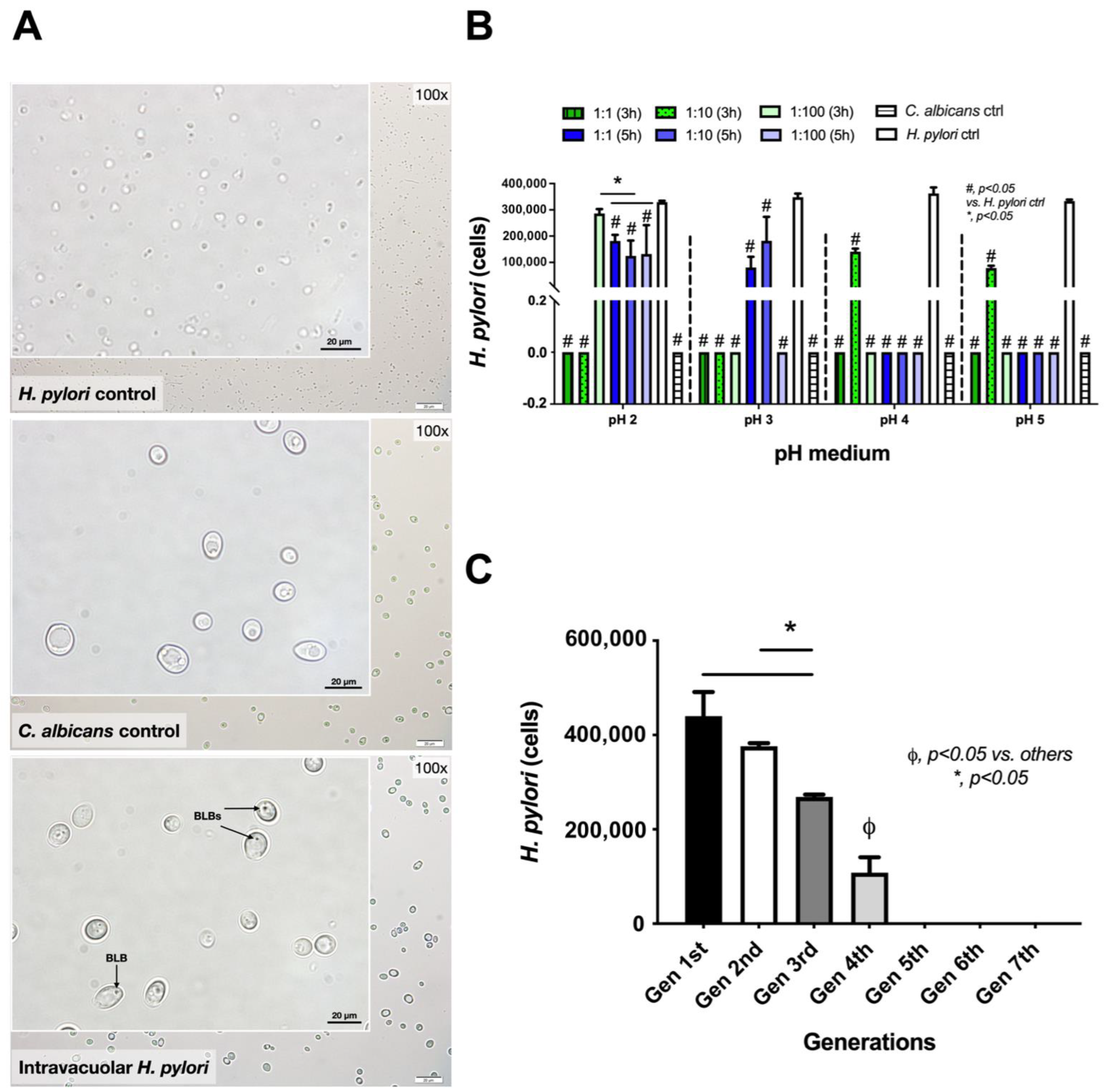
2.1. Induction of Intravacuolar H. pylori in Candida Yeast Cells

The endosymbiosis of H. pylori in yeast cells was indicated by the presence of H. pylori in Candida cytosols, referred to as “intravacuolar H. pylori”, as previously mentioned [45,46]. As such, H. pylori and C. albicans were co-incubated under different conditions, including the bacteria–Candida ratio, pH of the media, and duration of incubation); the result was the identification of H. pylori inside Candida yeast cells, as illustrated by bacteria-like bodies (BLBs) (dense black dots in a state of movement) inside the yeast cells [40,41,43,47,48,49,50], using bright-field microscopy (Figure 1). With continuous time-frame illustrations, the movements of the dense black dots could be observed (Figure 1), supporting the possible existence of BLBs inside the yeast cells. Interestingly, BLBs were detectable after the H. pyloriC. albicans co-incubation but not in the C. albicans culture alone (Figure 1 and Figure 2A). The higher density of H. pylori in BLBs may be responsible for the dense, black-colored dots observed in the cytosol of the co-incubated culture (Figure 2A lower), whereas the lower density of Candida’s organelles may explain the non-colored cytosol of the Candida culture without bacteria (Figure 2A middle). The highest abundance of intravacuolar H. pylori was demonstrated in a 1:100 ratio of H. pylori to Candida (1 × 10⁸ and 1 × 10⁶ CFU/mL of H. pylori and C. albicans, respectively) in Sabouraud dextrose broth (SB) media at pH 2 after 3 h incubation. There was a decrease in intravacuolar H. pylori in the sub-cultures starting from the 2nd generation (Figure 2B,C). At pH 2, intravacuolar H. pylori were detectable for all H. pylori: Candida ratios after a 5 h incubation, but only at the 1:100 ratio after a 3 h incubation (Figure 2B). At pH 3, intravacuolar H. pylori were detectable only after a 5 h incubation, and only for the ratios 1:1 and 1:10 (Figure 2B). Meanwhile, at pH 4 and 5, the 1:10 ratio was only the condition that could induce intravacuolar H. pylori (Figure 2B). Hence, the stomach (pH 1.5–3.5) is possibly the most suitable organ for the formation of intravacuolar H. pylori in Candida. This formation may depend on the abundance of Candida in the stomach and the gastric emptying time. The intravacuolar H. pylori were able to pass through the daughter yeast cells as there were similar abundances of H. pylori in the 1st and 2nd generations of the culture; however, H. pylori abundance was reduced in the 3rd and 4th generations (Figure 2C). Notably, incubation by SB (a Candida-enrichment media) in aerobic conditions (conditions for enhancing Candida growth) was the environmental stress for H. pylori.

2.2. Release of Intravacuolar H. pylori from Candida Yeast Cells

While the endosymbiosis of H. pylori in Candida yeast cells was inducible by several stresses (high oxygen levels and a non-H. pylori-enrichment media) (Figure 2A–C), we further tested whether the intravacuolar H. pylori could be released. Without any further manipulations of the Candida containing intravacuolar H. pylori, the culture of these Candida yeast cells in urea-based agar demonstrated urease activity (i.e., turning the color of the media from yellow into pink) at 3 and 5 days after the culture (Figure 3A) with H. pylori detectable by qPCR (Figure 3B). However, the breakdown of the yeast cell wall (with sonication) facilitated the release of H. pylori from Candida. There was a higher abundance of H. pylori released from sonicated Candida yeast cells with intravacuolar H. pylori; bacterial abundance after a 3-day-culture in urea-based agar using the sonicated H. pylori-containing Candida cells was higher than it was in the non-sonicated samples, as determined by the color of the agar (urease test) and qPCR assay (Figure 3C,D).

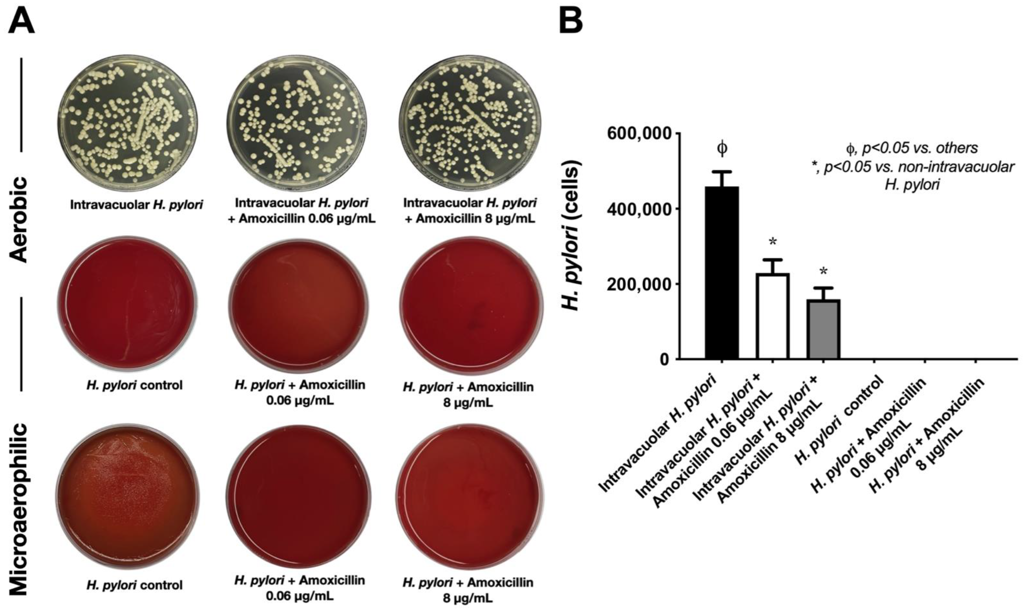
2.3. Intravacuolar H. pylori Was Protected from Antibiotics and Stress Conditions

Because a possible benefit of the H. pylori endosymbiosis inside C. albicans may be protection from antibiotics and stresses, the culture conditions of Candida using SB (the non-enrichment media of H. pylori) in aerobic conditions (a stress factor for H. pylori) with or without amoxicillin (a representative antibiotic) were tested against Candida containing H. pylori. Amoxicillin had no effect on Candida containing H. pylori as the antibiotic could not reduce the yeast colonies (Figure 4A upper), even though the selected strain of H. pylori was sensitive to aerobic conditions (H. pylori in Columbia blood agar in aerobic conditions) (Figure 4A middle) and to amoxicillin (no bacterial colony in Columbia blood agar in microaerophilic conditions) (Figure 4A lower). Notably, H. pylori were sensitive to amoxicillin in either low or high concentrations (Figure 4A lower). Likewise, the bacterial abundance of H. pylori based on CagA gene expression demonstrated the highest H. pylori abundance in Candida with intravacuolar H. pylori without amoxicillin, despite an aerobic environment (an environment toxic to H. pylori) (Figure 4B). In Candida containing intravacuolar H. pylori with amoxicillin, H. pylori abundance was lower than in the non-antibiotic group, but there was a non-difference between high versus low concentrations of amoxicillin (Figure 4B). Meanwhile, H. pylori were non-detectable in the samples of free H. pylori (H. pylori alone) under aerobic conditions and microaerophilic conditions with amoxicillin (Figure 4B). The reduced H. pylori abundance in amoxicillin-incubated Candida containing H. pylori may reflect the effect of amoxicillin on spontaneously released H. pylori.

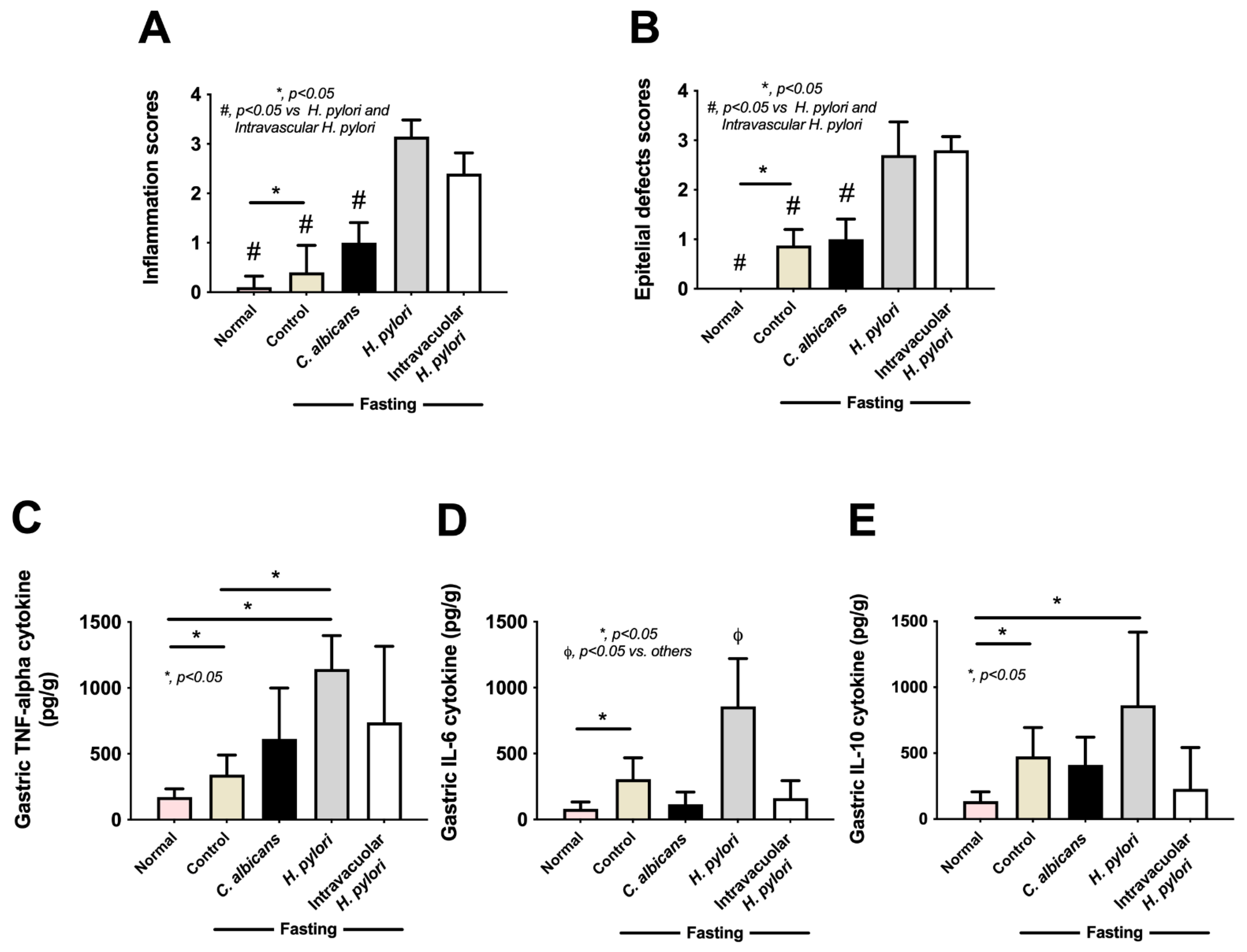
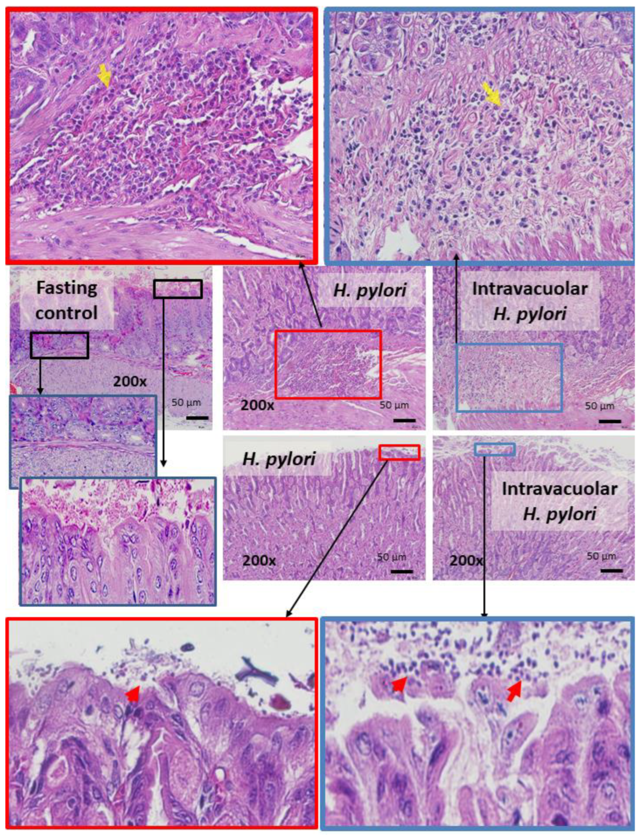
2.4. Candida Containing Intravacuolar H. pylori Caused Peptic Ulcer Disease

Although H. pylori contained inside Candida yeast cells have been previously reported [40,41,42], the clinical importance is still not clear. To test the clinical impacts of intravacuolar H. pylori in Candida yeast cells, a gastritis mouse model was conducted using the daily oral administration of vehicle control, H. pylori alone, C. albicans alone, and Candida containing intravacuolar H. pylori for 12 weeks (Figure 5A, schema). Results showed that H. pylori administration induced transient weight loss, especially at 2–6 weeks of administration; then, body weight increased to a level similar to pre-administration weight (Figure 5B). Meanwhile, there was no weight change in the other groups (control, Candida alone, and Candida containing H. pylori) during the 12 weeks of the experiment (Figure 5B). Despite the improved body weight at 12 weeks after H. pylori administration, colonization by H. pylori was detected in the stomach, as indicated by urease activity (a pink color change in urea-based agar) and CagA gene expression in samples taken from the mouse stomach (Figure 5C,D). Likewise, H. pylori were also detected in the stomach of mice after 12 weeks of the administration of Candida containing intravacuolar H. pylori (Figure 5C,D). In parallel, H. pylori-administered mice demonstrated gastritis with inflammatory responses (Figure 6A–E), as indicated by the prominent histological scores based on inflammatory cell infiltration and epithelial damage (Figure 6A,B and Figure 7A,B) and increased inflammatory cytokines (IL-6 and TNF-α, but not IL-10) in the stomach (Figure 6C–E). On the other hand, the administration of Candida containing H. pylori caused less extreme histological scores and levels of tissue cytokines in the stomach than the administration of H. pylori alone after 12 weeks of the experiment (Figure 6A,B). There were no gastritis lesions in mice with daily fasting with 1xPBS (control) or Candida administration, although the levels of stomach pro-inflammatory cytokines (IL-6 and TNF-α) were higher in fasting mice with PBS or C. albicans than in the non-fasting control mice (normal mice) (Figure 6C–E); nevertheless, the levels observed in the fasting mice were lower than those of mice administered with either H. pylori or Candida containing H. pylori (Figure 6C–E). There was an abundance of inflammatory cells in the gut (submucosa and mucosa) in mice with either H. pylori alone or Candida containing H. pylori compared to the absence of injury in other groups (Figure 6A,B), despite a tendency toward more prominent submucosal neutrophils and epithelial mononuclear cells (Figure 7).

3. Discussion

Intravacuolar H. pylori inside C. albicans were protected from antibiotics (and environmental stresses) and could cause gastritis after the release of H. pylori from the yeast cells, illustrating the importance of natural bacteria–fungi symbiosis.

3.1. The Transfer of H. pylori Inside the Vacuoles of Candida Yeast Cells

H. pylori are vulnerable to a non-acidic micro-environment, and they cannot be isolated from food, water, or non-stomach specimens (saliva and feces). Indeed, the gastric epithelium and immune cells (macrophages and dendritic cells) are the only eukaryotic cells that host H. pylori (the facultative intracellular bacteria) [41]. As such, viable intracellular prokaryotes inside eukaryotes are a prokaryotic adaptation to a wide range of environmental stresses [41]. H. Pylori can be established not only in eukaryotic human cells but also in eukaryotic microorganisms (e.g., Candida spp.) as a form of “prokaryotic–eukaryotic endosymbiosis” [41,45,47] in which the endosymbiotic bacteria are localized inside the membrane-bound vacuole [40,41,47,48]. Likewise, several examples of the endosymbiosis of endobacteria inside fungi have been described [51]. For example, Rhizopus microcarpus, a plant pathogen fungus, provide micronutrients, such as phosphate, to Burkholderia rhizoxinica that stay inside the fungi [51]. Perhaps H. pylori’s specific ergosterol interacts with the yeast vacuole to allow the viability of “intravacuolar H. pylori” [41]. Because Candida yeast is exceptionally resistant to stressful conditions [52,53], the yeast cell can be used as an alternative host to protect H. pylori from stress outside the stomach, offer nourishment, and serve as a conduit for the bacterium’s spread within human populations and in the environment [41]. Indeed, the mutual cooperation between Candida spp. and H. pylori was previously mentioned; the exosymbiosis between the extracellular form of Candida yeast and H. pylori results in the synergistic production of an interkingdom biofilm [45]. However, the endosymbiosis of intravacuolar H. pylori inside yeast cells may be even more complex than the synergy observed in biofilm production because it indicates an evolution in the symbiosis between the structures of these organisms.
Here, within the culture media of fungi that were too acidic for H. pylori, had fewer nutrients, or were non-microaerophilic, H. pylori could form bacteria-like bodies (BLBs) inside C. albicans vacuoles, as supported by a previous publication [46]. Notably, the differences between the BLBs and the non-specific components of the yeast cell’s cytosol were the movement and the density of the particles, as indicated in Figure 1 and Figure 2A. While C. albicans can easily adapt to acidic pH levels by secreting acids out of the cell after approximately 50 h of incubation [46], H. pylori are more vulnerable to extreme acidity, despite their ability to enhance pH using urease [54,55]. Moreover, fungi can also adapt to alkali environments; some pathogenic fungi secrete acids that are used for the induction of host tissue injury [56]. Interestingly, the H. pylori-specific gene (CagA) was detectable in C. albicans with intravacuolar H. pylori; this finding supports the likelihood of interaction between these two microorganisms. Due to the effectiveness of the protection, H. pylori’s presence inside C. albicans may be another mechanism explaining the persistence of H. pylori in the human GI tract [41,47]. Because the CagA gene was detectable in subsequent generations of C. albicans, showing vertical transmission into the daughter cells where it continued to express proteins of H. pylori [41], the yeast cell may be both a protective vehicle and a transmission method. Despite the use of antibiotics, including amoxicillin, together with a proton pump inhibitor for H. pylori eradication [57,58,59], and despite the amoxicillin susceptibility of the selected H. pylori in our experiments, amoxicillin could not inhibit the intravacuolar H. pylori, as indicated both by a culture on urea-based agar and by CagA expression. However, Candida may not protect intravacuolar H. pylori against the function of other antibiotics as some antibiotics may be able to diffuse inside the yeast cells. Nevertheless, the protection against amoxicillin provides proof of the concept of antibiotic protection inside yeast cells. Our data suggest that the intravacuolar H. pylori inside C. albicans were protected from antimicrobials. Hence, the development of intravacuolar H. pylori was very helpful for H. pylori in terms of resistance to treatments, extra-stomach transfers, person-to-person transmission, and environmental spread. Likewise, H. pylori can transform from a spiral-shaped bacillary form into a coccoid form in stress environments, similar to other Gram-negative bacteria; in this form, they are nonculturable and referred to as VBNC (viable but nonculturable) bacteria, with fewer activities than the spiral form, but they may revert to active regrowth conditions [60]. While H. pylori’s transformation into a coccoid form is a well-known mechanism to adapt to stresses before a regrowth that is often mentioned as an underlying mechanism for the detection of Helicobacter in the environment [60], intravacuolar H. pylori is possibly another strategy for survival in harsh environments. However, the putative infectious capability and the regrowth ability of this form of H. pylori are still controversial. Here, we also demonstrated that the development of intravacuolar H. pylori depended on pH and the abundance of Candida, as a higher fungal abundance at lower pH levels facilitated the intravacuolar form of Helicobacter. Hence, an evaluation of Candida abundance in the gut and the use of fungicidal drugs in the patients in whom H. pylori eradication has failed might be helpful. Moreover, our data also suggest that H. pylori inside Candida can be spontaneously released from the yeast cells on the 2nd and 3rd days of the symbiosis with biological activity intact (positive urease test). The prospect of additional studies on this topic is interesting.

3.2. Intravacuolar H. pylori Inside C. albicans as a Cause of Gastritis

Although the administration of C. albicans alone does not cause gastritis, the worsening of H. pylori-induced gastritis by Candida through synergistically elevated gastric inflammation has been previously described [13,43,45]. Likewise, Candida alone did not induce stomach injury in our study, but gastritis was demonstrated in the mice orally administered both with intravacuolar H. pylori (the bacteria inside Candida) and with H. pylori alone, as indicated by inflammatory lesions, CagA expression, a urea-based culture, and tissue cytokine levels in the stomach. Notably, the spontaneous bodyweight regains observed in the H. pylori mouse model [61,62] and the absence of weight gain in mice during the fasting experiments have been previously mentioned [63]. Our data suggest that Candida yeast is a reservoir of H. pylori and that the bacteria can be released outside the yeast cells and regrow in the stomach epithelium. Although the mechanisms of H. pylori’s release from the yeast cells are still unknown, vesicular release has been identified as a new fungal secretory pathway that might play a role here [64]. As such, some yeast fungi, including C. albicans, Cryptococcus neoformans, and Saccharomyces cerevisiae, can produce vesicles containing RNA or microRNA (miRNA) (the non-coding RNA sequence with gene regulatory effects) [65,66,67] that can alter the gene expression of target cells [68]. In addition, the fungal cell wall has a dynamic structure with flexible viscoelastic qualities that allow the release of vesicles [69], as well as fungal–host communication with some specific environmental factors (pH, temperature, presence of serum, and lack of some amino acids) that trigger the production and release of vesicles [70]. Thus, these data suggest that the large vesicles of intravacuolar H. pylori possibly pass through the cell wall of C. albicans yeast cells. With the release of intravacuolar H. pylori, Helicobacter bacteria can induce gastritis through decreases in mucus and mucosal blood flow, prostaglandin synthesis, cell healing processes, and gastric emptying time, similar to regular H. pylori [7,8,12,71]. Notably, the abundance of H. pylori in the stomach of H. pylori-administered mice was higher than in mice with Candida containing intravacuolar H. pylori (Figure 5C,D), suggesting that some intravacuolar Helicobacter could not be released from the Candida yeast cells. In contrast, Candida administration alone did not cause ulcers. The severity of the gastritis caused by H. pylori alone versus that caused by H. pylori inside Candida was not different, indicating a lack of synergy between H. pylori and the Candida hosting it; this finding differs from previous reports of enhanced pro-inflammation with the co-presence of H. pylori and Candida [13,43,45]. Perhaps the Candida yeasts that host H. pylori lose their pro-inflammatory properties after the release of H. pylori. We hypothesize that the release of H. pylori from C. albicans vesicles enhances the pro-inflammatory response of enterocytes (Figure 8A,B) that activate the gastric inflammation of mucosa through Toll-like receptors, especially TLR4 and TLR2, with several adapter molecules (MyD88, IRAK1, IRAK4, and NF-κB) [72]. Hence, our data suggest that Candida yeast cells could be the eukaryotic hosts that protect the prokaryotic bacteria from antibiotics and stressful environments and that the hosted bacteria are ready to be spontaneously released or let out by the breakdown of the yeast cells and/or other mechanisms. More mechanistic studies on this topic would be interesting.
For the clinical translation, the spontaneous development of intravacuolar H. pylori inside Candida yeast cells may depend on the ratio of the abundances of Candida and H. pylori in each patient; such development would possibly induce resistance to the standard treatment. Currently, the causes of treatment failure for H. pylori focus on (i) increased antibiotic resistance [73] through the genes that affect the alteration of proton pump inhibitor pharmacokinetics and/or the host cytochrome CYP2C19 and (ii) host factors, such as poor adherence, insufficient duration of therapy, and smoking [74,75,76]. On the other hand, our data support another underlying mechanism of treatment failure through an increased abundance of fungi in the stomach. Indeed, advanced age and the consumption of proton pump inhibitors correlate with fungal colonization in the stomach, which may be associated with H. pylori-positive status and dyspepsia [13]. Hence, reducing the abundance of Candida (anti-fungal treatments) together with H. pylori treatment (triple therapy) may improve the outcome of treatment and reduce the recurrent rate of gastritis, especially in cases of treatment failure. Unfortunately, the correlation between Candida abundance and the treatment resistance of H. pylori is still unknown. In contrast, an interkingdom correlation between Candida and H. pylori may be clinically possible because the generation of intravacuolar H. pylori was easily induced by the co-incubation of both organisms in a fungal culture media, and the intravacuolar H. pylori could pass through the daughter yeast cells, protected from antibiotics, and possibly caused resistance to the standard treatment. More studies are warranted.

4. Materials and Methods

4.1. Inducing Intravacuolar H. pylori in Candida Yeast Cells

H. pylori ATCC 43504 (Manassas, VA, USA) was cultured on Columbia agar (HiMedia, Mumbai, India) supplemented with 5% sheep blood and 7% horse serum (Invitrogen, Waltham, MA, USA) under microaerophilic conditions (5% O2, 15% CO2, and 80% N2) at 37 °C for 3 days. In parallel, C. albicans ATCC 90028 (Microbiologics, Saint Cloud, MN, USA), a fluconazole-susceptible strain (minimal inhibitory concentration 0.25–1 μL/mL), was cultured on Sabouraud dextrose agar (SDA) (Oxoid, Basingstoke, Hampshire, UK) in aerophilic conditions at 37 °C for 2 days before using. Then, H. pylori at 1 × 10⁶, 1 × 10⁷, and 1 × 10⁸ CFU/mL and 1 × 10⁶ CFU/mL of C. albicans were co-incubated with shaking in Sabouraud dextrose broth (SB) (Oxoid) at pH 2, pH 3, pH 4, and pH 5 for 3 h and 5 h. Then, the bacteria-like bodies (BLBs) inside Candida yeast cells were observed under a bright-field microscope (100× magnification), and 100 μL of these co-incubated microorganisms were plated on SDA with chloramphenicol (Oxoid) and incubated at 37 °C for 3 days for the C. albicans growth.

4.2. Determination of Intravacuolar H. pylori Inside the Candida Yeast Cells

Candida yeast cells in each experimental group were sub-cultured to produce other generations in SDA with chloramphenicol, and a specific H. pylori gene inside each Candida generation was examined, following a previous protocol [77]. In brief, 1 × 10⁶ CFU/mL of Candida yeast cells were sonicated with a Beadbeater (OK 74005) (BioSpec Products Inc., Bartlesville, OK, USA) for 15 s to destroy the fungal cell wall, and phenol-chloroform was used for the extraction of DNA from the samples. Real-time polymerase chain reaction (PCR) assays were run on a QuantStudio 5 Real-Time PCR (qPCR) system (Thermo Fisher Scientific, Waltham, MA, USA) using a CagA gene primer to identify H. pylori inside C. albicans with the following primer sequence: forward, 5′-AGT AAG GAG AAA CAA TGA-3′ and reverse, 5′-AAT AAG CCT TAG AGT CTT TTT GGA AAT C-3′ [77]. The H. pylori gene expression was calculated and translated into H. pylori cell-abundance by the gene reference from the National Center for Biotechnology Information (NCBI; NIH, Bethesda, MD, USA). To demonstrate the release of intravacuolar H. pylori from the Candida yeast cells, the second generation of Candida yeast cells from the sub-culture passages was incubated in aerophilic conditions at 37 °C for 5 days using a urea agar-based culture media (400,086) (HiMedia, Mumbai, India) to demonstrate the activities of intravacuolar H. pylori. To see whether damaged yeast cell walls can release more H. pylori than non-damaged yeast cells, the CagA gene in the second generation of Candida yeast cells with or without sonication was extracted by a Beadbeater machine for whole DNA, and the presence of the CagA gene was determined by a qPCR system (Thermo Fisher Scientific).

4.3. Antimicrobial Incubation and Stresses against Intravacuolar H. pylori

To explore the possible benefits of the endosymbiotic H. pylori inside C. albicans in terms of protection from antibiotics and environmental stresses, amoxicillin, and an aerobic (high-oxygen) condition, which is a stress factor for microaerophilic H. pylori (i.e., it has the ability to grow in 5–15% oxygen), were tested. As such, the second generation of Candida yeast cells was cultured into SB with or without amoxicillin (0.06 and 8 ug/mL) (Tianjin TEDA Steyuan Pharm Co., Ltd., Shijiazhuang, Hebei, China) in aerophilic conditions (21% oxygen) at 37 °C overnight. Then, 100 μL of each sample was plated onto SDA and incubated in aerophilic conditions at 37 °C overnight. After that, the whole DNA was extracted to identify CagA gene expression in each experimental group using a qPCR system (Thermo Fisher Scientific).

4.4. Animal and Peptic Ulcer Model

The animal study (SST 018/2562) was approved by the Institutional Animal Care and Use Committee of Chulalongkorn University’s Faculty of Medicine following the animal care and use procedure of the National Institutes of Health (NIH). Male 8-week-old C57BL/6 mice weighing 20–25 g was purchased from Nomura Siam International, Pathumwan, Bangkok, Thailand. The mice were housed in a temperature-controlled environment (24 ± 2 °C), with 50% relative humidity and a 12 h light–dark cycle (light from 7:00 a.m. to 7:00 p.m.). All mice received food and water ad libitum. Animal procedures were performed in adherence with U.S. National Institutes of Health guidelines and followed the 8th Edition of the Guide for Care and Use of Experimental Animals, published by the National Research Council of the National Academies (2011; available at https://grants.nih.gov/grants/olaw/guide-for-the-care-and-use-of-laboratory animals.pdf, accessed on 12 November 2021), as well as the Animal Research: Reporting of In Vivo Experiments (ARRIVE) guidelines.
A peptic ulcer model of H. pylori infection was modified from a previous study [78]. Briefly, H. pylori at 3.7 × 10⁶ CFU/mL, C. albicans at 1 × 10⁸ CFU/mL, or C. albicans with intravacuolar H. pylori at 1 × 10⁸ CFU/mL (with approximately 3.7 × 10⁶ CFU/mL of intravacuolar H. pylori inside the C. albicans) in 1 mL of 1x phosphate buffer solution (PBS) was once daily orally administered using a stainless-steel feeding tube (18-gauge size and 1.5 inches in length, with a rounded tip attached to a 1 mL syringe). Notably, all groups of mice fasted for 5 h before microorganismal administration. All mice were observed and sacrificed 12 weeks after the beginning of the experiment. Then, the stomach was divided longitudinally through the greater and lesser curvature into several parts, washed with 1xPBS, weighed, and used for determining inflammatory responses and fungal–bacterial interactions.

4.5. Mouse Gastric Analysis

Mouse stomachs were divided into 4 small pieces for performing (i) a H. pylori culture, (ii) an analysis of CagA gene expression, (iii) the measurement of gastric cytokine levels, and (iv) a histopathology analysis (fixed in 10% formaldehyde). To determine H. pylori abundance in mouse stomachs, the gastric tissues (0.01 g per sample) were cultured on urea-based agar and incubated at 37 °C overnight before colony enumeration. In addition, the CagA gene was identified by qPCR (Thermo Fisher Scientific) from gastric tissues (1 mL PBS per g tissue) that were sonicated with the setting of pulse-on for 20 s and pulse-off for 5 s in 30 min on ice using the Sonics Vibra Cell machine (VCX 750) (Sonics & Materials Inc., Newtown, CT, USA) until a homogeneous solution was formed. The supernatant, after the centrifugation, was used for the detection of gastric cytokines (TNF-α, IL-6, and IL-10) and CagA expression by ELISA assays (Invitrogen, Waltham, MA, USA) [79,80] and PCR, respectively. For histology, the stomach tissues were rinsed with PBS, fixed in 10% (weight by volume (W/V)) formaldehyde, embedded in paraffin, and stained with hematoxylin and eosin (H and E) color in 5.0 mm thickness sections. The histological analyses of 200× magnification slides were performed by 2 observers who were blinded to the experiments; the analyses used semi-quantitative scores based on inflammatory cell infiltration (macrophages and neutrophils), mucosal damage, and ulceration on a scale of 0 to 4 as modified from a previous publication [81].

4.6. Statistical Analysis

All data were analyzed by Statistical Package for Social Sciences software (SPSS 22.0, SPSS Inc., Chicago, IL, USA) and Graph Pad Prism version 7.0 software (La Jolla, CA, USA). Results were presented as mean ± standard error (SE). The differences between multiple groups were examined for statistical significance by one-way analysis of variance (ANOVA) with Tukey’s analysis. The survival analysis and time-point data were determined by the log-rank test and repeated measures ANOVA, respectively. A p-value < 0.05 was considered statistically significant.

5. Conclusions

In conclusion, intravacuolar H. pylori benefit from increased transmissibility, and the fungal host protects the bacteria from stressful micro-environments, including antibiotics. Our mouse model provides evidence that intravacuolar H. pylori were able to induce gastric infection, inflammatory cell infiltration, and tissue damage. Future studies on the role of intravacuolar H. pylori in yeast cells are needed to extend our understanding of intravacuolar H. pylori colonization in humans in order to optimize and individualize health strategies.

Author Contributions

Conceptualization, A.L., P.H. and W.P.; validation, formal analysis, and investigation, A.L., P.H. and W.P.; resources, A.L., P.H. and W.P.; data curation, P.H.; writing—original draft preparation, A.L. and P.H.; writing—review and editing, A.L., P.H. and A.C.; visualization, A.L. and A.C.; supervision and funding acquisition, A.L. and A.C. All authors have read and agreed to the published version of the manuscript.

Funding

This research was supported by Chulalongkorn University through Fundamental Fund 65 [CUFRB65_hea [33] _040_30_21], the National Research Council of Thailand (grant numbers NRCT-N41A640076 and 811/2563), along with the NSRF via the Program Management Unit for Human Resources & Institutional Development, Research, and Innovation (B16F640175 and B05F640144). P.H. was supported by the Second Century Fund (C2F) for Ph.D. students, Chulalongkorn University.

Institutional Review Board Statement

The study was conducted according to the animal study protocol approved by The Institutional Animal Care and Use Committee of the Faculty of Medicine, Chulalongkorn University (ASP SST 018/2562).

Informed Consent Statement

Not applicable.

Data Availability Statement

Data are contained within the article.

Acknowledgments

A.L. is affiliated with the Center of Excellence on Translational Research in Inflammation and Immunology (CETRII), Department of Microbiology, Chulalongkorn University, Bangkok 10330, Thailand.

Conflicts of Interest

The authors declare no conflict of interest.

References

  1. Kuna, L.; Jakab, J.; Smolic, R.; Raguz-Lucic, N.; Vcev, A.; Smolic, M. Peptic ulcer disease: A brief review of conventional therapy and herbal treatment options. J. Clin. Med. 2019, 8, 179. [Google Scholar] [CrossRef] [Green Version]
  2. Narayanan, M.; Reddy, K.M.; Marsicano, E. Peptic ulcer disease and Helicobacter pylori infection. Mo. Med. 2018, 115, 219–224. [Google Scholar]
  3. Ramakrishnan, K.; Salinas, R.C. Peptic ulcer disease. Am. Fam. Physician 2007, 76, 1005–1012. [Google Scholar]
  4. Kavitt, R.T.; Lipowska, A.M.; Anyane-Yeboa, A.; Gralnek, I.M. Diagnosis and treatment of peptic ulcer disease. Am. J. Med. 2019, 132, 447–456. [Google Scholar] [CrossRef]
  5. Azhari, H.; Underwood, F.; King, J.; Coward, S.; Shah, S.; Chan, C.; Ho, G.; Ng, S.; Kaplan, G. The global incidence of peptic ulcer disease and its complications at the turn of the 21st century: A systematic review: 1199. Off. J. Am. Coll. Gastroenterol. ACG 2018, 113, S684–S685. [Google Scholar] [CrossRef]
  6. Woolf, A.; Rose, R. Gastric ulcer. In StatPearls; StatPearls Publishing: Treasure Island, FL, USA, 2022. [Google Scholar]
  7. Bazzoli, F.; de Luca, L.; Graham, D.Y. Helicobacter pylori infection and the use of NSAIDs. Best Pract. Res. Clin. Gastroenterol. 2001, 15, 775–785. [Google Scholar] [CrossRef] [PubMed]
  8. De Brito, B.B.; da Silva, F.A.F.; Soares, A.S.; Pereira, V.A.; Santos, M.L.C.; Sampaio, M.M.; Neves, P.H.; de Melo, F.F. Pathogenesis and clinical management of Helicobacter pylori gastric infection. World J. Gastroenterol. 2019, 25, 5578–5589. [Google Scholar] [CrossRef]
  9. Diaconu, S.; Predescu, A.; Moldoveanu, A.; Pop, C.S.; Fierbinteanu-Braticevici, C. Helicobacter pylori infection: Old and new. J. Med. Life 2017, 10, 112–117. [Google Scholar]
  10. Denic, M.; Touati, E.; de Reuse, H. Review: Pathogenesis of Helicobacter pylori infection. Helicobacter 2020, 25 (Suppl. S1), e12736. [Google Scholar] [CrossRef]
  11. Kayali, S.; Manfredi, M.; Gaiani, F.; Bianchi, L.; Bizzarri, B.; Leandro, G.; di Mario, F.; de’Angelis Gian, L. Helicobacter pylori, transmission routes and recurrence of infection: State of the art. Acta Biomed. 2018, 89 (Suppl. S8), 72–76. [Google Scholar]
  12. Chang, W.L.; Yeh, Y.C.; Sheu, B.S. The impacts of H. pylori virulence factors on the development of gastroduodenal diseases. J. Biomed. Sci. 2018, 25, 68. [Google Scholar] [CrossRef] [PubMed] [Green Version]
  13. Massarrat, S.; Saniee, P.; Siavoshi, F.; Mokhtari, R.; Mansour-Ghanaei, F.; Khalili-Samani, S. The effect of Helicobacter pylori infection, aging, and consumption of proton pump inhibitor on fungal colonization in the stomach of dyspeptic patients. Front. Microbiol. 2016, 7, 801. [Google Scholar] [CrossRef] [PubMed]
  14. Roesler, B.M.; Rabelo-Gonçalves, E.M.; Zeitune, J.M. Virulence factors of Helicobacter pylori: A review. Clin. Med. Insights Gastroenterol. 2014, 7, 9–17. [Google Scholar] [CrossRef] [PubMed]
  15. Shiota, S.; Suzuki, R.; Yamaoka, Y. The significance of virulence factors in Helicobacter pylori. J. Dig. Dis. 2013, 14, 341–349. [Google Scholar] [CrossRef] [PubMed] [Green Version]
  16. Sterbenc, A.; Jarc, E.; Poljak, M.; Homan, M. Helicobacter pylori virulence genes. World J. Gastroenterol. 2019, 25, 4870–4884. [Google Scholar] [CrossRef]
  17. Javed, S.; Skoog, E.C.; Solnick, J.V. Impact of Helicobacter pylori virulence factors on the host immune response and gastric pathology. Curr. Top Microbiol. Immunol. 2019, 421, 21–52. [Google Scholar]
  18. Dunne, C.; Dolan, B.; Clyne, M. Factors that mediate colonization of the human stomach by Helicobacter pylori. World J. Gastroenterol. 2014, 20, 5610–5624. [Google Scholar] [CrossRef]
  19. Wang, Z.; Ren, R.; Yang, Y. Mucosa microbiome of gastric lesions: Fungi and bacteria interactions. Prog. Mol. Biol. Transl. Sci. 2020, 171, 195–213. [Google Scholar]
  20. Gow, N.A.; van de Veerdonk, F.L.; Brown, A.J.; Netea, M.G. Candida albicans morphogenesis and host defence: Discriminating invasion from colonization. Nat. Rev. Microbiol. 2011, 10, 112–122. [Google Scholar] [CrossRef] [Green Version]
  21. Mayer, F.L.; Wilson, D.; Hube, B. Candida albicans pathogenicity mechanisms. Virulence 2013, 4, 119–128. [Google Scholar] [CrossRef] [Green Version]
  22. Dadar, M.; Tiwari, R.; Karthik, K.; Chakraborty, S.; Shahali, Y.; Dhama, K. Candida albicans—Biology, molecular characterization, pathogenicity, and advances in diagnosis and control—An update. Microb. Pathog. 2018, 117, 128–138. [Google Scholar] [CrossRef] [PubMed]
  23. Hiengrach, P.; Visitchanakun, P.; Finkelman, M.A.; Chancharoenthana, W.; Leelahavanichkul, A. More prominent inflammatory response to pachyman than to whole-glucan particle and oat-β-glucans in dextran sulfate-induced mucositis mice and mouse injection through proinflammatory macrophages. Int. J. Mol. Sci. 2022, 23, 4026. [Google Scholar] [CrossRef] [PubMed]
  24. Saithong, S.; Worasilchai, N.; Saisorn, W.; Udompornpitak, K.; Bhunyakarnjanarat, T.; Chindamporn, A.; Tovichayathamrong, P.; Torvorapanit, P.; Chiewchengchol, D.; Chancharoenthana, W.; et al. Neutrophil extracellular traps in severe SARS-CoV-2 infection: A possible impact of LPS and (1→3)-β-d-glucan in blood from gut translocation. Cells 2022, 11, 1103. [Google Scholar] [CrossRef] [PubMed]
  25. Panpetch, W.; Somboonna, N.; Bulan, D.E.; Issara-Amphorn, J.; Worasilchai, N.; Finkelman, M.; Chindamporn, A.; Palaga, T.; Tumwasorn, S.; Leelahavanichkul, A. Gastrointestinal colonization of Candida albicans increases serum (1→3)-β-d-glucan, without candidemia, and worsens cecal ligation and puncture sepsis in murine model. Shock 2018, 49, 62–70. [Google Scholar] [CrossRef] [PubMed]
  26. Saithong, S.; Saisorn, W.; Dang, C.P.; Visitchanakun, P.; Chiewchengchol, D.; Leelahavanichkul, A. Candida administration worsens neutrophil extracellular traps in renal ischemia reperfusion injury mice: An impact of gut fungi on acute kidney injury. J. Innate Immun. 2022, 1–16, Online First. [Google Scholar] [CrossRef]
  27. Amornphimoltham, P.; Yuen, P.S.T.; Star, R.A.; Leelahavanichkul, A. Gut leakage of fungal-derived inflammatory mediators: Part of a gut-liver-kidney axis in bacterial sepsis. Dig. Dis. Sci. 2019, 64, 2416–2428. [Google Scholar] [CrossRef]
  28. Issara-Amphorn, J.; Surawut, S.; Worasilchai, N.; Thim-Uam, A.; Finkelman, M.; Chindamporn, A.; Palaga, T.; Hirankarn, N.; Pisitkun, P.; Leelahavanichkul, A. The synergy of endotoxin and (1→3)-β-d-glucan, from gut translocation, worsens sepsis severity in a lupus model of Fc gamma receptor IIb-deficient mice. J. Innate Immun. 2018, 10, 189–201. [Google Scholar] [CrossRef]
  29. Thim-Uam, A.; Surawut, S.; Issara-Amphorn, J.; Jaroonwitchawan, T.; Hiengrach, P.; Chatthanathon, P.; Wilantho, A.; Somboonna, N.; Palaga, T.; Pisitkun, P.; et al. Leaky-gut enhanced lupus progression in the Fc gamma receptor-IIb deficient and pristane-induced mouse models of lupus. Sci. Rep. 2020, 10, 777. [Google Scholar] [CrossRef] [PubMed] [Green Version]
  30. Visitchanakun, P.; Saisorn, W.; Wongphoom, J.; Chatthanathon, P.; Somboonna, N.; Svasti, S.; Fucharoen, S.; Leelahavanichkul, A. Gut leakage enhances sepsis susceptibility in iron-overloaded β-thalassemia mice through macrophage hyperinflammatory responses. Am. J. Physiol.-Gastrointest. Liver Physiol. 2020, 318, G966–G979. [Google Scholar] [CrossRef] [PubMed]
  31. Hiengrach, P.; Panpetch, W.; Worasilchai, N.; Chindamporn, A.; Tumwasorn, S.; Jaroonwitchawan, T.; Wilantho, A.; Chatthanathon, P.; Somboonna, N.; Leelahavanichkul, A. Administration of Candida albicans to dextran sulfate solution treated mice causes intestinal dysbiosis, emergence and dissemination of intestinal Pseudomonas aeruginosa and lethal sepsis. Shock 2020, 53, 189–198. [Google Scholar] [CrossRef]
  32. Panpetch, W.; Kullapanich, C.; Dang, C.P.; Visitchanakun, P.; Saisorn, W.; Wongphoom, J.; Wannigama, D.L.; Thim-Uam, A.; Patarakul, K.; Somboonna, N.; et al. Candida administration worsens uremia-induced gut leakage in bilateral nephrectomy mice, an impact of gut fungi and organismal molecules in uremia. mSystems 2021, 6, e01187-20. [Google Scholar] [CrossRef] [PubMed]
  33. Panpetch, W.; Sawaswong, V.; Chanchaem, P.; Ondee, T.; Dang, C.P.; Payungporn, S.; Tumwasorn, S.; Leelahavanichkul, A. Candida administration worsens cecal ligation and puncture-induced sepsis in obese mice through gut dysbiosis enhanced systemic inflammation, impact of pathogen-associated molecules from gut translocation and saturated fatty acid. Front. Immunol. 2020, 11, 2278. [Google Scholar]
  34. Panpetch, W.; Hiengrach, P.; Nilgate, S.; Tumwasorn, S.; Somboonna, N.; Wilantho, A.; Chatthanathon, P.; Prueksapanich, P.; Leelahavanichkul, A. Additional Candida albicans administration enhances the severity of dextran sulfate solution induced colitis mouse model through leaky gut-enhanced systemic inflammation and gut-dysbiosis but attenuated by Lactobacillus rhamnosus L34. Gut Microbes 2020, 11, 465–480. [Google Scholar] [CrossRef] [PubMed]
  35. Saithong, S.; Saisorn, W.; Visitchanakun, P.; Sae-Khow, K.; Chiewchengchol, D.; Leelahavanichkul, A. A synergy between endotoxin and (1→3)-β-d-glucan enhanced neutrophil extracellular traps in Candida administered dextran sulfate solution induced colitis in FcGRIIB-/-lupus mice, an impact of intestinal fungi in lupus. J. Inflamm. Res. 2021, 14, 2333–2352. [Google Scholar] [CrossRef] [PubMed]
  36. Issara-Amphorn, J.; Dang, C.P.; Saisorn, W.; Limbutara, K.; Leelahavanichkul, A. Candida administration in bilateral nephrectomy mice elevates serum (1→3)-β-d-glucan that enhances systemic inflammation through energy augmentation in macrophages. Int. J. Mol. Sci. 2021, 22, 5031. [Google Scholar] [CrossRef] [PubMed]
  37. Tungsanga, S.; Panpetch, W.; Bhunyakarnjanarat, T.; Udompornpitak, K.; Katavetin, P.; Chancharoenthana, W.; Chatthanathon, P.; Somboonna, N.; Tungsanga, K.; Tumwasorn, S.; et al. Uremia-induced gut barrier defect in 5/6 nephrectomized mice is worsened by Candida administration through a synergy of uremic toxin, lipopolysaccharide, and (1→3)-β-d-glucan, but is attenuated by Lacticaseibacillus rhamnosus L34. Int. J. Mol. Sci. 2022, 23, 2511. [Google Scholar] [CrossRef]
  38. Phuengmaung, P.; Panpetch, W.; Singkham-In, U.; Chatsuwan, T.; Chirathaworn, C.; Leelahavanichkul, A. Presence of Candida tropicalis on Staphylococcus epidermidis biofilms facilitated biofilm production and Candida dissemination: An impact of fungi on bacterial biofilms. Front. Cell. Infect. Microbiol. 2021, 11, 763239. [Google Scholar] [CrossRef] [PubMed]
  39. Phuengmaung, P.; Somparn, P.; Panpetch, W.; Singkham-In, U.; Wannigama, D.L.; Chatsuwan, T.; Leelahavanichkul, A. Coexistence of Pseudomonas aeruginosa with Candida albicans enhances biofilm thickness through alginate-related extracellular matrix but is attenuated by N-acetyl-l-cysteine. Front. Cell. Infect. Microbiol. 2020, 10, 594336. [Google Scholar] [CrossRef]
  40. Saniee, P.; Siavoshi, F.; Nikbakht Broujeni, G.; Khormali, M.; Sarrafnejad, A.; Malekzadeh, R. Localization of H. pylori within the vacuole of Candida yeast by direct immunofluorescence technique. Arch. Iran Med. 2013, 16, 705–710. [Google Scholar]
  41. Siavoshi, F.; Saniee, P. Vacuoles of Candida yeast as a specialized niche for Helicobacter pylori. World J. Gastroenterol. 2014, 20, 5263–5273. [Google Scholar] [CrossRef]
  42. Saniee, P.; Siavoshi, F.; Nikbakht Broujeni, G.; Khormali, M.; Sarrafnejad, A.; Malekzadeh, R. Immunodetection of Helicobacter pylori-specific proteins in oral and gastric Candida yeasts. Arch. Iran Med. 2013, 16, 624–630. [Google Scholar] [PubMed]
  43. Karczewska, E.; Wojtas, I.; Sito, E.; Trojanowska, D.; Budak, A.; Zwolinska-Wcislo, M.; Wilk, A. Assessment of co-existence of Helicobacter pylori and Candida fungi in diseases of the upper gastrointestinal tract. J. Physiol. Pharmacol. 2009, 60 (Suppl. 6), 33–39. [Google Scholar] [PubMed]
  44. Abrantes, P.; Africa, C.W.J. Measuring Streptococcus mutans, Streptococcus sanguinis and Candida albicans biofilm formation using a real-time impedance-based system. J. Microbiol. Methods 2020, 169, 105815. [Google Scholar] [CrossRef] [PubMed]
  45. Chen, X.; Zhou, X.; Liao, B.; Zhou, Y.; Cheng, L.; Ren, B. The cross-kingdom interaction between Helicobacter pylori and Candida albicans. PLoS Pathog. 2021, 17, e1009515. [Google Scholar] [CrossRef] [PubMed]
  46. Sánchez-Alonzo, K.; Parra-Sepúlveda, C.; Vega, S.; Bernasconi, H.; Campos, V.L.; Smith, C.T.; Sáez, K.; García-Cancino, A. In vitro incorporation of Helicobacter pylori into Candida albicans caused by acidic pH stress. Pathogens 2020, 9, 489. [Google Scholar] [CrossRef] [PubMed]
  47. Matamala-Valdés, L.; Sánchez-Alonzo, K.; Parra, C.; Saez, K.; Aguayo-Reyes, A.; García, A. Detection of intracellular Helicobacter pylori in Candida. SPP from neonate oral swabs. Rev. Assoc. Méd. Bras. 2018, 64, 928–935. [Google Scholar] [CrossRef] [PubMed]
  48. Sanchez-Alonzo, K.; Parra-Sepulveda, C.; Vergara, L.; Bernasconi, H.; Garcia-Cancino, A. Detection of Helicobacter pylori in oral yeasts from students of a Chilean university. Rev. Assoc. Méd. Bras. 2020, 66, 1509–1514. [Google Scholar] [CrossRef] [PubMed]
  49. Salmanian, A.H.; Siavoshi, F.; Akbari, F.; Afshari, A.; Malekzadeh, R. Yeast of the oral cavity is the reservoir of Heliobacter pylori. J. Oral Pathol. Med. 2008, 37, 324–328. [Google Scholar] [CrossRef] [PubMed]
  50. Heydari, S.; Siavoshi, F.; Jazayeri, M.H.; Sarrafnejad, A.; Saniee, P. Helicobacter pylori release from yeast as a vesicle-encased or free bacterium. Helicobacter 2020, 25, e12725. [Google Scholar] [CrossRef]
  51. Pinto-Tomás, A.A.; Anderson, M.A.; Suen, G.; Stevensonm, D.M.; Chun, F.S.; Cleland, W.W.; Weimer, P.J.; Currie, C.R. Symbiotic nitrogen fixation in the fungus gardens of leaf-cutter ants. Science 2009, 326, 1120–1123. [Google Scholar] [CrossRef]
  52. Dantas Ada, S.; Day, A.; Ikeh, M.; Kos, I.; Achan, B.; Quinn, J. Oxidative stress responses in the human fungal pathogen, Candida albicans. Biomolecules 2015, 5, 142–165. [Google Scholar] [CrossRef] [PubMed] [Green Version]
  53. Lourenço, A.; Pedro, N.A.; Salazar, S.B.; Mira, N.P. Effect of acetic acid and lactic acid at low pH in growth and azole resistance of Candida albicans and Candida glabrata. Front. Microbiol. 2019, 9, 3265. [Google Scholar] [CrossRef] [Green Version]
  54. Ansari, S.; Yamaoka, Y. Survival of Helicobacter pylori in gastric acidic territory. Helicobacter 2017, 22, e12386. [Google Scholar] [CrossRef]
  55. Clyne, M.; Dolan, B.; Reeves, E.P. Bacterial factors that mediate colonization of the stomach and virulence of Helicobacter pylori. FEMS Microbiol. Lett. 2007, 268, 135–143. [Google Scholar] [CrossRef] [PubMed]
  56. Vylkova, S. Environmental pH modulation by pathogenic fungi as a strategy to conquer the host. PLoS Pathogens 2017, 13, e1006149-e. [Google Scholar] [CrossRef] [PubMed] [Green Version]
  57. Scott, D.R.; Sachs, G.; Marcus, E.A. The role of acid inhibition in Helicobacter pylori eradication. F1000Res. 2016, 5, F1000, Faculty Rev-747. [Google Scholar] [CrossRef] [Green Version]
  58. Yang, J.-C.; Lu, C.-W.; Lin, C.-J. Treatment of Helicobacter pylori infection: Current status and future concepts. World J. Gastroenterol. 2014, 20, 5283–5293. [Google Scholar] [CrossRef]
  59. Chen, M.-C.; Lei, W.-Y.; Lin, J.-S.; Yi, C.-H.; Wu, D.-C.; Hu, C.-T. Levofloxacin-amoxicillin/clavulanate-rabeprazole versus a standard seven-day triple therapy for eradication of Helicobacter pylori infection. BioMed Res. Int. 2014, 2014, 158520. [Google Scholar]
  60. Mazaheri Assadi, M.; Chamanrokh, P.; Whitehouse, C.A.; Huq, A. Methods for detecting the environmental coccoid form of Helicobacter pylori. Front. Public Health 2015, 3, 147. [Google Scholar] [CrossRef] [Green Version]
  61. He, C.; Yang, Z.; Cheng, D.; Xie, C.; Zhu, Y.; Ge, Z.; Luo, Z.; Lu, N. Helicobacter pylori infection aggravates diet-induced insulin resistance in association with gut microbiota of mice. EBioMedicine 2016, 12, 247–254. [Google Scholar] [CrossRef] [Green Version]
  62. Holland, R.L.; Bosi, K.D.; Harpring, G.H.; Luo, J.; Wallig, M.; Phillips, H.; Blanke, S.R. Chronic in vivo exposure to Helicobacter pylori VacA: Assessing the efficacy of automated and long-term intragastric toxin infusion. Sci. Rep. 2020, 10, 9307. [Google Scholar] [CrossRef] [PubMed]
  63. Sujan, K.; Islam, K. Effects of fasting on body weight, serum glucose and creatinine and histotexture of liver and kidney in Swiss albino mice. Res. Agric. Livest. Fish. 2020, 7, 421–430. [Google Scholar]
  64. Nosanchuk, J.D.; Nimrichter, L.; Casadevall, A.; Rodrigues, M.L. A role for vesicular transport of macromolecules across cell walls in fungal pathogenesis. Commun. Integr. Biol. 2008, 1, 37–39. [Google Scholar] [CrossRef] [PubMed] [Green Version]
  65. Dang, C.P.; Leelahavanichkul, A. Over-expression of miR-223 induces M2 macrophage through glycolysis alteration and attenuates LPS-induced sepsis mouse model, the cell-based therapy in sepsis. PLoS ONE 2020, 15, e0236038. [Google Scholar]
  66. Visitchanakun, P.; Tangtanatakul, P.; Trithiphen, O.; Soonthornchai, W.; Wongphoom, J.; Tachaboon, S.; Srisawat, N.; Leelahavanichkul, A. Plasma miR-370-3P as a biomarker of sepsis-associated encephalopathy, the transcriptomic profiling analysis of microrna-arrays from mouse brains. Shock 2020, 54, 347–357. [Google Scholar] [CrossRef]
  67. Leelahavanichkul, A.; Somparn, P.; Panich, T.; Chancharoenthana, W.; Wongphom, J.; Pisitkun, T.; Hirankarn, N.; Eiam-Ong, S. Serum miRNA-122 in acute liver injury induced by kidney injury and sepsis in CD-1 mouse models. Hepatol. Res. 2015, 45, 1341–1352. [Google Scholar] [CrossRef]
  68. Peres da Silva, R.; Puccia, R.; Rodrigues, M.L.; Oliveira, D.L.; Joffe, L.S.; Cesar, G.V.; Nimrichter, L.; Goldenberg, S.; Alves, L.R. Extracellular vesicle-mediated export of fungal RNA. Sci. Rep. 2015, 5, 7763. [Google Scholar] [CrossRef] [PubMed] [Green Version]
  69. Walker, L.; Sood, P.; Lenardon, M.D.; Milne, G.; Olson, J.; Jensen, G.; Wolf, J.; Casadevall, A.; Adler-Moore, J.; Gow, N.A. The viscoelastic properties of the fungal cell wall allow traffic of AmBisome as intact liposome vesicles. mBio 2018, 9, e02383-17. [Google Scholar] [CrossRef] [Green Version]
  70. Deatherage, B.L.; Cookson, B.T. Membrane vesicle release in bacteria, eukaryotes, and archaea: A conserved yet underappreciated aspect of microbial life. Infect. Immun. 2012, 80, 1948–1957. [Google Scholar] [CrossRef] [Green Version]
  71. Ge, Z.; Sheh, A.; Feng, Y.; Muthupalani, S.; Ge, L.; Wang, C.; Kurnick, S.; Mannion, A.; Whary, M.T.; Fox, J.G. Helicobacter pylori-infected C57BL/6 mice with different gastrointestinal microbiota have contrasting gastric pathology, microbial and host immune responses. Sci. Rep. 2018, 8, 8014. [Google Scholar] [CrossRef]
  72. Cadamuro, A.C.T.; Rossi, A.F.T.; Maniezzo, N.M.; Silva, A.E. Helicobacter pylori infection: Host immune response, implications on gene expression and microRNAs. World J. Gastroenterol. 2014, 20, 1424–1437. [Google Scholar] [CrossRef] [Green Version]
  73. O’Morain, C.; Smith, S. Helicobacter pylori treatment failure: The rationale for alternative antibiotics. Digestion 2016, 93, 309–310. [Google Scholar] [CrossRef] [PubMed]
  74. Kuo, C.H.; Lu, C.Y.; Shih, H.Y.; Liu, C.J.; Wu, M.C.; Hu, H.M.; Hsu, W.H.; Yu, F.J.; Wu, D.C.; Kuo, F.C. CYP2C19 polymorphism influences Helicobacter pylori eradication. World J. Gastroenterol. 2014, 20, 16029–16036. [Google Scholar] [CrossRef]
  75. Jaka, H.; Mueller, A.; Kasang, C.; Mshana, S.E. Predictors of triple therapy treatment failure among H. pylori infected patients attending at a tertiary hospital in Northwest Tanzania: A prospective study. BMC Infect. Dis. 2019, 19, 447. [Google Scholar] [CrossRef] [PubMed]
  76. Huang, J.Q.; Hunt, R.H. Treatment after failure: The problem of “non-responders”. Gut 1999, 45 (Suppl. S1), I40–I44. [Google Scholar] [CrossRef] [PubMed] [Green Version]
  77. Moaddeb, A.; Fattahi, M.R.; Firouzi, R.; Derakhshandeh, A.; Farshad, S. Genotyping of the Helicobacter pylori cagA gene isolated from gastric biopsies in Shiraz, Southern Iran: A PCR-RFLP and sequence analysis approach. Jundishapur J. Microbiol. 2016, 9, e30046-e. [Google Scholar] [CrossRef] [Green Version]
  78. Surawut, S.; Panpetch, W.; Makjaroen, J.; Tangtanatakul, P.; Thim-Uam, A.; Wongphoom, J.; Tumwasorn, S.; Leelahavanichkul, A. Helicobacter pylori infection increased anti-dsDNA and enhanced lupus severity in symptomatic FcγRIIb-deficient lupus mice. Front. Microbiol. 2018, 9, 1488. [Google Scholar] [CrossRef]
  79. Rogers, A.B. Histologic scoring of gastritis and gastric cancer in mouse models. Methods Mol. Biol. 2012, 921, 189–203. [Google Scholar] [PubMed]
  80. Pennelli, G.; Grillo, F.; Galuppini, F.; Ingravallo, G.; Pilozzi, E.; Rugge, M.; Fiocca, R.; Fassan, M.; Mastracci, L. Gastritis: Update on etiological features and histological practical approach. Pathologica 2020, 112, 153–165. [Google Scholar] [CrossRef] [PubMed]
  81. Rogers, A.B.; Taylor, N.S.; Whary, M.T.; Stefanich, E.D.; Wang, T.C.; Fox, J.G. Helicobacter pylori but not high salt induces gastric intraepithelial neoplasia in B6129 mice. Cancer Res. 2005, 65, 10709–10715. [Google Scholar] [CrossRef] [Green Version]
Figure 1. Characteristics of bacteria-like bodies (BLBs). The motile property of intravacuolar H. pylori in the cytosols of Candida yeast cells (BLBs) was demonstrated by the time-frame pictures.
Ijms 23 08568 g001
Figure 2. Characteristics of intravacuolar H. pylori inside Candida yeast cells under a bright-field microscope, 10 fields/slide (100× magnification), compared with the controls H. pylori and C. albicans alone (A). The real-time polymerase chain reaction (PCR) based on CagA gene expression demonstrates intravacuolar H. pylori in different conditions (the H. pylori vs. Candida ratio, pH media, and duration of incubation) compared with control (B). Intravacuolar H. pylori in each generation after the sub-culture of Candida yeast cells (C) (n = 9/group). Independent triplicate experiments were performed. *, p < 0.05; ϕ, p < 0.05 vs. others; BLBs, bacteria-like bodies; ctrl, control; Gen, generation of the Candida culture.
Ijms 23 08568 g002
Figure 3. Characteristics of intravacuolar H. pylori released from Candida yeast cells. The activities of intravacuolar H. pylori inside C. albicans were demonstrated by culture on urea-based agar and quantitative real-time polymerase chain reaction (qPCR) after the culture (1, 3, and 5 days) (A,B). The results of 3 days of urea-based agar culture using sonicated samples (ruptured cells) vs. non-sonicated (unruptured cells) when compared with the H. pylori control or Candida control (C,D). Independent triplicate experiments were performed. *, p < 0.05; ϕ, p < 0.05 vs. others, as calculated by ANOVA with Tukey’s analysis.
Ijms 23 08568 g003
Figure 4. The protection of H. pylori by Candida. Representative pictures of the culture plates of second-generation intravacuolar H. pylori inside C. albicans co-incubated on SDA for 24 h with or without amoxicillin at 0.06 and 8 ug/mL (A upper); in addition, the culture plates of extracellular H. pylori in Columbia blood agar after 24 h of aerobic conditions (A middle) and microaerophilic conditions with amoxicillin (A lower) are shown. Additionally, H. pylori bacterial abundance based on CagA gene expression in these conditions is also shown (B). Independent triplicate experiments were performed. *, p < 0.05 vs. non-intravacuolar H. pylori; ϕ, p < 0.05 vs. others, as calculated by ANOVA with Tukey’s analysis.
Ijms 23 08568 g004
Figure 5. Characteristics of gastritis in mice after intravacuolar H. pylori administration. The schema of the experiments indicates the once-daily oral administration of 1× phosphate buffer solution (PBS) control, C. albicans alone, H. pylori alone, or Candida containing H. pylori for 12 weeks (A). The time-points of weight loss (%) of the mice in each group (B) and the abundance of H. pylori in the stomach at 12 weeks post-experiment, as indicated by the representative pictures of the urea-based culture (C) and CagA-gene expression (D) are shown (n = 6–9/group). #, p < 0.05 vs. control; ϕ, p < 0.05 vs. others, as calculated by ANOVA with Tukey’s analysis.
Ijms 23 08568 g005
Figure 6. Representative pictures of histological scores; inflammatory and epithelial defects in mice stomachs (A,B) at 12 weeks after fasting and the administration of H. pylori, C. albicans (control), or Candida containing intravacuolar H. pylori are demonstrated by histological scores (A,B) and levels of gastric cytokines (IL-6, TNF-α, and IL-10) (CE) (n = 10/group). *, p < 0.05; #, p < 0.05 vs. H. pylori control and intravacuolar H. pylori groups; ϕ, p < 0.05 vs. others, as calculated by ANOVA with Tukey’s analysis.
Ijms 23 08568 g006
Figure 7. Representative pictures of the hematoxylin and eosin (H and E) staining of mouse stomachs at 12 weeks with fasting control, H. pylori administration, and Candida containing intravacuolar H. pylori gavage demonstrate neutrophil infiltration (yellow arrows) and mucosal mononuclear cells (red arrows) in the H. pylori and intravacuolar H. pylori groups but not in the fasting control group. The histological pictures of control mice and those who received Candida administration alone are not shown due to the non-difference compared to the fasting control group. The injury scores of these pictures are displayed in Figure 6A,B. A more prominent submucosal accumulation of neutrophils (polymorphonuclear cells with eosinophilic color staining) (yellow arrows) and epithelial mononuclear cells (red arrows) was observed in the H. pylori group than in the intravacuolar H. pylori mice.
Ijms 23 08568 g007
Figure 8. The proposed working hypothesis demonstrates the role of H. pylori with and without Candida in gastritis. Without Candida, H. pylori activate immune cells and enhance gastric inflammation (A). With Candida, the intravacuolar H. pylori are released from the yeast cells and induce gastric inflammation (B). This picture was created by BioRender (https://app.biorender.com/, accessed on 5 March 2022).
Ijms 23 08568 g008
Publisher’s Note: MDPI stays neutral with regard to jurisdictional claims in published maps and institutional affiliations.

