Review

Clickable Biomaterials for Modulating Neuroinflammation

Department of Biomedical Engineering, University of Massachusetts Amherst, Amherst, MA 01003, USA
*
Author to whom correspondence should be addressed.
Int. J. Mol. Sci. 2022, 23(15), 8496; https://doi.org/10.3390/ijms23158496
Submission received: 23 June 2022 / Revised: 27 July 2022 / Accepted: 27 July 2022 / Published: 31 July 2022

Abstract

:
Crosstalk between the nervous and immune systems in the context of trauma or disease can lead to a state of neuroinflammation or excessive recruitment and activation of peripheral and central immune cells. Neuroinflammation is an underlying and contributing factor to myriad neuropathologies including neurodegenerative diseases like Alzheimer’s disease and Parkinson’s disease; autoimmune diseases like multiple sclerosis; peripheral and central nervous system infections; and ischemic and traumatic neural injuries. Therapeutic modulation of immune cell function is an emerging strategy to quell neuroinflammation and promote tissue homeostasis and/or repair. One such branch of ‘immunomodulation’ leverages the versatility of biomaterials to regulate immune cell phenotypes through direct cell-material interactions or targeted release of therapeutic payloads. In this regard, a growing trend in biomaterial science is the functionalization of materials using chemistries that do not interfere with biological processes, so-called ‘click’ or bioorthogonal reactions. Bioorthogonal chemistries such as Michael-type additions, thiol-ene reactions, and Diels-Alder reactions are highly specific and can be used in the presence of live cells for material crosslinking, decoration, protein or cell targeting, and spatiotemporal modification. Hence, click-based biomaterials can be highly bioactive and instruct a variety of cellular functions, even within the context of neuroinflammation. This manuscript will review recent advances in the application of click-based biomaterials for treating neuroinflammation and promoting neural tissue repair.

1. Introduction

Neuroinflammation is an inflammatory state generally considered to be specific to the central nervous system (CNS). While communication between neural and immune cells can be beneficial, such as during tissue development and synapse maintenance [1], the connotation of ‘neuroinflammation’ is detrimental immune cell involvement in CNS tissue that induces neurotoxicity and/or neural dysfunction. Events like traumatic injury, neurodegenerative disease, neurotoxin exposure, local or systemic infection, metabolic syndromes like diabetes and obesity, and even natural aging can induce a state of neuroinflammation [2,3,4,5]. The resulting pathological effects are mediated and propagated by CNS-resident agents of the immune system, such as microglia and astrocytes, as well as those from the periphery, like circulating monocytes and leukocytes [3]. These cells undergo spatiotemporal changes in morphology, proliferation rate, and expression of inflammatory cytokines and markers of activation, which drive neuropathological outcomes [2,6]. Advancing our understanding of the dynamics of neuroinflammation and how to modulate or redirect immune-CNS interactions will be critical for developing therapeutic strategies for neural tissue stabilization or regeneration.
The manipulation of the immune system for therapeutic purposes is an emerging field known as immunomodulation or immunotherapy. In general, therapeutic molecules are delivered into the body to instruct or redirect the functions of immune cells to promote natural repair processes. Immunomodulation strategies have already provided promising clinical results for the treatment of recalcitrant tumors [7,8] as well as some autoimmune diseases [9]. The potential for off-target effects or dosage limitations with systemic delivery has motivated the use of localized delivery as a preferred route of administration [10]. Toward this goal, injectable or implantable biomaterials offer several advantages over free-drug formulations, namely controlled, sustained release; increased cell or tissue targeting; and enhanced payload delivery [10,11,12]. Historically, implantable biomaterials were bioinert to avoid mounting a foreign body response by the host immune system. More contemporary biomaterial strategies have sought to actively engage the immune system, becoming a powerful tool for augmenting tissue integration and repair [10,13]. Materials based on so-called “click” chemistry are of particular interest because click reactions are bioorthogonal and proceed in the presence of biological molecules or within living systems without interfering with native biochemistry [14,15].
Bioorthogonal chemistries were initially applied to fundamental studies of biomolecules in live cells but have been rapidly expanded for biomaterial polymerization, crosslinking, and functional decoration [16,17,18]. While several such bioorthogonal biomaterials have been applied toward treating neuroinflammation or promoting neural tissue regeneration, it can be difficult to identify many of the references due to variations in diction or approach descriptions. Hence, our goal is to aggregate resources describing the implementation of bioorthogonal biomaterials for modulating neuroinflammation, prior to which we review the contexts and challenges of treating neuroinflammation and the therapeutic potential of using biomaterials for immunotherapy.

2. Effectors, Stimuli, and Outcomes of Neuroinflammation

Neuroinflammation is driven by the expression of pro-inflammatory cytokines and reactive oxygen species which collectively act to recruit immune cells to the site of pathology; stimulate proliferation, inflammatory activation, and debris clearance; and potentially induce unwanted tissue damage [3]. While inflammation has a negative connotation, immune cell involvement is both necessary and beneficial for early tissue protection in the CNS. In fact, inhibiting immune cell involvement early after CNS injury exacerbates tissue damage [19,20,21,22,23,24]. Nonetheless, sustained neuroinflammation at chronic stages also inhibits tissue repair [2,25,26]. This inflammatory environment is potentiated by both resident glial cells and extravasated peripheral immune cells. The primary glial cells known as astrocytes and microglia both directly contribute to the inflammatory milieu and modulate the response of infiltrating peripheral immune cells, namely monocytes/macrophages, T cell, mast cells, and neutrophils [24,27].

2.1. Cell Types and Phenotypes in Neuroinflammation

Astrocytes are the most common glial cell in the CNS and perform a wide range of functions, including maintenance of the blood-brain and blood-spinal cord barriers, transport of nutrients and neurotransmitters to neurons, and communication with other neural cells. Microglia are the tissue-resident macrophages of the CNS and therefore act as the first line of defense against neural trauma, infection, or disease. Peripheral immune cells are also often recruited to the CNS by the release of chemotactic factors and the breakdown of the blood–brain barrier (BBB) following an injury or onset of disease [28]. Both macrophages and microglia are derived from a monocyte lineage and have long been known to exhibit a spectrum of phenotypes ranging from neuroprotective to neurotoxic [2,29]. Recently, astrocytes were also shown to exist on a similar spectrum of phenotypes [30,31,32]. At end of the spectrum lies pro-inflammatory or classical activation, denoted as M1 and A1 for microglia/macrophages and astrocytes, respectively. The converse is anti-inflammatory or alternative activation, designated as M2 and A2, respectively. While this notation implies a dichotomy, the same cell can simultaneously express markers for both pro-and anti-inflammatory phenotypes. Hence, more research is needed to better characterize how cells adopt intermediate phenotypes and the functional impacts of such.
Pro-inflammatory (M1 and A1) phenotypes are activated mainly by pathogens or pro-inflammatory stimuli, such as bacterial lipopolysaccharide (LPS), tumor necrosis factor-alpha (TNFα), and interferon-gamma (IFNγ) [24,33]. For microglia/macrophages, the M1 phenotype is characterized by activation of the signal transducer and activator of transcription 1 (STAT1) pathway and expression of pro-inflammatory cytokines such as TNFα; interleukins IL-16, IL-1β, IL-18; and reactive oxygen and nitrogen species [2,34,35]. The M1 phenotype is often identified by the expression of membrane receptors CD16, CD32, and CD80, and the chemokines CCL5, CCL20, CXCL1, and CXCL9 [35]. The neurotoxic A1 phenotype is associated with pro-inflammatory pathways such as mitogen-activated protein kinase (MAPK) and nuclear factor kappa b (NF-κB) and is induced by signaling from pro-inflammatory microglia via IL-1α, TNF-α, and C1q [30,32,36,37]. A1 astrocytes express soluble factors like complement C3, IL-1β, TNF-α, and nitric oxide, and genes such as Serping1, Srgn, and Amigo2 [30]. It was also shown that A1 astrocytes promote neurotoxicity via saturated lipids. Other pro-inflammatory cytokines include GM-CSF, IL-6, and several CXCLs and CCLs. Unfortunately, both astrocytes and microglia display characteristics of pro-inflammatory activation with normal aging [38,39,40], suggesting M1/A1 phenotypes may contribute to age-related cognitive decline. Inflammatory mediators, such as LPS, therefore can act directly on microglia and still indirectly stimulate astrocyte activation [41].
In contrast, the M2 phenotype is activated by anti-inflammatory pathways such as STAT6 via anti-inflammatory cytokines: IL-4, and IL-13, or by activation of STAT3 via the expression of IL-10 [2]. Activation of the anti-inflammatory M2 phenotype is generally characterized by the expression of markers such as CD206, ARG1, IL-10, IL-4, IL-13, and transforming growth factor-ꞵ (TGFꞵ) [2,35]. A2 astrocytes may be regulated by the Janus kinase/STAT3 pathways and produce IL-4, IL-10, and TGF-β following exposure to the anti-inflammatory cytokines, IL-4, IL-13, and IL-10. These protective pathways can be activated by glycoprotein gp130, a signal transducer for the IL-6 cytokine, as well as cytokines such as TGFβ and IFN-γ. Astrocytes also coordinate with neurons for A2 activation, wherein neuronal EphB1 promotes A2 astrocyte phenotypes associated with BDNF secretion. This neuronal-activated pathway was found to activate neuronal STAT3, ultimately preventing neuronal loss and demyelination and disrupting glial scarring [2,31].

2.2. Neural Pathologies and the Role of Glial Cells

Inflammation is an underlying condition of neural responses to trauma, autoimmune diseases, and neurodegenerative diseases such as multiple sclerosis, Alzheimer’s disease, and Parkinson’s disease [2,6]. There are shared characteristics across these diverse pathologies, but each type can also have disease-specific factors or functional consequences. Alzheimer’s disease is associated with intracellular tau accumulation and extracellular accumulation of Aβ protein, which induce microglial and astrocyte activation and secretion of pro-inflammatory cytokines [42]. The inflammatory stimulus in Parkinson’s disease is related to intracellular accumulation of Lewy bodies clumps and α-synuclein protein [43]. Multiple sclerosis is characterized by spinal infiltration of myelin-autoreactive T cells which seek and destroy the myelin sheath [44]. In this context, astrocytes, microglia, and even neurons also play direct and indirect roles in regulating immune cell phenotypes and functions [2,27,45,46]. The disease-specific contexts are therefore important to consider when designing a strategy to treat or overcome neuroinflammation.
The CNS exhibits a unique response to traumatic injury. Astrocytes respond to trauma by enhanced proliferation and higher expression of proteins like glial fibrillary acidic protein (GFAP), vimentin, nestin, and synemin than when resting [32,35]. While these cells are not traditionally considered immune cells, astrocytes do function as phagocytes and antigen-presenting cells [47,48]. Infiltrating macrophages and resident microglia acutely exhibit an anti-inflammatory phase that transitions into a prolonged, persistent pro-inflammatory phase [49,50]. This response is contrary to that of peripheral tissues and is an active barrier to recovery [51]. One contributing factor to such a contradictory response in the CNS is the so-called glial scar. In the acute phase of injury, the glial scar protects adjacent healthy tissue and limits injury expansion; but the scar also physically and chemically inhibits neuronal regeneration at chronic times [52,53]. Additionally, the matrix components of the scar, such as collagen IV, laminin, and chondroitin sulfate proteoglycans, actively stimulate pro-inflammatory activation of infiltrating immune cells [54,55]. Nonetheless, depletion of scar-producing astrocytes five weeks after injury leads to lesion expansion and additional tissue degeneration [22]. A2 astrocytes are indeed present two weeks after injury, and expression of A2 genes positively correlates with neurological recovery [56]. Microglia are also vital for neuroprotection, in part by actively promoting proliferation and activation of scar-forming astrocytes [19,20]. More research is needed to understand how glial scarring relates to astrocyte phenotypes and if it is possible to promote eventual scar resolution.
The above data highlight the complex orchestra that is the post-injury neural tissue environment and the nuance requirements for therapeutically combating neuroinflammation. It is important to consider temporal and disease-specific contexts of both the immune and glial cell compartments when attempting to modulate neuroinflammation, and therapeutic strategies to control cellular phenotypes will advance the ability to target or redirect detrimental inflammatory responses. Future strategies to combat neuroinflammation will benefit from the use of biomaterials given their versatility and effectiveness as either passive vectors or active agents for local drug delivery and immune cell modulation.

2.3. Role of Peripheral Immune Cells

Similar to microglia and astrocytes, the functions of macrophages depend on the surrounding environment, which can depend on the type and stage of injury or disease [57]. In many contexts, the inflamed CNS experiences an ingress of bone marrow-derived monocytes, which differentiate into monocyte-derived macrophages and play a pleiotropic role in neuroinflammation [58]. Macrophage interactions with myelin debris and chondroitin sulfate proteoglycans in the glial scar induce the production of pro-inflammatory cytokines and reactive species that propagate neurotoxic inflammation [59,60]. Conversely, the presence of bone marrow-derived macrophages also mitigates microglial reactivity and ultimately reduces inflammation after CNS injury [61,62]. Studies have also shown that inhibiting macrophage migration disturbs glial scar formation and leads to enhanced neuronal and functional losses [52]. Administration of IL-4 and IL-10 after injury facilitates neuronal protection, myelination, and functional recovery [63]. Following the injury, activated macrophages and microglia must undergo apoptosis or they will likely persist into the chronic phase [58,64].
T cells are another important cellular element in neuroinflammation [65,66]. Naïve T cells surveil the CNS from within the dural sinuses of the cerebral and spinal meninges and also traffic within the meningeal lymphatic vessels [67]. In fact, meningeal lymphatics play a major role in regulating CNS immunity in health and disease [68,69,70]. T cell egress from the meningeal lymphatics is mediated by the C-C chemokine receptor type 7 (CCR7), expression of which decreases with age and is linked to an increase in detrimental T cell subsets [71]. This age-associated decrease in T cell trafficking is also directly linked to cognitive impairment. Once a T cell is activated, it becomes a cytotoxic CD8+ cell or a helper CD4+ cell, which further differentiates into different subsets: Th1, Th2, Th17, Treg (regulatory), and Tfh (follicular helper) [65].
Each T cell subset has a specific phenotype, function, and role in inflammation, with either a pro- or anti-inflammatory effect. For example, Th1 cells are triggered by IL-12 signaling, STAT4, and STAT1, and produce IFN-γ, IL-6, IL-10, TNF-β, and IL-2. The resulting pro-inflammatory state is therefore denoted as type 1 immunity (i.e., associated with Th1 cells). The Th2 phenotype is induced by the GATA-3 pathway, which is enhanced by NF-kB signaling and results in inhibitory cytokines such as IL-4, IL-13, IL-31, and IL-5 that trigger a type 2 inflammatory response. Treg cells tend to exert immunosuppressive effects, which can be beneficial in cases of trauma [72] but detrimental in cancer [73]. Lastly, despite the ‘helper’ moniker, Th17 and Tfh cells are implicated in autoimmune disorders such as multiple sclerosis and directly damage the myelin sheath and oligodendrocytes [74,75]. While it is possible for tissue-resident microglia to engulf living, pathogenic T cells [76], this ability is likely overwhelmed at high rates of T cell infiltration. Other immune cells with an active role in neuroinflammation include neutrophils [77,78,79], mast cells [80], and antibody-producing B cells [81].

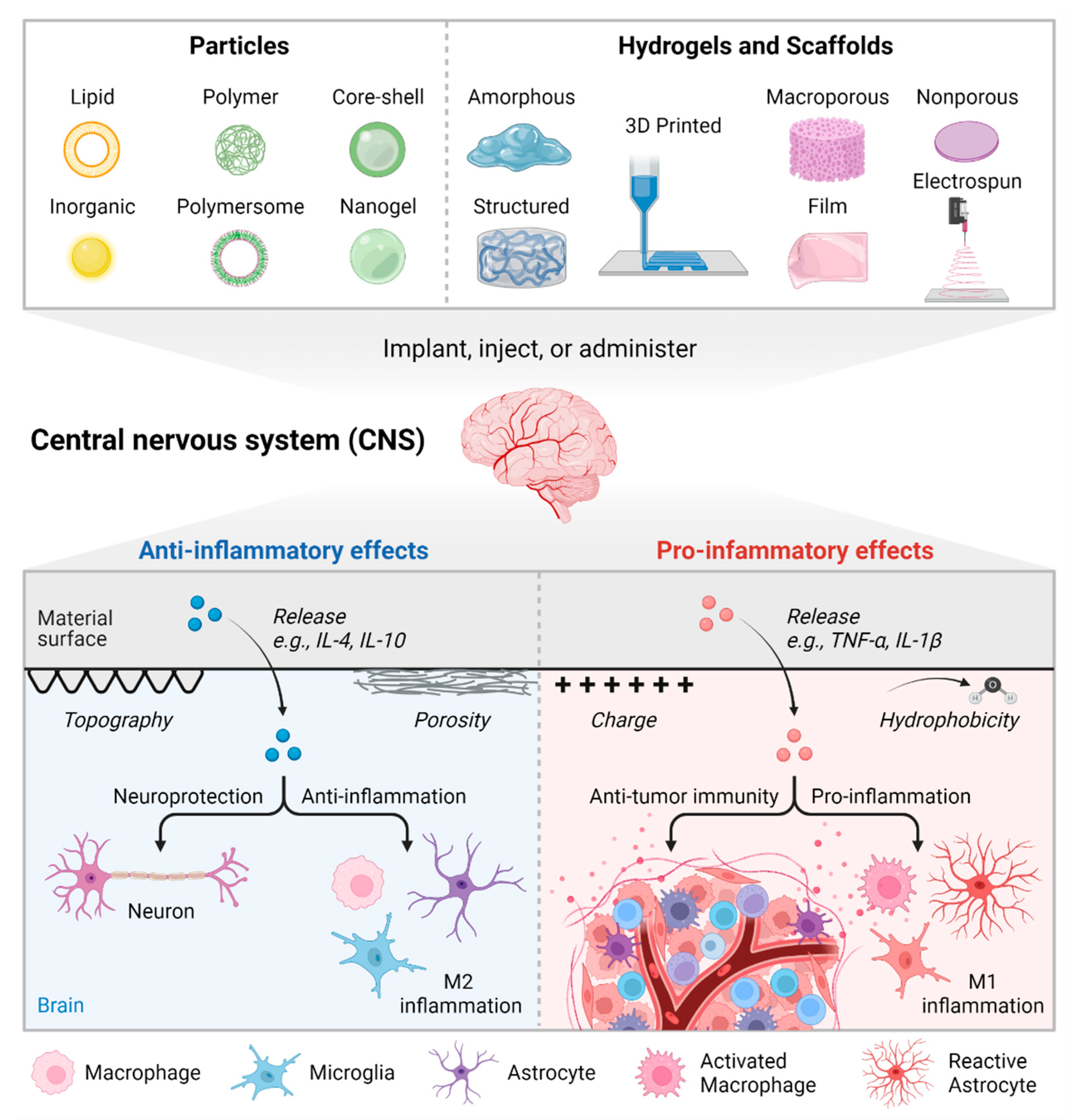
3. Biomaterial Strategies to Modulate Immune Cell Phenotypes

Regulating or manipulating the immune response is a promising strategy to halt or treat neurological damage. Early iterations of immunomodulation, or immunotherapy, used systemic delivery of small molecules or protein therapeutics [10]. The most notable example may be PD1 inhibitors in cancer, which prevent the induction of T cell apoptosis and immunosuppression by cancer cells [82,83]. Specific to the CNS, the FDA approved the small molecule fingolimod for the treatment of multiple sclerosis in 2018. The drug fingolimod prevents T cell infiltration into the spinal cord by blocking sphingosine 1-phosphate (S1P) signaling [84]. Fingolimod also alters the function of astrocytes, pericytes, and oligodendrocytes [85,86,87,88,89], suggesting systemic delivery may have secondary or off-target effects. Localized or triggered release of such a therapeutic cargo can be accomplished using biomaterials. Biomaterials have been used for prophylactic vaccines [90], cancer therapy [91,92], treating autoimmune diseases like diabetes [93], and promoting tissue regeneration [94,95,96]. We briefly review a few notable examples, but further review of CNS-specific or biomaterial-based immunomodulation is available [10,18,97,98,99,100,101,102,103].
Implanting a biomaterial inherently stimulates interactions with the host immune system, dictated in part by the preceding cascade of protein adsorption and provisional matrix formation. These interactions at the cell-biomaterial interface provide an opportunity to steer the resulting immune response. Biomaterials can be formulated as particles, scaffolds, or cross-linkable hydrogels [102,104,105,106]. Engineering the characteristics of a material and/or encapsulating drug cargo can enable control over cellular recruitment and phenotypic biasing, including immune cells. Additionally, conjugation or encapsulation of active molecules like peptides and proteins protects this fragile cargo from premature clearance or degradation by harsh physiological conditions [107,108]. Of course, it is critically important to choose an appropriate biomaterial and drug cargo to achieve a desired stromal, stem cell, or immune cell outcome [10,109,110,111]. Therapies for immunomodulation often leverage known pro- or anti-inflammatory pathways for cancer therapy and regenerative medicine, respectively. For example, anti-cancer therapies may deliver IL-1β, TNF-α, or agonists of Toll-like receptors, while anti-inflammatory approaches may include TGF-b, IFNγ, or interleukins IL-4, IL-6, IL-10, and IL-13. One consideration is that anti-inflammatory approaches may also benefit from small doses of pro-inflammatory signals—at least initially—since M2 macrophages derived from M1 cells are more effective at promoting angiogenesis than M2 cells derived from unstimulated M0 cells [112]. Figure 1 summarizes how materials can be engineered to impact neuroinflammation.

3.1. Particulate Biomaterials

Nano- and microparticle formulations leverage the ability of many immune cells to engulf or phagocytose particulate materials loaded with therapeutic drugs. Biomaterial particles are therefore an effective strategy to modulate immune cell phenotype from within the cell itself [113]. Phagocytosis is influenced by the size, shape, charge, stiffness, and fluidity of the biomaterial particle [107]. Round particles are more readily phagocytosed, but rod-like particles induce a larger immune response [114]. Rod-shaped particles also enhance the inflammatory response against decorated ligands, regardless of the ligand-receptor targets [115]. Particles smaller than 3 microns are good for intracellular drug delivery, while particles larger than 10 microns are better for extracellular delivery [114,116]. Combining multiple particle sizes can enable a multifaceted approach [116], as can co-loading multiple stimulating factors into the same particle. For example, co-loading agonists of the stimulator of interferon genes (STING) pathway and Toll-like receptor 4 yields a stronger type I IFNβ immune response for eliminating cancer cells [92].
In contrast to inducing phagocytosis, coating particles with poly(ethylene glycol) (PEG) will protect them from immune recognition and lengthen blood circulation time. Nonetheless, PEGylated particles must strike a balance between immune evasion, avoiding off-site accumulation, and desirable uptake by target immune cells, promoting the desired effect [117]. Nanoparticles can also be attached to living cells as a “cellular backpack”, which enhances particle homing and accumulation at target sites. Cellular backpacks are designed to stick to trafficking cells without being phagocytosed or undesirably altering cell function. Importantly, immune cells with backpacks can still cross the BBB [118]. Backpack-laden macrophages address some disadvantages of conventional carriers by enabling less cargo to be loaded, reaching the target site during inflammation, and protecting and sustaining the release of the drug cargo [118,119,120].
One common polymer used for particle development is poly lactic-co-glycolic acid or PLGA. This copolymer has been used to deliver an array of immunomodulatory agents to redirect immune cell phenotype and function, including TGFꞵ [116,121], granulocyte-macrophage colony-stimulating factor (GM-CSF) [116,122]; agonists of the STING pathway [123]; steroid hormone vitamin D3 [122]; vaccine adjuvant [124]; and the antioxidant N-acetylcysteine [125]. The PLGA polymer itself may be immunosuppressive as it degrades into lactic acid, which causes resistance to pro-inflammatory stimulation by LPS [126]. Other engineered particle formulations include those based on lipids [92], gold [127,128], and metal oxides [129], to name a few.
Cells naturally produce lipid-based nanoparticles called extracellular vesicles (EVs), which enable long-distance cell–cell communication. The most widely recognized type of EV is exosomes, which are emerging as powerful tools to assess tissue damage, deliver therapeutic molecules, and directly or indirectly modulate immune responses [130,131,132]. EVs play a key role in immunomodulation and have been shown to be the mechanism through which implanted cells like mesenchymal stem cells promote immunosuppression and wound resolution [133,134]. One key advantage to exosomes for neural applications is their innate ability to cross the BBB, an ability which is further enhanced under inflammatory conditions [135]. EVs can therefore leverage the conditions of neuroinflammation as a driving force for targeted drug delivery in the CNS.
EVs contain surface receptors and intracellular cargo derived from the source cell and can be engineered using genetic manipulation or exogenous chemistries for enhanced drug delivery or cellular targeting [136,137,138,139]. For further compositional control, engineered liposomes can be developed in vitro using sonication, forced filtration, or microfluidic methods [140]. A user controls what lipids are contained with the bilayer, the contents of the liposome interior or surface, which route through which the particles will be taken up, and the ability to target specific cell types. While standalone EVs and liposomes have many advantages, loading the particles into scaffolds can deter unintended phagocytosis and enable controlled or sustained release, and preferential accumulation at the target site [141,142].

3.2. Solid Scaffolds and Hydrogels

Biomaterial scaffolds also have great prospects for immunomodulation, as reviewed by [102,143]. Physicochemical properties of the scaffold direct immune cell adhesion, morphology, proliferation, and polarization and can be tuned by altering material surface chemistry or topography; mechanical properties; geometry, shape, and pore size; and degradation rates or byproducts [96,144]. Materials with properties similar to native tissue tend to promote more anti-inflammatory or pro-regenerative immune cell outcomes. For example, scaffolds comprising polymer fibers promote more M2 macrophages, potentially as a result of mimicking the fibrous qualities of collagen-based tissues [145,146]. Fibrous scaffolds are also inherently porous, and both fiber size and pore size are positively correlated with increased expression of M2 markers [145,147]. Related to mechanical properties, materials with Young’s moduli on the order of most native tissues (1–100 kPa) also promote an anti-inflammatory phenotype, while stiffer matrices prime the cells toward a pro-inflammatory phenotype [148]. Nonetheless, this macrophage response can depend on the crosslinking agent used to modulate the material stiffness, since genipin crosslinking appears to suppress all immune responses while crosslinking with carbodiimide enhances pro-inflammatory responses, specifically [149,150,151].
The chemical properties of a material, such as bulk and surface composition, surface charge and density, and hydrophilicity/phobicity influence protein adsorption and therefore immune cell interactions [103]. Again, a distinct advantage may be afforded to materials chemically similar to—or derived from—native tissue, since these materials mimic the physiological environment. Native tissue can be rendered non-immunogenic through decellularization—the process of treating tissue with detergents, enzymes, and/or other reagents to remove cellular components and isolate the extracellular matrix (ECM). Tissue-derived ECM is a natural source of instructive cues for many cellular processes and has shown vast potential as an immunomodulatory biomaterial [152,153]. Decellularized tissues induce a T helper 2 cell response that guides IL-4–dependent (anti-inflammatory) macrophage polarization [95,154]. Complete removal of cellular debris is required, however, since incomplete decellularization promotes a pro-inflammatory response [155].
ECM-driven induction of anti-inflammatory phenotypes has been ascribed to the presence of residual hyaluronan [156] as well as matrix-bound nanovesicles [157,158]. Interestingly, the phenotype induced by exposure to ECM scaffolds appears functionally distinct from canonical anti-inflammatory phenotypes, suggesting the response is more nuanced and requires additional study [157]. Nonetheless, one aspect is clear: decellularized ECM in its native, structured form or as an injectable hydrogel is an effective scaffold for immunomodulation and tissue repair, including in the CNS [159,160,161,162].

3.3. Materials for Glial Cell Modulation

Material implantation into the CNS will recruit and bias the phenotype of not only peripheral immune cells but also resident glial cells. Intentionally designing materials to positively interact with, and dictate the phenotype of, glial cells may be an impactful approach to sustained immunomodulation since these cells play direct roles in regulating and propagating inflammatory responses. As a baseline, nonionic and anionic materials appear to be best for avoiding a detrimental foreign body response in vivo [163]. In vitro studies have informed much of what is known about astrocyte responses to biomaterials [164,165,166,167], but the recent description of A1/A2 astrocyte phenotypes means much more work is necessary to understand how to engineer materials that intentionally harness astrocyte for repair. In mechanically dynamic materials, matrix stiffening limits reactive astrogliosis while softening of the material enhances astrocyte activation in 3D [168]. These mechanical effects may be related to the astrocytic scar, which was once assumed to be stiff but more recently described as being softer than healthy CNS tissue [55]. Fibrous and porous materials enable astrocyte invasion into the material, and aligned fibers promote bipolar or stellate morphologies common to quiescent astrocytes. Cultured astrocytes on larger fiber diameters appear to support more neurite outgrowth compared to smaller fiber diameters [169]. Nonetheless, electrospun fibers of PLLA were found to the enhance expression of A1, pro-inflammatory genes, requiring controlled release of TGF-β to promote neuroprotective A2 phenotypes [167]. For further review of how engineered biomaterials modulate glial responses and neuroinflammation, see the recent review by Tsui et al. [98].
Next-generation therapies will leverage temporal and spatial control over bulk or local material properties, ligand presentation, and drug release profiles. Enacting control over one or more of these axes can be difficult in vivo, but leveraging chemistries that do not interact with biology—known as bioorthogonal reactions—can enable spatial and temporal alterations in material composition, even after implantation. This versatility can enable therapeutics to combine dynamic mechanical or chemical changes, incorporation of other materials such as liposomes or exosomes, and/or triggered the release, exchange, or refilling of multiple drug cargos. In the next section, we introduce a range of bioorthogonal chemistries currently used and discuss their applications in biomaterials science.

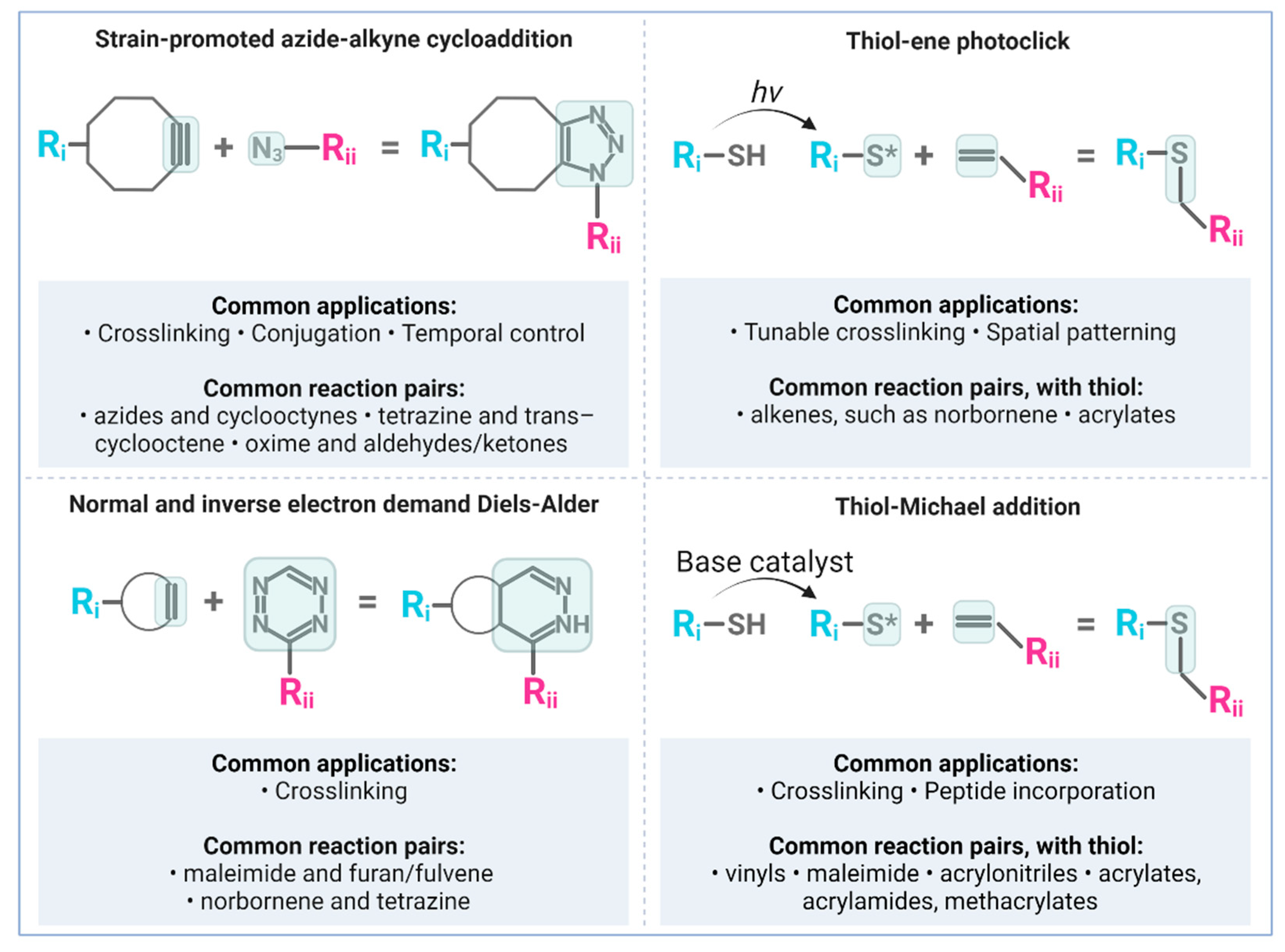
4. Bioorthogonal ‘Click’ Chemistry and Neural Biomaterials Applications

Click reactions are generally ligation reactions conducted under ambient conditions which rapidly and selectively produce the desired ligation product in high yield [14,170]. Several click reactions have been developed to date, enabling an array of chemical ligation strategies for bioorthogonal polymerization, crosslinking, or conjugation [14,17]. The earliest and most widely used click reaction in polymer science is the copper-catalyzed azide-alkyne cycloaddition (CuAAC). Given concerns about copper toxicity, numerous copper-free click reactions have also been developed and seen wider adoption in the biomaterials field. These reactions are used for tuning crosslinking kinetics and dynamics [171,172,173]; directing site-specific conjugation [174,175,176,177]; polymerizing and functionalizing hydrogel microparticles [178]; controlling small molecule loading and reloading [179], and investigating cellular mechanobiology [180]. Typically, the reaction scheme involves simply mixing polymers and biomolecules containing the click reactive groups. Endogenous proteins or glycans can also be labeled through the cellular metabolism of non-canonical amino acids or unnatural sugar residues, making it possible to engineer otherwise less-controllable materials like cell-derived matrices or decellularized tissues [175,181]. Below we highlight the most common copper-free click chemistries and highlight key biomaterial applications. Figure 2 depicts the most common reaction schemes discussed here. For further review of click reactions in biomaterials, refer to Anseth and Klok [14]; Madl and Heilshorn [170]; Arkenberg, Nguyen, and Lin [171]; and Idiago-López et al. [182].

4.1. Copper-Free Strain-Promoted Azide-Alkyne Cycloaddition (SPAAC)

Since the initial discovery by Bertozzi and colleagues [183], strain-promoted azide-alkyne cycloaddition (SPAAC) has proven to be an effective alternative to CuAAC, maintaining the high specificity and reaction rate while limiting cytotoxicity. Known SPAAC reactions commonly used in biomaterials include the 1,3-dipolar cycloaddition between azides (a 1,3-dipole, -N3) and cyclooctynes, like dibenzocyclooctyne (DBCO) [184,185,186]; the tetrazine ligation [187,188]; and oxime chemistry with aldehydes and ketones [189]. Other reactions include the isocyanide-based click reaction [190] and the quadricyclane ligation [191].
SPAAC reactions can be used as the primary mechanism of hydrogel crosslinking for gentle and efficient cell encapsulation. For example, Hardy, Lin, and Schmidt used oxime click chemistry to crosslink linear, homo-bifunctional aminooxy PEG with aldehyde-functionalized hyaluronic acid (HA) [189]. Additionally, engineered elastin-like proteins have been developed to crosslink via SPAAC, which gelled completely within minutes and supported encapsulation and maintenance of murine neural progenitor cells. The stiffness and cell adhesion properties of the SPAAC-ELP gels could be tuned independently through the incorporation of RGD peptides. The same group later modified the elastin-like protein to contain azide-functionalized tyrosine residues for SPAAC to overcome the reliance on amine-reactive chemistry [192].
SPAAC reactions are commonly used to functionalize hydrogel biomaterials with molecules of interest. Azide-bearing nerve growth factor has been selectively grafted onto an acrylate-terminated, poly(β-amino ester) form of PEG-diacrylate [193]. The resulting hydrogels were then subsequently conjugated with thiol-containing CYIGSR adhesive peptides through a thio-ene reaction (discussed below) to further promote neuroregeneration. Adil et al. used a SPAAC reaction to first incorporate azide-containing RGD peptide into DBCO-functionalized HA, then used the remaining unreacted DBCO groups to crosslink the hydrogel with homobifunctional PEG-azide [186]. This approach yielded a 5.6-fold improvement in transplanted cell survival compared to 2D cultured cells and a 5.4-fold increase in dopaminergic neuron differentiation and survival at 4 months post-injection. Similarly, Li and colleagues decorated DBCO-functionalized PEG with azide-modified laminin and interferon proteins prior to crosslinking with four-arm PEG-tetraazide [194]. This approach enabled in vitro culture of neural stem cells over a two-week period and specified for primarily neuronal differentiation without supplementing differentiation factors in the medium.
Leveraging the bioorthogonal nature of these reactions can also provide temporal control over biomolecule presentation. Micropatterned brushes of PEG-azide have been modified after the initiation of cell culture simply by adding peptide–DBCO conjugates into the media [184]. Additionally, azide-modified alginate hydrogels have been filled and refilled long after in vivo implantation using intravenous delivery of cyclooctyne-conjugated small molecules [179]. One can envision using this approach to alter drug dosage or completely change to a new drug based on the patient’s therapeutic response. Hence, temporal control over drug-loaded biomaterials is a step in the right direction for personalized therapy.

4.2. Diels-Alder and Inverse Electron-Demand Diels-Alder

Diels-Alder reactions occur between a diene (a molecule with two adjacent double bonds) and a substituted alkene (one double bond), which is commonly called the dienophile [195]. These are cycloadditions promoted not by strain but by electron rearrangement, either by normal or inverse electron demand. Normal electron-demand reactions occur between electron-poor dienophiles and electron-rich dienes, while inverse electron-demand reactions occur between an electron-rich dienophile and an electron-poor diene. Common diene-dienophile pairs used in biomaterials are maleimide-furan and tetrazine-norbornene for normal and inverse electron-demand Diels-Alder reactions (IEDDA), respectively. For a more in-depth review of these reactions and applications in biomaterials, see [196,197].
Furan modified-HA is a common Diels-Alder polymer backbone used for neural applications, which is typically crosslinked using PEG-bismaleimide. The mechanical and degradation properties of the resulting Diels−Alder cross-linked hydrogels can be tuned by varying the molar ratio of furan to maleimide [172]. These hydrogels can be used to encapsulate and deliver cells in vivo, such as V2a interneurons derived from mouse embryonic stem cells [198]. Another application is the production of cryogels, in which the uncrosslinked polymer mixture is frozen to induce ice crystal formation and then thawed to induce crosslinking and preservation of the porous architecture [199]. After crosslinking, maleimide-functionalized peptides can still be clicked into the gel via remaining unreacted furan groups to add cell adhesion sites or other bioactive motifs. Nonetheless, crosslinking between furan and maleimide can be relatively slow, on the order of hours. To improve the gelation rate, Madl and Heilshorn replaced the furan group with fulvene, which crosslinked with maleimide an order of magnitude faster than other common Diels-Alder reaction pairs while remaining stable for months [200]. Yet, another caveat is that many Diels-Alder reactions are most efficient at acidic pH, which is not optimal for cell encapsulation. Smith et al. addressed this concern by replacing the furan group on hyaluronic acid with the more electron-rich methylfuran to achieve faster, more efficient gelation at physiological pH [201].
IEDDA reactions can also be used as the primary mode of hydrogel gelation for cell encapsulation. Hydrogel properties like gelation rate and stiffness can be adjusted by varying the degree of polymer modification and molar ratio of tetrazine to norbornene. Stiffer, faster gelling materials are achieved at higher percent functionalization and an equimolar ratio of reactive groups [202]. Notable applications of IEDDA crosslinked biomaterials include assembling microporous annealed particle hydrogels for injection after stroke [203,204]; enabling the formation of low polymer content (and therefore low viscosity) methylcellulose for co-delivery of neural progenitor cells and scar-degrading enzyme [205]; and demonstrating maintenance of neural stemness depends on hydrogel degradation and cell–cell contacts regardless of stiffness [192].

4.3. Photo-Click Thiol-Ene Reactions

Photo-induced chemistries can be used to enable more control over temporal and spatial hydrogel dynamics [206]. Only locations exposed to light will react, and the extent of the reaction depends on the exposure time. There are photo-induced reactions for many bioorthogonal chemistries, termed ‘photo-click’ reactions [207], though the most notable in biomaterials literature is the thiol-ene reaction. Thiol-ene reactions occur between alkenes and thiol groups in the presence of a photoinitiator and light irradiation. The thiol groups (-SH) are initiated to thiyl radicals (-S*) following exposure to ultraviolet or visible light, depending on the photoinitiator. Irgacure 2959 is a common photoinitiator for ultraviolet activation, while Eosin Y can be used with the more cytocompatible range of visible light. Because photo-click reactions require a light stimulus of a specific wavelength, it is possible to perform multiple photo-click reactions within the same material by combining UV and visible light stimuli [208]. Additional control may be gained by adding photo-cleavage reactions to unmask or degrade chemical moieties with a different light wavelength [209,210]. One caveat is that thiol–based reactions are not perfectly biorthogonal, since thiol groups are naturally present in the amino acid cysteine. Nonetheless, these reactions often do not interfere with biological processes since cysteine is relatively low in abundance, and thiols are mostly present as disulfide bridges [207].
Polymers functionalized with norbornene are commonly used to enable photo-click crosslinking. For example, Jivan et al. used PEG-norbornene crosslinked with dithiothreitol (DTT) to form PEG hydrogel microparticles varying in size from 8 to 30 μm in diameter [178]. Given the potential for norbornene to also participate in IEDDA reactions, the researchers subsequently added tetrazine-functionalized proteins for conjugation with the remaining unreacted norbornene groups. Instead of using thiol groups on the crosslinker, Sawicki and Kloxin created hydrogels of thiolated PEG crosslinked with vinyl-containing peptides. The vinyl group was incorporated by functionalizing the peptides with alloxycarbonyl, a common protective group used during peptide synthesis. Using the photoinitiator lithium acylphosphinate, the hydrogels formed rapidly under cytocompatible doses of long-wavelength ultraviolet light (365 nm) [173]. Alternatively, it was recently shown that the thiyl radicals needed to initiate thiol-ene crosslinking can be generated using hydrogen peroxide in the presence of the enzyme horseradish peroxidase [211]. This approach preserves temporal control over the reaction while averting photodamage to live cells.

4.4. Thiol-Michael Addition

The thiol-Michael addition is a type of thiol-ene reaction that is base-catalyzed as opposed to radically mediated [212]. These reactions occur between nucleophilic thiol groups, known as the Michael donor, and electrophilic-ene compounds, or Michael acceptors, without the need for light radiation [212]. There are also Michael-type addition reactions not involving thiols, but the key attribute of the thiol-Michael addition reaction for biomaterials science is the wide array and availability of possible reactive macromers. Thiols react with several moieties commonly used in biomaterials, including maleimides, vinyl sulfones, acrylates and acrylamides, acrylonitriles, and methacrylates [177]. For example, a cyclized vinyl polymer of PEG was used as a precursor for hydrogel nanoparticle synthesis crosslinked with DTT [213]. After crosslinking, there is also potential for a retro Michael-type reaction with thiol exchange, which imparts degradability in response to reducing conditions in the microenvironment [210]. More about the thiol-Michael addition and primary uses for each reactive pairing in biomaterials can be found in [212].
Another major benefit to the thiol-Michael addition is the potential to use cysteine-containing peptides for easy and versatile biofunctionalization or crosslinking. In their study, Zhao et al. found that the inclusion of dithiol-containing peptides with laminin-derived sequences of IKVAV and YIGSR leads to a slower gelation rate but also improves the viability and metabolic activity of murine neural progenitor cells [213]. As with other examples, acryl hydrazide HA was loaded with thiol-containing RGD peptides and then crosslinked using an enzyme-cleavable di-thiol peptide [214]. Tam and colleagues leveraged thiol-maleimide click chemistry as well as biotin-streptavidin chemistry to covalently immobilize two separate bioactive factors into thiolated methylcellulose: maleimide–GRGDS for cell adhesion, and maleimide-streptavidin for subsequent binding with biotinylated platelet-derived growth factor-s (rPDGF-A), an oligodendrocyte differentiation factor [215].
The same group later created thiolated MC for injection into the intrathecal space on top of the spinal cord, functionalization with maleimide-modified peptide and crosslinked using PEG-bismaleimide [216]. The peptide exhibited homology to a region on an enzyme of interest, chondroitinase ABC, released over time to degrade the glial scar tissue and promote neuronal differentiation of transplanted human neural stem cells. Similarly, HA hydrogels based on thiol-alkyne Michael type addition promoted in vivo survival of encapsulated human neural progenitor cells after transplantation into a stroke cavity [214]. The HA backbone was functionalized with cell-adhesive peptides to support the survival of encapsulated neural progenitor cells as well as thiol-modified heparin, to facilitate growth factor retention. These authors used a design of experiments approach to optimize the concentration of the chosen peptides, but others have directly evaluated the landscape of integrin-binding and MMP-degradable sequences in neural tissue [217]. Proteomic analysis of the adult human brain tissue identified nine different integrin binding sites and five different MMP-cleavage sites of high interest. These sites were then incorporated into PEG-maleimide hydrogels using cysteine-containing peptides and sequential Michael-type addition reactions, thereby creating brain ECM-mimicking hydrogels that maintained astrocytes in a stellate, quiescent morphology.

4.5. Click Applications for Immunomodulation

The chemo-selective nature of biorthogonal reactions makes them an ideal choice for incorporating functional groups that modulate cell-material interactions and instruct immune cell responses. For example, Glass and colleagues optimized a process to functionalize mannose onto the surface of polymeric nanoparticles using a CuAAC reaction. The conjugated sugar acted as a targeting moiety on the nanoparticle surface by promoting specific interactions with the mannose receptor CD206 that is preferentially expressed on M2 macrophages [218]. The authors demonstrated an optimal range for minimizing copper-induced cytotoxicity while maintaining high levels of conjugation efficiency and increased cell targeting. This approach enabled the successful delivery of therapeutic siRNA and subsequent reprogramming of the M2 macrophages toward an anti-cancer, M1 phenotype. A similar but opposite approach may therefore also work to redirect M1 macrophages toward M2 phenotypes to promote tissue repair.
Bioorthogonal conjugation can add functionality or targeting moieties onto surfaces. Inert beads were chemo-selectively conjugated with a modified TGF-β1 via a Staudinger ligation between an azide and triarylphosphine [219], and the engineered surfaces efficiently converted naïve T cells to functional Treg cells for immune suppression. Dendritic cells were also labeled in vivo using azide-modified glycans [220]. The cells were recruited to the implantation site using alginate gel releasing GM-CSF as well as an azide-modified sugar, which was taken up by the dendritic cells and used to improve the generation of antigen-specific, CD8+ T cells through covalent capture of DBCO-bearing antigens and adjuvants like IL-15 and IL-15Rα. Hence, biorthogonal chemistries can advance the therapeutic efficacy of immunomodulation across a range of biomaterial-based systems.

5. Click-Based Materials for Neuroimmune Modulation

Traditional immunotherapy often uses a biomaterial as drug delivery vector, missing out on the opportunity to leverage cell-material interactions as an active therapy element. Combining the efficacy of biomaterial-based immunotherapy with the versatility of bioorthogonal chemistries is an excellent strategy to create the more intentional or synergistic effects that may be required to treat complex conditions like neuroinflammation. Current literature approaches have implemented click chemistry in two primary ways: bioorthogonal crosslinking and/or selective bioconjugation. Such applications include the incorporation of cell-adhesion sites, bioactive cytokines, and degradable crosslinks toward modulating immune, glial, or neuronal cell outcomes. Nonetheless, there remains a vast unexplored space in which to leverage bioorthogonal reactions for enhancing material functionality and regulating neuroinflammatory environments.

5.1. Biomaterial Scaffolds and Hydrogels

Hydrogel-based strategies have seen the highest use of bioorthogonal chemistries for directly or indirectly treating neuroinflammation. It is worth noting that each study highlighted below used a foundation of hyaluronic acid, the primary matrix component of the CNS. One notable example is Thompson and collaborators, who developed a modified HA hydrogel for treating spinal cord injuries. The authors used a mixture of furan- and methylfuran-modified high molecular weight HA (250 MDa) crosslinked with PEG-bismaleimide by a Diels-Alder reaction [198]. Prior to crosslinking the gels, the researchers physically blended in cell-derived ECM from either fibrous or protoplasmic populations of astrocytes (differentiated from RW.4 mouse embryonic stem cells). Upon implantation into the hemisected rat spinal cord, ECM from protoplasmic astrocytes decreased the size of the glial scar, decreased macrophage and microglia infiltration, and increased axonal ingrowth. Additional encapsulation of mouse embryonic stem cell-derived V2a interneurons further increased neuronal processes in and around the lesion. Hence, these bioorthogonal hydrogels enabled gentle but efficient encapsulation of ECM molecules and motoneuron cells to encourage spinal cord regeneration.
A di-functional HA was created by modifying the primary hydroxyl and carbonyl groups of the HA backbone with thiol and azide groups, respectively [221]. The hydrogels were then crosslinked with methacrylated HA by a photo-click reaction between the thiol and methacrylate. The ratio of methacrylate HA was varied to determine the effects of difunctionalization on mouse embryonic stem cell differentiation. A 1:2 ratio for difunctional to methalcrylate HA was selected for testing in a rat model of subacute contusion spinal cord injury. In vivo gelation relied solely on the Michael-type addition reaction for crosslinking. Compared to saline controls, the 1:2 hydrogel significantly reduced infiltration of macrophages into the spinal cord lesion one week after implantation but did not significantly influence the lesion volume, astrocyte activation, or the presence of axonal staining. While not conducted in this study, the free azide functional side groups could be reacted with DBCO-modified cytokines or adhesive peptides as additional bioactive signaling molecules, which may lead to further enhancements of axonal in-growth and tissue sparing.
The introduction of macroporosity will further facilitate vascularization and immune cell modulation after injury. One such method to produce macroporous hydrogel scaffolds is by annealing together microgels [105]. Microgels are crosslinked hydrogel microparticles that can be used to encapsulate and deliver cells, proteins, and/or small molecules. One study used click chemistry to incorporate three peptides into HA-acrylamide: one peptide of cell-adhesive RGD and two others for the active clotting enzyme Factor XIIIa [203,222]. The precursor solution could be fluorescently labeled using a maleimide-containing fluorophore, and the decorated HA-acrylamide was crosslinked by a Michael-type addition with di-cysteine-containing, MMP-cleavable peptides. Finally, the microgels were linked together using Factor XIIIa, forming an annealed-solid hydrogel scaffold with macroporous void space. Applying the annealed HA microgels into a stroke cavity lowered the percentage of both astrocytes and microglia in the peri-infarct area. There was also a significantly higher percentage of vessels in the peri-infarct area and more proliferating neural progenitor cells migrating toward the damaged tissue. Collectively, the annealed HA microgels accelerated brain repair after stroke by modifying the extent of astrogliosis, inflammation, vascularization, and neural progenitor cell recruitment to the lesion.

5.2. Therapeutic Particles

The BBB is a major hurdle for delivering therapies to the CNS, even following injury or ischemia [223]. Cationic, BBB-penetrating nanoparticles comprising poly(β-amino esters) were created by reacting excessive PEG-diacrylate with 2-propynylamine via a Michael-type addition reaction [224]. The poly(β-amino esters) were then functionalized with an azide-containing nerve growth factor mimetic by azide-alkyne cycloaddition. Further, thiol-containing adhesive peptide (CYIGSR) was conjugated to the terminal acrylate groups through a thio-ene photo-click reaction. The functionalized polymeric nanoparticles had a mean diameter size of ~30 nm and promoted neurite outgrowth of cultured sensory neurons below 100 ng/mL. The nanoparticle micelles were able to passively penetrate the BBB, potentially due to the positive charge of the particles and the negative charge of the endothelial cell layer [225]. Moreover, the incorporation of YIGSR peptides positively increased the surface zeta potential, further enhancing BBB permeation. To test the effects on the neuroinflammatory response, the authors used a cryogenic lesion model of brain injury with tail vein injection of the micelle formulation every other day. The dual-functional micelles promoted decreased lesion area and BBB leakage by day 8. The treatment group also had the lowest number of GFAP+ cells in the injured tissue, suggesting low astrocyte reactivity.
A glycoengineering approach can be used to attach reactive groups or drug cargo onto cells for on-demand control of phenotype polarization [226]. Li and colleagues engineered cultured microglia to home to damaged vasculature by fusion with platelet-derived vesicles, and the hybrid cells were fed an azide-modified glycan that became metabolically incorporated into mucin-type glycoproteins [227]. Separately, the anti-inflammatory cytokine IL-4 was encapsulated into liposomes containing both a DBCO-PEG-modified lipid and a molecule that sensitizes the membrane to ultrasound, known as protoporphyrin IX. Excessive azide-PEG was added to quench unreacted DBCO groups and avoid liposome aggregation, and the sono-sensitive DBCO-liposomes were linked to the hybrid microglia as a composite cellular therapy. In cell culture studies, exposure to ultrasound-induced the release of the IL-4, causing upregulation of M2 markers such as Arg-1 and TGF-β. The composite therapy significantly reduced the lesion volume in a mouse model of ischemic stroke, as well as decreased astrocyte activation and increased microglia recruitment. Ultimately, triggered release of IL-4 in the stroke cavity increased the proportion of M2 phenotypes, thereby remodeling the neuroinflammatory environment to promote neuroprotection and improve functional recovery.
While the above platelet-derived vesicles had to be generated ex vivo, extracellular vesicles can be easily isolated for tethering to biomaterials or the cell surface [228]. One study used CuAAC to conjugate cell-adhesive RGD to the exosome surface [136]. The RGD-conjugated exosomes were found to accumulate more within an ischemic brain lesion owing to the upregulation of the integrin αvβ3 in reactive cerebral vascular endothelial cells. Using the engineered exosomes to deliver curcumin, a natural anti-inflammatory agent, significantly suppressed the inflammatory response and cellular apoptosis in the lesion region compared to soluble curcumin or unloaded exosomes alone. Mice treated with the exosomes expressed significantly less TNF-α, IL-1β, and IL-6, demonstrating the ability to harness the intrinsic immunosuppressive potential of mesenchymal stem cell-derived exosomes for targeted delivery of additional anti-inflammatory agents.

5.3. Hydrogel Coatings

Lastly, biomaterials can be used as coatings to create a new, amenable cell-material interface. A major hurdle for sustained use and recording from neural electrodes is that the required stiff materials cause extensive inflammatory scarring. Matching the mechanical properties of an electrode to that of brain tissue can minimize the scarring response [229]. The unique qualities of thiol-ene photo-click chemistry offer precise control over the mechanical properties and thermal glass-transition temperature of a polymer, making it an ideal electrode coating. By varying the monomer composition, a thiol-ene shape memory polymer coating can be designed to be stiffer at room temperature for tissue insertion but softer once equilibrated with brain tissue. Dip coating a uniform layer of thiol-ene shape memory polymer onto silicon microelectrode reduced astrocytic scarring after 16 weeks [230]. Using the thiol-ene polymer coating to reduce the mechanical mismatch between the device and tissue was comparable to, if not better than, the bare silicon microelectrodes. While the recording efficacy needs to be evaluated, this work suggests bioorthogonal biomaterials could lengthen the allowable time for intracortical measurements.
Bioorthogonal coatings can also be used to encapsulate single cells in a hydrogel casing for enhanced cellular protection and control of the microenvironment [231]. Azide-modified sugars were metabolically incorporated onto the surface of neural stem cells in culture for subsequent reaction with DBCO-PEG chains of various molecular weights. The polymeric coating improved cell survival and modulated growth factor release. Varying the PEG molecular weight also enabled control over the stiffness of the immediate pericellular environment. Specifically, higher molecular weight PEG (20–30 kDa) produced a softer polymeric ECM and significantly increased the release of neurotrophic factors such as VEGF and CNTF. Similar single-cell hydrogel approaches may therefore be useful for protecting transplanted cells from hostile or mechanically stiff environments, favoring neuronal differentiation over astrocytic differentiation, or sustaining an anti-inflammatory phenotype in pre-stimulated cellular therapies [143,148,232].

6. Conclusions and Future Directions

Pathological inflammation of the CNS is complex and difficult to regulate, but the versatility and effectiveness of biomaterial-based strategies suggest this will become the next frontier of neuro-immunomodulation. Therapeutic material approaches will benefit from the specificity and utility afforded by bioorthogonal or ‘click’ chemistry. A majority of previous bioorthogonal materials applied to the CNS have assessed neuroinflammation as a secondary outcome opposed to an intentional target, creating vast opportunities for further impactful advancements. Exciting future directions not yet applied to neuroinflammation include pre-targeted imaging, personalized or modular drug delivery, theranostics, and temporally dynamic therapies. Consider bioorthogonal chemistry as the perfect toolbox for realizing dynamic materials that may one day enable resolving the chronic glial scar, recapitulating an M1 → M2 transition of successful wound healing, or reciprocally interacting and guiding the CNS toward regeneration.

Author Contributions

S.F. and C.C. both contributed to reviewing, collecting, and summarizing the literature and writing the review manuscript. All authors have read and agreed to the published version of the manuscript.

Funding

We thank the National Institute of Biomedical Imaging and Bioengineering (NIH R21 RB031435-01) for partially funding this work.

Institutional Review Board Statement

Not applicable.

Informed Consent Statement

Not applicable.

Data Availability Statement

Not applicable.

Conflicts of Interest

The authors declare no conflict of interest.

References

  1. Ransohoff, R.M.; Schafer, D.; Vincent, A.; Blachère, N.E.; Bar-Or, A. Neuroinflammation: Ways in Which the Immune System Affects the Brain. Neurotherapeutics 2015, 12, 896–909. [Google Scholar] [CrossRef] [PubMed] [Green Version]
  2. Kwon, H.S.; Koh, S.H. Neuroinflammation in neurodegenerative disorders: The roles of microglia and astrocytes. Transl. Neurodegener. 2020, 9, 42. [Google Scholar] [CrossRef] [PubMed]
  3. DiSabato, D.J.; Quan, N.; Godbout, J.P. Neuroinflammation: The devil is in the details. J. Neurochem. 2016, 139 (Suppl. S2), 136–153. [Google Scholar] [CrossRef] [PubMed] [Green Version]
  4. Van Dyken, P.; Lacoste, B. Impact of Metabolic Syndrome on Neuroinflammation and the Blood–Brain Barrier. Front. Neurosci. 2018, 12, 930. [Google Scholar] [CrossRef] [PubMed]
  5. Erickson, M.A.; Banks, W.A. Age-Associated Changes in the Immune System and Blood–Brain Barrier Functions. Int. J. Mol. Sci. 2019, 20, 1632. [Google Scholar] [CrossRef] [Green Version]
  6. Yang, Q.Q.; Zhou, J.W. Neuroinflammation in the central nervous system: Symphony of glial cells. Glia 2019, 67, 1017–1035. [Google Scholar] [CrossRef] [PubMed]
  7. Locy, H.; de Mey, S.; de Mey, W.; De Ridder, M.; Thielemans, K.; Maenhout, S.K. Immunomodulation of the Tumor Microenvironment: Turn Foe Into Friend. Front. Immunol. 2018, 9, 2909. [Google Scholar] [CrossRef]
  8. Osipov, A.; Saung, M.T.; Zheng, L.; Murphy, A.G. Small molecule immunomodulation: The tumor microenvironment and overcoming immune escape. J. Immunother. Cancer 2019, 7, 224. [Google Scholar] [CrossRef] [Green Version]
  9. Varadé, J.; Magadán, S.; González-Fernández, Á. Human immunology and immunotherapy: Main achievements and challenges. Cell. Mol. Immunol. 2020, 18, 805–828. [Google Scholar] [CrossRef]
  10. Dellacherie, M.O.; Seo, B.R.; Mooney, D.J. Macroscale biomaterials strategies for local immunomodulation. Nat. Rev. Mater. 2019, 4, 379–397. [Google Scholar] [CrossRef]
  11. Riley, R.S.; June, C.H.; Langer, R.; Mitchell, M.J. Delivery technologies for cancer immunotherapy. Nat. Rev. Drug Discov. 2019, 18, 175–196. [Google Scholar] [CrossRef]
  12. Adu-Berchie, K.; Mooney, D.J. Biomaterials as local niches for immunomodulation. Acc. Chem. Res. 2020, 53, 1749–1760. [Google Scholar] [CrossRef] [PubMed]
  13. Vasconcelos, D.P.; Águas, A.P.; Barbosa, M.A.; Pelegrín, P.; Barbosa, J.N. The inflammasome in host response to biomaterials: Bridging inflammation and tissue regeneration. Acta Biomater. 2019, 83, 1–12. [Google Scholar] [CrossRef] [PubMed]
  14. Anseth, K.S.; Klok, H.A. Click Chemistry in Biomaterials, Nanomedicine, and Drug Delivery. Biomacromolecules 2016, 17, 1–3. [Google Scholar] [CrossRef] [Green Version]
  15. Li, Y.; Wang, X.; Han, Y.; Sun, H.Y.; Hilborn, J.; Shi, L. Click chemistry-based biopolymeric hydrogels for regenerative medicine. Biomed. Mater. 2021, 16, 022003. [Google Scholar] [CrossRef] [PubMed]
  16. Bird, R.E.; Lemmel, S.A.; Yu, X.; Zhou, Q.A. Bioorthogonal Chemistry and Its Applications. Bioconjug. Chem. 2021, 32, 2457–2479. [Google Scholar] [CrossRef] [PubMed]
  17. Zou, Y.; Zhang, L.; Yang, L.; Zhu, F.; Ding, M.; Lin, F.; Wang, Z.; Li, Y. “Click” chemistry in polymeric scaffolds: Bioactive materials for tissue engineering. J. Control. Release 2018, 273, 160–179. [Google Scholar] [CrossRef] [PubMed]
  18. Rebelo, A.L.; Chevalier, M.T.; Russo, L.; Pandit, A. Sweet tailoring of glyco-modulatory extracellular matrix-inspired biomaterials to target neuroinflammation. Cell Rep. Phys. Sci. 2021, 2, 100321. [Google Scholar] [CrossRef]
  19. Bellver-Landete, V.; Bretheau, F.; Mailhot, B.; Vallières, N.; Lessard, M.; Janelle, M.E.; Vernoux, N.; Tremblay, M.È.; Fuehrmann, T.; Shoichet, M.S.; et al. Microglia are an essential component of the neuroprotective scar that forms after spinal cord injury. Nat. Commun. 2019, 10, 518. [Google Scholar] [CrossRef] [PubMed] [Green Version]
  20. Fu, H.; Zhao, Y.; Hu, D.; Wang, S.; Yu, T.; Zhang, L. Depletion of microglia exacerbates injury and impairs function recovery after spinal cord injury in mice. Cell Death Dis. 2020, 11, 528. [Google Scholar] [CrossRef] [PubMed]
  21. Poulen, G.; Aloy, E.; Bringuier, C.M.; Mestre-Francés, N.; Artus, E.V.F.; Cardoso, M.; Perez, J.C.; Goze-Bac, C.; Boukhaddaoui, H.; Lonjon, N.; et al. Inhibiting microglia proliferation after spinal cord injury improves recovery in mice and nonhuman primates. Theranostics 2021, 11, 8640–8659. [Google Scholar] [CrossRef] [PubMed]
  22. Anderson, M.A.; Burda, J.E.; Ren, Y.; Ao, Y.; O’Shea, T.M.; Kawaguchi, R.; Coppola, G.; Khakh, B.S.; Deming, T.J.; Sofroniew, M.V. Astrocyte scar formation aids CNS axon regeneration. Nature 2016, 532, 195. [Google Scholar] [CrossRef] [PubMed] [Green Version]
  23. Matejuk, A.; Vandenbark, A.A.; Offner, H. Cross-Talk of the CNS With Immune Cells and Functions in Health and Disease. Front. Neurol. 2021, 12, 750. [Google Scholar] [CrossRef]
  24. Vandenbark, A.A.; Offner, H.; Matejuk, S.; Matejuk, A. Microglia and astrocyte involvement in neurodegeneration and brain cancer. J. Neuroinflamm. 2021, 18, 298. [Google Scholar] [CrossRef]
  25. Li, Y.; Ritzel, R.M.; Khan, N.; Cao, T.; He, J.; Lei, Z.; Matyas, J.J.; Sabirzhanov, B.; Liu, S.; Li, H.; et al. Delayed microglial depletion after spinal cord injury reduces chronic inflammation and neurodegeneration in the brain and improves neurological recovery in male mice. Theranostics 2020, 10, 11376. [Google Scholar] [CrossRef] [PubMed]
  26. Banjara, M.; Ghosh, C. Sterile Neuroinflammation and Strategies for Therapeutic Intervention. Int. J. Inflam. 2017, 2017, 8385961. [Google Scholar] [CrossRef] [Green Version]
  27. Linnerbauer, M.; Wheeler, M.A.; Quintana, F.J. Astrocyte Crosstalk in CNS Inflammation. Neuron 2020, 108, 608–622. [Google Scholar] [CrossRef]
  28. Marchetti, L.; Engelhardt, B. Immune cell trafficking across the blood-brain barrier in the absence and presence of neuroinflammation. Vasc. Biol. 2020, 2, H1. [Google Scholar] [CrossRef] [Green Version]
  29. Skaper, S.D.; Facci, L.; Zusso, M.; Giusti, P. An Inflammation-Centric View of Neurological Disease: Beyond the Neuron. Front. Cell. Neurosci. 2018, 12, 72. [Google Scholar] [CrossRef]
  30. Liddelow, S.A.; Guttenplan, K.A.; Clarke, L.E.; Bennett, F.C.; Bohlen, C.J.; Schirmer, L.; Bennett, M.L.; Münch, A.E.; Chung, W.S.; Peterson, T.C.; et al. Neurotoxic reactive astrocytes are induced by activated microglia. Nature 2017, 541, 481–487. [Google Scholar] [CrossRef]
  31. Tyzack, G.E.; Hall, C.E.; Sibley, C.R.; Cymes, T.; Forostyak, S.; Carlino, G.; Meyer, I.F.; Schiavo, G.; Zhang, S.C.; Gibbons, G.M.; et al. A neuroprotective astrocyte state is induced by neuronal signal EphB1 but fails in ALS models. Nat. Commun. 2017, 8, 1164. [Google Scholar] [CrossRef] [PubMed]
  32. Ding, Z.B.; Song, L.J.; Wang, Q.; Kumar, G.; Yan, Y.Q.; Ma, C.G. Astrocytes: A double-edged sword in neurodegenerative diseases. Neural Regen. Res. 2021, 16, 1702–1710. [Google Scholar] [PubMed]
  33. Liu, L.R.; Liu, J.C.; Bao, J.S.; Bai, Q.Q.; Wang, G.Q. Interaction of Microglia and Astrocytes in the Neurovascular Unit. Front. Immunol. 2020, 11, 1024. [Google Scholar] [CrossRef] [PubMed]
  34. Cherry, J.D.; Olschowka, J.A.; O’Banion, M.K. Neuroinflammation and M2 microglia: The good, the bad, and the inflamed. J. Neuroinflamm. 2014, 11, 98. [Google Scholar] [CrossRef] [Green Version]
  35. Jurga, A.M.; Paleczna, M.; Kadluczka, J.; Kuter, K.Z. Beyond the GFAP-Astrocyte Protein Markers in the Brain. Biomolecules 2021, 11, 1361. [Google Scholar] [CrossRef] [PubMed]
  36. Guttenplan, K.A.; Weigel, M.K.; Prakash, P.; Wijewardhane, P.R.; Hasel, P.; Rufen-Blanchette, U.; Münch, A.E.; Blum, J.A.; Fine, J.; Neal, M.C.; et al. Neurotoxic reactive astrocytes induce cell death via saturated lipids. Nature 2021, 599, 102–107. [Google Scholar] [CrossRef]
  37. Matejuk, A.; Ransohoff, R.M. Crosstalk Between Astrocytes and Microglia: An Overview. Front. Immunol. 2020, 11, 1416. [Google Scholar] [CrossRef]
  38. Clarke, L.E.; Liddelow, S.A.; Chakraborty, C.; Münch, A.E.; Heiman, M.; Barres, B.A. Normal aging induces A1-like astrocyte reactivity. Proc. Natl. Acad. Sci. USA 2018, 115, E1896–E1905. [Google Scholar] [CrossRef] [PubMed] [Green Version]
  39. Niraula, A.; Sheridan, J.F.; Godbout, J.P. Microglia Priming with Aging and Stress. Neuropsychopharmacology 2017, 42, 318–333. [Google Scholar] [CrossRef] [Green Version]
  40. Marschallinger, J.; Iram, T.; Zardeneta, M.; Lee, S.E.; Lehallier, B.; Haney, M.S.; Pluvinage, J.V.; Mathur, V.; Hahn, O.; Morgens, D.W.; et al. Lipid-droplet-accumulating microglia represent a dysfunctional and proinflammatory state in the aging brain. Nat. Neurosci. 2020, 23, 194–208. [Google Scholar] [CrossRef] [PubMed]
  41. Holm, T.H.; Draeby, D.; Owens, T. Microglia are required for astroglial toll-like receptor 4 response and for optimal TLR2 and TLR3 response. Glia 2012, 60, 630–638. [Google Scholar] [CrossRef] [PubMed]
  42. Hampel, H.; Hardy, J.; Blennow, K.; Chen, C.; Perry, G.; Kim, S.H.; Villemagne, V.L.; Aisen, P.; Vendruscolo, M.; Iwatsubo, T.; et al. The Amyloid-β Pathway in Alzheimer’s Disease. Mol. Psychiatry 2021, 26, 5481–5503. [Google Scholar] [CrossRef] [PubMed]
  43. Kam, T.I.; Hinkle, J.T.; Dawson, T.M.; Dawson, V.L. Microglia and Astrocyte Dysfunction in Parkinson’s Disease. Neurobiol. Dis. 2020, 144, 105028. [Google Scholar] [CrossRef] [PubMed]
  44. Ponath, G.; Park, C.; Pitt, D. The Role of Astrocytes in Multiple Sclerosis. Front. Immunol. 2018, 9, 217. [Google Scholar] [CrossRef] [PubMed]
  45. Marinelli, S.; Basilico, B.; Marrone, M.C.; Ragozzino, D. Microglia-neuron crosstalk: Signaling mechanism and control of synaptic transmission. Semin. Cell Dev. Biol. 2019, 94, 138–151. [Google Scholar] [CrossRef]
  46. Cardoso, V.; Chesné, J.; Ribeiro, H.; Garcia-Cassani, B.; Carvalho, T.; Bouchery, T.; Shah, K.; Barbosa-Morais, N.L.; Harris, N.; Veiga-Fernandes, H. Neuronal regulation of type 2 innate lymphoid cells via neuromedin U. Nature 2017, 549, 277–281. [Google Scholar] [CrossRef] [Green Version]
  47. Rostami, J.; Fotaki, G.; Sirois, J.; Mzezewa, R.; Bergström, J.; Essand, M.; Healy, L.; Erlandsson, A. Astrocytes have the capacity to act as antigen-presenting cells in the Parkinson’s disease brain. J. Neuroinflamm. 2020, 17, 119. [Google Scholar] [CrossRef]
  48. Damisah, E.C.; Hill, R.A.; Rai, A.; Chen, F.; Rothlin, C.V.; Ghosh, S.; Grutzendler, J. Astrocytes and microglia play orchestrated roles and respect phagocytic territories during neuronal corpse removal in vivo. Sci. Adv. 2020, 6, eaba3239. [Google Scholar] [CrossRef]
  49. Kigerl, K.A.; Gensel, J.C.; Ankeny, D.P.; Alexander, J.K.; Donnelly, D.J.; Popovich, P.G. Identification of Two Distinct Macrophage Subsets with Divergent Effects Causing either Neurotoxicity or Regeneration in the Injured Mouse Spinal Cord. J. Neurosci. 2009, 29, 13435–13444. [Google Scholar] [CrossRef] [Green Version]
  50. Chen, Y.J.; Zhu, H.; Zhang, N.; Shen, L.; Wang, R.; Zhou, J.S.; Hu, J.G.; Lü, H.Z. Temporal kinetics of macrophage polarization in the injured rat spinal cord. J. Neurosci. Res. 2015, 93, 1526–1533. [Google Scholar] [CrossRef]
  51. Hesketh, M.; Sahin, K.B.; West, Z.E.; Murray, R.Z. Macrophage Phenotypes Regulate Scar Formation and Chronic Wound Healing. Int. J. Mol. Sci. 2017, 18, 1545. [Google Scholar] [CrossRef] [PubMed] [Green Version]
  52. Yang, T.; Dai, Y.J.; Chen, G.; Cui, S. Sen Dissecting the Dual Role of the Glial Scar and Scar-Forming Astrocytes in Spinal Cord Injury. Front. Cell. Neurosci. 2020, 14, 78. [Google Scholar] [CrossRef] [PubMed] [Green Version]
  53. He, Y.; Liu, X.; Chen, Z. Glial Scar—A Promising Target for Improving Outcomes After CNS Injury. J. Mol. Neurosci. 2020, 70, 340–352. [Google Scholar] [CrossRef]
  54. Bartus, K.; James, N.D.; Didangelos, A.; Bosch, K.D.; Verhaagen, J.; Yáñez-Muñoz, R.J.; Rogers, J.H.; Schneider, B.L.; Muir, E.M.; Bradbury, E.J. Large-Scale Chondroitin Sulfate Proteoglycan Digestion with Chondroitinase Gene Therapy Leads to Reduced Pathology and Modulates Macrophage Phenotype following Spinal Cord Contusion Injury. J. Neurosci. 2014, 34, 4822–4836. [Google Scholar] [CrossRef] [PubMed]
  55. Moeendarbary, E.; Weber, I.P.; Sheridan, G.K.; Koser, D.E.; Soleman, S.; Haenzi, B.; Bradbury, E.J.; Fawcett, J.; Franze, K. The soft mechanical signature of glial scars in the central nervous system. Nat. Commun. 2017, 8, 14787. [Google Scholar] [CrossRef]
  56. Kisucká, A.; Bimbová, K.; Bačová, M.; Gálik, J.; Lukáčová, N. Activation of Neuroprotective Microglia and Astrocytes at the Lesion Site and in the Adjacent Segments Is Crucial for Spontaneous Locomotor Recovery after Spinal Cord Injury. Cells 2021, 10, 1943. [Google Scholar] [CrossRef]
  57. Watanabe, S.; Alexander, M.; Misharin, A.V.; Budinger, G.R.S. The role of macrophages in the resolution of inflammation. J. Clin. Investig. 2019, 129, 2619–2628. [Google Scholar] [CrossRef] [Green Version]
  58. Sevenich, L. Brain-resident microglia and blood-borne macrophages orchestrate central nervous system inflammation in neurodegenerative disorders and brain cancer. Front. Immunol. 2018, 9, 697. [Google Scholar] [CrossRef]
  59. Francos-Quijorna, I.; Sánchez-Petidier, M.; Burnside, E.R.; Badea, S.R.; Torres-Espin, A.; Marshall, L.; De Winter, F.; Verhaagen, J.; Moreno-Manzano, V.; Bradbury, E.J.; et al. Chondroitin sulfate proteoglycans prevent immune cell phenotypic conversion and inflammation resolution via TLR4 in rodent models of spinal cord injury. Nat. Commun. 2022, 13, 2933. [Google Scholar] [CrossRef]
  60. Kopper, T.J.; Zhang, B.; Bailey, W.M.; Bethel, K.E.; Gensel, J.C. The effects of myelin on macrophage activation are phenotypic specific via cPLA2 in the context of spinal cord injury inflammation. Sci. Rep. 2021, 11, 6341. [Google Scholar] [CrossRef]
  61. Greenhalgh, A.D.; David, S.; Bennett, F.C. Immune cell regulation of glia during CNS injury and disease. Nat. Rev. Neurosci. 2020, 21, 139–152. [Google Scholar] [CrossRef] [PubMed]
  62. Greenhalgh, A.D.; Zarruk, J.G.; Healy, L.M.; Baskar Jesudasan, S.J.; Jhelum, P.; Salmon, C.K.; Formanek, A.; Russo, M.V.; Antel, J.P.; McGavern, D.B.; et al. Peripherally derived macrophages modulate microglial function to reduce inflammation after CNS injury. PLoS Biol. 2018, 16, e2005264. [Google Scholar] [CrossRef] [PubMed] [Green Version]
  63. Park, J.; Decker, J.T.; Margul, D.J.; Smith, D.R.; Cummings, B.J.; Anderson, A.J.; Shea, L.D. Local Immunomodulation with Anti-inflammatory Cytokine-Encoding Lentivirus Enhances Functional Recovery after Spinal Cord Injury. Mol. Ther. 2018, 26, 1756–1770. [Google Scholar] [CrossRef] [Green Version]
  64. Tay, T.L.; Mai, D.; Dautzenberg, J.; Fernández-Klett, F.; Lin, G.; Sagar, S.; Datta, M.; Drougard, A.; Stempfl, T.; Ardura-Fabregat, A.; et al. A new fate mapping system reveals context-dependent random or clonal expansion of microglia. Nat. Neurosci. 2017, 20, 793–803. [Google Scholar] [CrossRef] [PubMed]
  65. González, H.; Pacheco, R. T-cell-mediated regulation of neuroinflammation involved in neurodegenerative diseases. J. Neuroinflamm. 2014, 11, 201. [Google Scholar] [CrossRef] [Green Version]
  66. Williams, G.P.; Schonhoff, A.M.; Jurkuvenaite, A.; Gallups, N.J.; Standaert, D.G.; Harms, A.S. CD4 T cells mediate brain inflammation and neurodegeneration in a mouse model of Parkinson’s disease. Brain 2021, 144, 2047–2059. [Google Scholar] [CrossRef]
  67. Rustenhoven, J.; Drieu, A.; Mamuladze, T.; de Lima, K.A.; Dykstra, T.; Wall, M.; Papadopoulos, Z.; Kanamori, M.; Salvador, A.F.; Baker, W.; et al. Functional characterization of the dural sinuses as a neuroimmune interface. Cell 2021, 184, 1000–1016.e27. [Google Scholar] [CrossRef]
  68. Louveau, A.; Herz, J.; Alme, M.N.; Salvador, A.F.; Dong, M.Q.; Viar, K.E.; Herod, S.G.; Knopp, J.; Setliff, J.C.; Lupi, A.L.; et al. CNS lymphatic drainage and neuroinflammation are regulated by meningeal lymphatic vasculature. Nat. Neurosci. 2018, 21, 1380–1391. [Google Scholar] [CrossRef]
  69. Hu, X.; Deng, Q.; Ma, L.; Li, Q.; Chen, Y.; Liao, Y.; Zhou, F.; Zhang, C.; Shao, L.; Feng, J.; et al. Meningeal lymphatic vessels regulate brain tumor drainage and immunity. Cell Res. 2020, 30, 229–243. [Google Scholar] [CrossRef] [Green Version]
  70. Alves De Lima, K.; Rustenhoven, J.; Kipnis, J. Meningeal Immunity and Its Function in Maintenance of the Central Nervous System in Health and Disease. Annu. Rev. Immunol. 2020, 38, 597–620. [Google Scholar] [CrossRef]
  71. Da Mesquita, S.; Herz, J.; Wall, M.; Dykstra, T.; de Lima, K.A.; Norris, G.T.; Dabhi, N.; Kennedy, T.; Baker, W.; Kipnis, J. Aging-associated deficit in CCR7 is linked to worsened glymphatic function, cognition, neuroinflammation, and β-amyloid pathology. Sci. Adv. 2021, 7, 4601–4622. [Google Scholar] [CrossRef] [PubMed]
  72. Ma, X.; Qin, C.; Chen, M.; Yu, H.H.; Chu, Y.H.; Chen, T.J.; Bosco, D.B.; Wu, L.J.; Bu, B.T.; Wang, W.; et al. Regulatory T cells protect against brain damage by alleviating inflammatory response in neuromyelitis optica spectrum disorder. J. Neuroinflamm. 2021, 18, 201. [Google Scholar] [CrossRef]
  73. Ooi, Y.C.; Tran, P.; Ung, N.; Thill, K.; Trang, A.; Fong, B.M.; Nagasawa, D.T.; Lim, M.; Yang, I. The role of regulatory T-cells in glioma immunology. Clin. Neurol. Neurosurg. 2014, 119, 125–132. [Google Scholar] [CrossRef] [PubMed]
  74. Larochelle, C.; Wasser, B.; Jamann, H.; Löffel, J.T.; Cui, Q.L.; Tastet, O.; Schillner, M.; Luchtman, D.; Birkenstock, J.; Stroh, A.; et al. Pro-inflammatory T helper 17 directly harms oligodendrocytes in neuroinflammation. Proc. Natl. Acad. Sci. USA 2021, 118, e2025813118. [Google Scholar] [CrossRef] [PubMed]
  75. Quinn, J.L.; Kumar, G.; Agasing, A.; Ko, R.M.; Axtell, R.C. Role of TFH cells in promoting T Helper 17-induced neuroinflammation. Front. Immunol. 2018, 9, 382. [Google Scholar] [CrossRef] [Green Version]
  76. Wasser, B.; Luchtman, D.; Löffel, J.; Robohm, K.; Birkner, K.; Stroh, A.; Vogelaar, C.F.; Zipp, F.; Bittner, S. CNS-localized myeloid cells capture living invading T cells during neuroinflammation. J. Exp. Med. 2020, 217, e20190812. [Google Scholar] [CrossRef] [Green Version]
  77. Margul, D.J.; Park, J.; Boehler, R.M.; Smith, D.R.; Johnson, M.A.; McCreedy, D.A.; He, T.; Ataliwala, A.; Kukushliev, T.V.; Liang, J.; et al. Reducing neuroinflammation by delivery of IL-10 encoding lentivirus from multiple-channel bridges. Bioeng. Transl. Med. 2016, 1, 136–148. [Google Scholar] [CrossRef]
  78. Pierson, E.R.; Wagner, C.A.; Goverman, J.M. The contribution of neutrophils to CNS autoimmunity. Clin. Immunol. 2018, 189, 23–28. [Google Scholar] [CrossRef]
  79. Manda-Handzlik, A.; Demkow, U. The Brain Entangled: The Contribution of Neutrophil Extracellular Traps to the Diseases of the Central Nervous System. Cells 2019, 8, 1477. [Google Scholar] [CrossRef] [Green Version]
  80. Jones, M.K.; Nair, A.; Gupta, M. Mast cells in neurodegenerative disease. Front. Cell. Neurosci. 2019, 13, 171. [Google Scholar] [CrossRef]
  81. Ahn, J.J.; Abu-Rub, M.; Miller, R.H. B Cells in Neuroinflammation: New Perspectives and Mechanistic Insights. Cells 2021, 10, 1605. [Google Scholar] [CrossRef] [PubMed]
  82. Han, Y.; Liu, D.; Li, L. PD-1/PD-L1 pathway: Current researches in cancer. Am. J. Cancer Res. 2020, 10, 727–742. [Google Scholar] [PubMed]
  83. Iwai, Y.; Hamanishi, J.; Chamoto, K.; Honjo, T. Cancer immunotherapies targeting the PD-1 signaling pathway. J. Biomed. Sci. 2017, 24, 26. [Google Scholar] [CrossRef] [PubMed] [Green Version]
  84. Chun, J.; Giovannoni, G.; Hunter, S.F. Sphingosine 1-phosphate Receptor Modulator Therapy for Multiple Sclerosis: Differential Downstream Receptor Signalling and Clinical Profile Effects. Drugs 2021, 81, 207. [Google Scholar] [CrossRef] [PubMed]
  85. Hunter, S.F.; Bowen, J.D.; Reder, A.T. The Direct Effects of Fingolimod in the Central Nervous System: Implications for Relapsing Multiple Sclerosis. CNS Drugs 2016, 30, 135–147. [Google Scholar] [CrossRef] [PubMed] [Green Version]
  86. Vermersch, P.; Radue, E.-W.; Putzki, N.; Ritter, S.; Merschhemke, M.; Freedman, M.S. A comparison of multiple sclerosis disease activity after discontinuation of fingolimod and placebo. Mult. Scler. J.—Exp. Transl. Clin. 2017, 3, 2055217317730096. [Google Scholar] [CrossRef] [PubMed] [Green Version]
  87. Subei, A.M.; Cohen, J.A. Sphingosine 1-Phosphate Receptor Modulators in Multiple Sclerosis. CNS Drugs 2015, 29, 565. [Google Scholar] [CrossRef] [PubMed] [Green Version]
  88. Grassi, S.; Mauri, L.; Prioni, S.; Cabitta, L.; Sonnino, S.; Prinetti, A.; Giussani, P. Sphingosine 1-Phosphate Receptors and Metabolic Enzymes as Druggable Targets for Brain Diseases. Front. Pharmacol. 2019, 10, 807. [Google Scholar] [CrossRef] [PubMed] [Green Version]
  89. Zhang, S.Q.; Xiao, J.; Chen, M.; Zhou, L.Q.; Shang, K.; Qin, C.; Tian, D.S. Sphingosine-1-Phosphate Signaling in Ischemic Stroke: From Bench to Bedside and Beyond. Front. Cell. Neurosci. 2021, 15, 781098. [Google Scholar] [CrossRef]
  90. Kelly, S.H.; Shores, L.S.; Votaw, N.L.; Collier, J.H. Biomaterial strategies for generating therapeutic immune responses. Adv. Drug Deliv. Rev. 2017, 114, 3–18. [Google Scholar] [CrossRef] [Green Version]
  91. Gu, L.; Mooney, D.J. Biomaterials and Emerging Anticancer Therapeutics: Engineering the Microenvironment. Nat. Rev. Cancer 2016, 16, 56. [Google Scholar] [CrossRef] [Green Version]
  92. Atukorale, P.U.; Raghunathan, S.P.; Raguveer, V.; Moon, T.J.; Zheng, C.; Bielecki, P.A.; Wiese, M.L.; Goldberg, A.L.; Covarrubias, G.; Hoimes, C.J.; et al. Nanoparticle encapsulation of synergistic immune agonists enables systemic co-delivery to tumor sites and interferon β-driven anti-tumor immunity. Cancer Res. 2019, 79, 5394. [Google Scholar] [CrossRef] [PubMed] [Green Version]
  93. Stabler, C.L.; Li, Y.; Stewart, J.M.; Keselowsky, B.G. Engineering immunomodulatory biomaterials for type 1 diabetes. Nat. Rev. Mater. 2019, 4, 429. [Google Scholar] [CrossRef]
  94. Abdulghani, S.; Mitchell, G.R. Biomaterials for In Situ Tissue Regeneration: A Review. Biomolecules 2019, 9, 750. [Google Scholar] [CrossRef] [PubMed] [Green Version]
  95. Sadtler, K.; Estrellas, K.; Allen, B.W.; Wolf, M.T.; Fan, H.; Tam, A.J.; Patel, C.H.; Luber, B.S.; Wang, H.; Wagner, K.R.; et al. Developing a pro-regenerative biomaterial scaffold microenvironment requires T helper 2 cells. Science 2016, 352, 366–370. [Google Scholar] [CrossRef] [PubMed] [Green Version]
  96. Andorko, J.I.; Jewell, C.M. Designing biomaterials with immunomodulatory properties for tissue engineering and regenerative medicine. Bioeng. Transl. Med. 2017, 2, 139–155. [Google Scholar] [CrossRef] [PubMed] [Green Version]
  97. Maclean, F.L.; Horne, M.K.; Williams, R.J.; Nisbet, D.R. Review: Biomaterial systems to resolve brain inflammation after traumatic injury. APL Bioeng. 2018, 2, 021502. [Google Scholar] [CrossRef]
  98. Tsui, C.; Koss, K.; Churchward, M.A.; Todd, K.G. Biomaterials and glia: Progress on designs to modulate neuroinflammation. Acta Biomater. 2019, 83, 13–28. [Google Scholar] [CrossRef] [PubMed]
  99. Letko Khait, N.; Ho, E.; Shoichet, M.S.; Letko Khait, N.; Ho, E.; Shoichet, M.S. Wielding the Double-Edged Sword of Inflammation: Building Biomaterial-Based Strategies for Immunomodulation in Ischemic Stroke Treatment. Adv. Funct. Mater. 2021, 31, 2010674. [Google Scholar] [CrossRef]
  100. Hotaling, N.A.; Tang, L.; Irvine, D.J.; Babensee, J.E. Biomaterial strategies for immunomodulation. Annu. Rev. Biomed. Eng. 2015, 17, 317. [Google Scholar] [CrossRef] [PubMed] [Green Version]
  101. Ben-Akiva, E.; Est Witte, S.; Meyer, R.A.; Rhodes, K.R.; Green, J.J. Polymeric micro- and nanoparticles for immune modulation. Biomater. Sci. 2018, 7, 14–30. [Google Scholar] [CrossRef] [PubMed]
  102. Liu, Y.; Segura, T. Biomaterials-Mediated Regulation of Macrophage Cell Fate. Front. Bioeng. Biotechnol. 2020, 8, 609297. [Google Scholar] [CrossRef] [PubMed]
  103. Whitaker, R.; Hernaez-Estrada, B.; Hernandez, R.M.; Santos-Vizcaino, E.; Spiller, K.L. Immunomodulatory Biomaterials for Tissue Repair. Chem. Rev. 2021, 121, 11305–11335. [Google Scholar] [CrossRef]
  104. Sharma, P.; Alakesh, A.; Jhunjhunwala, S. The consequences of particle uptake on immune cells. Trends Pharmacol. Sci. 2022, 43, 305–320. [Google Scholar] [CrossRef] [PubMed]
  105. Darling, N.J.; Sideris, E.; Hamada, N.; Carmichael, S.T.; Segura, T. Injectable and Spatially Patterned Microporous Annealed Particle (MAP) Hydrogels for Tissue Repair Applications. Adv. Sci. 2018, 5, 1801046. [Google Scholar] [CrossRef]
  106. Soni, S.S.; Rodell, C.B. Polymeric materials for immune engineering: Molecular interaction to biomaterial design. Acta Biomater. 2021, 133, 139–152. [Google Scholar] [CrossRef] [PubMed]
  107. Getts, D.R.; Shea, L.D.; Miller, S.D.; King, N.J.C. Harnessing nanoparticles for immune modulation. Trends Immunol. 2015, 36, 419–427. [Google Scholar] [CrossRef] [Green Version]
  108. Malachowski, T.; Hassel, A. Engineering nanoparticles to overcome immunological barriers for enhanced drug delivery. Eng. Regen. 2020, 1, 35–50. [Google Scholar] [CrossRef]
  109. Singh, A.; Peppas, N.A. Hydrogels and scaffolds for immunomodulation. Adv. Mater. 2014, 26, 6530–6541. [Google Scholar] [CrossRef] [PubMed]
  110. Cerqueira, S.R.; Ayad, N.G.; Lee, J.K. Neuroinflammation Treatment via Targeted Delivery of Nanoparticles. Front. Cell. Neurosci. 2020, 14, 329. [Google Scholar] [CrossRef] [PubMed]
  111. Paris, J.L.; Baeza, A. Nano- and Microscale Drug Delivery Approaches for Therapeutic Immunomodulation. ChemNanoMat 2021, 7, 773–788. [Google Scholar] [CrossRef]
  112. O’Brien, E.M.; Spiller, K.L. Pro-inflammatory polarization primes Macrophages to transition into a distinct M2-like phenotype in response to IL-4. J. Leukoc. Biol. 2022, 111, 989–1000. [Google Scholar] [CrossRef] [PubMed]
  113. Wofford, K.L.; Cullen, D.K.; Spiller, K.L. Modulation of macrophage phenotype via phagocytosis of drug-loaded microparticles. J. Biomed. Mater. Res. Part A 2019, 107, 1213–1224. [Google Scholar] [CrossRef] [PubMed]
  114. Champion, J.A.; Walker, A.; Mitragotri, S. Role of Particle Size in Phagocytosis of Polymeric Microspheres. Pharm. Res. 2008, 25, 1815. [Google Scholar] [CrossRef] [Green Version]
  115. Garapaty, A.; Champion, J.A. Shape of ligand immobilized particles dominates and amplifies the macrophage cytokine response to ligands. PLoS ONE 2019, 14, e0217022. [Google Scholar]
  116. Lewis, J.S.; Stewart, J.M.; Marshall, G.P.; Carstens, M.R.; Zhang, Y.; Dolgova, N.V.; Xia, C.; Brusko, T.M.; Wasserfall, C.H.; Clare-Salzler, M.J.; et al. Dual-Sized Microparticle System for Generating Suppressive Dendritic Cells Prevents and Reverses Type 1 Diabetes in the Nonobese Diabetic Mouse Model. ACS Biomater. Sci. Eng. 2019, 5, 2631–2646. [Google Scholar] [CrossRef] [Green Version]
  117. Becicka, W.M.; Bielecki, P.A.; Lorkowski, M.E.; Moon, T.J.; Zhang, Y.; Atukorale, P.U.; Covarrubias, G.; Karathanasis, E. The effect of PEGylation on the efficacy and uptake of an immunostimulatory nanoparticle in the tumor immune microenvironment. Nanoscale Adv. 2021, 3, 4961–4972. [Google Scholar] [CrossRef] [PubMed]
  118. Klyachko, N.L.; Polak, R.; Haney, M.J.; Zhao, Y.; Gomes Neto, R.J.; Hill, M.C.; Kabanov, A.V.; Cohen, R.E.; Rubner, M.F.; Batrakova, E.V. Macrophages with cellular backpacks for targeted drug delivery to the brain. Biomaterials 2017, 140, 79. [Google Scholar] [CrossRef]
  119. Doshi, N.; Swiston, A.J.; Gilbert, J.B.; Alcaraz, M.L.; Cohen, R.E.; Rubner, M.F.; Mitragotri, S. Cell-Based Drug Delivery Devices Using Phagocytosis-Resistant Backpacks. Adv. Mater. 2011, 23, H105–H109. [Google Scholar] [CrossRef] [PubMed]
  120. Anselmo, A.C.; Gilbert, J.B.; Kumar, S.; Gupta, V.; Cohen, R.E.; Rubner, M.F.; Mitragotri, S. Monocyte-mediated delivery of polymeric backpacks to inflamed tissues: A generalized strategy to deliver drugs to treat inflammation. J. Control. Release 2015, 199, 29–36. [Google Scholar] [CrossRef] [PubMed]
  121. Li, Y.; Frei, A.W.; Labrada, I.M.; Rong, Y.; Liang, J.P.; Samojlik, M.M.; Sun, C.; Barash, S.; Keselowsky, B.G.; Bayer, A.L.; et al. Immunosuppressive PLGA TGF-β1 Microparticles Induce Polyclonal and Antigen-Specific Regulatory T Cells for Local Immunomodulation of Allogeneic Islet Transplants. Front. Immunol. 2021, 12, 1484. [Google Scholar] [CrossRef]
  122. Allen, R.; Chizari, S.; Ma, J.A.; Raychaudhuri, S.; Lewis, J.S. Combinatorial, Microparticle-Based Delivery of Immune Modulators Reprograms the Dendritic Cell Phenotype and Promotes Remission of Collagen-Induced Arthritis in Mice. ACS Appl. Bio Mater. 2019, 2, 2388–2404. [Google Scholar] [CrossRef] [PubMed]
  123. Lu, X.; Miao, L.; Gao, W.; Chen, Z.; McHugh, K.J.; Sun, Y.; Tochka, Z.; Tomasic, S.; Sadtler, K.; Hyacinthe, A.; et al. Engineered PLGA microparticles for long-term, pulsatile release of STING agonist for cancer immunotherapy. Sci. Transl. Med. 2020, 12, 6606. [Google Scholar] [CrossRef]
  124. Koerner, J.; Horvath, D.; Herrmann, V.L.; MacKerracher, A.; Gander, B.; Yagita, H.; Rohayem, J.; Groettrup, M. PLGA-particle vaccine carrying TLR3/RIG-I ligand Riboxxim synergizes with immune checkpoint blockade for effective anti-cancer immunotherapy. Nat. Commun. 2021, 12, 2935. [Google Scholar] [CrossRef] [PubMed]
  125. Murphy, N.P.; Lampe, K.J. Fabricating PLGA microparticles with high loads of the small molecule antioxidant N-acetylcysteine that rescue oligodendrocyte progenitor cells from oxidative stress. Biotechnol. Bioeng. 2018, 115, 246–256. [Google Scholar] [CrossRef]
  126. Allen, R.P.; Bolandparvaz, A.; Ma, J.A.; Manickam, V.A.; Lewis, J.S. Latent, Immunosuppressive Nature of Poly(lactic-co-glycolic acid) Microparticles. ACS Biomater. Sci. Eng. 2018, 4, 900. [Google Scholar] [CrossRef] [PubMed]
  127. Spinelli, A.; Girelli, M.; Arosio, D.; Polito, L.; Podini, P.; Martino, G.; Seneci, P.; Muzio, L.; Menegon, A. Intracisternal delivery of PEG-coated gold nanoparticles results in high brain penetrance and long-lasting stability. J. Nanobiotechnol. 2019, 17, 49. [Google Scholar] [CrossRef] [PubMed] [Green Version]
  128. Moyano, D.F.; Liu, Y.; Ayaz, F.; Hou, S.; Puangploy, P.; Duncan, B.; Osborne, B.A.; Rotello, V.M. Immunomodulatory effects of coated gold nanoparticles in LPS-stimulated in vitro and in vivo murine model systems. Chem 2016, 1, 320–327. [Google Scholar] [CrossRef] [PubMed] [Green Version]
  129. Roach, K.A.; Stefaniak, A.B.; Roberts, J.R. Metal nanomaterials: Immune effects and implications of physicochemical properties on sensitization, elicitation, and exacerbation of allergic disease. J. Immunotoxicol. 2019, 16, 87–124. [Google Scholar] [CrossRef] [PubMed]
  130. Haney, M.J.; Klyachko, N.L.; Zhao, Y.; Gupta, R.; Plotnikova, E.G.; He, Z.; Patel, T.; Piroyan, A.; Sokolsky, M.; Kabanov, A.V.; et al. Exosomes as drug delivery vehicles for Parkinson’s disease therapy. J. Control. Release 2015, 207, 18–30. [Google Scholar] [CrossRef] [Green Version]
  131. Veerman, R.E.; Güçlüler Akpinar, G.; Eldh, M.; Gabrielsson, S. Immune Cell-Derived Extracellular Vesicles—Functions and Therapeutic Applications. Trends Mol. Med. 2019, 25, 382–394. [Google Scholar] [CrossRef]
  132. Ratajczak, M.Z.; Ratajczak, J. Extracellular microvesicles/exosomes: Discovery, disbelief, acceptance, and the future? Leukemia 2020, 34, 3126–3135. [Google Scholar] [CrossRef] [PubMed]
  133. Riazifar, M.; Mohammadi, M.R.; Pone, E.J.; Yeri, A.; Lasser, C.; Segaliny, A.I.; McIntyre, L.L.; Shelke, G.V.; Hutchins, E.; Hamamoto, A.; et al. Stem Cell-Derived Exosomes as Nanotherapeutics for Autoimmune and Neurodegenerative Disorders. ACS Nano 2019, 13, 6670–6688. [Google Scholar] [CrossRef] [PubMed]
  134. Gorabi, A.M.; Kiaie, N.; Barreto, G.E.; Read, M.I.; Tafti, H.A.; Sahebkar, A. The Therapeutic Potential of Mesenchymal Stem Cell–Derived Exosomes in Treatment of Neurodegenerative Diseases. Mol. Neurobiol. 2019, 56, 8157–8167. [Google Scholar] [CrossRef] [PubMed]
  135. Yuan, D.; Zhao, Y.; Banks, W.A.; Bullock, K.M.; Haney, M.; Batrakova, E.; Kabanov, A.V. Macrophage exosomes as natural nanocarriers for protein delivery to inflamed brain. Biomaterials 2017, 142, 1–12. [Google Scholar] [CrossRef]
  136. Tian, T.; Zhang, H.X.; He, C.P.; Fan, S.; Zhu, Y.L.; Qi, C.; Huang, N.P.; Xiao, Z.D.; Lu, Z.H.; Tannous, B.A.; et al. Surface functionalized exosomes as targeted drug delivery vehicles for cerebral ischemia therapy. Biomaterials 2018, 150, 137–149. [Google Scholar] [CrossRef]
  137. Lin, Y.; Lu, Y.; Li, X. Biological characteristics of exosomes and genetically engineered exosomes for the targeted delivery of therapeutic agents. J. Drug Targetin 2019, 28, 129–141. [Google Scholar] [CrossRef] [PubMed]
  138. Khan, H.; Pan, J.J.; Li, Y.; Zhang, Z.; Yang, G.Y. Native and Bioengineered Exosomes for Ischemic Stroke Therapy. Front. Cell Dev. Biol. 2021, 9, 450. [Google Scholar] [CrossRef]
  139. You, D.G.; Lim, G.T.; Kwon, S.; Um, W.; Oh, B.H.; Song, S.H.; Lee, J.; Jo, D.G.; Cho, Y.W.; Park, J.H. Metabolically engineered stem cell–derived exosomes to regulate macrophage heterogeneity in rheumatoid arthritis. Sci. Adv. 2021, 7, eabe0083. [Google Scholar] [CrossRef]
  140. Has, C.; Sunthar, P. A comprehensive review on recent preparation techniques of liposomes. J. Liposome Res. 2020, 30, 336–365. [Google Scholar] [CrossRef]
  141. Brennan, M.; Layrolle, P.; Mooney, D.J. Biomaterials Functionalized with MSC Secreted Extracellular Vesicles and Soluble Factors for Tissue Regeneration. Adv. Funct. Mater. 2020, 30, 1909125. [Google Scholar] [CrossRef] [PubMed]
  142. Murali, V.P.; Holmes, C.A. Biomaterial-based extracellular vesicle delivery for therapeutic applications. Acta Biomater. 2021, 124, 88–107. [Google Scholar] [CrossRef] [PubMed]
  143. Sridharan, R.; Cameron, A.R.; Kelly, D.J.; Kearney, C.J.; O’Brien, F.J. Biomaterial based modulation of macrophage polarization: A review and suggested design principles. Mater. Today 2015, 18, 313–325. [Google Scholar] [CrossRef]
  144. Veiseh, O.; Vegas, A.J. Domesticating the Foreign Body Response: Recent Advances and Applications. Adv. Drug Deliv. Rev. 2019, 144, 148. [Google Scholar] [CrossRef] [PubMed]
  145. Garg, K.; Pullen, N.A.; Oskeritzian, C.A.; Ryan, J.J.; Bowlin, G.L. Macrophage functional polarization (M1/M2) in response to varying fiber and pore dimensions of electrospun scaffolds. Biomaterials 2013, 34, 4439–4451. [Google Scholar] [CrossRef] [Green Version]
  146. Tylek, T.; Blum, C.; Hrynevich, A.; Schlegelmilch, K.; Schilling, T.; Dalton, P.D.; Groll, J. Precisely defined fiber scaffolds with 40 μm porosity induce elongation driven M2-like polarization of human macrophages. Biofabrication 2020, 12, 025007. [Google Scholar] [CrossRef] [PubMed]
  147. Li, W.; Dai, F.; Zhang, S.; Xu, F.; Xu, Z.; Liao, S.; Zeng, L.; Song, L.; Ai, F. Pore Size of 3D-Printed Polycaprolactone/Polyethylene Glycol/Hydroxyapatite Scaffolds Affects Bone Regeneration by Modulating Macrophage Polarization and the Foreign Body Response. ACS Appl. Mater. Interfaces 2022, 14, 20693–20707. [Google Scholar] [CrossRef]
  148. Sridharan, R.; Cavanagh, B.; Cameron, A.R.; Kelly, D.J.; O’Brien, F.J. Material stiffness influences the polarization state, function and migration mode of macrophages. Acta Biomater. 2019, 89, 47–59. [Google Scholar] [CrossRef]
  149. Sridharan, R.; Ryan, E.J.; Kearney, C.J.; Kelly, D.J.; O’Brien, F.J. Macrophage Polarization in Response to Collagen Scaffold Stiffness Is Dependent on Cross-Linking Agent Used to Modulate the Stiffness. ACS Biomater. Sci. Eng. 2019, 5, 544–552. [Google Scholar] [CrossRef]
  150. Wang, Y.; Bao, J.; Wu, X.; Wu, Q.; Li, Y.; Zhou, Y.; Li, L.; Bu, H. Genipin crosslinking reduced the immunogenicity of xenogeneic decellularized porcine whole-liver matrices through regulation of immune cell proliferation and polarization. Sci. Rep. 2016, 6, 24779. [Google Scholar] [CrossRef]
  151. Badylak, S.F.; Gilbert, T.W. Immune response to biologic scaffold materials. Semin. Immunol. 2008, 20, 109–116. [Google Scholar] [CrossRef] [PubMed] [Green Version]
  152. Dziki, J.L.; Huleihel, L.; Scarritt, M.E.; Badylak, S.F. Extracellular Matrix Bioscaffolds as Immunomodulatory Biomaterials. Tissue Eng. Part A 2017, 23, 1152–1159. [Google Scholar] [CrossRef] [PubMed]
  153. Rowley, A.T.; Nagalla, R.R.; Wang, S.W.; Liu, W.F. Extracellular matrix-based strategies for immunomodulatory biomaterials engineering. Adv. Healthc. Mater. 2019, 8, e1801578. [Google Scholar] [CrossRef] [PubMed]
  154. Sicari, B.M.; Peter Rubin, J.; Dearth, C.L.; Wolf, M.T.; Ambrosio, F.; Boninger, M.; Turner, N.J.; Weber, D.J.; Simpson, T.W.; Wyse, A.; et al. An acellular biologic scaffold promotes skeletal muscle formation in mice and humans with volumetric muscle loss. Sci. Transl. Med. 2014, 6, 234ra58. [Google Scholar] [CrossRef] [Green Version]
  155. Keane, T.J.; Londono, R.; Turner, N.J.; Badylak, S.F. Consequences of ineffective decellularization of biologic scaffolds on the host response. Biomaterials 2012, 33, 1771–1781. [Google Scholar] [CrossRef]
  156. Meng, F.W.; Slivka, P.F.; Dearth, C.L.; Badylak, S.F. Solubilized extracellular matrix from brain and urinary bladder elicits distinct functional and phenotypic responses in macrophages. Biomaterials 2015, 46, 131–140. [Google Scholar] [CrossRef] [PubMed]
  157. Huleihel, L.; Dziki, J.L.; Bartolacci, J.G.; Rausch, T.; Scarritt, M.E.; Cramer, M.C.; Vorobyov, T.; LoPresti, S.T.; Swineheart, I.T.; White, L.J.; et al. Macrophage phenotype in response to ECM bioscaffolds. Semin. Immunol. 2017, 29, 2. [Google Scholar] [CrossRef]
  158. Hussey, G.S.; Molina, C.P.; Cramer, M.C.; Tyurina, Y.Y.; Tyurin, V.A.; Lee, Y.C.; El-Mossier, S.O.; Murdock, M.H.; Timashev, P.S.; Kagan, V.E.; et al. Lipidomics and RNA sequencing reveal a novel subpopulation of nanovesicle within extracellular matrix biomaterials. Sci. Adv. 2020, 6, eaay4361. [Google Scholar] [CrossRef] [PubMed] [Green Version]
  159. Damian, C.; Ghuman, H.; Mauney, C.; Azar, R.; Reinartz, J.; Badylak, S.F.; Modo, M. Post-Stroke Timing of ECM Hydrogel Implantation Affects Biodegradation and Tissue Restoration. Int. J. Mol. Sci. 2021, 22, 11372. [Google Scholar] [CrossRef] [PubMed]
  160. Cornelison, R.C.; Gonzalez-Rothi, E.J.; Porvasnik, S.L.; Wellman, S.M.; Park, J.H.; Fuller, D.D.; Schmidt, C.E. Injectable hydrogels of optimized acellular nerve for injection in the injured spinal cord. Biomed. Mater. 2018, 13, 034110. [Google Scholar] [CrossRef] [PubMed]
  161. Ghuman, H.; Mauney, C.; Donnelly, J.; Massensini, A.R.; Badylak, S.F.; Modo, M. Biodegradation of ECM hydrogel promotes endogenous brain tissue restoration in a rat model of stroke. Acta Biomater. 2018, 80, 66–84. [Google Scholar] [CrossRef] [PubMed]
  162. Yaldiz, B.; Saglam-Metiner, P.; Yesil-Celiktas, O. Decellularised extracellular matrix-based biomaterials for repair and regeneration of central nervous system. Expert Rev. Mol. Med. 2021, 23, e25. [Google Scholar] [CrossRef] [PubMed]
  163. OʼShea, T.M.; Wollenberg, A.L.; Kim, J.H.; Ao, Y.; Deming, T.J.; Sofroniew, M.V. Foreign body responses in mouse central nervous system mimic natural wound responses and alter biomaterial functions. Nat. Commun. 2020, 11, 6203. [Google Scholar] [CrossRef] [PubMed]
  164. Zuidema, J.M.; Gilbert, R.J.; Gottipati, M.K. Biomaterial Approaches to Modulate Reactive Astroglial Response. Cells. Tissues. Organs 2018, 205, 372. [Google Scholar] [CrossRef] [PubMed]
  165. Thompson, R.; Sakiyama-Elbert, S. Using Biomaterials to Promote Pro-Regenerative Glial Phenotypes After Nervous System Injuries. Biomed. Mater. 2018, 13, 024104. [Google Scholar] [CrossRef] [PubMed]
  166. Gottipati, M.K.; Zuidema, J.M.; Gilbert, R.J. Biomaterial strategies for creating in vitro astrocyte cultures resembling in vivo astrocyte morphologies and phenotypes. Curr. Opin. Biomed. Eng. 2020, 14, 67. [Google Scholar] [CrossRef]
  167. Gottipati, M.K.; D’Amato, A.R.; Ziemba, A.M.; Popovich, P.G.; Gilbert, R.J. TGFβ3 is Neuroprotective and Alleviates the Neurotoxic Response Induced by Aligned Poly-L-Lactic Acid Fibers on Naïve and Activated Primary Astrocytes. Acta Biomater. 2020, 117, 273. [Google Scholar] [CrossRef]
  168. Hu, Y.; Huang, G.; Tian, J.; Qiu, J.; Jia, Y.; Feng, D.; Wei, Z.; Li, S.; Xu, F. Matrix stiffness changes affect astrocyte phenotype in an in vitro injury model. NPG Asia Mater. 2021, 13, 35. [Google Scholar] [CrossRef]
  169. Johnson, C.D.L.; Zuidema, J.M.; Kearns, K.R.; Maguire, A.B.; Desmond, G.P.; Thompson, D.M.; Gilbert, R.J. The Effect of Electrospun Fiber Diameter on Astrocyte-Mediated Neurite Guidance and Protection. ACS Appl. Bio Mater. 2019, 2, 104–117. [Google Scholar] [CrossRef] [PubMed]
  170. Madl, C.M.; Heilshorn, S.C. Bioorthogonal Strategies for Engineering Extracellular Matrices. Adv. Funct. Mater. 2018, 28, 1706046. [Google Scholar] [CrossRef]
  171. Arkenberg, M.R.; Nguyen, H.D.; Lin, C.C. Recent advances in bio-orthogonal and dynamic crosslinking of biomimetic hydrogels. J. Mater. Chem. B 2020, 8, 7835–7855. [Google Scholar] [CrossRef] [PubMed]
  172. Nimmo, C.M.; Owen, S.C.; Shoichet, M.S. Diels-Alder Click cross-linked hyaluronic acid hydrogels for tissue engineering. Biomacromolecules 2011, 12, 824–830. [Google Scholar] [CrossRef] [Green Version]
  173. Sawicki, L.A.; Kloxin, A.M. Design of thiol–ene photoclick hydrogels using facile techniques for cell culture applications. Biomater. Sci. 2014, 2, 1612–1626. [Google Scholar] [CrossRef] [PubMed] [Green Version]
  174. Braun, A.C.; Gutmann, M.; Lühmann, T.; Meinel, L. Bioorthogonal strategies for site-directed decoration of biomaterials with therapeutic proteins. J. Control. Release 2018, 273, 68–85. [Google Scholar] [CrossRef]
  175. Ren, X.; Evangelista-Leite, D.; Wu, T.; Rajab, K.T.; Moser, P.T.; Kitano, K.; Economopoulos, K.P.; Gorman, D.E.; Bloom, J.P.; Tan, J.J.; et al. Metabolic glycan labeling and chemoselective functionalization of native biomaterials. Biomaterials 2018, 182, 127–134. [Google Scholar] [CrossRef] [PubMed]
  176. Gutmann, M.; Bechold, J.; Seibel, J.; Meinel, L.; Lühmann, T. Metabolic Glycoengineering of Cell-Derived Matrices and Cell Surfaces: A Combination of Key Principles and Step-by-Step Procedures. ACS Biomater. Sci. Eng. 2019, 5, 215–233. [Google Scholar] [CrossRef] [PubMed]
  177. Spicer, C.D.; Pashuck, E.T.; Stevens, M.M. Achieving Controlled Biomolecule-Biomaterial Conjugation. Chem. Rev. 2018, 118, 7702–7743. [Google Scholar] [CrossRef] [PubMed]
  178. Jivan, F.; Yegappan, R.; Pearce, H.; Carrow, J.K.; McShane, M.; Gaharwar, A.K.; Alge, D.L. Sequential Thiol-Ene and Tetrazine Click Reactions for the Polymerization and Functionalization of Hydrogel Microparticles. Biomacromolecules 2016, 17, 3516–3523. [Google Scholar] [CrossRef] [PubMed]
  179. Brudno, Y.; Pezone, M.J.; Snyder, T.K.; Uzun, O.; Moody, C.T.; Aizenberg, M.; Mooney, D.J. Replenishable drug depot to combat post-resection cancer recurrence. Biomaterials 2018, 178, 373–382. [Google Scholar] [CrossRef]
  180. Hui, E.; Sumey, J.L.; Caliari, S.R. Click-functionalized hydrogel design for mechanobiology investigations. Mol. Syst. Des. Eng. 2021, 6, 670–707. [Google Scholar] [CrossRef]
  181. Gutmann, M.; Braun, A.; Seibel, J.; Lühmann, T. Bioorthogonal Modification of Cell Derived Matrices by Metabolic Glycoengineering. ACS Biomater. Sci. Eng. 2018, 4, 1300–1306. [Google Scholar] [CrossRef] [PubMed]
  182. Idiago-López, J.; Moreno-Antolín, E.; de la Fuente, J.M.; Fratila, R.M. Nanoparticles and bioorthogonal chemistry joining forces for improved biomedical applications. Nanoscale Adv. 2021, 3, 1261–1292. [Google Scholar] [CrossRef]
  183. Agard, N.J.; Prescher, J.A.; Bertozzi, C.R. A strain-promoted [3 + 2] azide-alkyne cycloaddition for covalent modification of biomolecules in living systems. J. Am. Chem. Soc. 2004, 126, 15046–15047. [Google Scholar] [CrossRef] [PubMed]
  184. Knight, G.T.; Sha, J.; Ashton, R.S. Micropatterned, clickable culture substrates enable in situ spatiotemporal control of human PSC-derived neural tissue morphology. Chem. Commun. 2015, 51, 5238–5241. [Google Scholar] [CrossRef] [Green Version]
  185. Knight, G.T.; Lundin, B.F.; Iyer, N.; Ashton, L.M.T.; Sethares, W.A.; Willett, R.M.; Ashton, R.S. Engineering induction of singular neural rosette emergence within hPSC-derived tissues. eLife 2018, 7, e37549. [Google Scholar] [CrossRef]
  186. Adil, M.M.; Vazin, T.; Ananthanarayanan, B.; Rodrigues, G.M.C.; Rao, A.T.; Kulkarni, R.U.; Miller, E.W.; Kumar, S.; Schaffer, D.V. Engineered hydrogels increase the post-transplantation survival of encapsulated hESC-derived midbrain dopaminergic neurons. Biomaterials 2017, 136, 1–11. [Google Scholar] [CrossRef]
  187. Desai, R.M.; Koshy, S.T.; Hilderbrand, S.A.; Mooney, D.J.; Joshi, N.S. Versatile click alginate hydrogels crosslinked via tetrazine-norbornene chemistry. Biomaterials 2015, 50, 30–37. [Google Scholar] [CrossRef]
  188. Alge, D.L.; Azagarsamy, M.A.; Donohue, D.F.; Anseth, K.S. Synthetically tractable click hydrogels for three-dimensional cell culture formed using tetrazine-norbornene chemistry. Biomacromolecules 2013, 14, 949–953. [Google Scholar] [CrossRef]
  189. Hardy, J.G.; Lin, P.; Schmidt, C.E. Biodegradable hydrogels composed of oxime crosslinked poly(ethylene glycol), hyaluronic acid and collagen: A tunable platform for soft tissue engineering. J. Biomater. Sci. Polym. Ed. 2015, 26, 143–161. [Google Scholar] [CrossRef] [Green Version]
  190. Stöckmann, H.; Neves, A.A.; Stairs, S.; Brindle, K.M.; Leeper, F.J. Exploring isonitrile-based click chemistry for ligation with biomolecules. Org. Biomol. Chem. 2011, 9, 7303–7305. [Google Scholar] [CrossRef]
  191. Sletten, E.M.; Bertozzi, C.R. A bioorthogonal quadricyclane ligation. J. Am. Chem. Soc. 2011, 133, 17570–17573. [Google Scholar] [CrossRef] [PubMed]
  192. Madl, C.M.; Lesavage, B.L.; Dewi, R.E.; Dinh, C.B.; Stowers, R.S.; Khariton, M.; Lampe, K.J.; Nguyen, D.; Chaudhuri, O.; Enejder, A.; et al. Maintenance of neural progenitor cell stemness in 3D hydrogels requires matrix remodelling. Nat. Mater. 2017, 16, 1233–1242. [Google Scholar] [CrossRef] [PubMed] [Green Version]
  193. Zhang, C.Y.; Lin, W.; Gao, J.; Shi, X.; Davaritouchaee, M.; Nielsen, A.E.; Mancini, R.J.; Wang, Z. PH-Responsive Nanoparticles Targeted to Lungs for Improved Therapy of Acute Lung Inflammation/Injury. ACS Appl. Mater. Interfaces 2019, 11, 16380–16390. [Google Scholar] [CrossRef] [PubMed]
  194. Li, H.; Zheng, J.; Wang, H.; Becker, M.L.; Leipzig, N.D. Neural stem cell encapsulation and differentiation in strain promoted crosslinked polyethylene glycol-based hydrogels. J. Biomater. Appl. 2018, 32, 1222–1230. [Google Scholar] [CrossRef] [PubMed]
  195. Gregoritza, M.; Brandl, F.P. The Diels-Alder reaction: A powerful tool for the design of drug delivery systems and biomaterials. Eur. J. Pharm. Biopharm. 2015, 97, 438–453. [Google Scholar] [CrossRef]
  196. Cadamuro, F.; Russo, L.; Nicotra, F. Biomedical Hydrogels Fabricated Using Diels-Alder Crosslinking. European J. Org. Chem. 2021, 2021, 374–382. [Google Scholar] [CrossRef]
  197. Wu, H.; Devaraj, N.K. Inverse Electron-Demand Diels-Alder Bioorthogonal Reactions. In Cycloadditions in Bioorthogonal Chemistry. Topics in Current Chemistry Collections; Vrabel, M., Carell, T., Eds.; Springer: Cham, Switzerland, 2016; pp. 109–130. [Google Scholar]
  198. Thompson, R.E.; Pardieck, J.; Smith, L.; Kenny, P.; Crawford, L.; Shoichet, M.; Sakiyama-Elbert, S. Effect of hyaluronic acid hydrogels containing astrocyte-derived extracellular matrix and/or V2a interneurons on histologic outcomes following spinal cord injury. Biomaterials 2018, 162, 208–223. [Google Scholar] [CrossRef]
  199. Tam, R.Y.; Fisher, S.A.; Baker, A.E.G.; Shoichet, M.S. Transparent Porous Polysaccharide Cryogels Provide Biochemically Defined, Biomimetic Matrices for Tunable 3D Cell Culture. Chem. Mater. 2016, 28, 3762–3770. [Google Scholar] [CrossRef]
  200. Madl, C.M.; Heilshorn, S.C. Rapid Diels-Alder Cross-linking of Cell Encapsulating Hydrogels. Chem. Mater. 2019, 31, 8035–8043. [Google Scholar] [CrossRef]
  201. Smith, L.J.; Taimoory, S.M.; Tam, R.Y.; Baker, A.E.G.; Binth Mohammad, N.; Trant, J.F.; Shoichet, M.S. Diels-Alder Click-Cross-Linked Hydrogels with Increased Reactivity Enable 3D Cell Encapsulation. Biomacromolecules 2018, 19, 926–935. [Google Scholar] [CrossRef]
  202. Contessi Negrini, N.; Angelova Volponi, A.; Sharpe, P.T.; Celiz, A.D. Tunable Cross-Linking and Adhesion of Gelatin Hydrogels via Bioorthogonal Click Chemistry. ACS Biomater. Sci. Eng. 2021, 7, 4330–4346. [Google Scholar] [CrossRef] [PubMed]
  203. Nih, L.R.; Sideris, E.; Thomas Carmichael, S.; Segura, T.; Nih, L.R.; Sideris, E.; Segura, T.; Carmichael, S.T. Injection of Microporous Annealing Particle (MAP) Hydrogels in the Stroke Cavity Reduces Gliosis and Inflammation and Promotes NPC Migration to the Lesion. Adv. Mater. 2017, 29, 1606471. [Google Scholar] [CrossRef]
  204. Nih, L.R.; Sideris, E.; Carmichael, S.T.; Segura, T. Brain Transplantation of Microporous Particle Hydrogel After Stroke Promotes Tissue Repair and Endogenous NPC Recruitment to the Lesion. Adv. Mater. 2017, 29, 1606471. [Google Scholar] [CrossRef]
  205. Delplace, V.; Pickering, A.J.; Hettiaratchi, M.H.; Zhao, S.; Kivijärvi, T.; Shoichet, M.S. Inverse Electron-Demand Diels-Alder Methylcellulose Hydrogels Enable the Co-delivery of Chondroitinase ABC and Neural Progenitor Cells. Biomacromolecules 2020, 21, 2421–2431. [Google Scholar] [CrossRef]
  206. Ruskowitz, E.R.; Deforest, C.A. Photoresponsive biomaterials for targeted drug delivery and 4D cell culture. Nat. Rev. Mater. 2018, 3, 17087. [Google Scholar] [CrossRef]
  207. Fairbanks, B.D.; Macdougall, L.J.; Mavila, S.; Sinha, J.; Kirkpatrick, B.E.; Anseth, K.S.; Bowman, C.N. Photoclick Chemistry: A Bright Idea. Chem. Rev. 2021, 121, 6915–6990. [Google Scholar] [CrossRef] [PubMed]
  208. Wu, D.; Lei, H.; Xie, X.; Zhou, L.; Zheng, P.; Cao, Y.; Zhang, Y. Self-sorting double network hydrogels with photo-definable biochemical cues as artificial synthetic extracellular matrix. Nano Res. 2022, 15, 4294–4301. [Google Scholar] [CrossRef]
  209. Grim, J.C.; Marozas, I.A.; Anseth, K.S. Thiol-ene and photo-cleavage chemistry for controlled presentation of biomolecules in hydrogels. J. Control. Release 2015, 219, 95–106. [Google Scholar] [CrossRef] [Green Version]
  210. Kharkar, P.M.; Kiick, K.L.; Kloxin, A.M. Design of thiol- and light-sensitive degradable hydrogels using Michael-type addition reactions. Polym. Chem. 2015, 6, 5565–5574. [Google Scholar] [CrossRef] [Green Version]
  211. Nguyen, H.D.; Liu, H.Y.; Hudson, B.N.; Lin, C.C. Enzymatic Cross-Linking of Dynamic Thiol-Norbornene Click Hydrogels. ACS Biomater. Sci. Eng. 2019, 5, 1247–1256. [Google Scholar] [CrossRef] [Green Version]
  212. Nair, D.P.; Podgórski, M.; Chatani, S.; Gong, T.; Xi, W.; Fenoli, C.R.; Bowman, C.N. The Thiol-Michael addition click reaction: A powerful and widely used tool in materials chemistry. Chem. Mater. 2014, 26, 724–744. [Google Scholar] [CrossRef]
  213. Zhao, T.; Sellers, D.L.; Cheng, Y.; Horner, P.J.; Pun, S.H. Tunable, Injectable Hydrogels Based on Peptide-Cross-Linked, Cyclized Polymer Nanoparticles for Neural Progenitor Cell Delivery. Biomacromolecules 2017, 18, 2723–2731. [Google Scholar] [CrossRef] [PubMed]
  214. Moshayedi, P.; Nih, L.R.; Llorente, I.L.; Berg, A.R.; Cinkornpumin, J.; Lowry, W.E.; Segura, T.; Carmichael, S.T. Systematic optimization of an engineered hydrogel allows for selective control of human neural stem cell survival and differentiation after transplantation in the stroke brain. Biomaterials 2016, 105, 145–155. [Google Scholar] [CrossRef] [Green Version]
  215. Tam, R.Y.; Cooke, M.J.; Shoichet, M.S. A covalently modified hydrogel blend of hyaluronan-methyl cellulose with peptides and growth factors influences neural stem/progenitor cell fate. J. Mater. Chem. 2012, 22, 19402–19411. [Google Scholar] [CrossRef]
  216. Führmann, T.; Anandakumaran, P.N.; Payne, S.L.; Pakulska, M.M.; Varga, B.V.; Nagy, A.; Tator, C.; Shoichet, M.S. Combined delivery of chondroitinase ABC and human induced pluripotent stem cell-derived neuroepithelial cells promote tissue repair in an animal model of spinal cord injury. Biomed. Mater. 2018, 13, 024103. [Google Scholar] [CrossRef] [PubMed]
  217. Galarza, S.; Crosby, A.J.; Pak, C.H.; Peyton, S.R. Control of Astrocyte Quiescence and Activation in a Synthetic Brain Hydrogel. Adv. Healthc. Mater. 2020, 9, 1901419. [Google Scholar] [CrossRef] [PubMed] [Green Version]
  218. Glass, E.B.; Masjedi, S.; Dudzinski, S.O.; Wilson, A.J.; Duvall, C.L.; Yull, F.E.; Giorgio, T.D. Optimizing Mannose “click” Conjugation to Polymeric Nanoparticles for Targeted siRNA Delivery to Human and Murine Macrophages. ACS Omega 2019, 4, 16756–16767. [Google Scholar] [CrossRef] [PubMed] [Green Version]
  219. Yang, E.Y.; Kronenfeld, J.P.; Gattás-Asfura, K.M.; Bayer, A.L.; Stabler, C.L. Engineering an “infectious” Treg biomimetic through chemoselective tethering of TGF-β1 to PEG brush surfaces. Biomaterials 2015, 67, 20–31. [Google Scholar] [CrossRef] [PubMed] [Green Version]
  220. Wang, H.; Sobral, M.C.; Zhang, D.K.Y.; Cartwright, A.N.; Li, A.W.; Dellacherie, M.O.; Tringides, C.M.; Koshy, S.T.; Wucherpfennig, K.W.; Mooney, D.J. Metabolic labeling and targeted modulation of dendritic cells. Nat. Mater. 2020, 19, 1244–1252. [Google Scholar] [CrossRef]
  221. Lim, H.J.; Perera, T.H.; Wilems, T.S.; Ghosh, S.; Zheng, Y.Y.; Azhdarinia, A.; Cao, Q.; Smith Callahan, L.A. Response to di-functionalized hyaluronic acid with orthogonal chemistry grafting at independent modification sites in rodent models of neural differentiation and spinal cord injury. J. Mater. Chem. B 2016, 4, 6865–6875. [Google Scholar] [CrossRef] [PubMed]
  222. Griffin, D.R.; Weaver, W.M.; Scumpia, P.O.; Di Carlo, D.; Segura, T. Accelerated wound healing by injectable microporous gel scaffolds assembled from annealed building blocks. Nat. Mater. 2015, 14, 737. [Google Scholar] [CrossRef] [PubMed] [Green Version]
  223. Copeland, C.; Stabenfeldt, S.E. Leveraging the Dynamic Blood-Brain Barrier for Central Nervous System Nanoparticle-based Drug Delivery Applications. Curr. Opin. Biomed. Eng. 2020, 14, 1. [Google Scholar] [CrossRef]
  224. Zhang, L.; Yao, K.; Wang, Y.; Zhou, Y.L.; Fu, Z.; Li, G.; Ling, J.; Yang, Y. Brain-Targeted Dual Site-Selective Functionalized Poly(β-Amino Esters) Delivery Platform for Nerve Regeneration. Nano Lett. 2021, 21, 3007–3015. [Google Scholar] [CrossRef]
  225. Walter, F.R.; Santa-Maria, A.R.; Mészáros, M.; Veszelka, S.; Dér, A.; Deli, M.A. Surface charge, glycocalyx, and blood-brain barrier function. Tissue Barriers 2021, 9, 1904773. [Google Scholar] [CrossRef] [PubMed]
  226. Critcher, M.; O’Leary, T.; Huang, M.L. Glycoengineering: Scratching the surface. Biochem. J. 2021, 478, 703–719. [Google Scholar] [CrossRef] [PubMed]
  227. Li, Y.; Teng, X.; Yang, C.; Wang, Y.; Wang, L.; Dai, Y.; Sun, H.; Li, J. Ultrasound Controlled Anti-Inflammatory Polarization of Platelet Decorated Microglia for Targeted Ischemic Stroke Therapy. Angew. Chemie Int. Ed. 2021, 60, 5083–5090. [Google Scholar] [CrossRef] [PubMed]
  228. Pham, T.C.; Jayasinghe, M.K.; Pham, T.T.; Yang, Y.; Wei, L.; Usman, W.M.; Chen, H.; Pirisinu, M.; Gong, J.; Kim, S.; et al. Covalent conjugation of extracellular vesicles with peptides and nanobodies for targeted therapeutic delivery. J. Extracell. Vesicles 2021, 10, e12057. [Google Scholar] [CrossRef] [PubMed]
  229. Gutowski, S.M.; Templeman, K.L.; South, A.B.; Gaulding, J.C.; Shoemaker, J.T.; LaPlaca, M.C.; Bellamkonda, R.V.; Lyon, L.A.; García, A.J. Host response to microgel coatings on neural electrodes implanted in the brain. J. Biomed. Mater. Res. Part A 2014, 102, 1486–1499. [Google Scholar] [CrossRef] [PubMed] [Green Version]
  230. Shoffstall, A.J.; Ecker, M.; Danda, V.; Joshi-Imre, A.; Stiller, A.; Yu, M.; Paiz, J.E.; Mancuso, E.; Bedell, H.W.; Voit, W.E.; et al. Characterization of the Neuroinflammatory Response to Thiol-ene Shape Memory Polymer Coated Intracortical Microelectrodes. Micromachines 2018, 9, 486. [Google Scholar] [CrossRef] [Green Version]
  231. Oh, B.; Swaminathan, V.; Malkovskiy, A.; Santhanam, S.; McConnell, K.; George, P.M.; Oh, B.; Swaminathan, V.; Santhanam, S.; McConnell, K.; et al. Single-Cell Encapsulation via Click-Chemistry Alters Production of Paracrine Factors from Neural Progenitor Cells. Adv. Sci. 2020, 7, 1902573. [Google Scholar] [CrossRef]
  232. Kang, P.H.; Kumar, S.; Schaffer, D.V.; Schmidt, C.E. Novel biomaterials to study neural stem cell mechanobiology and improve cell-replacement therapies. Curr. Opin. Biomed. Eng. 2017, 4, 13–20. [Google Scholar] [CrossRef] [PubMed]
Figure 1. Overview of engineering material properties to instruct inflammatory outcomes. Created with Biorender.com and exported under a paid license.
Ijms 23 08496 g001
Figure 2. Bioorthogonal reaction schemes commonly used in biomaterials, including their respective applications and reactive functional groups. Created with Biorender.
Ijms 23 08496 g002
Publisher’s Note: MDPI stays neutral with regard to jurisdictional claims in published maps and institutional affiliations.

