Article

GTP Binding Protein Gtr1 Cooperating with ASF1 Regulates Asexual Development in Stemphylium eturmiunum

1
Shandong Provincial Key Laboratory for Biology of Vegetable Diseases and Insect Pests, College of Plant Protection, Shandong Agricultural University, Taian 271018, China
2
College of Life Sciences, Shandong Normal University, Jinan 250014, China
*
Author to whom correspondence should be addressed.
Int. J. Mol. Sci. 2022, 23(15), 8355; https://doi.org/10.3390/ijms23158355
Submission received: 13 June 2022 / Revised: 23 July 2022 / Accepted: 25 July 2022 / Published: 28 July 2022
(This article belongs to the Section Molecular Biology)

Abstract

:
The Gtr1 protein was a member of the RagA subfamily of the Ras-like small GTPase superfamily and involved in phosphate acquisition, ribosome biogenesis and epigenetic control of gene expression in yeast. However, Gtr1 regulation sexual or asexual development in filamentous fungi is barely accepted. In the study, SeGtr1, identified from Stemphylium eturmiunum, could manipulate mycelial growth, nuclear distribution of mycelium and the morphology of conidia in Segtr1 silenced strains compared with its overexpression transformants, while the sexual activity of Segtr1 silenced strains were unchanged. SeASF1, a H3/H4 chaperone, participated in nucleosome assembly/disassembly, DNA replication and transcriptional regulation. Our experiments showed that deletion Seasf1 mutants produced the hyphal fusion and abnormal conidia. Notably, we characterized that Segtr1 was down-regulated in Se∆asf1 mutants and Seasf1 was also down-regulated in SiSegtr1 strains. We further confirmed that SeGtr1 interacted with SeASF1 or SeH4 in vivo and vitro, respectively. Thus, SeGtr1 can cooperate with SeASF1 to modulate asexual development in Stemphylium eturmiunum.

1. Introduction

The fungal hyphae are in a vegetative growth state under normal conditions. The hyphae will form various propagules and then enter the reproduction stage under a certain period. The propagation of filamentous ascomycetes mainly includes asexual reproduction (formation of asexual spores) and sexual reproduction. The main method of asexual reproduction of filamentous fungi is to produce multicellular structures called conidiophores, each bearing asexual spores called conidia [1]. Asexual spores are generated when the surroundings are favorable for growth and development. However, when nutrient availability become scarce, the sexual reproduction occurs and fruit bodies are produced to resist adverse environment [2,3]. Although it is more complicated, it has been found that the evidence of sexual reproduction is confirmed in some eukaryotic groups [4].
Previous studies have shown that fungi are a group historically considered to present a high proportion of asexual species [5]. Stemphylium, a genus of ascomycetes in Pleosporales (Dothideomycetes), is known to reproduce asexually [6]. Stemphylium is closely related to Alternaria and Ulocladium [7,8]. The conidia of Stemphylium forming on proliferating conidiophores in which produced apically swollen conidiogenous cells, which is principal distinguished it from two closely related genera [6,9]. In addition, the evolution, differentiation and asexual development of filamentous fungi are regulated by several genes, such as velvet family [10,11] and phytochromes [12]. However, another example of the GTP binding proteins with a role in biological development is the Gtr1. The Gtr1 protein is a member of the RagA subfamily of the Ras-like small GTPase superfamily [13]. Moreover, the Gtr1, a multifunctional GTP-binding protein, is involved in phosphate acquisition [14], ribosome biogenesis [13] and epigenetic control of gene expression [15] in Saccharomyces cerevisiae. Notably, Gtr1 may be involved in stamen development via active GA supply in Arabidopsis [16]. However, Gtr1 regulation sexual or asexual development in filamentous fungi is barely accepted.
In recent years, several genes with potential functions in chromatin modification were found to be involved in asexual and sexual development [17,18,19]. One such factor is the histone chaperone ASF1 that was first identified in Saccharomyces cerevisiae [20]. ASF1, a H3-H4 chaperone, is highly conserved from yeast to mammals and involved in nucleosome assembly/disassembly [21,22,23], DNA replication, repair and transcriptional regulation [24]. Interestingly, in our experiments, deletion Seasf1 mutants produced the hyphal fusion and abnormal conidia. To further investigate asexual development in S. eturmiunum, we hypothesized that SeGtr1 could be involved in SeASF1 regulation asexual development. Here, to address the hypothesis, we analyzed RNA transcript levels of the Se∆asf1 mutant and wild type strain at various stages, and found that SeGtr1 interacted with SeASF1 and SeH4 in S. eturmiunum. We confirmed that SeGtr1 could cooperate SeASF1 involved in central regulatory pathway to modulate asexual development.

2. Results

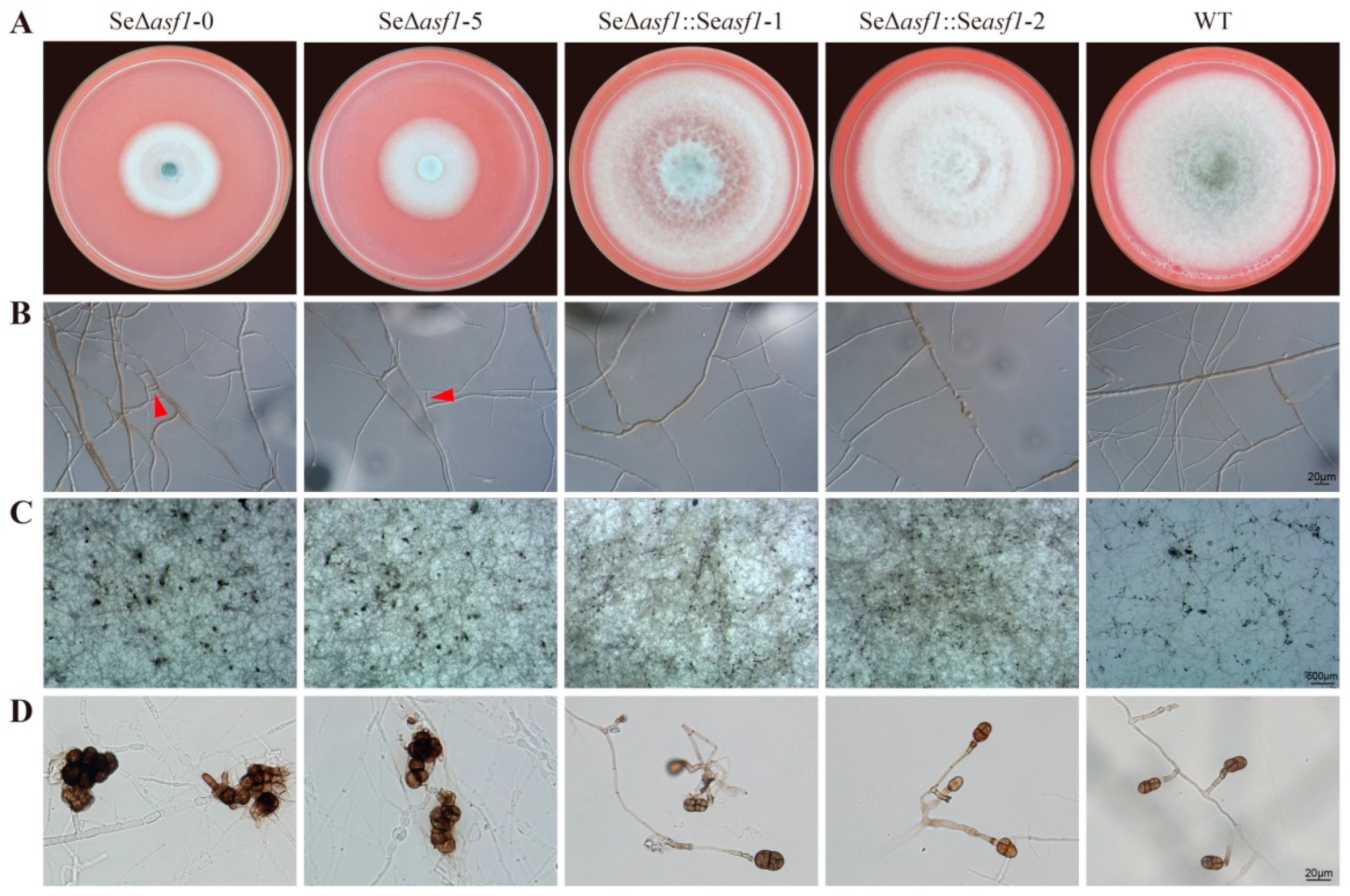
2.1. SeASF1 Regulates Asexual Development in S. eturmiunum

To understand the biological functions of the SeASF1 during asexual development of S. eturmiunum, we obtained two Se∆asf1 mutants and two complemented transformants. To determine the role of Seasf1 in hyphal and colonial growth, these four mutants and WT strains were inoculated on CM medium and then photographed after 9 days. Colony growth rates of the two Se∆asf1 mutants were distinguishable from the WT while two complemented transformants returned to normal growth (Figure 1A). Additionally, two Se∆asf1 mutants produced the hyphal fusion and abnormal conidia compared to the complemented transformants and WT strains (Figure 1B,D). These results suggest that SeASF1 is involved in asexual development of S. eturmiunum.

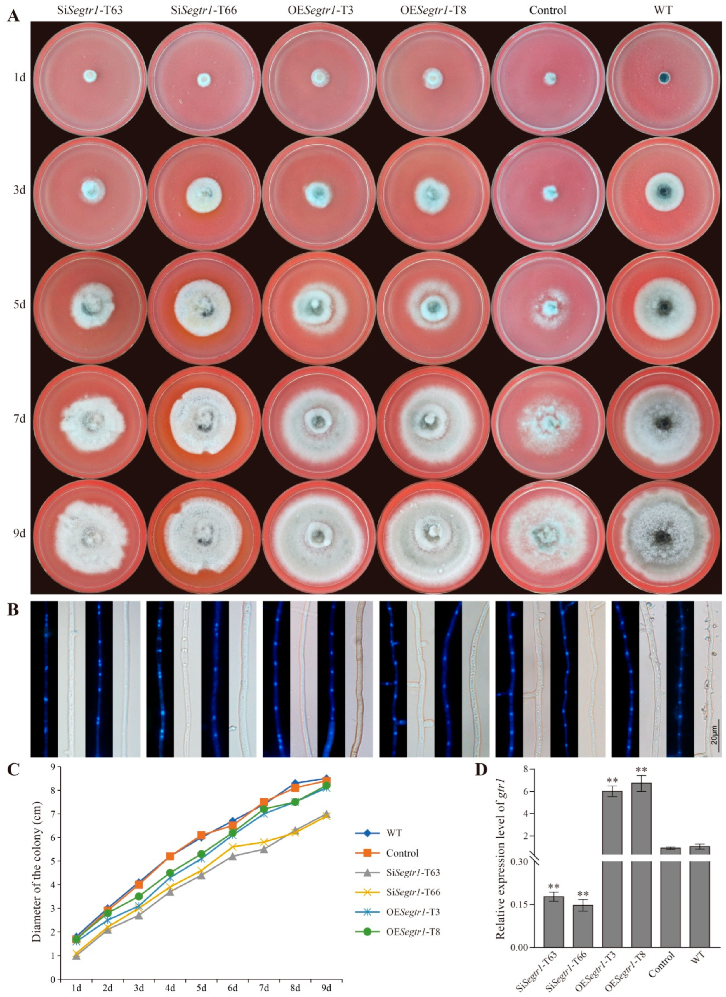
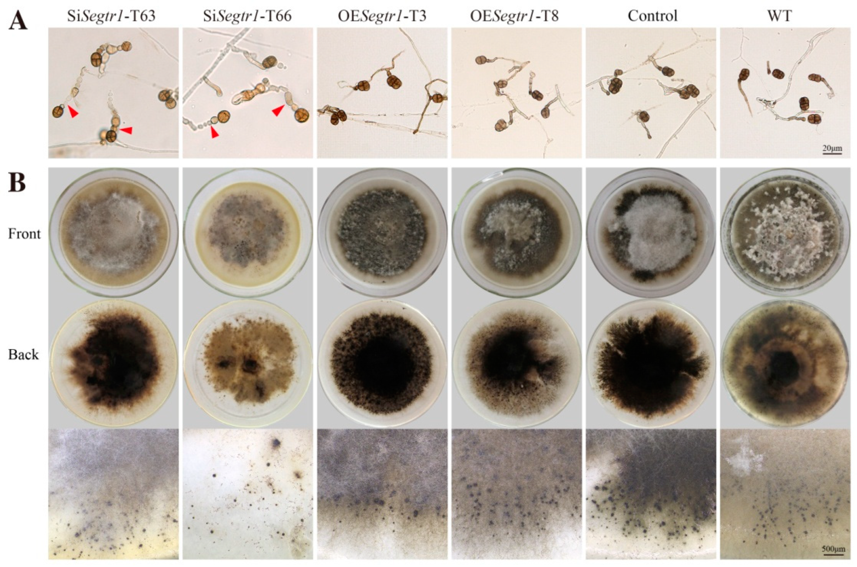
2.2. SeGtr1 Plays a Role in Asexual Development

Gtr1 (SeGtr1) was cloned from S. eturmiunum. SeGtr1 contains 335 amino acids with a calculated molecular mass of 37 kDa. To verify the roles of Segtr1 during the growth and development of S. eturmiunum, we obtained two Segtr1-silenced transformants (SiSegtr1-T63 and SiSegtr1-T65) and two overexpression transformants (OESegtr1-T3 and OESegtr1-T8) by A. tumefaciens mediated method (Figure 2D). Control was a negative control strain. As a result, two silenced transformants appeared the slow growth rate of colonies related to overexpression transformants or control strains (Figure 2A,C). In addition, the nuclei were anomalously distributed in mycelia of SiSegtr1 strains (Figure 2B). To further observe the roles of Segtr1 during the asexual and sexual development of S. eturmiunum, all transformants and WT strains were in the dark condition at 25 °C for 5 weeks on CM medium by inserting double slides. For two silenced strains, the conidiophores turned into bead-like and the conidia grew subglobose, which were significantly different from overexpression transformants and WT strains (Figure 3A). Furthermore, the expression levels of genes involved in the central regulatory pathway, including the brl, aba, and wet, were significantly down-regulated in the two Se∆asf1 mutants and two Segtr1-silenced transformants (Suppplementary Figure S1). However, two silenced strains still produced perithecia that were unanimous with overexpression transformants and WT strains (Figure 3B). These results indicate that SeGtr1 can affect the expression of related genes (brl, aba and wet) in the central regulatory pathway and modulate asexual development.

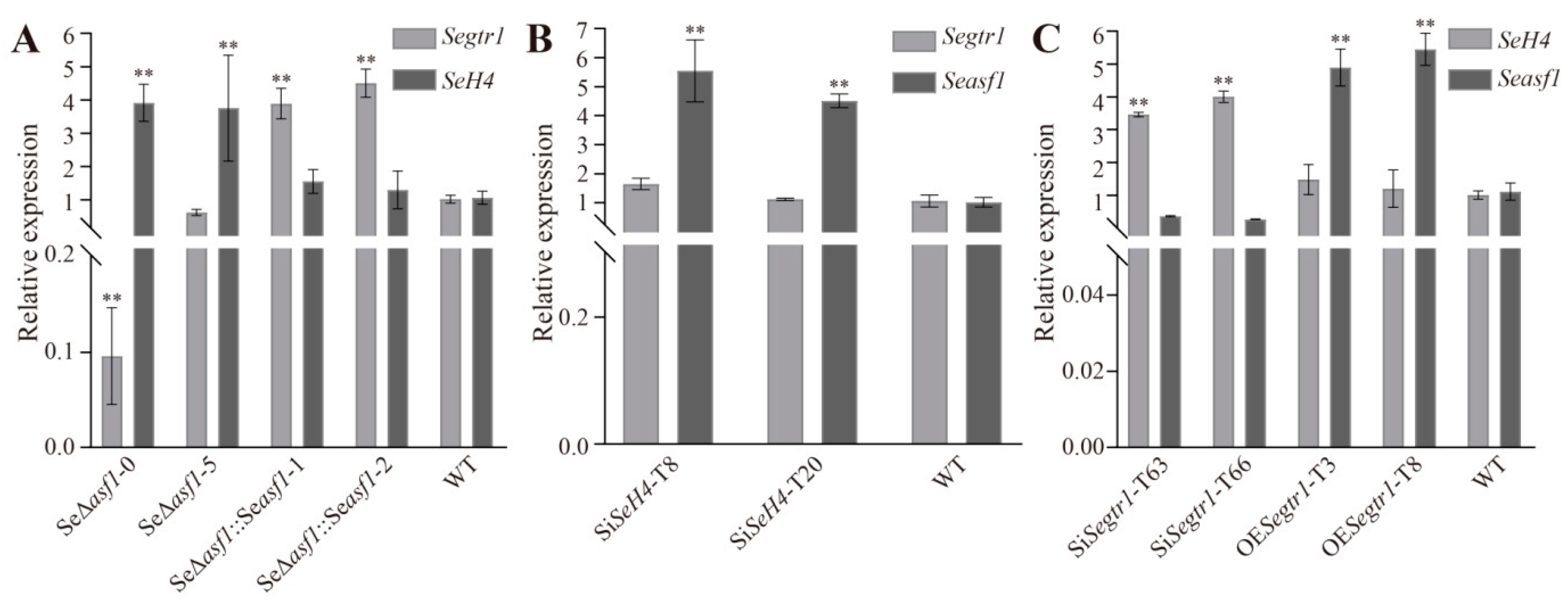
2.3. The Expression Patterns of Seasf1, SeH4, and Segtr1 in Knockout Mutants, Silenced Lines or Overexpression Strains

Due to SeGtr1 can also control the asexual development in S. eturmiunum, we verify whether can occur the relation among SeASF1, SeH4 and SeGtr1. Two SeH4-silenced strains were SiSeH4-T8 and SiSeH4-T20. The transcript levels of Segtr1 and SeH4 were detected in two Se∆asf1 mutants and two complemented transformants. The expressions of Segtr1 and Seasf1 were measured in two SiSeH4 lines, and those of Seasf1 and SeH4 examined in two SiSegtr1 lines and two OESegtr1 strains. As a result, Segtr1 and SeH4 showed down-regulation and up-regulation in two Se∆asf1 mutants, while Segtr1 was up-regulation in Se∆asf1::Seasf1 strains, respectively (Figure 4A). At the same time, Seasf1 displayed up-regulation in two SiSeH4 lines, but Segtr1 did not change (Figure 4B). Furthermore, Seasf1 and SeH4 showed down-regulation and up-regulation in two SiSegtr1 lines, respectively. However, Seasf1 showed up-regulation in two OESegtr1 strains (Figure 4C). Summary, SeGtr1 could interact with SeASF1 or SeH4.

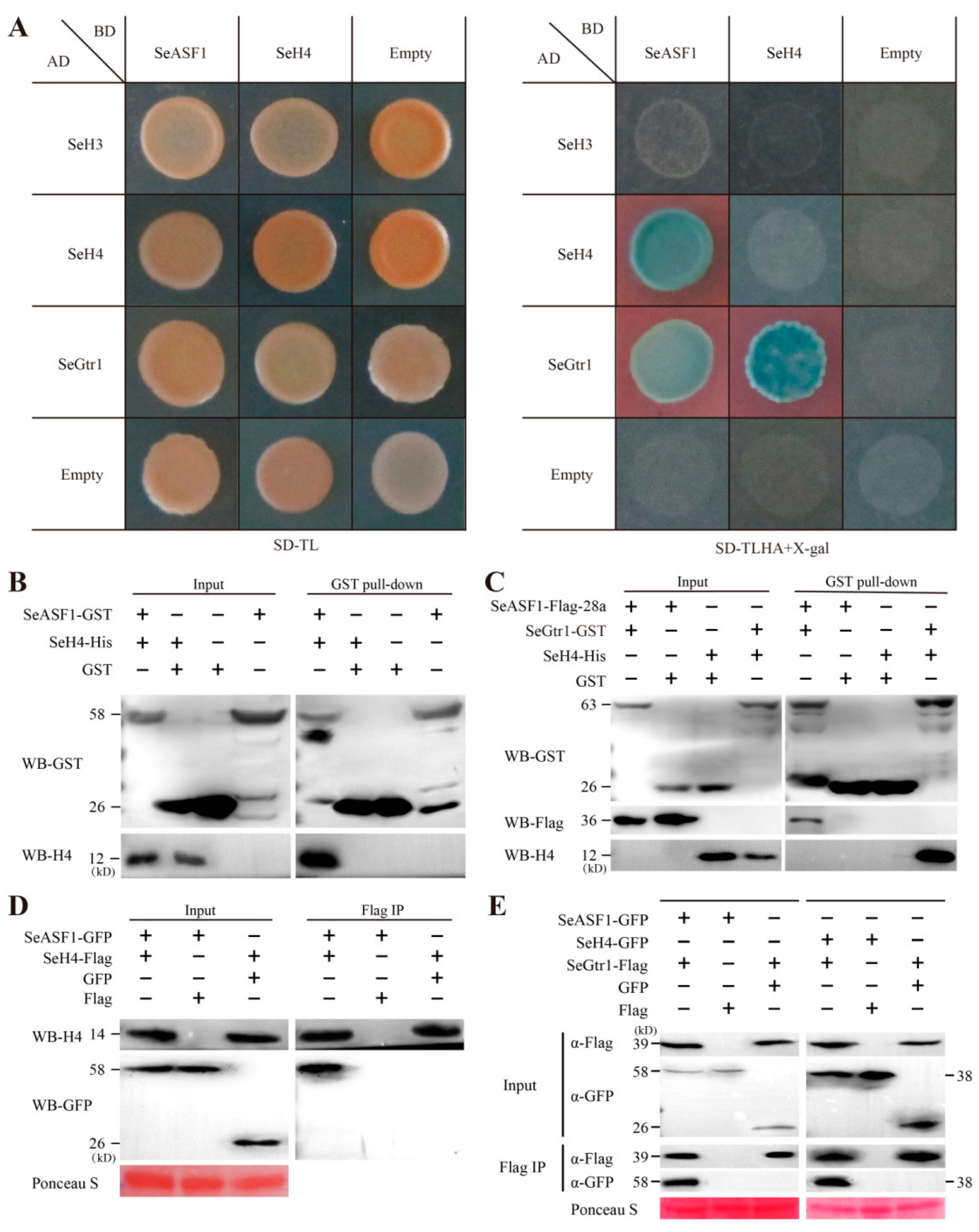
2.4. SeASF1 Interaction with SeH4, and SeGtr1 Interaction with SeASF1 and SeH4
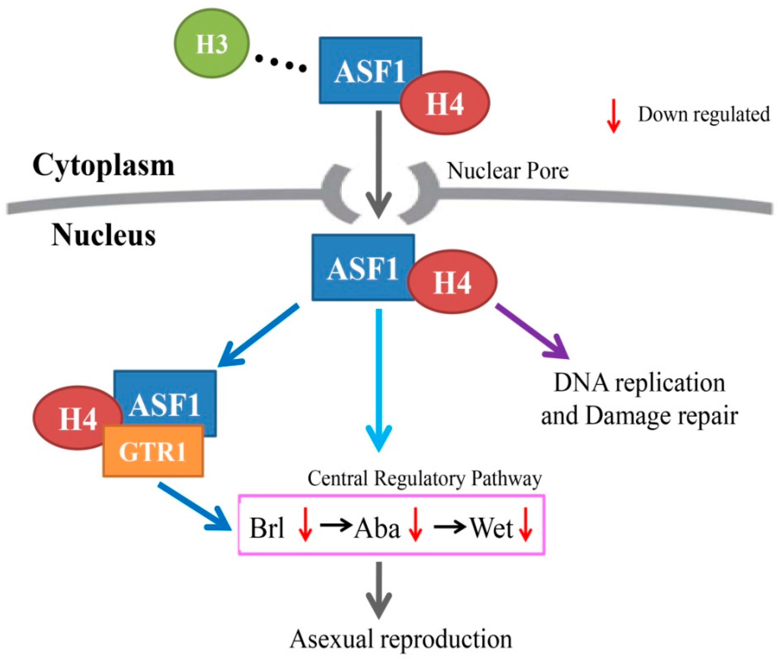
To test whether can occur the interaction between SeASF1 and SeGtr1, SeH4 and SeGtr1. Y2H revealed that SeASF1 interacted with SeH4, and SeGtr1 interacted with SeASF1 and SeH4 (Figure 5A). On the basis of GST pull-down, SeASF1 was specifically interacted with SeH4 (Figure 5B), while SeGtr1 could interact with SeASF1 and SeH4, respectively (Figure 5C). To further assure those results of the Y2H and pull-down experiments, SeASF1-GFP and SeH4-Flag, SeGtr1-Flag and SeASF1-GFP, and SeGtr1-Flag and SeH4-GFP were expressed in F. graminearum protoplasts, respectively, and then the immune complexes were estimated using Co-IP assays (Figure 5D,E). Thus, SeASF1 interacted with SeH4, and SeGtr1 interacted with SeASF1 and SeH4. Altogether, SeGtr1 could cooperate with SeASF1 and SeH4 to modulate asexual development of S. eturmiunum.

3. Discussion

Stemphylium was a dematiaceous hyphomycete that was established with S. botryosum as type species [25]. Until now, there were more than 150 Stemphylium species had been described [26,27,28,29]. S. eturmiunum, a typical species of Stemphylium genus, was an important homothallic filamentous fungus, and it could produce both conidia and perithecia. In a previous study, ASF1 could manipulate the sexual reproduction in Sordaria macrospora effectively [30]. However, in this study, deletion of Seasf1 carried out asexual development characters, such as hyphal fusion and abnormal conidia. A possible explanation for the SeASF1 in asexual development was functional redundancy because some genes were down-regulated or up-regulated expression when Seasf1 was deleted. Therefore, we hypothesized that other proteins might be involved in SeASF1 regulation asexual development.
In summary, through the comparative analysis of transcriptome data (Supplementary Table S1), we characterized that the expression of Segtr1 (No. TR23646-c0_g1) and Seasf1 (No. KX033515) was down-regulated in Se∆asf1 and SiSegtr1 strains, respectively. Meanwhile, we found that SeASF1 or SeGtr1 participated in the central regulatory pathway and regulated the expression of related genes, such as brl, aba and wet. In addition, we verified that SeGtr1 could effectively stimulate asexual activity of S. eturmiunum in Segtr1 silenced strains compared with its overexpression strains. A model of the process is shown in Figure 6. SeASF1 coupled to SeH4 is translocated into the nucleus. In previous studies, ASF1 was reported to regulate DNA replication and damage repair in nucleosome. In this study, we found that SeGtr1 interacted with SeASF1 or SeH4 in vivo and vitro, and SeGtr1 cooperated with SeASF1, which is involved in the central regulatory pathway, to regulate asexual development in S. eturmiunum.

4. Materials and Methods

4.1. Strains and Culture Conditions

Stemphylium eturmiunum strain (EGS 29-099) (WT) and all transformants strains were cultured in the dark condition at 25 °C on complete medium (CM), or potato dextrose agar (PDA) medium for mycelial growth assays. Escherichia coli DH5α or Agrobacterium tumefaciens AGL-1 was incubated in LB (Luria-Bertani) medium at 37 °C or 28 °C, respectively [31].

4.2. Plasmid Construction

Deletion strains for Seasf1 was generated by homologous recombination. The Seasf1 flanking regions, 1500 bp upstream and 1500 bp downstream, were amplified using primer pairs (Table 1). The resulting PCR products were ligated to the Hygromycin cassette and then transformed into WT. Transformants were screened by PCR with primers (Table 1). In addition, Seasf1 was cloned into eGPF-pHDT vector for complementation analysis (Table 2). The recombinant plasmid eGFP-pHDT-Seasf1 was transformed into the Seasf1 mutants by A. tumefaciens mediated transformation (ATMT) method [32]. Transformants were screened by PCR and western blot.
RNA interference [33] was used for Segtr1 silencing. The 525 bp cDNA fragments of Segtr1 was amplified from S. eturmiunum with primers (Table 1) and inserted into vector pCIT that flanked to the intron to form silencing construct, respectively [33] (Table 2). The constructed plasmid pCH-Segtr1 was transformed into S. eturmiunum strain by A. tumefaciens mediated transformation (ATMT) method [32].
For overexpression analysis, Segtr1 was cloned from S. eturmiunum with primers (Table 1), and then cloned into eGPF-pHDT vector. Subsequently, recombinant plasmid eGFP-pHDT-Segtr1 was transformed into the SiSegtr1 lines by ATMT method. Overexpression transformants were screened by qRT-PCR and western blot.
For co-immunoprecipitation (Co-IP) analysis, SeASF1, SeH4, and SeGtr1 were amplified from S. eturmiunum with primers (Table 1), and cloned into the pDL2 or pFL7 in yeast (XK125) by recombination approach [34] (Table 2). Recombinant plasmids were then co-transformed into the protoplasts of Fusarium graminearum wild-type strain (PH-1). Transformants were also screened by western blot.

4.3. RNA Extraction and qRT-PCR

Total RNA was extracted from mycelia of S. eturmiunum growing in PDB (Potato Dextrose Broth) cultures using the Fungal RNA Kit (OMEGA Biotechnology, USA). cDNA was generated using the HiScript II QRT SuperMix for qPCR (Vazyme, Nanjing, China). The qRT-PCR was carried out using the 2 × ChamQ SYBR Color qPCR Master Mix (Vazyme, China) and performed on an ABI QuantStudioTM 6 Quantitative Real-Time PCR System (Applied Biosystems). The specific primers of qRT-PCR listed in the Table 1. Changes in the relative expression level of each gene were calculated by the 2−∆∆CT method [35]. Gene expression levels were normalized using the housekeeping gene actin. This experiment was repeated at least three times.

4.4. Yeast Two-Hybrid

To test whether SeASF1 and SeH4 interact with SeGtr1, Y2H assay was performed according to the Yeast Protocols Handbook (Clontech) using the Y2H Gold yeast reporter strain (Clontech). The Seasf1 or SeH4 was amplified and cloned into pGBKT7 (BD). The Segtr1 or SeH4 was amplified and cloned into pGADT7 (AD) (Table 2). The primers used are listed in Table 1. Pairwise interaction was tested using AD and BD to transform the yeast strain Y2H gold. The yeast transformants were grown on SD/-Trp/-Leu mediums (TaKaRa Bio) for 3–5 days and then cultured on selection mediums (SD/-Trp/-Leu/-His/-Ade/X-α-gal) to detect the protein-protein interaction. Each experiment was repeated at least three times.

4.5. GST Pull-Down

The Seasf1 was cloned into the pET28a vector after adding a 1×FLAG tag to the 5′-terminal of Seasf1 to make the Flag-SeASF1-His fusion protein. The Segtr1 was cloned into the pGEX-6P-1 vector to make the GST-SeGtr1 fusion protein (Table 2). Flag-SeASF1-His, GST-SeGtr1, pET28a or pGEX-6P-1 was expressed in BL21 strain of E. coli, and were then affinity purified with a Ni-affinity column (GE) or GST-affinity column (glutathione sepharoseTM 4B beads GE Healthcare, Little Chalfont, Buckinghamshire, UK). For glutathione S-transferase (GST) pull-down in vitro, GST-SeGtr1 and Flag-SeASF1-28a were expressed in E. coli strain BL21 (DE3). Total proteins of GST-SeGtr1 and Flag-SeASF1-His were then incubated with 4000 μL of glutathione sepharoseTM 4B beads at 4 °C for 2 h. The supernatant was removed, and the beads was washed by GST-lysis buffer three times. Finally, the beads were eluted by GST-elution buffer. Pull-down of GST-SeGtr1 with Flag-SeASF1-His was detected using an anti-Flag (Invitrogen, Waltham, MA, USA). Each experiment was repeated at least three times.

4.6. Co-IP

Fusarium graminearum protoplasts were transfected with the indicated combination plasmids and empty construct. Proteins of F. graminearum were extracted in an extraction buffer (50 mM HEPES, 130 mM NaCl, 10% glycerin, pH 7.4) with 25 mM Glycerol phosphate, 1 mM Sodium orthovanada and protease inhibitor (100 mM PMSF). For FLAG IP, protein extracts were incubated with 30 μL of Anti-Flag® M2 Affinity Gel beads (Sigma-Aldrich, St. Louis, MO, USA) at 4 °C for 4 h. The beads were washed five times with by Co-IP washing buffer (50 mM HEPES, 130 mM NaCl, 10% glycerin, pH 7.4). The bound proteins retained on the beads were separated by 12% SDS–PAGE gels and detected using immunoblotting with anti-FLAG (Sigma-Aldrich) or anti-GFP antibody (Invitrogen). Each experiment was repeated at least three times.

4.7. Western Blot

Proteins were separated by 12% sodium dodecyl sulfate polyacrylamide gel electrophoresis (SDS-PAGE), and transferred to Immobilon®-P PVDF membrane for 1.5 h at 230 mA. The PVDF membranes were blocked with TBST (0.02 M Tris-base, 0.14 M NaCl, 0.1% Tween-20, pH 7.4) with 5% non-fat milk for 1 h at room temperature. Co-immunoprecipitated proteins were analyzed by incubating the membranes with 1:5000 diluted GFP or FLAG antibodies (Sigma-Aldrich) at room temperature for 1–1.5 h. The membranes were washed three times with TBST and then were incubated for 1 h with Goat anti-Mouse-HRP secondary antibody (Thermo Fisher Scientific, Waltham, MA, USA, no. 31430) at 1:7500 dilution. The specific proteins were visualized by the ECL Chemiluminescence Detection Kit (Vazyme) and imaged using a Tanon-5200 System. Each experiment was repeated at least three times.

4.8. Microscopy

To observe the morphology of conidia and conidiophores, all the transformants and WT strains were grown in the dark condition at 25 °C for 4 weeks on PDA medium by inserting double slides. Microscopic examination of nuclear distribution in mycelia, the transformants and WT strains were stained using 4,6-diamidino-2-phenylindole (DAPI). To image the sexual structures including perithecia and asci, all these test strains were cultured on CM medium at 25 °C for 6 weeks in dark condition. Perithecia were sectioned by using a double-edged blade in a dissecting microscope (Olympus, SZX10). The asci, conidia and conidiophores were all captured with 20× or 40× objectives of Olympus microscope (Olympus BX53, Tokyo, Japan) using differential interference contrast (DIC) and fluorescence illumination. Microscopic characters of asexual structures were further determined by measurements of 50 mature conidia and 50 conidiophores. The experiment was repeated at least three times.

4.9. Statistical Analysis

All determinations were carried out in triplicate and the results are expressed as mean ± standard deviation (SD). The data were subjected to one-way analysis of variance (ANOVA). Statistically significant differences were determined by two-tailed Student’s t test: * p < 0.05, ** p < 0.01.

Supplementary Materials

The following supporting information can be downloaded at: https://www.mdpi.com/article/10.3390/ijms23158355/s1.

Author Contributions

Conceptualization, S.W. and X.Z.; methodology, S.W. and X.Z.; validation, Z.L. and X.Z.; formal analysis, C.S., L.Z. and W.X.; investigation, L.Z. and X.L.; resources, S.W. and X.Z.; data curation, X.Z.; writing—original draft preparation, S.W. and X.Z.; writing—review and editing, X.L.; visualization, X.L.; supervision, Z.L., X.L. and X.Z.; project administration, X.Z.; funding acquisition, Z.L., X.L. and X.Z. All authors have read and agreed to the published version of the manuscript.

Funding

This research was supported by grants from the National Natural Science Foundation of China (31230001, U200220015).

Institutional Review Board Statement

Not applicable.

Informed Consent Statement

Not applicable.

Data Availability Statement

The data supporting our study results are included in the article.

Acknowledgments

We thank Jingze Zhang (Zhejiang University) for transcriptome analysis and Daohong Jiang (Huazhong Agricultural University) for providing the plasmid.

Conflicts of Interest

There are no conflict of interest for all authors and organizations that mentioned in this manuscript.

References

  1. Wu, M.Y.; Mead, M.E.; Lee, M.K.; Neuhaus, G.F.; Adpressa, D.A.; Martien, J.I.; Son, Y.E.; Moon, H.; Amador-Noguez, D.; Han, K.H.; et al. Transcriptomic, protein-DNA interaction, and metabolomic studies of VosA, VelB, and WetA in Aspergillus nidulans asexual spores. Mol. Biol. 2021, 12, e03128-20. [Google Scholar] [CrossRef] [PubMed]
  2. Dyer, P.S.; Ingram, D.S.; Johnstone, K. The control of sexual morphogenesis in the Ascomycotina. Biol. Rev. 1992, 67, 421–458. [Google Scholar] [CrossRef]
  3. Milgroom, M.G. Recombination and the multilocus structure of fungal populations. Annu. Rev. Phytopathol. 1996, 34, 457–477. [Google Scholar] [CrossRef]
  4. Grigsby, I.F.; Rutledge, E.M.; Morton, C.A.; Finger, F.P. Functional redundancy of two C. elegans homologs of the histone chaperone Asf1 in germline DNA replication. Dev. Biol. 2009, 329, 64–79. [Google Scholar] [CrossRef] [Green Version]
  5. Taylor, J.; Jacobson, D.; Fisher, M. The volution of asexual fungi: Reproduction, speciation and classification. Annu. Rev. Phytopathol. 1999, 37, 197–246. [Google Scholar] [CrossRef] [PubMed] [Green Version]
  6. Simmons, E.G. Typification of Alternaria, Stemphylium, and Ulocladium. Mycologia 1967, 59, 67–92. [Google Scholar] [CrossRef] [PubMed]
  7. Geng, Y.; Li, Z.; Xia, L.Y.; Wang, Q.; Hu, X.M.; Zhang, X.G. Characterization and phylogenetic analysis of the mating-type loci in the asexual ascomycete genus Ulocladium. Mycologia 2014, 106, 649–665. [Google Scholar] [CrossRef]
  8. Wang, Q.; Wang, S.; Xiong, C.L.; James, T.Y.; Zhang, X.G. Mating-type genes of the anamorphic fungus Ulocladium botrytis affect both asexual sporulation and sexual reproduction. Sci. Rep. 2017, 7, 7932. [Google Scholar] [CrossRef] [PubMed] [Green Version]
  9. Simmons, E.G. Perfect states of Stemphylium. Mycologia 1969, 61, 1–26. [Google Scholar] [CrossRef]
  10. Bayram, O.; Krappmann, S.; Ni, M.; Bok, J.W.; Helmstaedt, K.; Valerius, O.; Braus-Stromeyer, S.; Kwon, N.J.; Keller, N.P.; Yu, J.H.; et al. VelB/VeA/LaeA complex coordinates light signal with fungal development and secondary metabolism. Science 2008, 320, 1504–1506. [Google Scholar] [CrossRef] [PubMed]
  11. Sarikaya Bayram, O.; Bayram, O.; Valerius, O.; Park, H.S.; Irniger, S.; Gerke, J.; Ni, M.; Han, K.H.; Yu, J.H.; Braus, G.H. LaeA control of velvet family regulatory proteins for light-dependent development and fungal cell-type specificity. PLoS Genet. 2010, 6, e1001226. [Google Scholar] [CrossRef] [PubMed] [Green Version]
  12. Blumenstein, A.; Vienken, K.; Tasler, R.; Purschwitz, J.; Veith, D.; Frankenberg-Dinkel, N.; Fischer, R. The Aspergillus nidulans phytochrome FphA represses sexual development in red light. Curr. Biol. 2005, 15, 1833–1838. [Google Scholar] [CrossRef] [PubMed]
  13. Schürmann, A.; Brauers, A.; Massmann, S.; Becker, W.; Joost, H.G. Cloning of a novel family of mammalian GTP-binding proteins (RagA, RagBs, RagB1) with remote similarity to the Ras-related GTPases. J. Biol. Chem. 1995, 270, 28982–28988. [Google Scholar] [CrossRef] [PubMed] [Green Version]
  14. Masanori, B.Y.; Harashima, S.; Oshima, Y. Putative GTP-Binding protein, Gtr1, associated with the function of the Pho84 inorganic-phosphate transporter in Saccharomyces cerevisiae. Mol. Cell. Biol. 1992, 12, 2958–2966. [Google Scholar]
  15. Sekiguchi, T.; Hayashi, N.; Wang, Y.; Kobayashi, H. Genetic evidence that Raslike GTPases, Gtr1p, and Gtr2p, are involved in epigenetic control of gene expression in Saccharomyces cerevisiae. Biochem. Biophys. Res. Commun. 2008, 368, 748–754. [Google Scholar] [CrossRef]
  16. Saito, H.; Oikawa, T.; Hamamoto, S.; Ishimaru, Y.; Kanamori-Sato, M.; Sasaki-Sekimoto, Y.; Utsumi, T.; Chen, J.; Kanno, Y.; Masuda, S.; et al. The jasmonate-responsive GTR1 transporter is required for gibberellin-mediated stamen development in Arabidopsis. Nat. Commun. 2015, 6, 6095. [Google Scholar] [CrossRef] [Green Version]
  17. Li, Y.; Wang, C.; Liu, W.; Wang, G.; Kang, Z.; Kistler, H.C.; Xu, J.R. The HDF1 histone deacetylase gene is important for conidiation, sexual reproduction, and pathogenesis in Fusarium graminearum. Mol. Plant Microbe Interact. 2011, 24, 487–496. [Google Scholar] [CrossRef] [PubMed] [Green Version]
  18. Ramamoorthy, V.; Shantappa, S.; Dhingra, S.; Calvo, A.M. VeA-dependent RNApol II transcription elongation factor-like protein, RtfA, is associated with secondary metabolism and morphological development in Aspergillus nidulans. Mol. Microbiol. 2012, 85, 795–814. [Google Scholar] [CrossRef] [Green Version]
  19. Sarikaya Bayram, O.; Bayram, O.; Feussner, K.; Kim, J.H.; Kim, H.S.; Kaever, A.; Feussner, I.; Chae, K.S.; Han, D.M.; Han, K.H.; et al. Membrane-bound methyltransferase complex VapA-VipC-VapB guides epigenetic control of fungal development. Dev. Cell 2014, 29, 406–420. [Google Scholar] [CrossRef] [PubMed] [Green Version]
  20. Le, S.; Davis, C.; Konopka, J.B.; Sternglanz, R. Two new S-phase-specific genes from Saccharomyces cerevisiae. Yeast 1997, 13, 1029–1042. [Google Scholar] [CrossRef]
  21. Eitoku, M.; Sato, L.; Senda, T.; Horikoshi, M. Histone chaperones: 30 years from isolation to elucidation of the mechanisms of nucleosome assembly and disassembly. Cell. Mol. Life Sci. 2008, 65, 414–444. [Google Scholar] [CrossRef]
  22. Min, Y.; Frost, J.M.; Choi, Y. Gametophytic abortion in heterozygotes but not in homozygotes: Implied chromosome rearrangement during T-DNA insertion at the ASF1 locus in Arabidopsis. Mol. Cells 2020, 43, 448–458. [Google Scholar]
  23. Sanematsu, F.; Takami, Y.; Barman, H.K.; Fukagawa, T.; Ono, T.; Shibahara, K.I.; Nakayama, T. Asf1 is required for viability and chromatin assembly during DNA replication in vertebrate cells. J. Biol. Chem. 2006, 281, 13817–13827. [Google Scholar] [CrossRef] [Green Version]
  24. Mousson, F.; Ochsenbein, F.; Mann, C. The histone chaperone Asf1 at the crossroads of chromatin and DNA checkpoint pathways. Chromosoma 2007, 116, 79–93. [Google Scholar] [CrossRef] [PubMed]
  25. Wallroth, F.G. Flora Cryptogamica Germaniae, pars. Post. Nurenberg; J. L. Schrag: Madison, WI, USA, 1833; 923p. [Google Scholar]
  26. Câmara, M.P.; O’Neill, N.R.; van Berkum, P. Phylogeny of Stemphylium spp. based on ITS and glyceraldehyde-3-phosphate dehydrogenase gene sequences. Mycologia 2002, 94, 660–672. [Google Scholar] [CrossRef]
  27. Wang, Y.; Zhang, X.G. Three new species of Stemphylium from China. Mycotaxon 2006, 96, 77–81. [Google Scholar]
  28. Wang, Y.; Geng, Y.; Pei, Y.F.; Zhang, X.G. Molecular and morphological description of two new species of Stemphylium from China and France. Mycologia 2010, 102, 708–717. [Google Scholar] [CrossRef] [PubMed]
  29. Wang, Y.; Fu, H.B.; O’Neill, N.R.; Zhang, X.G. Two new species of Stemphylium from Northwest China. Mycol. Prog. 2009, 8, 301–304. [Google Scholar] [CrossRef]
  30. Gesing, S.; Schindler, D.; Fränzel, B.; Wolters, D.; Nowrousian, M. The histone chaperone ASF1 is essential for sexual development in the filamentous fungus Sordaria macrospora. Mol. Microbiol. 2012, 84, 748–765. [Google Scholar]
  31. Lennox, E.S. Transduction of linked genetic characters of the host by bacteriophage P1. Virology 1995, 1, 190–206. [Google Scholar] [CrossRef]
  32. Bernardi Wenzel, J.; Quecine, M.C.; Azevedo, J.L.; Pamphile, J.A. Agrobacterium-mediated transformation of Fusarium proliferatum. Genet. Mol. Res. 2016, 15, 1–12. [Google Scholar] [CrossRef]
  33. Zhao, Y.; He, M.; Ding, J.; Xi, Q.; Loake, G.J.; Zheng, W. Regulation of anticancer styrylpyrone biosynthesis in the medicinal mushroom Inonotus obliquus requires thioredoxin mediated transnitrosylation of S-nitrosoglutathione reductase. Sci. Rep. 2016, 6, 37601. [Google Scholar] [CrossRef] [PubMed]
  34. Zhou, X.; Li, G.; Xu, J.R. Efficient approaches for generating GFP fusion and epitope-tagging constructs in filamentous fungi. Methods Mol. Biol. 2011, 722, 199–212. [Google Scholar]
  35. Livak, K.J.; Schmittgen, T.D. Analysis of relative gene expression data using real-time quantitative PCR and the 2−∆∆CT method. Methods 2001, 25, 402–408. [Google Scholar] [CrossRef] [PubMed]
Figure 1. Seasf1 regulates asexual developmental characterization in S. eturmiunum. (A) Growth of two Se∆asf1 mutants, two Se∆asf1::Seasf1 transformants, and WT strains on CM medium. The cultures were photographed after 9 days of incubation. (B) Characterizations of hyphal fusion in two Se∆asf1 mutants, two Se∆asf1::Seasf1 transformants, and WT strains. The images were photographed after growing on PDA medium for 8 days. The fusions in the hyphae were marked with red arrows. (C,D) Conidia morphology of four mutants and WT strains were cultured on CM medium for 4 weeks. Bar = 20 μm, 500 μm.
Ijms 23 08355 g001
Figure 2. The colonial phenotypes and nuclear distribution of Segtr1 silenced transformants. (A) Colonial growth of two Segtr1 silenced transformants (SiSegtr1-T63 and SiSegtr1-T66) and two Segtr1 overexpression transformants (OESegtr1-T3 and OESegtr1-T8) was observed on PDA medium. WT was S. eturmiunum strain and Control was a negative control strain. The cultures were photographed after 1 day, 3 days, 5 days, 7 days and 9 days. (B) The mycelium of these four transformants were grown on PDA medium for 6 days and examined by DIC and fluorescence microscopy. The nuclei of the mycelia were discovered under the fluorescence microscopy after staining by DAPI. Bar = 20 μm. (C) Colony diameters were measured in each independent biological experiment at 1–9 days of growth on PDA medium. Rates of colonial growth were calculated for each treatment. (D) qRT-PCR was used to measure the expression levels of Segtr1 in silenced transformants, overexpression transformants, Control and WT. The degree of WT was assigned to value 1.0. Actin gene of S. eturmiunum was used as endogenous control. The bars indicated statistically significant differences (ANOVA; ** p < 0.01). Each experiment was repeated at least three times.
Ijms 23 08355 g002
Figure 3. SeGtr1 plays a role in asexual development, but the sexual activity was unchanged. (A) For the microscopic investigation of conidiophores and conidia development, two silenced transformants (SiSegtr1-T63 and SiSegtr1-T66), two overexpression transformants (OESegtr1-T3 and OESegtr1-T8), Control and WT strains were grown on CM medium for 35 days, respectively. Control was a negative control strain. Red arrowheads indicated abnormal conidiophores. (B) To further observe the role of Segtr1 during the sexual development of S. eturmiunum, all transformants and WT strains were cultured on PDA medium for inducing perithecia production. At 35 days, all transformant strains produced abundant perithecia. Bar = 20 μm and 500 μm.
Ijms 23 08355 g003
Figure 4. The expression patterns of Seasf1, SeH4, and Segtr1 in knockout mutants, silenced lines or overexpression strains. (A) The expression levels of Segtr1 and SeH4 in two Se∆asf1 mutants and two complemented transformants were measured by qRT-PCR. (B) The expression levels of Segtr1 and Seasf1 in two SiSeH4 lines were measured by qRT-PCR. (C) The expression levels of Seasf1 and SeH4 in two SiSegtr1 lines and two OESegtr1 lines were measured by qRT-PCR. The degree of WT was assigned to value 1.0. Two Seasf1 deleted mutants were Se∆asf1-0 and Se∆asf1-5, two complemented transformants were Se∆asf1::Seasf1-1 and Se∆asf1::Seasf1-2. Two SeH4-silenced lines were SiSeH4-T8 and SiSeH4-T20. Two Segtr1-silenced lines were SiSegtr1-T63 and SiSegtr1-T66, two OESegtr1 lines were OESegtr1-T3 and OESegtr1-T8. The Actin in S. eturmiunum was used as endogenous control. The bars indicated statistically significant differences (ANOVA; ** p < 0.01).
Ijms 23 08355 g004
Figure 5. SeASF1 interaction with SeH4, and SeGtr1 interaction with SeASF1 or SeH4. (A) SeASF1 interacted with SeH4, and SeGtr1 interaction with SeASF1 or SeH4 using Y2H. SeASF1 or SeH4 was cloned into plasmid pGBKT7 (BD). SeH4 or SeGtr1 was cloned into plasmid pGADT7 (AD). Yeast transformants were first grown on SD/-Trp/-Leu, and selected on SD/-Trp/-Leu/-His/-Ade/X-α-gal. A positive interaction results in the activation of the lacZ reporter, which turned the blue in the presence of X-α-galactosidase. The images were photographed at 3 days after incubation. (B) SeASF1 was cloned into plasmid pGEX-6P-1. SeH4 was cloned into plasmid pET28a. SeASF1-GST was expressed in E. coli and incubated with SeH4-His, purified (pull-down) by glutathione sepharose beads. Recombinant GST was control. SeH4-His was pulled down by SeASF1-GST. (C) SeGtr1 was cloned into plasmid pGEX-6P-1. Flag-SeASF1 or SeH4 was cloned into plasmid pET28a. SeH4-His and Flag-SeASF1-His were both retained by SeGtr1-GST. (D) SeASF1 was cloned into plasmid pDL2, SeH4 was cloned into plasmid pFL7. Total proteins were extracted from F. graminearum protoplasts expressing SeASF1-GFP and SeH4-Flag. Recombinant GFP or Flag was control. The immune complexes were immunoprecipitated with α-Flag antibody (α-Flag IP). Coprecipitation of SeH4-Flag was detected by immunoblotting. (E) SeH4 was cloned into plasmid pDL2. SeGtr1 was cloned into plasmid pFL7. Total proteins were extracted from F. graminearum protoplasts expressing SeASF1-GFP, SeH4-GFP, and SeGtr1-Flag. Coprecipitation of SeGtr1-Flag was detected by immunoblotting. Membranes were stained with Ponceau S to confirm equal loading. Protein sizes are indicated in kDa. Each experiment was repeated at least three times.
Ijms 23 08355 g005
Figure 6. A model for ASF1 binding H4 (ASF1-H4) to interact with GTR1 and then to mediate asexual reproduction in Stemphylium eturmiunum. ASF1 interacts with H4 and then they are translocated into nuclei through the nuclear pore. The dimer of ASF1-H4 regulates DNA replication and damage repair in previous studies. Herein, ASF1-H4 combines with GTR1 to constitute a trimeric complex which in turn is involved in the central regulatory pathway to modulate asexual reproduction.
Ijms 23 08355 g006
Table 1. Primers used in this study.
PrimerSequence(5′-3′)Application
Seasf1-FATGTCTGTCGTTTCGCTTCAmplify Seasf1 sequence
Seasf1-RCTAGTGAACCATGACATCTGC
Seasf1-5fCCGCTCGAGCAATCCAGGGGCGATAAAGAmplify Seasf1 upstream sequence
Seasf1-5rGGAAGATCTGCTTGGCGGGGTAGATAGAG
Seasf1-3fCGCGGATCCGCCGCCGTCTGTTAGTCTTAmplify Seasf1 downstream sequence
Seasf1-3rCTAGTCTAGACAGAGGAGATGCTTGCTTGTC
Seasf1-fTCTGTCGTTTCGCTTCTCGFor identification of Seasf1 deletion strains
Seasf1-rTGAACCATGACATCTGCGC
Seasf1-3DCCTCGTCGTCTTCCTGATCACTFor identification
of Hph in deletion transformants
Hph-3DGAGATTCTTCGCCCTCCGAG
Hph-5DAATTTCGATGATGCAGCTTGGG
Seasf1-5DCAAGACTGCTTCCTTCTCATCGT
Hph-FCGACAGCGTCTCCGACCTGAFor identification of Hph
For identification of
Hph-RCGCCCAAGCTGCATCATCGAA
Seasf1-PHDT-FCCGCTCGAGATGTCTGTCGTTTCGCTTCTCGSeasf1complementation
expression
Seasf1-PHDT-RGCTCTAGACTAGTGAACCATGACATCTGCGC
PHDT-FGATCACATGGTCCTGCTGVector construction of sequencing primer
PHDT-R CACCAACGATCTTATATCCAG
Segtr1-pCIT-F(BamHI/ClaI)CGCGGATCCATCGATGTAGTGCACCTGTTGA
ATGAGAGC
Primer for
Segtr1 silence
Segtr1-pCIT-R(PstI/EcoRV)AACTGCAG GATATCGCCCAGCC TACGTAC
ATCCTT
Seasf1-QRT-FACAACGAGTACACAGATGAGGQRT-PCR for
Seasf1
Seasf1-QRT-RTCGTCAGAGTCCCATTTGATAG
SeH4-QRT-FAGCCATGATCTACGAGGAAACQRT-PCR for
SeH4
SeH4-QRT-RCGAGAGAAGTGACAGTCTTACG
Segtr1-QRT-FAAGCTAGCGAGGGTTTCAAGQRT-PCR for
Segtr1
Segtr1-QRT-RGGCGTTTGGTATCAGGTAGTAG
Seactin-FGTCGATTGGAGAAGGAGCTAAAQRT-PCR for
Seactin
Seactin-RGTTCTCCTTGTCGGCCATAAT
Seasf1-BD-F(NdeI) GGAATTCCATATGATGTCTGTCGTTTCGCTTCTCGSeasf1 recombined into pGBKT7
Seasf1-BD-R(BamHI)CGCGGATCCCTAGTGAACCATGACATCTGCG
SeH4-BD-F(SmaI)TCCCCCGGGATGACTGGTCGCGGTAAAGGTSeH4 recombined into pGBKT7
SeH4-BD-R(BamHI)CGCGGATCCCTAACCACCGAAACCGTAAAGGG
SeH4-AD-F(SmaI)TCCCCCGGGATGACTGGTCGCGGTAAAGGTSeH4 recombined into
pGADT7
SeH4-AD-R(BamHI)CGGGATCCCTAACCACCGAAACCGTAAAGGG
SeH3-AD-F(SmaI)TCCCCCGGGATGCCGCCAAAATCCCCTACCAGSeH3 recombined into
pGADT7
SeH3-AD-R(BamHI)CGCGGATCCTCAGACAGGCGCCCCCCAAG
Segtr1-AD-F(SmaI)TCCCCCGGGATGAATTCAGTCAAGCGTCAGASegtr1 recombined into
pGADT7
Segtr1-AD-R(BamHI)CGCGGATCCCTACATTCCAGAGCCATGC
Seasf1-pET28a-Flag-F(NdeI)GGAATTCCATATGGATTACAAGGACGACG
ATGACAAGATGTCTGTCGTTTCGCTTCTC
Seasf1 recombined
into
pET28a
Seasf1-pET28a-Flag-R(HindIII)CCCAAGCTTCTAGTGAACCATGACATCTGC
Seasf1-pGEX-F(BamHI)CGCGGATCCATGTCTGTCGTTTCGCTTCTCG Seasf1 recombined
into
pGEX
Seasf1-pGEX-R(NotI)ATAAGAATGCGGCCGCCTAGTGAACCATG
ACATCTGCG
Segtr1-pGEX-F(NdeI)GGAATTCCATATGATGAATTCAGTCAAGCG
TCAGA
Segtr1 recombined
into
pGEX
Segtr1-pGEX-R(NotI)ATAAGAATGCGGCCGCCTACATTCCAGAG
CCATGC
Seasf1-pDL2-GFP-FCAGATCTTGGCTTTCGTAGGAACCCAATCTTCA
ATGTCTGTCGTTTCGCTTCTCG
Seasf1 recombined
into
pDL2
Seasf1-pDL2-GFP-RCACCACCCCGGTGAACAGCTCCTCGCCCTTGCT
CACGTGAACCATGACATCTGCGC
SeH4-pDL2-GFP-FCAGATCTTGGCTTTCGTAGGAACCCAATCTTCA
ATGACTGGTCGCGGTAAAGGT
SeH4 recombined
into
pDL2
SeH4-pDL2-GFP-RCACCACCCCGGTGAACAGCTCCTCGCCCTTGCT
CACACCACCGAAACCGTAAAGGG
SeH4-pFL7-Flag-FCAGATCTTGGCTTTCGTAGGAACCCAATCTTCA
ATGACTGGTCGCGGTAAAGGT
SeH4 recombined
into
pFL7
SeH4-pFL7-Flag-RCTTTATAATCACCGTCATGGTCTTTGTAGT
CACCACCGAAACCGTAAAGGG
Segtr1-pFL7-Flag-FCAGATCTTGGCTTTCGTAGGAACCCAATCTTCA
ATGAATTCAGTCAAGCGTCAGA
Segtr1 recombined
into
pFL7
Segtr1-pFL7-Flag-RCTTTATAATCACCGTCATGGTCTTTGTAGT
CATTCCAGAGCCATGC
Segtr1-PHDT-F(SnaBI)CCTACGTA ATGAATTCAGTCAAGCGTCAGAPrimer for
Segtr1 overexpression
Segtr1-PHDT-R(XbaI)GCTCTAGACATTCCAGAGCCATGC
Table 2. Plasmids used in this study.
NameOrigin of Target GenesConstruction PathPurpose
Seasf1-pXEHS. eturmiunumSeasf1 recombined into pXEHKnockout asf1
Seasf1-pHDTS. eturmiunumSeasf1 recombined into pHDTasf1complementation expression
Segtr1-pCITS. eturmiunumSegtr1 recombined into pCITSegtr1 silence
Seasf1-pGBKT7S. eturmiunumSeasf1 recombined into
pGBKT7
Yeast two-hybrid assays
SeH4-pGBKT7S. eturmiunumSeH4 recombined into
pGBKT7
Yeast two-hybrid assays
Segtr1-pGADT7S. eturmiunumSegtr1 recombined into
pGADT7
Yeast two-hybrid assays
SeH4-pGADT7S. eturmiunumSeH4 recombined into
pGADT7
Yeast two-hybrid assays
SeH3-pGADT7S. eturmiunumSeH3 recombined into
pGADT7
Yeast two-hybrid assays
Seasf1-pET28aS. eturmiunumSeasf1 recombined into
pET28a
Pull-down assays
Segtr1-pGEX-6P-1S. eturmiunumSegtr1 recombined into
pGEX-6P-1
Pull-down assays
Seasf1-pDL2S. eturmiunumSeasf1 recombined into pDL2CO-IP assays
SeH4-pDL2S. eturmiunumSeH4 recombined into pDL2CO-IP assays
Segtr1-pFL7S. eturmiunumSegtr1 recombined into pFL7CO-IP assays
SeH4-pFL7S. eturmiunumSeH4 recombined into pFL7CO-IP assays
Segtr1-pHDTS. eturmiunumSegtr1 recombined into pHDTSegtr1 over expression
Publisher’s Note: MDPI stays neutral with regard to jurisdictional claims in published maps and institutional affiliations.

