Review

New Frontiers in Neurodegeneration and Regeneration Associated with Brain-Derived Neurotrophic Factor and the rs6265 Single Nucleotide Polymorphism

by
Carlye A. Szarowicz
1,2,
Kathy Steece-Collier
1 and
Margaret E. Caulfield
1,*
1
Department of Translational Neuroscience, College of Human Medicine, Michigan State University, Grand Rapids, MI 49503, USA
2
Department of Pharmacology and Toxicology, Michigan State University, East Lansing, MI 48824, USA
*
Author to whom correspondence should be addressed.
Int. J. Mol. Sci. 2022, 23(14), 8011; https://doi.org/10.3390/ijms23148011
Submission received: 8 June 2022 / Revised: 11 July 2022 / Accepted: 12 July 2022 / Published: 20 July 2022
(This article belongs to the Special Issue Neurotrophins: Roles and Function in Human Diseases)

Abstract

:
Brain-derived neurotrophic factor is an extensively studied neurotrophin implicated in the pathology of multiple neurodegenerative and psychiatric disorders including, but not limited to, Parkinson’s disease, Alzheimer’s disease, Huntington’s disease, traumatic brain injury, major de-pressive disorder, and schizophrenia. Here we provide a brief summary of current knowledge on the role of BDNF and the common human single nucleotide polymorphism, rs6265, in driving the pathogenesis and rehabilitation in these disorders, as well as the status of BDNF-targeted therapies. A common trend has emerged correlating low BDNF levels, either detected within the central nervous system or peripherally, to disease states, suggesting that BDNF replacement therapies may hold clinical promise. In addition, we introduce evidence for a distinct role of the BDNF pro-peptide as a biologically active ligand and the need for continuing studies on its neurological function outside of that as a molecular chaperone. Finally, we highlight the latest research describing the role of rs6265 expression in mechanisms of neurodegeneration as well as paradoxical advances in the understanding of this genetic variant in neuroregeneration. All of this is discussed in the context of personalized medicine, acknowledging there is no “one size fits all” therapy for neurodegenerative or psychiatric disorders and that continued study of the multiple BDNF isoforms and genetic variants represents an avenue for discovery ripe with therapeutic potential.

1. Introduction

Brain-derived neurotrophic factor (BDNF) is a neurotrophin that functions to regulate and promote neuronal survival, differentiation, and outgrowth of central and peripheral neurons. Other members of the mammalian neurotrophin family include nerve growth factor (NGF), neurotrophin 3 (NT-3), and neurotrophin 4/5 (NT-4/5), and they share more than a 50% sequence homology in their primary structure with BDNF [1]. NGF was the first neurotrophin to be discovered by Rita Levi-Montalcini and Viktor Hamburger in the 1950s [2,3]. Using chick embryos, their work described the observation that neurons die when they lack their targets; research which led to their later revelation that the target was a critical source of a diffusible growth factor eventually identified as NGF [2,3]. In 1982, a few decades following this discovery, BDNF was isolated by Yves-Alain Barde and Hans Thoenen from pig brain [4]. Their research demonstrated that this novel growth factor could induce neuronal outgrowth and survival of cultured embryonic chick sensory neurons [4], supporting the “neurotrophic hypothesis” developed by Levi-Montalcini and Hamburger [2]. Although BDNF had a similar molecular weight to NGF, its functional capacities were distinct, and NGF neutralizing antibodies were not able to block its survival-promoting activity [4]. Follow-up cloning experiments established the identity of BDNF with a unique sequence and structure [5].
Nearly all brain regions have been reported to contain BDNF at varying concentrations, but its specific function depends on stage of development as well as the composition of neuronal, glial, and vascular constituents in the anatomical region [6]. BDNF is abundant in the cortex, hippocampus, and visual cortex. It is also found in the striatum, the substantia nigra, and ventral tegmental areas, though BDNF found in the striatum is supplied by cortical and nigral dopamine neuron afferent projections and not the local neurons themselves [7]. This trophic factor is not solely abundant in the central nervous system (CNS) but is also released in appreciable amounts in the peripheral nervous system and by other non-neuronal cells including lymphocytes, microglia, megakaryocytes, endothelial cells, and smooth muscle cells [8]. BDNF production and signaling is critical for a vast array of neurophysiological processes including, but not limited to, neuronal survival, dendritic spine development, synaptogenesis, neurite outgrowth, neuroprotection, long-term potentiation (LTP), and long-term depression (LTD) (for review [6,9,10,11,12]). BDNF has also been found to be a necessary factor in neurogenesis and osteogenesis in human bone both in vitro and in vivo [13,14].

2. Brain-Derived Neurotrophic Factor (BDNF)

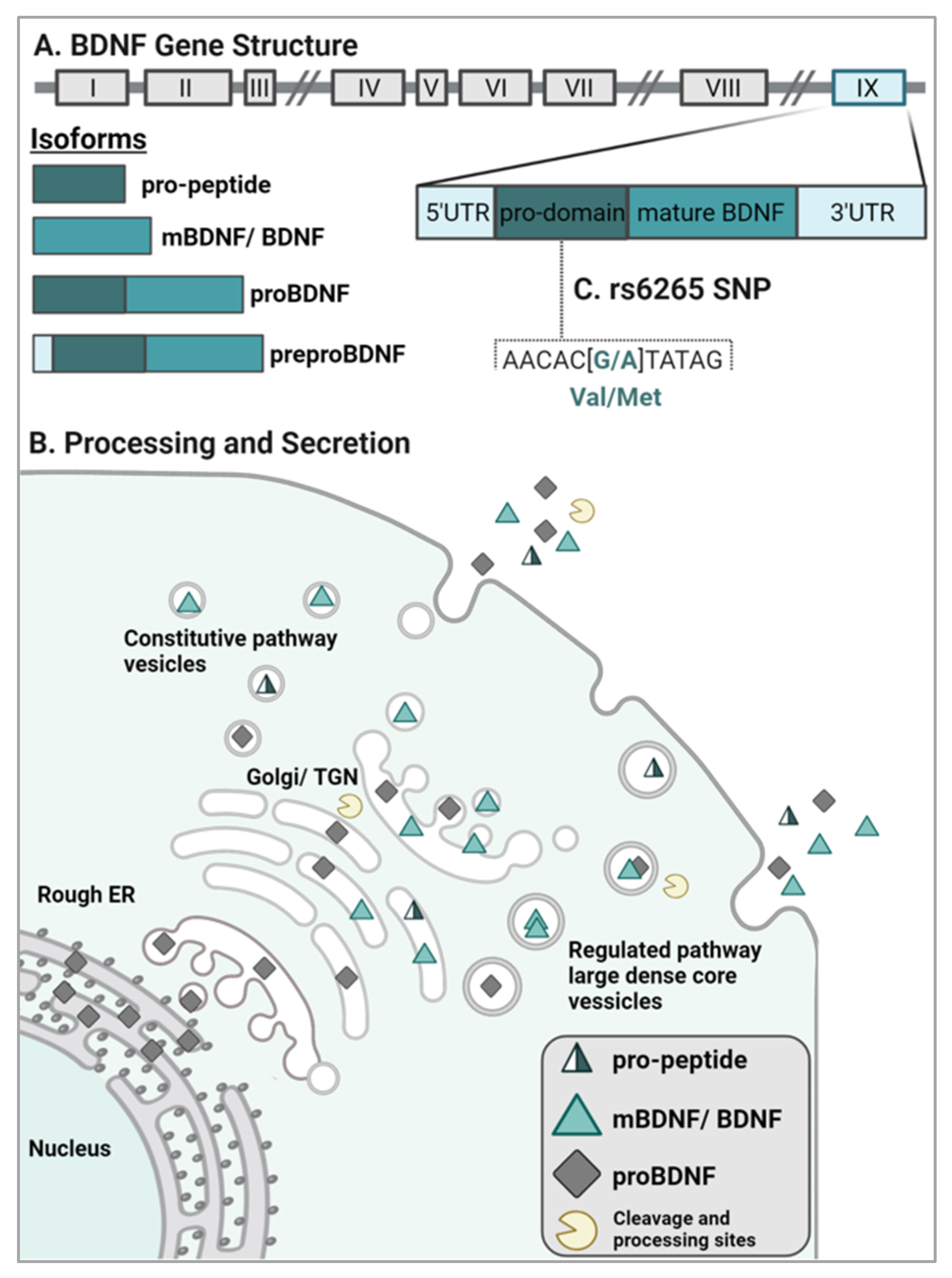
2.1. BDNF Gene Structure and Isoform Processing

The human BDNF gene is located on chromosome 11p13-14 and is composed of multiple noncoding exons and one coding exon. There are 11 exons that can be alternatively spliced to produce a minimum of 17 transcripts, but each transcript generates the same final protein product [15,16,17]. Of the 11 exons, 9 fall within the 5’ region [18]. The BDNF mRNA transcripts that contain exons II and VII are exclusively expressed in the brain, whereas the transcripts containing exons I, IV, and V are expressed in peripheral tissue; exons VI and IX are broadly expressed [14]. BDNF transcription terminates at two polyadenylation sites within exon IX, thus giving rise to two distinct mRNA populations including short (0.35 kb) or long (2.85 kb) 3’ untranslated regions (UTR) [14,18,19]. These two distinct populations have differing localizations: short UTR BDNF (exon I and IV) transcripts are found in the cell soma, whereas long UTR BDNF transcripts (exon II and IV) are trafficked to dendrites to regulate dendritic morphology and affect LTP [18,20].
The major coding sequence of BDNF is present in exon IX at the 3’ end and is translated into an inactive precursor polypeptide (i.e., preproBDNF) in the rough endoplasmic reticulum (ER) [8,16,21]. Within the rough ER, the signal sequence is immediately cleaved to yield the 28- to 32-kDa isoform proBDNF [8,18] which is comprised of an N-terminal prodomain and a C-terminal mature domain (Figure 1A). Post-translational modifications including N-linked glycosylation of the prodomain as well as sulfation of the N-linked oligosaccharides can take place as the proBDNF neurotrophins migrate from the Golgi apparatus to the trans-Golgi network (TGN). The processing of proBDNF continues via cleavage by intracellular proteolytic enzymes in the TGN (i.e., furin) or by convertases present in intracellular secretory vesicles for extracellular export [22]. A portion of full-length proBDNF proteins is also released and can subsequently bind the high affinity receptor, p75NTR [23]. After release from the cell, extracellular processing of proBDNF by plasmin or matrix metalloproteases (e.g., MMP-2, MMP-9) can also occur (Figure 1B) [8,23,24,25]. Processing of the preproBDNF yields three distinct active isoforms: the ~30kDa proBDNF, the ~13kDa mature BDNF (mBDNF), and the ~17kDa BDNF pro-peptide [26] (Figure 1).

2.2. BDNF Sorting and Release

Two distinct pathways of secretion exist for proBDNF and mBDNF: the constitutive and the regulated pathways. The constitutive pathway involves packaging BDNF into small-diameter granules that release BDNF independently of calcium fluctuation [1]. The majority of BDNF is packaged for release via the regulated pathway into larger granules that fuse to the plasma membrane in response to a calcium-dependent trigger (Figure 1B). Thus, the regulated release of BDNF occurs during activity-dependent depolarization [1,8,28,29] (Figure 1B). Proper sorting and secretion of BDNF is critical for the maintenance of synaptic plasticity, neuronal survival, and CNS homeostasis [1,8,30,31]. As such, disruption of BDNF sorting and/or secretion has been implicated in various neurodegenerative and psychiatric diseases. While the specific molecular mechanisms associated with improper BDNF secretion remain largely uncertain, current evidence correlates reductions of hippocampal and cortical volumes [32], formation of abnormal synapses [33], and decreases in dendritic complexity [34,35] as consequences of dysfunctional BDNF sorting and reduced secretion.
For the regulated pathway, two binding interactions drive sorting of BDNF into vesicles. The BDNF prodomain/pro-peptide region binds directly to either sortilin, a vacuolar protein sorting 10 (Vps10) domain-containing molecule, or carboxypeptidase E [8,18]. Sortilin contains a transmembrane region and a cytoplasmic tail responsible for signaling endosome sorting in the Golgi apparatus [18]. Sortilin and BDNF have been observed to colocalize within large dense-core vesicles, and sortilin truncation mutations result in impaired sorting of BDNF to the regulated pathway, subsequently decreasing activity-dependent release [36]. Similarly, membrane-bound carboxypeptidase E is a glycoprotein that binds BDNF, and knockdown of carboxypeptidase E in mice has also demonstrated a reduction of downstream activity-dependent BDNF release [18,37]. After being sorted into large dense-core vesicles of the regulated pathway, BDNF is generally trafficked to the axon where it can be degraded by the lysosome [38] or secreted into the synaptic cleft in response to neuronal activation where it can activate two classes of receptors, TrkB and p75NTR [39,40,41]. While the majority of BDNF is transported anterogradely, approximately 23% of BDNF is retrogradely transported to dendrites, although the biological significance of its retrograde trafficking has yet to be elucidated [18,42,43].

2.3. BDNF Signaling

Neurotrophins are known to bind to two classes of receptors: a tropomyosin receptor kinase (Trk) and a pan neurotrophin receptor (p75NTR) which is a member of the tumor necrosis factor super family [44] (Figure 2). More specifically, mBDNF preferentially binds to its high affinity receptor, TrkB, following its release into the synapse [39,40] (Figure 2B). In contrast, proBDNF binds with high affinity to p75NTR [44,45] (Figure 2A). While mBDNF can also bind p75NTR, it does so with low affinity [46]. Additionally, the BDNF prodomain/pro-peptide region binds directly to sortilin, thereby participating in proper sorting of this molecule to its regulated pathway [36].

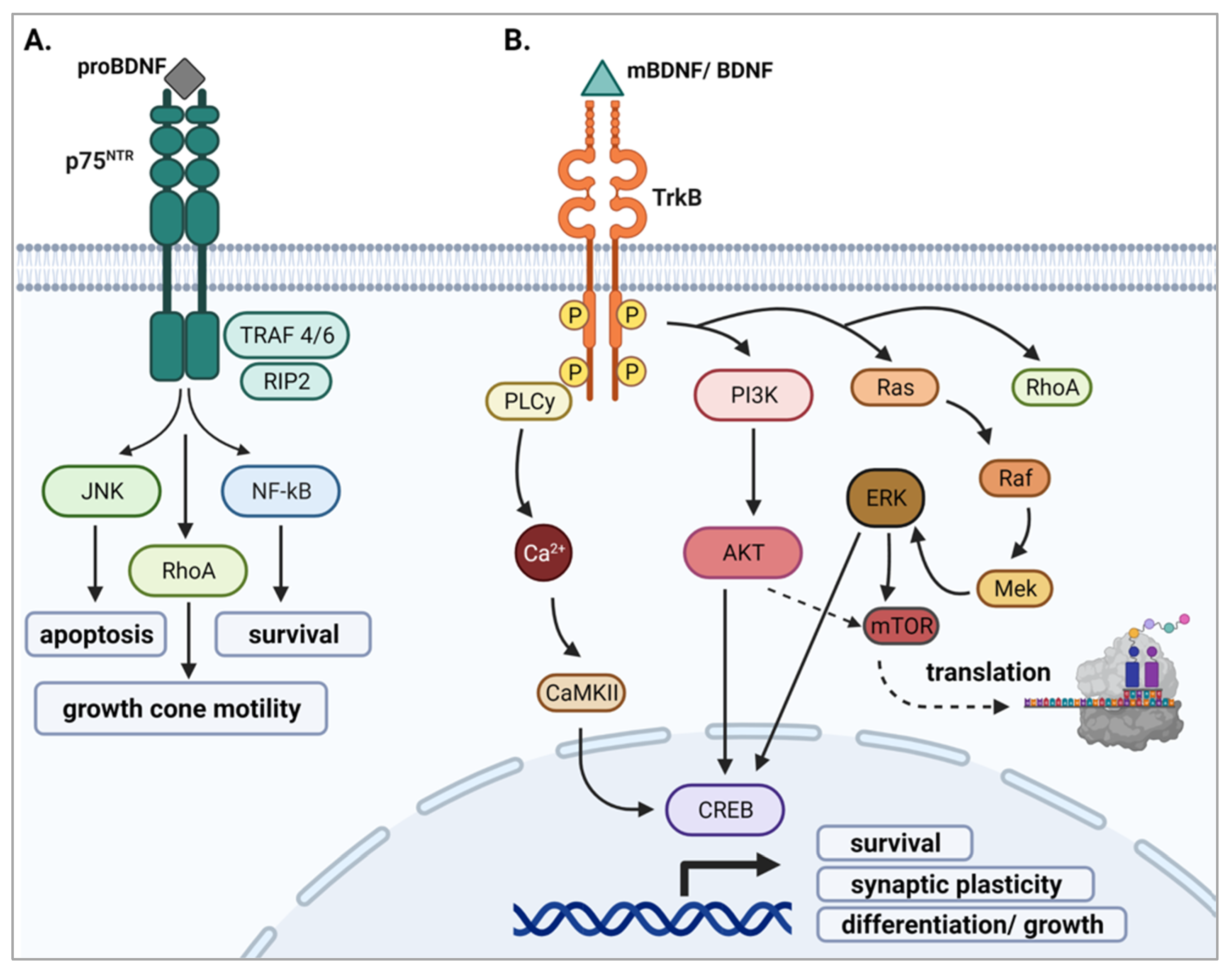
2.3.1. proBDNF and p75NTR

ProBDNF binds to p75NTR upon release, stimulating nuclear factor kappa B (NF-κB), c-Jun N-terminal Kinases (JNKs), and Ras homolog family member A (RhoA) signaling that modulate survival, apoptosis, and growth cone motility, respectively [6,44,47,48] (Figure 2A). The specific cascade that is activated is dependent on which receptors are complexed with p75NTR. For instance, when complexed with sortilin, pro-apoptotic pathways are activated [45,49]. Recent evidence indicates that signaling through p75NTR can also synergistically aid in TrkB activation [50,51,52]. Specifically, p75NTR can heterodimerize with TrkB, increasing TrkB binding affinity for mBDNF, thus promoting neuronal growth and survival [45,50,52].

2.3.2. mBDNF and TrkB

Upon mBDNF binding to full-length TrkB, TrkB dimerizes and autophosphorylates several of its tyrosine kinase residues including Y705 and Y706 in the cytoplasmic loop of the kinase domain, as well as Y515 and Y816 [18,53]. Multiple signaling pathways can be triggered once TrkB is activated including the phosphatidylinositol 3-kinase (PI3K), the phospholipase-C-γ1 (PLC-γ1), the guanosine triphosphate hydrolases of RhoA, and the mitogen-activated protein kinase (MAPK)/Ras cascades (reviewed in [44,47,54]). The PI3K pathway engages in pro-survival activity and enhances dendritic growth and branching [55,56]. The MAPK/Ras signaling cascade controls protein synthesis during neuronal differentiation [57]. Lastly, growth of neuronal fibers is activated via activation of RhoA (Figure 2B) [6,44].
Figure 2. Schematic representations of conventional proBDNF and mBDNF signaling cascades. (A) ProBDNF binds with high affinity to p75NTR, initiating downstream JNK, RhoA, and NF-kB signaling [6,44,47,48]. (B) mBDNF (mature BDNF) binds with high affinity to TrkB, inducing its dimerization and autophosphorylation, thus activating three main signaling pathways, PI3K, PLCγ, and Ras/MAPK, all of which lead to activation of the transcription factor CREB, driving transcription of genes crucial for neuronal growth and survival [44,47,54]. RhoA signaling and mTOR pathways can also be activated leading to growth cone modulation and translation of proteins involved in the regulation of cellular proliferation [18,45,53,56].
Ijms 23 08011 g002
It is widely accepted that the proBDNF and mBDNF ligands induce opposing outcomes through their preferential binding to different receptors in order to promote neurological homeostasis [6]. Specifically, mBDNF-TrkB signaling stimulates neuronal growth and synaptic plasticity, whereas signaling through p75NTR tends to initiate apoptosis thought to be important in development for eliminating inessential neurons [48,49]. Moreover, while mBDNF signaling is instrumental in driving hippocampal LTP, proBDNF promotes LTD [58,59,60,61]. Because of homeostatic regulation, the expression of p75NTR and TrkB are known to be tightly linked where they are co-expressed on the surface of the cell to establish signaling between cell survival and cell death [18]. Homeostasis can therefore be disrupted when there is an imbalance in the expression of these receptors or an imbalance in the levels of proBDNF and mBDNF isoforms. For example, research conducted by Suelves and colleagues [62] examined the consequences of BDNF/TrkB/p75NTR imbalance in a Huntington’s disease (HD) mouse model, showing that the reduction of BDNF and TrkB levels, along with an increase in p75NTR expression, correlated with striatal neuropathology and motor dysfunction. Pharmacological normalization of p75NTR levels rescued neuropathology (e.g., dendritic spine density) and motor deficits [62,63].
In addition to changes in receptor levels/balance, increased proBDNF levels have been correlated with adverse outcomes in neurodegenerative disorders. Specifically, in mice expressing one BDNF allele with a mutated cleavage site, hippocampal proBDNF levels rose and promoted a decrease in dendritic arborization as well as hippocampal volume [53,61]. Further reinforcing the importance of homeostatic balance in brain health, in Parkinson’s disease (PD), serum levels of proBDNF have been reported to be significantly higher in individuals with early PD as compared to heathy controls, whereas mBDNF levels were significantly lower [64]. Collectively, an abundance of data indicate that tight control of both BDNF ligands and their receptors is critical for proper neuronal function and/or survival.

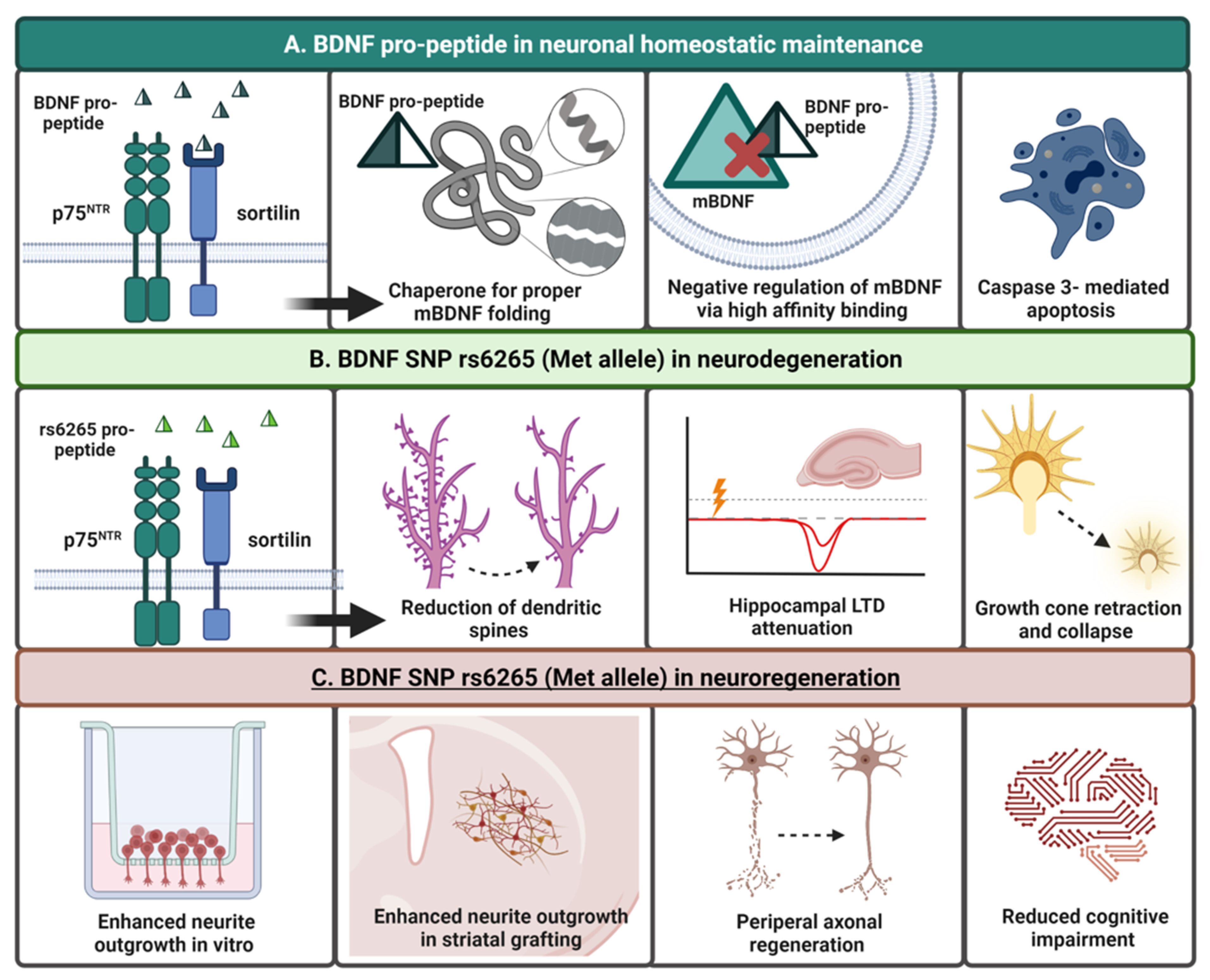
2.3.3. BDNF Pro-Peptide and Sortilin

It has been demonstrated that BDNF pro-peptide binding to sortilin drives proper sorting of BDNF into vesicles of the regulated secretory pathway [36]. In addition, the BDNF prodomain-peptide (pro-peptide), once cleaved from proBDNF, appears to function as an independent ligand similar to proBDNF and mBDNF isoforms [27,65,66]. Upon cleavage from proBDNF and its subsequent release, the BDNF pro-peptide binds to sortilin and complexes with p75NTR, resulting in various effects on the BDNF signaling cascade, neuronal survival, and synaptic plasticity [27,36,65,67,68], although specific mechanisms and downstream pathways remain to be elucidated.

3. Genetic Polymorphisms of BDNF

Remarkably, more than one hundred polymorphisms have been described in the BDNF gene [14,69]. While many known variants exist within non-coding regions, understanding of their functional consequences remains limited. However, the most extensively studied single nucleotide polymorphism (SNP) is the Val66Met (G196A, rs6265) polymorphism within the prodomain region of the BDNF gene. Other less well-studied variants exist within this region including Thr2I1e (rs8192466), Gln75His (rs1048221), Arg125Met (rs1048220), and Arg127Leu (rs1048221) and are reviewed elsewhere [14,18,70,71].

rs6265 (Val66Met)

The rs6265 BDNF SNP, or Val66Met, results from a nucleotide exchange from guanine to adenine at position 196 (G196A). This change results in a substitution of valine to methionine at codon 66, thus referred to as Val66Met [27] (Figure 1C). An individual can be heterozygous (Val66Met) or homozygous (Met66Met) for this SNP. The prevalence of this SNP worldwide is approximately 20% with certain populations in East Asia reporting an incidence up to 72% [33,72,73]. Found in the prodomain region of the BDNF gene, this substitution creates binding interference between the BDNF prodomain/pro-peptide of proBDNF to sortilin. The consequential result, and subsequent hallmark of this polymorphism, is a decrease in activity-dependent release of BDNF, with no reported alterations in constitutive release [14,35]. The reduction in BDNF release is dose-dependent with homozygous subjects showing significantly less release compared to heterozygous subjects (Met/Met > Val/Met > Val/Val) [33].
A wealth of studies have documented a variety of neuropathologies associated with the decrease in secreted mBDNF linked to rs6265 including reduction of hippocampal and cortical volume, abnormal synaptic connections, and decreased dendritic complexity and arborization [20,32,33,34,35,74]. The functional consequences of this common genetic variant are wide-reaching and have been documented to impact memory and cognition, anxiety, and depression, and have been associated with obsessive compulsive disorder (OCD), attention deficit hyperactivity disorder (ADHD), schizophrenia, multiple sclerosis, blepharospasm, and migraine [32,34,35,74,75,76,77,78,79]. Such pathology may be linked to evidence demonstrating that the BDNF Val66Met substitution can result in binding disruption of the translin/trax complex to BDNF mRNA transcripts, subsequently compromising transport of transcripts to dendrites which is critical for synaptic plasticity and dendritic complexity [18,19,20]. As a consequence, decreased BDNF trafficking to dendrites may have negative implications in multiple neurodevelopmental and neurological disorders [20,76].
In addition, decreased BDNF levels/signaling have been implicated in neurodegenerative disorders including Alzheimer’s disease (AD), Parkinson’s disease (PD), and Huntington’s disease (HD) (Figure 3A). While it is beyond the scope of this review to provide an extensive analysis of what is known about rs6265 in the aforementioned brain maladies, we briefly highlight how the expression of this common human genetic variant impacts PD, AD, HD, major depressive disorder (MDD), and schizophrenia in the following paragraphs.

4. BDNF and the rs6265 SNP in Neurodegeneration

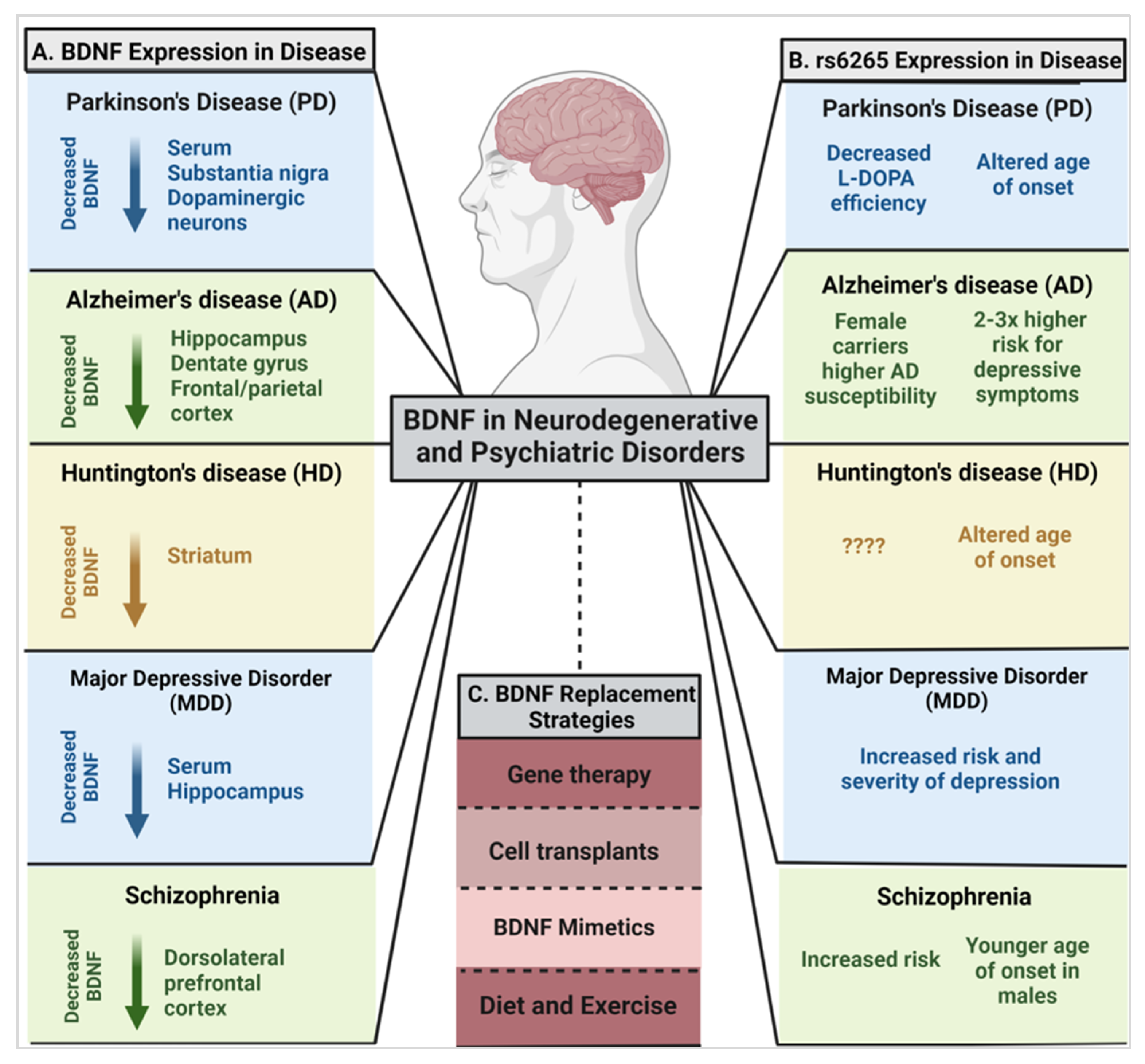
4.1. Parkinson’s Disease (PD) and BDNF

PD is a relentlessly progressive neurodegenerative disorder with typical motor symptoms that include rigidity, tremor, and akinesia/bradykinesia, in addition to a host of non-motor symptoms. Motor symptoms are caused by progressive and selective degeneration of dopamine (DA) neurons in the substantia nigra pars compacta (SNpc) and the accompanying loss of afferent nigrostriatal DA innervation. With the exception of monogenic forms of PD, the etiology of idiopathic PD remains unknown (for review [119]).
While dysfunction in BDNF signaling is not considered a primary cause of PD, it has long been known to be important for survival and development of SNpc DA neurons [120,121]. In addition, there is abundant literature demonstrating that, in the aged brain, there is diminished BDNF, diminished upregulation in response to stress, reduced expression of several BDNF transcription factors, and decreased expression of its TrkB receptor (for review [122]). Given that the primary risk factor for PD is aging, and given the critical role of BDNF in the well-being of SNpc DA neurons, BDNF dysfunction has been abundantly explored in PD.
Current evidence has demonstrated reduced expression of BDNF mRNA transcripts in the SNpc in PD [84,123] as well as lower levels of BDNF protein specifically in the SN of individuals with PD compared to other brain regions, and significantly reduced serum BDNF [81]. In addition to decreases in BDNF transcript levels, Scalzo and colleagues [81] have demonstrated that decreased BDNF levels are also detectible in serum of individuals with PD compared to healthy individuals and that concentrations were correlated with PD symptom severity [81] (Figure 3A). However, as the disease progresses, BDNF levels have been shown to increase [81,90,124], thought to be a compensatory mechanism in later diseased states.
In addition to changes in BDNF in PD, expression of TrkB receptors, which have high expression in SNpc neurons [125], has been shown to be altered in individuals with PD with evidence of isoform-specific alterations. For instance, levels of truncated TrkB have been reported to decrease in axons of the striatum, whereas levels were reported to increase in the striatal soma and distal dendrites of the SN in individuals with PD [126]. Full-length TrkB levels, in contrast, were found to be decreased in striatal neurites and in the cell soma of dendrites, but levels were higher in cell somas and axons of the striatum and SNpc, respectively [126,127]
These findings are corroborated in mouse models of PD where reducing levels of BDNF protein in the SNpc results in a reduction in DA neurons as well as a subsequent decrease in striatal dopamine [83,128]. Further, haplo insufficiency of the BDNF receptor, TrkB, in transgenic mice has been associated with degeneration of SNpc DA neurons over time and in association with aging [122].

rs6265 in PD

Although expression of the BDNF rs6265 Met allele is not correlated with an increased incidence of PD, it may contribute to worsening non-motor symptomology [71,129,130]. For example, apathy is one of the most common non-motor neuropsychiatric symptoms of PD [130], and although not statistically significant, PD individuals who were homozygous for the Met allele (Met/Met) were reportedly more likely to display apathetic emotions compared to those without the Met/Met genotype. Moreover, the risks of impulsive-compulsive and related behavioral disorders are also statistically correlated in individuals with PD when expressing the rs6265 SNP [129].
An important distinction of Met allele carriers with PD has been in their response to certain pharmacotherapies including levodopa treatment, the gold-standard symptomatic therapy for PD. Specifically, it has recently been reported that Met allele carriers, homozygous or heterozygous, reported worse United Parkinson’s Disease Rating Scale (UPDRS) scores when administered levodopa monotherapy compared to their homozygous Val allele carrier counterparts [102,131]. Individuals expressing the Met allele were also found to have a higher risk of developing the often debilitating side-effect known as levodopa-induced dyskinesia (LID) earlier in their treatment compared to homozygous Val allele carriers [104,132].
To contrast these negative correlations of the Met allele, in unmedicated PD patients, a lower severity of motor symptoms has been observed in the initial stages of the disease in BDNF variant individuals [103]. Although homozygous Met allele carriers tended to have more tremor-like symptoms, the progression of the disease was slower, with delayed need for levodopa administration compared to Val allele carriers [103]. Along with this notable decrease in severity of motor symptoms, a later age of onset of PD was reported in homozygous Met allele individuals compared to their Val/Val and Val/Met counterparts with one cohort reporting a 5.3-year later age of onset [105,133] (Figure 3B). In contrast, Svetel et al., 2013 reported that the presence of the Met allele was not associated with clinical characteristics of PD including age of onset and disease severity [134].
This dichotomy of data on the role of rs6265 in PD points to the need for continuing research in this area. In addition, it highlights that there is a significant advantage to tailoring therapeutics in neurodegenerative diseases with the goal of increasing efficacy. Indeed, the inherent heterogeneity in response to therapeutics in individuals with PD [102,131,135] is a significant impediment to implementation of the overall goal of “delivering the right treatment to the right person at the right time to implement safe, effective, and precise interventions with minimal complications” [136]. One approach to deconstructing the complexity of PD and response to therapy is the identification of common genetic variants, like rs6265, that influence these variables as a means to predict disease characteristics and tailor treatments to those most effective for subpopulations of patients.

4.2. Alzheimer’s Disease (AD) and BDNF

AD is the leading cause of dementia characterized by severe cognitive decline with age (e.g., [137]). The two pathological hallmarks of AD are extracellular β-amyloid plaques (APs) and intracellular neurofibrillary tangles (NFTs) [138]. There also is significant neuron loss in the hippocampus, entorhinal cortex, and basal forebrain neurons, eventually affecting large portions of the cortex, areas involved with memory and cognition, and eventually language, reasoning, and social behaviors [138,139]. Based on the importance of BDNF in neuron viability, synaptic plasticity, and learning and memory [30,140], this neurotrophin has been of great interest in AD.
As recently reviewed, dysfunction in BDNF and TrkB have been found in AD [141]. Nearly three decades ago, Phillips and colleagues [85] reported lower levels of BDNF mRNA transcripts in hippocampal tissue from AD patients compared to age-matched controls [85]. While limited research suggests variable effects of BDNF mRNA and protein levels in the hippocampus of individuals with AD [123,142], consensus supports that there is an overall decrease of BDNF protein and mRNA in the hippocampus, dentate gyrus, and the frontal and parietal cortex [85,86,87,88,127]. Additionally, lower expression levels of TrkB receptors were found in the hippocampus and frontal cortex in individuals with AD [14,143]. Neurons with NFTs, a major pathological hallmark of AD, were also found to have specifically lower intracellular BDNF protein levels [127], and individuals’ serum levels of mBDNF and proBDNF negatively correlated with AD disease severity [14,118,144] (Figure 3A). While downregulation of BDNF and TrkB have been observed in AD, whether AD-specific pathology (i.e., APs and NFTs) is related to these changes has only recently been examined. Specifically, Ginsberg and colleagues [141] have found that, in individuals with AD, downregulation of BDNF is associated with increased APs in the entorhinal cortex, and downregulation of TrkB is associated with increased NFTs in the entorhinal cortex and APs in the hippocampus.

rs6265 in AD

Because BDNF is critical for cell survival and synaptic plasticity, especially in the hippocampus, it is not surprising that expression of a genetic variant (rs6265) and decreased release of this neurotrophin have been associated with AD pathology [108]. A meta-analysis conducted by Fukumoto et al., 2010 reported that women who were Met allele carriers were more susceptible to AD, whereas male Met allele carriers were unaffected [73,106]. This work, and additional supporting studies [107,145,146], demonstrate that rs6265 expression may drive a sex-specific susceptibility to cognitive decline in AD for women but not men [107]. Despite the general paradigm suggesting a correlation between the expression of rs6265 and AD susceptibility, not all research groups have been able to establish the same correlation of rs6265 as a susceptibility factor for AD [147,148,149,150]. Thus, the utility of therapeutically targeting BDNF in AD requires additional investigation (Figure 3B). In addition to a potential association with susceptibility, heterozygous and homozygous carriers of the Met allele with AD have a two-fold and three-fold higher risk for AD-related depressive symptoms, respectively [108].

4.3. Huntington’s Disease (HD) and BDNF

HD is an autosomal dominant, progressive neurodegenerative disorder in which striatal medium spiny neurons (MSNs) degenerate with eventual shrinkage of the entire brain, resulting in debilitating motor, cognitive, and psychiatric symptoms [151]. Low levels of BDNF have been considered to play a significant role in the pathogenesis of HD. In healthy individuals, corticostriatal neurons innervate the striatum and release BDNF onto MSNs, and successive BDNF activation of TrkB drives MSNs survival [7]. In HD, however, the mutated huntingtin protein results in altered transcription of the BDNF gene, affects the axonal transport and release of BDNF, and alters TrkB and p75NTR receptors, resulting in a decreased corticostriatal release of BDNF and deficits in neuronal survival signaling [151,152]. In postmortem brain tissue from patients with HD, a distinct reduction of BDNF levels in the caudate and putamen was observed as compared to healthy controls [89]. Yet, in the cerebral cortex and hippocampus, normal BDNF levels were found [89,153]. Levels of BDNF mRNA expression were also affected. For instance, in the cortex of 20 subjects with HD, a significant decrease in both BDNF mRNA and protein levels was reported [153]. Furthermore, TrkB mRNA levels were decreased in the caudate and not the cortex. Its truncated isoform and p75NTR, however, were increased in the caudate [153]. Multiple research groups have also reported similar findings in animal models of HD [152,153,154]. For example, in mouse models of early symptomatic HD, TrkB receptors were found to be dysfunctional, failing to engage postsynaptic signaling mechanisms, an effect that was rescued by inhibiting p75NTR signaling [155]. Further, in huntingtin-knockout mice, BDNF mRNA levels were reduced in the cerebral cortex due to its physiological dependence on functional huntingtin protein for proper cellular localization [152] (Figure 3A). This evidence suggests that the imbalance of TrkB and p75NTR expression, along with a reduction of BDNF, may compound and exacerbate HD pathogenesis [153].

rs6265 in HD

There are currently few published studies that implicate the expression of the rs6265 SNP in HD pathogenesis. Interestingly, individuals with HD who have the heterozygous Val/Met genotype were reported to have a later age of onset when compared to those who were homozygous for the Val or homozygous for the Met genotype [109]. In contrast, four other studies were unable to confirm any relationship between the expression of this polymorphism and the age of onset in individuals with HD [156,157,158,159]. To the best of our knowledge, no additional analyses have been reported correlating rs6265 and HD, and this potential link represents a prospective area of research development (Figure 3B).

5. BDNF and rs6265 in Psychiatric Disorders

5.1. Major Depressive Disorder (MDD)

MDD is characterized by lack of pleasure in enjoyable activities, cognitive impairment, sleep and dietary issues, abnormal behavior, and suicidal tendencies [92]. Most studies suggest that there is an association between low levels of BDNF and underlying MDD pathophysiology. Meta-analysis in depressed patients has demonstrated that serum levels of BDNF were much lower than in non-depressed control individuals [91,160]. Additionally, in a French population of individuals who were 65 or older, those who had late-life depression also had elevated methylation of the BDNF gene, leading to a reduction in BDNF gene transcription [94]. These data correlate with other findings that suggest levels of BDNF are low in both the serum and the hippocampus of patients with MDD [93,161,162]. Research involving animal models of depression have demonstrated that BDNF serum levels are reduced, and that this is correlated with the duration of the condition, albeit not symptom severity in MDD (reviewed in [92]) (Figure 3A). BDNF levels are also detrimentally implicated in the context of suicide. Specifically, in the hippocampus and prefrontal cortex of teenagers who committed suicide, postmortem tissue analyses showed significant reductions of BDNF mRNA and protein as well as decreased levels of full-length TrkB expression [95,96,160]. These studies indicate that therapeutic BDNF supplementation may have the potential to alleviate the burden of depression, MDD, and suicide. Indeed, antidepressants are thought to elicit their therapeutic effects, in part, by increasing trophic factors in the brain including significant elevations in BDNF (e.g., [163]). Given the fact that individual responses to antidepressants are widely variable, genetic factors may be involved.

rs6265 in MDD

An increased risk and severity of depression is common in individuals who are rs6265 Met allele carriers [110,111] (Figure 3B). In postmortem tissue from victims of suicide with the Met allele, there were lower BDNF levels in the anterior cingulate cortex and caudal brainstem compared with non-depressed subjects, although this did not correlate with an increase in suicidal tendency [164]. Further, an increased instance of BDNF gene methylation was reported in heterozygous (Val/Met) older women with anxiety/depression compared to those who were homozygous (Val/Val) [73,165], again implying that there may be sex-specific consequences of Met allele expression. DNA methylation can upregulate or downregulate corresponding gene expressions and modify related phenotypes [165] with rs6265-associated DNA methylation presumably resulting in loss of function in BDNF signaling [164].

5.2. Schizophrenia

Affecting approximately 1% of the worldwide population, schizophrenia is a psychiatric disorder characterized by positive symptoms, negative symptoms, and cognitive impairment [166,167] with an onset of symptoms generally in the mid- to late 20s. These symptoms include visual and auditory hallucinations, delusions, depression, self-neglect, confused cognition, and memory impairments. Physiologically, schizophrenia involves the reduction of gamma-aminobutyric acid (GABA) signaling [92,168]; however, glutamate, acetylcholine, serotonin, and DA are also involved in the pathology of schizophrenia [169].
Given the essential role of BDNF in neurodevelopment, synapse regulation, and synaptic plasticity, this neurotrophin has been proposed to contribute, in part, to the pathogenesis of schizophrenia [170]. Most meta-analyses concur that serum levels of BDNF in unmedicated and medicated individuals with schizophrenia are reduced and that BDNF levels decrease further with age [92,171,172]. In the brain of schizophrenia individuals, the dorsolateral prefrontal cortex was shown to contain significantly lower BDNF protein and mRNA, along with decreased TrkB expression, compared to healthy controls [97,98,99,100,101] (Figure 3A). The DNA methylation status of the BDNF gene was reportedly altered in a prenatal stress mouse model of schizophrenia with a consequent decrease in BDNF mRNA and enrichment of 5-methylcytosine and 5-hydroxymethylcytosine at BDNF gene regulatory regions [173]. Increased inflammatory factors such as proinflammatory cytokines have also been reported to be present in postmortem brain tissue from the dorsolateral prefrontal cortex of individuals with schizophrenia, suggesting there may be interplay between DNA methylation, decreased BDNF production, and inflammation in these individuals [92,174]. One contradictory analysis in post-mortem schizophrenic brains demonstrated that BDNF levels were elevated in the hippocampus and anterior cingulate cortex, while the expression level of TrkB was decreased in the hippocampus and prefrontal cortex [175]. Although a correlation between low BDNF levels and/or altered gene regulation and schizophrenia is generally supported in the literature, further studies are necessary to establish the specific mechanisms by which these alterations contribute to schizophrenic pathology and the therapeutic utility of BDNF.

rs6265 in Schizophrenia

Currently, conflicting evidence exists regarding the relationship of rs6265 SNP expression and its alterations in BDNF levels and schizophrenia (for review [176]). In at least two studies, no significant effect of rs65265 genotype profiles were observed for levels of BDNF in serum [166,177]; however, one report found serum levels of BDNF were reduced significantly in individuals with schizophrenia [166], and the other found elevated serum BDNF in schizophrenic subjects [177] compared to control subjects. Indeed, one meta-analysis of approximately 39 case-control studies established that the homozygous Met genotype increased the risk of schizophrenia in Asian, European, and Chinese populations [113]. More specifically, homozygous Met allele carriers had an increased risk of schizophrenia by approximately 19% when compared to Val/Met carriers [113,114]. In contrast, several studies have failed to find an association of rs6265 and schizophrenia (for review [176]). There is, however, some evidence to suggest an association between rs6265 and age of onset in schizophrenia patients with Val/Met males having an earlier age of onset; this association was not noted in females [115,116,117]. It was observed that, in a Caucasian (Armenian) population of chronic schizophrenia individuals, homozygous Met allele carriers, regardless of sex, had an earlier age of onset compared to the individuals who carried Val/Val and Val/Met genotypes [178]. Interestingly, the Val/Val genotype predisposed patients to symptoms that were more severe, exhibiting more clinical symptoms compared to Met allele carriers [117,179] (Figure 3B). Based on the ambiguous nature of current published meta-analyses, additional studies are warranted and should include a focus on additional BDNF polymorphisms as well as sex, age, and ethnic variations.

6. Targeting BDNF in the Brain: Therapeutic Challenges and Potential

Overall, BDNF levels are negatively correlated in neurodegenerative and psychiatric disorders. Therefore, many BDNF-targeted therapies aim to raise the levels of BDNF either exogenously or endogenously. Exogenous application of BDNF through direct infusion has been demonstrated to be beneficial to varying degrees in numerous animal studies [180,181,182,183]. As a neuroprotective agent in PD models against DA neuron toxins such as 6-hyroxydopamine (6-OHDA) or 1-methyl-4-phenyl-1,2,3,6-tetrahydropyridine (MPTP), BDNF is effective at protecting SH-SY5Y neuroblastoma neurons in vitro and can modestly protect against 6-OHDA in vivo [183]. Additionally, in an AD rat model, exogenous BDNF application showed dose-dependent neuroprotection against amyloid-β-induced neurotoxicity in cortical neurons in vitro and, when administered intracerebrally, protected cholinergic neurons in the forebrain and diminished the concentrations of amyloid-β peptides [181,182]. Despite promising outcomes from select research conducted in preclinical animal models, a large-scale clinical trial involving oral BDNF supplementation in patients with amyotrophic lateral sclerosis (ALS) did not significantly increase patient survival at dosages of 50–100 mg/day [184]. In a clinical trial involving intrathecal delivery of BDNF to ALS patients, doses of 150 mg/day were well tolerated; however, conclusions about treatment efficacy were unable to be drawn due to small sample sizes [185]. However, a later trial also using intrathecal BDNF for ALS found a lack of clinical efficacy [186]. These disappointing clinical trial results could, in part, be due to the poor pharmacokinetics of BDNF.
The pharmacokinetics of neurotrophins are complex, making BDNF administration for brain therapeutics especially difficult. Neurotrophins are large, sticky molecules that cannot readily cross the blood-brain-barrier, have short half-lives reported to be 30 min or less [187], inefficiently diffuse into tissues [118], and approaches like intrathecal delivery result in broad exposure to nontargeted structures, thus limiting their scope of effectiveness [118]. If pharmacokinetic barriers could be overcome, consideration needs to be given to therapeutic concentrations of BDNF intended for delivery as well as the availability and status of TrkB receptors. Specifically, exogenous administration of BDNF in regions with significant reductions in TrkB expression, which is known to occur in PD and AD, could severely limit therapeutic benefit. In addition, excessive levels of BDNF could also have a negative impact. Not only can higher concentrations of BDNF downregulate TrkB expression, but excessive amounts of BDNF can lead to unwanted side effects such as seizures, fever, weight loss, fatigue, and diarrhea [127]. Molecularly, excess BDNF can likewise have a negative effect on synaptic circuitry, learning, and memory by inducing hyper-excitation in regions such as the hippocampus [188]. Keeping the above challenges in mind, non-pharmacological methods of BDNF delivery bear potential.

6.1. Gene- and Cell-Based Therapy

A promising non-pharmacological therapeutic technique is in vivo BDNF gene de-livery. This technique involves utilizing viral vectors to transduce host cells with the BDNF gene for downstream endogenous in situ mRNA and protein production. In this way, the high concentrations of local BDNF production in specific regions will ideally protect degenerating neurons in diseases such as PD, HD, and AD [189]. Preclinically, in a post-stroke depression rat model, intranasal delivery of a BDNF-encoding adeno-associated viral vector (AAV-BDNF) increased BDNF mRNA and protein in the prefrontal cortex, alleviating depressive-like symptoms [190]. Additionally, preventative intrastriatal injections of AAV-BDNF reduced the loss of NeuN, a pan neuronal maker, in a lesioned rat model of HD, therefore providing neural protection [191]. Although clinical trials of gene therapy that intended to supplement another neurotrophic factor (i.e., GDNF or neurturin) for neuroprotection against PD have been conducted, results are not yet promising [192,193,194]. Moreover, it remains unknown if it is clinically viable to target low BDNF levels in neurodegenerative or psychiatric disorders via gene therapy.
Another available BDNF-targeting gene therapy involves an ex vivo autologous approach for neuroregeneration. Briefly, cells such as fibroblasts are taken from the subject, genetically modified to produce BDNF, and then transplanted back into the cell donor’s brain. Like in vivo methods, this strategy could allow for the sustained release of BDNF locally in specific brain regions but advantageously would be poised to avoid immune rejection. Levivier et al., 1995 showed that genetically modified fibroblasts were able to prevent degeneration induced by 6-OHDA in a rat model of PD [195]. Likewise, in a quinolinic acid toxin model of HD, rat fibroblasts were genetically engineered to produce BDNF and transplanted back into the rat brain, resulting in the protection of striatal neurons as compared to control animals [191]. Similarly, mesenchymal stem cells (MSCs) genetically altered to overexpress BDNF have been shown to reduce neuropathological and behavioral deficits in rodent models of HD, suggesting that these approaches have considerable potential for clinical use (for review [196]).
In 2005, a clinical trial was conducted using this ex vivo method in the context of AD for the supplementation of NGF. Eight patients with mild AD received implants of autologous fibroblasts that were genetically engineered to produce NGF and were transplanted into the nucleus basalis of Meynert via stereotaxic surgery [197]. In follow-up studies after 22 months, no long-term side effects occurred, and the rate of cognitive decline was improved in these patients [197]. Considering that this ex vivo autologous treatment was well tolerated, and symptom improvement was demonstrated in AD [189,197], additional clinical trials would seem to be warranted involving BDNF-centric modifications.
Gene therapy, whether viral vector-mediated or autologous transplantation of genetically modified cells, holds strong promise but is not without caveats [198,199]. In general, local release of BDNF is difficult to tightly regulate genetically, and as introduced above, overproduction of BDNF can be detrimental to the circuitry of the brain [118,188]. In addition, both approaches involve invasive surgical protocols; however, in the scope of neurosurgery methods that are much more aggressive (e.g., tumor resection), the approach for vector or cell graft delivery is minimally invasive and straightforward. Of additional concern is immune response to viral vectors and the associated products of foreign transgenes [200]. However, as recently reviewed, current efforts and advances in clinical trials have led to advances to circumvent immune obstacles including modifying AAV capsids to evade pre-existing neutralizing antibodies and development of new methods for clearing of antibodies from circulation (for review [200]). With the advent of new DNA modification techniques, it is not beyond the realm of possibilities that novel gene therapy approaches could be applied in the future. In addition, given that ex vivo autologous treatment was well tolerated, and symptom improvement was demonstrated in AD [189,197], this approach remains hopeful to those suffering from neurodegenerative or neurological disorders.

6.2. BDNF Mimetics

One of the most promising BDNF-related administration strategies involves the use of BDNF mimetics. These are small molecules designed to mimic the binding loops of BDNF, resulting in the phosphorylation and activation of TrkB and its downstream effectors, AKT and ERK [118,201,202]. The use of small molecules allows for the delivery of controlled dosages with improved pharmacokinetics compared to full-length BDNF. Mimetics have shown improved diffusivity, blood-brain-barrier permeability, and augmented receptor specificity with less promiscuity [201,202,203,204]. These compounds, however, would require repeat dosing and would not be brain region-specific in targeting, potentially trafficking to areas where their engagement is not advantageous [202,205].
Two common BDNF mimetics are 7,8-dihyrodxyflavone (DHF) and GSB-106. 7,8-DHF is a naturally occurring flavonoid responsible for binding and initiating TrkB signaling pathways. 7,8-DHF application has been investigated in many neurodegenerative and neurological disorders including PD and AD [206,207,208]. For example, in a mouse model of AD, cognitive deficits were restored after 7,8-DHF administration [206,207]. In a comprehensive report by Jang and colleagues [208], 7,8-DHF was documented in mice to specifically activate TrkB in the brain, to diminish kainic acid-induced toxicity in the hippocampus, to decrease infarct volumes in a middle cerebral artery occlusion model of stroke, and it was neuroprotective in a MPTP model of Parkinson’s disease [208]. Cognitive deficits were also rescued with 7,8-DHF treatment in a mouse model of Down syndrome [209]. Collectively, these studies support the idea that 7,8-DHF may be a therapeutic mimetic worth implementing in a wide range of disorders.
Another common mimetic is bis-(N-monosuccinyl-L-seryl-L-lysine) hexameth-ylenediamide, also referred to as GSB-106, and it mimics the interaction between the TrkB receptor and BDNF via loop 4 of BDNF. Like 7,8-DHF, GSB-106 administration elicits neuroprotective properties by preventing apoptosis in SH-SY5Y cells through the suppression of caspase-3 activity [204]. As reviewed in [210], GSB-106 has also been shown to have a variety of TrkB-mediated neuroprotective effects as well as reduce depressive-like symptoms in a mouse model of depression where administration increased locomotor activity and reduced signs of anhedonia [210,211]. Studies focused on these two BDNF mimetics demonstrate that these small molecules represent potentially useful treatment approaches for those with neurodegenerative diseases and cognitive disabilities. Continued preclinical and clinical development are needed so that their therapeutic effects can be optimized to the greatest extent.

6.3. Diet and Exercise

Diet and exercise are widely accessible, non-invasive, low-cost treatments that are of interest for neurodegenerative and neurological conditions. Preclinical studies in various animal models confirm that dietary and exercise regimens increase BDNF levels in the brain and improve cognitive and behavioral functions [118,152,212,213,214,215]. For example, Fahnestock and colleagues [215] demonstrated that implementing a diet high in antioxidants in aged dogs increased BDNF transcripts to levels which were comparative to the young dog cohort [215]. Additionally, restricting the diet of 3-month-old male Sprague Dawley rats to an alternate day feeding regimen compared to ad libitum increased BDNF levels in multiple brain regions including the cortex, striatum, and hippocampus [213]. There also is a wealth of data suggesting that exercise provides neuroprotection in multiple animal models of PD [216,217,218,219,220,221,222] with additional indications that it improves motor symptoms and quality of life in individuals with PD [223,224,225]. Studies using heterozygous deletion of BDNF [226] or inhibition of BDNF TrkB receptors [227] demonstrate that BDNF is essential for the beneficial effects of exercise on the neuroprotection of the nigrostriatal DA system in PD rodent models.
In patients with depression, exercise was found to induce significant increases in serum levels of BDNF levels in all assessed participants [228]. After sprint interval training, BDNF levels were increased directly afterward, then returned to baseline within 90 minutes in eight male subjects [229]. A number of genes, including BDNF, are associated with risk for post-traumatic stress disorder (PTSD), and rs6265 is associated with the psychotic symptoms of PTSD (for review [230]). Intriguingly, in combat veterans with PTSD, active exercise reduced methylation of the BDNF gene at specific CpG sites, resulting in normalized gene expression of BDNF as compared to those without active exercise [230]. Although there are many studies reporting that diet and exercise lead to increased BDNF levels [213,215,229], the specific mechanisms responsible have yet to be elucidated. In the context of BDNF as a therapeutic target, understanding and harnessing the benefits of diet and exercise on BDNF function could lead to vital non-invasive treatments geared toward improving not only neurodegenerative or psychiatric conditions but general patient quality of life.

7. The BDNF Pro-Peptide: A Functional “Third Ligand”

Neurotrophin pro-peptides functionally assist in the folding of mature neurotrophin proteins, regulating their localization and bioavailability [231], and the BDNF pro-peptide is no exception. Historically, the BDNF pro-peptide was thought to exclusively function as a molecular chaperone, supporting the folding of mBDNF. Recent studies, however, reveal that the BDNF pro-peptide may also act as an independent functional “third” ligand [27,66,232]. The detection of the BDNF pro-peptide within the laboratory or clinic was previously limited, leading to the conclusion that it may be immediately degraded after proteolytic processing [231,233]. However, in 2012, Dieni and collaborators [43] successfully detected the BDNF pro-peptide and found that both mBDNF and the BDNF pro-peptide were present at equimolar ratios in large dense-core vesicles of excitatory presynaptic termini in hippocampal lysates; an abundance tenfold of that of proBDNF [43,231]. From primary mouse hippocampal neurons, Anastasia and colleagues [27] were also able to isolate the BDNF pro-peptide. The pro-peptide was shown to be released in an activity-dependent manner upon depolarization with potassium chloride like that of the other BDNF isoforms [27]. In addition to its interaction with the sortilin/p75NTR complex, the BDNF pro-peptide has been shown to bind specifically to mBDNF with high affinity in intracellular compartments of trafficking vesicles, representing a potential regulatory mechanism of the mBDNF signaling pathways [67,232] (Figure 4A).
The BDNF pro-peptide functions as a negative regulator of neuronal structure and survival in multiple circumstances. In hippocampal slices, in the absence of mBDNF, in vitro application of the BDNF pro-peptide facilitated LTD by promoting NMDA-induced AMPA receptor internalization only in p75NTR-positive cells [31]. In C6 glioma cells, stimulation with the BDNF pro-peptide decreased cell growth and promoted caspase-3 mediated apoptosis [234]. Comparable results were also found in mature rat hippocampal neurons exposed to BDNF pro-peptide in culture which induced a significant caspase-3-dependent reduction of spine density [235] (Figure 4A). All of these effects were seen using the wild-type (Val/Val) BDNF pro-peptide.
Clinically, the levels of BDNF pro-peptide are altered in individuals with neurodegenerative or psychiatric disorders, but the trends are disease and sex-specific. In human cerebral spinal fluid (CSF), BDNF pro-peptide levels were much lower in individuals with MDD compared to controls, and male patients exhibited significantly lower levels than their female counterparts [241]. Comparatively, in individuals with schizophrenia, the ratio of BDNF pro-peptide to total protein level in CSF was also markedly lower in male patients compared to female patients [242]. In contrast, BDNF pro-peptide levels were significantly increased in postmortem hippocampal extracts from patients with AD; a greater than 30-fold increase [243]. These distinct changes in pro-peptide concentrations may lead to its use as a potential biomarker found in blood and CSF, although additional studies are needed to determine the biological consequences of BDNF pro-peptide flux and clinical feasibility of longitudinal monitoring of pro-peptide levels.
There are also prominent anatomical differences in BDNF pro-peptide expression throughout the body which may determine the role it plays in neurodegenerative or psychiatric disease. In a learned helplessness rat model of depression, the levels of the BDNF pro-peptide were significantly higher in the medial prefrontal cortex (mPFC) of susceptible rats compared to controls, but levels were much lower in the nucleus accumbens (NAc) [244,245]. In the postmortem parietal cortex in patients with MDD, schizophrenia, and bipolar disorder, the levels of mBDNF were significantly lower, whereas pro-peptide levels were significantly higher compared to the control group [245]. In contrast, the cerebellum of these groups with psychiatric disorders contained significantly lower levels of BDNF pro-peptide compared to controls [245]. A negative correlation was also observed between levels of mBDNF in the parietal cortex and mBDNF in the liver of these subjects [245].
Disease and region-specific alterations in mBDNF and pro-peptide levels imply that these molecules contribute to the pathology of various psychiatric disorders. In imaging studies of individuals with schizophrenia, the parietal cortex exhibited reduced gray matter [246,247]. Similarly, a reduction in gray matter and dendritic spine number was reported in the mPFC of patients with MDD [248,249,250]. Due to the current evidence demonstrating high levels of the pro-peptide in these brain regions, paired with its negative modulatory effect in various in vitro studies, it is reasonable to hypothesize that therapeutic regulation of BDNF pro-peptide expression would be beneficial, although this strategy would be complicated. Inhibiting pro-peptide expression in the mPFC and parietal cortex, for example, could exacerbate low pro-peptide levels in the NAc [244]. Since one of the many proposed mechanisms of neuropathology in MDD, schizophrenia, and bipolar disorder involve DA neuron malfunction, low levels of pro-peptide in the NAc may be a source of dysfunction in some individuals, and worsening this phenomenon with non-specific pro-peptide lowering strategies may increase disease severity. Conversely, increasing pro-peptide levels in the NAc specifically could be therapeutically beneficial but with potential negative consequences in the mPFC and parietal cortex [251].

Harnessing the Neurogenerative Benefits of the BDNF Pro-Peptide and rs6265 SNP

Because the BDNF rs6265 SNP involves a substitution of valine to methionine at codon 66, which occurs in this prodomain/pro-peptide region of BDNF that binds directly to sortilin, the Val pro-peptide appears to function differently than that of the pro-peptide containing the Met allele, at least in relation to activity-dependent release of BDNF [252]. As expected, in the hippocampus of homozygous mice expressing the Met allele, lower levels of the Met pro-peptide are released compared to Val pro-peptide secretion [27]. The Met pro-peptide also has a slightly different structure that could impact function. Specifically, in a study conducted by Anastasia and colleagues [27], a shift in conformation was found in the Met pro-peptide using nuclear magnetic resonance [27]. A strong preference for the β-strand conformation is typical; however, the Met pro-peptide tends to demonstrate a helical confirmation. This conformational change resulted in high affinity binding of unique residues in the Met pro-peptide to sortilin with a subsequent downregulation of the Rac pathway, a pathway known to play a role in regulating extension of axons and dendrites, neurite branching, axonal navigation, and synapse formation [27,253] (Figure 4B). In contrast, others have shown that stronger binding of the Val pro-peptide to sortilin occurs as compared to the Met pro-peptide in vitro [36].
In a study that involved treating primary mouse hippocampal neurons with Val and Met pro-peptides, administration of the Met pro-peptide induced growth cone retraction in sortilin- and p75NTR-positive cells after a period of approximately 20 min [27]. Similarly, infusing the Met pro-peptide into ventral CA1 hippocampal neurons of mice expressing the Val/Val, Val/Met, and Met/Met genotypes resulted in disassembly of dendritic spines and synaptic elimination, both of which were also dependent upon co-expression of sortilin and p75NTR [68]. Transcript levels of Rac1 were additionally affected after Met pro-peptide infusion and were decreased to half of basal levels, contrasting a robust increase in Rac1 expression levels of mBDNF-treated cells and no change in Val/Val pro-peptide-treated cells [68] (Figure 4B).
Although studies support the paradigm that the proBDNF and the BDNF pro-peptides, whether Val- or Met-type, decrease dendritic spine density, induce growth cone retraction, and promote LTD, these studies have only been conducted thus far in limited brain regions (i.e., hippocampus) (see Table A1). The production and signaling via various BDNF isoforms may elicit distinct biological effects based on isoform ratios and anatomical brain regions. Therefore, further research is necessary to fully elucidate the specific roles of both the Val and Met pro-peptide.
Despite the tenants of BDNF pro-peptide function discussed above, recent reports paradoxically suggest that the expression of the BDNF rs6265 SNP may confer protective, or neuroregenerative, effects in injury or disease. For instance, expression of the Met allele correlates with improved recovery from brain injury and promotion of axon regeneration in combat veterans who were exposed to traumatic brain injury (TBI) [237,238,239]. A reduction in cognitive decline in individuals with multiple sclerosis and risk of late-onset AD in individuals carrying the Met allele have also been reported [240,254,255]. Multiple rodent studies have likewise reported positive contributions of the Met allele on neuroregeneration [26,236]. Murine Met allele carriers showed enhanced peripheral axon regeneration in vivo and neurite outgrowth in culture compared to the homozygous Val allele carriers [26,236]. Moreover, in a rat model of PD, Met/Met host animals demonstrated robust enhancement of neurite outgrowth derived from intrastriatally placed wild-type midbrain DA grafts and significantly enhanced behavioral efficacy compared to Val/Val hosts despite uniform survival of grafted DA neurons between groups [33]. While the functional benefit of the graft was much greater in Met/Met animals, these animals uniquely developed an aberrant side effect known as graft-induced dyskinesias (GID) seen in a subset of human PD graft recipients and under some conditions in parkinsonian rats (for review [33]). This GID behavior was correlated with grafted neurons maintaining an “immature” neurochemical phenotype possibly attributed to decreased BDNF in the environment of the grafted embryonic DA neurons [33]. The increased neurite outgrowth could be speculated to be due to potential growth enhancing properties of the Met pro-peptide per the data discussed above [33] (Figure 4C).
Overall, these findings contradict the paradigm that the Met allele is solely a “risk” allele through its impact of reducing mBDNF release responsible for detrimental effects seen in many neurodegenerative diseases. Instead, accumulating studies suggest that rs6265 may confer differing mechanisms in varying contexts under normal and pathological conditions [236,237,238,239]. It is interesting to speculate that a reduction in activity-dependent release of the pool of BDNF isoforms may be neuroprotective due, for example, to a proportional decrease in release of proBDNF, BDNF pro-peptide, or altered isoform ratios that interact with p75NTR receptors that can initiate apoptosis. It is likely that, with the reduction of activity-dependent release of BDNF in individuals expressing rs6265, there is parallel reduction in the release of the pro-peptide. This reduction could decrease the negative regulation of mBDNF when bound by the BDNF pro-peptide, amplifying neurite and axonal growth. Finally, because the rs6265 SNP is in the BDNF prodomain/pro-peptide, the unexpected benefit of this genetic variation in TBI [237,238,239], stroke [256], and neural grafting [33] suggests that there is likely an important role for the Met pro-peptide in neuroregeneration. Understanding the “good” and the “bad” associated with rs6265 will doubtless aid in the development of safe and optimized therapeutic approaches of neurodegeneration and other brain disorders. Indeed, a growing collective body of evidence suggests a protective role for the rs6265 SNP in various neurological conditions and could explain the significant prevalence of this genetic mutation in the human population.

8. Summary

It is well accepted that mBDNF via interactions with TrkB and p75NTR is critical for the development and maintenance of the numerous populations of neurons in the central and peripheral nervous systems with direct effects on neuronal survival, synaptic plasticity, and the development and maturation of neurite outgrowth. Pro-apoptotic pathways activated through proBDNF- p75NTR engagement are also critical for normal brain homeostasis via neuronal elimination of non-essential neurons which is crucial for proper circuitry development and function. To date, preclinical and clinical studies have reported a generalized decrease in serum and brain BDNF levels in multiple neurodegenerative and psychiatric disorders, including PD, AD, HD, MDD, and schizophrenia. However, contradictory results do exist regarding these trends and their pathological consequences. The paradoxical role of the BDNF prodomain/pro-peptide in brain health and disease, especially in individuals with the rs6265 genotype, warrants additional investigation. The heterogeneity in current findings could be attributed to sex differences, environmental exposures, and co-expression of multiple genetic variants or gene-gene interactions that are yet to be appreciated.
This gap in our understanding of BDNF in the context of neurodegeneration and neuroregeneration is an attractive target for therapeutic advancement with individualized medicine. In a precision-medicine-based climate, successful treatment options will not be accomplished by a “one-size fits all” method. In heterogenous disorders such as PD and AD, a patient would more likely benefit from having their individual characteristics such as genetics, age, and sex taken into consideration in treatment plan development. The status of BDNF is no exception. With current BDNF-related research being relatively novel and somewhat contradictory, particularly with regard to the function of proBDNF and the BDNF pro-peptide, continued exploration of BDNF signaling mechanisms in various genetic and environmental contexts is critical for the advancement of BDNF-based therapy. If specific contextual aspects of BDNF and its isoforms can be established, then these mechanisms can be manipulated in a beneficial way to successfully treat patients affected with a range of neurodegenerative and psychiatric disorders.

Author Contributions

C.A.S. and M.E.C. conceptualized, wrote, and illustrated this article. K.S.-C. reviewed and contributed to portions of writing this article. All authors have read and agreed to the published version of the manuscript.

Funding

Support provided by National Institute of Neurological Disorders and Stroke NS110398 (K.S.-C., M.E.C.) and NS105826 (K.S.-C., C.A.S.).

Institutional Review Board Statement

Not applicable.

Informed Consent Statement

Not applicable.

Data Availability Statement

Not applicable.

Acknowledgments

Authors would like to acknowledge Molly J. Vander Werp for her editorial assistance. All figures were created with BioRender.com.

Conflicts of Interest

The authors declare no conflict of interest.

Appendix A

Table A1. Evidence of varied BDNF pro-peptide activity associated with rs6265 SNP expression.
Study Subjects/ModelRegion/SourceEffect
Dieni et al. (2012) [43]C57BL/6, Bdnf-Myc, cbdnf ko, Bsn mutant mice all 8 weeks of age.Hippocampus Mature BDNF and the BDNF pro-peptide are stored at equimolar ratios in large dense core vesicles in presynaptic terminals of excitatory neurons.
Anastasia et al. (2013) [27]Cultures prepared from E18 BDNFVal/Val and BDNFMet/Met knock-in mice


Primary neurons isolated from E15 C57BL/6 mouse embryos.
Hippocampal-cortical neurons


Hippocampus
In hippocampal-cortical neurons, secreted levels of Met prodomain was significantly lower compared to Val prodomain secretion.

In hippocampal neurons, growth cone retraction was induced by Met prodomain application in p75+ cells; Val prodomain was inactive. Met prodomain only interacted with SorCS2 receptor.
Lim et al. (2015) [243]SH-SY5Y neuroblastoma cells


Extracts from post-mortem tissue (AD patients)
HippocampusIn culture, application of the Met prodomain negatively affected cell viability only in the presence of Aβ; Val prodomain had no effect.

Levels of pro-peptide were 16-fold higher in AD patients and correlated with Aβ accumulation.
Mizui et al. (2015) [31]Slices prepared from 3–4-week-old C57BL/6 and Bdnf KO mice.


DIV21 cultures prepared from E18 Wistar rats.
Hippocampal tissue slices


Hippocampus
Application of the Val pro-peptide facilitated LTD in hippocampal slices and required the activation of GluN2B-containing NMDA receptors.

In cultured neurons, Val pro-peptide also induced endocytosis of AMPA receptors.

In cultured neurons, the presence of the Val66Met SNP in the pro-peptide inhibited LTD.
Guo et al. (2016) [235]DIV16 rat neuronal cultures electroporated with plasmid-expressing eGFP.Hippocampus Val prodomain application reduced spine density and increased spine length.

Val prodomain increased caspase-3 activity and mitochondria elongation. *Met prodomain was not studied.
Yang et al. (2016) [244]7-week-old male Sprague Dawley rats of learned helplessness (LH) model of depression (WT and Bdnf KO).Medial prefrontal cortex (mPFC), CA3 and dentate gyrus of hippocampus, nucleus accumbens.Significantly higher expression of BDNF pro-peptide in mPFC and CA3 regions of LH rats compared to controls.

Significantly lower expression of BDNF pro-peptide in nucleus accumbens and dentate gyrus compared to controls.
Uegaki et al. (2017) [67]BIAcore sensor chip and recombinant human BDNF protein

Slices prepared from male C57BL/6J mice (3-4-weeks-old)
HippocampusUsing BIAcore chip, the BDNF pro-peptide binds to mature BDNF with high affinity.

Using BIAcore chip, The Met pro-peptide is more stable in acidic and neutral pH environments compared to Val pro-peptide.

In hippocampal slices, pre-incubation of the Val pro-peptide reduced the ability of mBDNF to inhibit LTD.
Yang et al. (2017) [245]Patients with MDD, SCZ, and bipolar disorder (BD)Postmortem samples of cerebellum, parietal cortex, liver, and spleenBDNF pro-peptide levels were significantly lower in the cerebellum and the spleen of MDD, SCZ, and BD patients compared to control groups.

BDNF pro-peptide levels were significantly higher in the parietal cortex of MDD, SCZ, and BD patients compared to control groups.
Yang et al. (2017) [234]Cell cultureC6 glioma cellsApplication of the WT BDNF pro-peptide promoted C6 glioma cell apoptosis and decreased cell growth through caspase-3 activation.
Giza et al. (2018) [68]DIV21 primary neurons prepared from C57BL/6 mice.


BDNFVal/Val, BDNFVal/Met, BDNFMet/Met P23-P60 male mice.
Hippocampus (ventral CA1 neurons)


Hippocampus
In culture, the Met prodomain decreased mushroom spines and reduced PSD95 density in p75+ and SorCS2+ cells; Val prodomain had no effect.

Increased freezing behavior/decreased fear extinction was demonstrated in Met-prodomain injected mice. Fewer spines were also found in Met-prodomain treated mice compared with the Val-prodomain injected mice.
Mizui et al. (2019) [242]Japanese patients with Major depressive disorder (MDD) or Schizophrenia (SCZ)Cerebral spinal fluidThe ratio of BDNF pro-peptide to total protein in MDD patients was lower in males and not females compared to controls.

The ratio of BDNF pro-peptide to total protein was lower in SCZ patients, but it was not statistically significant.
Abbreviations: HC: hippocampus, mPFC: medial pre-frontal cortex, CSF: cerebral spinal fluid, MDD: major depressive disorder, SCZ: schizophrenia, B6: C57BL/6, NAc: nucleus accumbens, BD: bipolar disorder, E: embryonic, DIV: Day in vitro, SD: Sprague Dawley, AD: Alzheimer’s disease, CBM: cerebellum, PC: parietal cortex, P: postnatal.

References

  1. Al-Qudah, M.A.; Al-Dwairi, A. Mechanisms and regulation of neurotrophin synthesis and secretion. Neurosciences 2016, 21, 306–313. [Google Scholar] [CrossRef] [Green Version]
  2. Levi-Montalcini, R.; Hamburger, V. Selective growth stimulating effects of mouse sarcoma on the sensory and sympathetic nervous system of the chick embryo. J. Exp. Zool. 1951, 116, 321–362. [Google Scholar] [CrossRef]
  3. Levi-Montalcini, R.; Hamburger, V. A diffusible agent of mouse sarcoma, producing hyperplasia of sympathetic ganglia and hyperneurotization of viscera in the chick embryo. J. Exp. Zool. 1953, 123, 233–287. [Google Scholar] [CrossRef]
  4. Barde, Y.A.; Edgar, D.; Thoenen, H. Purification of a new neurotrophic factor from mammalian brain. EMBO J. 1982, 1, 549–553. [Google Scholar] [CrossRef]
  5. Leibrock, J.; Lottspeich, F.; Hohn, A.; Hofer, M.; Hengerer, B.; Masiakowski, P.; Thoenen, H.; Barde, Y.A. Molecular cloning and expression of brain-derived neurotrophic factor. Nature 1989, 341, 149–152. [Google Scholar] [CrossRef]
  6. Kowiański, P.; Lietzau, G.; Czuba, E.; Waśkow, M.; Steliga, A.; Moryś, J. BDNF: A Key Factor with Multipotent Impact on Brain Signaling and Synaptic Plasticity. Cell. Mol. Neurobiol. 2018, 38, 579–593. [Google Scholar] [CrossRef] [PubMed]
  7. Baydyuk, M.; Xu, B. BDNF signaling and survival of striatal neurons. Front. Cell. Neurosci. 2014, 8, 254. [Google Scholar] [CrossRef] [PubMed] [Green Version]
  8. Brigadski, T.; Leßmann, V. The physiology of regulated BDNF release. Cell Tissue Res. 2020, 382, 15–45. [Google Scholar] [CrossRef] [PubMed]
  9. Zagrebelsky, M.; Tacke, C.; Korte, M. BDNF signaling during the lifetime of dendritic spines. Cell Tissue Res. 2020, 382, 185–199. [Google Scholar] [CrossRef] [PubMed]
  10. Park, H.; Poo, M.M. Neurotrophin regulation of neural circuit development and function. Nat. Rev. Neurosci. 2013, 14, 7–23. [Google Scholar] [CrossRef] [PubMed]
  11. Gonzalez, A.; Moya-Alvarado, G.; Gonzalez-Billaut, C.; Bronfman, F.C. Cellular and molecular mechanisms regulating neuronal growth by brain-derived neurotrophic factor. Cytoskeleton 2016, 73, 612–628. [Google Scholar] [CrossRef] [Green Version]
  12. Sasi, M.; Vignoli, B.; Canossa, M.; Blum, R. Neurobiology of local and intercellular BDNF signaling. Pflugers Arch. 2017, 469, 593–610. [Google Scholar] [CrossRef] [Green Version]
  13. Liu, Q.; Lei, L.; Yu, T.; Jiang, T.; Kang, Y. Effect of Brain-Derived Neurotrophic Factor on the Neurogenesis and Osteogenesis in Bone Engineering. Tissue Eng. Part A 2018, 24, 1283–1292. [Google Scholar] [CrossRef]
  14. Urbina-Varela, R.; Soto-Espinoza, M.I.; Vargas, R.; Quiñones, L.; del Campo, A. Influence of BDNF genetic polymorphisms in the pathophysiology of aging-related diseases. Aging Dis. 2020, 11, 1513. [Google Scholar] [CrossRef]
  15. Aid, T.; Kazantseva, A.; Piirsoo, M.; Palm, K.; Timmusk, T. Mouse and rat BDNF gene structure and expression revisited. J. Neurosci. Res. 2007, 85, 525–535. [Google Scholar] [CrossRef]
  16. Cattaneo, A.; Cattane, N.; Begni, V.; Pariante, C.M.; Riva, M.A. The human BDNF gene: Peripheral gene expression and protein levels as biomarkers for psychiatric disorders. Transl. Psychiatry 2016, 6, e958. [Google Scholar] [CrossRef]
  17. Vaghi, V.; Polacchini, A.; Baj, G.; Pinheiro, V.L.M.; Vicario, A.; Tongiorgi, E. Pharmacological profile of brain-derived neurotrophic factor (BDNF) splice variant translation using a novel drug screening assay b. J. Biol. Chem. 2014, 289, 27702–27713. [Google Scholar] [CrossRef] [Green Version]
  18. Notaras, M.; van den Buuse, M. Brain-Derived Neurotrophic Factor (BDNF): Novel Insights into Regulation and Genetic Variation. Neuroscientist 2019, 25, 434–454. [Google Scholar] [CrossRef]
  19. Cohen-Cory, S.; Kidane, A.H.; Shirkey, N.J.; Marshak, S. Brain-derived neurotrophic factor and the development of structural neuronal connectivity. Dev. Neurobiol. 2010, 70, 271–288. [Google Scholar] [CrossRef] [Green Version]
  20. Chiaruttini, C.; Vicario, A.; Li, Z.; Baj, G.; Braiuca, P.; Wu, Y.; Lee, F.S.; Gardossi, L.; Baraban, J.M.; Tongiorgi, E. Dendritic trafficking of BDNF mRNA is mediated by translin and blocked by the G196A (Val66Met) mutation. Proc. Natl. Acad. Sci. USA 2009, 106, 16481–16486. [Google Scholar] [CrossRef] [Green Version]
  21. Pruunsild, P.; Kazantseval, A.; Aid, T.; Palm, K.; Timmusk, T. Dissecting the human BDNF locus: Bidirectional transcription, complex splicing, and multiple promoters. Genomics 2007, 90, 397–406. [Google Scholar] [CrossRef] [Green Version]
  22. Pang, P.T.; Nagappan, G.; Guo, W.; Lu, B. Extracellular and intracellular cleavages of proBDNF required at two distinct stages of late-phase LTP. NPJ Sci. Learn. 2016, 1, 16003. [Google Scholar] [CrossRef] [Green Version]
  23. Lee, R.; Kermani, P.; Teng, K.K.; Hempstead, B.L. Regulation of cell survival by secreted proneurotrophins. Science 2001, 294, 1945–1948. [Google Scholar] [CrossRef] [Green Version]
  24. Pang, P.T.; Teng, H.K.; Zaitsev, E.; Woo, N.T.; Sakata, K.; Zhen, S.; Teng, K.K.; Yung, W.H.; Hempstead, B.L.; Lu, B. Cleavage of proBDNF by tPA/plasmin is essential for long-term hippocampal plasticity. Science 2004, 306, 487–491. [Google Scholar] [CrossRef]
  25. Mizoguchi, H.; Nakade, J.; Tachibana, M.; Ibi, D.; Someya, E.; Koike, H.; Kamei, H.; Nabeshima, T.; Itohara, S.; Takuma, K.; et al. Matrix metalloproteinase-9 contributes to kindled seizure development in pentylenetetrazole-treated mice by converting pro-BDNF to mature BDNF in the hippocampus. J. Neurosci. 2011, 31, 12963–12971. [Google Scholar] [CrossRef]
  26. McGregor, C.E.; English, A.W. The role of BDNF in peripheral nerve regeneration: Activity-dependent treatments and Val66Met. Front. Cell. Neurosci. 2019, 12, 522. [Google Scholar] [CrossRef] [Green Version]
  27. Anastasia, A.; Deinhardt, K.; Chao, M.V.; Will, N.E.; Irmady, K.; Lee, F.S.; Hempstead, B.L.; Bracken, C. Val66Met polymorphism of BDNF alters prodomain structure to induce neuronal growth cone retraction. Nat. Commun. 2013, 4, 2490. [Google Scholar] [CrossRef] [Green Version]
  28. Leßmann, V.; Brigadski, T. Mechanisms, locations, and kinetics of synaptic BDNF secretion: An update. Neurosci. Res. 2009, 65, 11–22. [Google Scholar] [CrossRef]
  29. Wong, Y.H.; Lee, C.M.; Xie, W.; Cui, B.; Poo, M.M. Activity-dependent BDNF release via endocytic pathways is regulated by synaptotagmin-6 and complexin. Proc. Natl. Acad. Sci. USA 2015, 112, E4475–E4484. [Google Scholar] [CrossRef] [Green Version]
  30. Cunha, C.; Brambilla, R.; Thomas, K.L. A simple role for BDNF in learning and memory? Front. Mol. Neurosci. 2010, 3, 1. [Google Scholar] [CrossRef] [Green Version]
  31. Mizui, T.; Ishikawa, Y.; Kumanogoh, H.; Lume, M.; Matsumoto, T.; Hara, T.; Yamawaki, S.; Takahashi, M.; Shiosaka, S.; Itami, C.; et al. BDNF pro-peptide actions facilitate hippocampal LTD and are altered by the common BDNF polymorphism Val66Met. Proc. Natl. Acad. Sci. USA 2015, 112, E3067–E3074. [Google Scholar] [CrossRef] [PubMed] [Green Version]
  32. Frodl, T.; Schaub, A.; Banac, S.; Charypar, M.; Jäger, M.; Kümmler, P.; Bottlender, R.; Zetzsche, T.; Born, C.; Leinsinger, G.; et al. Reduced hippocampal volume correlates with executive dysfunctioning in major depression. J. Psychiatry Neurosci. 2006, 31, 316–323. [Google Scholar] [PubMed]
  33. Mercado, N.M.; Stancati, J.A.; Sortwell, C.E.; Mueller, R.L.; Boezwinkle, S.A.; Duffy, M.F.; Fischer, D.L.; Sandoval, I.M.; Manfredsson, F.P.; Collier, T.J.; et al. The BDNF Val66Met polymorphism (rs6265) enhances dopamine neuron graft efficacy and side-effect liability in rs6265 knock-in rats. Neurobiol. Dis. 2021, 148, 105175. [Google Scholar] [CrossRef] [PubMed]
  34. Chen, Z.Y.; Jing, D.; Bath, K.G.; Ieraci, A.; Khan, T.; Siao, C.J.; Herrera, D.G.; Toth, M.; Yang, C.; McEwen, B.S.; et al. Genetic variant BDNF (Val66Met) polymorphism alters anxiety-related behavior. Science 2006, 314, eaay8477. [Google Scholar] [CrossRef] [Green Version]
  35. Egan, M.F.; Kojima, M.; Callicott, J.H.; Goldberg, T.E.; Kolachana, B.S.; Bertolino, A.; Zaitsev, E.; Gold, B.; Goldman, D.; Dean, M.; et al. The BDNF val66met polymorphism affects activity-dependent secretion of BDNF and human memory and hippocampal function. Cell 2003, 112, 257–269. [Google Scholar] [CrossRef] [Green Version]
  36. Chen, Z.Y.; Ieraci, A.; Teng, H.; Dall, H.; Meng, C.X.; Herrera, D.G.; Nykjaer, A.; Hempstead, B.L.; Lee, F.S. Sortilin controls intracellular sorting of brain-derived neurotrophic factor to the regulated secretory pathway. J. Neurosci. 2005, 25, 6156–6166. [Google Scholar] [CrossRef]
  37. Lou, H.; Kim, S.K.; Zaitsev, E.; Snell, C.R.; Lu, B.; Loh, Y.P. Sorting and activity-dependent secretion of BDNF require interaction of a specific motif with the sorting receptor carboxypeptidase E. Neuron 2005, 45, 585–595. [Google Scholar] [CrossRef] [Green Version]
  38. Evans, S.F.; Irmady, K.; Ostrow, K.; Kim, T.; Nykjaer, A.; Saftig, P.; Blobel, C.; Hempstead, B.L. Neuronal brain-derived neurotrophic factor is synthesized in excess, with levels regulated by sortilin-mediated trafficking and lysosomal degradation. J. Biol. Chem. 2011, 286, 29556–29567. [Google Scholar] [CrossRef] [Green Version]
  39. Carvalho, A.L.; Caldeira, M.V.; Santos, S.D.; Duarte, C.B. Role of the brain-derived neurotrophic factor at glutamatergic synapses. Br. J. Pharmacol. 2008, 153, S310–S324. [Google Scholar] [CrossRef] [Green Version]
  40. Skaper, S.D. Neurotrophic factors: An overview. In Methods in Molecular Biology; Humana Press: New York, NY, USA, 2018; Volume 1727. [Google Scholar]
  41. Lu, J.J.; Yang, M.; Sun, Y.; Zhou, X.F. Synthesis, trafficking and release of BDNF. In Handbook of Neurotoxicity; Springer: New York, NY, USA, 2014; Volume 3. [Google Scholar]
  42. Adachi, N.; Kohara, K.; Tsumoto, T. Difference in trafficking of brain-derived neurotrophic factor between axons and dendrites of cortical neurons, revealed by live-cell imaging. BMC Neurosci. 2005, 6, 42. [Google Scholar] [CrossRef] [Green Version]
  43. Dieni, S.; Matsumoto, T.; Dekkers, M.; Rauskolb, S.; Ionescu, M.S.; Deogracias, R.; Gundelfinger, E.D.; Kojima, M.; Nestel, S.; Frotscher, M.; et al. BDNF and its pro-peptide are stored in presynaptic dense core vesicles in brain neurons. J. Cell Biol. 2012, 196, 775–788. [Google Scholar] [CrossRef] [Green Version]
  44. Reichardt, L.F. Neurotrophin-regulated signalling pathways. Philos. Trans. R. Soc. B Biol. Sci. 2006, 361, 1545–1564. [Google Scholar] [CrossRef] [Green Version]
  45. Meeker, R.B.; Williams, K.S. The p75 neurotrophin receptor: At the crossroad of neural repair and death. Neural Regen. Res. 2015, 10, 721. [Google Scholar] [CrossRef]
  46. Binder, D.K.; Scharfman, H.E. Brain-derived neurotrophic factor. Growth Factors 2004, 22, 123. [Google Scholar] [CrossRef] [Green Version]
  47. Chao, M.V. Neurotrophins and their receptors: A convergence point for many signalling pathways. Nat. Rev. Neurosci. 2003, 4, 299–309. [Google Scholar] [CrossRef]
  48. Teng, H.K.; Teng, K.K.; Lee, R.; Wright, S.; Tevar, S.; Almeida, R.D.; Kermani, P.; Torkin, R.; Chen, Z.Y.; Lee, F.S.; et al. ProBDNF induces neuronal apoptosis via activation of a receptor complex of p75NTR and sortilin. J. Neurosci. 2005, 25, 5455–5463. [Google Scholar] [CrossRef]
  49. Friedman, W.J. Neurotrophins induce death of hippocampal neurons via the p75 receptor. J. Neurosci. 2000, 20, 6340–6346. [Google Scholar] [CrossRef]
  50. Meeker, R.; Williams, K. Dynamic Nature of the p75 Neurotrophin Receptor in Response to Injury and Disease. J. Neuroimmune Pharmacol. 2014, 9, 615–628. [Google Scholar] [CrossRef] [Green Version]
  51. Hempstead, B. Dissecting the Diverse Actions of Pro- and Mature Neurotrophins. Curr. Alzheimer Res. 2006, 3, 19–24. [Google Scholar] [CrossRef]
  52. Zanin, J.P.; Montroull, L.E.; Volosin, M.; Friedman, W.J. The p75 Neurotrophin Receptor Facilitates TrkB Signaling and Function in Rat Hippocampal Neurons. Front. Cell. Neurosci. 2019, 13, 485. [Google Scholar] [CrossRef]
  53. Diniz, C.R.A.F.; Casarotto, P.C.; Resstel, L.; Joca, S.R.L. Beyond good and evil: A putative continuum-sorting hypothesis for the functional role of proBDNF/BDNF-propeptide/mBDNF in antidepressant treatment. Neurosci. Biobehav. Rev. 2018, 90, 70–83. [Google Scholar] [CrossRef] [PubMed] [Green Version]
  54. Segal, R.A. Selectivity in neurotrophin signaling: Theme and variations. Annu. Rev. Neurosci. 2003, 26, 845–850. [Google Scholar] [CrossRef] [PubMed]
  55. Jaworski, J.; Spangler, S.; Seeburg, D.P.; Hoogenraad, C.C.; Sheng, M. Control of dendritic arborization by the phosphoinositide-3′-kinase- Akt-mammalian target of rapamycin pathway. J. Neurosci. 2005, 25, 11300–11312. [Google Scholar] [CrossRef] [PubMed]
  56. Kumar, V.; Zhang, M.X.; Swank, M.W.; Kunz, J.; Wu, G.Y. Regulation of dendritic morphogenesis by Ras-PI3K-Akt-mTOR and Ras-MAPK signaling pathways. J. Neurosci. 2005, 25, 11288–11299. [Google Scholar] [CrossRef] [Green Version]
  57. Molina, J.R.; Adjei, A.A. The Ras/Raf/MAPK Pathway. J. Thorac. Oncol. 2006, 1, 7–9. [Google Scholar] [CrossRef] [Green Version]
  58. Sakuragi, S.; Tominaga-Yoshino, K.; Ogura, A. Involvement of TrkB- and p75MNTR -signaling pathways in two contrasting forms of long-lasting synaptic plasticity. Sci. Rep. 2013, 3, 3185. [Google Scholar] [CrossRef] [Green Version]
  59. Woo, N.H.; Teng, H.K.; Siao, C.J.; Chiaruttini, C.; Pang, P.T.; Milner, T.A.; Hempstead, B.L.; Lu, B. Activation of p75NTR by proBDNF facilitates hippocampal long-term depression. Nat. Neurosci. 2005, 8, 1069–1077. [Google Scholar] [CrossRef]
  60. Deinhardt, K.; Kim, T.; Spellman, D.S.; Mains, R.E.; Eipper, B.A.; Neubert, T.A.; Chao, M.V.; Hempstead, B.L. Neuronal growth cone retraction relies on proneurotrophin receptor signaling through rac. Sci. Signal. 2011, 4, ra82. [Google Scholar] [CrossRef] [Green Version]
  61. Yang, J.; Harte-Hargrove, L.C.; Siao, C.J.; Marinic, T.; Clarke, R.; Ma, Q.; Jing, D.; LaFrancois, J.J.; Bath, K.G.; Mark, W.; et al. ProBDNF Negatively Regulates Neuronal Remodeling, Synaptic Transmission, and Synaptic Plasticity in Hippocampus. Cell Rep. 2014, 7, 796–806. [Google Scholar] [CrossRef] [Green Version]
  62. Suelves, N.; Miguez, A.; López-Benito, S.; Barriga, G.G.D.; Giralt, A.; Alvarez-Periel, E.; Arévalo, J.C.; Alberch, J.; Ginés, S.; Brito, V. Early Downregulation of p75 NTR by Genetic and Pharmacological Approaches Delays the Onset of Motor Deficits and Striatal Dysfunction in Huntington’s Disease Mice. Mol. Neurobiol. 2019, 56, 935–953. [Google Scholar] [CrossRef]
  63. Brito, V.; Puigdellívol, M.; Giralt, A.; Del Toro, D.; Alberch, J.; Ginés, S. Imbalance of p75NTR/TrkB protein expression in Huntington’s disease: Implication for neuroprotective therapies. Cell Death Dis. 2013, 4, e595. [Google Scholar] [CrossRef]
  64. Yi, X.; Yang, Y.; Zhao, Z.; Xu, M.; Zhang, Y.; Sheng, Y.; Tian, J.; Xu, Z. Serum mBDNF and ProBDNF Expression Levels as Diagnosis Clue for Early Stage Parkinson’s Disease. Front. Neurol. 2021, 12, 1200. [Google Scholar] [CrossRef]
  65. Mizui, T.; Ohira, K.; Kojima, M. BDNF pro-peptide: A novel synaptic modulator generated as an N-terminal fragment from the BDNF precursor by proteolytic processing. Neural Regen. Res. 2017, 12, 1024–1027. [Google Scholar] [CrossRef]
  66. Mizui, T.; Ishikawa, Y.; Kumanogoh, H.; Kojima, M. Neurobiological actions by three distinct subtypes of brain-derived neurotrophic factor: Multi-ligand model of growth factor signaling. Pharmacol. Res. 2016, 105, 93–98. [Google Scholar] [CrossRef] [Green Version]
  67. Uegaki, K.; Kumanogoh, H.; Mizui, T.; Hirokawa, T.; Ishikawa, Y.; Kojima, M. BDNF binds its pro-peptide with high affinity and the common val66met polymorphism attenuates the interaction. Int. J. Mol. Sci. 2017, 18, 1042. [Google Scholar] [CrossRef]
  68. Giza, J.I.; Kim, J.; Meyer, H.C.; Anastasia, A.; Dincheva, I.; Zheng, C.I.; Lopez, K.; Bains, H.; Yang, J.; Bracken, C.; et al. The BDNF Val66Met Prodomain Disassembles Dendritic Spines Altering Fear Extinction Circuitry and Behavior. Neuron 2018, 99, 163–178. [Google Scholar] [CrossRef] [Green Version]
  69. Tudor, L.; Konjevod, M.; Perkovic, M.N.; Strac, D.S.; Erjavec, G.N.; Uzun, S.; Kozumplik, O.; Sagud, M.; Petrovic, Z.K.; Pivac, N. Genetic variants of the brain-derived neurotrophic factor and metabolic indices in veterans with posttraumatic stress disorder. Front. Psychiatry 2018, 9, 637. [Google Scholar] [CrossRef] [Green Version]
  70. Huang, R.; Huang, J.; Cathcart, H.; Smith, S.; Poduslo, S.E. Genetic variants in brain-derived neurotrophic factor associated with Alzheimer’s disease. J. Med. Genet. 2007, 44, e66. [Google Scholar] [CrossRef] [Green Version]
  71. Shen, T.; You, Y.; Joseph, C.; Mirzaei, M.; Klistorner, A.; Graham, S.L.; Gupta, V. BDNF polymorphism: A review of its diagnostic and clinical relevance in neurodegenerative disorders. Aging Dis. 2018, 9, 523. [Google Scholar] [CrossRef] [Green Version]
  72. Petryshen, T.L.; Sabeti, P.C.; Aldinger, K.A.; Fry, B.; Fan, J.B.; Schaffner, S.F.; Waggoner, S.G.; Tahl, A.R.; Sklar, P. Population genetic study of the brain-derived neurotrophic factor (BDNF) gene. Mol. Psychiatry 2010, 15, 810–815. [Google Scholar] [CrossRef] [Green Version]
  73. Tsai, S.J. Critical issues in BDNF Val66met genetic studies of neuropsychiatric disorders. Front. Mol. Neurosci. 2018, 11, 156. [Google Scholar] [CrossRef] [Green Version]
  74. Lee, Y.; Lim, S.W.; Kim, S.Y.; Chung, J.W.; Kim, J.; Myung, W.; Song, J.; Kim, S.; Carroll, B.J.; Kim, D.K. Association between the BDNF Val66Met Polymorphism and Chronicity of Depression. Psychiatry Investig. 2013, 10, 56. [Google Scholar] [CrossRef] [Green Version]
  75. Mei, S.; Chen, W.; Chen, S.; Hu, Y.; Dai, X.; Liu, X. Evaluation of the Relationship Between BDNF Val66Met Gene Polymorphism and Attention Deficit Hyperactivity Disorder: A Meta-Analysis. Front. Psychiatry 2022, 13, 827658. [Google Scholar] [CrossRef]
  76. Di Carlo, P.; Punzi, G.; Ursini, G. Brain-derived neurotrophic factor and schizophrenia. Psychiatr. Genet. 2019, 29, 200–210. [Google Scholar] [CrossRef]
  77. Shang, Y.; Wang, N.; Zhang, E.; Liu, Q.; Li, H.; Zhao, X. The Brain-Derived Neurotrophic Factor Val66Met Polymorphism Is Associated With Female Obsessive-Compulsive Disorder: An Updated Meta-Analysis of 2765 Obsessive-Compulsive Disorder Cases and 5558 Controls. Front. Psychiatry 2022, 12, 685041. [Google Scholar] [CrossRef]
  78. Cai, X.; Shi, X.; Zhang, X.; Zhang, A.; Zheng, M.; Fang, Y. The association between brain-derived neurotrophic factor gene polymorphism and migraine: A meta-analysis. J. Headache Pain 2017, 18, 13. [Google Scholar] [CrossRef] [Green Version]
  79. Siokas, V.; Kardaras, D.; Aloizou, A.M.; Asproudis, I.; Boboridis, K.G.; Papageorgiou, E.; Hadjigeorgiou, G.M.; Tsironi, E.E.; Dardiotis, E. BDNF rs6265 (Val66Met) Polymorphism as a Risk Factor for Blepharospasm. NeuroMol. Med. 2019, 21, 68–74. [Google Scholar] [CrossRef]
  80. Huang, Y.; Yun, W.; Zhang, M.; Luo, W.; Zhou, X. Serum concentration and clinical significance of brain-derived neurotrophic factor in patients with Parkinson’s disease or essential tremor. J. Int. Med. Res. 2018, 46, 1477–1485. [Google Scholar] [CrossRef] [Green Version]
  81. Scalzo, P.; Kümmer, A.; Bretas, T.L.; Cardoso, F.; Teixeira, A.L. Serum levels of brain-derived neurotrophic factor correlate with motor impairment in Parkinson’s disease. J. Neurol. 2010, 257, 540–545. [Google Scholar] [CrossRef]
  82. Razgado-Hernandez, L.F.; Espadas-Alvarez, A.J.; Reyna-Velazquez, P.; Sierra-Sanchez, A.; Anaya-Martinez, V.; Jimenez-Estrada, I.; Bannon, M.J.; Martinez-Fong, D.; Aceves-Ruiz, J. The transfection of BDNF to dopamine neurons potentiates the effect of dopamine D3 receptor agonist recovering the striatal innervation, dendritic spines and motor behavior in an aged rat model of Parkinson’s disease. PLoS ONE 2015, 10, e0117391. [Google Scholar] [CrossRef]
  83. Baquet, Z.C.; Gorski, J.A.; Jones, K.R. Early Striatal Dendrite Deficits followed by Neuron Loss with Advanced Age in the Absence of Anterograde Cortical Brain-Derived Neurotrophic Factor. J. Neurosci. 2004, 24, 1169–1178. [Google Scholar] [CrossRef] [PubMed]
  84. Howells, D.W.; Porritt, M.J.; Wong, J.Y.F.; Batchelor, P.E.; Kalnins, R.; Hughes, A.J.; Donnan, G.A. Reduced BDNF mRNA expression in the Parkinson’s disease substantia nigra. Exp. Neurol. 2000, 166, 127–135. [Google Scholar] [CrossRef] [PubMed]
  85. Phillips, H.S.; Hains, J.M.; Armanini, M.; Laramee, G.R.; Johnson, S.A.; Winslow, J.W. BDNF mRNA is decreased in the hippocampus of individuals with Alzheimer’s disease. Neuron 1991, 7, 695–702. [Google Scholar] [CrossRef]
  86. Hock, C.; Heese, K.; Hulette, C.; Rosenberg, C.; Otten, U. Region-Specific Neurotrophin Imbalances in Alzheimer Disease. Arch. Neurol. 2000, 57, 846–851. [Google Scholar] [CrossRef] [Green Version]
  87. Narisawa-Saito, M.; Wakabayashi, K.; Tsuji, S.; Takahashi, H.; Nawa, H. Regional specificity of alterations in NGF, BDNF and NT-3 levels in Alzheimer’s disease. Neuroreport 1996, 7, 2925–2928. [Google Scholar] [CrossRef]
  88. Peng, S.; Wuu, J.; Mufson, E.J.; Fahnestock, M. Precursor form of brain-derived neurotrophic factor and mature brain-derived neurotrophic factor are decreased in the pre-clinical stages of Alzheimer’s disease. J. Neurochem. 2005, 93, 1412–1421. [Google Scholar] [CrossRef]
  89. Ferrer, I.; Goutan, E.; Marín, C.; Rey, M.J.; Ribalta, T. Brain-derived neurotrophic factor in Huntington disease. Brain Res. 2000, 866, 257–261. [Google Scholar] [CrossRef]
  90. Knott, C.; Stern, G.; Kingsbury, A.; Welcher, A.A.; Wilkin, G.P. Elevated glial brain-derived neurotrophic factor in Parkinson’s diseased nigra. Park. Relat. Disord. 2002, 8, 329–341. [Google Scholar] [CrossRef]
  91. Molendijk, M.L.; Spinhoven, P.; Polak, M.; Bus, B.A.A.; Penninx, B.W.J.H.; Elzinga, B.M. Serum BDNF concentrations as peripheral manifestations of depression: Evidence from a systematic review and meta-analyses on 179 associations (N = 9484). Mol. Psychiatry 2014, 19, 791–800. [Google Scholar] [CrossRef]
  92. Lima Giacobbo, B.; Doorduin, J.; Klein, H.C.; Dierckx, R.A.J.O.; Bromberg, E.; de Vries, E.F.J. Brain-Derived Neurotrophic Factor in Brain Disorders: Focus on Neuroinflammation. Mol. Neurobiol. 2019, 56, 3295–3312. [Google Scholar] [CrossRef] [Green Version]
  93. Shimizu, E.; Hashimoto, K.; Okamura, N.; Koike, K.; Komatsu, N.; Kumakiri, C.; Nakazato, M.; Watanabe, H.; Shinoda, N.; Okada, S.I.; et al. Alterations of serum levels of brain-derived neurotrophic factor (BDNF) in depressed patients with or without antidepressants. Biol. Psychiatry 2003, 54, 70–75. [Google Scholar] [CrossRef]
  94. Januar, V.; Ancelin, M.L.; Ritchie, K.; Saffery, R.; Ryan, J. BDNF promoter methylation and genetic variation in late-life depression. Transl. Psychiatry 2015, 5, e619. [Google Scholar] [CrossRef]
  95. Pandey, G.N.; Ren, X.; Rizavi, H.S.; Conley, R.R.; Roberts, R.C.; Dwivedi, Y. Brain-derived neurotrophic factor and tyrosine kinase B receptor signalling in post-mortem brain of teenage suicide victims. Int. J. Neuropsychopharmacol. 2008, 11, 1047–1061. [Google Scholar] [CrossRef] [Green Version]
  96. Dwivedi, Y.; Rizavi, H.S.; Conley, R.R.; Roberts, R.C.; Tamminga, C.A.; Pandey, G.N. Altered gene expression of brain-derived neurotrophic factor and receptor tyrosine kinase B in postmortem brain of suicide subjects. Arch. Gen. Psychiatry 2003, 60, 804–815. [Google Scholar] [CrossRef] [Green Version]
  97. Weickert, C.S.; Hyde, T.M.; Lipska, B.K.; Herman, M.M.; Weinberger, D.R.; Kleinman, J.E. Reduced brain-derived neurotrophic factor in prefrontal cortex of patients with schizophrenia. Mol. Psychiatry 2003, 8, 592–610. [Google Scholar] [CrossRef] [Green Version]
  98. Weickert, C.S.; Ligons, D.L.; Romanczyk, T.; Ungaro, G.; Hyde, T.M.; Herman, M.M.; Weinberger, D.R.; Kleinman, J.E. Reductions in neurotrophin receptor mRNAs in the prefrontal cortex of patients with schizophrenia. Mol. Psychiatry 2005, 10, 637–650. [Google Scholar] [CrossRef]
  99. Reinhart, V.; Bove, S.E.; Volfson, D.; Lewis, D.A.; Kleiman, R.J.; Lanz, T.A. Evaluation of TrkB and BDNF transcripts in prefrontal cortex, hippocampus, and striatum from subjects with schizophrenia, bipolar disorder, and major depressive disorder. Neurobiol. Dis. 2015, 77, 220–227. [Google Scholar] [CrossRef]
  100. Hashimoto, T.; Bergen, S.E.; Nguyen, Q.L.; Xu, B.; Monteggia, L.M.; Pierri, J.N.; Sun, Z.; Sampson, A.R.; Lewis, D.A. Relationship of brain-derived neurotrophic factor and its receptor TrkB to altered inhibitory prefrontal circuitry in schizophrenia. J. Neurosci. 2005, 25, 372–383. [Google Scholar] [CrossRef] [Green Version]
  101. Xiu, M.H.; Hui, L.; Dang, Y.F.; De Hou, T.; Zhang, C.X.; Zheng, Y.L.; Chen, D.C.; Kosten, T.R.; Zhang, X.Y. Decreased serum BDNF levels in chronic institutionalized schizophrenia on long-term treatment with typical and atypical antipsychotics. Prog. Neuro-Psychopharmacol. Biol. Psychiatry 2009, 33, 1508–1512. [Google Scholar] [CrossRef]
  102. Sortwell, C.E.; Hacker, M.L.; Fischer, D.L.; Konrad, P.E.; Davis, T.L.; Neimat, J.S.; Wang, L.; Song, Y.; Mattingly, Z.R.; Cole-Strauss, A.; et al. BDNF rs6265 Genotype Influences Outcomes of Pharmacotherapy and Subthalamic Nucleus Deep Brain Stimulation in Early-Stage Parkinson’s Disease. Neuromodulation 2021, in press. [CrossRef]
  103. Fischer, D.L.; Auinger, P.; Goudreau, J.L.; Paumier, K.L.; Cole-Strauss, A.; Kemp, C.J.; Lipton, J.W.; Sortwell, C.E. Bdnf variant is associated with milder motor symptom severity in early-stage Parkinson’s disease. Park. Relat. Disord. 2018, 53, 70–75. [Google Scholar] [CrossRef]
  104. Drozdzik, M.; Bialecka, M.; Kurzawski, M. Pharmacogenetics of Parkinson’s Disease—Through Mechanisms of Drug Actions. Curr. Genomics 2014, 14, 568–577. [Google Scholar] [CrossRef] [Green Version]
  105. Karamohamed, S.; Latourelle, J.C.; Racette, B.A.; Perlmutter, J.S.; Wooten, G.F.; Lew, M.; Klein, C.; Shill, H.; Golbe, L.I.; Mark, M.H.; et al. BDNF genetic variants are associated with onset age of familial Parkinson disease: GenePD Study. Neurology 2005, 65, 1823–1825. [Google Scholar] [CrossRef]
  106. Fukumoto, N.; Fujii, T.; Combarros, O.; Kamboh, M.I.; Tsai, S.J.; Matsushita, S.; Nacmias, B.; Comings, D.E.; Arboleda, H.; Ingelsson, M.; et al. Sexually dimorphic effect of the Val66Met oolymorphism of BDNF on susceptibility to Alzheimer’s disease: New data and meta-analysis. Am. J. Med. Genet. Part B Neuropsychiatr. Genet. 2010, 153, 235–242. [Google Scholar] [CrossRef]
  107. Laing, K.R.; Mitchell, D.; Wersching, H.; Czira, M.E.; Berger, K.; Baune, B.T. Brain-derived neurotrophic factor (BDNF) gene: A gender-specific role in cognitive function during normal cognitive aging of the MEMO-Study? Age 2012, 34, 1011–1022. [Google Scholar] [CrossRef] [Green Version]
  108. Borroni, B.; Grassi, M.; Archetti, S.; Costanzi, C.; Bianchi, M.; Caimi, L.; Caltagirone, C.; Di Luca, M.; Padovani, A. BDNF genetic variations increase the risk of Alzheimer’s disease-related depression. J. Alzheimer’s Dis. 2009, 18, 867–875. [Google Scholar] [CrossRef]
  109. Alberch, J.; López, M.; Badenas, C.; Carrasco, J.L.; Milà, M.; Muñoz, E.; Canals, J.M. Association between BDNF Val66Met polymorphism and age at onset in Huntington disease. Neurology 2005, 65, 964–965. [Google Scholar] [CrossRef]
  110. Losenkov, I.S.; Mulder, N.J.V.; Levchuk, L.A.; Vyalova, N.M.; Loonen, A.J.M.; Bosker, F.J.; Simutkin, G.G.; Boiko, A.S.; Bokhan, N.A.; Wilffert, B.; et al. Association Between BDNF Gene Variant Rs6265 and the Severity of Depression in Antidepressant Treatment-Free Depressed Patients. Front. Psychiatry 2020, 11, 38. [Google Scholar] [CrossRef] [Green Version]
  111. Pei, Y.; Smith, A.K.; Wang, Y.; Pan, Y.; Yang, J.; Chen, Q.; Pan, W.; Bao, F.; Zhao, L.; Tie, C.; et al. The brain-derived neurotrophic-factor (BDNF) val66met polymorphism is associated with geriatric depression: A meta-analysis. Am. J. Med. Genet. Part B Neuropsychiatr. Genet. 2012, 159B, 560–566. [Google Scholar] [CrossRef] [Green Version]
  112. Hosang, G.M.; Shiles, C.; Tansey, K.E.; McGuffin, P.; Uher, R. Interaction between stress and the BDNF Val66Met polymorphism in depression: A systematic review and meta-analysis. BMC Med. 2014, 12, 7. [Google Scholar] [CrossRef] [Green Version]
  113. Kheirollahi, M.; Kazemi, E.; Ashouri, S. Brain-Derived Neurotrophic Factor Gene Val66Met Polymorphism and Risk of Schizophrenia: A Meta-analysis of Case–Control Studies. Cell. Mol. Neurobiol. 2016, 36, 131–142. [Google Scholar] [CrossRef] [PubMed]
  114. Gratacòs, M.; González, J.R.; Mercader, J.M.; de Cid, R.; Urretavizcaya, M.; Estivill, X. Brain-Derived Neurotrophic Factor Val66Met and Psychiatric Disorders: Meta-Analysis of Case-Control Studies Confirm Association to Substance-Related Disorders, Eating Disorders, and Schizophrenia. Biol. Psychiatry 2007, 61, 911–922. [Google Scholar] [CrossRef] [PubMed]
  115. Suchanek, R.; Owczarek, A.; Paul-Samojedny, M.; Kowalczyk, M.; Kucia, K.; Kowalski, J. BDNF val66met polymorphism is associated with age at onset and intensity of symptoms of paranoid schizophrenia in a Polish population. J. Neuropsychiatry Clin. Neurosci. 2013, 25, 88–94. [Google Scholar] [CrossRef] [PubMed]
  116. Yi, Z.; Zhang, C.; Wu, Z.; Hong, W.; Li, Z.; Fang, Y.; Yu, S. Lack of effect of brain derived neurotrophic factor (BDNF) Val66Met polymorphism on early onset schizophrenia in Chinese Han population. Brain Res. 2011, 1417, 146–150. [Google Scholar] [CrossRef] [PubMed]
  117. Chao, H.M.; Kao, H.T.; Porton, B. BDNF Val66Met variant and age of onset in schizophrenia. Am. J. Med. Genet. Part B Neuropsychiatr. Genet. 2008, 147, 505–506. [Google Scholar] [CrossRef] [Green Version]
  118. Zuccato, C.; Cattaneo, E. Brain-derived neurotrophic factor in neurodegenerative diseases. Nat. Rev. Neurol. 2009, 5, 311–322. [Google Scholar] [CrossRef]
  119. Bloem, B.R.; Okun, M.S.; Klein, C. Parkinson’s disease. Lancet 2021, 397, 2284–2303. [Google Scholar] [CrossRef]
  120. Yurek, D.M.; Fletcher-Turner, A. Differential expression of GDNF, BDNF, and NT-3 in the aging nigrostriatal system following a neurotoxic lesion. Brain Res. 2001, 891, 228–235. [Google Scholar] [CrossRef]
  121. Hyman, C.; Hofer, M.; Barde, Y.A.; Juhasz, M.; Yancopoulos, G.D.; Squinto, S.P.; Lindsay, R.M. BDNF is a neurotrophic factor for dopaminergic neurons of the substantia nigra. Nature 1991, 350, 230–232. [Google Scholar] [CrossRef]
  122. Mercado, N.M.; Collier, T.J.; Sortwell, C.E.; Steece-Collier, K. BDNF in the Aged Brain: Translational Implications for Parkinson’s Disease. Austin Neurol. Neurosci. 2017, 2, 1021. [Google Scholar]
  123. Murer, M.G.; Yan, Q.; Raisman-Vozari, R. Brain-derived neurotrophic factor in the control human brain, and in Alzheimer’s disease and Parkinson’s disease. Prog. Neurobiol. 2001, 63, 71–124. [Google Scholar] [CrossRef]
  124. Ventriglia, M.; Zanardini, R.; Bonomini, C.; Zanetti, O.; Volpe, D.; Pasqualetti, P.; Gennarelli, M.; Bocchio-Chiavetto, L. Serum brain-derived neurotrophic factor levels in different neurological diseases. Biomed Res. Int. 2013, 2013, 901082. [Google Scholar] [CrossRef]
  125. Jin, W. Regulation of bdnf-trkb signaling and potential therapeutic strategies for parkinson’s disease. J. Clin. Med. 2020, 9, 257. [Google Scholar] [CrossRef] [Green Version]
  126. Fenner, M.E.; Achim, C.L.; Fenner, B.M. Expression of full-length and truncated trkB in human striatum and substantia nigra neurons: Implications for Parkinson’s disease. J. Mol. Histol. 2014, 45, 349–361. [Google Scholar] [CrossRef]
  127. Mitre, M.; Mariga, A.; Chao, M.V. Neurotrophin signalling: Novel insights into mechanisms and pathophysiology. Clin. Sci. 2017, 131, 13–23. [Google Scholar] [CrossRef] [Green Version]
  128. Porritt, M.J.; Batchelor, P.E.; Howells, D.W. Inhibiting BDNF expression by antisense oligonucleotide infusion causes loss of nigral dopaminergic neurons. Exp. Neurol. 2005, 192, 226–234. [Google Scholar] [CrossRef]
  129. Fedosova, A.; Titova, N.; Kokaeva, Z.; Shipilova, N.; Katunina, E.; Klimov, E. Genetic markers as risk factors for the development of impulsive-compulsive behaviors in patients with parkinson’s disease receiving dopaminergic therapy. J. Pers. Med. 2021, 11, 1321. [Google Scholar] [CrossRef]
  130. Gorzkowska, A.; Cholewa, J.; Cholewa, J.; Wilk, A.; Klimkowicz-Mrowiec, A. Risk factors for apathy in Polish patients with parkinson’s disease. Int. J. Environ. Res. Public Health 2021, 18, 10196. [Google Scholar] [CrossRef]
  131. Fischer, D.L.; Auinger, P.; Goudreau, J.L.; Cole-Strauss, A.; Kieburtz, K.; Elm, J.J.; Hacker, M.L.; Charles, P.D.; Lipton, J.W.; Pickut, B.A.; et al. BDNF rs6265 Variant Alters Outcomes with Levodopa in Early-Stage Parkinson’s Disease. Neurotherapeutics 2020, 17, 1785–1795. [Google Scholar] [CrossRef]
  132. Foltynie, T.; Cheeran, B.; Williams-Gray, C.H.; Edwards, M.J.; Schneider, S.A.; Weinberger, D.; Rothwell, J.C.; Barker, R.A.; Bhatia, K.P. BDNF val66met influences time to onset of levodopa induced dyskinesia in Parkinson’s disease. J. Neurol. Neurosurg. Psychiatry 2009, 80, 141–144. [Google Scholar] [CrossRef] [Green Version]
  133. Białecka, M.; Kurzawski, M.; Roszmann, A.; Robowski, P.; Sitek, E.J.; Honczarenko, K.; Mak, M.; Deptuła-Jarosz, M.; Gołab-Janowska, M.; Droździk, M.; et al. BDNF G196A (Val66Met) polymorphism associated with cognitive impairment in Parkinson’s disease. Neurosci. Lett. 2014, 561, 86–90. [Google Scholar] [CrossRef]
  134. Svetel, M.; Pekmezovic, T.; Markovic, V.; Novaković, I.; Dobričić, V.; Djuric, G.; Stefanova, E.; Kostić, V. No association between brain-derived neurotrophic factor g196a polymorphism and clinical features of parkinson’s disease. Eur. Neurol. 2013, 70, 257–262. [Google Scholar] [CrossRef]
  135. Hauser, R.A.; Auinger, P.; Oakes, D. Levodopa response in early Parkinson’s disease. Mov. Disord. 2009, 24, 2328–2336. [Google Scholar] [CrossRef]
  136. Parkinson’s Disease 2014: Advancing Research, Improving Lives, U.S. Dep. Heal. Hum. Serv. (n.d.). Available online: https://www.ninds.nih.gov/parkinsons-disease-2014-advancing-research-improving-lives (accessed on 6 June 2022).
  137. Schroeder, S.K.; Joly-Amado, A.; Gordon, M.N.; Morgan, D. Tau-Directed Immunotherapy: A Promising Strategy for Treating Alzheimer’s Disease and Other Tauopathies. J. Neuroimmune Pharmacol. 2016, 11, 9–25. [Google Scholar] [CrossRef] [Green Version]
  138. Mufson, E.J.; Ikonomovic, M.D.; Counts, S.E.; Perez, S.E.; Malek-Ahmadi, M.; Scheff, S.W.; Ginsberg, S.D. Molecular and cellular pathophysiology of preclinical Alzheimer’s disease. Behav. Brain Res. 2016, 311, 54–69. [Google Scholar] [CrossRef] [Green Version]
  139. Deture, M.A.; Dickson, D.W. The neuropathological diagnosis of Alzheimer’s disease. Mol. Neurodegener. 2019, 14, 32. [Google Scholar] [CrossRef] [PubMed] [Green Version]
  140. Bathina, S.; Das, U.N. Brain-derived neurotrophic factor and its clinical Implications. Arch. Med. Sci. 2015, 11, 1164–1178. [Google Scholar] [CrossRef] [PubMed]
  141. Ginsberg, S.D.; Malek-Ahmadi, M.H.; Alldred, M.J.; Chen, Y.; Chen, K.; Chao, M.V.; Counts, S.E.; Mufson, E.J. Brain-derived neurotrophic factor (BDNF) and TrkB hippocampal gene expression are putative predictors of neuritic plaque and neurofibrillary tangle pathology. Neurobiol. Dis. 2019, 132, 104540. [Google Scholar] [CrossRef] [PubMed]
  142. Murer, M.G.; Boissiere, F.; Yan, Q.; Hunot, S.; Villares, J.; Faucheux, B.; Agid, Y.; Hirsch, E.; Raisman-Vozari, R. An immunohistochemical study of the distribution of brain-derived neurotrophic factor in the adult human brain, with particular reference to Alzheimer’s disease. Neuroscience 1999, 88, 1015–1032. [Google Scholar] [CrossRef]
  143. Ferrer, I.; Marín, C.; Rey, M.J.; Ribalta, T.; Goutan, E.; Blanco, R.; Tolosa, E.; Martí, E. BDNF and full-length and truncated TrkB expression in Alzheimer disease. Implications in therapeutic strategies. J. Neuropathol. Exp. Neurol. 1999, 58, 729–739. [Google Scholar] [CrossRef]
  144. Laske, C.; Stransky, E.; Leyhe, T.; Eschweiler, G.W.; Maetzler, W.; Wittorf, A.; Soekadar, S.; Richartz, E.; Koehler, N.; Bartels, M.; et al. BDNF serum and CSF concentrations in Alzheimer’s disease, normal pressure hydrocephalus and healthy controls. J. Psychiatr. Res. 2007, 41, 387–394. [Google Scholar] [CrossRef]
  145. Fisher, D.W.; Bennett, D.A.; Dong, H. Sexual dimorphism in predisposition to Alzheimer’s disease. Neurobiol. Aging 2018, 70, 308–324. [Google Scholar] [CrossRef]
  146. Li, G.D.; Bi, R.; Zhang, D.F.; Xu, M.; Luo, R.; Wang, D.; Fang, Y.; Li, T.; Zhang, C.; Yao, Y.G. Female-specific effect of the BDNF gene on Alzheimer’s disease. Neurobiol. Aging 2017, 53, 192-e11. [Google Scholar] [CrossRef] [Green Version]
  147. Bagnoli, S.; Nacmias, B.; Tedde, A.; Guarnieri, B.M.; Cellini, E.; Petruzzi, C.; Bartoli, A.; Ortenzi, L.; Sorbi, S. Brain-Derived Neurotrophic Factor Genetic Variants Are Not Susceptibility Factors to Alzheimer’s Disease in Italy. Ann. Neurol. 2004, 55, 447–448. [Google Scholar] [CrossRef]
  148. Chuu, J.Y.J.; Taylor, J.L.; Tinklenberg, J.; Noda, A.; Yesavage, J.; Murphy, G.M. The brain-derived neurotrophic factor Val66Met polymorphism and rate of decline in Alzheimer’s disease. J. Alzheimer’s Dis. 2006, 9, 43–49. [Google Scholar] [CrossRef]
  149. He, X.M.; Zhang, Z.X.; Zhang, J.W.; Zhou, Y.T.; Tang, M.N.; Wu, C.B.; Hong, Z. Lack of association between the BDNF gene Val66Met polymorphism and Alzheimer disease in a Chinese Han population. Neuropsychobiology 2007, 55, 151–155. [Google Scholar] [CrossRef]
  150. Saarela, M.S.; Lehtimäki, T.; Rinne, J.O.; Huhtala, H.; Rontu, R.; Hervonen, A.; Röyttä, M.; Ahonen, J.P.; Mattila, K.M. No association between the brain-derived neurotrophic factor 196G>A or 270C>T polymorphisms and Alzheimer’s or Parkinson’s disease. Folia Neuropathol. 2006, 44, 12–16. [Google Scholar]
  151. Jimenez-Sanchez, M.; Licitra, F.; Underwood, B.R.; Rubinsztein, D.C. Huntington’s disease: Mechanisms of pathogenesis and therapeutic strategies. Cold Spring Harb. Perspect. Med. 2017, 7, a024240. [Google Scholar] [CrossRef] [Green Version]
  152. Zuccato, C.; Cattaneo, E. Role of brain-derived neurotrophic factor in Huntington’s disease. Prog. Neurobiol. 2007, 81, 294–330. [Google Scholar] [CrossRef]
  153. Zuccato, C.; Marullo, M.; Conforti, P.; MacDonald, M.E.; Tartari, M.; Cattaneo, E. Systematic assessment of BDNF and its receptor levels in human cortices affected by Huntington’s disease. Brain Pathol. 2008, 18, 399–411. [Google Scholar] [CrossRef]
  154. Zuccato, C.; Ciammola, A.; Rigamonti, D.; Leavitt, B.R.; Goffredo, D.; Conti, L.; MacDonald, M.E.; Friedlander, R.M.; Silani, V.; Hayden, M.R.; et al. Loss of huntingtin-mediated BDNF gene transcription in Huntington’s disease. Science 2001, 293, 493–498. [Google Scholar] [CrossRef] [Green Version]
  155. Plotkin, J.L.; Day, M.; Peterson, J.D.; Xie, Z.; Kress, G.J.; Rafalovich, I.; Kondapalli, J.; Gertler, T.S.; Flajolet, M.; Greengard, P.; et al. Impaired TrkB receptor signaling underlies corticostriatal dysfunction in Huntington’s disease. Neuron 2014, 83, 178–188. [Google Scholar] [CrossRef] [Green Version]
  156. Mai, M.; Akkad, A.D.; Wieczorek, S.; Saft, C.; Andrich, J.; Kraus, P.H.; Epplen, J.T.; Arning, L. No association between polymorphisms in the BDNF gene and age at onset in Huntington disease. BMC Med. Genet. 2006, 7, 10. [Google Scholar] [CrossRef] [Green Version]
  157. Di Maria, E.; Marasco, A.; Tartari, M.; Ciotti, P.; Abbruzzese, G.; Novelli, G.; Bellone, E.; Cattaneo, E.; Mandich, P. No evidence of association between BDNF gene variants and age-at-onset of Huntington’s disease. Neurobiol. Dis. 2006, 24, 274–279. [Google Scholar] [CrossRef]
  158. Kishikawa, S.; Li, J.L.; Gillis, T.; Hakky, M.M.; Warby, S.; Hayden, M.; MacDonald, M.E.; Myers, R.H.; Gusella, J.F. Brain-derived neurotrophic factor does not influence age at neurologic onset of Huntington’s disease. Neurobiol. Dis. 2006, 24, 280–285. [Google Scholar] [CrossRef]
  159. Metzger, S.; Bauer, P.; Tomiuk, J.; Laccone, F.; DiDonato, S.; Gellera, C.; Mariotti, C.; Lange, H.W.; Weirich-Schwaiger, H.; Wenning, G.K.; et al. Genetic analysis of candidate genes modifying the age-at-onset in Huntington’s disease. Hum. Genet. 2006, 120, 285–292. [Google Scholar] [CrossRef]
  160. Levy, M.J.F.; Boulle, F.; Steinbusch, H.W.; van den Hove, D.L.A.; Kenis, G.; Lanfumey, L. Neurotrophic factors and neuroplasticity pathways in the pathophysiology and treatment of depression. Psychopharmacology 2018, 235, 2195–2220. [Google Scholar] [CrossRef] [Green Version]
  161. Elfving, B.; Buttenschøn, H.N.; Foldager, L.; Poulsen, P.H.P.; Andersen, J.H.; Grynderup, M.B.; Hansen, Å.M.; Kolstad, H.A.; Kaerlev, L.; Mikkelsen, S.; et al. Depression, the Val66Met polymorphism, age, and gender influence the serum BDNF level. J. Psychiatr. Res. 2012, 46, 1118–1125. [Google Scholar] [CrossRef]
  162. Ray, M.T.; Weickert, C.S.; Wyatt, E.; Webster, M.J. Decreased BDNF, trkB-TK+ and GAD67 mRNA expression in the hippocampus of individuals with schizophrenia and mood disorders. J. Psychiatry Neurosci. 2011, 36, 195–203. [Google Scholar] [CrossRef] [PubMed] [Green Version]
  163. Paumier, K.L.; Sortwell, C.E.; Madhavan, L.; Terpstra, B.; Celano, S.L.; Green, J.J.; Imus, N.M.; Marckini, N.; Daley, B.; Steece-Collier, K.; et al. Chronic amitriptyline treatment attenuates nigrostriatal degeneration and significantly alters trophic support in a rat model of parkinsonism. Neuropsychopharmacology 2015, 40, 874–883. [Google Scholar] [CrossRef] [PubMed] [Green Version]
  164. Youssef, M.M.; Underwood, M.D.; Huang, Y.Y.; Hsiung, S.C.; Liu, Y.; Simpson, N.R.; Bakalian, M.J.; Rosoklija, G.B.; Dwork, A.J.; Arango, V.; et al. Association of BDNF Val66MET polymorphism and brain BDNF levels with major depression and suicide. Int. J. Neuropsychopharmacol. 2018, 21, 528–538. [Google Scholar] [CrossRef] [PubMed]
  165. Chagnon, Y.C.; Potvin, O.; Hudon, C.; Préville, M. DNA methylation and single nucleotide variants in the brain-derived neurotrophic factor (BDNF) and oxytocin receptor (OXTR) genes are associated with anxiety/depression in older women. Front. Genet. 2015, 6, 230. [Google Scholar] [CrossRef] [PubMed] [Green Version]
  166. Kumar, P.K.; Mitra, P.; Ghosh, R.; Sharma, S.; Nebhinani, N.; Sharma, P. Association of circulating BDNF levels with BDNF rs6265 polymorphism in schizophrenia. Behav. Brain Res. 2020, 394, 112832. [Google Scholar] [CrossRef]
  167. Lee, M.T.; Mouri, A.; Kubota, H.; Lee, H.-J.; Chang, M.-H.; Wu, C.-Y.; Knutson, D.E.; Mihovilovic, M.; Cook, J.; Sieghart, W.; et al. Targeting α6GABAA receptors as a novel therapy for schizophrenia: A proof-of-concept preclinical study using various animal models. Biomed. Pharmacother. 2022, 150, 113022. [Google Scholar] [CrossRef]
  168. Miyazawa, A.; Kanahara, N.; Shiko, Y.; Ozawa, Y.; Kawasaki, Y.; Komatsu, H.; Masumo, Y.; Nakata, Y.; Iyo, M. The cortical silent period in schizophrenia: A systematic review and meta-analysis focusing on disease stage and antipsychotic medication. J. Psychopharmacol. 2022, 36, 479–488. [Google Scholar] [CrossRef]
  169. Brisch, R.; Saniotis, A.; Wolf, R.; Bielau, H.; Bernstein, H.G.; Steiner, J.; Bogerts, B.; Braun, K.; Kumaratilake, J.; Henneberg, M.; et al. The role of dopamine in schizophrenia from a neurobiological and evolutionary perspective: Old fashioned, but still in vogue. Front. Psychiatry 2014, 5, 47. [Google Scholar] [CrossRef]
  170. Nieto, R.; Kukuljan, M.; Silva, H. BDNF and schizophrenia: From neurodevelopment to neuronal plasticity, learning, and memory. Front. Psychiatry 2013, 4, 45. [Google Scholar] [CrossRef] [Green Version]
  171. Green, M.J.; Matheson, S.L.; Shepherd, A.; Weickert, C.S.; Carr, V.J. Brain-derived neurotrophic factor levels in schizophrenia: A systematic review with meta-analysis. Mol. Psychiatry 2011, 16, 960–972. [Google Scholar] [CrossRef] [Green Version]
  172. Toyooka, K.; Asama, K.; Watanabe, Y.; Muratake, T.; Takahashi, M.; Someya, T.; Nawa, H. Decreased levels of brain-derived neurotrophic factor in serum of chronic schizophrenic patients. Psychiatry Res. 2002, 110, 249–257. [Google Scholar] [CrossRef]
  173. Dong, E.; Dzitoyeva, S.G.; Matrisciano, F.; Tueting, P.; Grayson, D.R.; Guidotti, A. Brain-derived neurotrophic factor epigenetic modifications associated with schizophrenia-like phenotype induced by prenatal stress in mice. Biol. Psychiatry 2015, 77, 589–596. [Google Scholar] [CrossRef] [Green Version]
  174. Fillman, S.G.; Cloonan, N.; Catts, V.S.; Miller, L.C.; Wong, J.; Mccrossin, T.; Cairns, M.; Weickert, C.S. Increased inflammatory markers identified in the dorsolateral prefrontal cortex of individuals with schizophrenia. Mol. Psychiatry 2013, 18, 206–214. [Google Scholar] [CrossRef] [Green Version]
  175. Takahashi, M.; Shirakawa, O.; Toyooka, K.; Kitamura, N.; Hashimoto, T.; Maeda, K.; Koizumi, S.; Wakabayashi, K.; Takahashi, H.; Someya, T.; et al. Abnormal expression of brain-derived neurotrophic factor and its receptor in the corticolimbic system of schizophrenic patients. Mol. Psychiatry 2000, 5, 293–300. [Google Scholar] [CrossRef] [Green Version]
  176. Xia, H.; Zhang, G.; Du, X.; Zhang, Y.; Yin, G.; Dai, J.; He, M.X.; Soares, J.C.; Li, X.; Zhang, X.Y. Suicide attempt, clinical correlates, and BDNF Val66Met polymorphism in chronic patients with schizophrenia. Neuropsychology 2018, 32, 199. [Google Scholar] [CrossRef]
  177. Skibinska, M.; Groszewska, A.; Kapelski, P.; Rajewska-Rager, A.; Pawlak, J.; Dmitrzak-Weglarz, M.; Szczepankiewicz, A.; Twarowska-Hauser, J. Val66Met functional polymorphism and serum protein level of brain-derived neurotrophic factor (BDNF) in acute episode of schizophrenia and depression. Pharmacol. Rep. 2018, 70, 55–59. [Google Scholar] [CrossRef]
  178. Zakharyan, R.; Boyajyan, A.; Arakelyan, A.; Gevorgyan, A.; Mrazek, F.; Petrek, M. Functional variants of the genes involved in neurodevelopment and susceptibility to schizophrenia in an Armenian population. Hum. Immunol. 2011, 72, 746–748. [Google Scholar] [CrossRef]
  179. Numata, S.; Ueno, S.I.; Iga, J.I.; Yamauchi, K.; Hongwei, S.; Ohta, K.; Kinouchi, S.; Shibuya-Tayoshi, S.; Tayoshi, S.; Aono, M.; et al. Brain-derived neurotrophic factor (BDNF) Val66Met polymorphism in schizophrenia is associated with age at onset and symptoms. Neurosci. Lett. 2006, 401, 1–5. [Google Scholar] [CrossRef]
  180. Hung, H.C.; Lee, E.H.Y. The mesolimbic dopaminergic pathway is more resistant than the nigrostriatal dopaminergic pathway to MPTP and MPP+ toxicity: Role of BDNF gene expression. Mol. Brain Res. 1996, 41, 16–26. [Google Scholar] [CrossRef]
  181. Arancibia, S.; Silhol, M.; Moulière, F.; Meffre, J.; Höllinger, I.; Maurice, T.; Tapia-Arancibia, L. Protective effect of BDNF against beta-amyloid induced neurotoxicity in vitro and in vivo in rats. Neurobiol. Dis. 2008, 31, 316–326. [Google Scholar] [CrossRef]
  182. Deng, P.; Anderson, J.D.; Yu, A.S.; Annett, G.; Fink, K.D.; Nolta, J.A. Engineered BDNF producing cells as a potential treatment for neurologic disease. Expert Opin. Biol. Ther. 2016, 16, 1025–1033. [Google Scholar] [CrossRef] [Green Version]
  183. Altar, C.A.; Boylan, C.B.; Fritsche, M.; Jones, B.E.; Jackson, C.; Wiegand, S.J.; Lindsay, R.M.; Hyman, C. Efficacy of Brain-Derived Neurotrophic Factor and Neurotrophin-3 on Neurochemical and Behavioral Deficits Associated with Partial Nigrostriatal Dopamine Lesions. J. Neurochem. 1994, 63, 1021–1032. [Google Scholar] [CrossRef] [PubMed]
  184. Bradley, W.G. A controlled trial of recombinant methionyl human BDNF in ALS. Neurology 1999, 52, 1427–1433. [Google Scholar] [CrossRef]
  185. Ochs, G.; Penn, R.D.; York, M.; Giess, R.; Beck, M.; Tonn, J.; Haigh, J.; Malta, E.; Traub, M.; Sendtner, M.; et al. A phase I/II trial of recombinant methionyl human brain derived neurotrophic factor administered by intrathecal infusion to patients with amyotrophic lateral sclerosis. Amyotroph. Lateral Scler. 2000, 1, 201–206. [Google Scholar] [CrossRef] [PubMed]
  186. Kalra, S.; Genge, A.; Arnold, D.L. A prospective, randomized, placebo-controlled evaluation of corticoneuronal response to intrathecal BDNF therapy in ALS using magnetic resonance spectroscopy: Feasibility and results. Amyotroph. Lateral Scler. Other Mot. Neuron Disord. 2003, 4, 22–26. [Google Scholar] [CrossRef] [PubMed]
  187. Habtemariam, S. The brain-derived neurotrophic factor in neuronal plasticity and neuroregeneration: New pharmacological concepts for old and new drugs. Neural Regen. Res. 2018, 13, 983. [Google Scholar] [CrossRef]
  188. Yeom, C.W.; Park, Y.J.; Choi, S.W.; Bhang, S.Y. Association of peripheral BDNF level with cognition, attention and behavior in preschool children. Child Adolesc. Psychiatry Ment. Health 2016, 10, 10. [Google Scholar] [CrossRef] [Green Version]
  189. Nagahara, A.H.; Tuszynski, M.H. Potential therapeutic uses of BDNF in neurological and psychiatric disorders. Nat. Rev. Drug Discov. 2011, 10, 209–219. [Google Scholar] [CrossRef]
  190. Chen, C.; Dong, Y.; Liu, F.; Gao, C.; Ji, C.; Dang, Y.; Ma, X.; Liu, Y. A study of antidepressant effect and mechanism on intranasal delivery of BDNF-HA2TAT/AAV to rats with post-stroke depression. Neuropsychiatr. Dis. Treat. 2020, 16, 637. [Google Scholar] [CrossRef] [Green Version]
  191. Kells, A.P.; Fong, D.M.; Dragunow, M.; During, M.J.; Young, D.; Connor, B. AAV-mediated gene delivery of BDNF or GDNF is neuroprotective in a model of Huntington disease. Mol. Ther. 2004, 9, 682–688. [Google Scholar] [CrossRef]
  192. Manfredsson, F.P.; Polinski, N.K.; Subramanian, T.; Boulis, N.; Wakeman, D.R.; Mandel, R.J. The Future of GDNF in Parkinson’s Disease. Front. Aging Neurosci. 2020, 12, 593572. [Google Scholar] [CrossRef]
  193. Marks, W.J.; Bartus, R.T.; Siffert, J.; Davis, C.S.; Lozano, A.; Boulis, N.; Vitek, J.; Stacy, M.; Turner, D.; Verhagen, L.; et al. Gene delivery of AAV2-neurturin for Parkinson’s disease: A double-blind, randomised, controlled trial. Lancet Neurol. 2010, 9, 1164–1172. [Google Scholar] [CrossRef]
  194. Merola, A.; Van Laar, A.; Lonser, R.; Bankiewicz, K. Gene therapy for Parkinson’s disease: Contemporary practice and emerging concepts. Expert Rev. Neurother. 2020, 20, 577–590. [Google Scholar] [CrossRef]
  195. Levivier, M.; Przedborski, S.; Bencsics, C.; Kang, U.J. Intrastriatal implantation of fibroblasts genetically engineered to produce brain-derived neurotrophic factor prevents degeneration of dopaminergic neurons in a rat model of Parkinson’s disease. J. Neurosci. 1995, 15, 7810–7820. [Google Scholar] [CrossRef] [Green Version]
  196. Crane, A.T.; Rossignol, J.; Dunbar, G.L. Use of genetically altered stem cells for the treatment of Huntington’s disease. Brain Sci. 2014, 4, 202–219. [Google Scholar] [CrossRef]
  197. Tuszynski, M.H.; Thal, L.; Pay, M.; Salmon, D.P.; Sang, U.H.; Bakay, R.; Patel, P.; Blesch, A.; Vahlsing, H.L.; Ho, G.; et al. A phase 1 clinical trial of nerve growth factor gene therapy for Alzheimer disease. Nat. Med. 2005, 11, 551–555. [Google Scholar] [CrossRef]
  198. Baum, C.; Düllmann, J.; Li, Z.; Fehse, B.; Meyer, J.; Williams, D.A.; Von Kalle, C. Side effects of retroviral gene transfer into hematopoietic stem cells. Blood 2003, 101, 2099–2113. [Google Scholar] [CrossRef] [Green Version]
  199. Baum, C.; von Kalle, C.; Staal, F.J.T.; Li, Z.; Fehse, B.; Schmidt, M.; Weerkamp, F.; Karlsson, S.; Wagemaker, G.; Williams, D.A. Chance or necessity? Insertional mutagenesis in gene therapy and its consequences. Mol. Ther. 2004, 9, 5–13. [Google Scholar] [CrossRef]
  200. Bulaklak, K.; Gersbach, C.A. The once and future gene therapy. Nat. Commun. 2020, 11, 5820. [Google Scholar] [CrossRef]
  201. Du, X.; Hill, R.A. 7,8-Dihydroxyflavone as a pro-neurotrophic treatment for neurodevelopmental disorders. Neurochem. Int. 2015, 89, 170–180. [Google Scholar] [CrossRef]
  202. Kazim, S.F.; Iqbal, K. Neurotrophic factor small-molecule mimetics mediated neuroregeneration and synaptic repair: Emerging therapeutic modality for Alzheimer’s disease. Mol. Neurodegener. 2016, 11, 50. [Google Scholar] [CrossRef] [Green Version]
  203. Cardenas-Aguayo, M.d.C.; Kazim, S.F.; Grundke-Iqbal, I.; Iqbal, K. Neurogenic and Neurotrophic Effects of BDNF Peptides in Mouse Hippocampal Primary Neuronal Cell Cultures. PLoS ONE 2013, 8, e53596. [Google Scholar] [CrossRef]
  204. Zainullina, L.F.; Vakhitova, Y.V.; Lusta, A.Y.; Gudasheva, T.A.; Seredenin, S.B. Dimeric mimetic of BDNF loop 4 promotes survival of serum-deprived cell through TrkB-dependent apoptosis suppression. Sci. Rep. 2021, 11, 14336. [Google Scholar] [CrossRef]
  205. Longo, F.M.; Massa, S.M. Small-molecule modulation of neurotrophin receptors: A strategy for the treatment of neurological disease. Nat. Rev. Drug Discov. 2013, 12, 507–525. [Google Scholar] [CrossRef]
  206. Bollen, E.; Vanmierlo, T.; Akkerman, S.; Wouters, C.; Steinbusch, H.M.W.; Prickaerts, J. 7,8-Dihydroxyflavone improves memory consolidation processes in rats and mice. Behav. Brain Res. 2013, 257, 8–12. [Google Scholar] [CrossRef]
  207. Devi, L.; Ohno, M. 7,8-dihydroxyflavone, a small-molecule TrkB agonist, reverses memory deficits and BACE1 elevation in a mouse model of alzheimer’s disease. Neuropsychopharmacology 2012, 37, 434–444. [Google Scholar] [CrossRef] [Green Version]
  208. Jang, S.W.; Liu, X.; Yepes, M.; Shepherd, K.R.; Miller, G.W.; Liu, Y.; Wilson, W.D.; Xiao, G.; Blanchi, B.; Sun, Y.E.; et al. A selective TrkB agonist with potent neurotrophic activities by 7,8-dihydroxyflavone. Proc. Natl. Acad. Sci. USA 2010, 107, 2687–2692. [Google Scholar] [CrossRef] [Green Version]
  209. Parrini, M.; Ghezzi, D.; Deidda, G.; Medrihan, L.; Castroflorio, E.; Alberti, M.; Baldelli, P.; Cancedda, L.; Contestabile, A. Aerobic exercise and a BDNF-mimetic therapy rescue learning and memory in a mouse model of Down syndrome. Sci. Rep. 2017, 7, 16825. [Google Scholar] [CrossRef]
  210. Gudasheva, T.A.; Povarnina, P.Y.; Tarasiuk, A.V.; Seredenin, S.B. Low-molecular mimetics of nerve growth factor and brain-derived neurotrophic factor: Design and pharmacological properties. Med. Res. Rev. 2021, 41, 2746–2774. [Google Scholar] [CrossRef]
  211. Gudasheva, T.A.; Tallerova, A.V.; Mezhlumyan, A.G.; Antipova, T.A.; Logvinov, I.O.; Firsova, Y.N.; Povarnina, P.Y.; Seredenin, S.B. Low-molecular weight bdnf mimetic, dimeric dipeptide GSB-106, reverses depressive symptoms in mouse chronic social defeat stress. Biomolecules 2021, 11, 252. [Google Scholar] [CrossRef]
  212. Maswood, N.; Young, J.; Tilmont, E.; Zhang, Z.; Gash, D.M.; Gerhardt, G.A.; Grondin, R.; Roth, G.S.; Mattison, J.; Lane, M.A.; et al. Caloric restriction increases neurotrophic factor levels and attenuates neurochemical and behavioral deficits in a primate model of Parkinson’s disease. Proc. Natl. Acad. Sci. USA 2004, 101, 18171–18176. [Google Scholar] [CrossRef] [Green Version]
  213. Duan, W.; Guo, Z.H.; Mattson, M.P. Brain-derived neurotrophic factor mediates an excitoprotective effect of dietary restriction in mice. J. Neurochem. 2001, 76, 619–626. [Google Scholar] [CrossRef] [Green Version]
  214. Mattson, M.P.; Chan, S.L.; Duan, W. Modification of brain aging and neurodegenerative disorders by genes, diet, and behavior. Physiol. Rev. 2002, 82, 637–672. [Google Scholar] [CrossRef] [PubMed] [Green Version]
  215. Fahnestock, M.; Marchese, M.; Head, E.; Pop, V.; Michalski, B.; Milgram, W.N.; Cotman, C.W. BDNF increases with behavioral enrichment and an antioxidant diet in the aged dog. Neurobiol. Aging 2012, 33, 553-e1. [Google Scholar] [CrossRef] [PubMed] [Green Version]
  216. Tuon, T.; Valvassori, S.S.; Lopes-Borges, J.; Luciano, T.; Trom, C.B.; Silva, L.A.; Quevedo, J.; Souza, C.T.; Lira, F.S.; Pinho, R.A. Physical training exerts neuroprotective effects in the regulation of neurochemical factors in an animal model of Parkinson’s disease. Neuroscience 2012, 227, 305–312. [Google Scholar] [CrossRef] [PubMed]
  217. Wu, S.Y.; Wang, T.F.; Yu, L.; Jen, C.J.; Chuang, J.I.; Wu, F.S.; Wu, C.W.; Kuo, Y.M. Running exercise protects the substantia nigra dopaminergic neurons against inflammation-induced degeneration via the activation of BDNF signaling pathway. Brain. Behav. Immun. 2011, 25, 135–146. [Google Scholar] [CrossRef]
  218. Fredriksson, A.; Stigsdotter, I.M.; Hurtig, A.; Ewalds-Kvist, B.; Archer, T. Running wheel activity restores MPTP-induced functional deficits. J. Neural Transm. 2011, 118, 407–420. [Google Scholar] [CrossRef]
  219. Tajiri, N.; Yasuhara, T.; Shingo, T.; Kondo, A.; Yuan, W.; Kadota, T.; Wang, F.; Baba, T.; Tayra, J.T.; Morimoto, T.; et al. Exercise exerts neuroprotective effects on Parkinson’s disease model of rats. Brain Res. 2010, 1310, 200–207. [Google Scholar] [CrossRef] [Green Version]
  220. Lau, Y.S.; Patki, G.; Das-Panja, K.; Le, W.D.; Ahmad, S.O. Neuroprotective effects and mechanisms of exercise in a chronic mouse model of Parkinson’s disease with moderate neurodegeneration. Eur. J. Neurosci. 2011, 33, 1264–1274. [Google Scholar] [CrossRef] [Green Version]
  221. Toy, W.A.; Petzinger, G.M.; Leyshon, B.J.; Akopian, G.K.; Walsh, J.P.; Hoffman, M.V.; Vučković, M.G.; Jakowec, M.W. Treadmill exercise reverses dendritic spine loss in direct and indirect striatal medium spiny neurons in the 1-methyl-4-phenyl-1,2,3,6-tetrahydropyridine (MPTP) mouse model of Parkinson’s disease. Neurobiol. Dis. 2014, 63, 201–209. [Google Scholar] [CrossRef] [Green Version]
  222. Petzinger, G.M.; Walsh, J.P.; Akopian, G.; Hogg, E.; Abernathy, A.; Arevalo, P.; Turnquist, P.; Vučković, M.; Fisher, B.E.; Togasaki, D.M.; et al. Effects of treadmill exercise on dopaminergic transmission in the 1-methyl-4-phenyl-1,2,3,6-tetrahydropyridine-lesioned mouse model of basal ganglia injury. J. Neurosci. 2007, 27, 5291–5300. [Google Scholar] [CrossRef] [Green Version]
  223. Herman, T.; Giladi, N.; Gruendlinger, L.; Hausdorff, J.M. Six Weeks of Intensive Treadmill Training Improves Gait and Quality of Life in Patients With Parkinson’s Disease: A Pilot Study. Arch. Phys. Med. Rehabil. 2007, 88, R713–R715. [Google Scholar] [CrossRef]
  224. Stuckenschneider, T.; Helmich, I.; Raabe-Oetker, A.; Feodoroff, B.; Froböse, I.; Schneider, S. Parkinson’s Disease Patients Show Long-term Gait Improvements After Active Assistive Forced Exercise Training. Med. Sci. Sport. Exerc. 2016, 48, 983. [Google Scholar] [CrossRef]
  225. Fisher, B.E.; Wu, A.D.; Salem, G.J.; Song, J.; Lin, C.H. (Janice); Yip, J.; Cen, S.; Gordon, J.; Jakowec, M.; Petzinger, G. The Effect of Exercise Training in Improving Motor Performance and Corticomotor Excitability in People with Early Parkinson’s Disease. Arch. Phys. Med. Rehabil. 2008, 89, 1221–1229. [Google Scholar] [CrossRef] [Green Version]
  226. Gerecke, K.M.; Jiao, Y.; Pagala, V.; Smeyne, R.J. Exercise does not protect against MPTP-induced neurotoxicity in BDNF happloinsufficent mice. PLoS ONE 2012, 7, e43250. [Google Scholar] [CrossRef]
  227. Real, C.C.; Ferreira, A.F.B.; Chaves-Kirsten, G.P.; Torrão, A.S.; Pires, R.S.; Britto, L.R.G. BDNF receptor blockade hinders the beneficial effects of exercise in a rat model of Parkinson’s disease. Neuroscience 2013, 237, 118–129. [Google Scholar] [CrossRef]
  228. Szuhany, K.L.; Bugatti, M.; Otto, M.W. A meta-analytic review of the effects of exercise on brain-derived neurotrophic factor. J. Psychiatr. Res. 2015, 60, 56–64. [Google Scholar] [CrossRef] [Green Version]
  229. Reycraft, J.T.; Islam, H.; Townsend, L.K.; Hayward, G.C.; Hazell, T.O.M.J.; MacPherson, R.E.K. Exercise Intensity and Recovery on Circulating Brain-derived Neurotrophic Factor. Med. Sci. Sports Exerc. 2020, 52, 1210–1217. [Google Scholar] [CrossRef]
  230. Voisey, J.; Lawford, B.; Bruenig, D.; Harvey, W.; Morris, C.P.; Young, R.M.D.; Mehta, D. Differential BDNF methylation in combat exposed veterans and the association with exercise. Gene 2019, 698, 107–112. [Google Scholar] [CrossRef]
  231. Zanin, J.P.; Unsain, N.; Anastasia, A. Growth factors and hormones pro-peptides: The unexpected adventures of the BDNF prodomain. J. Neurochem. 2017, 141, 330–340. [Google Scholar] [CrossRef]
  232. Kojima, M.; Mizui, T. BDNF Propeptide: A Novel Modulator of Synaptic Plasticity. Vitam. Horm. 2017, 104, 19–28. [Google Scholar]
  233. Yang, J.; Siao, C.J.; Nagappan, G.; Marinic, T.; Jing, D.; McGrath, K.; Chen, Z.Y.; Mark, W.; Tessarollo, L.; Lee, F.S.; et al. Neuronal release of proBDNF. Nat. Neurosci. 2009, 12, 113–115. [Google Scholar] [CrossRef]
  234. Yang, B.; Qin, J.; Nie, Y.; Li, Y.; Chen, Q. Brain-derived neurotrophic factor propeptide inhibits proliferation and induces apoptosis in C6 glioma cells. Neuroreport 2017, 28, 726. [Google Scholar] [CrossRef] [Green Version]
  235. Guo, J.; Ji, Y.; Ding, Y.; Jiang, W.; Sun, Y.; Lu, B.; Nagappan, G. BDNF pro-peptide regulates dendritic spines via caspase-3. Cell Death Dis. 2016, 7, e2264. [Google Scholar] [CrossRef]
  236. McGregor, C.E.; Irwin, A.M.; English, A.W. The Val66Met BDNF Polymorphism and Peripheral Nerve Injury: Enhanced Regeneration in Mouse Met-Carriers Is Not Further Improved With Activity-Dependent Treatment. Neurorehabil. Neural Repair 2019, 33, 407–418. [Google Scholar] [CrossRef]
  237. Barbey, A.K.; Colom, R.; Paul, E.; Forbes, C.; Krueger, F.; Goldman, D.; Grafman, J. Preservation of general intelligence following traumatic brain injury: Contributions of the Met66 brain-derived neurotrophic factor. PLoS ONE 2014, 9, e88733. [Google Scholar] [CrossRef] [Green Version]
  238. Krueger, F.; Pardini, M.; Huey, E.D.; Raymont, V.; Solomon, J.; Lipsky, R.H.; Hodgkinson, C.A.; Goldman, D.; Grafman, J. The role of the met66 brain-derived neurotrophic factor allele in the recovery of executive functioning after combat-related traumatic brain injury. J. Neurosci. 2011, 31, 598–606. [Google Scholar] [CrossRef] [Green Version]
  239. Finan, J.D.; Udani, S.V.; Patel, V.; Bailes, J.E. The Influence of the Val66Met Polymorphism of Brain-Derived Neurotrophic Factor on Neurological Function after Traumatic Brain Injury. J. Alzheimer’s Dis. 2018, 65, 1055–1064. [Google Scholar] [CrossRef]
  240. Zivadinov, R.; Weinstock-Guttman, B.; Benedict, R.; Tamaño-Blanco, M.; Hussein, S.; Abdelrahman, N.; Durfee, J.; Ramanathan, M. Preservation of gray matter volume in multiple sclerosis patients with the Met allele of the rs6265 (Val66Met) SNP of brain-derived neurotrophic factor. Hum. Mol. Genet. 2007, 16, 2659–2668. [Google Scholar] [CrossRef] [Green Version]
  241. Kojima, M.; Matsui, K.; Mizui, T. BDNF pro-peptide: Physiological mechanisms and implications for depression. Cell Tissue Res. 2019, 377, 73–79. [Google Scholar] [CrossRef]
  242. Mizui, T.; Hattori, K.; Ishiwata, S.; Hidese, S.; Yoshida, S.; Kunugi, H.; Kojima, M. Cerebrospinal fluid BDNF pro-peptide levels in major depressive disorder and schizophrenia. J. Psychiatr. Res. 2019, 113, 190–198. [Google Scholar] [CrossRef]
  243. Lim, J.Y.; Reighard, C.P.; Crowther, D.C. The pro-domains of neurotrophins, including BDNF, are linked to Alzheimer’s disease through a toxic synergy with Aβ. Hum. Mol. Genet. 2015, 24, 3929–3938. [Google Scholar] [CrossRef] [PubMed] [Green Version]
  244. Yang, B.; Yang, C.; Ren, Q.; Zhang, J.C.; Chen, Q.X.; Shirayama, Y.; Hashimoto, K. Regional differences in the expression of brain-derived neurotrophic factor (BDNF) pro-peptide, proBDNF and preproBDNF in the brain confer stress resilience. Eur. Arch. Psychiatry Clin. Neurosci. 2016, 266, 765–769. [Google Scholar] [CrossRef] [PubMed]
  245. Yang, B.; Ren, Q.; Zhang, J.C.; Chen, Q.X.; Hashimoto, K. Altered expression of BDNF, BDNF pro-peptide and their precursor proBDNF in brain and liver tissues from psychiatric disorders: Rethinking the brain-liver axis. Transl. Psychiatry 2017, 7, e1128. [Google Scholar] [CrossRef] [PubMed]
  246. Bellani, M.; Ferro, A.; Brambilla, P. The potential role of the parietal lobe in schizophrenia. Epidemiol. Psichiatr. Soc. 2010, 19, 118–119. [Google Scholar] [CrossRef] [PubMed]
  247. Yildiz, M.; Borgwardt, S.J.; Berger, G.E. Parietal Lobes in Schizophrenia: Do They Matter? Schizophr. Res. Treatment 2011, 2011, 581686. [Google Scholar] [CrossRef] [Green Version]
  248. Bittar, T.P.; Labonté, B. Functional Contribution of the Medial Prefrontal Circuitry in Major Depressive Disorder and Stress-Induced Depressive-Like Behaviors. Front. Behav. Neurosci. 2021, 15, 128. [Google Scholar] [CrossRef]
  249. Drevets, W.C.; Price, J.L.; Furey, M.L. Brain structural and functional abnormalities in mood disorders: Implications for neurocircuitry models of depression. Brain Struct. Funct. 2008, 213, 93–118. [Google Scholar] [CrossRef] [Green Version]
  250. Kang, H.J.; Voleti, B.; Hajszan, T.; Rajkowska, G.; Stockmeier, C.A.; Licznerski, P.; Lepack, A.; Majik, M.S.; Jeong, L.S.; Banasr, M.; et al. Decreased expression of synapse-related genes and loss of synapses in major depressive disorder. Nat. Med. 2012, 18, 1413–1417. [Google Scholar] [CrossRef]
  251. Mavridis, I. The role of the nucleus accumbens in psychiatric disorders. Psychiatrike 2015, 25, 282–294. [Google Scholar]
  252. Hempstead, B.L. Brain-Derived Neurotrophic Factor: Three Ligands, Many Actions. Trans. Am. Clin. Climatol. Assoc. 2015, 126, 9. [Google Scholar]
  253. Hall, A.; Lalli, G. Rho and Ras GTPases in axon growth, guidance, and branching. Cold Spring Harb. Perspect. Biol. 2010, 2, a001818. [Google Scholar] [CrossRef]
  254. Kailainathan, S.; Piers, T.M.; Yi, J.H.; Choi, S.; Fahey, M.S.; Borger, E.; Gunn-Moore, F.J.; O’Neill, L.; Lever, M.; Whitcomb, D.J.; et al. Activation of a synapse weakening pathway by human Val66 but not Met66 pro-brain-derived neurotrophic factor (proBDNF). Pharmacol. Res. 2016, 104, 97–107. [Google Scholar] [CrossRef]
  255. Voineskos, A.N.; Lerch, J.P.; Felsky, D.; Shaikh, S.; Rajji, T.K.; Miranda, D.; Lobaugh, N.J.; Mulsant, B.H.; Pollock, B.G.; Kennedy, J.L. The brain-derived neurotrophic factor Val66Met polymorphism and prediction of neural risk for alzheimer disease. Arch. Gen. Psychiatry 2011, 68, 198–206. [Google Scholar] [CrossRef]
  256. Qin, L.; Jing, D.; Parauda, S.; Carmel, J.; Ratan, R.R.; Lee, F.S.; Cho, S. An adaptive role for BDNF Val66Met polymorphism in motor recovery in chronic stroke. J. Neurosci. 2014, 34, 2493–2502. [Google Scholar] [CrossRef]
Figure 1. BDNF Gene Structure, Processing, and Secretion. (A) Schematic representation of human BDNF gene structure and isoforms. Grey boxes represent exons; exon IX (blue) contains the major coding sequence of BDNF [8,16,21]. (B) Following translation into preproBDNF in the ER, the signaling sequence is cleaved, and proBDNF is transported through the Golgi apparatus to the trans-Golgi network. Here, proBDNF can be cleaved by intracellular proteolytic enzymes sorting into the constitutive or regulated pathways [8,22]. ProBDNF can also be cleaved within the vesicles or extracellularly, generating mBDNF and the BDNF pro-peptide [26]. (C) The common SNP rs6265 (aka: Val66Met) is located within the prodomain region of the BDNF gene and results in a substitution of valine (Val) for methionine (Met) at codon (G/A) 66 [8,18,27]. Abbreviations: pro-peptide = cleaved BDNF pro-peptide; mBDNF/BDNF = mature BDNF; proBDNF = BDNF isoform with pro-domain and mature domain.
Ijms 23 08011 g001
Figure 3. Summary of altered BDNF expression levels and consequences of the rs6265 SNP in neurodegenerative and psychiatric disorders. (A) Decreased BDNF mRNA and protein expression in various regions of the brain in PD [80,81,82,83,84], AD [85,86,87,88], HD [89,90], MDD [91,92,93,94,95,96], and schizophrenia [97,98,99,100,101]. (B) Associations of rs6265 SNP expression and disease state including therapeutic efficacy, age of onset, and susceptibility to the disease: PD [102,103,104,105], AD [106,107,108], HD [109], MDD [110,111,112], Schizophrenia [113,114,115,116,117]. (C) BDNF replacement strategies currently being implemented preclinically and clinically (reviewed in [118]).
Ijms 23 08011 g003
Figure 4. Proposed mechanisms of the BDNF pro-peptide and the rs6265 SNP binding and action in neurodegeneration and neuroregeneration. (A) Although it was widely accepted that the BDNF pro-peptide functions as a molecular chaperone, it can also play a role in homeostatic maintenance via binding to or inducing apoptosis through caspase-3 mediate pathways [68,234,235]. (B) Application of the Met pro-peptide, and not the Val pro-peptide, reduced dendritic spine density in cultured hippocampal cells [27]. Growth cone retraction was induced by application of Met pro-peptide to hippocampal neurons in culture [27], as was an attenuation of Val pro-peptide-induced hippocampal LTD [31]. (C) It is hypothesized that application of the Met pro-peptide may directly enhance neurite outgrowth [33], improve peripheral axonal regeneration [236], and reduce cognitive impairment [237,238]. These hypotheses are built on studies from rodent models of PD as well as studies involving veterans suffering from TBI [237,238,239] and patients with MS [240].
Ijms 23 08011 g004
Publisher’s Note: MDPI stays neutral with regard to jurisdictional claims in published maps and institutional affiliations.

