Article

Synuclein Analysis in Adult Xenopus laevis

1
Department of Biochemical Sciences, Sapienza University, 00185 Rome, Italy
2
Department of Biology and Biotechnology “Charles Darwin”, Sapienza University, 00161 Rome, Italy
3
Department of Anatomical, Histological, Forensic Medicine and Orthopedic Sciences, Sapienza University, 00161 Rome, Italy
*
Authors to whom correspondence should be addressed.
Int. J. Mol. Sci. 2022, 23(11), 6058; https://doi.org/10.3390/ijms23116058
Submission received: 9 April 2022 / Revised: 23 May 2022 / Accepted: 26 May 2022 / Published: 27 May 2022
(This article belongs to the Special Issue Neurobiology of Protein Synuclein)

Abstract

:
The α-, β- and γ-synucleins are small soluble proteins expressed in the nervous system of mammals and evolutionary conserved in vertebrates. After being discovered in the cartilaginous fish Torpedo californica, synucleins have been sequenced in all vertebrates, showing differences in the number of genes and splicing isoforms in different taxa. Although α-, β- and γ-synucleins share high homology in the N-terminal sequence, suggesting their evolution from a common ancestor, the three isoforms also differ in molecular characteristics, expression levels and tissue distribution. Moreover, their functions have yet to be fully understood. Great scientific interest on synucleins mainly derives from the involvement of α-synuclein in human neurodegenerative diseases, collectively named synucleinopathies, which involve the accumulation of amyloidogenic α-synuclein inclusions in neurons and glia cells. Studies on synucleinopathies can take advantage of the development of new vertebrate models other than mammals. Moreover, synuclein expression in non-mammalian vertebrates contribute to clarify the physiological role of these proteins in the evolutionary perspective. In this paper, gene expression levels of α-, β- and γ-synucleins have been analysed in the main organs of adult Xenopus laevis by qRT-PCR. Moreover, recombinant α-, β- and γ-synucleins were produced to test the specificity of commercial antibodies against α-synuclein used in Western blot and immunohistochemistry. Finally, the secondary structure of Xenopus synucleins was evaluated by circular dichroism analysis. Results indicate Xenopus as a good model for studying synucleinopathies, and provide a useful background for future studies on synuclein functions and their evolution in vertebrates.

Graphical Abstract

1. Introduction

The synuclein (syn) family is composed of α-, β- and γ-syn, that are small soluble proteins particularly expressed in the central (α-syn and β-syn) and peripheral (γ-syn) nervous system of mammals. Syn family members were initially discovered in the cartilaginous fish Torpedo californica [1], and later sequenced in representative vertebrates. Comparative sequence analysis suggested these proteins were evolutionary conserved, although several differences in the number of genes encoding syn proteins have been identified in different taxa [2].
Among syn isoforms, the α-syn is especially studied as it is involved in neurodegenerative disorders (synucleinopathies) characterized by the presence of amyloidogenic α-syn inclusions that occur in neurons and glia cells [3,4,5,6,7,8]. These high social impact diseases include Parkinson’s disease (PD), dementia with Lewy bodies, PD dementia, multiple system atrophy, and several less well-characterized neuroaxonal dystrophies [1,9,10,11,12,13,14]. The direct involvement of α-syn in PD was demonstrated by the identification of point mutations in the human α-syn gene (snca) that results in autosomal-dominant PD [15,16,17,18,19,20,21,22,23,24,25].
Experimental evidence collected in the last 15 years support the existence of a “prion-like” mechanism in synucleinopathies, and Prusiner and co-workers defined α-syn as a new human prion [26]. Human α-syn is a natively unfolded protein of 140 aa (about 14 kDa) that can adopt different secondary structure shifting from a random conformation to α-helix or to anti-parallel β-sheet conformation when bound to phospholipid vesicles [27] or aggregated into fibrils [27,28,29,30], respectively. Furthermore, experimental evidence suggests a prion-like neuron-to-neuron spread of α-syn [31,32].
The exact physiological functions of α-syn have not yet been fully clarified, but experimental evidence demonstrate its involvement in vesicular trafficking and neurotransmitter release for its ability to bind to phospholipid vesicles assuming an α-helix conformation and to interact with the SNARE complex [33,34]. The β- and γ-syn isoforms are expressed also in peripheral tissues and the understanding of their physiological function is still quite limited.
The study of syns in non-mammalian vertebrates would help in understanding the evolution and physiological role of these proteins and in developing animal models for the study of synucleinopathies. However, information on syns of fish [2,35,36], amphibians [37,38,39,40], reptiles [41] and birds [42,43,44] is limited to a few studies.
The study of syns could take advantage of model species used in scientific research which can be easily obtained from animal facilities and whose genome has been sequenced such as zebrafish Danio rerio for teleosts, African clawed frog Xenopus laevis for amphibians or lizard Anolis carolinensis for reptiles. However, not all organisms are suitable for the study of the physiological expression of α-syn, such as zebrafish, which lacks the snca gene [45,46,47].
Xenopus laevis (hereafter in this article referred as “Xenopus”) has been used as a model organism in various scientific research fields including neuroscience [48,49,50,51,52], and it could be a good model for the study of synucleinopathies. The knowledge of syns in Xenopus is limited to the analysis of the gene expression of the three isoforms during embryogenesis [39], and to the attempt to analyse β-syn protein expression [37]. Since synucleinopathies occur in adulthood in humans and their onset is related to the accumulation and spread of misfolded α-syn proteins, it is important to study syn proteins in the adult stage. For these reasons, in the present work, α-, β- and γ-syn gene expression was analysed in the central nervous system (CNS) and in the main organs of adult Xenopus by qRT-PCR. Furthermore, recombinant α-, β- and γ-syns were produced to investigate the secondary structure by circular dichroism and to test the ability of a commercial antibody against human α-syn to recognize and discriminate Xenopus α-syn from β- and γ-syn by Western blot analysis. Finally, the same antibody was used in Western blot and immunohistochemical experiments to analyse α-syn expression and distribution in Xenopus samples.

2. Results

The information on the nucleotide and amino acid sequences of Xenopus syns currently present in the NCBI database consisted of V10.1 Primary assembly. Unlike mammals in which three syn genes (snca, sncb and sncg) are present, six genes (two for each isoform) were identified in the Xenopus genome. This depends on the tetraploid condition of this species, which is characterized by L and S homologous chromosomes [53]. The genes coding for α-syn are located on chromosomes 1L (snca L, gene ID: 380315) and 1S (snca S, gene ID: 100037108), for β-syn on chromosomes 3L (sncb L; gene ID: 495448) and 3S (sncb S; gene ID: 443875) and for γ-syn on chromosomes 7L (sncg L; gene ID: 432294) and 7S (sncg S; gene ID: 380522). All the mRNA sequences of Xenopus syns available in NCBI were complete, except for S α-syn, which is missing the initial part of the coding sequence (Figures S1–S3). The comparative analysis showed a good degree of homology between the L and S mRNA coding sequence of the same isoform, and consequently of their amino acid sequence (Figures S1–S3 and Table 1).
The α-, β- and γ-syns showed a high homology with their respective human isoforms (Figure 1a–c), as expected from previous studies [39]. Key amino acids, such as the apolipoprotein lipid-binding motif ([EGS]-KTK-[EQ]-[GQ]-V-XXXX), most of the phosphorylatable tyrosines and serines, and two methionines representing binding sites for Mn and other metals, are conserved in the Xenopus and human α-syn (Figure 1a). Furthermore, the C-terminal region of Xenopus α-syn contains most of the negative amino acids present in human α-syn. Interestingly, both L and S α-syn contain a threonine instead of alanine in position 53 of the human protein (Figure 1a), and this substitution may be biologically relevant since A53T mutation in humans is linked to PD [54].
As in human syns, a high homology of the N-terminal region was observed among Xenopus α-, β- and γ-syns, while a lower degree of homology was observed in the C-terminal region (Figure 1d,e).

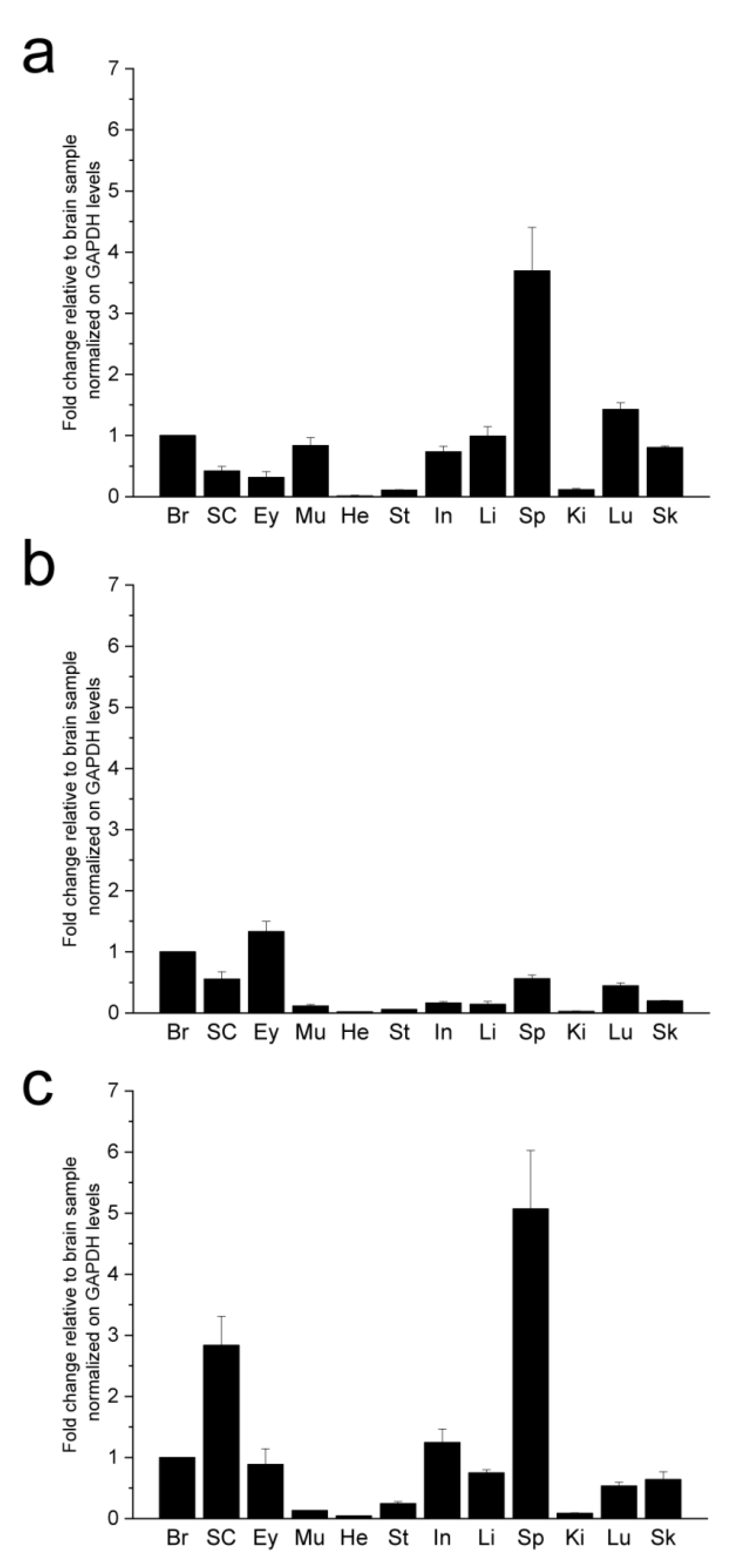
2.1. Synuclein Gene Expression

Syn gene expression was evaluated in the major organs of the Xenopus by qRT-PCR (Figure 2). The availability of the mRNA sequences coding for syn isoforms allowed us to design specific primers able to discriminate α-, β- and γ-syn expression (Table 2). The three isoforms were expressed both in nervous (brain, spinal cord and eye) and non-nervous (intestine, kidney, liver, lung, muscle, skin, stomach, heart and spleen) organs (Figure 2). In the nervous system, syns showed differences in the relative expression pattern as α-syn was more expressed in the brain, β-syn in the eye and γ-syn in the spinal cord. Among non-nervous tissues, high levels of α-syn mRNA were detected in the spleen and lung, discrete levels in the muscle, intestine, liver, and skin, whereas low or undetectable levels were detected in the kidney, stomach and heart. Discrete expression of β-syn mRNA was detected in the spleen and lung, and γ-syn expression was particularly high in the spleen and discrete in intestine, liver, lung and skin (Figure 2).

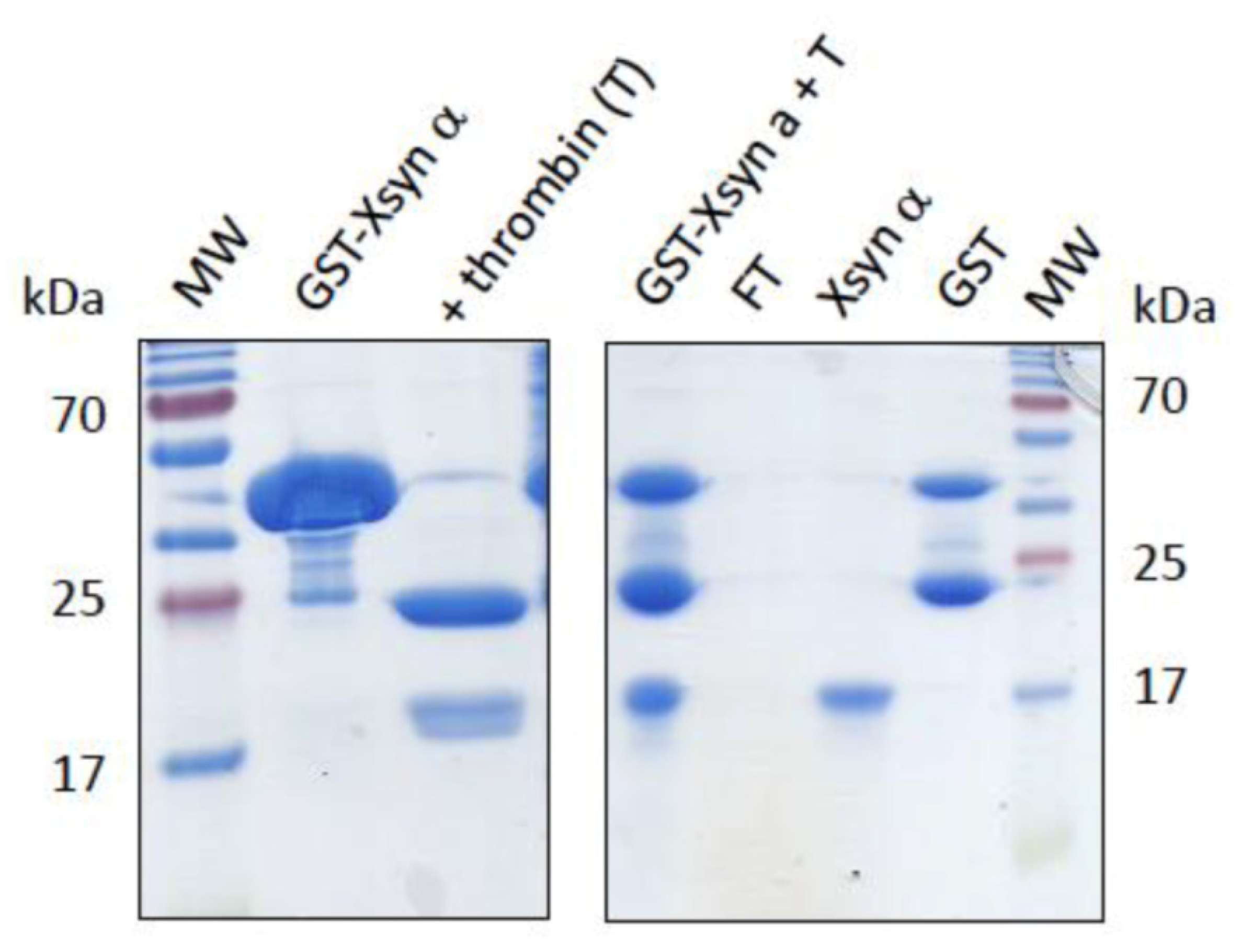
2.2. Synuclein Recombinant Protein Production

The coding sequences of the three Xenopus syn isoforms were cloned in pGEX-2T and the recombinant GST-syn fusion proteins were expressed in E. coli BL21 (DE3) and purified by affinity chromatography on GSH-Sepharose. The GST-syn fusion protein was recovered with high yield and high purity (Figure 3, left panel). The GST tag was removed by treatment with thrombin followed by separation on GSH-Sepharose and recovery of pure syn in the wash fraction. Figure 3 shows representative results obtained for α-syn; all syn isoforms were obtained and purified in the same conditions with similar yield and purity.

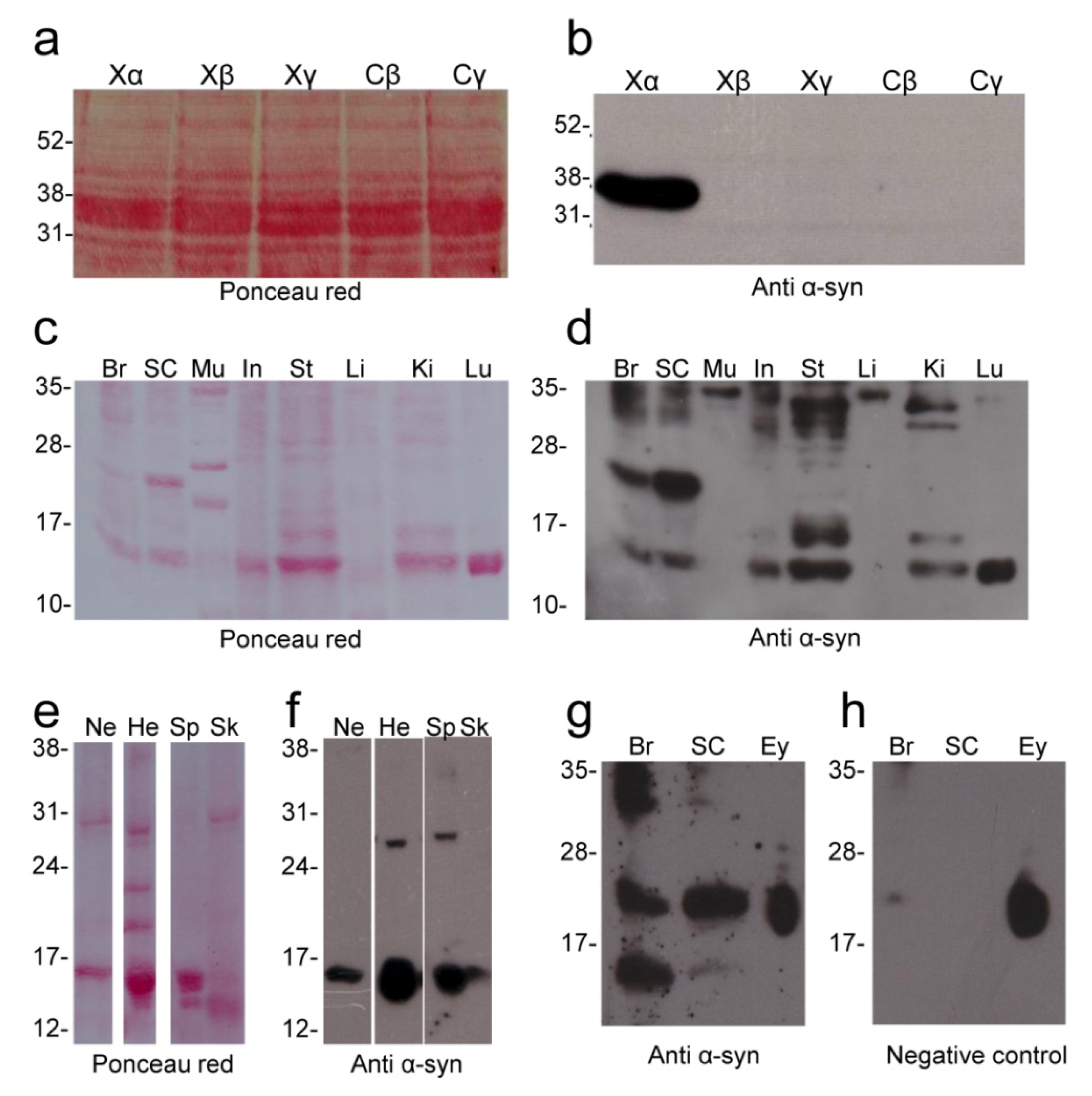
2.3. Alpha Synuclein Antibody Validation

Antibodies able to recognize and discriminate the different syn isoforms are needed to study their expression and distribution. Given the high level of homology between human and Xenopus α-syn (Figure 1a–c), commercial antibodies against human α-syn might be successfully used also in the Xenopus. However, the selection of suitable commercial antibodies specific for α-syn is not easy due to the high homology of the first 100 amino acids of Xenopus syns (Figure 1d,e) and to the low homology between the C-terminus of human and Xenopus α-syn (Figure 1a).
In this work, Xenopus recombinant α-, β- and γ-syns were used to test, by Western blot, the ab27766 (abcam, UK) antibody directed against the 115–122 amino acid region of human α-syn (Figure 1a,b). The ab27766 antibody intensely labelled a band corresponding to the recombinant α-syn fused to glutathione S-transferase (GST, GST-tagged α-syn), while it does not recognize Xenopus and carp β- and γ-syn (Figure 4b). The results identify this antibody as suitable for the recognition and discrimination of α-syn in Xenopus.

2.4. Alpha Synuclein Protein Expression

The same antibody (ab27766) was used to verify the α-syn protein expression in main Xenopus organs by Western blot (Figure 4b–h). The main purpose of these analyses was to confirm and corroborate the data obtained from qRT-PCR experiments demonstrating the effective expression of α-syn protein in the brain and other organs analysed.
An immunolabelled band at 14–15 kDa (corresponding to the α-syn predicted MW) was detected in most of the organs examined (brain, spinal cord, nerve, intestine, stomach, kidney, lung, heart, spleen and skin) (Figure 4d,f,g). Moreover, immunolabelled bands at higher molecular weight that could correspond to oligomers were detected. An intense immunolabelled band was also detected at 26–27 kDa in the CNS (brain and spinal cord) and in the heart and spleen. Bands at higher molecular weight (range 31–35 kDa) were also detected in most organs. Interestingly, the skeletal muscle and liver showed only an immunolabelled band at 35 kDa. In the eye, no immunolabelled bands were observed at 14–15 kDa. It was not possible to establish the presence of any bands at 26–27 kDa in the eye due to the presence of intense non-specific immunolabelling in the range 20–27 kDa, as shown by negative control in which the primary antibody was omitted (Figure 4h).

2.5. Immunohistochemical Analysis

The ab27766 antibody was also tested in a preliminary immunohistochemical experiment (Figure 5). The results showed α-syn positive soma and nerve fibres in the brain (Figure 5a–d). By way of example, positive neurons were detected in the interpeduncular nucleus (Figure 5c) and immunolabelled fibres in the tractus opticus marginalis (Figure 5d). Moreover, α-syn immunostaining was detected in the inner and outer plexiform layer of the retina (Figure 5e). The immunohistochemical analysis of skeletal muscle (Figure 5g,h), heart (Figure 5i,j) and stomach (Figure 5k,l) samples revealed α-syn immunolabelling mainly limited to the nerve fibres and neuromuscular junctions. Based on these results, it will be possible in future studies to carry out an in-depth study of α-syn distribution in the CNS and organs of Xenopus.

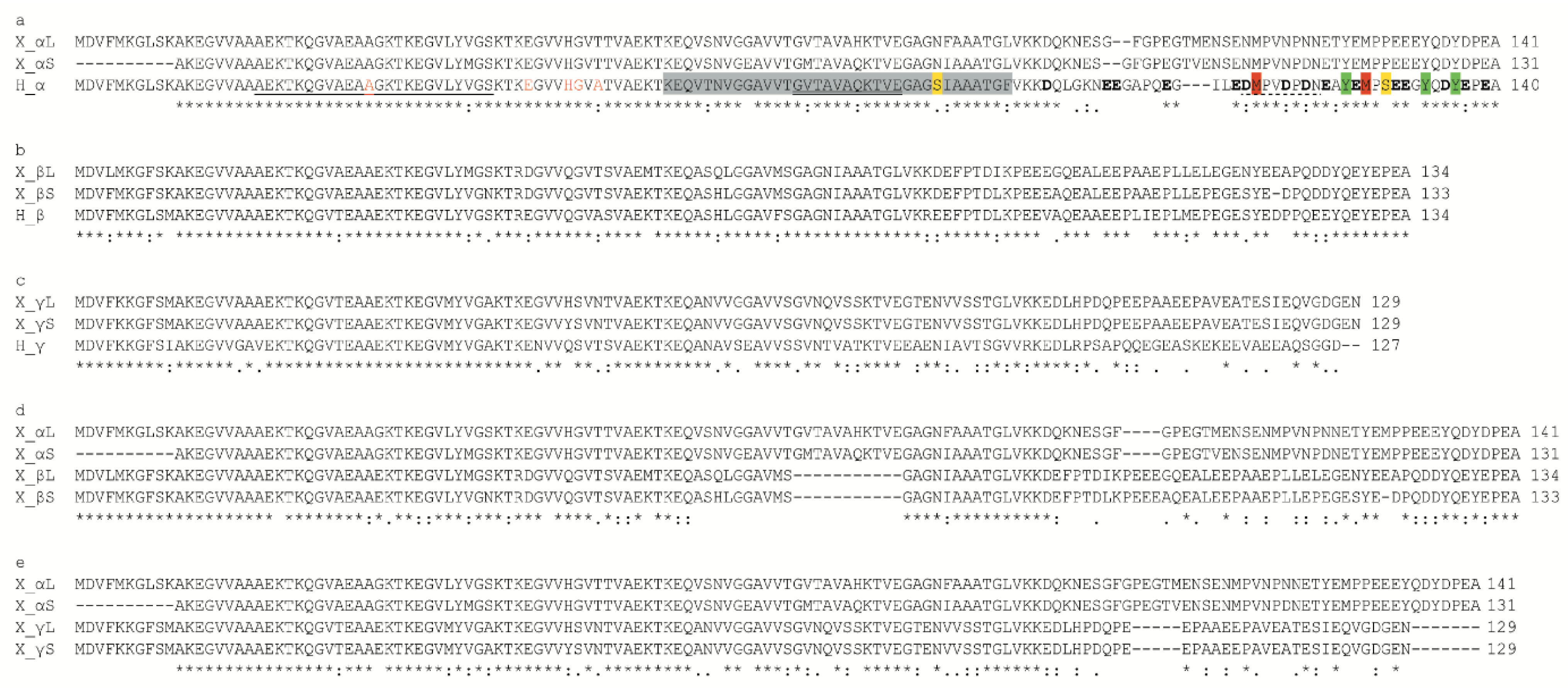
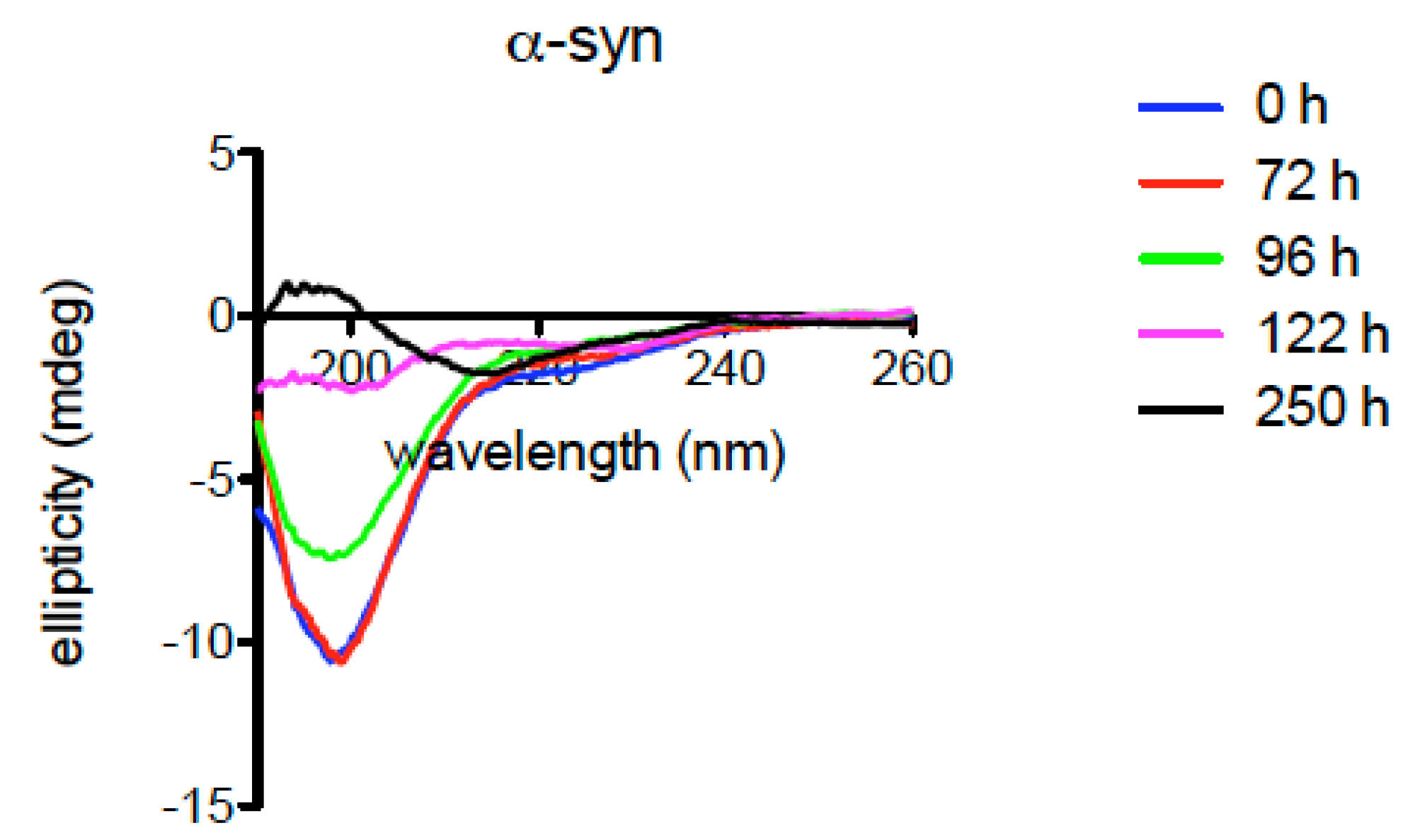
2.6. Structural Characteristics of Xenopus Synucleins

Spectroscopic analyses of Xenopus recombinant syns were performed to evaluate the properties of the purified proteins. The fluorescence spectra presented in Figure 6 were characterized by tyrosine emission due to the lack of tryptophan residues in the amino acid sequence of all isoforms of the protein. Four tyrosines are present in α- and β-syn sequences and one in γ-syn, all of which are conserved in the corresponding human isoforms. The α- and β-syn spectra were very similar and presented an intense peak at about 302 nm, typical of tyrosine. The γ-syn spectrum displayed a broader peak with a shoulder between 340–360 nm, which was weakly visible also in the β-syn spectrum. This feature has been suggested to depend on the tyrosinate form of this residue due to the possible proton transfer to nearby acidic residues [55].
Far-UV CD spectra of the proteins confirmed the predominantly unfolded state of all the three isoforms (Figure 7a) with a typical intense negative peak at about 200 nm. The anionic detergent SDS has been used to test the interaction of syns with micelles/membranes and to evaluate the conformational transition from unfolded to α-helical secondary structure [56]. After the addition of SDS, the CD spectra clearly showed a shift towards an α-helical structure (Figure 7b), in line with previous reports on mammalian syn isoforms [57,58].
Syns have the ability to bind metals such as copper, which may be physiologically relevant [59,60,61,62]. The addition of copper did not change the CD features of all three Xenopus syn isoforms (Figure 7c). These results suggest that there are no gross differences among syn isoforms, at least as far as secondary structure and response to membranes and copper are concerned. The presence of the PD-linked variant A53T in Xenopus α-syn does not appear to impact the secondary structure of the protein. It should be reminded that threonine in position 53 occurs naturally in most mammalian species [63] and its role in toxicity can be significantly altered/decreased by combination with other substitutions. The results of the CD analyses indicate that Xenopus syns appear to be similar to mammalian syns as far as their behaviour in the presence of SDS or copper is concerned.
To further investigate the properties of Xenopus α-syn, the protein was incubated at 37 °C for 250 h, and CD spectra were recorded at the indicated times (Figure 8). The results demonstrate progressive conversion from the unfolded state to a β-sheet secondary structure, typical of aggregated syn (Figure 8). Further experiments are needed to fully evaluate the propensity of Xenopus syn isoforms to aggregate and form fibrils.

3. Discussion

Syn proteins are expressed in representatives of all vertebrates, although differences in the number of coding genes and isoforms can be identified, especially in teleosts [2], in which a major variability in the number of syn isoforms has been observed. This has been attributed to the whole genome duplication that occurred in the ray-finned fishes (Osteichthyes, Actinopterygii) about 230 million years ago, which was followed by the subsequent loss of some duplicated genes, depending on the species [2]. For example, the zebrafish Danio rerio, a teleost model organism in neuroscience, possesses a gene for β-syn, two genes for γ-syn (γ1 and γ2), but no gene for α-syn [2].
Among amphibians, Xenopus is a model organism widely used in scientific research. It is an allotetraploid organism with 36 chromosomes (2n = 36), which is believed to derive from the interspecific hybridization of diploid progenitors with 2n = 18, which occurred 17–18 million years ago [53]. The chromosomes deriving from each diploid progenitor are referred to as L (longer) and S (shorter) due to their different lengths. Due to the tetraploid condition, most Xenopus genes have two copies, defined as L and S. Consequently, six genes coding for syns (two genes for each isoform) have been sequenced in the Xenopus genome. The amino acid sequences of the two proteins encoded by each pair of L and S genes showed a high homology degree, suggesting that both homologous isoforms may have similar physiological functions, although specific studies must be carried out to verify whether L and S isoforms can be involved in different physiological processes.
Xenopus and human syns show a good degree of homology, suggesting that the conservation of the amino acid sequences may correspond to conserved cellular and physiological roles. In particular, the conservation of the apolipoprotein lipid-binding motif, putative sites of phosphorylation or interaction with metals, suggests that α-syn can have similar biological functions in Xenopus and humans. However, some differences that could impact the properties of the protein were observed. In fact, the comparison between human and Xenopus α-syn showed the A53T substitution in both Xenopus L and S proteins. This is not surprising since a threonine at position 53 occurs naturally in α-syn of most animals [54,63]. However, in humans and other mammals, the physiological α-syn isoform has an alanine at position 53, and the A53T substitution is associated with familial forms of PD. Moreover, the A53T α-syn has been shown to exhibit a greater tendency to aggregate than the physiological isoform [64,65]. Since synucleinopathies are related to α-syn aggregation and accumulation in the CNS, it is believed that the presence of the A53T substitution may facilitate these processes and PD onset. In this respect, Xenopus can be a potential model of synucleinopathy since it physiologically expresses α-syn with the A53T substitution, and the tetraploid condition could increase the intracellular amount of α-syn, both conditions potentially favouring protein aggregation. CD analyses confirm that Xenopus α-syn converts to a β-sheet secondary structure, indicative of aggregation. Further studies are needed to investigate in detail the tendency of Xenopus α-syn to aggregate in vitro and in vivo.
Studies on syn gene expression during Xenopus development have reported that α-syn gene (snca) is expressed since the gastrula stage and it is localized in the olfactory placode, brain, otic vesicle, branchial arches, and somites in the bud stage [39], thus suggesting a wide distribution in the embryos. These results have been confirmed in adult Xenopus by present qRT-PCR results, revealing α-syn gene expression in most of the organs analysed. High levels were detected in the CNS (brain and spinal cord), eye and spleen, consistent with results obtained in mice [66]. Discrete levels were also observed in the lung, skin, liver, skeletal muscle and intestines. The α-syn expression in skeletal muscle and lung was consistent with results obtained in humans [67], whereas α-syn expression in Xenopus heart appeared lower compared to lizards [41] and humans [67].
The β-syn expression in humans has a more restricted distribution compared to α-syn [67,68,69], and this was also evident in Xenopus embryos where β-syn gene expression was limited mainly to the brain and spinal cord at the tail bud and tadpole stages [39]. Present results demonstrate that β-syn expression remains limited to the brain, spinal cord, eyes, spleen, lung and skin, even in the adult frog. In contrast, moderate β-syn levels were observed in a higher number of organs in the lizard Anolis carolinensis [41].
The γ-syn gene expression in Xenopus embryos became detectable at the gastrula stage and was mainly localized in cranial nerves, cranial and dorsal root ganglia and pineal gland in the tail bud stage [39]. In adult Xenopus, high levels of γ-syn expression are detected in the CNS (brain and spinal cord), eye, spleen, liver, intestine, lung and skin by qRT-PCR analysis, whereas low levels are detected in muscle and the heart. The reduced syn expression in the heart is consistent with results obtained in the lizard [41].
As is well known, the analysis of syn protein expression requires specific antibodies capable of discriminating one specific isoform from the others. For this purpose, in the present work we have produced Xenopus α-, β- and γ-syn recombinant proteins that were used in Western blot experiments to test the ability of the ab27766 monoclonal antibody against mammalian α-syn, to recognize Xenopus α-syn and not β- and γ-syns. The same antibody was then used to verify α-syn expression in the main organs of adult Xenopus. The ab27766 antibody (abcam, UK) tested by immunohistochemistry on nervous and non-nervous tissues has proved to be a suitable tool to localize Xenopus α-syn. Western blotting showed the presence of a band at about 15 kDa (consistent with α-syn predicted molecular weight, MW) in most of the organs examined, confirming the wide distribution of α-syn, consistent with results in humans and monkey, showing that α-syn is highly enriched in the brain, but also widely distributed in other tissues, especially at fetal stages [70,71]. Immunolabelled bands were also detected at higher molecular weight that could represent α-syn dimers or oligomers. In interpreting Western blot results, it must be considered that two different genes, encoding L- and S-α-syn, respectively, are present in the Xenopus genome. The complete L-α-syn sequence encoding a protein with a predicted MW of 14.7 kDa is available in the current genome assembly, while the S-α-syn sequence is incomplete, and consequently its MW cannot be accurately estimated. Furthermore, both L- and S-α-syn are characterized by the A53T substitution, which could make the proteins more prone to aggregate into oligomers. These features could explain the presence of immunolabelled bands with MW higher than 17 kDa. Interestingly, an intense immunolabelled band at 26–27 kDa was observed mainly in the CNS (brain and spinal cord), that could result from the high expression of specific α-syn isoforms in these tissues. A precise analysis of α-syn expression in the eye was not possible by Western blot due to the presence of a non-specific labelling in the 20–27 kDa range. Surprisingly, the non-specific labelling was not observed in the immunohistochemical analysis of the eye. The α-syn immunolabelling was observed both in neuronal soma and nerve fibres in the CNS. In the eye, immunopositivity was observed in the inner and outer plexiform layer confirming previous studies [72], and suggesting an evolutionarily conserved role of α-syn in this organ. Immunopositive labelling for α-syn was also detected in muscle tissues. In the cross section of skeletal muscle fibres, immunolabelled spots were observed at the sarcolemmal level, suggesting α-syn localization in neuromuscular junctions. This is consistent with results demonstrating α-syn presence, especially in the postsynaptic domain of neuromuscular junctions in humans [73]. However, TEM observations are necessary to detail α-syn distribution at this level. As observed in humans [74], α-syn positive fibres were also observed in Xenopus stomach.
Similar to present data, previous experiments by Yuan and collaborators [37] used a Xenopus β-syn recombinant protein to produce a specific antibody against β-syn, through which they revealed β-syn expression in the brain but not in the heart and liver. Interestingly, these results are consistent with the scarce or absent β-syn gene expression detected in the same organs by qRT-PCR.
Human syns are natively unfolded proteins [75,76] which can undergo conformational changes following interaction with membranes [76,77], or alteration of physicochemical parameters such as temperature and pH [55]. The protein conformation strongly influences syn tendency to aggregate [55,78] and, especially for α-syn, to contribute to the onset of synucleinopathies. The production of purified recombinant proteins in Xenopus allowed us to begin the characterization of the structural features of amphibian syns.
In line with the high sequence homology with human syns, the fluorescence and CD data indicate general conservation of the secondary structure and response to detergents and copper. Moreover, like human α-syn, the Xenopus α isoform changes its conformation over time in experiments of in vitro incubation at 37 °C, converting to a β-sheet secondary structure. Overall, these results reinforce the proposition that Xenopus may be a good model for the study of synucleinopathies.
The availability of recombinant syns will make it possible to carry out further in-depth analyses to highlight possible subtle differences between amphibian and human syn that could corroborate the goodness of Xenopus as a model for the study of synucleinopathies, and provide new insights into the role and evolution of vertebrate syn proteins.

4. Materials and Methods

4.1. Animals and Sampling

Nervous (brain, eye, spinal cord and nerve) and non-nervous organs (intestine, kidney, liver, lung, muscle, skin, stomach, heart, spleen) from 14 adult individuals of Xenopus were used. Tissues from 12 animals were stored in RNA Later (Ambion, Austin, TX, USA) at −70 °C until they were processed for RNA or protein extraction, and those from 2 animals were fixed in PFA fixative (4% paraformaldehyde in 0.1 M phosphate buffer) and then stored at 4 °C in 0.01 M phosphate buffer (PB) containing 15% of sucrose until they were processed for immunohistochemistry.

4.2. qRT-PCR

Total RNA was isolated from tissues pooled from 6 animals with the use of PureLink RNA® Mini Kit (Ambion), according to the manufacturer’s instructions, and quantified spectrophotometrically by Optizen Pop Bio (Mecasys). The mRNAs obtained were reverse-transcribed into cDNAs using oligo dT and SuperScript™ II Reverse Transcriptase (Invitrogen); then, cDNA was stored at − 20 °C until use. The qPCR was performed in 10 µL with a primer concentration of 1 μM, 10 ng cDNA and 1× SYBR Green Qpcr Master Mix (EURx) and carried out in the BIOER Line-Gene K PCR. The amplification setup consisted of an initial denaturation step at 95 °C for 2 min and 40 cycles of denaturation at 95 °C for 5 s, annealing at 66 °C for 30 s and extension at 72 °C for 30 s. Samples were analysed in triplicate on separate reactions to avoid technical measurement errors. Primer pairs used for qPCR analyses were designed by using the Primer3 software (version 4.1.0 [79]) [80]. Primers sequences are reported in Table 2. The relative expression levels for each gene were calculated by the 2−ΔΔCT method, and normalized using the relative expression of GAPDH.

4.3. Cloning and Production of Recombinant Proteins

Coding sequences for α-, β- and γ-syns were obtained by PCR on an aliquot of the same cDNAs employed for qRT-PCR. Primer sequences are reported in Table 3. Xenopus α-, β- and γ-syn isoforms were cloned BamHI-EcoRI in pGEX-2T to produce GST-fusion proteins. All plasmids were sequence-verified before transformation into E. coli BL21(DE3) cells that were grown in LB medium supplemented with ampicillin to OD600 0.5–0.6 when GST-syn expression was induced with 0.1 mM IPTG at 37 °C for 2–3 h. Cells were harvested and stored frozen at −80 °C until use. Cells were resuspended in lysis buffer (25 mM Mops pH 7, containing 150 mM NaCl, 1 mM PMSF, 1 mg/mL lysozyme) and sonicated to obtain a lysate that was clarified by centrifugation at 20,000× g for 20 min. All GST-syn isoforms were purified on GSH-Sepharose Fast Flow (GE Healthcare, Chicago, IL, USA) according to the manufacturer’s instructions. To remove the GST tag, the purified fusion protein was treated with thrombin (GE Healthcare, Chicago, IL, USA) for 2 h and repurified on GSH-Sepharose. Syn was recovered in the unbound and wash fractions. The purified protein was concentrated by ultrafiltration with Vivaspin10K filters (Sartorius). Protein content was measured with the microBCA assay and spectrophotometrically. CD spectra were recorded on a Jasco J-810 spectropolarimeter in the range 260–190 nm, with 0.1 cm cuvettes. All spectra are the average of at least 4 scans with buffer subtracted. For CD analyses, the proteins were exchanged in 10 mM potassium phosphate buffer pH 7, containing 50 mM Na2SO4 to remove chloride. Fluorescence spectra were recorded on a Fluoromax Jobin Yvon spectrofluorimeter at 20 °C with a 0.4 × 1 cm cuvette (excitation along the 0.4 cm path-length). Excitation was at 270 nm, emission spectra were collected between 280 and 500 nm, and the excitation and emission slit width was 5 nm.

4.4. Western Blot Experiments

Samples previously collected and constituted of tissues pooled from 6 animals were homogenized in a denaturing lysis buffer containing 30 mM Tris/HCl (pH 7.4), 1.5% sodium dodecyl sulphate (SDS, w/v), 8 mM EDTA (v/v) and 50 mM dithiothreitol (DTT, v/v) [41] and protease inhibitors (Roche, Indianapolis, IN, USA); then, the particulate matter was removed by centrifugation at 14,000× g for 20 min. The protein concentration was determined by the Bradford assay. For SDS-PAGE analysis, proteins were denatured by boiling in Laemmli Sample Buffer for 5 min. Then, 50 μg of protein was loaded in each lane and separated in 15% SDS-polyacrylamide gels according to Laemmli [81]. After electrophoresis, gels were transferred to nitrocellulose paper (Hybond C+ Extra, GE Healthcare, Chicago, IL, USA) and membranes were stained with Ponceau S to confirm the transfer of proteins. The saturation step was performed by incubating membranes in 5% bovine serum albumin (BSA) in TBS-Tween for 2 h at room temperature [35]. Then membranes were incubated overnight in the anti-α syn primary antibody ab27766 (abcam, Cambridge, UK) diluted at 1:1000, and for 1 h at room temperature with the HRP-conjugated anti-mouse secondary antibody (Sigma-Aldrich Cat# A9044). Detection was done using the Westar μC Ultra enhanced chemiluminescent HRP substrate (Cyanagen, Bologna, Italy) and Kodak X Omat LS films (Sigma-Aldrich, St. Louis, MO, USA).

4.5. Immunohistochemical Analysis

Samples were fixed by immersion in PFA fixative (4% paraformaldehyde in 0.1 M phosphate buffer), pH 7 at 4 °C for 24 h, then stored at 4 °C in 0.01 M phosphate buffer (PB) containing 15% of sucrose, embedded in PB containing 10% gelatin and frozen. Samples were frozen and cut on a cryostat (HM 505 E, Microm, Walldorf, Germany) into 30-µm-thick coronal serial sections that were stored until use in 24-well plates containing cold 15% sucrose PB. Sections were enumerated to avoid misplacement, maintaining the seriality. Before immunohistochemical staining, the free-floating sections were treated with 0.01 M phosphate-buffered saline (PBS) containing 0.3% Triton X-100 (PBST) at 4 °C for 2 or 3 days to improve tissue permeability. Sections were pre-treated for 1 h at room temperature with PBST containing 0.1% sodium azide and 0.5% H2O2. To avoid the non-specific antibody binding, sections were pre-incubated with normal horse serum (Vector Laboratories, Newark, NJ, USA) diluted 1:50 in PBST, containing 1% bovine serum albumin (BSA, Sigma-Aldrich, St. Louis, MO, USA). Free-floating sections were then incubated for 5 days at 4 °C with ab27766 antibody (abcam, Cambridge, UK) dilution 1:10,000. Sections were then incubated for 1 h at room temperature with biotinylated horse anti-mouse immunoglobulin (Vector Laboratories) and then incubated for 45 minutes at room temperature with avidin-biotin-peroxidase complex (ABC, Elite Kit; Vector Laboratories, Newark, NJ, USA) diluted 1:2000 with PBST. The peroxidase activity was evidenced by a reaction with a solution containing 0.04% of 3.3-diaminobenzidine-tetrahydrochloride (DAB, Fluka, Buchs, Switzerland), 0.4% of nickel ammonium sulphate, and 0.003% of H2O2 in 0.5 M Tris-HCl buffer, pH 7.6 for 3 minutes at room temperature. Some sections were counterstained with the Nuclear Fast Red (Kernechtrot) solution (Sigma-Aldrich, St. Louis, MO, USA) after the immunohistochemistry procedure. For control experiments, the primary antiserum was substituted with buffer or normal rabbit serum. None of the control sections showed positive immuno-staining. The stained sections were mounted on glass slides maintaining the seriality, dehydrated, cleared, and cover-slipped with Permount (Fisher Scientific, Pittsburgh, PA, USA).

4.6. Preparation of Figures

Microsoft Power Point and Corel Draw software and BioRender [82] were used for the preparation of the figures.

5. Conclusions

The analysis of the sequence, secondary structure, expression and distribution of α-syn in Xenopus laevis (Figure 9) suggests that this amphibian, historically widely used in scientific research, may constitute a good model for the study of synucleinopathies.

Supplementary Materials

The following supporting information can be downloaded at: www.mdpi.com/article/10.3390/ijms23116058/s1.

Author Contributions

Conceptualization, M.C.B.d.P. and M.T.; methodology, M.C.B.d.P., E.A., A.C., R.V. and M.T.; formal analysis, M.C.B.d.P. and M.T.; investigation, M.C.B.d.P. and M.T.; resources, M.C.B.d.P. and M.T.; data curation, M.C.B.d.P. and M.T.; writing—original draft preparation, M.C.B.d.P. and M.T.; writing—review and editing, M.C.B.d.P., E.A., A.C., R.V., C.C. and M.T.; supervision, M.T.; project administration, M.T.; funding acquisition, M.T. All authors have read and agreed to the published version of the manuscript.

Funding

This research was funded by Sapienza University of Rome, Progetto Ateneo 2015 (C26A15N7NF) and 2020 (RM1201729CB8436F).

Institutional Review Board Statement

All procedures performed in studies involving animals were in accordance with the ethical standards of the institution or practice at which the studies were conducted and were approved by the Italian administrative order DM 70/96 of Italian Ministry of Health.

Acknowledgments

We thank Chiara Adamo and Alessandra Di Concilio for assistance in some experiments, and Eleonora Palma for kindly providing some samples. All applicable international, national, and/or institutional guidelines for the care and use of animals were followed.

Conflicts of Interest

The authors declare no conflict of interest. The funders had no role in the design of the study; in the collection, analyses, or interpretation of data; in the writing of the manuscript, or in the decision to publish the results.

References

  1. Maroteaux, L.; Campanelli, J.T.; Scheller, R.H. Synuclein: A neuron-specific protein localized to the nucleus and presynaptic nerve terminal. J. Neurosci. 1988, 8, 2804–2815. [Google Scholar] [CrossRef] [PubMed] [Green Version]
  2. Toni, M.; Cioni, C. Fish Synucleins: An Update. Mar. Drugs 2015, 13, 6665–6686. [Google Scholar] [CrossRef] [PubMed]
  3. Cookson, M.R. The biochemistry of Parkinson’s disease. Annu. Rev. Biochem. 2005, 74, 29–52. [Google Scholar] [CrossRef] [PubMed] [Green Version]
  4. Gasser, T. Mendelian forms of Parkinson’s disease. Biochim. Biophys. Acta 2009, 1792, 587–596. [Google Scholar] [CrossRef] [PubMed] [Green Version]
  5. Goedert, M. Alpha-synuclein and neurodegenerative diseases. Nat. Rev. Neurosci. 2001, 2, 492–501. [Google Scholar] [CrossRef] [PubMed]
  6. Waxman, E.A.; Giasson, B.I. Molecular mechanisms of alpha-synuclein neurodegeneration. Biochim. Biophys. Acta 2009, 1792, 616–624. [Google Scholar] [CrossRef] [Green Version]
  7. Wakabayashi, K.; Mori, F.; Tanji, K.; Orimo, S.; Takahashi, H. Involvement of the peripheral nervous system in synucleinopathies, tauopathies and other neurodegenerative proteinopathies of the brain. Acta. Neuropathol. 2010, 120, 1–12. [Google Scholar] [CrossRef]
  8. Braak, H.; de Vos, R.A.; Bohl, J.; Del Tredici, K. Gastric alpha-synuclein immunoreactive inclusions in Meissner’s and Auerbach’s plexuses in cases staged for Parkinson’s disease-related brain pathology. Neurosci. Lett. 2006, 396, 67–72. [Google Scholar] [CrossRef]
  9. Pfefferkorn, C.M.; Jiang, Z.; Lee, J.C. Biophysics of alpha-synuclein membrane interactions. Biochim. Biophys. Acta 2012, 1818, 162–171. [Google Scholar] [CrossRef] [Green Version]
  10. Dikiy, I.; Eliezer, D. Folding and misfolding of alpha-synuclein on membranes. Biochim. Biophys. Acta 2012, 1818, 1013–1018. [Google Scholar] [CrossRef] [Green Version]
  11. Spillantini, M.G. Parkinson’s disease, dementia with Lewy bodies and multiple system atrophy are alpha-synucleinopathies. Parkinsonism Relat. Disord. 1999, 5, 157–162. [Google Scholar] [CrossRef]
  12. Luk, K.C.; Lee, V.M. Modeling Lewy pathology propagation in Parkinson’s disease. Parkinsonism Relat. Disord. 2014, 20, S85–S87. [Google Scholar] [CrossRef] [Green Version]
  13. Irwin, D.J.; Lee, V.M.; Trojanowski, J.Q. Parkinson’s disease dementia: Convergence of alpha-synuclein, tau and amyloid-beta pathologies. Nat. Rev. Neurosci. 2013, 14, 626–636. [Google Scholar] [CrossRef] [PubMed]
  14. Norris, E.H.; Giasson, B.I.; Lee, V.M. Alpha-synuclein: Normal function and role in neurodegenerative diseases. Curr. Top. Dev. Biol. 2004, 60, 17–54. [Google Scholar] [CrossRef] [PubMed]
  15. Hamilton, B.A. alpha-Synuclein A53T substitution associated with Parkinson disease also marks the divergence of Old World and New World primates. Genomics 2004, 83, 739–742. [Google Scholar] [CrossRef] [PubMed]
  16. Rospigliosi, C.C.; McClendon, S.; Schmid, A.W.; Ramlall, T.F.; Barre, P.; Lashuel, H.A.; Eliezer, D. E46K Parkinson’s-linked mutation enhances C-terminal-to-N-terminal contacts in alpha-synuclein. J. Mol. Biol. 2009, 388, 1022–1032. [Google Scholar] [CrossRef] [Green Version]
  17. Appel-Cresswell, S.; Vilarino-Guell, C.; Encarnacion, M.; Sherman, H.; Yu, I.; Shah, B.; Weir, D.; Thompson, C.; Szu-Tu, C.; Trinh, J.; et al. Alpha-synuclein p.H50Q, a novel pathogenic mutation for Parkinson’s disease. Mov. Disord. 2013, 28, 811–813. [Google Scholar] [CrossRef] [PubMed]
  18. Lesage, S.; Anheim, M.; Letournel, F.; Bousset, L.; Honore, A.; Rozas, N.; Pieri, L.; Madiona, K.; Durr, A.; Melki, R.; et al. G51D alpha-synuclein mutation causes a novel parkinsonian-pyramidal syndrome. Ann. Neurol. 2013, 73, 459–471. [Google Scholar] [CrossRef]
  19. Proukakis, C.; Dudzik, C.G.; Brier, T.; MacKay, D.S.; Cooper, J.M.; Millhauser, G.L.; Houlden, H.; Schapira, A.H. A novel alpha-synuclein missense mutation in Parkinson disease. Neurology 2013, 80, 1062–1064. [Google Scholar] [CrossRef] [Green Version]
  20. Mbefo, M.K.; Fares, M.B.; Paleologou, K.; Oueslati, A.; Yin, G.; Tenreiro, S.; Pinto, M.; Outeiro, T.; Zweckstetter, M.; Masliah, E.; et al. Parkinson disease mutant E46K enhances alpha-synuclein phosphorylation in mammalian cell lines, in yeast, and in vivo. J. Biol. Chem. 2015, 290, 9412–9427. [Google Scholar] [CrossRef] [Green Version]
  21. Kruger, R.; Kuhn, W.; Muller, T.; Woitalla, D.; Graeber, M.; Kosel, S.; Przuntek, H.; Epplen, J.T.; Schols, L.; Riess, O. Ala30Pro mutation in the gene encoding alpha-synuclein in Parkinson’s disease. Nat. Genet. 1998, 18, 106–108. [Google Scholar] [CrossRef] [PubMed]
  22. Polymeropoulos, M.H.; Lavedan, C.; Leroy, E.; Ide, S.E.; Dehejia, A.; Dutra, A.; Pike, B.; Root, H.; Rubenstein, J.; Boyer, R.; et al. Mutation in the alpha-synuclein gene identified in families with Parkinson’s disease. Science 1997, 276, 2045–2047. [Google Scholar] [CrossRef] [PubMed] [Green Version]
  23. Zarranz, J.J.; Alegre, J.; Gomez-Esteban, J.C.; Lezcano, E.; Ros, R.; Ampuero, I.; Vidal, L.; Hoenicka, J.; Rodriguez, O.; Atares, B.; et al. The new mutation, E46K, of alpha-synuclein causes Parkinson and Lewy body dementia. Ann. Neurol. 2004, 55, 164–173. [Google Scholar] [CrossRef] [PubMed]
  24. Singleton, A.B.; Farrer, M.; Johnson, J.; Singleton, A.; Hague, S.; Kachergus, J.; Hulihan, M.; Peuralinna, T.; Dutra, A.; Nussbaum, R.; et al. alpha-Synuclein locus triplication causes Parkinson’s disease. Science 2003, 302, 841. [Google Scholar] [CrossRef] [PubMed] [Green Version]
  25. Kiely, A.P.; Asi, Y.T.; Kara, E.; Limousin, P.; Ling, H.; Lewis, P.; Proukakis, C.; Quinn, N.; Lees, A.J.; Hardy, J.; et al. alpha-Synucleinopathy associated with G51D SNCA mutation: A link between Parkinson’s disease and multiple system atrophy? Acta Neuropathol. 2013, 125, 753–769. [Google Scholar] [CrossRef] [Green Version]
  26. Prusiner, S.B.; Woerman, A.L.; Mordes, D.A.; Watts, J.C.; Rampersaud, R.; Berry, D.B.; Patel, S.; Oehler, A.; Lowe, J.K.; Kravitz, S.N.; et al. Evidence for alpha-synuclein prions causing multiple system atrophy in humans with parkinsonism. Proc. Natl. Acad. Sci. USA 2015, 112, E5308–E5317. [Google Scholar] [CrossRef] [Green Version]
  27. Davidson, W.S.; Jonas, A.; Clayton, D.F.; George, J.M. Stabilization of alpha-synuclein secondary structure upon binding to synthetic membranes. J. Biol. Chem. 1998, 273, 9443–9449. [Google Scholar] [CrossRef] [Green Version]
  28. Weinreb, P.H.; Zhen, W.; Poon, A.W.; Conway, K.A.; Lansbury, P.T., Jr. NACP, a protein implicated in Alzheimer’s disease and learning, is natively unfolded. Biochemistry 1996, 35, 13709–13715. [Google Scholar] [CrossRef]
  29. El-Agnaf, O.M.; Jakes, R.; Curran, M.D.; Middleton, D.; Ingenito, R.; Bianchi, E.; Pessi, A.; Neill, D.; Wallace, A. Aggregates from mutant and wild-type alpha-synuclein proteins and NAC peptide induce apoptotic cell death in human neuroblastoma cells by formation of beta-sheet and amyloid-like filaments. FEBS Lett. 1998, 440, 71–75. [Google Scholar] [CrossRef] [Green Version]
  30. Narhi, L.; Wood, S.J.; Steavenson, S.; Jiang, Y.; Wu, G.M.; Anafi, D.; Kaufman, S.A.; Martin, F.; Sitney, K.; Denis, P.; et al. Both familial Parkinson’s disease mutations accelerate alpha-synuclein aggregation. J. Biol. Chem. 1999, 274, 9843–9846. [Google Scholar] [CrossRef] [Green Version]
  31. Burre, J.; Sharma, M.; Sudhof, T.C. Cell Biology and Pathophysiology of alpha-Synuclein. Cold Spring Harb. Perspect. Med. 2018, 8, a024091. [Google Scholar] [CrossRef] [PubMed]
  32. Toni, M.; Massimino, M.L.; De Mario, A.; Angiulli, E.; Spisni, E. Metal Dyshomeostasis and Their Pathological Role in Prion and Prion-Like Diseases: The Basis for a Nutritional Approach. Front Neurosci. 2017, 11, 3. [Google Scholar] [CrossRef] [PubMed] [Green Version]
  33. Sulzer, D.; Edwards, R.H. The physiological role of alpha-synuclein and its relationship to Parkinson’s Disease. J. Neurochem. 2019, 150, 475–486. [Google Scholar] [CrossRef] [PubMed] [Green Version]
  34. Khounlo, R.; Hawk, B.J.D.; Khu, T.M.; Yoo, G.; Lee, N.K.; Pierson, J.; Shin, Y.K. Membrane Binding of alpha-Synuclein Stimulates Expansion of SNARE-Dependent Fusion Pore. Front. Cell Dev. Biol. 2021, 9, 663431. [Google Scholar] [CrossRef] [PubMed]
  35. Vaccaro, R.; Toni, M.; Casini, A.; Vivacqua, G.; Yu, S.; D’Este, L.; Cioni, C. Localization of alpha-synuclein in teleost central nervous system: Immunohistochemical and Western blot evidence by 3D5 monoclonal antibody in the common carp, Cyprinus carpio. J. Comp. Neurol. 2015, 523, 1095–1124. [Google Scholar] [CrossRef] [Green Version]
  36. Matsui, H.; Kenmochi, N.; Namikawa, K. Age- and alpha-Synuclein-Dependent Degeneration of Dopamine and Noradrenaline Neurons in the Annual Killifish Nothobranchius furzeri. Cell Rep. 2019, 26, 1727–1733.e1726. [Google Scholar] [CrossRef] [Green Version]
  37. Yuan, Z.; Zhao, X.; Yan, F.; Zhao, J.; Liu, H.; Xiong, S.; Li, J.; Chen, L.; Wei, Y. Beta-synuclein protein from Xenopus laevis: Overexpression in Escherichia coli of the GST-tagged protein and production of polyclonal antibodies. Biochemistry 2007, 72, 1270–1278. [Google Scholar]
  38. Liu, H.Y.; Cao, K.; Zhao, X.Y.; Yuan, Z. Cloning, subcellular localization and in situ detection of Xenopus laevis beta-synnclein gene. Sichuan Da Xue Xue Bao Yi Xue Ban 2011, 42, 1–4. [Google Scholar]
  39. Wang, C.; Liu, Y.; Chan, W.Y.; Chan, S.O.; Grunz, H.; Zhao, H. Characterization of three synuclein genes in Xenopus laevis. Dev. Dyn 2011, 240, 2028–2033. [Google Scholar] [CrossRef]
  40. Seleem, A.A. Teratogenicity and neurotoxicity effects induced by methomyl insecticide on the developmental stages of Bufo arabicus. Neurotoxicol. Teratol. 2019, 72, 1–9. [Google Scholar] [CrossRef]
  41. Toni, M.; Cioni, C.; De Angelis, F.; Bonaccorsi di Patti, M.C. Synuclein expression in the lizard Anolis carolinensis. J. Comp. Physiol. A Neuroethol. Sens Neural Behav. Physiol. 2016, 202, 577–595. [Google Scholar] [CrossRef] [PubMed] [Green Version]
  42. Tiunova, A.A.; Anokhin, K.V.; Saha, A.R.; Schmidt, O.; Hanger, D.P.; Anderton, B.H.; Davies, A.M.; Ninkina, N.N.; Buchman, V.L. Chicken synucleins: Cloning and expression in the developing embryo. Mech. Dev. 2000, 99, 195–198. [Google Scholar] [CrossRef]
  43. Hartman, V.N.; Miller, M.A.; Clayton, D.F.; Liu, W.C.; Kroodsma, D.E.; Brenowitz, E.A. Testosterone regulates alpha-synuclein mRNA in the avian song system. Neuroreport 2001, 12, 943–946. [Google Scholar] [CrossRef] [PubMed]
  44. Li, M.; Zhou, S.; Wang, X.S.; Liu, C.; Li, S. Effects of in vitro and in vivo avermectin exposure on alpha synuclein expression and proteasomal activity in pigeons. Ecotoxicol. Environ. Saf. 2017, 135, 24–31. [Google Scholar] [CrossRef] [PubMed]
  45. Sun, Z.; Gitler, A.D. Discovery and characterization of three novel synuclein genes in zebrafish. Dev. Dyn. 2008, 237, 2490–2495. [Google Scholar] [CrossRef] [PubMed]
  46. Milanese, C.; Sager, J.J.; Bai, Q.; Farrell, T.C.; Cannon, J.R.; Greenamyre, J.T.; Burton, E.A. Hypokinesia and reduced dopamine levels in zebrafish lacking beta- and gamma1-synucleins. J. Biol. Chem. 2012, 287, 2971–2983. [Google Scholar] [CrossRef] [Green Version]
  47. Chen, Y.C.; Cheng, C.H.; Chen, G.D.; Hung, C.C.; Yang, C.H.; Hwang, S.P.; Kawakami, K.; Wu, B.K.; Huang, C.J. Recapitulation of zebrafish sncga expression pattern and labeling the habenular complex in transgenic zebrafish using green fluorescent protein reporter gene. Dev. Dyn. 2009, 238, 746–754. [Google Scholar] [CrossRef]
  48. Blum, M.; Ott, T. Xenopus: An Undervalued Model Organism to Study and Model Human Genetic Disease. Cells Tissues Organs 2018, 205, 303–313. [Google Scholar] [CrossRef]
  49. Slater, P.G.; Hayrapetian, L.; Lowery, L.A. Xenopus laevis as a model system to study cytoskeletal dynamics during axon pathfinding. Genesis 2017, 55, e22994. [Google Scholar] [CrossRef] [Green Version]
  50. Borodinsky, L.N. Xenopus laevis as a Model Organism for the Study of Spinal Cord Formation, Development, Function and Regeneration. Front. Neural Circuits 2017, 11, 90. [Google Scholar] [CrossRef] [Green Version]
  51. Lee-Liu, D.; Mendez-Olivos, E.E.; Munoz, R.; Larrain, J. The African clawed frog Xenopus laevis: A model organism to study regeneration of the central nervous system. Neurosci. Lett. 2017, 652, 82–93. [Google Scholar] [CrossRef] [PubMed]
  52. Straka, H.; Simmers, J. Xenopus laevis: An ideal experimental model for studying the developmental dynamics of neural network assembly and sensory-motor computations. Dev. Neurobiol. 2012, 72, 649–663. [Google Scholar] [CrossRef] [PubMed]
  53. Session, A.M.; Uno, Y.; Kwon, T.; Chapman, J.A.; Toyoda, A.; Takahashi, S.; Fukui, A.; Hikosaka, A.; Suzuki, A.; Kondo, M.; et al. Genome evolution in the allotetraploid frog Xenopus laevis. Nature 2016, 538, 336–343. [Google Scholar] [CrossRef] [PubMed] [Green Version]
  54. Larsen, K.; Hedegaard, C.; Bertelsen, M.F.; Bendixen, C. Threonine 53 in alpha-synuclein is conserved in long-living non-primate animals. Biochem. Biophys. Res. Commun. 2009, 387, 602–605. [Google Scholar] [CrossRef] [PubMed]
  55. Jain, M.K.; Singh, P.; Roy, S.; Bhat, R. Comparative Analysis of the Conformation, Aggregation, Interaction, and Fibril Morphologies of Human alpha-, beta-, and gamma-Synuclein Proteins. Biochemistry 2018, 57, 3830–3848. [Google Scholar] [CrossRef] [PubMed]
  56. Tulumello, D.V.; Deber, C.M. SDS micelles as a membrane-mimetic environment for transmembrane segments. Biochemistry 2009, 48, 12096–12103. [Google Scholar] [CrossRef]
  57. Bisaglia, M.; Tessari, I.; Pinato, L.; Bellanda, M.; Giraudo, S.; Fasano, M.; Bergantino, E.; Bubacco, L.; Mammi, S. A topological model of the interaction between alpha-synuclein and sodium dodecyl sulfate micelles. Biochemistry 2005, 44, 329–339. [Google Scholar] [CrossRef] [PubMed]
  58. Sung, Y.H.; Eliezer, D. Secondary structure and dynamics of micelle bound beta- and gamma-synuclein. Protein Sci. 2006, 15, 1162–1174. [Google Scholar] [CrossRef] [Green Version]
  59. Binolfi, A.; Rasia, R.M.; Bertoncini, C.W.; Ceolin, M.; Zweckstetter, M.; Griesinger, C.; Jovin, T.M.; Fernandez, C.O. Interaction of alpha-synuclein with divalent metal ions reveals key differences: A link between structure, binding specificity and fibrillation enhancement. J. Am. Chem. Soc. 2006, 128, 9893–9901. [Google Scholar] [CrossRef]
  60. Binolfi, A.; Lamberto, G.R.; Duran, R.; Quintanar, L.; Bertoncini, C.W.; Souza, J.M.; Cervenansky, C.; Zweckstetter, M.; Griesinger, C.; Fernandez, C.O. Site-specific interactions of Cu(II) with alpha and beta-synuclein: Bridging the molecular gap between metal binding and aggregation. J. Am. Chem. Soc. 2008, 130, 11801–11812. [Google Scholar] [CrossRef]
  61. Lu, Y.; Prudent, M.; Fauvet, B.; Lashuel, H.A.; Girault, H.H. Phosphorylation of alpha-Synuclein at Y125 and S129 alters its metal binding properties: Implications for understanding the role of alpha-Synuclein in the pathogenesis of Parkinson’s Disease and related disorders. ACS Chem. Neurosci. 2011, 2, 667–675. [Google Scholar] [CrossRef] [PubMed] [Green Version]
  62. Camponeschi, F.; Valensin, D.; Tessari, I.; Bubacco, L.; Dell’Acqua, S.; Casella, L.; Monzani, E.; Gaggelli, E.; Valensin, G. Copper(I)-alpha-synuclein interaction: Structural description of two independent and competing metal binding sites. Inorg. Chem. 2013, 52, 1358–1367. [Google Scholar] [CrossRef] [PubMed]
  63. Sahin, C.; Kjaer, L.; Christensen, M.S.; Pedersen, J.N.; Christiansen, G.; Perez, A.W.; Moller, I.M.; Enghild, J.J.; Pedersen, J.S.; Larsen, K.; et al. alpha-Synucleins from Animal Species Show Low Fibrillation Propensities and Weak Oligomer Membrane Disruption. Biochemistry 2018, 57, 5145–5158. [Google Scholar] [CrossRef] [PubMed]
  64. Ostrerova-Golts, N.; Petrucelli, L.; Hardy, J.; Lee, J.M.; Farer, M.; Wolozin, B. The A53T alpha-synuclein mutation increases iron-dependent aggregation and toxicity. J. Neurosci. 2000, 20, 6048–6054. [Google Scholar] [CrossRef] [PubMed] [Green Version]
  65. Giasson, B.I.; Uryu, K.; Trojanowski, J.Q.; Lee, V.M. Mutant and wild type human alpha-synucleins assemble into elongated filaments with distinct morphologies in vitro. J. Biol. Chem. 1999, 274, 7619–7622. [Google Scholar] [CrossRef] [PubMed] [Green Version]
  66. Hong, L.; Ko, H.W.; Gwag, B.J.; Joe, E.; Lee, S.; Kim, Y.T.; Suh, Y.H. The cDNA cloning and ontogeny of mouse alpha-synuclein. Neuroreport 1998, 9, 1239–1243. [Google Scholar] [CrossRef] [PubMed]
  67. Ueda, K.; Fukushima, H.; Masliah, E.; Xia, Y.; Iwai, A.; Yoshimoto, M.; Otero, D.A.; Kondo, J.; Ihara, Y.; Saitoh, T. Molecular cloning of cDNA encoding an unrecognized component of amyloid in Alzheimer disease. Proc. Natl. Acad. Sci. USA 1993, 90, 11282–11286. [Google Scholar] [CrossRef] [PubMed] [Green Version]
  68. Jakes, R.; Spillantini, M.G.; Goedert, M. Identification of two distinct synucleins from human brain. FEBS Lett. 1994, 345, 27–32. [Google Scholar] [CrossRef] [Green Version]
  69. Lavedan, C. The synuclein family. Genome Res. 1998, 8, 871–880. [Google Scholar] [CrossRef] [Green Version]
  70. Ltic, S.; Perovic, M.; Mladenovic, A.; Raicevic, N.; Ruzdijic, S.; Rakic, L.; Kanazir, S. Alpha-synuclein is expressed in different tissues during human fetal development. J. Mol. Neurosci. 2004, 22, 199–204. [Google Scholar]
  71. Yang, J.L.; Gao, J.H.; Du, T.F.; Yi, H.K.; Ma, K.L. Distribution of the Alpha-Synuclein in the Brain and the Primary Organs of the Rhesus Monkey. Appl. Biochem. Biotechnol. 2021, 193, 3187–3201. [Google Scholar] [CrossRef] [PubMed]
  72. Martinez-Navarrete, G.C.; Martin-Nieto, J.; Esteve-Rudd, J.; Angulo, A.; Cuenca, N. Alpha synuclein gene expression profile in the retina of vertebrates. Mol. Vis. 2007, 13, 949–961. [Google Scholar] [PubMed]
  73. Askanas, V.; Engel, W.K.; Alvarez, R.B.; McFerrin, J.; Broccolini, A. Novel immunolocalization of alpha-synuclein in human muscle of inclusion-body myositis, regenerating and necrotic muscle fibers, and at neuromuscular junctions. J. Neuropathol. Exp. Neurol. 2000, 59, 592–598. [Google Scholar] [CrossRef] [PubMed] [Green Version]
  74. Chung, S.J.; Kim, J.; Lee, H.J.; Ryu, H.S.; Kim, K.; Lee, J.H.; Jung, K.W.; Kim, M.J.; Kim, M.J.; Kim, Y.J.; et al. Alpha-synuclein in gastric and colonic mucosa in Parkinson’s disease: Limited role as a biomarker. Mov. Disord. 2016, 31, 241–249. [Google Scholar] [CrossRef] [PubMed]
  75. Uversky, V.N.; Li, J.; Souillac, P.; Millett, I.S.; Doniach, S.; Jakes, R.; Goedert, M.; Fink, A.L. Biophysical properties of the synucleins and their propensities to fibrillate: Inhibition of alpha-synuclein assembly by beta- and gamma-synucleins. J. Biol. Chem. 2002, 277, 11970–11978. [Google Scholar] [CrossRef] [PubMed] [Green Version]
  76. Mihajlovic, M.; Lazaridis, T. Membrane-bound structure and energetics of alpha-synuclein. Proteins 2008, 70, 761–778. [Google Scholar] [CrossRef]
  77. Jao, C.C.; Hegde, B.G.; Chen, J.; Haworth, I.S.; Langen, R. Structure of membrane-bound alpha-synuclein from site-directed spin labeling and computational refinement. Proc. Natl. Acad. Sci. USA 2008, 105, 19666–19671. [Google Scholar] [CrossRef] [Green Version]
  78. Mehra, S.; Sahay, S.; Maji, S.K. alpha-Synuclein misfolding and aggregation: Implications in Parkinson’s disease pathogenesis. Biochim. Biophys. Acta Proteins Proteom. 2019, 1867, 890–908. [Google Scholar] [CrossRef]
  79. Primer3 Software. Available online: https://primer3.ut.ee/ (accessed on 25 May 2021).
  80. Koressaar, T.; Lepamets, M.; Kaplinski, L.; Raime, K.; Andreson, R.; Remm, M. Primer3_masker: Integrating masking of template sequence with primer design software. Bioinformatics 2018, 34, 1937–1938. [Google Scholar] [CrossRef]
  81. Laemmli, U.K. Cleavage of Structural Proteins during the Assembly of the Head of Bacteriophage T4. Nature 1970, 227, 680–685. [Google Scholar] [CrossRef]
  82. BioRender. Available online: www.biorender.com (accessed on 25 April 2022).
Figure 1. Alignment of syn amino acid sequences. Comparisons among human and Xenopus α- (a), β- (b) and γ- (c) syns, among Xenopus α- and β-syns (d), and among α- and γ-syns (e). (a) Conserved repeats of the apolipoprotein lipid-binding motif [EGS]-KT-K-[EQ]-[GQ]-V-XXXX are underlined. The non-amyloid-component (NAC) region of α-syn, the phosphorylatabletyrosines and serines are highlighted in grey, green and yellow, respectively. The methionines representing binding sites for Mn(II) and other metals are highlighted in red. Negative amino acids in the CT region are indicated in bold. The amino acids that in humans are involved in the pathological mutations linked to Parkinson’s disease are shown in red. The aa stretch GVTAVAQKTVE that is directly involved in the formation of human amyloid fibrils is double underlined. The sequences were aligned with Clustal Omega. Asterisks indicate identity of amino acids; double dots indicate amino acids with the same polarity or size; dots indicate semiconserved substitutions. The epitope recognized by the ab27766 antibody is dotted underlined.
Ijms 23 06058 g001
Figure 2. Syn gene expression in the major organs of adult Xenopus. qRT-PCR analysis of α- (a), β- (b) and γ- (c) syn gene expression in the main organs of adult Xenopus. Expression levels were normalized against GAPDH and expressed as fold change relative to brain sample. Br: brain, SC: spinal cord, E: eye, Mu: muscle, He: heart, St: stomach, In: intestine, Li: liver, Sp: spleen Ki: kidney, Lu: lung, Sk: skin.
Ijms 23 06058 g002
Figure 3. SDS-PAGE analysis of purified recombinant Xenopus α-syn (Xsynα). (Left panel), purified GST-Xsynα before and after treatment with thrombin. (Right panel), GSH-Sepharose chromatography fractions: GST-Xsynα treated with thrombin, flow-through (FT), Xsynα and GST recovered in the wash and GSH-eluted fractions, respectively; MW: molecular weight markers.
Ijms 23 06058 g003
Figure 4. Western blot analysis of α-syn expression by ab27766 antibody. Validation of the antibody on Xenopus (Xα, Xβ, Xγ) and Cyprinus carpio (Cβ and Cγ) recombinant syns (a,b). α-syn expression in the main Xenopus organs (c,h). Red ponceau staining is shown in (a,c,e). α-syn immunolabelling in (b,d,f,g). Negative control (primary antibody omitted) in (h). Br: brain; Ey: eye; He: heart; In: intestine; Ki: kidney; Li: liver; Lu: lung; Mu: skeletal muscle; Ne: nerve; SC: spinal cord; Sk: skin; Sp: spleen; St: stomach; Xα, Xβ and Xγ: Xenopus recombinant α-, β- and γ-syn, respectively; Cβ and Cγ: carp recombinant β- and γ-syn, respectively.
Ijms 23 06058 g004
Figure 5. Immunohistochemical analysis of the α-syn distribution. Xenopus brain coronal sections (ad). Strong α-syn immunoreactivity was found in the interpeduncular nucleus (a,c) and in the visual projections, tractus opticus marginalis (d). Retina (e,f). The strongest α-syn immunoreactivity was found in the thick inner plexiform layer (white arrow) and in the outer plexiform layer (white arrowhead) (e). No immunoreactivity was found in control sections (b,f). The α-syn immunoreactivity was found in motor nerve endings within skeletal muscle (longitudinal (g), and transverse section (h), arrows) and heart muscle (i,j). α-syn immunolabelled nerve fibres were found also within all layers of the stomach wall (k,l). Some sections have been counterstained with Nuclear Fast Red Solution. IN: interpeduncular nucleus; optma: tractus opticus marginalis. Bar = 100 µm.
Ijms 23 06058 g005
Figure 6. Fluorescence spectra of purified recombinant Xenopus syns. Protein concentration was 0.11 mg/mL for α- and β-syn, and 0.24 mg/mL for γ-syn.
Ijms 23 06058 g006
Figure 7. CD spectra of purified recombinant Xenopus syns. The proteins were diluted in 10 mM potassium phosphate buffer pH 7, containing 50 mM Na2SO4 (a); SDS was added at 10 mM (b), while CuSO4 was added at 100 µM final concentration (c). The spectra are normalized for protein concentration.
Ijms 23 06058 g007
Figure 8. CD spectra of Xenopus α-syn. The protein (10 μM) in 10 mM potassium phosphate buffer pH 7, containing 50 mM Na2SO4, was incubated at 37 °C for the specified times.
Ijms 23 06058 g008
Figure 9. Xenopus laevis: a potential model for the study of synucleins.
Ijms 23 06058 g009
Table 1. mRNA coding sequences and amino acid sequence comparison between L and S syns. * indicates partial sequence.
mRNA Coding SequenceAmino Acid Sequence
L α-syn vs S α-syn *95.23%94.66%
L β-syn vs S β-syn93.53%92.48%
L γ-syn vs S γ-syn99.49%99.22%
Table 2. Oligonucleotide sequences used as qPCR primers for the analysis of syn transcripts.
Primer NameSequence (5′–3′)
Snca_Xn_FCGGCCCAGAAGGGACAATGG
Snca_Xn_RTCCTCCTCAGGCGGCATTTC
Sncb_Xn_FGTTCCCTACAGACATCAAGCCTG
Sncb_Xn_RGGGGCCTCTTCATAGTTCTCCC
Sncg_Xn_FGATCTACATCCAGATCAGCCAG
Sncg_Xn_RCCGACCTGCTCAATGCTTTCTGT
Gapdh_Xn_FGCTGGTGCCGTGTATGTGGTG
Gapdh_Xn_RCACCTCCCTTCAAGTGCAGAGA
Table 3. Oligonucleotide sequences used for cloning of syn coding sequences. Restriction sites used for cloning in pGEX-2T are underlined.
Primer NameSequence (5′–3′)
Snca_Xen_FCTGGATCCATGGATGTATTCATGAAAGG
Snca_Xen_RCTGAATTCTCATGCTTCAGGATCATAATCTTG
Sncb_Xen_FCAGGGATCCATGGATGTGCTTATGAAAGG
Sncb_Xen_RCTGAATTCTTATGCTTCAGGTTCATATTCC
Sncg_Xen_FCTGGATCCATGGATGTGTTTAAGAAAGGTT
Sncg_Xen_RCTGAATTCTTAATTCTCTCCATCACCGACC
Publisher’s Note: MDPI stays neutral with regard to jurisdictional claims in published maps and institutional affiliations.

