Article

The Cytoskeletal Protein Zyxin Inhibits Retinoic Acid Signaling by Destabilizing the Maternal mRNA of the RXRγ Nuclear Receptor

by
Elena A. Parshina
1,*,
Eugeny E. Orlov
1,
Andrey G. Zaraisky
1,2 and
Natalia Y. Martynova
1,2,*
1
Shemyakin-Ovchinnikov Institute of Bioorganic Chemistry of the Russian Academy of Sciences, 117997 Moscow, Russia
2
Pirogov Russian National Research Medical University, 117997 Moscow, Russia
*
Authors to whom correspondence should be addressed.
Int. J. Mol. Sci. 2022, 23(10), 5627; https://doi.org/10.3390/ijms23105627
Submission received: 19 April 2022 / Revised: 13 May 2022 / Accepted: 15 May 2022 / Published: 17 May 2022
(This article belongs to the Special Issue Molecular Biology of Nuclear Receptors 3.0)

Abstract

:
Zyxin is an LIM-domain-containing protein that regulates the assembly of F-actin filaments in cell contacts. Additionally, as a result of mechanical stress, Zyxin can enter nuclei and regulate gene expression. Previously, we found that Zyxin could affect mRNA stability of the maternally derived stemness factors of Pou5f3 family in Xenopus laevis embryos through binding to Y-box factor1. In the present work, we demonstrate that Zyxin can also affect mRNA stability of the maternally derived retinoid receptor Rxrγ through the same mechanism. Moreover, we confirmed the functional link between Zyxin and Rxrγ-dependent gene expression. As a result, Zyxin appears to play an essential role in the regulation of the retinoic acid signal pathway during early embryonic development. Besides, our research indicates that the mechanism based on the mRNA destabilization by Zyxin may take part in the control of the expression of a fairly wide range of maternal genes.

Graphical Abstract

1. Introduction

The Lim-domain protein Zyxin is mainly known for its function as a scaffolding protein involved into the assembly of the actin filaments in cell contacts [1,2,3,4]. Additionally, Zyxin can play a role of a mechanotransducer, which leaves cell contacts in case of mechanical stresses and enters the nucleus where it operates as a transcriptional regulator [5,6]. Recent works have also confirmed that such roles are played by Zyxin during embryogenesis [7,8,9]. All this make Zyxin as a promising candidate for the role of one of the proteins that couple the embryonic morphogenetic movements with gene expression.
Using RNA-seq, we previously analyzed the effects of Zyxin knock-down on the transcriptomes of the dorsal tissue cells of the Xenopus laevis neurula stage embryos [10]. We found out that Zyxin downregulation increases expression of the pou5f3 family genes (paralogs of Pou5f1/Oct4 in mammals), which are responsible for maintenance of stem cell status [10]. As we further demonstrated, this effect is based on stabilization of the maternal transcripts of pou5f3 genes by Y-box RNA-binding protein Ybx1. In the cytoplasm, Zyxin binds Ybx1 resulting in dissociation of the latter from pou5f3 mRNA, which increases its degradation. Thus, Zyxin in this context operates as a molecular switch between the undifferentiated totipotent state of the zygote and cell differentiation in the early embryo.
To verify whether the revealed mechanism of the mRNA destabilization by Zyxin could also operates in case of transcripts of other maternal genes, we investigated in the present work a possible role of Zyxin in the regulation of mRNA stability of the Rxrγ retinoid receptor. Along with pou5f3, Rxrγ was identified in our previous screening as a gene whose mRNA level was strongly increased in embryos with the downregulated Zyxin. Noteworthy, Rxrγ mRNA, such as pou5f3 transcripts, is accumulated maternally, but rapidly degrades after the mid-blastula transition (MBT) [11].
Retinoid X receptors (Rxrs) and retinoic acid receptors (Rars) belong to the class of Type II nuclear receptors [12]. They usually act as heterodimers and upon ligand binding occupy DNA as retinoic acid responsive elements (RARE) to drive retinoid responsive transcription [13]. Rxr-Rar heterodimers are activated by a group of naturally occurring retinoids, which bind Rar and/or Rxr with different affinities [14]. For instance, Rar binds 9-cis retinoic acid (9cRA) as well as all-trans retinoic acid (atRA) while Rxr binds only 9-cis retinoic acid (9cRA) [15]. Rxr alone can, however, form homodimers and drive transcription from Rxr-responsive elements (RXRE) upon binding with 9cRA [16]. In developmental processes, retinoic acid (RA) and its stereoisomers play an essential role as biologically active derivatives of retinol (vitamin A). Excess or deficiency of RA signaling during development leads to a variety of defects in morphogenesis, organogenesis, growth, apoptosis, or tissue homeostasis [17,18,19].
Six retinoid receptors are known in Xenopus laevis: xRars (Rarα, Rarβ, Rarγ), and xRxrs (Rxrα, Rxrβ, Rxrγ). At least four of them (xRxrα, xRxrγ, xRarα, and xRarγ) are expressed in the X. laevis egg. With exception of xrarγ, these maternal mRNAs are degraded before gastrulation. These four receptors provide a molecular basis for the pleiotropic effects of RA in early development. The differential response of these receptors to retinoid ligands and the expression patterns are consistent with their specific roles in early development [20,21].
As we demonstrate in the present work, Zyxin is indeed involved in the regulation of rxrγ mRNA stability in the Xenopus laevis embryo by the mechanism that we described previously for pou5f3 mRNAs [10]. Namely, Zyxin destabilizes rxrγ mRNA via sequestering Ybx1 that otherwise binds and stabilizes this mRNA. Thus, besides revealing a previously unknown role of Zyxin in the regulation of the rxrγ mRNA level, our data indicate that the previously identified mechanism of mRNA destabilization via the interaction of Zyxin with Ybx1 could play the role of a more general mechanism regulating mRNA levels of maternal transcripts in early embryogenesis.

2. Results

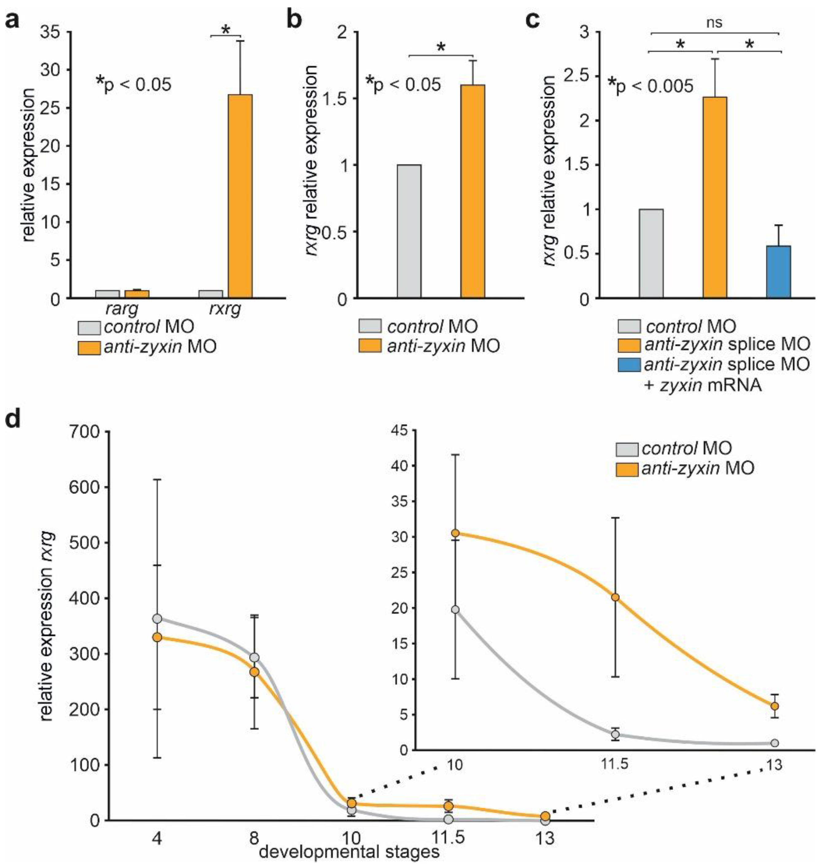
2.1. Downregulation of Zyxin Results in the Increase in rxrγ mRNA Level

First of all, to verify the data of our previous RNA-seq screening by another method, we compared rxrγ mRNA levels in the middle gastrula stage Xenopus laevis embryos with knocked down Zyxin and the control levels by qRT-PCR. To knock-down Zyxin, the embryos were injected with conventional antisense morpholino oligonucleotide (MO) to Zyxin mRNA. The control embryos were injected with control MO (see Supplementary Table S1).
As a result, more than 20-fold higher rxrγ mRNA levels were detected in embryos injected with any of the aforementioned anti-zyxin MO compared to the control levels (Figure 1a). In contrast, the expression of zygotic gene retinoid acid receptor gamma (rarγ), which was selected as an additional control, was not affected in this experiment. Importantly, a similar increase in rxrγ mRNA upon Zyxin down-regulation by MO was observed in the Danio rerio fish embryos, suggesting the evolutionary conservation of the effect (Figure 1b). The specificity of the MO activity on the Xenopus laevis rxrγ mRNA was confirmed in rescue experiments, in which we observed preservation of the basal level of the rxrγ mRNA concentration in embryos co-injected with anti-zyxin splice MO, which prevented maturation of this mRNA due to the binding to the exon3–intron3 boundary, and the synthetic zyxin mRNA (Figure 1c). In addition, we confirmed the efficiency of zyxin knock-down by MO, as well as the efficiency of its rescue by zyxin mRNA co-injection, analyzing the levels of Zyxin protein in these embryos (Figure S1).
To investigate changes of the rxrγ expression in embryos with downregulated Zyxin in more detail, we compared rxrγ expression in the control and anti-zyxin MO injected embryos at different developmental stages. At the stages prior to MBT, the mRNA levels of rxrγ were statistically indistinguishable in the control and anti-zyxin MO embryos (Figure 1d and Supplementary Figure S2). From MBT to early gastrula stage (stage 10), the mRNA levels also decrease similarly both in the controls and in anti-zyxin MO injected embryos (Figure 1d and Supplementary Figure S2). However, from gastrula stage 10 to stage 13 (early neurula), the decay of rxrγ mRNA in anti-zyxin MO embryos becomes significantly slower, eliciting significantly higher levels of rxrγ mRNA in the anti-zyxin MO injected embryos (Figure 1d and Supplementary Figure S2).
Thus, the data obtained suggest that Zyxin definitely plays a role in the process of the degradation of the maternally accumulated rxrγ mRNA.

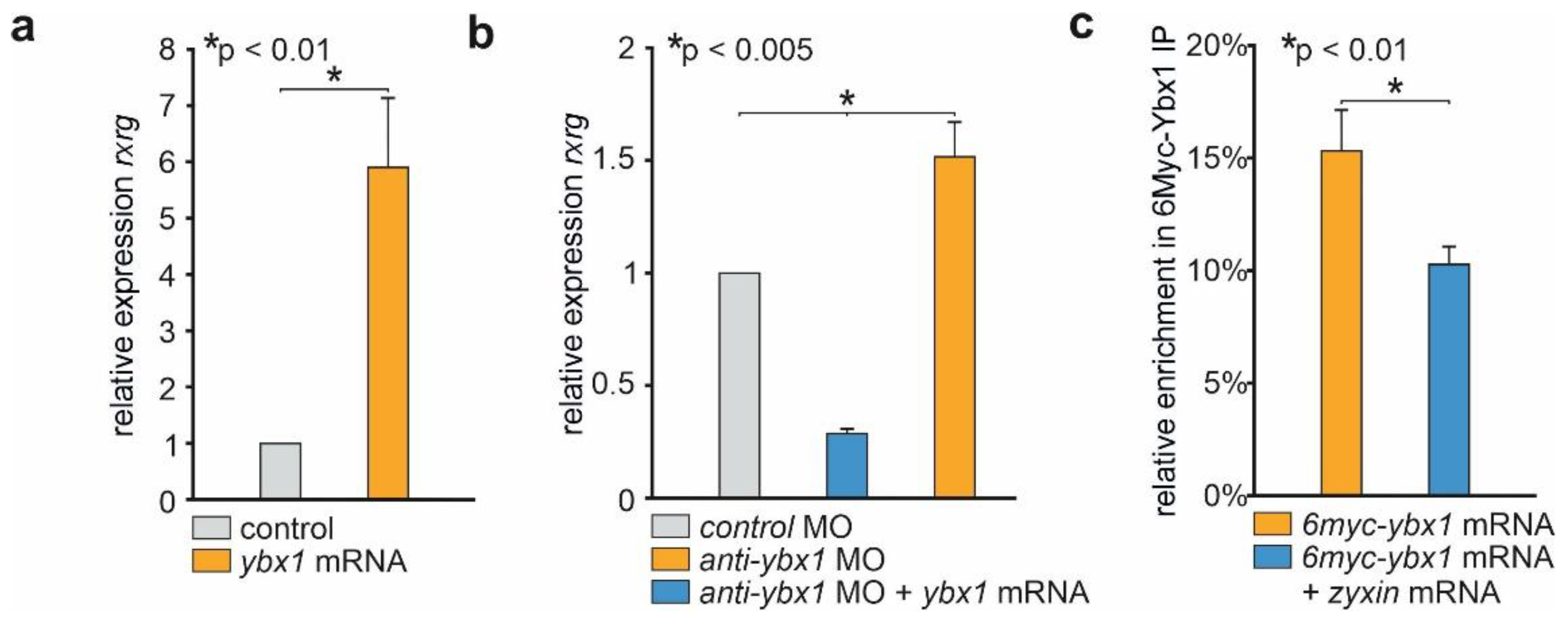
2.2. Zyxin Prevents Binding of Ybx1 to rxrγ mRNA

To test if Ybx1 could regulate the rxrγ mRNA level, we overexpressed it in embryos by injecting the synthetic ybx1 mRNA. Indeed, a strong increase in rxrγ transcripts concentration was revealed by qRT-PCR in these embryos comparing to the control ones (Figure 2a). The opposite effect was obtained when anti-ybx1 MO was injected (Figure 2b).
Next, we tested if Ybx1 is able to bind to rxrγ mRNA to protect it from degradation, using RNA-immunoprecipitation assay (RIP). Xenopus embryos were injected with myc-tagged ybx1 mRNA and zyxin mRNA, and lysed at stage 11. The lysates were incubated with anti-myc resin and the precipitated material was analyzed by qPCR as described in [22]. In the samples with expressed myc-Ybx1 we observed strong enrichment of rxrγ mRNA comparing to a control sample, injected with myc-tagged mRNA of a secreted morphogen Chordin. On the other hand, in the sample with co-expressed both myc-ybx1 and zyxin mRNA we observed a decrease in the amount of rxrγ mRNA. Therefore, we conclude that Ybx1 does form a complex with rxrγ mRNA, and that Zyxin can counteract to the complex formation (Figure 2c).

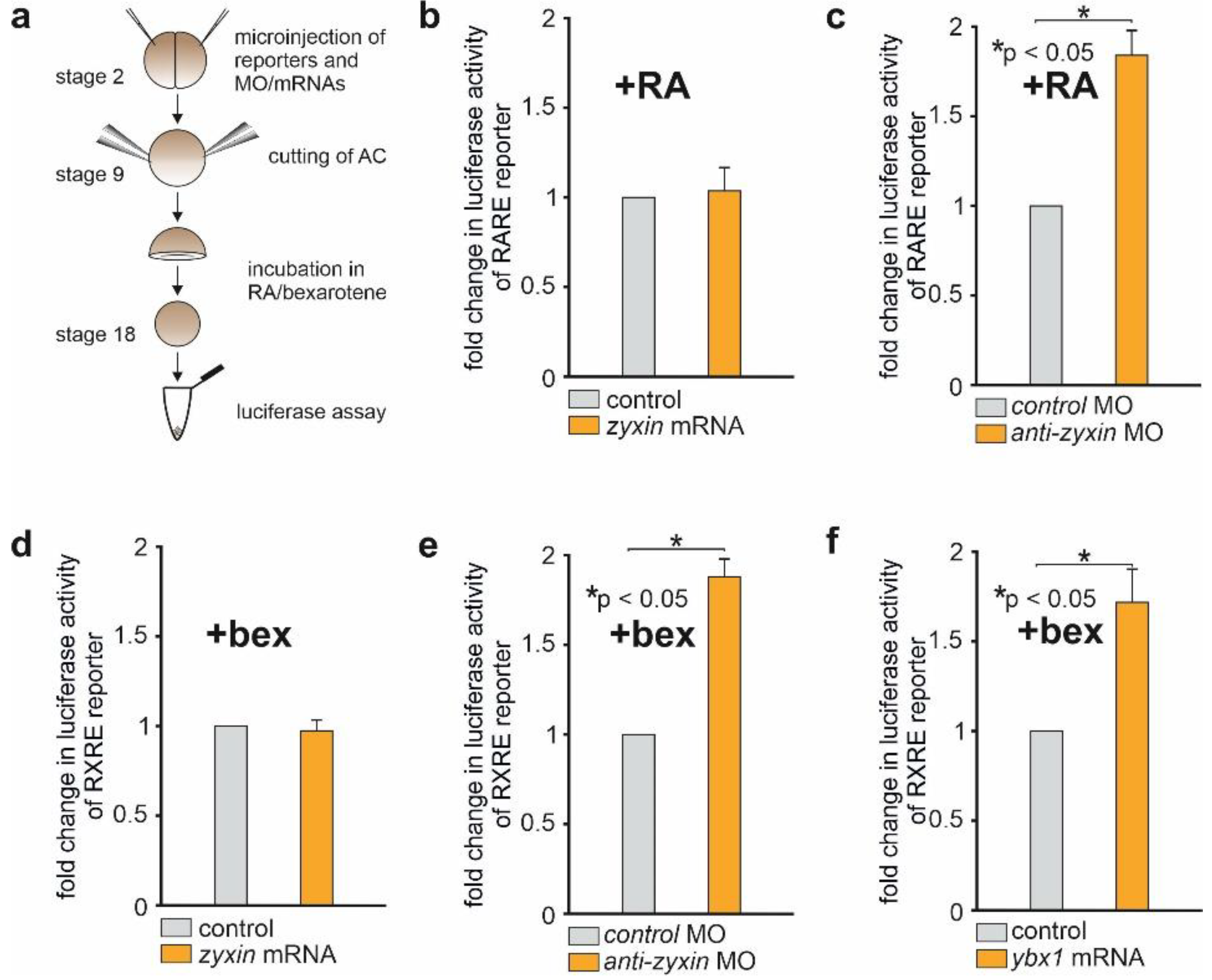
2.3. Downregulation of Zyxin Results in the Activation of Luciferase Reporters Containing the Cis-Regulatory Elements Essential for the RA-Dependent Signaling

In order to determine the Zyxin influence on the RA-dependent signaling cascade, we designed a luciferase reporter containing RA-responsive cis-regulatory elements (RARE) upstream of the luciferase gene. To do so, we cloned the 3xRARE sequence (3xGGGTAGGGTTCACCGAAAGTTCACTCG) [23] into the pGL3-Promoter Vector (Promega) plasmid containing the SV40 promoter upstream of the luciferase sequence. We also created similar constructs with 6xRARE and 12xRARE elements in the same plasmid. The efficiency of these constructs was tested as follows: a mixture of the specific reporter pGL3-Promoter Vector-3xRARE/6xRARE/12xRARE and the Renilla reporter under the control of a low-specific thymidine kinase promoter was injected into the animal region of each blastomere of Xenopus laevis embryos at the 2–4 blastomere stage. Animal caps (AC) were excised at the stage of late blastula (stage 9) and harvested at stage 18. ACs were incubated in 1x Modified Marc’s Ringer’s (MMR) with 10−5 M retinoic acid. Control ACs were incubated in 1xMMR only (Figure 3a). Interestingly, samples injected with pGL3-Promoter Vector-3xRARE demonstrated the strongest luciferase activity in response to retinoic acid. Multiplication of the RARE elements did not increase activity, but somewhat weakened it (Figure S3a). In the following experiments we used the 3xRARE construct.
To investigate the effect of Zyxin on this signaling, we co-injected pGL3-3xRARE and Renilla reference reporter with anti-zyxin MO, zyxin mRNA or control MO into the embryos, treated their AC with 10−5 M RA and then measured the luciferase signal (Figure 3a). As a result, we observed an increased expression of pGL3-3xRARE in AC co-injected with anti-zyxin MO, but not with zyxin mRNA (Figure 3b–d).
Although the data obtained by means of pGL3-3xRARE reporter were in agreement with the data indicating the role of Zyxin in the rxrγ mRNA destabilization, there was still some uncertainty in the direct involvement of Rxrγ in the RA signaling regulation by Zyxin, since the retinoic receptor heterodimer, which must be formed for RARE activation, includes besides Rxr its Rar counterpart [13]. To resolve this uncertainty, we created a reporter with the luciferase gene driven by γRXRE regulatory elements sensitive exclusively to the RXR homodimer [24]. To this end, we cloned 2xγRXRE sequences (2xGGTTGAaAGGTCA) into the pGL3-Promoter Vector. The effectiveness of the resulting construct (pGL3-2xγRXRE) was verified in the AC explants in the same manner as the effectiveness of pGL3-3xRARE reporter, except 10−5 M solution of the Rxrs agonist bexarotene was used for the activation of pGL3-2xγRXRE (Figure S3b). Accordingly, bexarotene was also used in all other experiments with this reporter.
As a result, we established that similarly to the pGL3-3xRARE, injections of anti-zyxin MO or synthetic ybx1 mRNA significantly increased pGL3-2xγRXRE expression, whereas Zyxin overexpression did not affect the activity of this reporter (Figure 3d–f).
In addition, the activity of reporters was only changed when retinoic acid (for RARE) or bexarotene (for RXRE) was added to the medium (compare Figure 3c with Figure S4b, and Figure 3e with Figure S4d). In the absence of an agonist, the receptor itself is unable to form dimers and bind to regulatory elements. It is evidently apparent from experiments with rxrγ overexpression, since only when bexarotene was added did the activity of the reporter increase severalfold; without the agonist, it increased slightly (Figure S3b).
In sum, all these results confirm the functional role of the previously found mechanism of Zyxin influence on the degradation of maternal rxrγ mRNA.

2.4. The Distribution of Zyxin between the Cytoplasm and the Nucleus Is Not Changed upon Treatment of AC with RA or Bexarotene

Recently, Youn et al., [25] have shown that in cell culture Zyxin can shuttle into the nucleus from cytoplasm upon stimulation with RA. In this regard, we decided to verify whether Zyxin also moves into the nucleus upon RA or the Rxrγ receptor agonist, bexarotene, stimulation in the animal cap (AC) explants of Xenopus embryos. To this end, we incubated AC explants in 10−5 M RA or bexarotene, lysed these explants to separate nuclei from cytoplasm, and analyzed the two fractions by Western blotting. As a result, we revealed that neither RA nor bexarotene affect Zyxin distribution between the cytoplasm and nucleus (Figure S5).

3. Discussion

In this study, we demonstrate that Zyxin down-regulation up-regulates the level of rarγ mRNA, but not rarγ mRNA, in the Xenopus laevis embryo. Zyxin-dependent regulation of rxrγ mRNA relies on the previously reported mechanism of mRNA stabilization by association with RNA-binding protein Y-box factor 1 (Ybx1) [10]. Zyxin can bind to Ybx1 and sequester it from the target rxrγ mRNA, resulting in its accelerated degradation. The following line of evidence for these conclusions was obtained. First, we demonstrated that Ybx1 can precipitate rxrγ mRNA, whereas addition of Zyxin significantly decreases the co-precipitation of rxrγ transcripts. Secondly, Ybx1 overexpression significantly increases the endogenous rxrγ mRNA level, while downregulation of Ybx1 has the opposite effect. Thirdly, the knock-down of Zyxin by MO, as well as Ybx1 overexpression, activates RARE and γRXRE-containing luciferase reporters. However, overexpression of Zyxin does not activate them. The latter is consistent with our previous data, showing that Zyxin overexpression has a minor effect on the developing embryo [7,10]. Finally, we demonstrated that the effect of rxrγ mRNA increase upon Zyxin down-regulation is also reproduced in the Danio rerio fish embryos, which indicates a conservation of the revealed mechanism of the regulation of rxrγ mRNA stability by Zyxin among vertebrates.
Evidence of the functional relationship between retinoic acid receptors (Rars), retinoid X receptors (Rxrs), and the cytoskeletal LIM domain proteins of Zyxin family has periodically appeared in the literature over the past decade [26]. The Zyxin family proteins Ajuba, Limd1, and WTIP have been shown to directly interact with RAR and RXR receptors as corepressors of RARE-dependent transcription. Generally, retinoid nuclear receptor corepressors bind directly to that receptor and are thought to act as scaffolds to recruit chromatin remodeling complexes to specific targets [27]. In the same work it was shown that Zyxin and LPP do not interact with RAR and RXR receptors and do not suppress the activity of RARE-driven promoters. In the present work, we also confirmed that Zyxin overexpression does not affect RARE and RXRE-dependent transcription.
At the same time, to our knowledge, there was the only evidence of interaction of Zyxin and RA signaling until hitherto. Namely, in H1299 cell culture, Zyxin moves into the nucleus in the presence of RA and represses RARE-dependent transcription forming a triple complex with PTOV1 and CBP, a co-activator of RARE, by sequestering CBP from RARE [25].
Obviously, in our present work, we have identified a completely different mechanism of the regulation of RA signaling by Zyxin based on the destabilization of rxrγ mRNA. In addition to all the functional data, this is also confirmed by the fact that, in contrast to the work of Youn et al., [25], we did not detect the movement of Zyxin into the cell nuclei in response to treatment of animal caps with RA or bexarotene. Taking into account that we recently showed the involvement of the same mechanism in the regulation of the expression of embryonic stemness genes [10], one may assume that it can generally take part in the control of the expression of a fairly wide range of maternal genes. In this regard, a large-scale search for genes whose expression is influenced by this mechanism would be an interesting prospect in the future.

4. Materials and Methods

The materials used in the work are outlined in Table 1.

4.1. Experimental Model and Subject Details

Embryo Manipulations

The care and handling of all animals was conducted in accordance with the regulations of the Animal Committee at the Shemyakin-Ovchinnikov Institute of Bioorganic Chemistry (Moscow, Russia), and the Animals (Scientific Procedures) Act of 1986, and the Helsinki Declaration.
Xenopus laevis embryos were obtained with HCG injection as described by [28]. The embryos were staged as described by Nieuwkoop and Faber [29].
The D. rerio (AB/TL strain) was raised in E3 medium (5 mM NaCl, 0.17 mM KCl, 0.33 mM CaCl2, 0.33 mM MgSO4, adjusted to pH 7.0) at 26.5 °C with 12 h/12 h of light and dark.

4.2. Method Details

4.2.1. Embryonic Microinjections

Microinjections were performed as described previously [10,22,28].

4.2.2. mRNA Extraction and qRT-PCR

Xenopus laevis embryos were extracted using ExtractRNA reagent (Evrogen) and CleanRNA Standard spin columns (Evrogen) following manufacturer’s instructions.
Nearly 250 ng of total RNA were transcribed into cDNA using MMLV RT kit (Evrogen), and PCR was performed with qPCRmix-HS SYBR kit (Evrogen). Amplifications were conducted using DTprime 4 qPCR amplifier (DNA-Technology). We also amplified the housekeeping genes odc1 and eef1a to evaluate RNA quality. See Table S1 for primers we used.
We used stage 11 to study the amount of endogenous maternal mRNAs, since after the activation of the embryonic genome at the blastula-early gastrula stage, maternal transcripts rapidly degrade, and the greatest difference in the number of transcripts is observed precisely at stage 11.

4.2.3. DNA Constructs

Previous studies have described the following plasmids: p35T-Zyxin and pCS2MT-Ybx1 [7,10].
In Table S1, we provide detailed cloning strategies for pGl3pv -RARE, pGl3pv -RXRE, and pCS2-rxrγ-3xmyc.

4.2.4. Synthetic mRNA

After linearizing pCS2-based plasmids with NotI, synthetic mRNAs (see Table S1) were prepared with the mMessage mMachine SP6 kit (Ambion) and injected into 2–4 cell embryo (3–4 nL of 100ng/mL mRNA water solution per embryo) into the whole embryo or into a particular blastomere depending on the experimental design.

4.2.5. RNA-Immunoprecipitation (RIP)

To study the ability of Ybx1 to bind rxrγ mRNA, 2-cell stage embryos were injected with myc-ybx1 RNA (35 pg/blastomere) and zyxin RNA (50 pg/blastomere). RIP analysis was performed according to RIP protocol (Martynova et al., 2021b).

4.2.6. Luciferase Reporter Assay

The Luciferase assay was performed following the instructions [30]. To check reporter activity, pGl3pv–RARE or pGl3pv–RXRE plasmid (50 pg/blastomere) and reference pRL-TK-renilla luciferase plasmid (Promega) (50 pg/blastomere) mixed with 0.3 mM anti-zyxin MO or ybx1 mRNA (35 pg/blastomere) were injected to each blastomere of 2-cell stage embryos. Incubation of the injected embryos in 0.1 × MMR was continued until the late blastula stage (stage 9). Then explants of animal caps (ACs) were cut out with two tweezers. The ACs were then placed in 1 × MMR for two hours. Then, depending on the experiment, 10 µM RA or bexarotene was added to the medium.
The AC were collected for luciferase activity detection at stage 18. The problem is that when using whole embryos, the yolk gets into the lysate, strongly dampening the luciferase signal unspecifically. For the purity of the experiment, we have to carry out all studies on luciferase reporters on animal caps. It takes some time to accumulate a sufficient amount of reporter protein (luciferase) since reporter constructs (plasmids) can begin to be transcribed only after the embryonic genome (MBT) has been activated. Stage 11 (middle gastrula) contains very little reporter protein, which makes it difficult to interpret the findings. Therefore, all luciferase tests are carried out on animal caps incubated until stages 16–18 of development.

4.2.7. Cytoplasmic and Nuclear Fractionation

For our purposes, Xenopus embryos were incubated in 0.1 × MMR with 10 μM RA/bexarotene from stage 9 to stage 18. AC were cut out at stage 9 and incubated in 1 × MMR with 10 μM RA/bexarotene till stage 18.
Embryos or AC were pretreated by incubation in 0.1 × MMR/1 × MMR containing 150 μg of cycloheximide per ml. After incubation for 1 h at 23 °C, nuclear and cytoplasmic fractions were prepared as described in [28].

4.2.8. Statistical Analysis

For single comparisons standard t-test was used. For multiple comparisons ANOVA test was used. The statistical analysis was performed in GraphPad Prism.

Supplementary Materials

The following supporting information can be downloaded at: https://www.mdpi.com/article/10.3390/ijms23105627/s1.

Author Contributions

E.A.P. performed the majority of experiments and participated in their planning and wrote the text, E.E.O. participated in the creation of luciferase reporters and corrected the text, A.G.Z. wrote the final draft and approved it. N.Y.M. generated the initial idea, organized the work, participated in experiments, and wrote the text. All authors have read and agreed to the published version of the manuscript.

Funding

This research was funded by RFBR grant number 20-34-90017 (N.Y.M.), Western blotting experiments were supported by Ministry of Science and Higher Education of the Russian Federation grant 075-15-2021-1344 (A.G.Z.).

Institutional Review Board Statement

The animal study protocol was approved by the Institutional Review Board (or Ethics Committee) of the Shemyakin-Ovchinnikov Institute of Bioorganic Chemistry (Moscow, Russia, protocol code IACUC 229 and 01.02.2018).

Informed Consent Statement

Not applicable.

Acknowledgments

The authors thank Bezuglov V.V. for providing bexarotene.

Conflicts of Interest

The authors declare no competing interests.

References

  1. Smith, M.A.; Hoffman, L.M.; Beckerle, M.C. LIM proteins in actin cytoskeleton mechanoresponse. Trends Cell Biol. 2014, 24, 575–583. [Google Scholar] [CrossRef] [PubMed] [Green Version]
  2. Wang, Y.; Gilmore, T.D. Zyxin and paxillin proteins: Focal adhesion plaque LIM domain proteins go nuclear. Biochim. Biophys. Acta 2003, 1593, 115–120. [Google Scholar] [CrossRef] [PubMed] [Green Version]
  3. Winkelman, J.D.; Anderson, C.A.; Suarez, C.; Kovar, D.R.; Gardel, M.L. Evolutionarily diverse LIM domain containing proteins bind stressed actin filaments through a conserved mechanism. Proc. Natl. Acad. Sci. USA 2020, 117, 25532–25542. [Google Scholar] [CrossRef] [PubMed]
  4. Wu, C. Focal adhesion: A focal point in current cell biology and molecular medicine. Cell Adh. Migr. 2007, 1, 13–18. [Google Scholar] [CrossRef] [Green Version]
  5. Sun, X.; Phua, D.Y.Z.; Axiotakis, L.; Smith, M.A.; Blankman, E.; Gong, R.; Cail, R.C.; Espinosa de los Reyes, S.; Beckerle, M.C.; Waterman, C.M. Mechanosensing through direct binding of tensed F-actin by LIM domains. Dev. Cell 2020, 55, 468–482. [Google Scholar] [CrossRef]
  6. Wang, Y.X.; Wang, D.Y.; Guo, Y.C.; Guo, J. Zyxin: A mechanotransductor to regulate gene expression. Eur. Rev. Med. Pharm. Sci. 2019, 23, 413–425. [Google Scholar] [CrossRef]
  7. Martynova, N.Y.; Eroshkin, F.M.; Ermolina, L.V.; Ermakova, G.V.; Korotaeva, A.L.; Smurova, K.M.; Gyoeva, F.K.; Zaraisky, A.G. The LIM-domain protein Zyxin binds the homeodomain factor Xanf1/Hesx1 and modulates its activity in the anterior neural plate of Xenopus laevis embryo. Dev. Dyn. 2008, 237, 736–749. [Google Scholar] [CrossRef]
  8. Martynova, N.Y.; Ermolina, L.V.; Ermakova, G.V.; Eroshkin, F.M.; Gyoeva, F.K.; Baturina, N.S.; Zaraisky, A.G. The cytoskeletal protein Zyxin inhibits Shh signaling during the CNS patterning in Xenopus laevis through interaction with the transcription factor Gli1. Dev. Biol. 2013, 380, 37–48. [Google Scholar] [CrossRef] [Green Version]
  9. Martynova, N.Y.; Parshina, E.A.; Zaraisky, A.G. Cytoskeletal protein Zyxin in embryonic development: From controlling cell movements and pluripotency to regulating embryonic patterning. FEBS J. 2021. [Google Scholar] [CrossRef]
  10. Parshina, E.A.; Eroshkin, F.M.; Orlov, E.E.; Gyoeva, F.K.; Shokhina, A.G.; Staroverov, D.B.; Belousov, V.V.; Zhigalova, N.A.; Prokhortchouk, E.B.; Zaraisky, A.G. Cytoskeletal protein zyxin inhibits the activity of genes responsible for embryonic stem cell status. Cell Rep. 2020, 33, 108396. [Google Scholar] [CrossRef]
  11. Session, A.M.; Uno, Y.; Kwon, T.; Chapman, J.A.; Toyoda, A.; Takahashi, S.; Fukui, A.; Hikosaka, A.; Suzuki, A.; Kondo, M.; et al. Genome evolution in the allotetraploid frog Xenopus laevis. Nature 2016, 538, 336–343. [Google Scholar] [CrossRef] [Green Version]
  12. Stunnenberg, H.G. Mechanisms of transactivation by retinoic acid receptors. Bioessays 1993, 15, 309–315. [Google Scholar] [CrossRef]
  13. Valcárcel, R.; Holz, H.; Jiménez, C.G.; Barettino, D.; Stunnenberg, H.G. Retinoid-dependent in vitro transcription mediated by the RXR/RAR heterodimer. Genes Dev. 1994, 8, 3068–3079. [Google Scholar] [CrossRef] [Green Version]
  14. Minucci, S.; Leid, M.; Toyama, R.; Saint-Jeannet, J.P.; Peterson, V.J.; Horn, V.; Ishmael, J.E.; Bhattacharyya, N.; Dey, A.; Dawid, I.B.; et al. Retinoid X receptor (RXR) within the RXR retinoic acid receptor heterodimer binds its ligand and enhances retinoid-dependent gene expression. Mol. Cell. Biol. 1997, 17, 644–655. [Google Scholar] [CrossRef] [Green Version]
  15. Allenby, G.; Bocquel, M.T.; Saunders, M.; Kazmer, S.; Speck, J.; Rosenberger, M.; Lovey, A.; Kastner, P.; Grippo, J.F.; Chambon, P. Retinoic acid receptors and retinoid X receptors: Interactions with endogenous retinoic acids. Proc. Natl. Acad. Sci. USA 1993, 90, 30–34. [Google Scholar] [CrossRef] [Green Version]
  16. Medin, J.A.; Minucci, S.; Driggers, P.H.; Lee, I.J.; Ozato, K. Quantitative increases in DNA binding affinity and positional effects determine 9-cis retinoic acid induced activation of the retinoid X receptor beta homodimer. Mol. Cell. Endocrinol. 1994, 105, 27–35. [Google Scholar] [CrossRef]
  17. Gudas, L.J.; Wagner, J.A. Retinoids regulate stem cell differentiation. J. Cell. Physiol. 2011, 226, 322–330. [Google Scholar] [CrossRef] [Green Version]
  18. Maden, M. Retinoids as endogenous components of the regenerating limb and tail. Wound Repair Regen. 1998, 6, 358–365. [Google Scholar] [CrossRef]
  19. Ghyselinck, N.B.; Duester, G. Retinoic acid signaling pathways. Development 2019, 146, dev167502. [Google Scholar] [CrossRef] [Green Version]
  20. Blumberg, B.; Mangelsdorf, D.J.; Dyck, J.A.; Bittner, D.A.; Evans, R.M.; De Robertis, E.M. Multiple retinoid-responsive receptors in a single cell: Families of retinoid “X” receptors and retinoic acid receptors in the Xenopus egg. Proc. Natl. Acad. Sci. USA 1992, 89, 2321–2325. [Google Scholar] [CrossRef] [Green Version]
  21. Sharpe, C.R. The expression of retinoic acid receptors in Xenopus development. Biochem. Soc. Trans. 1994, 22, 575–579. [Google Scholar] [CrossRef]
  22. Martynova, N.Y.; Parshina, E.A.; Zaraisky, A.G. Using RNA-binding proteins for immunoprecipitation of mRNAs from Xenopus laevis embryos. STAR Protoc. 2021, 2, 100552. [Google Scholar] [CrossRef]
  23. Srivastava, R.K.; Srivastava, A.R.; Cho-Chung, Y.S.; Longo, D.L. Synergistic effects of retinoic acid and 8-chloro-adenosine 3′,5′-cyclic monophosphate on the regulation of retinoic acid receptor beta and apoptosis: Involvement of mitochondria. Clin. Cancer Res. 1999, 5, 1892–1904. [Google Scholar] [PubMed]
  24. Barger, P.M.; Kelly, D.P. Identification of a retinoid/chicken ovalbumin upstream promoter transcription factor response element in the human retinoid X receptor gamma2 gene promoter. J. Biol. Chem. 1997, 272, 2722–2728. [Google Scholar] [CrossRef] [Green Version]
  25. Youn, H.; Kim, E.J.; Um, S.J. Zyxin cooperates with PTOV1 to confer retinoic acid resistance by repressing RAR activity. Cancer Lett. 2013, 331, 192–199. [Google Scholar] [CrossRef]
  26. Sala, S.; Ampe, C. An emerging link between LIM domain proteins and nuclear receptors. Cell Mol. Life Sci. 2018, 75, 1959–1971. [Google Scholar] [CrossRef]
  27. Hou, Z.; Peng, H.; White, D.E.; Negorev, D.G.; Maul, G.G.; Feng, Y.; Longmore, G.D.; Waxman, S.; Zelent, A.; Rauscher, F.J. LIM protein Ajuba functions as a nuclear receptor corepressor and negatively regulates retinoic acid signaling. Proc. Natl. Acad. Sci. USA 2010, 107, 2938–2943. [Google Scholar] [CrossRef] [Green Version]
  28. Martynova, N.Y.; Parshina, E.A.; Zaraisky, A.G. Protocol for separation of the nuclear and the cytoplasmic fractions of Xenopus laevis embryonic cells for studying protein shuttling. STAR Protoc. 2021, 2, 100449. [Google Scholar] [CrossRef]
  29. Nieuwkoop, P.D.; Faber, J. Normal Table of Xenopus Laevis (Daudin): A Systematical and Chronological Survey of the Development from the Fertilized Egg Till the End of Metamorphosis; North-Holland Pub. Co.: Amsterdam, The Netherlands, 1967. [Google Scholar]
  30. Eroshkin, F.; Kazanskaya, O.; Martynova, N.; Zaraisky, A. Characterization of cis-regulatory elements of the homeobox gene Xanf-1. Gene 2002, 285, 279–286. [Google Scholar] [CrossRef]
Figure 1. Downregulation of Zyxin results in an increase in the rxrγ mRNA level in the Xenopus laevis embryos. (a) Analysis by qRT-PCR of rxrγ and rarγ expression in the middle-gastrula stage Xenopus laevis embryos injected with the control and anti-zyxin MO. Here and below the transcript levels of the housekeeping genes odc1 and eef1a1 were used for the data normalization. Error bars indicate standard deviation. (b) Analysis by qRT-PCR of rxrγ expression at bud stage of Danio rerio embryos injected with the control and anti-zyxin splice MO. (c) Analysis by qRT-PCR of the rescue effect on rxrγ expression of zyxin mRNA. (d) Comparative analysis of the rxrγ transcripts levels in Zyxin knocked-down and control embryos at different stages of development. The inset shows at larger scale the data for stages from 10 to 13. The data presented are averaged from at least three independent experiments.
Ijms 23 05627 g001
Figure 2. Zyxin regulates the stability of rxrγ mRNA by preventing formation of the Ybx1/mRNA complex. (a) Overexpression of ybx1 leads to an increase in the rxrγ mRNA level. (b) Suppression of ybx1 mRNA translation by anti-ybx1 MO decreases the rxrγ mRNA level, while co-expression of ybx1 mRNA restores it, even with some overshooting. (c) RIP experiments demonstrate the ability of Ybx1 to bind rxrγ mRNA, whereas Zyxin reduces this ability. Error bars indicate standard deviation. p-value was measured by Student’s t-test. The data presented are averaged from at least three independent experiments.
Ijms 23 05627 g002
Figure 3. Effects of loss- and gain-of-function of Zyxin on γRXRE and RARE cis-regulatory elements in the luciferase reporter assay. (a) Experimental design. (b) Zyxin overexpression does not result in the activation of pGL3-3xRARE reporter in the presence of RA. (c) pGL3-3xRARE reporter activity induced by RA increased in AC with the downregulated zyxin. (d) Zyxin overexpression does not result in the activation of pGL3-2xγRXRE reporter in the presence of bexarotene. (e) pGL3-2xγRXRE reporter activity induced by bexarotene increased in AC with the downregulated Zyxin. (f) pGL3-2xγRXRE reporter activity increased in AC with the overexpressed ybx1 in the presence of bexarotene. Error bars indicate standard deviation. p-value was measured by Student’s t-test. The data presented are averaged from at least three independent experiments.
Ijms 23 05627 g003
Table 1. Key resources table.
Reagent or ResourceSourceIdentifier
Antibodies
Rabbit polyclonal anti-Zyxin A.G. Zaraisky lab, Moscow, RussiaN/A
Anti-rabbit IgG, AP-conjugated, produced in goatSigma-Aldrich, Missouri, USACat#A3937; RRID:AB_258122
Mouse monoclonal anti-alpha-TubulinSigma-Aldrich, Missouri, USACat#T9026; RRID:AB_477593
Anti-mouse IgG, AP-conjugatedSigma-Aldrich, Missouri, USACat#A3562; RRID:AB_258091
EZview Red Anti-c-Myc Affinity Gel Sigma-Aldrich, Missouri, USACat#E6654; RRID:AB_10093201
Bacterial and Virus Strains
DH5alpha E. coli strainN/AN/A
Chemicals, Peptides, and Recombinant Proteins
Human chorionic gonadotropinSigma-Aldrich, Missouri, USACat#CG10
Ethyl 3-aminobenzoate methanesulfonate saltSigma-Aldrich, Missouri, USACat#A5040
Western Blue® stabilized substrate for alkaline phosphatasePromega, Madison, Wisconsin, USACat#S3841
BM Purple AP-substrateRoche, Basel, Switzerland Cat#11442074001
Brilliant Blue G solutionSigma-Aldrich, Missouri, USACat#B8522
CycloheximideSigma-Aldrich, Missouri, USACat#C1988
CNBr-Activated SepharoseTM 4BGE Healthcare, Chicago, Illinois, USAN/A
Fluorescein Lysine Dextran (FLD) 40 kD Invitrogen, Waltham, Massachusetts, USACat#D1845
L-CysteineDia-m, Moscow, RussiaCat#M52904
FicollDia-m, Moscow, RussiaCat#PS400
Igepal Nonidet P-40 Sigma-Aldrich, Missouri, USACat#I-30201
DTTFermentas, Waltham, Massachusetts, USACat#RO861
RNase Out RNase inhibitor, 100 units/mlInvitrogen, Waltham, Massachusetts, USACat#10777-019
Vanadyl ribonucleoside complexes (VRC)New England Biolabs, Ipswich, Massachusetts, USACat#S1402S
Protease Inhibitor Coctail Sigma-Aldrich, Missouri, USACat#P8340
BexaroteneLLC “Gurus BioPharm”, Moscow, RussiaN/A
Retinoic acid (RA)Sigma-Aldrich, Missouri, USACat#R2625
Critical Commercial Assays
mMessage mMachineTM SP6kitThermo Fisher, Waltham, Massachusetts, USACat#AM1340
ExtractRNAEvrogen, Moscow, RussiaCat# BC032
CleanRNA StandardEvrogen, Moscow, RussiaCat# BC033
NucleoSpin RNA purification columnsMacherey Nagel, GermanyCat#REF 740955.50
MMLV RT Evrogen, Moscow, RussiaCat# SK021
qPCRmix-HS SYBR Evrogen, Moscow, RussiaCat# PK147L
Dual-Luciferase Reporter Assay System Promega, Madison, Wisconsin, USACat# E1910
Dynabeads mRNA DIRECT KitAmbion, Waltham, Massachusetts, USACat# 61011
Experimental Models: Organisms/Strains
Wild type Xenopus laevis frogsNasco, Chicago, USA Cat#LM00456; RRID:XEP_Xla100
Danio rerio AB/TL strainN/AN/A
Oligonucleotides
See Table S1 for all oligonucleotides used
Recombinant DNA
See Table S1 for all recombinant DNAs used
Software and Algorithms
ExcelMicrosoft, USAN/A
ImageJSchneider et al., 2012https://imagej.nih.gov/ij/
Other
See Table S1 for all recombinant DNAs and oligonucleotides used
Publisher’s Note: MDPI stays neutral with regard to jurisdictional claims in published maps and institutional affiliations.

