Article

Anti-Fungal Analysis of Bacillus subtilis DL76 on Conidiation, Appressorium Formation, Growth, Multiple Stress Response, and Pathogenicity in Magnaporthe oryzae

1
Department of Plant Pathology, College of Plant Protection, Nanjing Agricultural University, Nanjing 210095, China
2
Key Laboratory of Integrated Management of Crop Diseases and Pests, Nanjing Agricultural University, Ministry of Education, Nanjing 210095, China
*
Author to whom correspondence should be addressed.
Int. J. Mol. Sci. 2022, 23(10), 5314; https://doi.org/10.3390/ijms23105314
Submission received: 19 April 2022 / Revised: 3 May 2022 / Accepted: 6 May 2022 / Published: 10 May 2022
(This article belongs to the Section Molecular Microbiology)

Abstract

:
In recent years, biological control has gained more attention as a promising method to combat plant disease. Such severe diseases cited include rice blasts caused by Magnaporthe oryzae. However, more effective microbial strains with strong adaptability still need to be identified. Therefore, we sought to assess the conidia germination, and formation of appressorium of DL76 in Magnaporthe oryzae. Besides, we also aimed at understanding the growth, multiple stress response and pathogenicity in Magnaporthe oryzae. We isolated Bacillus subtilis DL76 from a rice farm, which observed a strong antimicrobial effect on M. oryzae. The sterilized culture filtrate of DL76 inhibited the growth of M. oryzae, which motivated us to deduce the influence of DL76 on the pathogenicity of M. oryzae. We screened the effect of Bacillus subtilis DL76 on M. oryzae guy11. It demonstrated that sterilized culture filtrate (1 × 10⁷ CFU/mL) of DL76 can delay and even suppress the germination of conidia and (1 × 10⁷ and 1 × 10⁶ CFU/mL) prevent the formation of appressorium in vitro and in vivo. DL76 became hypersensitive to osmotic, oxidative, and cell wall degrading agents. In addition, the relative transcript levels of stress-responsive genes oxidative and osmotic were down-regulated by DL76 except for sod1, cat1, and cat2. In vivo assessment of the antifungal activity of Bacillus subtilis using conidia suspension of DL76 reduced the incidence and severity of rice blast. Conclusively, our results show that DL76 is essential for controlling rice blast by inhibiting conidiation, growth, multiple stress tolerance, and pathogenicity in M. oryzae.

1. Introduction

Magnaporthe oryzae is one of the most destructive pathogens of rice worldwide, causing a significant yield loss [1]. Outbreaks of rice blast disease are a serious and recurrent problem in China and other rice-growing regions. Therefore, several farmers use antifungal pesticides and plant disease-resistant cultivars to reduce the loss caused by rice blasts [2]. However, physiological races can quickly evolve and adapt to the resistant rice varieties because of the complexity of rice blasts. It is also challenging to control rice blast by planting resistant rice varieties because of their genetic background. Moreover, the overuse of chemical fungicide pollutes the environment and enhances the drug resistance in rice blasts amidst other advantages it offers [3].
Biological control has emerged as an environmentally friendly and promising strategy to control plant pathogens due to the increasing focus on sustainable development in agriculture [4]. Recent literature projects an increasing interest in exploiting rice blasts for biological control using beneficial plant micro-organisms because of their low toxicity and a lack of pathogen resistance [5,6,7]. Biological control is more beneficial than chemical fungicides because it is simple, highly efficient, and has less environmental impact. As a result, it has been widely used for micro-organism control, including Bacillus spp., Actinomycetes, Pseudomonas spp. and other anti-fungal bacteria that produce a series of antibiotics and enzymes [8]. Bacillus is considered a biological control candidate in agriculture because of its high antifungal properties and resistance to extreme conditions. Bacillus spp. has attracted significant attention because it exists universally in nature and can produce a series of antifungal substances as a non-pathogenic bacterium. Bacillus can increase plant resistance to pathogenic fungi by colonizing plants or spraying a plant with its powder, exerting a biological control effect [9,10,11]. Besides, Bacillus also promotes growth and increases plants’ yield, equating its biological control as immeasurable.
Researchers have assessed the interactions between biocontrol agents and pathogens in rice and other commercially important crops [12,13]. Particularly, Yan et al. [14] explored the interactions between fungi and bacteria in rice and observed a competitive superiority between Burkholderia gladioli and Aspergillus flavus strains. Kumar et al. [15] analyzed interactions between Bacillus subtilis MBI 600 and Rhizoctonia solani and concluded that the MBI 600 strain suppressed the plant pathogen. Previous studies have demonstrated the antifungal activity of Pseudomonas fluorescens 2137 in suppressing Fusarium culmorum on barley roots [16]. The anti-fungal properties of Bacillus species, including B. subtilis, have been investigated for their biological control against many plant and animal diseases [17]. Bacillus strains act directly or indirectly to control plant pathogens by colonizing plants, forming bio-films, enhancing the provision of nutrients and phytohormones, or inducing systemic resistance.
For instance, Bacillus amyloliquefaciens has potential applications in the biological control of Colletotrichum dematium which causes Mulberry anthracnose. Therefore, it is crucial and urgent to identify more effective and safer methods to suppress rice blasts. As a result, we isolated Bacillus subtilis, DL76, from the rice rhizosphere in a field. The outcome indicated that it suppressed the conidia germination and appressorium formation with a biocontrol efficiency of 75% against Magnaporthe oryzae. In addition, our results suggested that DL76 is more sensitive to oxidants, osmotic and cell wall perturbing agents. Our study presents B. subtilis DL76 as a potential biocontrol agent against M. oryzae, the rice blast pathogen.

2. Experiment Results

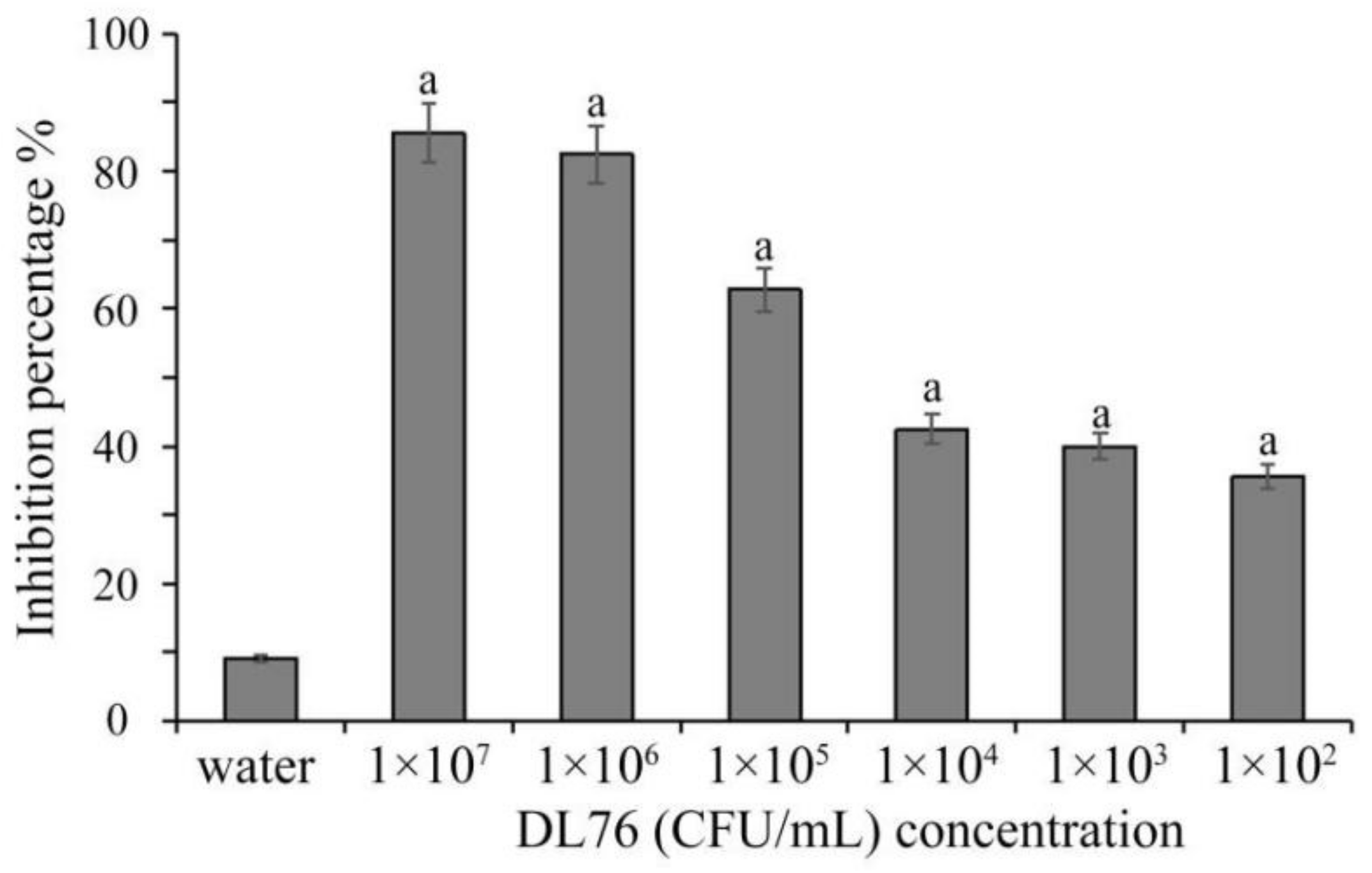
2.1. The Antifungal Activities of the Culture Filtrates DL76 at Different Concentration

According to the dual culture experiment, the endophytic bacteria DL76 showed salient antifungal activity against M. oryzae. Endophytic micro-organisms have been researched for decades as a potential source of biologically active metabolites in biological control [18]. Endophytic bacteria have been reported to have excellent anti-fungal activities. For instance, Gao et al. [19] identified an endophyte with significant antifungal activity against various fungi. The culture filtrate and fermentation broth of DL76 observed a significant suppression of M. oryzae growth at all concentrations (1 × 10², 1 × 10³, 1 × 10⁴, 1 × 10⁵, 1 × 10⁶, 1 × 10⁷ colony-forming units (CFU/mL) (Figure 1). The inhibition rate ranges from 65–85 CFU/mL. That is, the suppression activity increased as the concentration increased. The outcome could propose that strain DL76 strongly inhibits M. oryzae.

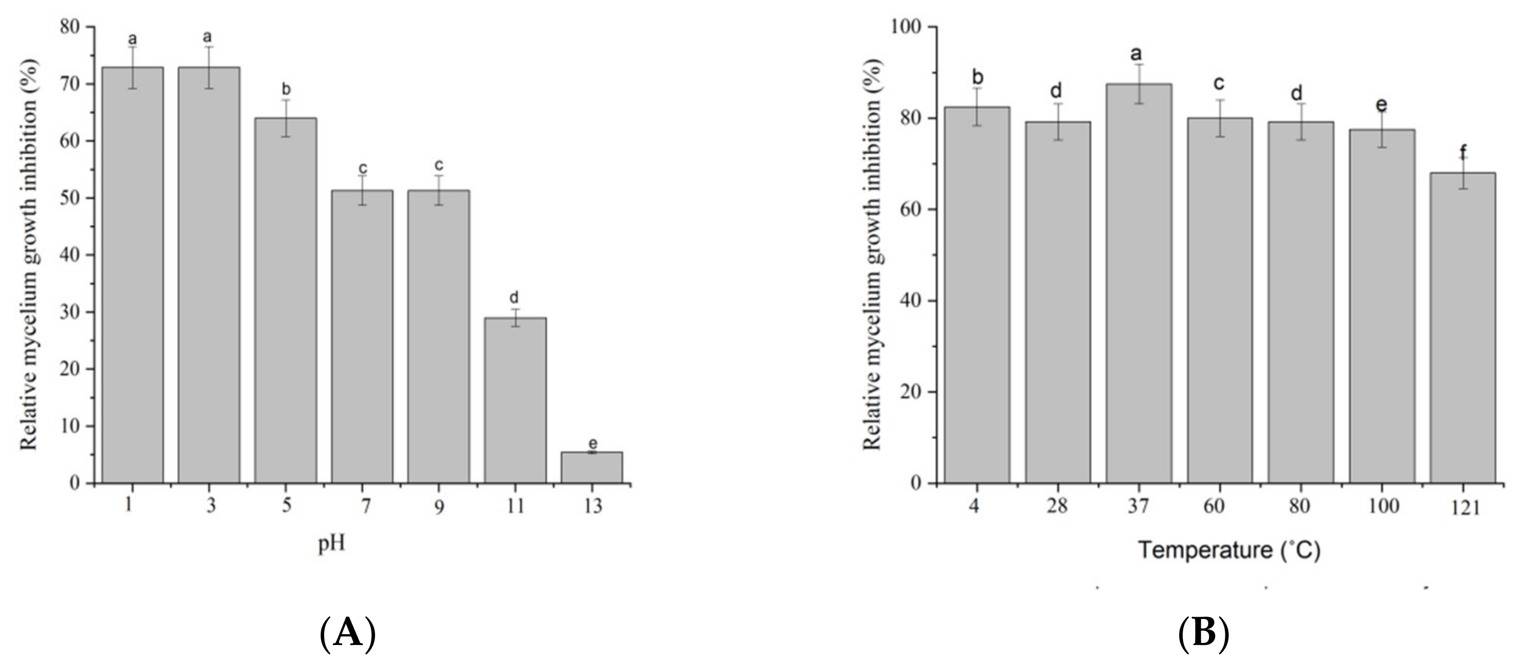
2.2. Stability of Antifungal Metabolites

The outcome from the acid-base stability assay observed a decline in the anti-fungal activity of culture filtrate DL76 after exposure to alkaline conditions at a pH of 11 or higher. The remaining inhibition for DL76 was significantly (p < 0.05) reduced to 29%, and 5.4% of the original level after alkali treatment at a pH of 11 and 13, respectively. However, the antifungal activity observed no significant difference at pHs between 1 and 9. The finding illustrated that the antifungal activity of the culture filtrate of DL76 against M. oryzae Guy11 was vital under strong acid, neutral and weak alkalinity conditions. Yet, the suppression activity diminished with the increase of alkaline (Figure 2A). Additionally, the antifungal properties exhibited by strain DL76 culture filtrate showed high thermal stability. The suppression rate reached 70% when the filtrate temperature ranged between 4 °C to 100 °C, reducing the suppression rate as the temperature increased (Figure 2B). The antifungal activity remained strong between 4 °C to 100 °C for 30 min but significantly decreased at 120 °C. Other studies, such as [20] observed similar results where Bacillus spp. produced antifungal metabolites for thermal stability and strong base instability. Therefore, high stability is necessary for developing anti-fungal metabolites as biocontrol pesticides. The fundamental stability of the anti-fungal metabolites produced by strain DL76 shows its potential in various environmental conditions.

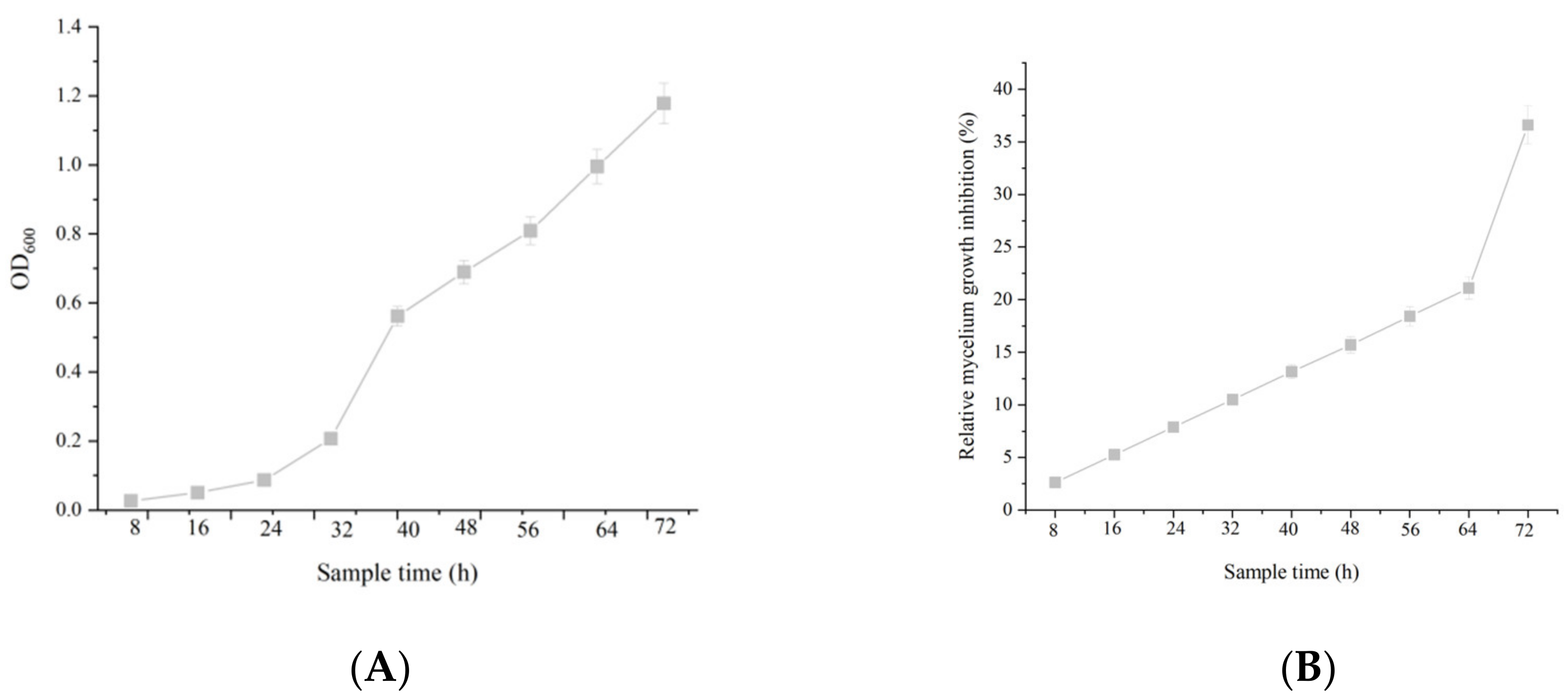
2.3. The Correlation between Cell Growth and Anti-Fungal Activity

Based on the experiment, the concentration of DL76 observed a positive correlation with the growth time. Growth curve analysis indicated that strains DL76 entered the logarithmic phase after 8 h of culture and reached the stationary state at 40 h. Further, the growth trend showed an S curve with increased cultivation time and concentration of DL76. The DL76 observed the fastest reproduction at 8 h. Nevertheless, there was no indication of the decline phase during the 72 h of incubation (Figure 3A). Similarly, there was a significant correlation between the culture filtrate bioactivity against M. oryzae and culture time. The anti-fungal activity increased rapidly over the entire logarithmic phase with the multiplication of bacteria (Figure 3B). Even more, we detected the strongest anti-fungal activity after forty-hours post-inoculation. Based on these findings, we could deduce that the optimal culture time to obtain strong anti-fungal metabolites occurs after forty hours. Various bacteria can produce anti-fungal metabolites, although they need a long fermentation time. Yet, strain DL76 can multiply and produce anti-fungal metabolites in a short time. However, DL76 can multiply and produce anti-fungal metabolites faster, thus it is convenient for production and application.

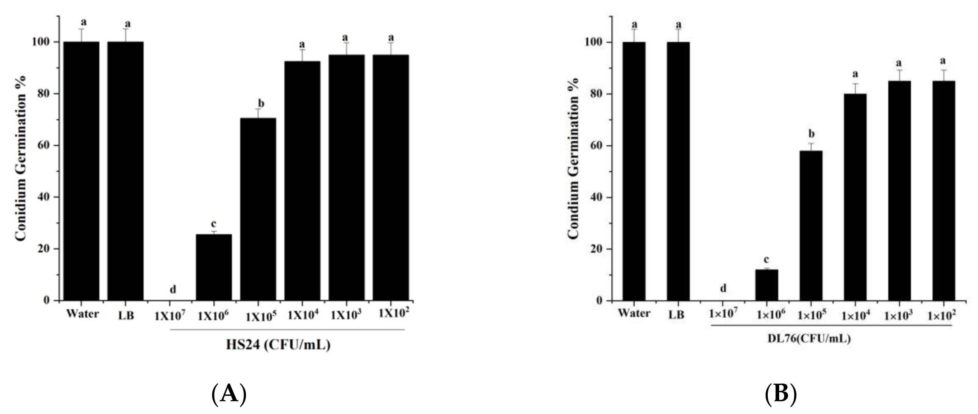
2.4. Effect of DL76 on Conidium Attachment

After treating conidium germination by HS24 at 105 CFU/mL, we observed an insignificant difference from the controls at 24 hpi. Germination increased from 17.9% at 8 hpi to 56.0% at 24 hpi when treated with HS24 at 106 CFU/mL. A few conidia (2.1%) germinated when treated with HS24 at 107 CFU/mL at 24 hpi (Figure 4A). The germination rate was 70% when DL76 105 CFU/mL was treated at 24 hpi. Additionally, the 106 CFU/mL germination rate increased from 12.5% at 8 hpi to 41.7% at 24 hpi (Figure 4B). Therefore, based on these results, we can conclude that the conidium germination gap between HS24 and DL76, indicates significantly different components of secondary metabolites between them. Hence, DL76 at the concentration of 1 × 10⁷ CFU/mL can significantly inhibit, delay, or even suppress the germination of conidia in M. oryzae.

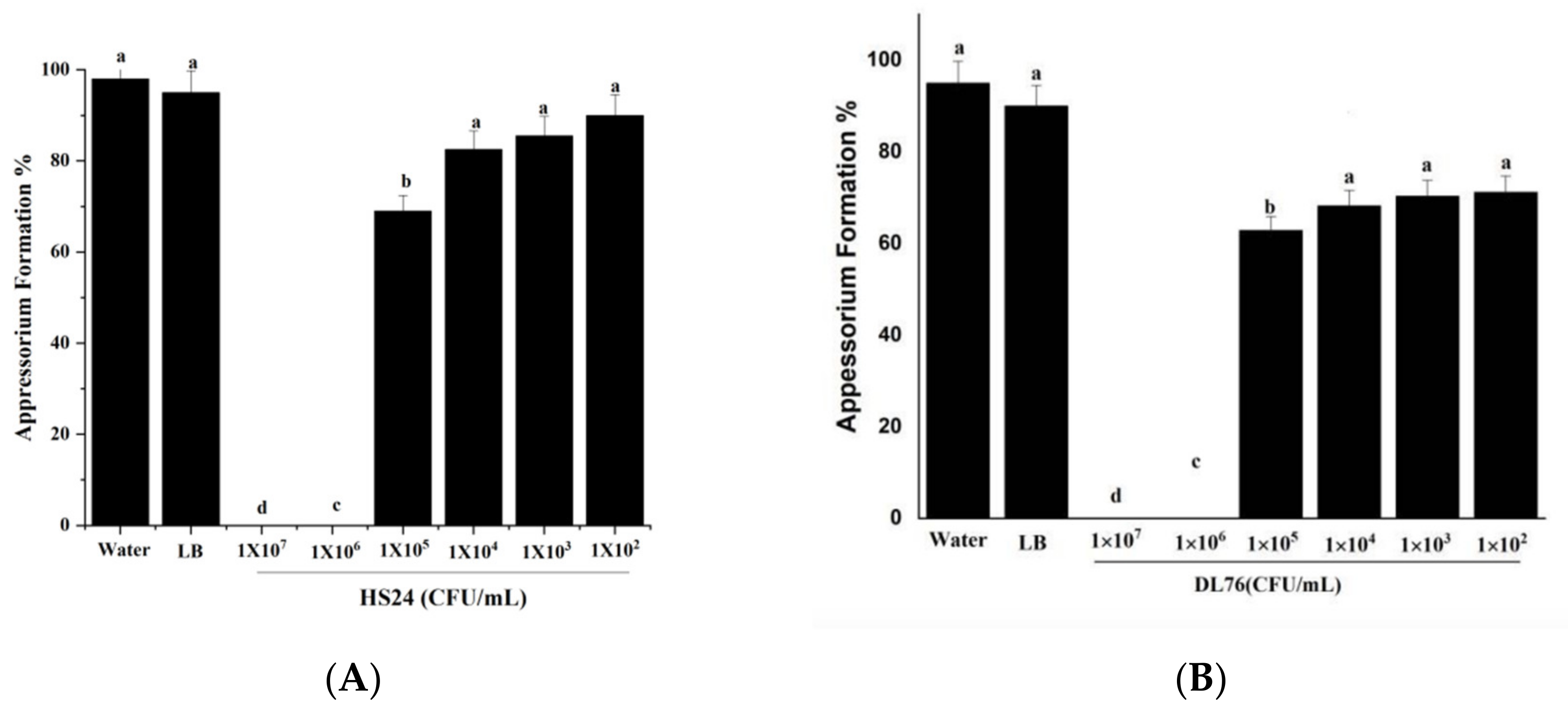
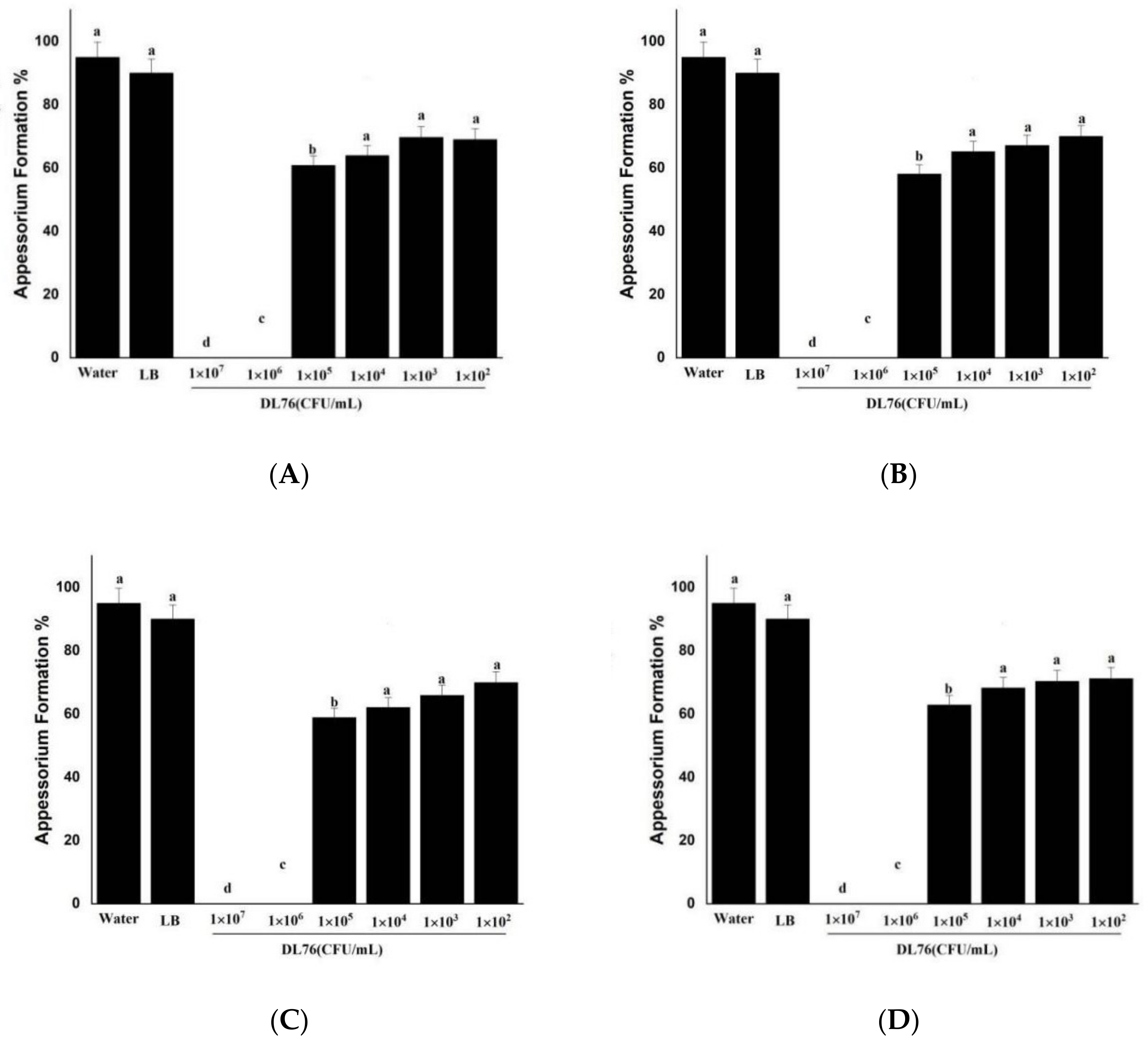
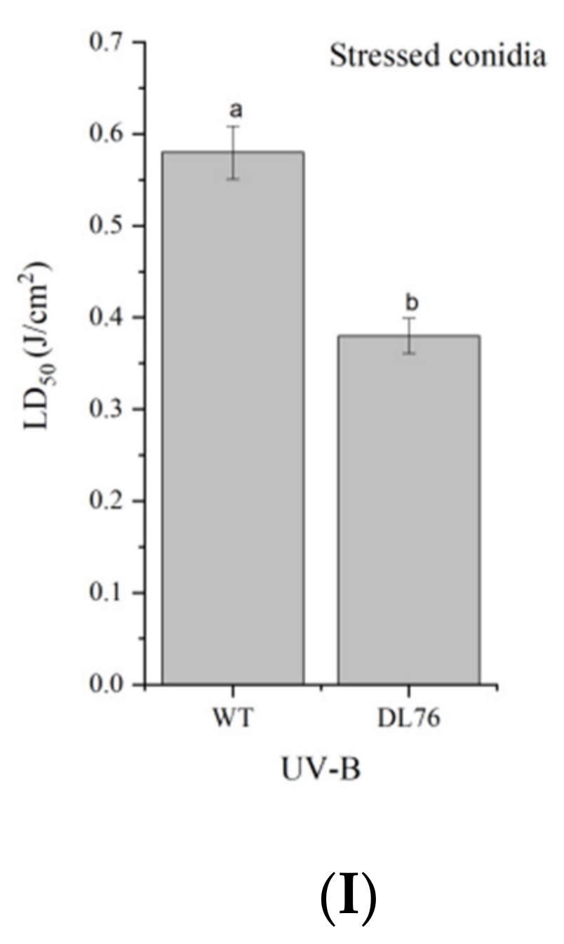
2.5. The DL76 Suppresses the Formation of Appressorium

An appressorium is the typical penetration structure that helps the rice blast invade the host cell [21,22]. Figure 5B observes the outcome when DL76 is assayed for appressorium formation on hydrophobic surfaces at 1 × 10⁷ CFU/mL, eight hpi. We observed similar results using HS24 at the same concentrations. However, after incubating for 8 h on hydrophobic surfaces, we observed that over 95% of wild-type and water germ tubes formed appressoria, as presented in Figure 5A,B. An increase in concentration resulted in a suppression of appressorium formation. In contrast, germlings developing appressoria were formed with a lesser concentration. Similarly, DL76 at 1 × 10⁵ CFU/mL allowed 79% of the germlings to develop appressoria, and in a lower concentration of 1× 10³ and 1 × 10² CFU/mL, over 85% appressorium formation was observed in the germ tubes, as illustrated in Figure 5B. The findings denote that at the concentration of 1 × 10⁷ and 1 × 10⁶ CFU/mL, most appressorium formation is delayed or inhibited, which is a critical infection structure for the pathogenicity ability of M. oryzae.

2.6. Differentiation of Appressoria

We further examined germlings for differentiation of the appressoria after 24, 36, 48, and 60 h of incubation on hydrophobic surfaces when most of the water and wild-type germlings had produced appressoria (Figure 6A–D). However, there was no observation of appressoria at 1 × 10⁷ and 1 × 10⁶ CFU/mL because the suppression of appressorium weakened with the decrease in the concentration of DL76. The appressorium formation developed significantly with more incubation time at a lower 1 × 10² and 1 × 10³ CFU/mL concentration. Additionally, there were no significant changes in turgor pressure between water and DL76-treated M. oryzae. We observed a delayed appressorium formation by DL76 at 1 × 10⁷ and 1 × 10⁶ CFU/mL because almost 0% of the germlings had not produced appressoria after 24, 36, 48, and 60 h.

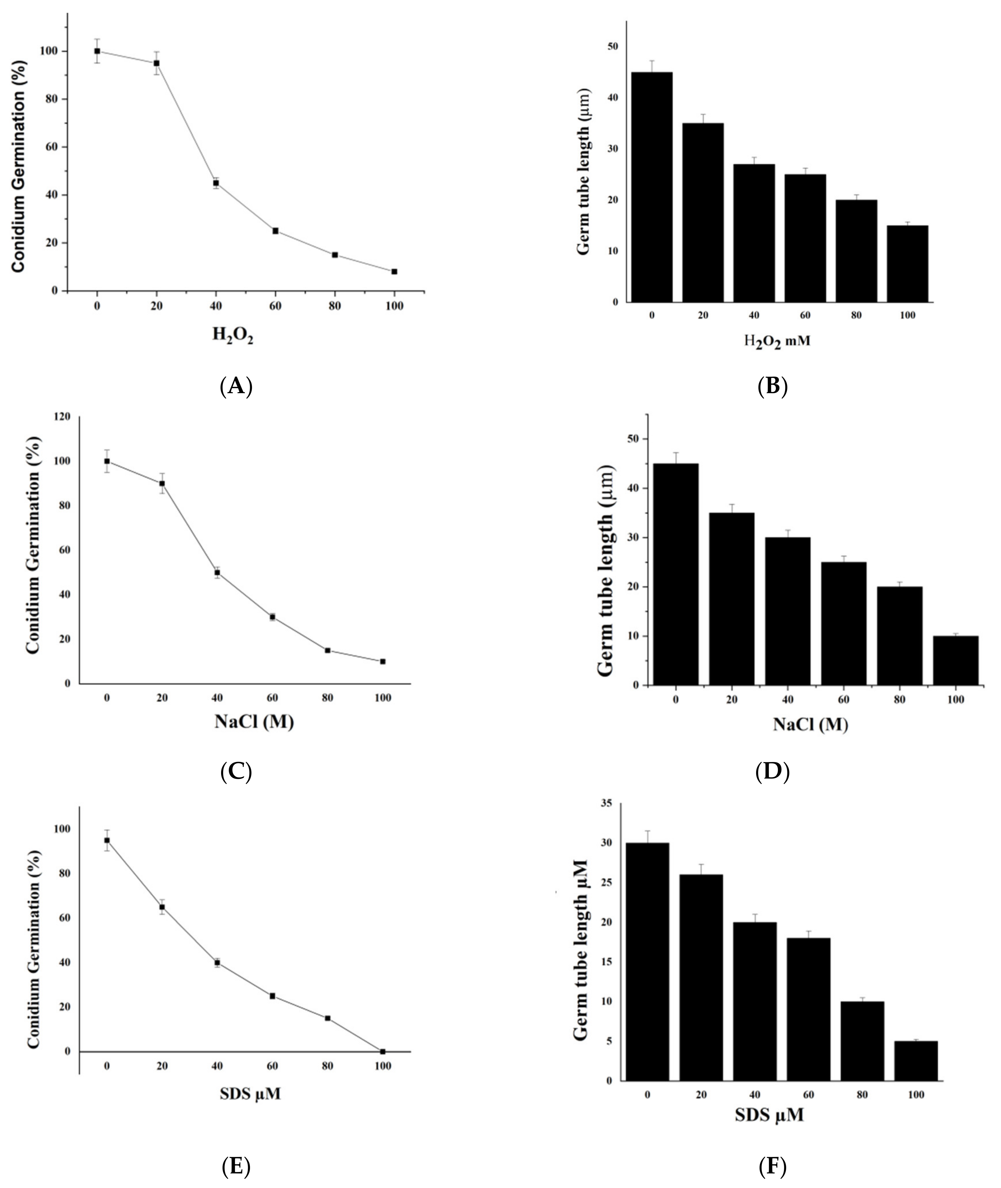
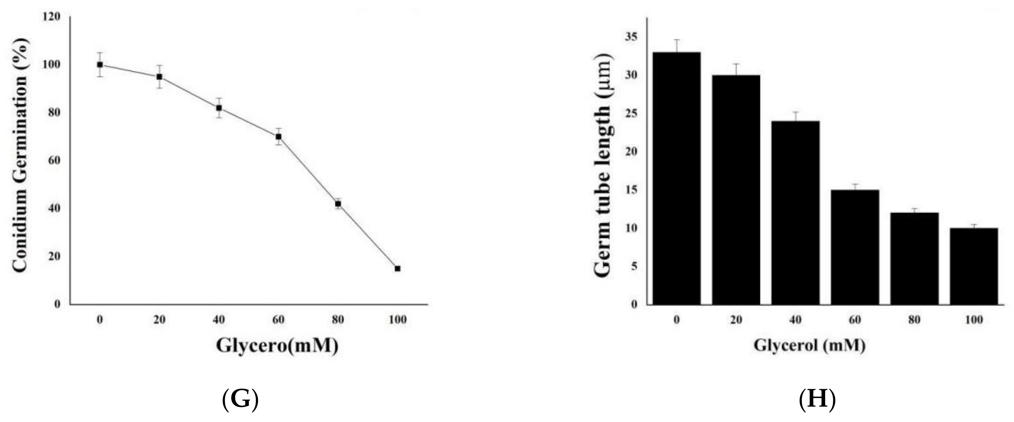
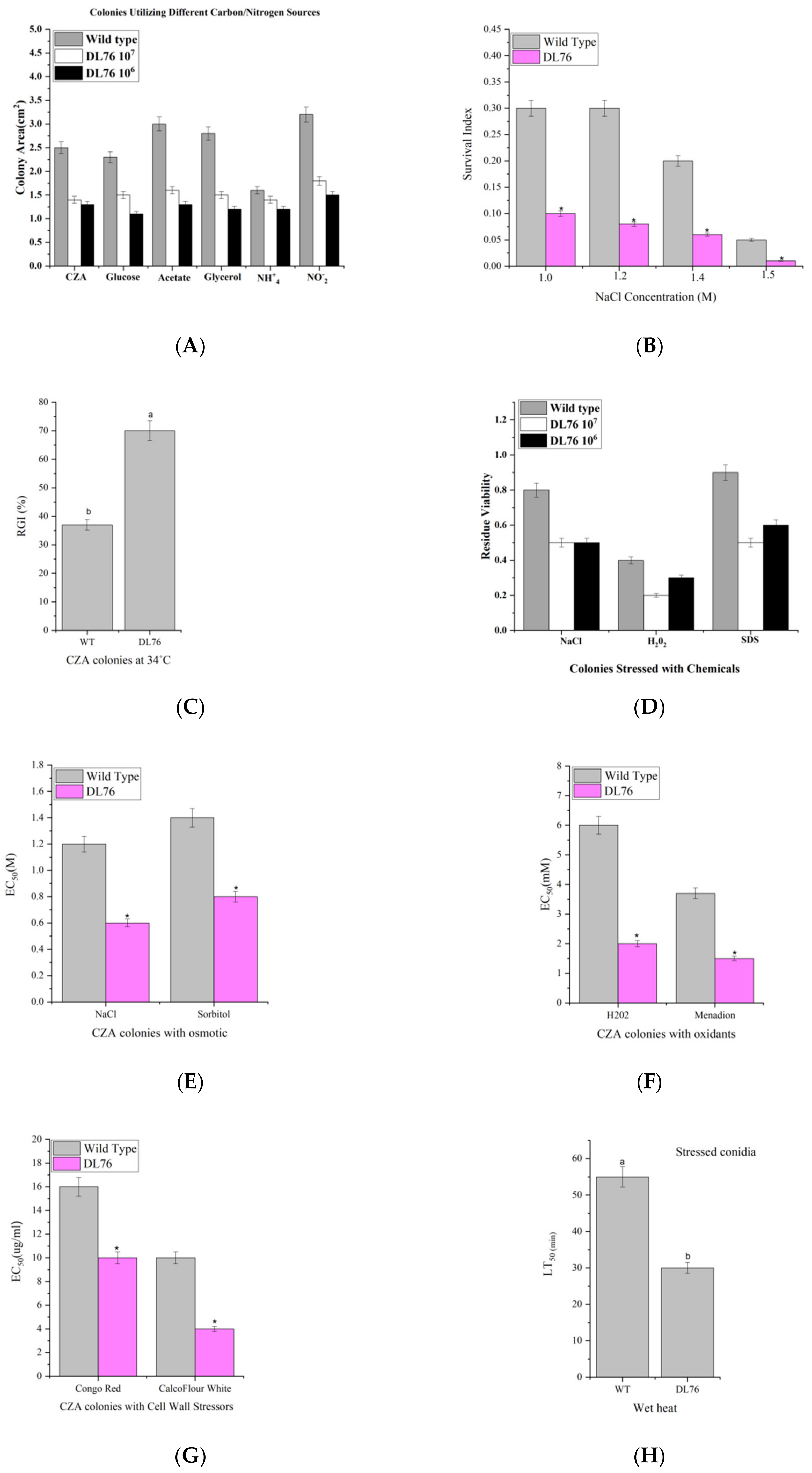
2.7. We Treated M. oryzae Conidia with Environmental Stress Inhibitors

We added different concentrations of environmental stresses of H2O2, glycerol, SDS, and NaCl (0, 20, 40, 60, 80, and 100 mΜ) to the conidial suspension. Further, we dripped twenty microliters of the mixture from each treatment onto a microscope cover glass with wet filter paper and incubated at 28 °C in darkness. We examined the conidium germination and germ tube length at six hpi. The observable results indicated that all the chemical stresses tested reduced the conidium germination with a more substantial effect observed with increased concentration of the chemicals (Figure 7A,C,E,G). Additionally, the chemicals affected germ tube elongation, with the effects becoming more pronounced with the increase of the chemical concentration (Figure 7B,D,F,H).

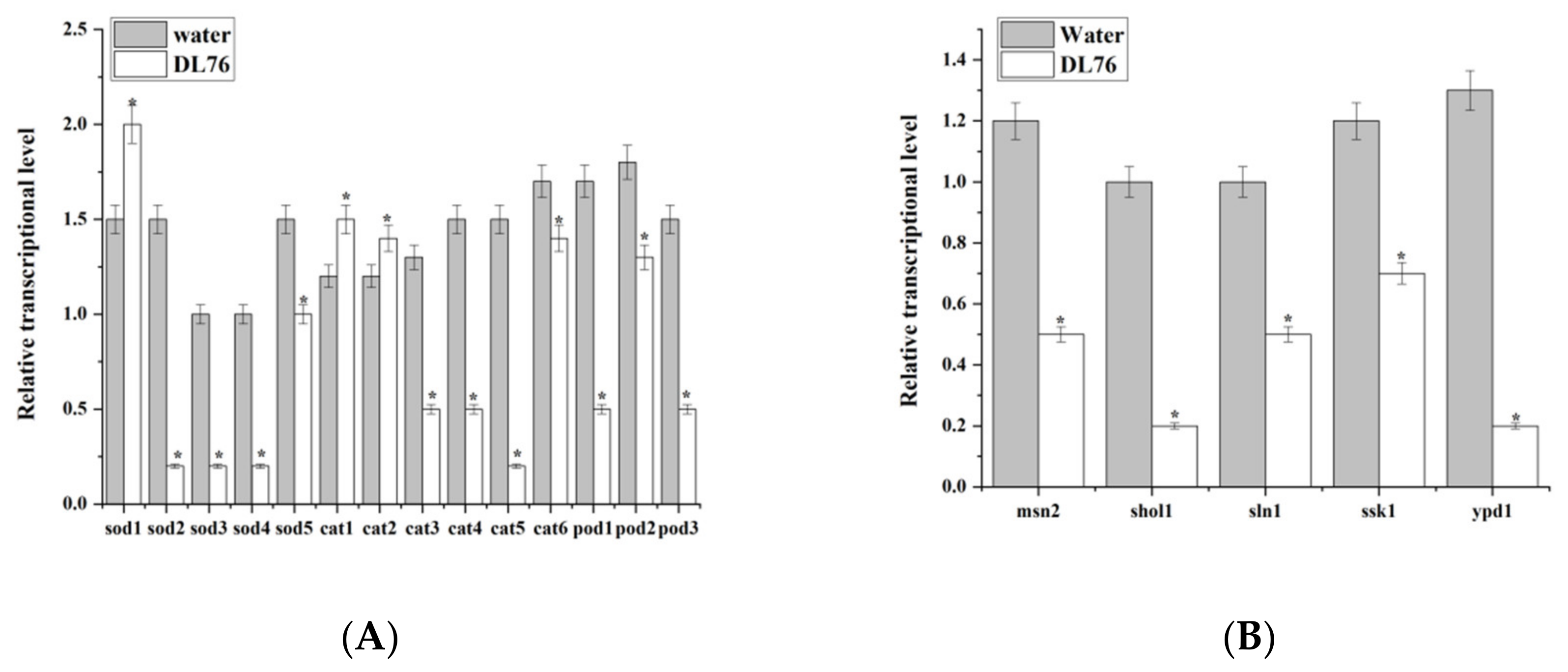
2.8. DL76 Reduced M. oryzae Adaptation to the Environment and Contributed to Cellular Response and Abiotic Stresses

DL76 grew significantly slower than the control strains during the 8-day incubation at 25 °C on minimal CZA (Czapek agar) and 5 CZAR-derived media. The final size of the emerging colony was reduced by 46–60% on the sucrose carbon source, glucose, acetate, or glycerol and 32–46% on the nitrogen source of NO2 or NH+4, and affected more by the starvation of carbon than nitrogen (Figure 8A). Consequently, we assayed the osmotic agents (NaCl) for their effects on the colony growth of each strain on the CZA Petri dish (Figure 8B) using the spotting method for colony initiation. We observed a significant difference between wild-type and DL76, based on the survival indices. Regarding the wild-type indices, germination was reduced from 0.3 to 0.05, whereas in DL76, it was 0.2 to 0.03. Hence, DL76 was more sensitive and less tolerant to stressful chemical NaCl than the wild-type. The stress tolerance of phytopathogens to osmotic stressors plays an essential role in pathogenicity [23,24]. Continuing using the spotting method, compared to wild type, DL76 was more sensitive to 34 °C with 27.5% during colony growth (Figure 8C). We used a sensitive concentration to assess the effects of NaCl, H2O2, and SDS on conidial germination. The results indicated that DL76 was significantly less than the wild-type, thus suggesting that DL76 is less tolerant to stressful chemicals (Figure 8D) based on the residual viability (germination). Further, we estimated EC50s for two oxidants, two osmotic agents, and two cell wall stressors to suppress 50% colony growth at 25 °C, respectively (Figure 8E–G). We initiated all colonies with the spotting method. In the two oxidants, H2O2 and menadion, DL76 decreased significantly and was more sensitive to all the chemicals (Figure 8E) than wild-type. In the two osmotic agents, NaCl and sorbitol, DL76 decreased significantly and was more sensitive to all the chemicals (Figure 8F) than wild-type. Comparing the colonies, DL76 was more sensitive compared to wild-type Guy11 (Figure 8G). In addition, conidial thermotolerance and UV-B resistance were reduced in DL76, as indicated by LT50 (min) and LD50 (J/cm2) (Figure 8H,I). The data indicates DL76 responses to oxidative, hyperosmotic, cell wall disturbing, thermal, and UV-B resistance.

2.9. DL76 Is Essential for Pathogenic Development in M. oryzae and Responsible for Appressorium-like Structure Formation

We observed that DL76 promotes fewer appressorium formation and suppresses appressorium formation in the previous results at both concentrations of 1 × 10⁷ CFU/mL and 1 × 10⁶ CFU/mL. We further checked if the two concentrations would produce appressorium-like structures by hyphal tips. We used four incubation times (24, 36, 48, and 60 hpi); at 24 hpi, DL76 in both concentrations generated fewer appressorium-like structures when compared with water. DL76 generated fewer appressorium-like structures than water as the incubation time increased from 24 hpi to 60 hpi (Figure 9A). Similarly, most of the appressorium-like structures formed by DL76 were abnormal and swollen compared to water. The deformation rate was higher in DL76 than in water (Figure 9B). Further, to investigate the pathogenic role of DL76 in M. oryzae, we inoculated 20 μL drops of conidial suspension from Guy11 and treated them with DL76 on wounded rice leaves.
The field and in vitro experiments indicated that DL76 significantly inhibits the growth of rice blast pathogen (Figure 9C). The wild-type Guy11 was fully pathogenic with extendible necrotic lesions, whereas DL76 caused small necrotic lesions on the inoculated site. The leaf lesion diameter in the DL76 group was 4.2 mm, whereas in wild type Guy 11 was 11.5 mm. We also compared the DL76 inhibitory pathogenicity against rice blasts with the control group. The lesion diameter on isolated leaves of the treatment group was smaller than that of the control group. This phenomenon indicated that the sterilized culture filtrate of DL76 had a strong ability to suppress the pathogenicity of M. oryzae.
On the other hand, we evaluated the DL76 inhibitory properties against rice blasts. We sprayed rice seedlings with five-fold dilutions of bacterial PDB cultures with 1 × 10⁷ and 1 × 10⁶ CFU/mL 36 h before inoculating with M. oryzae. The results indicated that the preventive application of DL76 at both concentrations effectively controlled rice blast disease. Further, we observed rare tiny lesions on rice leaves when we compared DL76 with the control group. The average lesion quantity on living leaves of the control group was 36 lesions per leaf compared, while the average lesion quantity of treatment was 15–20 lesions per leaf treated with DL76 (Figure 9D). This outcome informed that the sterilized culture filtrate of DL76 had a strong ability to suppress the pathogenicity of M. oryzae.
Similarly, we examined the appressorium turgor in the DL76 and wild-type Guy11 to further elucidate the mechanism underlying virulence in the DL76 in M. oryzae. Surprisingly, the appressoria of the DL76 observed a reduced collapse rate in 1, 2, 3, and 4 M glycerol compared to the wild-type. There were significant differences between the generated turgor pressures of the tested strains at these concentrations. For example, at 1 M glycerol, the appressorium collapse rate was 0.8% in wild-type Guy11 but was 45% in DL76. The collapse rates gradually increased from 0.6% to 65% in wild-type Guy11 and 45% to 83.5% in DL76, respectively, at 4 M glycerol (Figure 9E). The proportion of collapsed appressoria increased with the external glycerol concentration suggesting that DL76 reduces defects in maintaining appressorium turgor.

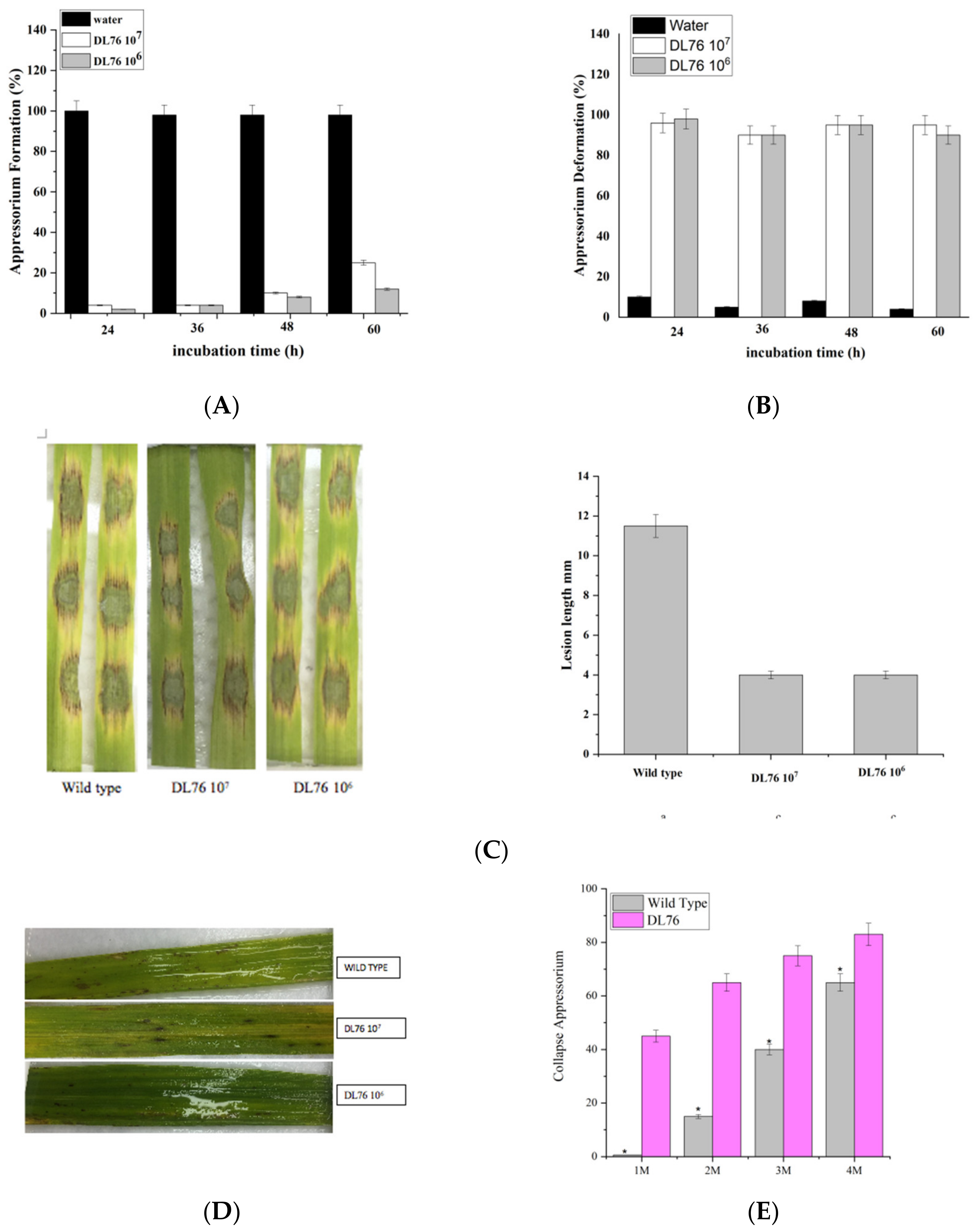
2.10. DL76 Is Involved in Down-Regulating the Expression of Oxidation and Osmotic Genes in M. oryzae

In contrast, many of the examined genes were down-regulated by DL76 versus water under oxidative stress; RT-qPCR examined 14 genes. The expression level of 11 genes was significantly down-regulated by DL76 in M. oryzae than in water (Figure 10A). The transcript levels of sod1, cat1, and cat2 were significantly increased in DL76. Under osmotic stress, the transcriptional level of five genes msn2, sln1, shol1, ssk1, and ypd1 were examined. Compared to water, all five osmosensitive genes were down-regulated in M. oryzae by DL76 (Figure 10B).

3. Discussion

Few potential biocontrol strains of rice blast suppression have been isolated from the plant rhizosphere in recent years [25,26]. Several species of bacteria, especially Bacillus, are known as biological control agents that suppress several phytopathogenic fungi due to cell wall-degrading enzymes and other anti-fungal metabolic creation [27,28,29]. Several studies observed that Bacillus strains could stimulate plant growth and disease resistance. Antecedent B. subtilis strains have been used as biological control agents, where they played a significant agricultural role in the biocontrol of several plant pathogens, including M. oryzae [30,31]. B. subtilis activity relies on unique biological control mechanisms that Bacillus spp. can compete for colonization sites in plants to form bio-films and enhance the provision of nutrients and phytohormones. It improves the interaction of pathogen and plant and suppresses the pathogen growth by antibiotics (Surfactin, Iturin, and Fengycin), toxins, and bio-surfactants. The mechanism associated with bacteriolysis involves the production of extracellular cell wall degrading enzymes, such as chitinase and β-1,3-glucanase [32,33]. Several Bacillus strains antagonistic to fungi have been isolated from soil; however, we isolated Bacillus subtilis DL76 from the field rice rhizosphere in this study [34]. Our results observed that culture filtrate of DL76 obtained a biocontrol deficiency of 75% under greenhouse conditions. Furthermore, the valuation of the antifungal activity of DL76 under different concentrations of the colony-forming unit was found to have a significant suppression growth of M. oryzae. The stability experiment suggested that the active metabolites have substantial stability for high temperature and acid, not alkaline conditions. This indicates that high stability is necessary for developing antifungal metabolites as a biocontrol agent. Similarly, B. subtilis DL76 achieved logarithmic growth after 8 h of cultivation at a given fermentation condition. The strongest antifungal activity was obtained during the stationary phase 40 h after inoculation. We also observed a direct association between inoculation time and antifungal activity. That is, an increase in the inoculation time increases the anti-fungal activity. In the previous study, the antibiotics usually start to be produced at the end of the exponential growth phase. It reaches maximum concentration after cell growth has ceased, which conforms with the production kinetics of the strain DL76. Germination of conidia and the formation of appressorium are two crucial processes of M. oryzae during plant infection. In conidium development, the concentration of 1 × 10⁷ CFU/mL of DL76 suppressed conidia germination at eight hpi. In contrast, a lower concentration (1 × 10⁶ CFU/mL) allowed some conidia to germinate, and the concentration of 105 CFU/mL observed no suppression effect at 24 hpi. The concentration of 1 × 10⁷ CFU/mL of DL76 completely suppressed conidium germination at 24, 36, 48, and 60 hpi with an extended time. Furthermore, the germ tubes of conidia treated with DL76 at high concentration were remarkably shorter than those treated at low concentration or with water. We monitored appressorium formation and the turgor pressure inside the appressoria. At both concentrations of 1 × 10⁷ and 1 × 10⁶ CFU/mL appressorium formation was suppressed, and no appressorium was observed. However, lower concentration permitted appressorium formation. These results suggested that B. subtilis DL76 at lower concentration does not affect the eventual appressorium formation and turgor pressure inside the appressoria. It indicates that the germination of conidia was delayed and suppressed and the fermentation filtrate of DL76 hindered the formation of appressorium. Therefore, we can conclude that the filtrate of DL76 can suppress the formation of infectious structures of M. oryzae in vitro.
We noted that the conidia treated with the culture filtrate of DL76 lost pathogenicity in the pathogenicity test. In contrast, the lesion of the wild type was bigger than that of DL76. The number of lesions in DL76 was much lower than that in the wild type. Conclusively, our results inform that in the infection situation, the pathogenicity of M. oryzae was significantly suppressed due to DL76-treatment. Furthermore, we suggest that DL76 can suppress growth, germination, and appressorium formation in vitro. That is the pathogenicity of M. oryzae in vitro.
Besides, various well-conserved signaling pathways have been verified to be involved in M. oryzae germ tube-derived appressorium formation and penetration [35,36]. The regulation mechanisms of appressorium formation and appressorium-like structure development differ in structure-function during penetration and pathogenicity [37,38,39]. In this research, we found that DL76 does not affect the formation of appressoria at 1 × 10⁷ and 1 × 10⁶ CFU/mL. We examined for appressorium-like structure (ALS) to understand its reasons. The results indicated that DL76 completely suppressed the appressorium-like structure at hyphal tips on the hydrophobic surface, and most of the appressorium-like structures formed by DL76 were deformed. Additionally, DL76 dramatically lost its ability to infect rice leaves and cause disease symptoms. This led us to examine the appressorium turgor pressure. We found out that the collapse rate of appressoria of DL76 was remarkably higher than that of wild-type Guy11 at all concentrations, implying that appressorium turgor in B. subtilis DL76 was remarkably decreased.
Cell wall integrity (CWI) plays an essential role in responding to external stress, fungal growth, and pathogenicity. The fungal cell wall provides mechanical protection against attack from the host during host-pathogen interactions. Thus, maintaining cell wall integrity is critical for phytopathogens to establish disease on their host [40,41,42]. This study observed that the B. subtilis DL76 was a more sensitive cell wall perturbing agent and showed gradient reductions in tolerance to high temperature, oxidation, high osmolarity, and cell wall perturbation during colony growth. Moreover, the relative transcript level of stress-responsive genes was examined. We found that in oxidation conditions, the expression level of the stress-responsive genes was significantly reduced by DL76 except for Sod1, which showed an increased expression level in DL76. However, in osmotic conditions, all the stress-responsive genes were remarkably reduced by DL76.

4. Materials and Methods

4.1. The Evaluation of Antifungal Activity

In this experiment, we tested the inhibition of M. oryzae against the strain DL76. The strain DL76 was inoculated to LB liquid medium and incubated at 28 °C on a rotary shaker Thermo Fisher Scientific, Waltham, MA, USA). For 48 h, followed by lysine and removal of bacterial cells. The supernatant was filtered through a 0.22 μm membrane filter to remove the bacteria cells, and the filtrate was mixed with solid LB at different concentrations (1 × 10², 1 × 10³, 1 × 10⁴, 1 × 10⁵, 1 × 10⁶, 1 × 10⁷ colony-forming units (CFU) per mL). Lastly, the mixture was poured into Petri dishes. The fungus mycelia plug of Guy11 was transferred to the center of the medium and incubated at 28 °C in the incubator (Fisher Scientific, Waltham, MA, USA).

4.2. The Stability of PH and Temperature of Filtrate on the Growth Suppression of M. oryzae

We prepared the sterilized culture filtrate of DL76 to determine the stability of pH and temperature against M. oryzae. We took out 70 mL to be placed in the seven Vitro, adjusted to various pH values to 1, 3, 5, 7, 11, 13, respectively, and stored in the refrigerator at 4 °C. After 24 h, the samples were readjusted to pH 7 and took out 1.5mL from each of the Vitro to mix up with molten potato dextrose agar (PDA) mediums making filtrate of concentration to 6.25 μL/mL (1:160, v/v). The fungus cake of Guy11 was transferred to a Petri dish. After incubation at 28 °C for seven days, the growth of Guy11 was observed, and the colony diameter of Guy11 was measured to assess the growth inhibition rate compared to control. Regarding the temperature test, the sterilized and broth culture filtrate were exposed to 4 °C, 28 °C, 37 °C, 60 °C, 80 °C, 100 °C, and 121 °C for 30 min. Similarly, this test set was repeated three times after cooling the sample to room temperature. The relative suppression rate was described as follows;
Relative inhibition (%) = [(K1 − K2)/K1] × 100
where K1 = M. oryzae colony diameter in the control and R2 = M. oryzae colony diameter in the dual culture Petri dishes with a mixture of sterilized culture filtrate of DL76.

4.3. Correlation between Cell Growth and Anti-Fungal Activity

We inoculated Bacillus subtilis DL76 into 500 mL of potato dextrose liquid medium and incubated it at 28 °C, 180 rpm for 108 h. During the incubation, samples were taken at eight hours to measure OD600 (Eppendorf BioPhotometer, Marshall Scientific LLC, Hampton, NH, USA) and antifungal activity against M. oryzae. Further, we used 1.5mL from each vitro to mix with molten potato dextrose agar mediums making a filtrate of 6.25 μL/mL (1:160, v/v) concentration. The fungus cake of Guy11 was transferred to the Petri dish (and incubated at 28 °C for seven days). The growth of Guy11 was observed, and its colony diameter was measured to assess the growth suppression rate compared to the control.
Relative inhibition% = [(K1 − K2)/K1] × 100
K1 represents M. oryzae colony diameter in control, and K2 represents M. oryzae colony diameter in the dual culture plates with sterilized culture filtrate of DL76 mixed with PDA.

4.4. Effect of the Supernatant at Different Concentrations on Conidium Germination, Appressorium Formation, Germ Tube Length, Turgor Pressure, Infection Process, Vegetative Growth and Conidiation, and Cell Activity

4.4.1. The Conidia Germination Test

We used 10-milliliters of water to scrape the conidia from a 10-day-old M. oryzae conidia-producing medium adjusted to a concentration of 1 × 10⁵ conidia/mL after filtration with two layers of lens paper. Further, we added one milliliter of sterile water, LB broth, and HS24 as the control groups and 1 mL of DL76 at different concentrations (1 × 10², 1 × 10³, 1 × 10⁴, 1 × 10⁵, 1 × 10⁶, 1 × 10⁷ CFU/mL) to each tube. Fifty micro-liters of conidia suspension were dropped on the hydrophobic cover slide. We observed the appressorium formation under a ZEISS fluorescence microscope (Tokyo, Japan) after incubation at room temperature in a moist Petri dish for 8, 12, 24, 36, 48, and 60 h. We then selected 100 conidia to compute the germination rate. The experiment was repeated three times.
The formula for the germination rate was as follows:
Germination rate (%) = [K1/K2] × 100%
where K1 = the number of germinated conidia and K2 = total conidia.

4.4.2. The Appressorium Formation

We scraped conidia with sterilized water and culture filtrate from a 10-day-old M. oryzae conidia-producing medium, then diluted the suspension to 1 × 10⁵ conidia/mL. Afterward, fifty micro-liters of conidia per droplet were dripped on a hydrophobic cover slide. We observed the appressorium formation under a ZEISS fluorescence microscope (Tokyo, Japan) after incubation at room temperature in a moist Petri dish for 8, 12, 24, 36, 48, and 60 h. The experiment was repeated three times.
The formula for appressorium formation rate was as follows:
Formation rate of appressorium (%) = [V1/V2] × 100%
V1: the number of conidia that formed appressorium;
V2: the number of total conidia.

4.5. Environmental Stresses at Different Concentrations of SDS, H2O2, Glycerol, and NaCl (0, 20, 40, 60, 80, and 100 µΜ) Were Added to the Conidial Suspension

Twenty microliters of the mixture from each treatment were dripped onto a microscope cover glass with wet filter paper and incubated at 28 °C in darkness. Conidium germination and germ tube length were examined at six hpi.

4.6. Measurement of Growth and Conidiation Parameters

Aliquots (1ml) of 1 × 10⁶ conidia/mL suspension treated with DL76 at various concentrations were spotted centrally on the Petri dishes (9 cm diameter) of nutrition-rich SDAY (4% glucose, 1% peptone, and 1.5% agar plus 1% yeast extract), minimal CZA and eight CZA-derived media. The derived media were prepared by excluding 3% sucrose (carbon starvation) from standard CZA, replacing 3% sucrose with 3% of glucose, galactose, glycerol, or acetate (NaAc) as the sole carbon source, and replacing 0.3% NaNO3 with 0.3% of NaNO2 or NH4Cl as sole nitrogen source, respectively. During the eight-day incubation at 25 °C and 12:12 h, colony diameters were cross-measured daily to compute the colony area as a growth rate index.
Further, 100 mL aliquots of 1 × 10⁷ conidia/mL suspension treated with DL76 at different concentrations were evenly spread on SDAY Petri dishes and incubated for 7-days at 25 °C and 12:12 h to assess the sporulation capacity of each strain. From the 3rd day onwards, 5-mm diameter colony discs were cut off daily from the Petri dishes, and the conidia on each disc were washed into 1 mL of 0.02% Tween 80 by vortexing. We removed the hyphal debris by filtration, determined the conidial concentration with microscopic counts, and converted it to the number of conidia per 2 cm colony. Further, the fungal mass samples taken from the colonies were evaluated under the microscope (Olympus, Tokyo, Japan) to observe possible morphological changes in their conidiophores during the incubation.

4.7. Assaying Cellular Responses to Chemical and Environmental Stresses

SDAY Petri dishes overlaid with cellophane were spread with 100 mL aliquots of conidial suspension treated with DL76 at different concentrations to produce uniform cultures after 3-day incubation at 25 °C. We cut a 5 mm diameter fungal mycelial discs from the culture of each strain and placed them centrally onto the Petri dishes (90 mm diameter) of 1/4 SDAY supplemented with the gradients of NaCl (0.4–2 M), menadione (2–8 mM), H2O2 (20–80 mM), Congo red (0.5–3 mg/mL), SDS (0.02–0.12%) and carbendazim (0.4–2 mg/mL), respectively. After 6-day stressful incubation at 25 °C and 12:12 h, the diameters of all colonies in the control and stress treatments were cross-measured to compute their net area increases.
Cellular responses to stressful chemicals were assayed by spotting aliquots of 1 μL of a 107 conidia/mL suspension onto the Petri dishes of CZA alone (control) or supplemented with the respective gradients of menadione (0–40 μM), H2O2 (04 mM), NaCl (0–0.7 M), sorbitol (0–1.5 M), Congo red (0–50 μg/mL), and calcofluor white (0–20 μg/mL), followed by seven days of incubation at 25 °C. Chemical-free Petri dishes incubated at 34 °C for seven days were included as a heat stress treatment. All colony diameters were measured on day 7. An effective concentration (EC50) for each chemical to suppress 50% colony growth was estimated by modeling growth trends over the gradient. The heat stress estimated relative growth inhibition of fungal colonies (TC − TS)/TS × 100, where TC and TS denote colony diameters at 25 °C (control) and 34 °C, respectively.
Additionally, we assayed the conidial tolerances to oxidation, hyperosmolarity, and cell wall disturbance by spreading 100 mL aliquots of conidial suspension treated with DL76 at different concentrations onto the Petri dishes of germination medium (GM: 2% sucrose and 0.5% peptone plus 1.5% agar) supplemented with menadione (0.2 mM), H2O2 (4 mM), NaCl (1.2 M), Congo red (1 mg/mL) and SDS (0.04%), respectively. We assessed the conidial germination on each of the plates, stressed or not, using three counts of germinated and ungerminated conidia under a microscope after 24 h of incubation at 25 °C. We calculated the residue viability as the ratio of percent germination under each stress over that in control. Similarly, we used the previous protocol to assay the conidial tolerances to wet-heat stress of 15–120 Min at 45 °C and UV-B irradiation (weighted 312 nm) of 0.1–0.8 J/cm 2 in the Bio-Sun 11 chamber (Vilber Lourmat, Marnela-Valle’e, France). Additionally, after exposure to a specific intensity of heat or UV-B stress, conidia were incubated for 24 h at 25 °C under saturated humidity. Percent germination was determined using microscopic counts.

4.8. Assays for Stress Tolerance of Antisense Transformants

Hyperosmotic stress assay for each strain, 10-μL aliquots of spore suspension (1 × 10⁶ conidia mL−1) were spread evenly on the Petri dishes of Czapek’s agar supplemented with 0–1.5 M NaCl for different degrees of osmotic stress (Zhang et al., 2009). After 24-h of incubation at 25 °C, percent germination on the Petri dishes was determined above. We estimated the osmotic survival index as the ratio of the percent germination on the salt-inclusive Petri dish over that on the salt-free Petri dishes.

4.9. Experiment on Isolated Leaves

Rice seedlings were grown in an artificial climate chamber (30 °C) for 30 days. Rice leaves without apparent disease symptoms were placed in Petri dishes with a solution of 6-benzylaminopurine. Before that, every leaf was poked slightly with a needle making it more susceptible to M. oryzae infection. Regarding the treatment group: a five μL volume of M. oryzae conidia suspension (1 × 10⁵ conidia/mL) droplets was diluted with a sterilized culture filtrate of Bacillus subtilis DL76 and applied to the slightly punctured sites of the leaves. The inoculated leaves were incubated at 25 °C in the dark for 24 h.

4.10. RNA Extraction and Transcripts of Stress-Responsive Gene Transcription Analysis

The strain guy11 conidia co-cultured with sterile water or DL76 were collected at two hpi for RNA extraction. The Prime Script RT reagent kit (TaKaRa Bio Inc., San Jose, CA, USA) performed a reverse transcription test. Transcription of the stress-responsive gene was examined by reverse transcription-quantitative PCR (RT-qPCR) on an ABI 7500 Fast Real-Time System (Applied Biosystems, (AB Ltd., Lincoln, NE, USA) following previously established procedures (Guo et al., 2010). The primers used in this study were designed using the NCBI (https://www.ncbi.nlm.nih.gov accessed on 23 June 2021), as shown in Table 1.

4.11. Statistics and Data Analysis

All treatments were repeated at least three times, and >100 conidia were observed in each replicate. Data were analyzed with SPSS 17.0 (IBM Corporation) by one-way analysis of variance followed by means separation using the least significance difference (LSD) test (p < 0.05). The ratio of colony size or conidial germination rate under stress over control was defined as relative viability (VR). For each of the tested strains, the VR trends over the concentrations (C) of each chemical, the time lengths (T) of wet-heat stress, and the doses (D) of UV-B irradiation were fitted to the equation VR = 1/[1 + exp (a + bx)]. Where x is C, D, or T, a and b are parameters to be estimated. When VR = 0.5, the fitted equations gave solutions (−a/b) to effective concentration (EC50) of each stressful chemical required to suppress 50% colony growth and median lethal responses of conidia to heat (LT50, min) and UV-B (LD50, J/cm2) stresses.

5. Conclusions

In summary, we have observed that B. subtilis DL76 can inhibit the growth, germination, and formation of appressorium in vitro and the pathogenicity of M. oryzae in vivo. DL76 significantly suppressed the conidial germination and appressorial formation of M. oryzae. Moreover, DL76 might effectively inhibit infection by influencing the formation of the infectious structure of M. oryzae. Conidial thermotolerance and UV-B resistance were reduced in DL76 disruption. Furthermore, DL76 also reduced large parts of conidial tolerances to both oxidative and cell wall perturbing stresses. Finally, the transcriptional level of DL76 was down-regulated both under osmotic and oxidative stress. We can conclude that Bacillus subtilis DL76 significantly contributes as a candidate biological control agent against M. oryzae and has furthered the progress of pathogenesis research.

Author Contributions

Conceptualization, V.T.K. and H.L.; writing—original draft preparation, V.T.K.; writing—review and editing, V.T.K. and B.T.; visualization, Z.Y.; supervision, H.L.; project administration, V.T.K. and H.L.; funding acquisition, H.L. All authors have read and agreed to the published version of the manuscript.

Funding

This research was supported by Natural Science Foundation of Jiangsu Province (BK20201324) and National Natural Science Foundation of China (31571992).

Institutional Review Board Statement

Not applicable.

Informed Consent Statement

Not applicable.

Data Availability Statement

Not applicable.

Conflicts of Interest

The authors declare no conflict of interest.

References

  1. Zhang, C.X.; Zhang, X.X.; Shen, S.H. Proteome Analysis for Antifungal Effects of Bacillus subtilis KB-1122 on Magnaporthe grisea P131. World J. Microbiol. Biotchnol. 2014, 30, 1763–1774. [Google Scholar] [CrossRef]
  2. Xiong, Z.Q.; Tu, X.R.; Wei, S.J.; Huang, L.; Li, X.H.; Lu, H. In Vitro Antifungal Activity of Antifungalmycin 702, A New Polyene Macrolide Antibiotic, Against the Rice Blast Fungus Magnaporthe-grisea. Biotechnol. Lett. 2013, 35, 1475. [Google Scholar] [CrossRef]
  3. Li, Q.; Jiang, Y.; Ning, P.; Zheng, L.; Huang, J.; Li, G. Suppression of Magnaporthe oryzae by Culture Filtrates of Streptomyces globisporus JK-1. Biol. Control 2011, 58, 139–148. [Google Scholar] [CrossRef]
  4. Ohtaka, N.; Kawamata, H.; Narisawa, K. Suppression of Rice Blast Using Freeze-Killed Mycelia of Biocontrol Fungal Candidate MKP5111B. J. Gen. Plant Pathol. 2008, 74, 101–108. [Google Scholar] [CrossRef]
  5. Leneveu-Jenvrin, C.; Charles, F.; Barba, F.J.; Remize, F. Role of Biological Control Agents and Physical Treatments in Maintaining the Quality of Fresh and Minimally-Processed Fruit and Vegetables. Crit. Rev. Food Sci. Nutr. 2019, 60, 2837–2855. [Google Scholar] [CrossRef]
  6. Moraes, B.J.; Belinato, J.R.; Costa, J.H.; Akiyama, D.Y.; Pontes, J.G.M.; Kupper, K.C.; Augusto, F.; De Carvalho, J.E.; Fill, T.P. Biological Control of Citrus Postharvest Phytopathogens. Toxins 2019, 11, 460. [Google Scholar] [CrossRef] [Green Version]
  7. Kohl, J.; Kolnaar, R.; Ravensberg, W.J. Mode of Action of Microbial Biological Control Agents against Plant Diseases: Relevance beyond Efficacy. Front. Plant Sci. 2019, 10, 845. [Google Scholar] [CrossRef] [Green Version]
  8. Hyakumachi, M.; Takahashi, H.; Matsubara, Y.; Someya, N.; Shimizu, M.; Kobayashi, K. Recent Studies on Biological Control of Plant Diseases in Japan. J. Gen. Plant Pathol. 2014, 80, 287–302. [Google Scholar] [CrossRef]
  9. Vidhyasekaran, P.; Rabindran, R.; Muthamilan, M.; Nayar, K.; Rajappan, K.; Subramanian, N. Development of a Powder Formulation of Pseudomonas fluorescens for Control of Rice Blast. Plant Pathol. 2010, 46, 291–297. [Google Scholar] [CrossRef]
  10. Sha, Y.; Wang, Q.; Li, Y. Suppression of Magnaporthe oryzae and Interaction between Bacillus subtilis and Rice Plants in the Control of Rice Blast. SpringerPlus 2016, 5, 1238. [Google Scholar] [CrossRef] [Green Version]
  11. Karthikeyan, V.; Gnanamanickam, S.S. Biological Control of Setaria Blast (Magnaporthe grisea) with Bacterial Strains. Crop Prot. 2008, 27, 263–267. [Google Scholar] [CrossRef]
  12. Zarandi, M.E.; Bonjar, G.H.S.; Dehkaei, F.P.; Moosavi, S.A.A.; Farokhi, P.R.; Aghighi, S. Biological Control of Rice Blast (Magnaporthe oryzae) By Use of Streptomyces sindeneusis Isolate 263 in Greenhouse. Am. J. Appl. Sci. 2009, 6, 194–199. [Google Scholar] [CrossRef] [Green Version]
  13. Anand, T.; Chandrasekaran, A.; Kuttalam, S.; Senthilraja, G.; Samiyappan, R. Integrated Control of Fruit Rot and Powdery Mildew of Chilli Using the Biocontrol Agent Pseudomonas fluorescens and A Chemical Fungicide. Biol. Control 2010, 52, 1–7. [Google Scholar] [CrossRef]
  14. Yan, Q.H.; Zhou, J.X.; Li, H.Z.; Zhi, Q.Q.; Zhou, X.P.; He, Z.M. Coexistence of and Interaction Relationships between Anaflatoxin-Producing Fungus and a Bacterium. Fungal Biol. 2015, 119, 605–614. [Google Scholar] [CrossRef]
  15. Kumar, K.V.K.; Yellareddygari, S.K.; Reddy, M.S.; Kloepper, J.W.; Lawrence, K.S.; Miller, M.E.; Sudini, H.; Reddy, E.C.S.; Zhou, X.G.; Groth, D.E. Ultrastructural Studies on The Interaction Between Bacillus subtilis MBI 600 (Integral) and the Rice Sheath Blight Pathogen, Rhizoctonia solani. Afr. J. Microbiol. Res. 2013, 7, 2078–2086. [Google Scholar]
  16. Strunnikova, O.K.; Vishnevskaya, N.A.; Ruchiy, A.S.; Shakhnazarova, V.Y.; Vorobyov, N.I.; Chebotar, V.K. The Influence of Soils with Different Textures on Development, Colonization Capacity, and Interactions Between Fusarium culmorum and Pseudomonas fluorescens in Soil and on Barley Roots. Plant Soil 2015, 389, 131–144. [Google Scholar] [CrossRef]
  17. Grover, M.; Nain, L.; Singh, S.B.; Saxena, A.K. Molecular and Biochemical Approaches for Characterization of Antifungal Trait of a Potent Biocontrol Agent Bacillus subtilis RP24. Curr. Microbiol. 2010, 60, 99–106. [Google Scholar] [CrossRef]
  18. Strobel, G.; Daisy, B.; Castillo, U.; Harper, J. Natural Products from Endophytic Microorganisms. J. Nat. Prod. 2004, 67, 257–268. [Google Scholar] [CrossRef]
  19. Gao, Z.; Zhang, B.; Liu, H.; Han, J.; Zhang, Y. Identification of Endophytic Bacillus velezensis ZSY-1 Strain and Antifungal Activity of Its Volatile Compounds Against Alternaria solani and Botrytis cinerea. Biol. Control 2017, 105, 27–39. [Google Scholar] [CrossRef]
  20. Ali, G.S.; El-Sayed, A.; Patel, J.S.; Green, K.B.; Ali, M.; Brennan, M.; Norman, D. Ex Vivo Application of Secreted Metabolites Produced by Soil-Inhabiting Bacillus Spp. Efficiently Controls Foliar Diseases Caused by Alternaria Spp. Appl. Environ. Microbiol. 2015, 82, 478–490. [Google Scholar] [CrossRef] [Green Version]
  21. Howard, R.J.; Ferrari, M.A.; Roach, D.H.; Money, N.P. Penetration of Hard Substrates by a Fungus Employing Enormous Turgor Pressures. Proc. Natl. Acad. Sci. USA 1991, 88, 11281–11284. [Google Scholar] [CrossRef] [PubMed] [Green Version]
  22. Talbot, N.J. On The Trail of A Cereal Killer: Exploring the biology of Magnaporthe grisea. Annu. Rev. Microbiol. 2003, 57, 177–202. [Google Scholar] [CrossRef] [PubMed] [Green Version]
  23. Andersson, R.A.; Koiv, V.; Norman-Setterblad, C.; Pirhonen, M. Role of Rpos in Virulence and Stress Tolerance of the Plant Pathogen Erwinia Carotovora Subsp. Carotovora. Microbiology 1999, 145, 3547–3556. [Google Scholar] [CrossRef] [Green Version]
  24. Guo, M.; Chen, Y.; Du, Y.; Dong, Y.H.; Guo, W.; Zhai, S.; Zhang, H.F.; Dong, S.M.; Zhang, Z.G.; Wang, Y.C.; et al. The Bzip Transcription Factor Moap1 Mediates the Oxidative Stress Response and Is Critical for Pathogenicity of the Rice Blast Fungus Magnaporthe oryzae. PLoS Pathog. 2011, 7, e1001302. [Google Scholar] [CrossRef] [Green Version]
  25. Tokpah, D.P.; Li, H.W.; Wang, L.Y.; Liu, X.Y.; Mulbah, Q.S.; Liu, H.X. An Assessment System for Screening Effective Bacteria as Biological Control Agents against Magnaporthe grisea on Rice. Biol. Control 2016, 103, 21–29. [Google Scholar] [CrossRef]
  26. Zhang, H.F.; Zhao, Q.; Liu, K.Y.; Zhang, Z.G.; Wang, Y.C.; Zheng, X.B. Mgcrz1, a Transcription Factor of Magnaporthe grisea, Controls Growth, Development and is Involved in Full Virulence. FEMS Microbiol. Lett. 2009, 293, 160–169. [Google Scholar] [CrossRef] [Green Version]
  27. Sellem, I.; Triki, M.A.; Elleuch, L.; Cheffi, M.; Chakchouk, A.; Smaoui, S. The Use of Newly Isolated Streptomyces Strain TN258 as a Potential Biocontrol Agent of Potato Tubers Leak Caused by Pythium Ultimum. J. Basic Microbiol. 2017, 57, 393. [Google Scholar] [CrossRef]
  28. Han, T.; You, C.; Zhang, L.; Feng, C.; Zhang, C.; Wang, J. Biocontrol Potential of Antagonist Bacillus subtilis Tpb55 Against Tobacco Black Shank. BiolControl 2016, 61, 195–205. [Google Scholar] [CrossRef]
  29. Calvo, H.; Marco, P.; Blanco, D.; Oria, R.; Venturini, M.E. Potential of A New Strain of Bacillus amyloliquefaciens BUZ-14 As A Biocontrol Agent of Postharvest Fruit Diseases. Food Microbiol. 2013, 63, 101–110. [Google Scholar] [CrossRef] [Green Version]
  30. Zhang, H.; Wu, Z.; Wang, C.; Li, Y.; Xu, J.R. Germination and Infectivity of Microconidia in the Rice Blast Fungus Magnaporthe oryzae. Nat. Commun. 2014, 5, 4518. [Google Scholar] [CrossRef]
  31. Leelasuphakul, W.; Sivanunsakul, P.; Phongpaichit, S. Purification, Characterization and Synergistic Activity of Beta-1,3-Glucanase and Antibiotic Extract from An Antagonistic Bacillus subtilis NSRS 89-24 Against Rice Blast and Sheath Blight. Enzyme Microb. Technol. 2006, 38, 990–997. [Google Scholar] [CrossRef]
  32. Chernin, L.; Chet, I. Microbial Enzymes in the Biocontrol of Plant Pathogens and Pests. In Enzymes in the Environment: Activity, Ecology, and Applications; Marcel Dekker, Inc.: New York, NY, USA, 2002; pp. 171–226. [Google Scholar]
  33. Li, R.Y.; Wu, X.M.; Yin, X.H.; Liang, J.N.; Li, M. The Natural Product Citral can Cause Significant Damage to the Hyphal Cell Walls of Magnaporthe grisea. Molecules 2014, 19, 10279–10290. [Google Scholar] [CrossRef] [PubMed] [Green Version]
  34. Whipps, J.M. Microbial Interactions and Biocontrol in the Rhizosphere. J. Exp. Bot. 2001, 52, 487–511. [Google Scholar] [CrossRef] [PubMed]
  35. Xu, J.R.; Hamer, J.E. MAP Kinase and Camp Signalling Regulate Infection Structure Formation and Pathogenic Growth in The Rice Blast Fungus Magnaporthe grisea. Genes Dev. 1996, 10, 2696–2706. [Google Scholar] [CrossRef] [Green Version]
  36. Wilson, R.A.; Talbot, N.J. Under Pressure: Investigating the biology of Plant Infection by Magnaporthe oryzae. Nat. Rev. Microbiol. 2009, 7, 185–195. [Google Scholar] [CrossRef]
  37. Kim, S.; Park, S.-Y.; Kim, K.S.; Rho, H.-S.; Chi, M.-H.; Choi, J.; Park, J.; Kong, S.; Park, J.; Goh, J.; et al. Homeobox Transcription Factors are Required for Conidiation and Appressorium Development in The Rice Blast Fungus Magnaporthe oryzae. PLoS Genet. 2009, 5, e1000757. [Google Scholar] [CrossRef] [Green Version]
  38. Zhou, Z.; Li, G.; Lin, C.; He, C. Conidiophore Stalk-Less1 Encodes a Putative Zinc-Finger Protein Involved in The Early Stage of Conidiation and Mycelial Infection in Magnaporthe oryzae. Mol. Plant Microbe Interact. 2009, 22, 402–410. [Google Scholar] [CrossRef] [Green Version]
  39. Kong, L.A.; Li, G.T.; Liu, Y.; Liu, M.G.; Zhang, S.J.; Yang, J.; Zhou, X.Y.; Peng, Y.L.; Xu, J.R. Differences Between Appressoria Formed by Germ Tubes and Appressorium-Like Structures Developed by Hyphal Tips in Magnaporthe oryzae. Fungal Genet. Biol. 2013, 56, 33–41. [Google Scholar] [CrossRef]
  40. Howard, R.J.; Valent, B. Breaking and Entering: Host Penetration by the Fungal Rice Blast Pathogen Magnaporthe grisea. Annu. Rev. Microbiol. 1996, 50, 491–512. [Google Scholar] [CrossRef]
  41. Skamnioti, P.; Gurr, S.J. Magnaporthe grisea Cutinase2 Mediates Appressorium Differentiation and Host Penetration and is required for Full Virulence. Plant Cell 2007, 19, 2674–2689. [Google Scholar] [CrossRef] [Green Version]
  42. Jeon, J.; Goh, J.; Yoo, S.; Chi, M.H.; Choi, J.; Rho, H.S.; Park, J.; Han, S.S.; Kim, B.R.; Park, S.Y.; et al. A Putative MAP Kinase, MCK1, is required for Cell Wall Integrity and Pathogenicity of the Rice Blast Fungus, Magnaporthe oryzae. Mol. Plant Microbe Interact. 2008, 21, 525–534. [Google Scholar] [CrossRef] [PubMed] [Green Version]
Figure 1. Effect of different concentrations on the suppression of M. oryzae. The formula of relative inhibition rate was described as Relative inhibition (%) = [(K1 − K2)/K1] × 100. Lower-case letters represent significant difference (p < 0.05).
Ijms 23 05314 g001
Figure 2. Antifungal activity of the pH-treated (A) and heat-treated (B) DL76 against M. oryzae. Error Bars represent the SEs of three replicates. Different letters indicate significant differences (p < 0.05) based on one-way analysis of variance followed by the least significant difference test.
Ijms 23 05314 g002
Figure 3. Correlation between cell growth and anti-fungal activity Correlation between strain DL76 growth and its inhibition percentage. (A) is means of the growth curve measured by OD600 and (B) indicates that antifungal activities of the sterilized culture filtrate of DL76 against M. oryzae. Relative inhibition (%) = [(K1 − K2)/K1] × 100.
Ijms 23 05314 g003
Figure 4. Effect of Bacillus subtilis DL76 at various concentrations on conidium germination at 8 h post-incubation. Magnaporthe oryzae conidia were treated with sterile water, Luria–Bertani (LB) broth, Bacillus cerus HS24, Bacillus subtilis DL76 at a concentration of 1 × 10⁷ CFU/mL. (A) represents conidium germination HS24, and (B) represents conidium germination DL76. Values are means and standard deviations of three replicates. Means separation letters indicate statistical significance (p < 0.05) based on one-way analysis of variance test. Scale bar = 30 µm.
Ijms 23 05314 g004
Figure 5. Effect of Bacillus subtilis DL76 at various concentrations on Appressorium formation at 8 h post-incubation. Magnaporthe oryzae conidia were treated with sterile water, Luria–Bertani (LB) broth, Bacillus cerus HS24, Bacillus subtilis DL76 at a concentration of 1 × 10⁷ CFU/mL (a negative control of bacterium). (A) represents Appressorium germination HS24, (B) represents Appressorium germination DL76. The values are means and standard deviations of three replicates. The means separation letters indicate statistical significance (p < 0.05) based on one-way analysis of variance test. Scale bar = 30 µm.
Ijms 23 05314 g005
Figure 6. Effect of Bacillus subtilis DL76 on Appressorium formation in Magnaporthe oryzae at different time points. Conidia were treated with Luria–Bertani (LB) broth, sterile water, (control), or DL76 at the concentrations of 1 × 10², 1 × 10³, 1 × 10⁴, 1 × 10⁵, 1 × 10⁶ or 1 × 10⁷ CFU/mL. (A) is the conidium germination evaluated with LB broth, sterile water, or DL76 at 24-h post-inoculation (hpi). (B) refers to conidium germination evaluated with LB broth, sterile water, or DL76 at 36-h post-inoculation (hpi). (C) refers to conidium germination evaluated with LB broth, sterile water, or DL76 at 48-h post-inoculation (hpi). (D) is the conidium germination evaluated with LB broth, sterile water, or DL76 at 60-h post-inoculation (hpi). The values are means and standard deviations of three replicates. The means separation letters indicate statistical significance (p < 0.05) based on one-way analysis of variance followed by the least significant difference test. Scale bar = 20 µm.
Ijms 23 05314 g006
Figure 7. The effect of exogenous addition of environmental stresses on conidium germination and germ tube length of Magnaporthe oryzae at 6 h post-incubation. Additionally, the effect of various concentrations of NaCl (A,C,E,G) conidium germination and (B,D,F,H) germ tube length.
Ijms 23 05314 g007aIjms 23 05314 g007b
Figure 8. (A) represents colony sizes on minimal CZA (3% sucrose as mere carbon and 0.3% NaNO3 as mere nitrogen) and CZA-derived media [altered carbon: glucose, glycerol, acetate; altered nitrogen: NaNO2, NH4Cl and (B) The conidial survival indices of the tested strains under the hyperosmotic stresses (C) Relative growth inhibition (RGI) of a fungal colony at 34 °C. (D) represents residue viability of conidia after 24 h incubation at 25 °C on germination medium supplemented with NaCl (1.2 M), H2O2 (4 mM), or SDS (0.04%). (EG) EC50s estimated for two oxidants, two osmotic agents, and two cell wall stressors to suppress 50% colony growth at 25 °C, respectively. (H,I) Median lethal time (LT50) for conidial tolerance to wet-heat stress at 45 °C and median lethal dose (LD50) for conidial UV-B resistance. Error bars represent the SEs of three replicates. Different letters indicate significant differences (p < 0.05) and * Significant difference (p < 0.05) based on one-way analysis of variance followed by the least significant difference test.
Ijms 23 05314 g008aIjms 23 05314 g008b
Figure 9. (A) is the statistical analysis of the appressorium like structure formation rate. More than 100 hyphal tips were observed each time. (B) is the appressorium deformity rate was measured and statistically analyzed. (C) Pathogenicity of conidia on isolated leaves (D) is the pathogenicity assay on abraded rice plant leaves. The tested strains were incubated on isolated rice leaves, and their virulence was evaluated 5 days after incubation. (E) is the measurement of collapsed appressoria. At least 100 appressoria were observed at each glycerol concentration. Error bars represent SD. * Significant difference (p < 0.05) based on one-way analysis of variance followed by the least significant difference test.
Ijms 23 05314 g009
Figure 10. Transcription oxidative and osmotic involved genes in Bacillus subtilis DL76-treated conidia of Magnaporthe oryzae. Conidia were treated with sterile water or DL76 (1 × 10⁷ CFU/mL) for 2 h, and then, total RNA was extracted. Reverse transcription quantitative PCR was used to measure transcription. (A) Oxidative genes used to measure transcription level. (B) Osmotic genes used to measure transcription level. * Significant difference (p < 0.05) based on one-way analysis of variance followed by the least significant difference test.
Ijms 23 05314 g010
Table 1. Primers used for quantitative real-time PCR.
Involved in Response to Oxidative Stress
sod1GCGGCTTCCACATCCACACCTTTGGGTCCAGCGTTGCCAGTCTTGAG
sod2CCAGTGTTTGGCATTGACATGTCAGCCGTCTTCCAGTTGATG
sod3TCTCCGGCAAGATTATGGAGCTTGGCGTCATTCTTGGCCT
cat1CCTCTGACGTTGGCGGCCCTTTCCCGTGTCCGTGCTGCCTCGTG
cat2CCGTCTGGGCATCAACTGGGAAGGCTGGGCGTGGTCGTGGTAG
cat3TCAAGTCGGTTCAGGAGATGGAGTTGTTGCGTCTTCAATCGGAGTG
cat4GAGGAGCCCAGCAACGCACAAGAGCTGAGGACGACAAGGCCGCCATTC
cat5GCTGGGCTGATCTGCTGGTCCTTGTCCTTGCTGTAACGGTGGCTGTCG
cat6CGGCTGCGGTGTCTTGTCCATACCCTTGTCGGCGTTCTGGCGAAG
Involved in Response to Osmotic Stress
sho1AGCGTCAACAACCTCGTCTACAGGCGTGGAGTTCTCAAAG
sln1TCGGTGCCATCACTCCTTCCAACGGCGGACGAGCAATGCCAACAGC
ssk1AGAACTTTAGCACCGAGCCCTTTCAGAAGCAGCAGCAGACGATTGG
ypd1TGACCAGGTCGAGGAGACGTTTGCTCGGCGTCGGGTTCTGATGAGC
msn2GCCCGCCACGCCCATCTACACCGAGGTCTCAACCGAGTCAAAC
Publisher’s Note: MDPI stays neutral with regard to jurisdictional claims in published maps and institutional affiliations.

