Review

Flubendazole Plays an Important Anti-Tumor Role in Different Types of Cancers

1
Institute of Nursing and Health, School of Nursing and Health, Henan University, Jinming Avenue, Kaifeng 475004, China
2
Henan International Joint Laboratory of Nuclear Protein Regulation, School of Basic Medical Sciences, Henan University, Kaifeng 475004, China
3
School of Clinical Medicine, Henan University, Kaifeng 475004, China
4
School of Stomatology, Henan University, Kaifeng 475004, China
*
Authors to whom correspondence should be addressed.
These authors contributed equally to this work.
Int. J. Mol. Sci. 2022, 23(1), 519; https://doi.org/10.3390/ijms23010519
Submission received: 29 November 2021 / Revised: 30 December 2021 / Accepted: 1 January 2022 / Published: 4 January 2022

Abstract

:
Flubendazole, belonging to benzimidazole, is a broad-spectrum insect repellent and has been repurposed as a promising anticancer drug. In recent years, many studies have shown that flubendazole plays an anti-tumor role in different types of cancers, including breast cancer, melanoma, prostate cancer, colorectal cancer, and lung cancer. Although the anti-tumor mechanism of flubendazole has been studied, it has not been fully understood. In this review, we summarized the recent studies regarding the anti-tumor effects of flubendazole in different types of cancers and analyzed the related mechanisms, in order to provide the theoretical reference for further studies in the future.

1. Introduction

Flubendazole(FLU)([5-(4-fluorobenzoyl)-1H-benzimidazole-2-y1]-carbamic acid methyl ester) (Figure 1), which was developed by Janssen in the 1970s, belongs to the group of benzimidazole anthelmintics and is widely used in the treatment of helminth and intestinal parasite infection [1,2,3,4,5]. It has a typical benzimidazole part, but the main structure is the added fluorine atom, which makes it different from other benzimidazoles [6]. In 1980, FLU was approved for the treatment of gastrointestinal nematode infection in veterinary and human medicine. It has also been known for a long time that the intravenous FLU can lead to a high level of filarial effect in many preclinical models and humans [7]. The anthelmintic effect of FLU is based on its ability to change microtubule structure, inhibit tubulin polymerization, destroy microtubule function, and interfere with the transport of secretory vesicles mediated by the microtubule in the absorptive tissues of helminths [4,8,9,10,11]. In recent years, FLU has been found to activate autophagy and to be an effective inducer of reactive oxygen species (ROS) [12,13]. It has been reported that FLU plays an important anti-tumor role in many kinds of cancers, such as breast cancer, melanoma, neuroblastoma, colorectal cancer, liver cancer, oral squamous cell cancer [1,10,14,15,16,17,18,19,20,21,22,23,24], and is regarded as a promising anti-tumor drug [1,22]. Recent studies have shown that FLU inhibits cell proliferation and delays tumor formation in xenograft models and shows preclinical activity by suppressing tubulin polymerization in lymphoma and leukemia [10,25,26]. Although some studies have described some anti-tumor mechanisms of FLU, including the inhibition of the tumor cell cycle, and the tumor cell proliferation and growth, the anti-tumor mechanism of FLU has not been fully understood [10,22,23,27]. Here, we reviewed and analyzed the studies about the anti-tumor effects and mechanisms of FLU in recent years, hoping to provide a theoretical reference for further studies on the anti-tumor effect of FLU in the future.

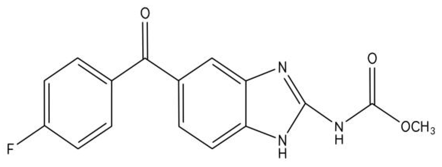
2. The Pharmacokinetics of Flubendazole

FLU is usually taken orally and absorbed through the gastrointestinal tract. Only a small amount of FLU is reabsorbed after the oral treatment in pigs, rats, sheep, dogs, and humans [28]. FLU is difficult to dissolve in the water system of the gastrointestinal tract, resulting in the low absorption rate of blood, so its bioavailability is very low. Even if the oral dose in humans was 2 g, the maximum plasma concentration level of FLU in humans was lower than 5 ng/mL. If FLU is used immediately after meals, its absorption increases significantly [20,29]. In order to ameliorate the bioavailability of the system, it has been reconstituted as an amorphous solid dispersion (ASD) oral preparation [3]. After subcutaneous administration, FLU showed high effectiveness in the animal model of filarial infection. FLU has obvious clinical effects on onchocerciasis by intramuscular injection in humans. However, the intramuscular injection leads to severe injection site reactions [30].
There are two possible biotransformation ways of FLU in vivo. The first metabolic pathway is the transformation of absorbed FLU in the first-pass metabolic process of the liver, in which carbamate hydrolysis or ketone reduction may occur in the liver. The reduction of ketones to methylcarbamate is the main metabolic pathway in chickens and turkeys. The second metabolic pathway is the hydrolysis of carbamate to ketone, which is the main metabolic pathway in pigs. The products of the above two pathways are then converted to 2-amino-α-(4-fluorophenyl)-1H-benzimidazole-5-methanol [4,21,31].
More than 80% of the oral dose of FLU is excreted through feces, only small amounts of unchanged drugs (less than 0.1%) are excreted in the urine, and its half-life in tissues is 1–2 days [4].

3. The Mechanism of Action of Flubendazole

Microtubules are the cytoskeletal components that are required for cell division, cell transport, and maintaining cell integrity [32]. Microtubules consist of α- and β-tubulin heterodimers, which are assembled into the linear filaments and polymerized into the hollow fibers and cylindrical structures [33]. The acquisition or loss of tubulin heterodimers leads to the microtubules elongation or microtubules shortening [34]. The action mechanism of FLU consists of the possible binding of the tubulin specifically to change the microtubule structure, inhibiting the tubulin polymerization, and destroying the microtubule function, and then interfering with the microtubule-mediated physiological activities [4]. Due to the crucial role of the microtubule structure in many important functions of parasites, such as proliferation, mitosis, intracellular transport of organelles, maintenance of cell shape or cell movement, the changes in the microtubule assembly and dynamics leads to the final destruction of parasites [9,32]. In addition, FLU also inhibits the energy metabolism of parasitic cells. It can disrupt glucose transport and metabolism, resulting in energy depletion, reduction in glycogen reserves, and loss of cell viability. Thus, this process eventually leads to the death of parasites [35,36]. Microtubules are also involved in the invasion and metastasis of tumor cells by playing an important role in the proliferation, migration, and transport of eukaryotic cells [37,38].

4. Anti-Tumor Role of Flubendazole in Breast Cancer

4.1. Breast CS-like Cells Inhibition via Suppressing Cellcycle Progression and Tubulin Polymerization

Breast cancer is the most common cancer in women and is also a common cause of cancer-related deaths [39,40,41]. Although the traditional therapies including surgery, radiotherapy, chemotherapy, and hormone therapy have improved the survival rate of patients with breast cancer, recurrence and metastasis are still unavoidable [42,43,44]. Cancer stem-like cells (CS-like cells) are considered to be the main cause of breast cancer recurrence and drug resistance, which makes it a potential target for the new cancer treatment strategies [45,46,47]. It has been reported that breast CS-like cells are enriched by chemotherapy and radiotherapy, indicating that CS-like cells have therapy resistance [48,49]. The results of Zhi-Jie Hou et al. showed that FLU suppressed the proliferation of breast cancer which enriched CS-like cells in a dose- and time-dependent manner and reduced the tumor volume of the xenograft model by intraperitoneal injection. The mechanism study revealed that FLU decreased CD44high/CD24low subpopulation, inhibited mammosphere formation, and reduced the expression of self-renewal-related genes such as c-Myc, Oct4, Sox2, Nanog and CyclinD1 in breast cancer cells, thus significantly reducing the properties of CS-like cells in breast cancer. Moreover, FLU also decreased the number of CS-like cells by inducing their differentiation. Collectively, it indicated that FLU suppressed breast cancer by reducing the properties and the number of CS-like cells in CS-like cells-enriched breast cancer. In addition, FLU suppressed the cell migration and reversed the epithelial-mesenchymal transition (EMT) phenotype by decreasing the expression of mesenchymal markers (β-catenin, N-cadherin, and Vimentin) and promoting the expression of the epithelial and differentiation marker (Keratin 18) in CS-like cells-enriched breast cancer, indicating that FLU inhibited breast cancer metastasis. The further study showed that FLU inhibited cell cycle at the G2/M phase and promoted the monopolar spindle formation by suppressing tubulin polymerization in breast cancer cells, which inhibited CS-like cells capability. FLU treatment significantly enhanced the cytotoxicity of fluorouracil and adriamycin on CS-like cells-enriched breast cancer and their inhibitory effect on the colony-forming ability of breast cancer cells, indicating that FLU could reduce the drug resistance of breast cancer. From the above, FLU could ameliorate breast cancer through inhibiting proliferation, metastasis, and drug resistance mainly by inhibiting breast CS-like cells in CS-like cells-enriched breast cancer via inhibiting cell cycle progression and tubulin polymerization [24].

4.2. Breast CS-like Cells Inhibition via Suppressing STAT3 Activation

In addition to the above mentioned that FLU suppressed breast CS-like cells proliferation by inhibiting the cell cycle at the G2/M phase and tubulin polymerization, the signal transducer and activator of transcription 3 (STAT3) signaling pathway is also involved in the inhibition of breast CS-like cells by FLU [23]. Three negative breast cancer (TNBC) is a particularly aggressive subtype of breast cancer with the negative expression of human epidermal growth factor receptor-2 (HER2), progesterone (PR), and estrogen (ER) [50,51,52]. The treatment of TNBC is challenging because of the lack of established molecular targets. At present, chemotherapy is still the main treatment for early and late TNBC patients [53,54,55]. The results of Oh et al. showed that FLU treatment could notably induce TNBC cells apoptosis and accumulation in the G2/M phase, and caspase-3/-7 activation and STAT3 inhibition in TNBC cells. Pretreatment with Z-VAD-fmk, the caspase inhibitor, completely inhibited FLU-induced apoptosis and caspase-3/-7, indicating that caspase-3/-7 mediated FLU-induced apoptosis. FLU impaired the properties of breast CS-like cells by decreasing CD24high/CD49fhigh, CD24low/CD44high subpopulation, mammosphere formation, and ALDH1 activity. FLU also reduced STAT3 phosphorylation and cyclin D1 expression in the mammosphere-forming cells. S3I-201 and LLL12, two pharmacological inhibitors of STAT3 phosphorylation, had the synergistic inhibitory effects with FLU on mammosphere formation, while interleukin-6(IL-6), a STAT3 activator, had the opposite effects, indicating that FLU suppressed the properties of breast CS-like cells by inhibiting STAT3 pathway. FLU administration also inhibited breast CS-like cells-enriched TNBC tumor growth, angiogenesis, and metastasis to lung and liver through suppressing STAT3 activation. Collectively, FLU could improve TNBC mainly by targeting breast CS-like cells through inhibiting STAT3 activation [23]. The mechanism of FLU inhibiting breast CS-like cells has not been fully understood and needs to be further studied. Traditional cancer treatment kills most of the proliferating cancer cells that make up the tumor volume but often cannot eliminate CS-like cells, which increases the chance of tumor recurrence [56,57]. Therefore, it can be concluded that the inhibitory effect of FLU on CS-like cells is of great importance and could be a leading new approach to breast cancer treatment.

4.3. Improvement of Drug Resistance by Inhibiting CS-like Cells Properties and HER2 Pathway, and Inducing Apoptosis and G2/M Phase Arrest in Breast Cancer

Human epidermal growth factor receptor-positive (HER2-positive) breast cancer accounts for about 15–20% of breast cancers. HER2 is associated with aggressive tumor behavior and poor prognosis [58,59,60]. Although trastuzumab has significant clinical effects on HER2-positive breast cancer, the emergence of drug resistance limits the efficacy [61,62]. The main potential mechanism of anti-trastuzumab involves the interaction between HER2 and other family members, such as EGFR and HER3 [63]. The HER2 pathway is also involved in trastuzumab resistance [64,65,66]. The results of Yoon-Jae Kim et al. showed that FLU promoted G2/M phase arrest and decreased cell viability in HER2-positive breast cancer cells in vitro. In HER2-positive breast cancer, FLU also induced caspase-dependent apoptosis by activating caspase-3, caspase-7, and caspase-8, and suppressed trastuzumab-resistant cells proliferation through decreasing cell viability and inducing apoptosis and G2/M phase arrest. The mechanism study revealed that FLU inhibited the HER2 pathway in both trastuzumab-sensitive and trastuzumab-resistant HER2-positive breast cancer cells by decreasing the levels of the truncated p95HER2, phospho-HER2, phospho HER3, and phospho-Akt, and preventing the hetero-dimerization of HER2/HER3. Moreover, FLU inhibited HER2-positive breast CS-like cells properties by suppressing ALDH1 activity, CD44high/CD24low phenotype, and mammosphere formation. FLU notably suppressed the tumor growth of trastuzumab-resistant xenografts by promoting apoptosis and downregulating the expression of p95HER2, ALDH1A1, and CD44 in vivo. Collectively, FLU surmounted the resistance to trastuzumab by inhibiting CS-like cells properties and HER2 pathway and induced apoptosis and G2/M phase arrest in HER2-positive breast cancer [22]. The effect of FLU on the HER2 pathway needs further study.

4.4. Promotion of Autophagic Cell Death of TNBC Cells through Upregulating EVA1A

It has been reported that FLU can also inhibit breast cancer cells proliferation by promoting autophagic cell death by significantly increasing ROS generation [12,13]. Eva-1 homolog A (EVA1A) is a lysosome and endoplasmic reticulum-associated protein. It regulates autophagy and apoptosis [67,68,69]. Yongqi Zhen and colleagues demonstrated that after treatment with FLU, the proliferation of TNBC cells was significantly inhibited and colony formation was reduced in vitro. Similar results were obtained in a mouse xenograft tumor model in vivo. The in-depth study showed that FLU increased the apoptosis of TNBC cells by upregulating the expression levels of bax and cleaved caspase 3 and downregulating bcl-2 expression. FLU also induced autophagy by increasing the levels of LC3-II/LC3-I and Beclin-1 and inducing p62 degradation. In addition, FLU could facilitate the formation of autolysosomes and autophagosomes. 3-MA (an autophagy inhibitor that inhibits autophagosome formation) inhibited autophagy and partially abolished TNBC cells apoptosis induced by FLU, indicating that FLU induced apoptosis of TNBC cells by promoting autophagy. FLU suppressed TNBC metastasis by increasing E-cadherin expression and reducing MMP-2 expression in vivo and in vitro, which was mitigated by the combined FLU treatment with 3-MA or ATG5 knockdown, indicating that FLU inhibited TNBC metastasis by promoting autophagy via ATG5. The expression of EVA1A was decreased in tumor tissues of TNBC, which was reversed by FLU in vivo and in vitro. EVA1A knockdown with siRNA mitigated the suppression of autophagy, metastasis, and apoptosis of TNBC cells by FLU, indicating that FLU induced autophagy and suppressed the proliferation and migration of TNBC cells through upregulating EVA1A. Furthermore, EVA1A overexpression also significantly inhibited TNBC cells’ growth and proliferation, which was alleviated in ATG5-depleted TNBC cells. Moreover, EVA1A overexpression could not induce LC3 aggregation in ATG5 knockdown TNBC cells, demonstrating that EVA1A-induced autophagy is dependent on ATG5. The above results indicated that FLU suppressed TNBC apoptosis and metastasis by inducing autophagy via ATG5. The studies on the combination of FLU and EVA1A revealed that the point mutation of Thr113 in EVA1A alleviated the suppression of autophagy, apoptosis, and metastasis of TNBC by FLU, indicating that FLU might promote EVA1A expression through binding Thr113 in EVA1A. Collectively, FLU could promote autophagic cell death of TNBC cells by promoting EVA1A, thus suppressing tumor proliferation and migration [70]. From the above, ATG5 mediated the EVA1A-regulated autophagic cell death of TNBC cells promoted by FLU. Furthermore, FLU has been reported to promote autophagy through ATG4B [13], therefore, whether ATG4B mediated the EVA1A-regulated autophagic cell death of TNBC cells promoted by FLU needs to be studied.

5. Anti-Tumor Role of Flubendazole in Melanoma

5.1. Suppression of Mitosis and Induction of Apoptosis in Melanoma Cells

Melanoma, an invasive cancer caused by melanocytes, not only locally invades the surrounding tissues but also metastasizes throughout the body [71,72,73]. It is a leading cause of cancer-related death [74]. It has been reported that FLU delays tumor growth by inhibiting the microtubule function in myeloma xenografts with no obvious cytotoxicity [10]. Čáňová K. and colleagues found that FLU suppressed cell growth and proliferation in malignant melanoma cell lines including A-375, BOWES, and RPMI-7951 cell lines. The mechanism study revealed that FLU induced melanoma cells accumulation at the G2/M phase of the cell cycle and destroyed the structure and function of microtubules, accompanied by the great changes in cell morphology. Moreover, FLU could cause abnormal mitosis, which is characterized by the formation of multinucleated giant cells and abnormal spindles. Lastly, FLU induced mitochondria and caspase-3/7-dependent apoptosis. Collectively, FLU suppressed melanoma by inhibiting mitosis and induced apoptosis in melanoma cells [14]. How FLU-induced abnormal mitosis caused mitochondria-mediated apoptosis remains to be clarified. p53 is a tumor suppressor gene, and its mutation is related to more than half of human tumors [75,76]. In the above studies, the effects of FLU on p53 are diverse in different melanoma cell lines, and its mechanism needs to be further studied.

5.2. Silence of the Immunosuppressive Effects of PD-1 and MDSC in Melanoma Cells

A study by Yue Li et al. showed that FLU could inhibit the growth and metastasis of melanoma when administered systemically rather than locally. FLU inhibited tumor angiogenesis—as evidenced by almost completely suppressing the level of CD31 and STAT3. The in-depth study revealed that FLU inhibited the expression of programmed death-1 (PD-1), not programmed death-ligand 1 (PD-L1), in cultured melanoma cells. Moreover, FLU also decreased the level of myeloid-derived suppressor cells (MDSC) and STAT3 in tumor tissues [20]. PD-1 and MDSC are negative regulators of the immune system [77,78]. STAT3 is an upstream regulator of PD-1 expression [79]. Therefore, these studies showed, for the first time, that FLU inhibited the growth and metastasis of melanoma by silencing the immunosuppressive effects of PD-1 and MDSC [20]. STAT3 regulates MDSC in head and neck tumors [80]. Therefore, the STAT3 pathway may mediate the FLU inhibition of PD-1 and MDSC in melanoma, which is further proven by using the STAT3 inhibitor.

6. Flubendazole Inhibits Prostate Cancer by Promoting P53 Signaling Pathway to Induce Ferroptosis and Cell Cycle Arrest

Prostate cancer (PCa) is the most common malignant tumor in men and the common cause of cancer-related deaths in western countries [81,82,83]. The current treatment for prostate cancer is androgen deprivation therapy (ADT). However, most patients treated with ADT develop castration-resistant human prostate cancer (CRPC) after 2–3 years. At present, there is no effective treatment for CRPC, therefore, it is particularly important to find an effective method to treat CRPC [84,85,86]. The results of Xumin Zhou et al. demonstrated that FLU inhibited the proliferation of CRPC cells in a dose- and time-dependent manner through upregulating P53 to induce cell cycle arrest in the G2/M phase. FLU also accelerated ferroptosis-induced cell death in CRPC cells by inhibiting ferroptosis-related contents, SLC7A11 and GPX4. The in-depth study showed that FLU improved the stability of p53 by binding to p53, indicating that p53 is the target gene of FLU. p53 can also bind to the promoter of SLC7A11. p53 depletion with siRNA upregulated SLC7A11 expression in CRPC cells, while p53 overexpression had the opposite effect. The above indicated that p53 regulated SLC7A11 by binding to its promoter. Therefore, it indicated that FLU promoted ferroptosis through upregulating P53 in CRPC cells. Moreover, FLU and 5-fluorouracil (5-FU) have synergistic effects in CRPC chemotherapy. p53 overexpression could enhance the synergistic effects of FLU and 5-FU, while p53 inhibition had the opposite effect, indicating that p53 mediated the synergistic effects of FLU and 5-FU in CRPC cells. In the in vivo xenograft models, FLU notably increased p53 expression and promoted ferroptosis, which was consistent with the experimental results in vitro. Overall, FLU suppressed CRPC growth by promoting the p53 signaling pathway to induce ferroptosis and cell cycle arrest [87]. In addition to the p53 signaling pathway, it has been reported that FLU inhibits tumor via NF-κB pathway and STAT3 pathway [1,15,23], therefore, whether the above two pathways are involved in the suppression of CRPC by FLU needs to be studied. From the above studies, we know that FLU has an inhibitory effect on CRPC cells at low concentrations. Therefore, the combination of FLU and other drugs in the treatment of CRPC should be the focus of future research.

7. Flubendazole Inhibits Lung Cancer Proliferation by Promoting Autophagic Cell Death

Lung cancer is the most common cause of cancer-related deaths in the world. It is estimated that 1.6 million people die from lung cancer every year. About 85% of lung cancer is non-small cell lung cancer (NSCLC) [88,89,90]. Tingjun Dong and colleagues found that FLU was a potential chemotherapeutic drug for lung cancer because it could inhibit the proliferation of NSCLC cells and reduce the viability of NSCLC A549 and H460 cells. A further study showed that FLU could promote autophagy by upregulating the ratio of LC3 II/I, reducing p62 protein, and promoting the activation of autophagy flow in A549 and H460 cells [27]. Whether FLU could suppress the proliferation of NSCLC cells through inducing autophagic cell death needs to be further elucidated by using an autophagy inhibitor. Moreover, the involved signaling pathways need to be determined.

8. Flubendazole Inhibits Colon Cancer by Suppressing Mitosis and Inducing Apoptosis and Cell Senescence

Colon cancer is the second leading digestive cancer worldwide. The recurrence and chemoresistance are very common in advanced colon cancer. Therefore, it is urgent to find effective drugs to treat colon cancer [91,92,93]. It has been reported that FLU significantly inhibited cell proliferation in a concentration-dependent and time-dependent manner by arresting the G2/M phase [25]. Vera Krlova and colleagues found that FLU significantly inhibited the proliferation of colon cancer cells. The mechanism study showed that FLU decreased the level of cyclin D1, increased the level of cyclin B1, activated caspase 2, caspase 3/7, and cleaved PARP. The morphological observation showed that the microtubule network was destroyed, with the irregular mitotic spindle, the formation of huge multinucleated cells, and the increase of nuclear area and DNA content. The above indicated that FLU induced apoptosis and mitotic arrest in colon cancer cells. FLU also induced senescence as evidenced by the increase in SA-β-galactosidase (an aging-related indicator) level in positive colon cancer cells. Collectively, FLU inhibited the proliferation of colon cancer cells by suppressing mitosis and inducing apoptosis and cell senescence [18]. Studies have shown that the interference with microtubules can induce tumor cells’ senescence to inhibit cancer cell growth, which is mediated by microtubule destruction [94]. Whether FLU can induce colon cancer cells’ senescence through its inhibition of microtubule needs to be elucidated.

9. Flubendazole Inhibits Oral Squamous Cell Cancer by Suppressing Cell Migration and EMT

Oral squamous cell cancer (OSCC) is one of the most common malignant tumors which affects more than 400,000 people a year. Because of its high recurrence and metastasis rate, OSCC has a poor prognosis and high mortality [95,96,97]. The process of EMT is closely related to the occurrence of cancer [98,99]. Vera Kralova and colleagues found that FLU decreased the viability of the oral squamous cancer cells (PE/CA-PJ15 cells and H376 cells) and the precancerous oral keratinocytes. While oral keratinocytes and normal gingival fibroblasts were not significantly influenced by FLU. FLU inhibited PE/CA-PJ15 cells migration by decreasing the levels of the proteins, which regulates cell migration, including focal adhesion kinase FAK, Rho-A, Rac1 GTPases, and Rho guanine nucleotide exchange factor GEF-H1. FLU inhibited EMT, as well as cadherin switching, induced by TGF-β by reducing the level of N-cadherin in oral keratinocytes DOK cells, indicating that FLU suppressed OSCC by inhibiting cellular migration, and EMT in oral squamous cancer cells [19].

10. Conclusions

As a member of benzimidazole anthelmintics, FLU has been reported to have anti-tumor effects. In this review, we summarized as follows:
(1) FLU suppresses breast cancer through the inhibition of proliferation, metastasis and drug resistance mainly by targeting breast CS-like cells via inhibiting cell cycle progression and tubulin polymerization; (2) FLU suppresses TNBC mainly through the inhibition of breast CS-like cells via inhibiting STAT3 activation; (3) FLU mitigates resistance to trastuzumab through the inhibition of CS-like cell properties and the HER2 pathway, and the induction of apoptosis and G2/M phase arrest in HER2-positive breast cancer; (4) FLU inhibits TNBC by promoting autophagic cell death of TNBC cells by promoting EVA1A; (5) FLU suppresses melanoma by mitosis inhibition and apoptosis induction in melanoma cells; (6) FLU inhibits the growth and metastasis of melanoma through silencing of the immunosuppressive effects of PD-1 and MDSC; (7) FLU inhibits CRPC growth through the promotion of the p53 signaling pathway to induce ferroptosis and cell cycle arrest; (8) FLU inhibits the proliferation of NSCLC cells by the induction of autophagic cell death (needs to be further elucidated); (9) FLU suppresses the proliferation of colon cancer cells through the inhibition of mitosis, and induction of apoptosis and cell senescence; (10) FLU suppresses OSCC through the inhibition of cellular migration, and EMT in oral squamous cancer cells (Table 1). It can be seen from the above that the mechanism of FLU inhibiting tumors is to: (1) inhibit cell microtubules, resulting in cell cycle disorder; (2) induce apoptosis; (3) silence the immunosuppressive effects of PD-1 and MDSC; (4) promote tumor cell senescence. These four anti-tumor mechanisms of FLU need to be further studied. FLU mainly plays a role in binding and inhibiting microtubules, so whether the other three anti-tumor mechanisms of FLU are related to its inhibition of microtubules needs to be further explored. Moreover, other anti-tumor mechanisms of FLU, such as glucose transport disorder, need to be further studied. The side effects of FLU in anti-tumor, such as promoting aging, also need to be clarified. It can be seen from the above that FLU can play an anti-tumor role through the p53 and STAT3 signaling pathways. Whether there are other signal pathways involved in the anti-tumor effect of FLU remains to be further studied. Furthermore, it can be seen from the above that FLU plays an anti-tumor role by inducing EVA1A-mediated autophagic cell death, therefore, the role of EVA1A in the anti-tumor process of FLU is worth studying.
It is believed that with in-depth studies of FLU and its anti-tumor effect, FLU will become a new anti-tumor drug.

Author Contributions

H.W. devised, wrote, and funded the manuscript; C.C., Y.D., D.W. and H.L. wrote the manuscript; M.S. drew the figure; H.W. and C.C. funded the manuscript. All authors have read and agreed to the published version of the manuscript.

Funding

This work was supported by the grants from key scientific and technological projects in Henan Province, China (Grant No. 202102310153), the grants from Construction of first-class medical disciplines in Medical College of Henan University from 2020 to 2021 for Hongggang Wang, and the innovation and Entrepreneurship Training Program for Henan University Students in 2021(Grant number: 20211021002 and 20217003002), the Henan Provincial Social Science Planning Decision Consulting Project (Grant No. 2018JC38) and the Graduate Education Reform and Quality Improvement Project of Henan Province (Grant No. YJS2021AL074).

Conflicts of Interest

The authors declare no conflict of interest.

References

  1. Lin, S.; Yang, L.; Yao, Y.; Xu, L.; Xiang, Y.; Zhao, H.; Wang, L.; Zuo, Z.; Huang, X.; Zhao, C. Flubendazole demonstrates valid antitumor effects by inhibiting STAT3 and activating autophagy. J. Exp. Clin. Cancer Res. 2019, 38, 293. [Google Scholar] [CrossRef] [PubMed] [Green Version]
  2. Ceballos, L.; Elissondo, M.; Bruni, S.S.; Denegri, G.; Alvarez, L.; Lanusse, C. Flubendazole in cystic echinococcosis therapy: Pharmaco-parasitological evaluation in mice. Parasitol. Int. 2009, 58, 354–358. [Google Scholar] [CrossRef] [PubMed] [Green Version]
  3. Mackenzie, C.D.; Geary, T.G. Flubendazole: A candidate macrofilaricide for lymphatic filariasis and onchocerciasis field programs. Expert Rev. Anti-Infect. Ther. 2011, 9, 497–501. [Google Scholar] [CrossRef]
  4. Canova, K.; Rozkydalova, L.; Rudolf, E. Anthelmintic Flubendazole and Its Potential Use in Anticancer Therapy. Acta Med. 2017, 60, 5–11. [Google Scholar]
  5. Furtado, L.F.V.; de Paiva Bello, A.C.P.; Rabelo, E.M.L. Benzimidazole resistance in helminths: From problem to diagnosis. Acta Trop. 2016, 162, 95–102. [Google Scholar] [CrossRef]
  6. Ceballos, L.; Elissondo, C.; Sanchez Bruni, S.; Denegri, G.; Lanusse, C.; Alvarez, L. Comparative performances of flubendazole and albendazole in cystic echinococcosis: Ex vivo activity, plasma/cyst disposition, and efficacy in infected mice. Antimicrob. Agents Chemother. 2011, 55, 5861–5867. [Google Scholar] [CrossRef] [Green Version]
  7. Geary, T.G.; Mackenzie, C.D.; Silber, S.A. Flubendazole as a macrofilaricide: History and background. PLoS Negl. Trop. Dis. 2019, 13, e0006436. [Google Scholar] [CrossRef] [Green Version]
  8. Jasmer, D.P.; Yao, C.; Rehman, A.; Johnson, S. Multiple lethal effects induced by a benzimidazole anthelmintic in the anterior intestine of the nematode Haemonchus contortus. Mol. Biochem. Parasitol. 2000, 105, 81–90. [Google Scholar] [CrossRef]
  9. Lacey, E. Mode of action of benzimidazoles. Parasitol. Today 1990, 6, 112–115. [Google Scholar] [CrossRef]
  10. Spagnuolo, P.A.; Hu, J.; Hurren, R.; Wang, X.; Gronda, M.; Sukhai, M.A.; Di Meo, A.; Boss, J.; Ashali, I.; Beheshti Zavareh, R.; et al. The antihelmintic flubendazole inhibits microtubule function through a mechanism distinct from Vinca alkaloids and displays preclinical activity in leukemia and myeloma. Blood 2010, 115, 4824–4833. [Google Scholar] [CrossRef] [Green Version]
  11. Friedman, P.A.; Platzer, E.G. Interaction of anthelmintic benzimidazoles with Ascaris suum embryonic tubulin. Biochim. Biophys. Acta 1980, 630, 271–278. [Google Scholar] [CrossRef]
  12. Rudolf, K.; Rudolf, E. An analysis of mitotic catastrophe induced cell responses in melanoma cells exposed to flubendazole. Toxicol. Vitr. 2020, 68, 104930. [Google Scholar] [CrossRef]
  13. Zhang, L.; Guo, M.; Li, J.; Zheng, Y.; Zhang, S.; Xie, T.; Liu, B. Systems biology-based discovery of a potential Atg4B agonist (Flubendazole) that induces autophagy in breast cancer. Mol. Biosyst. 2015, 11, 2860–2866. [Google Scholar] [CrossRef] [PubMed]
  14. Canova, K.; Rozkydalova, L.; Vokurkova, D.; Rudolf, E. Flubendazole induces mitotic catastrophe and apoptosis in melanoma cells. Toxicol. Vitr. 2018, 46, 313–322. [Google Scholar] [CrossRef]
  15. Tao, J.; Zhao, H.; Xie, X.; Luo, M.; Gao, Z.; Sun, H.; Huang, Z. The anthelmintic drug flubendazole induces cell apoptosis and inhibits NF-kappaB signaling in esophageal squamous cell carcinoma. Onco Targets Ther. 2019, 12, 471–478. [Google Scholar] [CrossRef] [PubMed] [Green Version]
  16. Michaelis, M.; Agha, B.; Rothweiler, F.; Loschmann, N.; Voges, Y.; Mittelbronn, M.; Starzetz, T.; Harter, P.N.; Abhari, B.A.; Fulda, S.; et al. Identification of flubendazole as potential anti-neuroblastoma compound in a large cell line screen. Sci. Rep. 2015, 5, 8202. [Google Scholar] [CrossRef] [PubMed]
  17. Hanusova, V.; Skalova, L.; Kralova, V.; Matouskova, P. The Effect of Flubendazole on Adhesion and Migration in SW480 and SW620 Colon Cancer Cells. Anticancer Agents Med. Chem. 2018, 18, 837–846. [Google Scholar] [CrossRef]
  18. Kralova, V.; Hanusova, V.; Rudolf, E.; Canova, K.; Skalova, L. Flubendazole induces mitotic catastrophe and senescence in colon cancer cells in vitro. J. Pharm. Pharmcol. 2016, 68, 208–218. [Google Scholar] [CrossRef]
  19. Kralova, V.; Hanusova, V.; Caltova, K.; Spacek, P.; Hochmalova, M.; Skalova, L.; Rudolf, E. Flubendazole and mebendazole impair migration and epithelial to mesenchymal transition in oral cell lines. Chem. Biol. Interact. 2018, 293, 124–132. [Google Scholar] [CrossRef]
  20. Li, Y.; Acharya, G.; Elahy, M.; Xin, H.; Khachigian, L.M. The anthelmintic flubendazole blocks human melanoma growth and metastasis and suppresses programmed cell death protein-1 and myeloid-derived suppressor cell accumulation. Cancer Lett. 2019, 459, 268–276. [Google Scholar] [CrossRef]
  21. Raisova Stuchlikova, L.; Kralova, V.; Lnenickova, K.; Zarybnicky, T.; Matouskova, P.; Hanusova, V.; Ambroz, M.; Subrt, Z.; Skalova, L. The metabolism of flubendazole in human liver and cancer cell lines. Drug Test. Anal. 2018, 10, 1139–1146. [Google Scholar] [CrossRef] [PubMed]
  22. Kim, Y.J.; Sung, D.; Oh, E.; Cho, Y.; Cho, T.M.; Farrand, L.; Seo, J.H.; Kim, J.Y. Flubendazole overcomes trastuzumab resistance by targeting cancer stem-like properties and HER2 signaling in HER2-positive breast cancer. Cancer Lett. 2018, 412, 118–130. [Google Scholar] [CrossRef] [PubMed]
  23. Oh, E.; Kim, Y.J.; An, H.; Sung, D.; Cho, T.M.; Farrand, L.; Jang, S.; Seo, J.H.; Kim, J.Y. Flubendazole elicits anti-metastatic effects in triple-negative breast cancer via STAT3 inhibition. Int. J. Cancer 2018, 143, 1978–1993. [Google Scholar] [CrossRef] [PubMed] [Green Version]
  24. Hou, Z.J.; Luo, X.; Zhang, W.; Peng, F.; Cui, B.; Wu, S.J.; Zheng, F.M.; Xu, J.; Xu, L.Z.; Long, Z.J.; et al. Flubendazole, FDA-approved anthelmintic, targets breast cancer stem-like cells. Oncotarget 2015, 6, 6326–6340. [Google Scholar] [CrossRef] [Green Version]
  25. Kralova, V.; Hanusova, V.; Stankova, P.; Knoppova, K.; Canova, K.; Skalova, L. Antiproliferative effect of benzimidazole anthelmintics albendazole, ricobendazole, and flubendazole in intestinal cancer cell lines. Anticancer Drugs 2013, 24, 911–919. [Google Scholar] [CrossRef]
  26. Son, D.S.; Lee, E.S.; Adunyah, S.E. The Antitumor Potentials of Benzimidazole Anthelmintics as Repurposing Drugs. Immune Netw. 2020, 20, e29. [Google Scholar] [CrossRef]
  27. Dong, T.; Lu, Z.; Li, J.; Liu, Y.; Wen, J. Flubendazole Inhibits the Proliferation of A549 and H460 Cells and Promotes Autophagy. Zhongguo Fei Ai Za Zhi 2020, 23, 306–313. [Google Scholar]
  28. Else, K.J.; Keiser, J.; Holland, C.V.; Grencis, R.K.; Sattelle, D.B.; Fujiwara, R.T.; Bueno, L.L.; Asaolu, S.O.; Sowemimo, O.A.; Cooper, P.J. Whipworm and roundworm infections. Nat. Rev. Dis. Primers 2020, 6, 44. [Google Scholar] [CrossRef]
  29. Feldmeier, H.; Bienzle, U.; Dohring, E.; Dietrich, M. Flubendazole versus mebendazole in intestinal helminthic infections. Acta Trop. 1982, 39, 185–189. [Google Scholar]
  30. Dominguez-Vazquez, A.; Taylor, H.R.; Greene, B.M.; Ruvalcaba-Macias, A.M.; Rivas-Alcala, A.R.; Murphy, R.P.; Beltran-Hernandez, F. Comparison of flubendazole and diethylcarbamazine in treatment of onchocerciasis. Lancet 1983, 1, 139–143. [Google Scholar] [CrossRef]
  31. Kubicek, V.; Skalova, L.; Skarka, A.; Kralova, V.; Holubova, J.; Stepankova, J.; Subrt, Z.; Szotakova, B. Carbonyl Reduction of Flubendazole in the Human Liver: Strict Stereospecificity, Sex Difference, Low Risk of Drug Interactions. Front. Pharmacol. 2019, 10, 600. [Google Scholar] [CrossRef] [Green Version]
  32. Jordan, M.A.; Wilson, L. Microtubules as a target for anticancer drugs. Nat. Rev. Cancer 2004, 4, 253–265. [Google Scholar] [CrossRef]
  33. Risinger, A.L.; Giles, F.J.; Mooberry, S.L. Microtubule dynamics as a target in oncology. Cancer Treat. Rev. 2009, 35, 255–261. [Google Scholar] [CrossRef] [Green Version]
  34. Westermann, S.; Weber, K. Post-translational modifications regulate microtubule function. Nat. Rev. Mol. Cell Biol. 2003, 4, 938–947. [Google Scholar] [CrossRef] [Green Version]
  35. Jasra, N.; Sanyal, S.N.; Khera, S. Effect of thiabendazole and fenbendazole on glucose uptake and carbohydrate metabolism in Trichuris globulosa. Vet. Parasitol. 1990, 35, 201–209. [Google Scholar] [CrossRef]
  36. Cumino, A.C.; Elissondo, M.C.; Denegri, G.M. Flubendazole interferes with a wide spectrum of cell homeostatic mechanisms in Echinococcus granulosus protoscoleces. Parasitol. Int. 2009, 58, 270–277. [Google Scholar] [CrossRef]
  37. Hanusova, V.; Skalova, L.; Kralova, V.; Matouskova, P. Potential anti-cancer drugs commonly used for other indications. Curr. Cancer Drug Targets 2015, 15, 35–52. [Google Scholar] [CrossRef]
  38. Yadav, S.; Narasimhan, B.; Kaur, H. Perspectives of Benzimidazole Derivatives as Anticancer Agents in the New Era. Anticancer Agents Med. Chem. 2016, 16, 1403–1425. [Google Scholar] [CrossRef]
  39. Harbeck, N.; Penault-Llorca, F.; Cortes, J.; Gnant, M.; Houssami, N.; Poortmans, P.; Ruddy, K.; Tsang, J.; Cardoso, F. Breast cancer. Nat. Rev. Dis. Primers 2019, 5, 66. [Google Scholar] [CrossRef]
  40. Liang, Y.; Zhang, H.; Song, X.; Yang, Q. Metastatic heterogeneity of breast cancer: Molecular mechanism and potential therapeutic targets. Semin. Cancer Biol. 2020, 60, 14–27. [Google Scholar] [CrossRef]
  41. Ganz, P.A.; Goodwin, P.J. Breast Cancer Survivorship: Where Are We Today? Adv. Exp. Med. Biol. 2015, 862, 1–8. [Google Scholar]
  42. Barzaman, K.; Karami, J.; Zarei, Z.; Hosseinzadeh, A.; Kazemi, M.H.; Moradi-Kalbolandi, S.; Safari, E.; Farahmand, L. Breast cancer: Biology, biomarkers, and treatments. Int. Immunopharmacol. 2020, 84, 106535. [Google Scholar] [CrossRef]
  43. Libson, S.; Lippman, M. A review of clinical aspects of breast cancer. Int. Rev. Psychiatry 2014, 26, 4–15. [Google Scholar] [CrossRef] [PubMed]
  44. Cancer Genome Atlas, N. Comprehensive molecular portraits of human breast tumours. Nature 2012, 490, 61–70. [Google Scholar] [CrossRef] [Green Version]
  45. Dittmer, J. Breast cancer stem cells: Features, key drivers and treatment options. Semin. Cancer Biol. 2018, 53, 59–74. [Google Scholar] [CrossRef] [PubMed]
  46. Shen, S.; Xu, X.; Lin, S.; Zhang, Y.; Liu, H.; Zhang, C.; Mo, R. A nanotherapeutic strategy to overcome chemotherapeutic resistance of cancer stem-like cells. Nat. Nanotechnol. 2021, 16, 104–113. [Google Scholar] [CrossRef]
  47. Phan, N.L.; Pham, K.D.; Le Minh, P.; Nguyen, M.T.; Kim, N.P.; Truong, K.D.; Van Pham, P. Hopea odorata Extract Can Efficiently Kill Breast Cancer Cells and Cancer Stem-Like Cells in Three-Dimensional Culture More Than in Monolayer Cell Culture. Adv. Exp. Med. Biol. 2020, 1292, 145–155. [Google Scholar] [PubMed]
  48. Li, X.; Lewis, M.T.; Huang, J.; Gutierrez, C.; Osborne, C.K.; Wu, M.F.; Hilsenbeck, S.G.; Pavlick, A.; Zhang, X.; Chamness, G.C.; et al. Intrinsic resistance of tumorigenic breast cancer cells to chemotherapy. J. Natl. Cancer Inst. 2008, 100, 672–679. [Google Scholar] [CrossRef] [PubMed]
  49. Phillips, T.M.; McBride, W.H.; Pajonk, F. The response of CD24(-/low)/CD44+ breast cancer-initiating cells to radiation. J. Natl. Cancer Inst. 2006, 98, 1777–1785. [Google Scholar] [CrossRef] [Green Version]
  50. Wolff, A.C.; Hammond, M.E.; Hicks, D.G.; Dowsett, M.; McShane, L.M.; Allison, K.H.; Allred, D.C.; Bartlett, J.M.; Bilous, M.; Fitzgibbons, P.; et al. Recommendations for human epidermal growth factor receptor 2 testing in breast cancer: American Society of Clinical Oncology/College of American Pathologists clinical practice guideline update. J. Clin. Oncol. 2013, 31, 3997–4013. [Google Scholar] [CrossRef]
  51. Sasidharan Nair, V.; Toor, S.M.; Ali, B.R.; Elkord, E. Dual inhibition of STAT1 and STAT3 activation downregulates expression of PD-L1 in human breast cancer cells. Expert Opin. Ther. Targets 2018, 22, 547–557. [Google Scholar] [CrossRef]
  52. Ko, E.S.; Lee, B.H.; Kim, H.A.; Noh, W.C.; Kim, M.S.; Lee, S.A. Triple-negative breast cancer: Correlation between imaging and pathological findings. Eur. Radiol. 2010, 20, 1111–1117. [Google Scholar] [CrossRef] [PubMed]
  53. Isakoff, S.J. Triple-negative breast cancer: Role of specific chemotherapy agents. Cancer J. 2010, 16, 53–61. [Google Scholar] [CrossRef] [Green Version]
  54. Kalimutho, M.; Parsons, K.; Mittal, D.; Lopez, J.A.; Srihari, S.; Khanna, K.K. Targeted Therapies for Triple-Negative Breast Cancer: Combating a Stubborn Disease. Trends Pharmacol. Sci. 2015, 36, 822–846. [Google Scholar] [CrossRef] [PubMed]
  55. Li, C.H.; Karantza, V.; Aktan, G.; Lala, M. Current treatment landscape for patients with locally recurrent inoperable or metastatic triple-negative breast cancer: A systematic literature review. Breast Cancer Res. 2019, 21, 143. [Google Scholar] [CrossRef] [Green Version]
  56. Zhou, B.B.; Zhang, H.; Damelin, M.; Geles, K.G.; Grindley, J.C.; Dirks, P.B. Tumour-initiating cells: Challenges and opportunities for anticancer drug discovery. Nat. Rev. Drug Discov. 2009, 8, 806–823. [Google Scholar] [CrossRef] [PubMed]
  57. Gupta, P.B.; Onder, T.T.; Jiang, G.; Tao, K.; Kuperwasser, C.; Weinberg, R.A.; Lander, E.S. Identification of selective inhibitors of cancer stem cells by high-throughput screening. Cell 2009, 138, 645–659. [Google Scholar] [CrossRef] [PubMed] [Green Version]
  58. Loibl, S.; Gianni, L. HER2-positive breast cancer. Lancet 2017, 389, 2415–2429. [Google Scholar] [CrossRef]
  59. Figueroa-Magalhaes, M.C.; Jelovac, D.; Connolly, R.; Wolff, A.C. Treatment of HER2-positive breast cancer. Breast 2014, 23, 128–136. [Google Scholar] [CrossRef]
  60. Montemurro, F.; Di Cosimo, S.; Arpino, G. Human epidermal growth factor receptor 2 (HER2)-positive and hormone receptor-positive breast cancer: New insights into molecular interactions and clinical implications. Ann. Oncol. 2013, 24, 2715–2724. [Google Scholar] [CrossRef]
  61. Von Minckwitz, G.; Huang, C.S.; Mano, M.S.; Loibl, S.; Mamounas, E.P.; Untch, M.; Wolmark, N.; Rastogi, P.; Schneeweiss, A.; Redondo, A.; et al. Trastuzumab Emtansine for Residual Invasive HER2-Positive Breast Cancer. N. Engl. J. Med. 2019, 380, 617–628. [Google Scholar] [CrossRef] [PubMed]
  62. Hunter, F.W.; Barker, H.R.; Lipert, B.; Rothe, F.; Gebhart, G.; Piccart-Gebhart, M.J.; Sotiriou, C.; Jamieson, S.M.F. Mechanisms of resistance to trastuzumab emtansine (T-DM1) in HER2-positive breast cancer. Br. J. Cancer 2020, 122, 603–612. [Google Scholar] [CrossRef] [PubMed]
  63. Wehrman, T.S.; Raab, W.J.; Casipit, C.L.; Doyonnas, R.; Pomerantz, J.H.; Blau, H.M. A system for quantifying dynamic protein interactions defines a role for Herceptin in modulating ErbB2 interactions. Proc. Natl. Acad. Sci. USA 2006, 103, 19063–19068. [Google Scholar] [CrossRef] [Green Version]
  64. Kwon, Y.S.; Nam, K.S.; Kim, S. Tamoxifen overcomes the trastuzumab-resistance of SK-BR-3 tumorspheres by targeting crosstalk between cytoplasmic estrogen receptor alpha and the EGFR/HER2 signaling pathway. Biochem. Pharmacol. 2021, 190, 114635. [Google Scholar] [CrossRef]
  65. Wu, Y.; Sarkissyan, M.; Ogah, O.; Kim, J.; Vadgama, J.V. Expression of MALAT1 Promotes Trastuzumab Resistance in HER2 Overexpressing Breast Cancers. Cancers 2020, 12, 1918. [Google Scholar] [CrossRef]
  66. Huang, Y.; Fu, P.; Fan, W. Novel targeted therapies to overcome trastuzumab resistance in HER2-overexpressing metastatic breast cancer. Curr. Drug Targets 2013, 14, 889–898. [Google Scholar] [CrossRef]
  67. Wang, L.; Yu, C.; Lu, Y.; He, P.; Guo, J.; Zhang, C.; Song, Q.; Ma, D.; Shi, T.; Chen, Y. TMEM166, a novel transmembrane protein, regulates cell autophagy and apoptosis. Apoptosis 2007, 12, 1489–1502. [Google Scholar] [CrossRef]
  68. Chang, Y.; Li, Y.; Hu, J.; Guo, J.; Xu, D.; Xie, H.; Lv, X.; Shi, T.; Chen, Y. Adenovirus vector-mediated expression of TMEM166 inhibits human cancer cell growth by autophagy and apoptosis in vitro and in vivo. Cancer Lett. 2013, 328, 126–134. [Google Scholar] [CrossRef]
  69. Xie, H.; Hu, J.; Pan, H.; Lou, Y.; Lv, P.; Chen, Y. Adenovirus vector-mediated FAM176A overexpression induces cell death in human H1299 non-small cell lung cancer cells. BMB Rep. 2014, 47, 104–109. [Google Scholar] [CrossRef] [PubMed] [Green Version]
  70. Zhen, Y.; Zhao, R.; Wang, M.; Jiang, X.; Gao, F.; Fu, L.; Zhang, L.; Zhou, X.L. Flubendazole elicits anti-cancer effects via targeting EVA1A-modulated autophagy and apoptosis in Triple-negative Breast Cancer. Theranostics 2020, 10, 8080–8097. [Google Scholar] [CrossRef] [PubMed]
  71. Haass, N.K.; Smalley, K.S. Melanoma biomarkers: Current status and utility in diagnosis, prognosis, and response to therapy. Mol. Diagn. Ther. 2009, 13, 283–296. [Google Scholar] [CrossRef]
  72. Niehues, N.B.; Evanson, B.; Smith, W.A.; Fiore, C.T.; Parekh, P. Melanoma patient notification and treatment timelines. Dermatol. Online J. 2019, 25, 13. [Google Scholar] [CrossRef]
  73. Moser, J.C.; Grossman, K.F. Adjuvant therapy for resected high-risk melanoma. Semin. Cutan. Med. Surg. 2018, 37, 109–111. [Google Scholar] [CrossRef] [PubMed]
  74. Abbas, O.; Miller, D.D.; Bhawan, J. Cutaneous malignant melanoma: Update on diagnostic and prognostic biomarkers. Am. J. Dermatol. 2014, 36, 363–379. [Google Scholar] [CrossRef]
  75. Sabapathy, K.; Lane, D.P. Understanding p53 functions through p53 antibodies. J. Mol. Cell Biol. 2019, 11, 317–329. [Google Scholar] [CrossRef] [Green Version]
  76. Derech-Haim, S.; Friedman, Y.; Hizi, A.; Bakhanashvili, M. p53 regulates its own expression by an intrinsic exoribonuclease activity through AU-rich elements. J. Mol. Med. 2020, 98, 437–449. [Google Scholar] [CrossRef]
  77. Loeuillard, E.; Yang, J.; Buckarma, E.; Wang, J.; Liu, Y.; Conboy, C.; Pavelko, K.D.; Li, Y.; O’Brien, D.; Wang, C.; et al. Targeting tumor-associated macrophages and granulocytic myeloid-derived suppressor cells augments PD-1 blockade in cholangiocarcinoma. J. Clin. Investig. 2020, 130, 5380–5396. [Google Scholar] [CrossRef]
  78. Grauers Wiktorin, H.; Nilsson, M.S.; Kiffin, R.; Sander, F.E.; Lenox, B.; Rydstrom, A.; Hellstrand, K.; Martner, A. Histamine targets myeloid-derived suppressor cells and improves the anti-tumor efficacy of PD-1/PD-L1 checkpoint blockade. Cancer Immunol. Immunother. 2019, 68, 163–174. [Google Scholar] [CrossRef] [PubMed] [Green Version]
  79. Celada, L.J.; Kropski, J.A.; Herazo-Maya, J.D.; Luo, W.; Creecy, A.; Abad, A.T.; Chioma, O.S.; Lee, G.; Hassell, N.E.; Shaginurova, G.I.; et al. PD-1 up-regulation on CD4(+) T cells promotes pulmonary fibrosis through STAT3-mediated IL-17A and TGF-beta1 production. Sci. Transl. Med. 2018, 10, eaar8356. [Google Scholar] [CrossRef] [Green Version]
  80. Liu, J.F.; Deng, W.W.; Chen, L.; Li, Y.C.; Wu, L.; Ma, S.R.; Zhang, W.F.; Bu, L.L.; Sun, Z.J. Inhibition of JAK2/STAT3 reduces tumor-induced angiogenesis and myeloid-derived suppressor cells in head and neck cancer. Mol. Carcinog. 2018, 57, 429–439. [Google Scholar] [CrossRef] [PubMed]
  81. Jemal, A.; Siegel, R.; Ward, E.; Murray, T.; Xu, J.; Thun, M.J. Cancer statistics, 2007. CA Cancer J. Clin. 2007, 57, 43–66. [Google Scholar] [CrossRef] [PubMed]
  82. Kaiser, A.; Haskins, C.; Siddiqui, M.M.; Hussain, A.; D’Adamo, C. The evolving role of diet in prostate cancer risk and progression. Curr. Opin. Oncol. 2019, 31, 222–229. [Google Scholar] [CrossRef] [PubMed]
  83. Scott, E. Prostate cancer. Sci. World J. 2011, 11, 749–750. [Google Scholar]
  84. Perner, S.; Cronauer, M.V.; Schrader, A.J.; Klocker, H.; Culig, Z.; Baniahmad, A. Adaptive responses of androgen receptor signaling in castration-resistant prostate cancer. Oncotarget 2015, 6, 35542–35555. [Google Scholar] [CrossRef] [Green Version]
  85. Teo, M.Y.; Rathkopf, D.E.; Kantoff, P. Treatment of Advanced Prostate Cancer. Annu. Rev. Med. 2019, 70, 479–499. [Google Scholar] [CrossRef]
  86. Komura, K.; Sweeney, C.J.; Inamoto, T.; Ibuki, N.; Azuma, H.; Kantoff, P.W. Current treatment strategies for advanced prostate cancer. Int. J. Urol. 2018, 25, 220–231. [Google Scholar] [CrossRef] [Green Version]
  87. Zhou, X.; Zou, L.; Chen, W.; Yang, T.; Luo, J.; Wu, K.; Shu, F.; Tan, X.; Yang, Y.; Cen, S.; et al. Flubendazole, FDA-approved anthelmintic, elicits valid antitumor effects by targeting P53 and promoting ferroptosis in castration-resistant prostate cancer. Pharmacol. Res. 2021, 164, 105305. [Google Scholar] [CrossRef]
  88. Torre, L.A.; Bray, F.; Siegel, R.L.; Ferlay, J.; Lortet-Tieulent, J.; Jemal, A. Global cancer statistics, 2012. CA Cancer J. Clin. 2015, 65, 87–108. [Google Scholar] [CrossRef] [Green Version]
  89. Molina, J.R.; Yang, P.; Cassivi, S.D.; Schild, S.E.; Adjei, A.A. Non-small cell lung cancer: Epidemiology, risk factors, treatment, and survivorship. Mayo Clin. Proc. 2008, 83, 584–594. [Google Scholar] [CrossRef]
  90. Hoy, H.; Lynch, T.; Beck, M. Surgical Treatment of Lung Cancer. Crit. Care Nurs. Clin. N. Am. 2019, 31, 303–313. [Google Scholar] [CrossRef] [PubMed]
  91. Wen, J.; Min, X.; Shen, M.; Hua, Q.; Han, Y.; Zhao, L.; Liu, L.; Huang, G.; Liu, J.; Zhao, X. ACLY facilitates colon cancer cell metastasis by CTNNB1. J. Exp. Clin. Cancer Res. 2019, 38, 401. [Google Scholar] [CrossRef] [PubMed] [Green Version]
  92. Siegel, R.L.; Miller, K.D.; Jemal, A. Cancer statistics, 2020. CA Cancer J. Clin. 2020, 70, 7–30. [Google Scholar] [CrossRef] [PubMed]
  93. Labianca, R.; Beretta, G.D.; Kildani, B.; Milesi, L.; Merlin, F.; Mosconi, S.; Pessi, M.A.; Prochilo, T.; Quadri, A.; Gatta, G.; et al. Colon cancer. Crit. Rev. Oncol. Hematol. 2010, 74, 106–133. [Google Scholar] [CrossRef]
  94. Tierno, M.B.; Kitchens, C.A.; Petrik, B.; Graham, T.H.; Wipf, P.; Xu, F.L.; Saunders, W.S.; Raccor, B.S.; Balachandran, R.; Day, B.W.; et al. Microtubule binding and disruption and induction of premature senescence by disorazole C(1). J. Pharmacol. Exp. Ther. 2009, 328, 715–722. [Google Scholar] [CrossRef]
  95. Dan, H.; Liu, S.; Liu, J.; Liu, D.; Yin, F.; Wei, Z.; Wang, J.; Zhou, Y.; Jiang, L.; Ji, N.; et al. RACK1 promotes cancer progression by increasing the M2/M1 macrophage ratio via the NF-kappaB pathway in oral squamous cell carcinoma. Mol. Oncol. 2020, 14, 795–807. [Google Scholar] [CrossRef] [Green Version]
  96. Sun, L.P.; Xu, K.; Cui, J.; Yuan, D.Y.; Zou, B.; Li, J.; Liu, J.L.; Li, K.Y.; Meng, Z.; Zhang, B. Cancerassociated fibroblastderived exosomal miR3825p promotes the migration and invasion of oral squamous cell carcinoma. Oncol. Rep. 2019, 42, 1319–1328. [Google Scholar]
  97. Dave, K.; Ali, A.; Magalhaes, M. Increased expression of PD-1 and PD-L1 in oral lesions progressing to oral squamous cell carcinoma: A pilot study. Sci. Rep. 2020, 10, 9705. [Google Scholar] [CrossRef]
  98. Hay, E.D. An overview of epithelio-mesenchymal transformation. Acta Anat. 1995, 154, 8–20. [Google Scholar] [CrossRef]
  99. Mittal, V. Epithelial Mesenchymal Transition in Tumor Metastasis. Annu. Rev. Pathol. 2018, 13, 395–412. [Google Scholar] [CrossRef]
Figure 1. The structure of flubendazole.
Ijms 23 00519 g001
Table 1. Summary of antitumor effects of flubendazole in different tumors.
Type of CancerAntitumor Mechanism of FlubendazoleExperimental ModelReference
CS-like cells-enriched breast cancer Inhibition of breast CS-like cells via suppressing cellcycle progression and tubulin polymerization in breast cancer cellsBreast cancer cell(MDA-MB-231, BT-549, MCF-7 and SK-BR-3 cells) and mouse allograft breast cancer model[24]
Triple-negative breast cancers (TNBC)Inhibition of proliferation and metastasis maily by suppressing breast CS-like cells via suppressing STAT3 activationTNBC cell lines: (MDA-MB-231,Hs578T, BT-549 and 4T1-Luc) and mouse allograft breast cancer model[23]
Human epidermal growth factor receptor-2 (HER2-positive breast cancer)Improvement in drug resistance through suppressing CS-like cells properties and HER2 pathway, and inducing apoptosis and G2/M phase arrestHuman breast cancer cell lines (BT474,SKBR3, MDA-MB-453) and mouse allograft breast cancer model[22]
Triple-negative breast cancers (TNBC)Promotion of autophagic cell death of TNBC cells by promoting EVA1ATNBC cell lines and mouse allograft breast cancer model[70]
MelanomaInhibition of mitosis and induction of apoptosisHuman melanoma cell lines [14]
MelanomaSilencing of immunosuppressive effects of PD-1 and MDSCMelanoma cell line (MDA-MB-435) and mouse xenograft melanoma model[20]
Castration-resistant human prostate cancer (CRPC)Promotion of p53 signaling pathway to induce ferroptosis and cell cycle arresthuman CRPC cell lines (PC3, DU145)[87]
Non-small cell lung cancer (NSCLC)Induction of autophagic cell death (needs to be further elucidated)NSCLC cell lines
(A549, H460)
[27]
Colon cancerInhibition of mitosis and induction of apoptosis and cells senescenceHuman colon cell lines (SW480 and SW620)[18]
Oral squamous cell cancer(OSCC)Inhibition of cell migration and epithelial–mesenchymal transition (EMT)oral squamous cancer cells (PE/CA-PJ15 cells and H376 cells) and precancerous oral keratinocytes[19]
Publisher’s Note: MDPI stays neutral with regard to jurisdictional claims in published maps and institutional affiliations.

