Article

Modulation by 17,20S(OH)2pD of Fibrosis-Related Mediators in Dermal Fibroblast Lines from Healthy Donors and from Patients with Systemic Sclerosis

1
Department of Pediatrics, University of Tennessee Health Science Center, 50 N. Dunlap, Rm. 461R, Memphis, TN 38103, USA
2
Department of Medicine, University of Tennessee Health Science Center, 956 Court Avenue, Memphis, TN 38163, USA
3
Department of Dermatology, University of Alabama at Birmingham, 500 22nd Street South, Birmingham, AL 35294, USA
4
Comprehensive Cancer Center, University of Alabama at Birmingham, 1824 6th Avenue, Birmingham, AL 35294, USA
5
Birmingham Veterans Affairs Medical Center, 700 19th Street South, Birmingham, AL 35233, USA
6
Memphis Veterans Affairs Medical Center, 1030 Jefferson Avenue, Memphis, TN 38104, USA
7
Department of Orthopaedic Surgery and Biomedical Engineering, University of Tennessee Health Science Center, 1211 Union Avenue, Suite 520, Memphis, TN 38104, USA
8
Department of Pharmaceutical Sciences, University of Tennessee Health Science Center, 881 Madison Avenue, Memphis, TN 38103, USA
9
School of Molecular Sciences, University of Western Australia, 35 Stirling Highway, Perth 6009, Australia
*
Author to whom correspondence should be addressed.
Int. J. Mol. Sci. 2022, 23(1), 367; https://doi.org/10.3390/ijms23010367
Submission received: 3 December 2021 / Revised: 16 December 2021 / Accepted: 27 December 2021 / Published: 29 December 2021
(This article belongs to the Special Issue Melatonin and Vitamin D: Molecular Mechanisms and Biological Function)

Abstract

:
We previously demonstrated that the non-calcemic pregnacalciferol (pD) analog 17,20S (OH)2pD suppressed TGF-β1-induced type I collagen production in cultured normal human dermal fibroblasts. In the present studies, we examined fibroblasts cultured from the lesional skin of patients with systemic sclerosis (scleroderma (SSc)) and assessed the effects of 17,20S(OH)2pD on fibrosis-related mediators. Dermal fibroblast lines were established from skin biopsies from patients with SSc and healthy controls. Fibroblasts were cultured with either 17,20S(OH)2pD or 1,25(OH)2D3 (positive control) with/without TGF-β1 stimulation and extracted for protein and/or mRNA for collagen synthesis and mediators of fibrosis (MMP-1, TIMP-1, PAI-1, BMP-7, PGES, GLI1, and GLI2). 1 7,20S(OH)2pD (similar to 1,25(OH)2D3) significantly suppressed net total collagen production in TGF-β1-stimulated normal donor fibroblast cultures and in cultures of SSc dermal fibroblasts. 17,20S(OH)2pD (similar to 1,25(OH)2D3) also increased MMP-1, BMP-7, and PGES and decreased TIMP-1 and PAI1 expression in SSc fibroblasts. Although 17,20S(OH)2pD had no effect on Gli1 or Gli2 in SSc fibroblasts, it increased Gli2 expression when cultured with TGF-β1 in normal fibroblasts. These studies demonstrated that 17,20S(OH)2pD modulates mediators of fibrosis to favor the reduction of fibrosis and may offer new noncalcemic secosteroidal therapeutic approaches for treating SSc and fibrosis.

1. Introduction

Systemic sclerosis (SSc) (scleroderma) is a chronic disease characterized by extensive fibrosis of the skin and internal organs. The pathogenesis of this disease has been reviewed by Pattanaik, Brown, and Postlethwaite [1] and includes the interplay of autoimmunity, fibroblast activation, increased collagen type I (CI) and III (CIII) deposition, and decreased collagen degradation by metalloproteinases (MMPs). Collagen regulation is tightly controlled by the surrounding extracellular matrix (ECM) [2,3]. TGF-β1 (transforming growth factor beta 1) a key player in fibrogenesis that promotes epithelial to mesenchymal transition (EMT), aids in the transitioning of fibroblast to a myofibroblast phenotype, promotes the conversion of adipocytes to fibroblasts, and promotes fibroblast chemotaxis, collagen synthesis, and plasminogen activator inhibitor (PAI-1) synthesis by fibroblasts [4,5,6,7].
TGF-β1 is effective in inducing fibrogenesis in part because it reduces the expression of matrix metalloproteinase-1 (MMP-1) while increasing the tissue inhibitor of metalloproteinase-1 (TIMP-1) [8,9]. Other factors play a key role in regulating fibrogenesis as well. The bone morphemic protein (BMP-7) is downregulated in fibrosing disorders [10,11], yet it can inhibit CI synthesis and the resulting matrix accumulation [4,12]. PGES (prostaglandin E synthase) can resolve inflammation by activating the release of prostaglandin E2, a direct vasodilator and inhibitor of platelet aggregation and T cell receptor signaling [13,14], although it can be downregulated by TGF-β1 [15,16]. Another contributor to fibrosis, plasminogen activator inhibitor-1 (PAI-1), is induced by TGF-β1, and sustained PAI-1 expression contributes to excessive collagen formation and fibrosis [7,17]. Finally, TGF-β1 induces the expression of glioma-associated oncogene homolog-1 (Gli-1) and -2 (Gli-2) in normal fibroblasts [18,19]. GLI1/GLI2 (the effector molecules of the Hedgehog pathway) promote tissue fibrosis by inducing the fibrotic phenotype of scleroderma.
In recent attempts to find therapeutic interventions for scleroderma, studies have shown that vitamin D (Vit D) deficiency occurs at a higher rate in patients with autoimmune diseases and fibrotic disorders such as SSc [20,21,22]. In SSc, more than 50% of patients have low serum 25-hydroxyvitamin D, and the extent of hypovitaminosis is directly related to the degree of fibrosis, disease severity [23,24], and mortality [25,26]. Dermal thickening, capillary damage, and decreased intestinal absorption contribute to Vit D deficiency [24]. Low serum vit D correlates with increased expression of TGF-β1 [27].
Exposing mesenchymal multipotent cells (MMCs) to 1,25(OH)2D3 decreases the expression of TGF-β1, CI, and PAI-1 [11]. In localized scleroderma (LS), supplemental treatment with Vit D shows modest positive effects on fibrotic skin lesions [28,29,30]. A randomized control trial (RCT) using paricalcitol, a derivative of Vit D, at 2 µg/d decreases serum levels of TGF-β and IL-6 in kidney transplant recipients [31]. Vit D improved endothelial cell injury and renal fibrosis and the slowed progression of disease in patients with diabetic nephropathy [32].
Although these data look promising, current pharmacologic levels are likely not attainable with 1,25 (OH)2D3 due to its hypercalcemic risk [33,34]. In order to reverse fibrosis, modest doses of Vit D are needed to reach pharmacologic levels and to have an impact on disease morbidity. We have previously published data showing that noncalcemic analogs of Vit D such as 20-hydroxyvitamin D3 (20-OHD3), a product of CYP11A1 (cytochrome P 450 11A1) action on vitamin D3, exhibited potent antifibrogenic activity in vivo and were effective in the inhibition of bleomycin-induced fibrosis [35]. We now study the pregnacalciferol analog 17,20S(OH)2pD, which has antifibrotic properties and inhibits TGF-β1-induced net collagen production, without inducing calcemia [36]. The effects are similar to those exerted by the precursors [37]. Functional studies and molecular modeling have demonstrated that it poorly binds to the vitamin D receptor (VDR) [38], indicating an alternative nuclear receptor for this secosteroid with a short sidechain that remains to be identified [37]. Although we previously used normal fibroblasts, we now demonstrate that 17,20S(OH)2pD can suppress the total net collagen production and reduces steady-state levels of Col1A1 mRNA and other mediators of fibrosis in SSc fibroblast cultures. We believe that the results of these studies may help place noncalcemic secosteroidal analogs with a short sidechain as a new class of therapeutic agents for the treatment of SSc.

2. Results

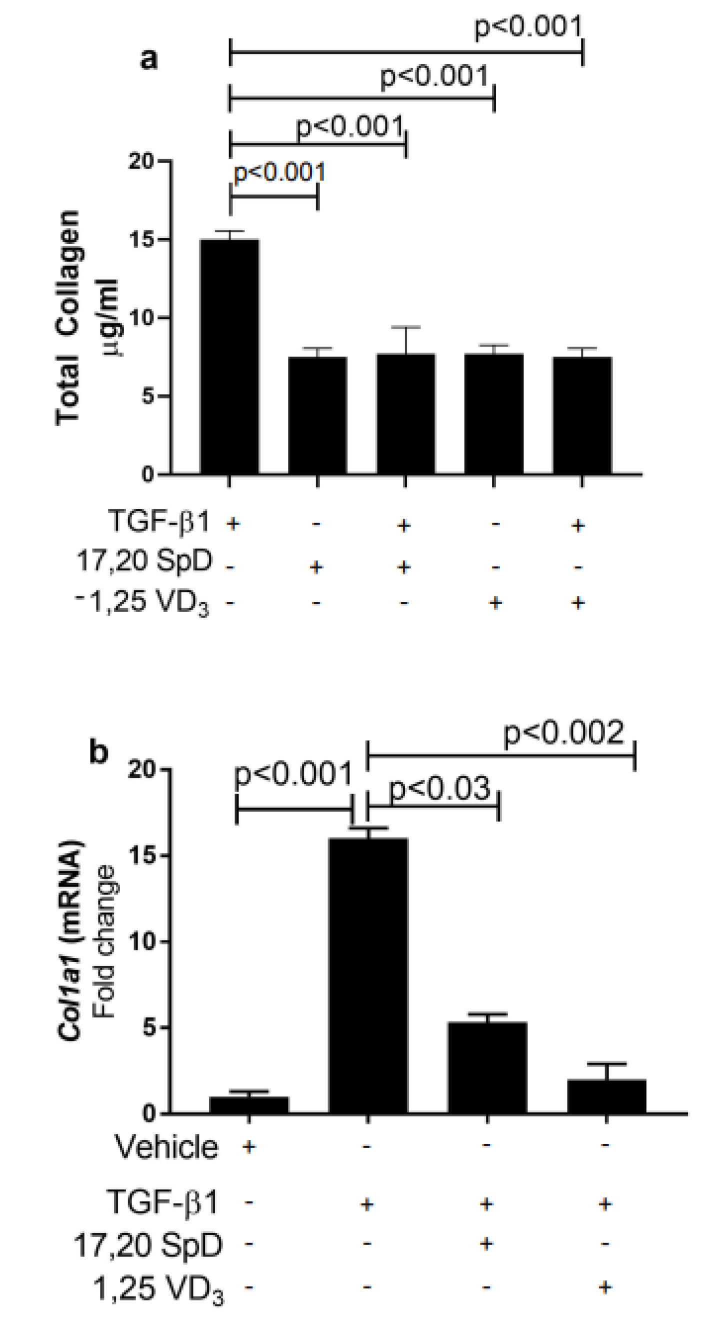
2.1. 17,20S(OH)2pD Suppressed COL1A1 Expression and Total Collagen Production in Healthy Normal and SSc Fibroblasts

Excessive accumulation of collagen has been associated with the thickening and fibrosis of the skin in SSc. We previously showed that 17,20S(OH)2pD inhibited TGF-β1-driven collagen synthesis and both type I collagen protein and Col1A1 mRNA in murine cells and normal human fibroblasts [36]. In this report, we examined total collagen protein synthesis in SSc dermal fibroblasts and found that baseline total collagen production was increased in SSc fibroblasts. On the other hand, culture with 17,20S(OH)2pD suppressed the total collagen formation (Figure 1a).
When we examined COL1a1 expression in SSc dermal fibroblasts, 17,20S(OH)2pD decreased COL1a1 expression when compared to TGF-β1-treated fibroblasts (Figure 1b). These results suggested that 17,20S(OH)2pD could suppress TGF-β1-mediated collagen synthesis at both the protein and mRNA level in SSc dermal fibroblasts.

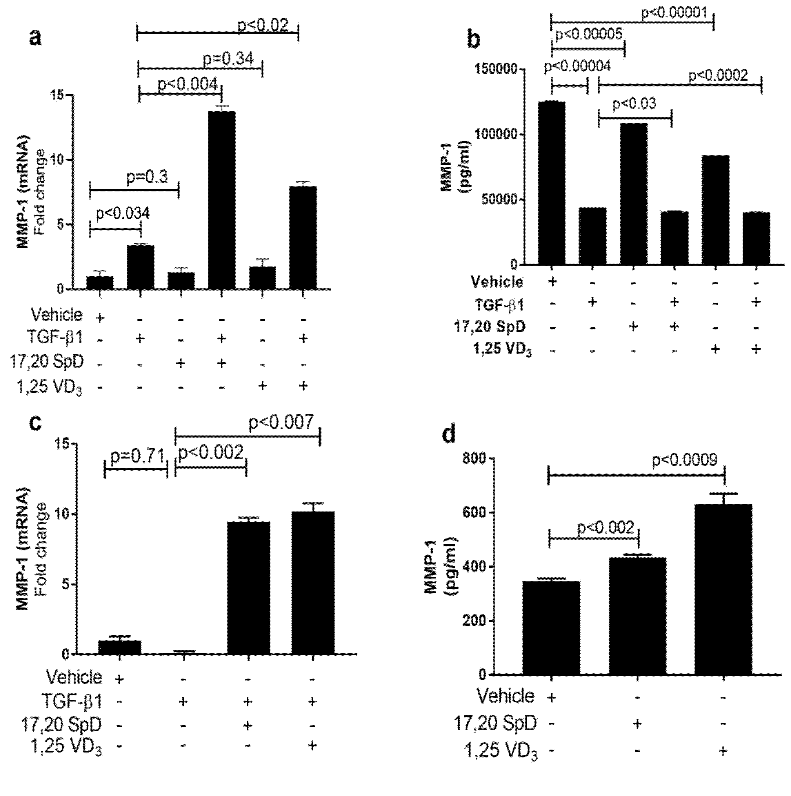
2.2. 17,20S(OH)2pD Increases MMP-1 Expression in SSc Fibroblasts

Excess tissue matrix accumulation in systemic sclerosis (SSc) contributes to fibrosis. It has been suggested that decreased serum levels of matrix metalloproteinases (MMPs) may aggravate this matrix overload. Normal and SSc fibroblasts were cultured with 17,20S(OH)2pD to determine the effect on MMP-1. While TGF-β1 slightly increased MMP-1 mRNA and protein expression in normal fibroblasts, the addition of 17,20S(OH)2pD to TGF-β increased MMP-1 RNA expression even more (Figure 2a). Normal fibroblasts were not affected by either 17,20S(OH)2pD or 1,25(OH)2D3 (Figure 2b). When we examined SSc fibroblasts, we found that dermal fibroblasts had active TGF-β signaling and spontaneously produced more collagen than cultured fibroblasts from healthy donors [39,40,41]. In SSc fibroblasts, the addition of 17,20S(OH)2pD or 1,25(OH)2D3 increased gene expression and protein synthesis of MMP-1 (Figure 2c,d). These data suggest that both 17,20S(OH)2pD and 1,25(OH)2D3 potentially can alleviate fibrosis by increasing MMP-1.

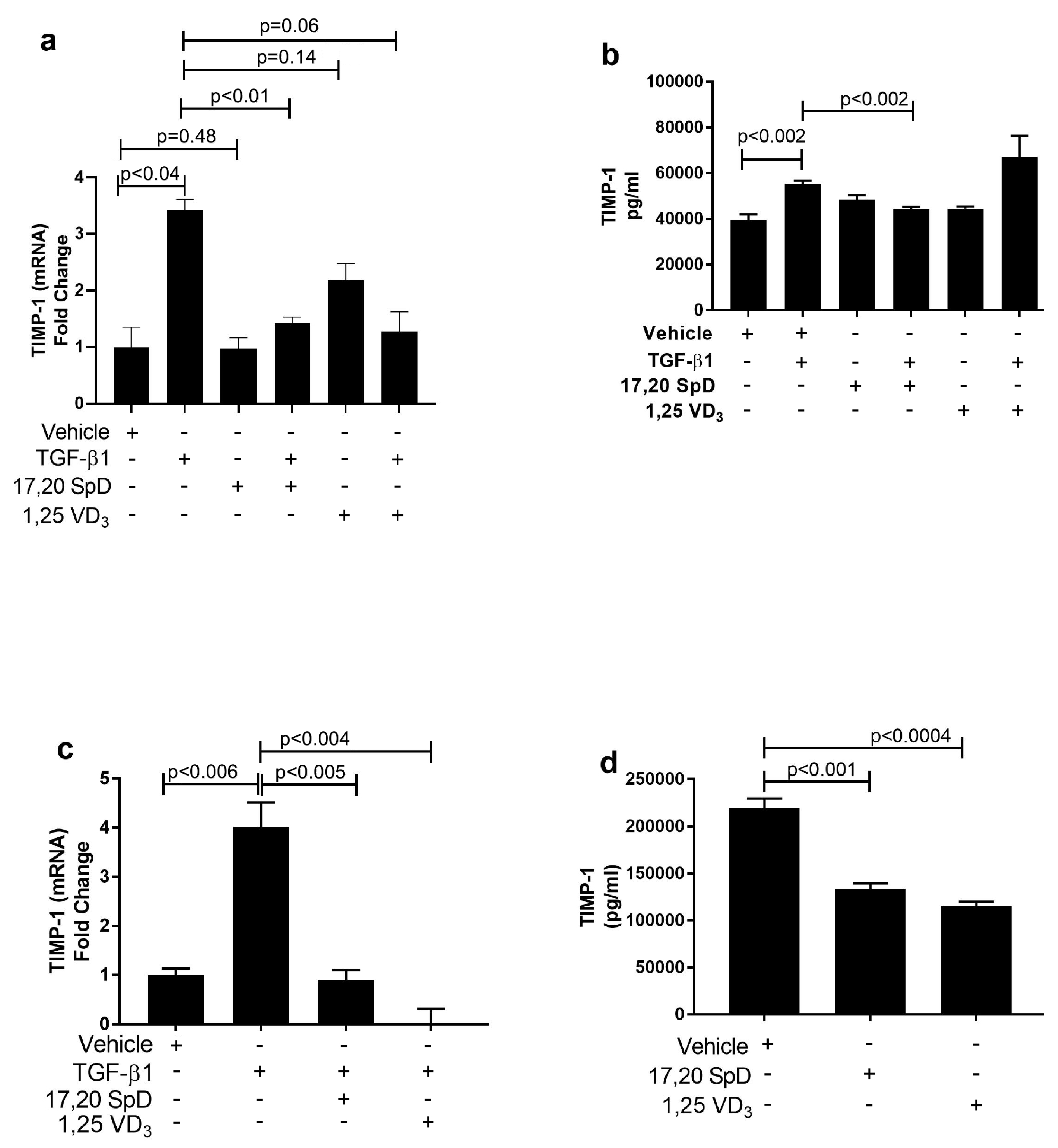
2.3. 17,20S(OH)2pD Decreases TIMP-1 Expression in SSc Fibroblasts

Elevated levels of tissue inhibitors of matrix metalloproteinases (TIMPs) may contribute to matrix accumulation in systemic sclerosis (SSc). In normal fibroblasts, TGF-β1 increased the expression of TIMP-1 mRNA and protein. 1,25(OH)2D3 in the presence of TGF-β1 suppressed TIMP-1 mRNA expression and 17,20S(OH)2pD and in the presence of TGF-β1 inhibited protein synthesis of TIMP-1 in normal fibroblasts (Figure 3a,b). In SSc fibroblasts, although basal TIMP-1 mRNA and protein levels were increased, 17,20S(OH)2pD and 1,25(OH)2D3 significantly decreased TIMP-1 protein and mRNA expression in SSc fibroblast lines (Figure 3c,d). These results suggested that 17,20S(OH)2pD has anti-fibrogenic properties including the inhibition of collagen synthesis by stimulation of MMP-1 mRNA and protein production and suppression of TIMP-1 protein production in both normal and SSc fibroblasts.

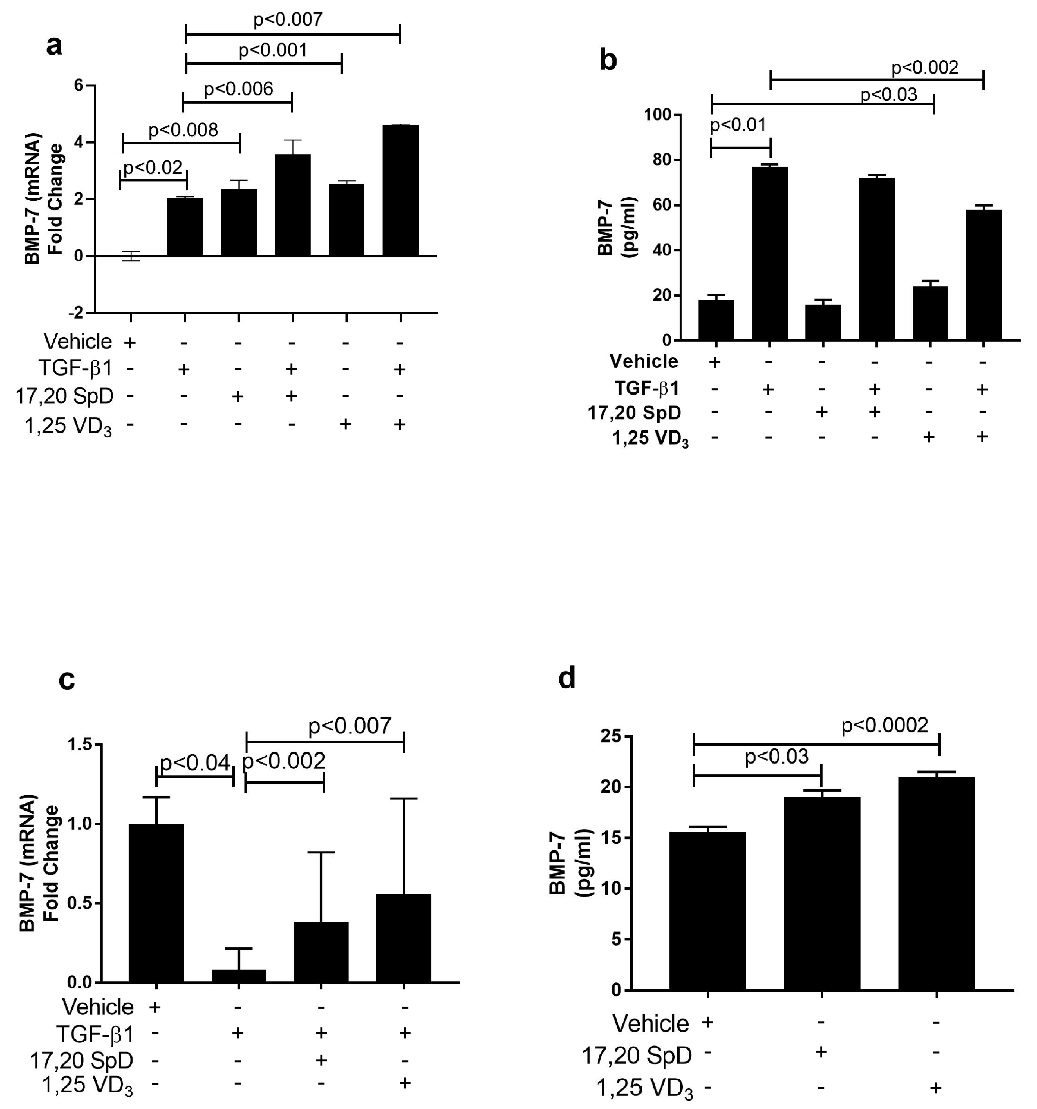
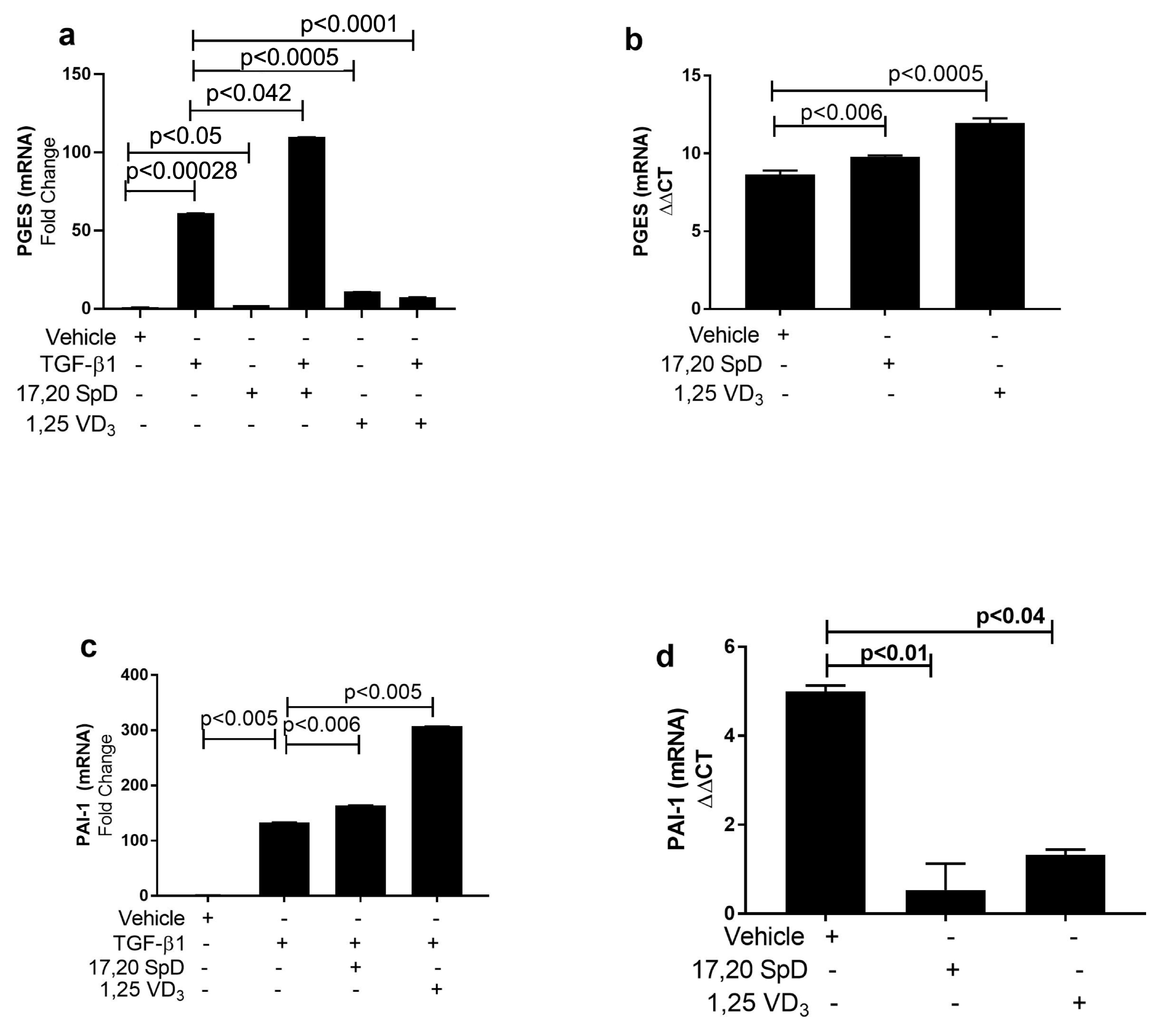
2.4. 17,20S(OH)2pD Modulates Other Mediators of Fibrosis In Vitro

TGFβ-1 is a potent stimulator of collagen synthesis by fibroblasts. On the other hand, even in the presence of TGF-β1, bone morphemic protein (BMP-7) can inhibit CI synthesis and the resulting matrix accumulation. Normal and SSc dermal fibroblasts were cultured with 17,20S(OH)2pD to examine its effect on BMP-7, PGES, and PAI-1 expression. Although BMP-7 mRNA expression was slightly increased in TGF-β1-stimulated normal fibroblasts, 17,20S(OH)2pD either alone or co-cultured with TGF-β1 further increased BMP-7 mRNA expression. BMP-7 expression was also increased with 1,25(OH)2D3 alone and in the presence of TGF-β1. Protein levels were not affected by 17,20S(OH)2pD in normal fibroblasts (Figure 4a,b). On the other hand, SSc fibroblasts had decreased basal expression of BMP-7, so that the addition of 17,20S(OH)2pD significantly increased the expression of BMP-7. A similar increase was seen when fibroblasts were cultured with 1,25(OH)2D3 (Figure 4c,d). These data suggest that in both normal and SSc fibroblasts, 17,20S(OH)2pD either alone or in the presence of TGF-β1 can increase BMP-7 expression, helping alleviate excessive fibrosis.
TGF-β1 ordinarily decreases prostaglandin E2 synthetase (PGES) production. PGES can resolve inflammation because it activates the release of prostaglandin E2, which acts as a direct vasodilator and has antifibrotic effects such as inhibition of fibroblast chemotaxis, proliferation, and collagen formation. In normal fibroblasts, 17,20S(OH)2pD together with TGF-β1 increased PGES expression, although there was no significant change in PGES when fibroblasts were co-cultured with 17,20S(OH)2pD alone or 1,25(OH)2D3 alone (Figure 5a). In scleroderma fibroblasts, PGES was induced when cultured with 17,20S(OH)2pD if compared to vehicle-treated fibroblasts. PGES expression was also increased in SSc fibroblasts when cultured with 1,25(OH)2D3 (Figure 5b). These data suggest that in both normal and scleroderma fibroblasts, 17,20S(OH)2pD increased PGES synthesis, suggesting that 17,20S(OH)2pD may have anti-inflammatory properties.
In inflammatory conditions in which fibrin is deposited in tissues, PAI-1 appears to play a significant role in the progression to fibrosis. Presumably, lower PAI levels would lead to less suppression of fibrinolysis and conversely a more rapid degradation of the fibrin. Sustained PAI-1 expression contributes to excessive collagen formation and fibrosis. We examined the effect of 17,20S(OH)2pD on PAI-1 mRNA expression in both normal and SSc fibroblasts. In normal fibroblasts, TGF-β1 increased PAI-1 expression as previously described [7] (Figure 5c). Normal fibroblasts cultured with 17,20S(OH)2pD or 1,25(OH)2D3 had no effect on PAI-1 when compared to vehicle-cultured fibroblasts. In the presence of TGF-β1, both 17,20S(OH)2pD and 1,25(OH)2D3 increased the expression of PAI-1 (Figure 5c). In SSc fibroblasts, PAI-1 expression was increased at baseline. Culturing SSc fibroblasts with 17,20S(OH)2pD significantly decreased PAI-1 expression. This same suppressive effect was seen in fibroblasts cultured with 1,25(OH)2D3 (Figure 5d). These results suggest that 17,20S(OH)2pD in the presence of TGF-β1 can effectively inhibit PAI-1 mRNA expression in SSc fibroblasts, leading to less fibrosis.
The induction of the fibrotic phenotype of scleroderma is mediated through GLI1/GLI2 (the effector molecules of the Hedgehog pathway) to promote tissue fibrosis. We analyzed the effect of 17,20S(OH)2pD on GLI1 and GLI2 mRNA expression in both normal and SSc fibroblasts. In normal fibroblasts, the addition of 17,20S(OH)2pD increased GLI1 mRNA expression, while the addition of TGF-β1 together with 17,20S(OH)2pD abrogated the stimulatory effect of the latter on GLI1 expression. The stimulation of GLI1 expression by 1,25(OH)2D3 alone did not reach the statistical significance, and concomitant addition of TGF-β1 significantly decreased this expression (Figure 6a). In the presence of TGF-β1, both 17,20S(OH)2pD and 1,25(OH)2D3 increased GLI2 mRNA expression (Figure 6b). When SSc fibroblasts were examined, neither 17,20S(OH)2pD, nor 1,25(OH)2D3 had a significant effect on either GLI1 or Gli2 mRNA expression (Figure 6c,d). Taken together, these data suggest that 17,20S(OH)2pD may partially inhibit GLI1 in normal fibroblasts, but is not effective in suppressing this pathway in SSc fibroblasts.

3. Discussion

Our results showed that 17,20S(OH)2pD suppressed TGF-β1-induced collagen protein production in cultured dermal normal and SSc fibroblasts. Moreover, 17,20S(OH)2pD effectively increased MMP-1 synthesis and suppressed TIMP-1 production, both factors allowing 17,20S(OH)2pD to enhance collagen degradation and decrease fibrosis. Since PGES and BMP-7 can help alleviate fibrosis, it is significant that in both normal and SSc fibroblasts, 17,20S(OH)2pD increased PGES and BMP-7. On the other hand, sustained PAI-1 aggravates fibrosis. 17,20S(OH)2pD was able to suppress PAI-1 mRNA expression in both normal and SSc fibroblasts, while it had no effect on Gli1 and Gli2 expression in SSc fibroblasts. Taken together, these results indicate that 17,20S(OH)2pD can modulate mediators of the TGF-β1 pathway in vitro, suggesting its potential to have anti-inflammatory properties in vivo and, by implication, anti-sclerotic effects.
In patients with SSc, dermal thickening, capillary damage, and decreased intestinal absorption contribute to vitamin D deficiency [24]. Vitamin D deficiency correlates with increased expression of TGF-β1 [27]. In vitro studies have shown that 1,25(OH)2D3 and analogs of vitamin D inhibited the TGF-β effect on collagen synthesis and topical treatment with a vitamin D analogue reduced skin fibrosis [42]. 1,25(OH)2D3 suppressed TGF-β1-induced collagen synthesis in lung fibroblasts, stellate cells from the liver, and renal cells [43,44]. Exposing mesenchymal multipotent cells (MMCs) to 1,25(OH)2D3 decreased the expression of TGF-β1, CI, CIII, and PAI-1 and increased BMP-7 expression [11]. In our study, we demonstrated for the first time that the secosteroidal analog with a short side chain, 17,20S(OH)2pD, inhibited collagen synthesis, TIMP-1, and PAI-1, while it increased MMP-1, PGES, and BMP-7 expression. In SSc fibroblast, 17,20S(OH)2pD suppressed collagen synthesis, TIMP-1, and PAI-1, while it increased the expression of MMP-1, BMP-7, and PGES and had no effect on Gli1 or Gli2 expression.
TGF-β1 is a major cytokine involved in fibrosing disorders. TGF-β1 increases collagen synthesis in dermal fibroblasts and contributes to the production of extracellular matrix proteins that favor fibrosis [5,45]. SSc fibroblasts express endogenous TGF-β1 [46]. Nephrogenic systemic fibrosis skin biopsies display increased expression of TIMP-1, but little to no MMP-1 [47]. The lack of metalloproteinases contributes to the collagen accumulation that is characteristic of fibrotic disorders [48]. Serum levels of MMP-1 have been shown to negatively correlate with the degree of fibrosis [49,50].
TGF-β also modulates the expression of BMP-7, PGES, PAI-1, GLI1, and GLI2, which also contribute to fibrosis [10,11,16,17,18]. BMP-7, a member of the TGF-β superfamily, is an antagonist of TGF-β1 and is downregulated in disorders of fibrosis in the presence of TGF-β1 upregulation [10,11]. BMP-7 has a role in preventing the TGF-induced EMT by decreasing CI synthesis [4,12]. Treatment with BMP-7 has been shown to inhibit renal, liver, and cardiac fibrosis [51,52]. In our study, BMP-7 synthesis was decreased in SSc fibroblasts and in normal fibroblasts cultured with TGF-β1.
Prostaglandin (PG) E2 has antiproliferative and antifibrotic properties. Its antifibrotic effects are manifested by the inhibition of fibroblasts’ chemotaxis, proliferation, and collagen formation [53,54,55]. TGF-β1 decreases PGE production [15]. In vitro studies using mesangial cells showed that TGF-β1 increased the proliferation of these mesangial cells in addition to decreasing PGE production. Li and coworkers found that following EMT, PGE2 inhibits chemotaxis by increasing the expression of E-prostanoid receptors 2 (EP2) and 4 (EP4) [16]. Prostaglandin E2 synthase (PGES) catalyzes the conversion of prostaglandin to PGE2. Although in SSc fibroblasts, the expression of PGES is mildly upregulated when compared to normal fibroblasts [13,14], we showed that treatment with 17,20S(OH)2pD further increased PGES expression in both normal and scleroderma fibroblasts. These data suggested that 17,20S(OH)2pD might have anti-inflammatory properties.
Urokinase/tissue-type plasminogen activator (uPA/tPA) and plasmin-dependent MMPs function in maintaining tissue homeostasis by degrading proteins of the ECM. uPA/tPA converts latent TGF-β and pro-MMP to their active forms [17]. PAI-1, a serine protease inhibitor, is a potent inhibitor of uPA/tPA by blocking uPA/tPA activation and plasmin-dependent MMP activation [17]. Although PAI-1 is a major contributor to efficient wound healing due to its ability to protect ECM proteins from proteolytic degradation, under pathologic conditions such as SSc, increased levels of PAI-1 contribute to collagen accumulation and increased expression of ECM proteins. TGF-β1 is a known inducer of PAI-1 [7]. Increased PAI-1 levels in SSc fibroblasts are due to increased TGF-β1 expression, SMAD2/3 activation, and decreased expression of inhibitory SMAD7. Sustained PAI-1 expression contributes to excessive collagen accumulation, leading to fibrosis [17]. Our results showed that in normal fibroblasts, TGF-β1 increased PAI-1 expression, while the addition of 17,20S(OH)2pD decreased the expression of PAI-1. In SSc fibroblasts, baseline expression of PAI-1 was increased, and 17,20S(OH)2pD decreased PAI-1 expression. Our results suggested that 17,20S(OH)2pD in the presence of TGF-β1 could effectively inhibit PAI-1 mRNA expression in both normal and SSc fibroblasts.
GLI1 and GLI2 are transcription factors in the Sonic Hedgehog (SHH) pathway. GLI1 and GLI2 have important roles in cell development and differentiation and are involved in myofibroblast formation. Lung tissues from patients with idiopathic pulmonary fibrosis have increased expression of GLI1 and GLI2 when compared to normal lung tissues. Lung epithelium and fibroblasts from these patients expressed increased GLI1 levels, while alveolar cells expressed increased GLI2 in a nuclear distribution. Fibroblasts that were treated with SHH had increased collagen synthesis and gene expression [56]. TGF-β1 induces the expression of GLI1 and GLI2 in normal fibroblasts and keratinocytes [18,19] and induces the expression of SHH and GLI1 in lung epithelial cells [18]. In our study, normal fibroblasts behaved differently from SSc fibroblasts. GLI1 mRNA expression decreased with TGF-β1 in normal fibroblasts, and culture with 17,20S(OH)2pD increased Gli1 mRNA expression. The addition of TGF-β1 with 17,20S(OH)2pD increased GLi2 expression, but not GL1 in normal fibroblasts. On the other hand, 17,20S(OH)2pD had no significant effect on either GLI1 or Gli2 mRNA expression in SSc fibroblasts. Taken together, these data demonstrate that 17,20S(OH)2pD affects normal and SSc fibroblasts differently in regard to its effect on Gli1 and Gli2. However, a precise mechanism of action remains unclear, since the corresponding nuclear receptor for 17,20S(OH)2pD remains to be identified. It must be noted that CYP11A1 hydroxyderivatives can act on alternative nuclear receptors including retinoic acid orphan receptors (RORs) [57], the aryl hydrocarbon receptor (AhR) [58], and liver X receptors (LXRs) [59], as well as their increased selectivity on the VDR requires hydroxylation on C1α [60]. Although 17,20S(OH)2pD can act as a weak agonist on LXRα and LXRβ [59], the high affinity receptor for hydroxysecosteroids with a short side chain remains to be defined.
Limitations of this study: Our study highlights a potential role for 17,20S(OH)2pD in the treatment of fibrosis. Our data are limited by the number of fibroblasts analyzed and treated with 17,20S(OH)2pD. We understand that differences in patient populations could alter the response of 17,20S(OH)2pD on collagen, TGF-β1, and other mediators of fibrosis. We understand that other pathways not related to TGF-β1 may be involved in 17,20S(OH)2pD suppression of collagen levels. Importantly, the nuclear receptor through which 17,20S(OH)2pD mediates its phenotypic effects remains to be defined, a future challenge to our laboratories. This report has discrepancies between mRNA levels and protein levels of MMP-1 and BMP7. Unfortunately, in some instances, the mRNA expression differs from protein expression levels due to: (a) complicated post-transcriptional mechanisms; (b) differences in the in vivo half-lives of proteins; (c) error and noise in both protein and mRNA experiments. Moreover, short-chain analogs of vitamin D, such as 17,20R(OH)2pD and 17,20S(OH)2pD, differ structurally from 1,25(OH)2D3. The biochemical differences in the side chains may explain their differing effects on the induction of calcemia and varying levels of PGES.

4. Materials and Methods

4.1. Secosteroids

1,25(OH)2D3 was obtained from Sigma-Aldrich, Co. LLC (St. Louis, MO, USA). Secosteroid: 17,20S(OH)2pD was synthesized as previously described [61]. For the tissue culture experiments, 1,25(OH)2D3 and 17,20S(OH)2pD were dissolved in absolute ethanol and diluted with Eagle’s minimal essential medium (EMEM) containing 1% charcoal-stripped fetal calf serum (FCS) at a stock concentration of 10−5 M for each.

4.2. Cell Lines and Culture

This study was conducted and approved by the University of Tennessee Health Science Center (UTHSC) Memphis Institutional Review Board (#07-08719). The Declaration of Helsinki protocols were followed, and normal healthy donors and SSc patients gave their written informed consent. Adult patients (ages 18–70 y) who met the American College of Rheumatology criteria for the diagnosis of SSc were used in this study. Punch biopsies from the lesional skin of patients with diffuse cutaneous systemic sclerosis (dcSSc) were taken from the volar forearm surface. Punch biopsies from the volar forearm surface were taken from normal healthy donors. Explanted biopsy specimens were allowed to grow into fibroblasts for 2–3 wk in Eagle’s minimal essential medium (EMEM) (Corning Cellgro, Manassas, VA, USA) with 9% fetal calf serum (FCS), 100 µL/mL penicillin, 100 µg/mL streptomycin, and amphotericin B 1 µg/mL and ascorbic acid (50 µg/mL) (Thermo Fisher, Waltham, MA, USA) as previously described by Brown Lobbins et al. [36,62]. In total, there were 5 normal fibroblast lines and 5 SSc lines. All were age, gender, and ethnicity matched. Each study used 3 cell lines per group, and passages 3–7 were used.
Fibroblasts were cultured and maintained in EMEM with 10% FCS, penicillin (100 units/mL), streptomycin (100 mg/mL), and 2 mM L-glutamine at 37 °C in a 5% CO2 humidified atmosphere. Fibroblasts between 10 and 16 passages were used for the experiments. Prior to the experiments, the fibroblast medium was changed to EMEM supplemented with 10% charcoal (Sigma-Aldrich, St. Louis, MO, USA)-stripped fetal calf serum (Atlanta Biologics, Flowery Branch, GA, USA) for 72 h. After a 72 h incubation period, the medium was changed to 1% charcoal-stripped FCS for 24 h. Following the 24 h incubation period, fibroblasts were incubated with 17,20S(OH)2pD or 1,25(OH)2D3 at either 10−8 M or 10−9 M or ethanol (ETOH) vehicle at 1:10,000 or 1:1 million dilution to control for final ETOH concentration in the 10−8 M and 10−9 M 17,20S(OH)2pD and 1,25(OH)2D3 preparations in culture. Fibroblasts were allowed to incubate under these conditions for a 2 h period before stimulation with TGF-β1 (R&D Systems, Minneapolis, Minnesota USA) dissolved in 0.1% bovine serum albumin (BSA) in 4 mM hydrochloric acid (Gibco, Life Technologies, Waltham MO, USA) to a final concentration of 5 ng/mL or BSA/HCL diluted 1:2000 to control for the final BSA-HCL in TGF-β1 concentration. Fibroblasts were cultured under these conditions either for an additional 24 h for total RNA extraction and 48 h for the analysis of total collagen or protein synthesis, respectively.

4.3. Quantitative Real-Time PCR

Total RNA was extracted from fibroblast monolayers using TRIzol reagent (Invitrogen-Thermo Fisher Scientific, Waltham, MA, USA) and reversed transcribed into cDNA using reverse transcriptase (Life Technologies-Applied Biosystems (ABI), Grand Island, NY, USA), for real-time -polymerase chain reaction (RT-PCR). RNAs were quantified using NanoDrop-2000 (Thermos, Franklin, NJ, USA). Fifteen nanograms of total RNA were used for these experiments. The TaqMan gene assay for human COL1A1 (Cat. No. HS01076777), MMP-1 (Cat. No. HS00899658), TIMP-1 (Cat. No. HS00171558), PAI-1 (Cat. No. HS01126606), BMP-7 (Cat. No. HS00233476), PGES (Cat. No. HS00153133), Gli1 (Cat. No. Hs01110766), Gli2 (Cat. No Hs01119974), and beta actin (Cat. No. HS01060665) (Applied Biosystems Life technologies, Grand Island, NY, USA) was used as a housekeeping gene. SDS Version 2.3 software was used to run the PCR, and the analysis was obtained with RQ manager (Life Technologies-ABI). 17,20S(OH)2pD and 1,25(OH)2D3 were normalized to β-actin (Applied Biosystems Life Technologies, Grand Island, NY, USA) mRNA expression levels. Data for 17,20S(OH)2pD- and 1,25(OH)2D3-treated fibroblast cultures are shown as fold change normalized to an endogenous reference (BSA + ETOH) vehicle control.

4.4. Protein Analysis

The supernatants from cultured fibroblasts were analyzed for protein concentrations using ELISA for MMP-1 (Cat No. DY901, R&D systems; Minneapolis, MN, USA), TIMP-1 (Cat No. DTM100, R&D systems; Minneapolis, MN, USA), and BMP-7 (Cat No. DBP700, R&D systems; Minneapolis, MN, USA).

4.5. Analysis of Total Collagen

Fibroblasts monolayers were extracted for total collagen levels using the Sircol Collagen Assay (Biocolor Ltd., Carickfergus, Northern Ireland, UK). Before performing these experiments, we compared the data generated by Sircol to other colorimetric methods to measure hydroxyproline and found the results to be comparable.

4.6. Statistical Analyses

The calculations to determine the statistical significance of the tests were carried out using the programs SAS and GraphPad Prism 4. Depending on the data, a one-way ANOVA or Student’s t-test analysis was performed. The comparison of the mean variable values with a distribution significantly different from normal in two unrelated groups was performed using the Mann–Whitney test, while in more than two unrelated groups, using the Kruskal–Wallis test. p ≤ 0.05 was considered statistically significant.

5. Conclusions

We showed that 17,20S(OH)2pD is able to suppress TGF-β1-induced collagen protein production in cultured dermal normal and SSc fibroblasts. 17,20S(OH)2pD inhibits collagen synthesis by effectively modulating mediators of the TGF-β1 pathway. The key players we studied were BMP-7, PAI-1, and PGES. These changes lead to altered metalloproteinase synthesis and the degradation of collagen. Although 17,20S(OH)2pD and 1,25(OH)2D3 are similar molecules, they do have important critical differences, such as their ability to induce hypercalcemia. The differential effects on TIMP-1 in normal fibroblasts appears to be another difference between the two secosteroids. Further work will be needed to determine why 1,25(OH)2D3 has such differing effects on normal and SSc dermal fibroblasts. Possibilities include the fact that 17,20S(OH)2pD has a shortened side chain and is lacking a hydroxyl group on C1α. Furthermore, it likely targets a different nuclear receptor than the VDR, which remains to be identified. In summary, our study supports that 17,20S(OH)2pD modulates mediators of fibrosis wherein a balance will favor the reduction of fibrosis and may offer a new therapeutic approach for treating SSc and other disorders of fibrosis.

Author Contributions

Writing, investigation, M.L.B.L.; assistance with writing and investigation, I.-S.O.S.; conceptualization, methodology, writing editor, and data analysis, A.T.S., L.K.M. and W.L.; methodology, K.A.H.; synthesized and purified 17,20S(OH)2pD—S.Z., W.L., R.C.T. and D.D.M.; investigation, T.-K.K. and Z.J.; Conceptualization, methodology, validation, and writing, A.E.P. All authors have read and agreed to the published version of the manuscript.

Funding

Supported by a grant from the National Scleroderma Foundation (M.L.B.L.) R073223552; the Arthritis Foundation (K.A.H.); the Department of Veterans Affairs Merit Review Grant (A.E.P.) and Department of Veterans Affairs Program Project Grant 5IP1BX001607-01 (A.E.P. and K.A.H.); the National Institutes of Health R21AR066505 (A.T.S. and AEP), R01AR071189 (A.T.S.), R01AR073004 (A.T.S.), R21 AI149267 (A.T.S.), and R21AR063242 (W.L. and D.D.M.); the Department of Veterans Affairs Merit Awards BX004293 (A.T.S.) and BX004151 (K.A.H.). Additional support was provided by the University of Tennessee College of Pharmacy Drug Discovery Center (W.L.).

Institutional Review Board Statement

The study was conducted according to the guidelines of the Declaration of Helsinki and approved by the Institutional Review Board of the University of Tennessee Health Science Center (UTHSC) at Memphis (#07-08719, approved 1 November 2017).

Informed Consent Statement

Informed consent was obtained from all subjects involved in the study.

Data Availability Statement

The datasets used and/or analyzed during the current study are available from the corresponding author upon reasonable request.

Acknowledgments

The authors would like to acknowledge the excellent contributions of Christy Patterson, Virginia Woo-Rasberry, and Patricia Wheller for their technical assistance and Angela Cody for help with the preparation of this manuscript. This article is dedicated to the late Professor Arnold E. Postlethwaite, in loving memory of his humanity, humility, integrity, and seminal contributions to the broad field of rheumatology for more than five decades.

Conflicts of Interest

The authors have no conflict of interest to disclose.

Abbreviations

1:25(OH)2D31:25-dihydroxyvitamin D3
17,20(OH)2pD17,20-dihydroxypregnacalciferol
BMP-7Bone morphogenic protein 7
Gli1Glioma-associated oncogene homolog 1
Gli2Glioma-associated oncogene homolog 2
MMP-1Metalloproteinase-1
PAI-1Plasminogen activator inhibitor-1
PGESProstaglandin E2 synthetase
TIMP-1Tissue inhibitor of metalloproteinase-1
TGF-β1Transforming growth factor-β1

References

  1. Pattanaik, D.; Brown, M.; Postlethwaite, B.C.; Postlethwaite, A.E. Pathogenesis of systemic sclerosis. Front. Immunol. 2015, 6, 272. [Google Scholar] [CrossRef] [PubMed] [Green Version]
  2. Varga, J.A.; Trojanowska, M. Fibrosis in systemic sclerosis. Rheum. Dis. Clin. N. Am. 2008, 34, 115–143. [Google Scholar] [CrossRef] [PubMed]
  3. Wynn, T.A.; Ramalingam, T.R. Mechanisms of fibrosis: Therapeutic translation for fibrotic disease. Nat. Med. 2012, 18, 1028–1040. [Google Scholar] [CrossRef] [PubMed] [Green Version]
  4. Postlethwaite, A.E.; Shigemitsu, H.; Kanangat, S. Cellular origins of fibroblasts: Possible implications for organ fibrosis in systemic sclerosis. Curr. Opin. Rheumatol. 2004, 16, 733–738. [Google Scholar] [CrossRef] [PubMed]
  5. Raghow, R.; Postlethwaite, A.E.; Keski-Oja, J.; Moses, H.L.; Kang, A.H. Transforming growth factor-beta increases steady state levels of type I procollagen and fibronectin messenger RNAs posttranscriptionally in cultured human dermal fibroblasts. J. Clin. Investig. 1987, 79, 1285–1288. [Google Scholar] [CrossRef] [PubMed] [Green Version]
  6. Postlethwaite, A.E.; Keski-Oja, J.; Moses, H.L.; Kang, A.H. Stimulation of the chemotactic migration of human fibroblasts by transforming growth factor beta. J. Exp. Med. 1987, 165, 251–256. [Google Scholar] [CrossRef] [Green Version]
  7. Keski-Oja, J.; Raghow, R.; Sawdey, M.; Loskutoff, D.J.; Postlethwaite, A.E.; Kang, A.H.; Moses, H.L. Regulation of mRNAs for type-1 plasminogen activator inhibitor, fibronectin, and type I procollagen by transforming growth factor-beta. Divergent responses in lung fibroblasts and carcinoma cells. J. Biol. Chem. 1988, 263, 3111–3115. [Google Scholar] [CrossRef]
  8. Yuan, W.; Varga, J. Transforming growth factor-beta repression of matrix metalloproteinase-1 in dermal fibroblasts involves Smad3. J. Biol. Chem. 2001, 276, 38502–38510. [Google Scholar] [CrossRef] [Green Version]
  9. Lin, P.S.; Chang, H.H.; Yeh, C.Y.; Chang, M.C.; Chan, C.P.; Kuo, H.Y.; Liu, H.C.; Liao, W.C.; Jeng, P.Y.; Yeung, S.Y.; et al. Transforming growth factor beta 1 increases collagen content, and stimulates procollagen I and tissue inhibitor of metalloproteinase-1 production of dental pulp cells: Role of MEK/ERK and activin receptor-like kinase-5/Smad signaling. J. Formos. Med. Assoc. 2017, 116, 351–358. [Google Scholar] [CrossRef]
  10. Khan, I.; Agarwal, P.; Thangjam, G.S.; Radhesh, R.; Rao, S.G.; Kondaiah, P. Role of TGF-beta and BMP7 in the pathogenesis of oral submucous fibrosis. Growth Factors 2011, 29, 119–127. [Google Scholar] [CrossRef]
  11. Artaza, J.N.; Norris, K.C. Vitamin D reduces the expression of collagen and key profibrotic factors by inducing an antifibrotic phenotype in mesenchymal multipotent cells. J. Endocrinol. 2009, 200, 207–221. [Google Scholar] [CrossRef] [PubMed] [Green Version]
  12. Zeisberg, M.; Shah, A.A.; Kalluri, R. Bone morphogenic protein-7 induces mesenchymal to epithelial transition in adult renal fibroblasts and facilitates regeneration of injured kidney. J. Biol. Chem. 2005, 280, 8094–8100. [Google Scholar] [CrossRef] [Green Version]
  13. McCann, M.R.; Monemdjou, R.; Ghassemi-Kakroodi, P.; Fahmi, H.; Perez, G.; Liu, S.; Shi-Wen, X.; Parapuram, S.K.; Kojima, F.; Denton, C.P.; et al. mPGES-1 null mice are resistant to bleomycin-induced skin fibrosis. Arthritis Res. Ther. 2011, 13, R6. [Google Scholar] [CrossRef] [PubMed] [Green Version]
  14. Kanekura, T.; Higashi, Y.; Kanzaki, T. Cyclooxygenase-2 expression and prostaglandin E2 biosynthesis are enhanced in scleroderma fibroblasts and inhibited by UVA irradiation. J. Rheumatol. 2001, 28, 1568–1572. [Google Scholar]
  15. Li, N.N.; Xu, Y.Y.; Chen, X.L.; Fan, Y.P.; Wu, J.H. Role of the prostaglandin E2/E-prostanoid 2 receptor signalling pathway in TGFbeta-induced mice mesangial cell damage. Biosci. Rep. 2014, 34, e00159. [Google Scholar] [CrossRef]
  16. Li, Y.J.; Kanaji, N.; Wang, X.Q.; Sato, T.; Nakanishi, M.; Kim, M.; Michalski, J.; Nelson, A.J.; Farid, M.; Basma, H.; et al. Prostaglandin E2 switches from a stimulator to an inhibitor of cell migration after epithelial-to-mesenchymal transition. Prostaglandins Other Lipid Mediat. 2015, 116, 1–9. [Google Scholar] [CrossRef]
  17. Ghosh, A.K.; Vaughan, D.E. PAI-1 in tissue fibrosis. J. Cell. Physiol. 2012, 227, 493–507. [Google Scholar] [CrossRef] [PubMed] [Green Version]
  18. Hu, B.; Liu, J.; Wu, Z.; Liu, T.; Ullenbruch, M.R.; Ding, L.; Henke, C.A.; Bitterman, P.B.; Phan, S.H. Reemergence of hedgehog mediates epithelial-mesenchymal crosstalk in pulmonary fibrosis. Am. J. Respir. Cell Mol. Biol. 2015, 52, 418–428. [Google Scholar] [CrossRef] [Green Version]
  19. Dennler, S.; Andre, J.; Alexaki, I.; Li, A.; Magnaldo, T.; ten Dijke, P.; Wang, X.J.; Verrecchia, F.; Mauviel, A. Induction of sonic hedgehog mediators by transforming growth factor-beta: Smad3-dependent activation of Gli2 and Gli1 expression in vitro and in vivo. Cancer Res. 2007, 67, 6981–6986. [Google Scholar] [CrossRef] [PubMed] [Green Version]
  20. Holick, M.F. Vitamin D deficiency. N. Engl. J. Med. 2007, 357, 266–281. [Google Scholar] [CrossRef]
  21. Bhakhri, B.K.; Debata, P.K. Nutritional rickets presenting with myelofibrosis. Indian J. Pediatr. 2010, 77, 1437–1439. [Google Scholar] [CrossRef]
  22. Pelajo, C.F.; Lopez-Benitez, J.M.; Miller, L.C. Vitamin D and autoimmune rheumatologic disorders. Autoimmun. Rev. 2010, 9, 507–510. [Google Scholar] [CrossRef]
  23. Vacca, A.; Cormier, C.; Piras, M.; Mathieu, A.; Kahan, A.; Allanore, Y. Vitamin D deficiency and insufficiency in 2 independent cohorts of patients with systemic sclerosis. J. Rheumatol. 2009, 36, 1924–1929. [Google Scholar] [CrossRef] [PubMed]
  24. Calzolari, G.; Data, V.; Carignola, R.; Angeli, A. Hypovitaminosis D in systemic sclerosis. J. Rheumatol. 2009, 36, 2845. [Google Scholar] [CrossRef] [PubMed] [Green Version]
  25. Gambichler, T.; Chrobok, I.; Hoxtermann, S.; Kreuter, A. Significantly decreased serum 25-hydroxyvitamin d levels in a large german systemic sclerosis cohort. J. Rheumatol. 2011, 38, 2494. [Google Scholar] [CrossRef] [PubMed]
  26. Arnson, Y.; Amital, H.; Agmon-Levin, N.; Alon, D.; Sanchez-Castanon, M.; Lopez-Hoyos, M.; Matucci-Cerinic, M.; Szucs, G.; Shapira, Y.; Szekanecz, Z.; et al. Serum 25-OH vitamin D concentrations are linked with various clinical aspects in patients with systemic sclerosis: A retrospective cohort study and review of the literature. Autoimmun. Rev. 2011, 10, 490–494. [Google Scholar] [CrossRef] [PubMed]
  27. Isik, S.; Ozuguz, U.; Tutuncu, Y.A.; Erden, G.; Berker, D.; Acar, K.; Aydin, Y.; Akbaba, G.; Helvaci, N.; Guler, S. Serum transforming growth factor-beta levels in patients with vitamin D deficiency. Eur. J. Intern. Med. 2012, 23, 93–97. [Google Scholar] [CrossRef]
  28. Humbert, P.; Dupond, J.L.; Agache, P.; Laurent, R.; Rochefort, A.; Drobacheff, C.; de Wazieres, B.; Aubin, F. Treatment of scleroderma with oral 1,25-dihydroxyvitamin D3: Evaluation of skin involvement using non-invasive techniques. Results of an open prospective trial. Acta Derm. Venereol. 1993, 73, 449–451. [Google Scholar]
  29. Elst, E.F.; Van Suijlekom-Smit, L.W.; Oranje, A.P. Treatment of linear scleroderma with oral 1,25-dihydroxyvitamin D3 (calcitriol) in seven children. Pediatr. Dermatol. 1999, 16, 53–58. [Google Scholar] [CrossRef] [Green Version]
  30. Caca-Biljanovska, N.G.; Vlckova-Laskoska, M.T.; Dervendi, D.V.; Pesic, N.P.; Laskoski, D.S. Treatment of generalized morphea with oral 1,25-dihydroxyvitamin D3. Adv. Exp. Med. Biol. 1999, 455, 299–304. [Google Scholar]
  31. Oblak, M.; Mlinsek, G.; Kandus, A.; Buturovic-Ponikvar, J.; Arnol, M. Effects of paricalcitol on biomarkers of inflammation and fibrosis in kidney transplant recipients: Results of a randomized controlled trial. Clin. Nephrol. 2017, 88, 119–125. [Google Scholar] [CrossRef]
  32. Hu, X.; Liu, W.; Yan, Y.; Liu, H.; Huang, Q.; Xiao, Y.; Gong, Z.; Du, J. Vitamin D protects against diabetic nephropathy: Evidence-based effectiveness and mechanism. Eur. J. Pharmacol. 2019, 845, 91–98. [Google Scholar] [CrossRef]
  33. van Etten, E.; Mathieu, C. Immunoregulation by 1,25-dihydroxyvitamin D3: Basic concepts. J. Steroid Biochem. Mol. Biol. 2005, 97, 93–101. [Google Scholar] [CrossRef]
  34. Marcus, J.F.; Shalev, S.M.; Harris, C.A.; Goodin, D.S.; Josephson, S.A. Severe hypercalcemia following vitamin d supplementation in a patient with multiple sclerosis: A note of caution. Arch. Neurol. 2012, 69, 129–132. [Google Scholar] [CrossRef] [PubMed] [Green Version]
  35. Slominski, A.; Janjetovic, Z.; Tuckey, R.C.; Nguyen, M.N.; Bhattacharya, K.G.; Wang, J.; Li, W.; Jiao, Y.; Gu, W.; Brown, M.; et al. 20S-hydroxyvitamin D3, noncalcemic product of CYP11A1 action on vitamin D3, exhibits potent antifibrogenic activity in vivo. J. Clin. Endocrinol. Metab. 2013, 98, E298–E303. [Google Scholar] [CrossRef] [Green Version]
  36. Slominski, A.T.; Li, W.; Bhattacharya, S.K.; Smith, R.A.; Johnson, P.L.; Chen, J.; Nelson, K.E.; Tuckey, R.C.; Miller, D.; Jiao, Y.; et al. Vitamin D analogs 17,20S(OH)2pD and 17,20R(OH)2pD are noncalcemic and exhibit antifibrotic activity. J. Investig. Dermatol. 2011, 131, 1167–1169. [Google Scholar] [CrossRef] [PubMed] [Green Version]
  37. Slominski, A.; Kim, T.K.; Zmijewski, M.A.; Janjetovic, Z.; Li, W.; Chen, J.; Kusniatsova, E.I.; Semak, I.; Postlethwaite, A.; Miller, D.D.; et al. Novel vitamin D photoproducts and their precursors in the skin. Dermatoendocrinol 2013, 5, 7–19. [Google Scholar] [CrossRef] [Green Version]
  38. Kim, T.K.; Wang, J.; Janjetovic, Z.; Chen, J.; Tuckey, R.C.; Nguyen, M.N.; Tang, E.K.; Miller, D.; Li, W.; Slominski, A.T. Correlation between secosteroid-induced vitamin D receptor activity in melanoma cells and computer-modeled receptor binding strength. Mol. Cell. Endocrinol. 2012, 361, 143–152. [Google Scholar] [CrossRef] [Green Version]
  39. Higley, H.; Persichitte, K.; Chu, S.; Waegell, W.; Vancheeswaran, R.; Black, C. Immunocytochemical localization and serologic detection of transforming growth factor beta 1. Association with type I procollagen and inflammatory cell markers in diffuse and limited systemic sclerosis, morphea, and Raynaud’s phenomenon. Arthritis Rheum. 1994, 37, 278–288. [Google Scholar] [CrossRef] [PubMed]
  40. Lu, J.; Liu, Q.; Wang, L.; Tu, W.; Chu, H.; Ding, W.; Jiang, S.; Ma, Y.; Shi, X.; Pu, W.; et al. Increased expression of latent TGF-beta-binding protein 4 affects the fibrotic process in scleroderma by TGF-beta/SMAD signaling. Lab. Investig. 2017, 97, 591–601. [Google Scholar] [CrossRef] [PubMed]
  41. Nikitorowicz-Buniak, J.; Denton, C.P.; Abraham, D.; Stratton, R. Partially evoked epithelial-mesenchymal transition (EMT) is associated with increased tgfbeta signaling within lesional scleroderma skin. PLoS ONE 2015, 10, e0134092. [Google Scholar]
  42. Usategui, A.; Criado, G.; Del Rey, M.J.; Fare, R.; Pablos, J.L. Topical vitamin D analogue calcipotriol reduces skin fibrosis in experimental scleroderma. Arch. Dermatol. Res. 2014, 306, 757–761. [Google Scholar] [CrossRef] [PubMed]
  43. Ramirez, A.M.; Wongtrakool, C.; Welch, T.; Steinmeyer, A.; Zugel, U.; Roman, J. Vitamin D inhibition of pro-fibrotic effects of transforming growth factor beta1 in lung fibroblasts and epithelial cells. J. Steroid Biochem. Mol. Biol. 2010, 118, 142–150. [Google Scholar] [CrossRef] [PubMed] [Green Version]
  44. Potter, J.J.; Liu, X.; Koteish, A.; Mezey, E. 1,25-dihydroxyvitamin D3 and its nuclear receptor repress human alpha1 (I) collagen expression and type I collagen formation. Liver Int. 2013, 33, 677–686. [Google Scholar] [CrossRef] [PubMed] [Green Version]
  45. Kudo, H.; Wang, Z.; Jinnin, M.; Nakayama, W.; Inoue, K.; Honda, N.; Nakashima, T.; Kajihara, I.; Makino, K.; Makino, T.; et al. EBI3 downregulation contributes to Type I collagen overexpression in scleroderma skin. J. Immunol. 2015, 195, 3565–3573. [Google Scholar] [CrossRef] [PubMed] [Green Version]
  46. Varga, J.; Whitfield, M.L. Transforming growth factor-beta in systemic sclerosis (scleroderma). Front. Biosci. 2009, 1, 226–235. [Google Scholar] [CrossRef] [PubMed]
  47. Kelly, B.C.; Markle, L.S.; Vickers, J.L.; Petitt, M.S.; Raimer, S.S.; McNeese, C. The imbalanced expression of matrix metalloproteinases in nephrogenic systemic fibrosis. J. Am. Acad. Dermatol. 2010, 63, 483–489. [Google Scholar] [CrossRef] [PubMed]
  48. Brown, M.; Postlethwaite, A.E.; Myers, L.K.; Hasty, K.A. Supernatants from culture of type I collagen-stimulated PBMC from patients with cutaneous systemic sclerosis versus localized scleroderma demonstrate suppression of MMP-1 by fibroblasts. Clin. Rheumatol. 2012, 31, 973–981. [Google Scholar] [CrossRef] [Green Version]
  49. Zhang, B.B.; Cai, W.M.; Weng, H.L.; Hu, Z.R.; Lu, J.; Zheng, M.; Liu, R.H. Diagnostic value of platelet derived growth factor-BB, transforming growth factor-beta1, matrix metalloproteinase-1, and tissue inhibitor of matrix metalloproteinase-1 in serum and peripheral blood mononuclear cells for hepatic fibrosis. World J. Gastroenterol. 2003, 9, 2490–2496. [Google Scholar] [CrossRef]
  50. Foronjy, R.F.; Sun, J.; Lemaitre, V.; D’Armiento, J.M. Transgenic expression of matrix metalloproteinase-1 inhibits myocardial fibrosis and prevents the transition to heart failure in a pressure overload mouse model. Hypertens. Res. 2008, 31, 725–735. [Google Scholar] [CrossRef] [PubMed] [Green Version]
  51. Zeisberg, E.M.; Tarnavski, O.; Zeisberg, M.; Dorfman, A.L.; McMullen, J.R.; Gustafsson, E.; Chandraker, A.; Yuan, X.; Pu, W.T.; Roberts, A.B.; et al. Endothelial-to-mesenchymal transition contributes to cardiac fibrosis. Nat. Med. 2007, 13, 952–961. [Google Scholar] [CrossRef] [PubMed]
  52. Zeisberg, M.; Yang, C.; Martino, M.; Duncan, M.B.; Rieder, F.; Tanjore, H.; Kalluri, R. Fibroblasts derive from hepatocytes in liver fibrosis via epithelial to mesenchymal transition. J. Biol. Chem. 2007, 282, 23337–23347. [Google Scholar] [CrossRef] [Green Version]
  53. Kohyama, T.; Ertl, R.F.; Valenti, V.; Spurzem, J.; Kawamoto, M.; Nakamura, Y.; Veys, T.; Allegra, L.; Romberger, D.; Rennard, S.I. Prostaglandin E(2) inhibits fibroblast chemotaxis. Am. J. Physiol. Lung Cell. Mol. Physiol. 2001, 281, L1257–L1263. [Google Scholar] [CrossRef] [Green Version]
  54. Kolodsick, J.E.; Peters-Golden, M.; Larios, J.; Toews, G.B.; Thannickal, V.J.; Moore, B.B. Prostaglandin E2 inhibits fibroblast to myofibroblast transition via E. prostanoid receptor 2 signaling and cyclic adenosine monophosphate elevation. Am. J. Respir. Cell Mol. Biol. 2003, 29, 537–544. [Google Scholar] [CrossRef]
  55. Kowal-Bielecka, O.; Kowal, K.; Distler, O.; Rojewska, J.; Bodzenta-Lukaszyk, A.; Michel, B.A.; Gay, R.E.; Gay, S.; Sierakowski, S. Cyclooxygenase- and lipoxygenase-derived eicosanoids in bronchoalveolar lavage fluid from patients with scleroderma lung disease: An imbalance between proinflammatory and antiinflammatory lipid mediators. Arthritis Rheum. 2005, 52, 3783–3791. [Google Scholar] [CrossRef] [PubMed]
  56. Bolanos, A.L.; Milla, C.M.; Lira, J.C.; Ramirez, R.; Checa, M.; Barrera, L.; Garcia-Alvarez, J.; Carbajal, V.; Becerril, C.; Gaxiola, M.; et al. Role of Sonic Hedgehog in idiopathic pulmonary fibrosis. Am. J. Physiol. Lung Cell. Mol. Physiol. 2012, 303, L978–L990. [Google Scholar] [CrossRef] [PubMed] [Green Version]
  57. Slominski, A.T.; Kim, T.K.; Takeda, Y.; Janjetovic, Z.; Brozyna, A.A.; Skobowiat, C.; Wang, J.; Postlethwaite, A.; Li, W.; Tuckey, R.C.; et al. RORalpha and ROR gamma are expressed in human skin and serve as receptors for endogenously produced noncalcemic 20-hydroxy- and 20,23-dihydroxyvitamin D. FASEB J. 2014, 28, 2775–2789. [Google Scholar] [CrossRef] [PubMed] [Green Version]
  58. Slominski, A.T.; Kim, T.K.; Janjetovic, Z.; Brozyna, A.A.; Zmijewski, M.A.; Xu, H.; Sutter, T.R.; Tuckey, R.C.; Jetten, A.M.; Crossman, D.K. Differential and overlapping effects of 20,23(OH)(2)D3 and 1,25(OH)(2)D3 on gene expression in human epidermal keratinocytes: Identification of ahr as an alternative receptor for 20,23(OH)(2)D3. Int. J. Mol. Sci. 2018, 19, 3072. [Google Scholar] [CrossRef] [Green Version]
  59. Slominski, A.T.; Kim, T.K.; Qayyum, S.; Song, Y.; Janjetovic, Z.; Oak, A.S.W.; Slominski, R.M.; Raman, C.; Stefan, J.; Mier-Aguilar, C.A.; et al. Vitamin D and lumisterol derivatives can act on liver X receptors (LXRs). Sci. Rep. 2021, 11, 8002. [Google Scholar] [CrossRef] [PubMed]
  60. Slominski, A.T.; Kim, T.K.; Hobrath, J.V.; Oak, A.S.W.; Tang, E.K.Y.; Tieu, E.W.; Li, W.; Tuckey, R.C.; Jetten, A.M. Endogenously produced nonclassical vitamin D hydroxy-metabolites act as “biased” agonists on VDR and inverse agonists on RORalpha and RORgamma. J. Steroid Biochem. Mol. Biol. 2017, 173, 42–56. [Google Scholar] [CrossRef] [PubMed] [Green Version]
  61. Zmijewski, M.A.; Li, W.; Zjawiony, J.K.; Sweatman, T.W.; Chen, J.; Miller, D.D.; Slominski, A.T. Photo-conversion of two epimers (20R and 20S) of pregna-5,7-diene-3beta, 17alpha, 20-triol and their bioactivity in melanoma cells. Steroids 2009, 74, 218–228. [Google Scholar] [CrossRef] [PubMed] [Green Version]
  62. Postlethwaite, A.E.; Smith, G.N., Jr.; Lachman, L.B.; Endres, R.O.; Poppleton, H.M.; Hasty, K.A.; Seyer, J.M.; Kang, A.H. Stimulation of glycosaminoglycan synthesis in cultured human dermal fibroblasts by interleukin 1. Induction of hyaluronic acid synthesis by natural and recombinant interleukin 1s and synthetic interleukin 1 beta peptide 163–171. J. Clin. Investig. 1989, 83, 629–636. [Google Scholar] [CrossRef] [PubMed] [Green Version]
Figure 1. 17,20S(OH)2pD suppressed total collagen synthesis and COL1A1 in human dermal fibroblasts. SSc human fibroblasts were treated with either 17,20S(OH)2pD 10−8 M or vehicle (EMEM + 1% stripped FCS + 0.1% BSA with 4 mM HCL (vehicle for TGF-β1) + ETOH (1:10,000 or 1:1 million (vehicle for 17,20S(OH)2pD) followed by stimulation with TGF-β1 5 ng/mL for either 24 h for COL1A1 analysis (b) or 48 h for total collagen protein analysis (a). mRNA data are expressed as fold change.
Ijms 23 00367 g001
Figure 2. 17,20S(OH)2pD increased MMP-1 in human dermal fibroblasts. Normal (Panels (a,b)) and SSc (Panels (c,d)) dermal fibroblast were treated with either 17,20S(OH)2pD 10−8 M, 1,25(OH)2D3 10−8 M, or vehicle (EMEM + 1% stripped FCS + 0.1% BSA with 4 mM HCL (vehicle for TGF-β1) + ETOH (1:10,000 or 1:1 million (vehicle for 17,20S(OH)2pD and 1,25(OH)2D3)), followed by stimulation with TGF-β1 5ng/mL for either 24 h or 48 h and then analyzed for MMP-1 mRNA (fold change) or ELISA (pg/mL).
Ijms 23 00367 g002
Figure 3. 17,20S(OH)2pD increased MMP-1 in human dermal fibroblasts. 17,20S(OH)2pD decreased TIMP-1 protein synthesis in normal and SSc dermal fibroblasts. Normal (Panels (a,b)) and SSc (Panels (c,d)) dermal fibroblast were treated with either 17,20S(OH)2pD 10−8 M, 1,25(OH)2D3 10−8 M, or vehicle (EMEM + 1% stripped FCS + 0.1% BSA with 4 mM HCL (vehicle for TGF-β1) + ETOH (1:10,000 or 1:1 million (vehicle for 17,20S(OH)2pD and 1,25(OH)2D3)), followed by stimulation with TGF-β1 5ng/mL for either 24 h or 48 h and then analyzed for TIMP-1 mRNA (fold change) or ELISA (pg/mL). p < 0.05 is significant.
Ijms 23 00367 g003
Figure 4. 17,20S(OH)2pD stimulated BMP-7 synthesis in SSc dermal fibroblasts. Normal (Panels (a,b)) and SSc (Panels (c,d)) dermal fibroblast were treated with either 17,20S(OH)2pD 10−8 M, 1,25(OH)2D3 10−8 M, or vehicle (EMEM + 1% stripped FCS + 0.1% BSA with 4 mM HCL (vehicle for TGF-β1) + ETOH (1:10,000 or 1:1 million (vehicle for 17,20S(OH)2pD and 1,25(OH)2D3)), followed by stimulation with TGF-β1 5ng/mL for either 24 h or 48 h and then analyzed for BMP-7 by either mRNA (fold change) or ELISA (pg/mL). p < 0.05 is significant.
Ijms 23 00367 g004
Figure 5. 17,20S(OH)2pD increases PGES and inhibits PAI-1 mRNA expression in normal and SSc dermal fibroblasts. Normal (Panels (a,c)) and SSc (Panels (b,d)) dermal fibroblast were treated with either 17,20S(OH)2pD 10−8 M, 1,25(OH)2D3 10−8 M, or vehicle (EMEM + 1% stripped FCS + 0.1% BSA with 4 mM HCL (vehicle for TGF-β1) + ETOH (1:10,000 or 1:1 million (vehicle for 17,20S(OH)2pD and 1,25(OH)2D3)), followed by stimulation with TGF-β1 5 ng/mL for 24 h and then analyzed for PGES or PAI-1 mRNA expression (fold change). p < 0.05 is significant.
Ijms 23 00367 g005
Figure 6. 17,20S(OH)2pD had no effect on GLi mRNA expression in SSc dermal fibroblasts, but in the presence of TGF-β1 increases Gli2 expression in normal fibroblast. Normal (Panels (a,b)) and SSc (Panels (c,d)) dermal fibroblast were treated with either 17,20S(OH)2pD 10−8 M, 1,25(OH)2D3 10−8 M, or vehicle (EMEM + 1% stripped FCS + 0.1% BSA with 4 mM HCL (vehicle for TGF-β1) + ETOH (1:10,000 or 1:1 million (vehicle for 17,20S(OH)2pD and 1,25(OH)2D3)), followed by stimulation with TGF-β1 5 ng/mL for 24 h and then analyzed for Gli1 or Gli2 mRNA expression (fold change). NS is not significant. p < 0.05 is significant.
Ijms 23 00367 g006
Publisher’s Note: MDPI stays neutral with regard to jurisdictional claims in published maps and institutional affiliations.

